research article antioxidant activity of ...downloads.hindawi.com/journals/jamc/2013/804504.pdf ·...
TRANSCRIPT
Hindawi Publishing CorporationJournal of Analytical Methods in ChemistryVolume 2013, Article ID 804504, 5 pageshttp://dx.doi.org/10.1155/2013/804504
Research ArticleIn Vivo Antioxidant Activity of DeacetylasperulosidicAcid in Noni
De-Lu Ma,1 Mai Chen,2 Chen X. Su,3 and Brett J. West3
1 Division of Pharmacology, Tianjin Medical University, Tianjin 300070, China2Quality Control, Tahitian Noni Beverages Company Ltd., Room A 12F, No. 789, Zhaojiabang Road, Shanghai 200032, China3 Research and Development, Morinda Inc., 737 East 1180 South, American Fork, UT 84003, USA
Correspondence should be addressed to Brett J. West; brett [email protected]
Received 10 September 2013; Accepted 15 October 2013
Academic Editor: Jian Yang
Copyright © 2013 De-Lu Ma et al. This is an open access article distributed under the Creative Commons Attribution License,which permits unrestricted use, distribution, and reproduction in any medium, provided the original work is properly cited.
Deacetylasperulosidic acid (DAA) is a major phytochemical constituent of Morinda citrifolia (noni) fruit. Noni juice hasdemonstrated antioxidant activity in vivo and in human trials. To evaluate the role of DAA in this antioxidant activity, Wistar ratswere fed 0 (control group), 15, 30, or 60mg/kg body weight per day for 7 days. Afterwards, serum malondialdehyde concentrationand superoxide dismutase and glutathione peroxidase activities were measured and compared among groups. A dose-dependentreduction in malondialdehyde was evident as well as a dose-dependent increase in superoxide dismutase activity. DAA ingestiondid not influence serum glutathione peroxidase activity. These results suggest that DAA contributes to the antioxidant activity ofnoni juice by increasing superoxide dismutase activity.The fact that malondialdehyde concentrations declined with increased DAAdose, despite the lack of glutathione peroxidase-inducing activity, suggests that DAAmay also increase catalase activity. It has beenpreviously reported that noni juice increases catalase activity in vivo but additional research is required to confirm the effect ofDAA on catalase. Even so, the current findings do explain a possible mechanism of action for the antioxidant properties of nonijuice that have been observed in human clinical trials.
1. Introduction
Morinda citrifolia, commonly known as noni, is a small treethat has been used as a traditional source of food andmedicine throughout the tropics [1, 2]. A variety of potentialhealth benefits have been reported for noni fruit juice [3].These include immunomodulation [4, 5] and antioxidantactivities in vitro and in vivo [6–8]. The antioxidant activityof noni juice was found to be associated with increasedendurance in athletes [9]. In a human clinical trial involvingheavy cigarette smokers, consumption of noni juice resultedin lowered plasma concentrations of superoxide anion radi-cals (SAR) and lipid hydroperoxides [10]. Further, consump-tion of noni juice also decreased the level of lipid peroxi-dation-derived DNA adducts in the lymphocytes of heavysmokers [11].
In vivo research has demonstrated that noni juiceincreases superoxide dismutase (SOD) and glutathione per-oxidase (GPx) enzyme activities [12]. The superoxide anion
radical (SAR) is a major cellular reactive oxygen species andmay be generated via enzymatic and nonenzymatic processor may come from exogenous sources, including cigarettesmoke [13]. SOD catalyzes the dismutation of SAR to hydro-gen peroxide and oxygen [14]. GPx is capable of reducingfree hydrogen peroxide to water [15]. GPx also reduces lipidhydroperoxides, as well as prevents free radical attack onpolyunsaturated fatty acids in cellular membranes [16]. Assuch, the effect of noni juice on these two enzymes may beat least two of the major antioxidant mechanisms of actionthrough which it protects lymphocyte DNA and lowersplasma concentration of tobacco smoke-induced free radicalsand peroxides.
Chemical studies of noni fruit have revealed that iridoidsare the main phytochemical constituents, with deacetylaspe-rulosidic acid (DAA) comprising the majority of the iridoidcontent [17]. DAA has anticlastogenic activity, suppressingthe induction of chromosome aberrations inChinese hamsterovary cells and in mice [18]. DAA is reported to inhibit the
2 Journal of Analytical Methods in Chemistry
release of tumor necrosis factor-alpha from cultured mouseperitoneal macrophages and inhibits low-density lipoproteinoxidation [19, 20]. DAA also prevented 4-nitroquinoline 1-oxide (4NQO) induced DNA damage in vitro [21]. 4NQOexposure leads to the formation of superoxide, hydrogenperoxide, and hydroxyl radicals, resulting in the productionof a substantial amount of 8-hydroxydeoxyguanosine, aproduct of DNA oxidation in mammalian and bacterial cells[22, 23]. TreatmentwithDAA reduced 4NQOgenotoxicity by98.96%, suggesting that iridoids are responsible for the DNAprotective effects of noni juice in cigarette smokers.
With demonstrated antioxidant activity of noni juice andthe potential bioactivities of its major phytochemical con-stituent, the current study was conducted to investigate therole of DAA on SOD and GPx activities in vivo.
2. Materials and Methods
2.1. Test Material. Deacetylasperulosidic acid (DAA) wasobtained from Chengdu Biopurify Phytochemicals Ltd.(Chengdu, China). The purity of DAA was 98%, and theidentity was confirmed by high performance liquid chro-matography [24]. DAA was dissolved in MeOH-H
2O (1 : 1) at
a concentration of 0.2mg/mL. Separation of the standard wasperformed with a HC-C18 column (25 cm × 4.6mm; 5𝜇m,Agilent Technologies, Santa Clara, CA, USA) in a Waters2690 separationsmodule (Waters Corporation,Milford, MA,USA) and detected with a Waters 2489 UV/Vis detector at235 nm.DAAwas eluted at a flow rate of 0.8mL/minwith twomobile phases: (A) MeCN and (B) 0.1% formic acid in H
2O
(v/v). The elution gradient was 5min with 100% B, 35minwith 30% A and 70% B, 12min 95% A and 5% B, and then8 minutes with 100% B. The retention time and absorbancespectrum were compared against those of a DAA standard.
2.2. Animals and Treatment. For this study, 40 Wistar rats(male and female, 180–200 g) were obtained from the Exper-imental Animal Center, Academy of Military Medical Sci-ences (Beijing), Approval no. SCXK-Army-2009-003. Thecare of animals and experimental procedures were compliantwith ethical and institutional animal welfare guidelines ofTianjin Medical University. Following acclimation, rats wererandomly divided into 4 groups of 10 each (5 male and 5female). Each group was provided feed and water ad libitum.DAAwas dissolved in saline and each animal was gavaged for7 days with 1mL/200 g body weight (bw) of one of four treat-ments, depending on group assignment.The treatments werenormal saline (control), 15mg DAA/kg bw (low dose), 30mgDAA/kg bw (mid dose), and 60mg DAA/kg bw (high dose).Individual animal weight and feed intake were recorded ondays 1, 4, and 7. On the 8th day, the rats were anesthetizedand 0.5mL blood was removed from the orbital sinus.Wholeblood was centrifuged at 3,000 rpm for 10min in a low speedcentrifuge (model BFX5-320, Baiyang Centrifuge Factory,Liulizhuang,Hebei, China).The resulting serumwas retainedfor assays.
2.3. Antioxidant Enzyme Activity Assays and Malondialde-hyde Assay. Using a commercial bioassay kit (Jiancheng
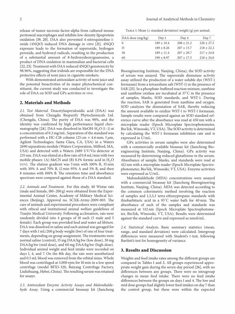
Table 1: Mean (± standard deviation) weight (g) per animal.
DAA dose (mg/kg) Day 1 Day 4 Day 70 189 ± 10.4 208 ± 21.2 220 ± 27.5
15 189 ± 8.26 207 ± 15.7 218 ± 22.2
30 189 ± 11.4 207 ± 20.7 217 ± 24.8
60 190 ± 8.97 207 ± 17.5 218 ± 24.0
Bioengineering Institute, Nanjing, China), the SOD activityof serum was assayed. The superoxide dismutase activityassay utilized the production of a water soluble dye (WST-1formazan) from a tetrazolium salt (WST-1) in the presence ofSAR [25]. In a phosphate-buffered reactionmixture, xanthineand xanthine oxidase are incubated at 37∘C in the presenceof samples, blanks, SOD standards, and WST-1. Duringthe reaction, SAR is generated from xanthine and oxygen.SOD catalyzes the dismutation of SAR, thereby reducingthe amount available to oxidize WST-1 to WST-1 formazan.Sample results were compared against an SOD standard ref-erence curve after the absorbance was read at 450 nm with amicroplate reader (Epoch Microplate Spectrophotometer,BioTek,Winooski, VT, USA).The SOD activity is determinedby calculating the WST-1 formazan inhibition rate and isexpressed in U/mL.
GPx activities in serum samples were also determinedwith a commercially available bioassay kit (Jiancheng Bio-engineering Institute, Nanjing, China). GPx activity wasmeasured by determining reduced glutathione in the serum.Absorbance of sample, blanks, and standards were read at412 nm with a microplate reader (Epoch Microplate Spectro-photometer, BioTek, Winooski, VT, USA). Enzyme activitieswere expressed as U/mL.
Malondialdehyde (MDA) concentrations were assayedwith a commercial bioassay kit (Jiancheng BioengineeringInstitute, Nanjing, China). MDA was detected according tothe common colorimetric method involving the reactionof samples and 1,3,3,3 tetra-ethoxypropane standards withthiobarbituric acid in a 95∘C water bath for 40min. Theabsorbance of each of the samples and standards wasmeasured at 532 nm (Epoch Microplate Spectrophotome-ter, BioTek, Winooski, VT, USA). Results were determinedagainst the standard curve and expressed as nmol/mL.
2.4. Statistical Analysis. Basic summary statistics (mean,range, and standard deviation) were calculated. Intergroupdifferences were measured with Student’s 𝑡-test, followingBartlett’s test for homogeneity of variance.
3. Results and Discussion
Weights and feed intake rates among the different groups arecompared in Tables 1 and 2. All groups experienced appro-priate weight gain during the seven-day period [26], with nodifferences between any groups. There were no intragroupchanges in mean feed intake. There were no feed intakedifferences between the groups on days 1 and 4. The low andmid dose groups had slightly lower feed intakes on day 7 thanthe control group, but these were within the expected
Journal of Analytical Methods in Chemistry 3
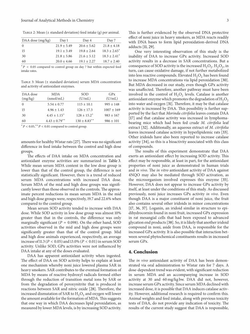
Table 2: Mean (± standard deviation) feed intake (g) per animal.
DAA dose (mg/kg) Day 1 Day 4 Day 70 21.9 ± 5.49 20.6 ± 5.62 21.8 ± 4.18
15 19.1 ± 3.49 19.8 ± 2.64 18.3 ± 2.65∗
30 21.8 ± 5.86 21.6 ± 5.12 18.3 ± 2.41∗
60 20.0 ± 4.66 19.1 ± 2.27 18.7 ± 2.40
∗𝑃 < 0.05 compared to control group on day 7 but within expected feed
intake rates.
Table 3: Mean (± standard deviation) serum MDA concentrationand activity of antioxidant enzymes.
DAA dose(mg/kg)
MDA(nmol/mL)
SOD(U/mL)
GPx(U/mL)
0 5.54 ± 0.77 115 ± 10.1 995 ± 148
15 4.96 ± 1.43 126 ± 17.3 1007 ± 169
30 4.45 ± 1.15∗
128 ± 15.2∗
983 ± 167
60 4.43 ± 0.79∗∗
130 ± 8.83∗∗
986 ± 101
∗𝑃 < 0.05,∗∗𝑃 < 0.01 compared to control group.
amounts for healthyWistar rats [27].There was no significantdifference in feed intake between the control and high dosegroups.
The effects of DAA intake on MDA concentration andantioxidant enzyme activities are summarized in Table 3.While mean serum MDA content in the low dose group islower than that of the control group, the difference is notstatistically significant. However, there is a trend of reducedserum MDA concentrations with increased DAA dose.Serum MDA of the mid and high dose groups was signifi-cantly lower than those observed in the controls.The approx-imate percent reductions in mean serum MDA in the midand high dose groups were, respectively, 19.7 and 22.6%whencompared to the control group.
Mean serum SOD activity tended to increase with DAAdose. While SOD activity in low dose group was almost 10%greater than that in the controls, the difference was onlymarginally significant (𝑃 = 0.098). On the other hand, SODactivities observed in the mid and high dose groups weresignificantly greater than that of the control group. Midand high dose animals experienced, respectively, an averageincrease of 11.3 (𝑃 < 0.05) and 13.0% (𝑃 < 0.01) in serumSODactivity. Unlike SOD, GPx activities were not influenced byDAA intake at any of the doses evaluated.
DAA has apparent antioxidant activity when ingested.The effect of DAA on SOD activity helps to explain at leastone mechanism whereby noni juice lowered plasma SAR inheavy smokers. SAR contributes to the eventual formation ofMDA by means of reactive hydroxyl radicals formed eitherthrough the reduction of transition metal ions by SAR orfrom the degradation of peroxynitrite that is produced inreactions between SAR and nitric oxide [28]. Therefore, theincreased dismutation of SAR to H
2O2and oxygen will lower
the amount available for the formation ofMDA.This suggeststhat one way in which DAA decreases lipid peroxidation, asmeasured by lowerMDA levels, is by increasing SOD activity.
This is further evidenced by the observed DNA protectiveeffect of noni juice in heavy smokers, as MDA reacts readilywith DNA bases to form lipid peroxidation-derived DNAadducts [11, 29].
One very interesting observation of this study is theinability of DAA to increase GPx activity. Increased SODactivity results in a decrease in SAR concentrations. But aconsequence of SOD activity is the increased H
2O2. H2O2, in
turn, can cause oxidative damage, if not further metabolizedinto less reactive compounds. Elevated H
2O2has been found
to increase MDA concentrations via lipid peroxidation [30].But MDA decreased in our study, even though GPx activitywas unaffected. Therefore, another pathway must have beeninvolved in the control of H
2O2levels. Catalase is another
antioxidant enzymewhich promotes the degradation ofH2O2
into water and oxygen [31]. Therefore, it may be that catalaseactivity is increased by DAA. This possibility is further sup-ported by the fact thatMorinda citrifolia leaves contain DAA[17] and that catalase activity was increased in lymphoma-bearing mice which had been fed crude M. citrifolia leafextract [32]. Additionally, an aqueous extract of M. citrifolialeaves increased catalase activity in hyperlipidemic rats [33].Other iridoids have also been reported to increase catalaseactivity [34], so this is a bioactivity associated with this classof compounds.
The results of this experiment demonstrate that DAAexerts an antioxidant effect by increasing SOD activity. Thiseffect may be responsible, at least in part, for the antioxidantproperties of noni juice as demonstrated in human trialsand in vivo. The in vitro antioxidant activity of DAA against4NQO may also be mediated through SOD activation, asthe microorganism involved expresses this enzyme [35].However, DAA does not appear to increase GPx activity byitself, at least under the conditions of this study. As discussedpreviously, noni juice increases GPx activity in vivo. Eventhough DAA is a major constituent of noni juice, the fruitalso contains several other iridoids in minor concentrations[17, 36, 37]. Loganin, an iridoid similar in structure to epi-dihydrocornin found in noni fruit, increased GPx expressionin rat mesangial cells that had been exposed to advancedglycation end products [38]. So, it is likely that another iridoidcompound in noni, aside from DAA, is responsible for theincreased GPx activity. It is also possible that interaction bet-ween several phytochemical constituents results in increasedserum GPx.
4. Conclusion
The in vivo antioxidant activity of DAA has been demon-strated via oral administration to Wistar rats for 7 days. Adose-dependent trendwas evident, with significant reductionin serum MDA and an accompanying increase in SODactivity at 30 and 60mg/kg bw. DAA did not, however,increase serumGPx activity. Since serumMDAdeclinedwithincreased dose, it is possible that DAA induces catalase activ-ity. However, additional research is required to confirm this.Animal weights and feed intake, along with previous toxicitytests of DAA, do not provide any indication of toxicity. Theresults of the current study suggest that DAA is responsible,
4 Journal of Analytical Methods in Chemistry
at least in part, for the antioxidant activities observed inhuman trials. While it is possible that DAA’s influence onSOD activity could explain the effect of noni juice on heavysmokers, the results also demonstrate that the effect of nonijuice on GPx activity in vivo is not associated with DAAalone. Induction of GPx is likely due to another iridoid or acombination of phytochemical constituents.
Acknowledgment
This research was supported financially by Morinda Inc., amanufacturer of noni juice.
References
[1] J. F. Morton, “The ocean-going noni, or Indian Mulberry(Morinda citrifolia, Rubiaceae) and some of its “colorful” rela-tives,” Economic Botany, vol. 46, no. 3, pp. 241–256, 1992.
[2] B. J. West, C. J. Jensen, J. Westendorf, and L. D. White, “A safetyreview of noni fruit juice,” Journal of Food Science, vol. 71, no. 8,pp. R100–R106, 2006.
[3] M. Y. Wang, B. J. West, C. J. Jensen et al., “Morinda citrifo-lia (Noni): a literature review and recent advances in Noniresearch,” Acta Pharmacologica Sinica, vol. 23, no. 12, pp. 1127–1141, 2002.
[4] A. Hirazumi and E. Furusawa, “An immunomodulatorypolysaccharide-rich substance from the fruit juice of Morindacitrifolia (noni) with antitumour activity,” PhytotherapyResearch, vol. 13, no. 5, pp. 380–387, 1999.
[5] A.K. Palu, A.H.Kim, B. J.West, S.Deng, J. Jensen, andL.White,“The effects of Morinda citrifolia L. (noni) on the immunesystem: its molecular mechanisms of action,” Journal of Ethno-pharmacology, vol. 115, no. 3, pp. 502–506, 2007.
[6] Z. Mohd-Zin, A. Abdul-Hamid, and A. Osman, “Antioxidativeactivity of extracts fromMengkudu (Morinda citrifolia L.) root,fruit and leaf,” Food Chemistry, vol. 78, no. 2, pp. 227–231, 2002.
[7] B. N. Su, A. D. Pawlus, H. Jung, W. J. Keller, J. L. McLaughlin,and A. D. Kinghorn, “Chemical constituents of the fruits ofMorinda citrifolia (Noni) and their antioxidant activity,” Journalof Natural Products, vol. 68, no. 4, pp. 592–595, 2005.
[8] M. Y. Wang and C. Su, “Cancer preventive effect of Morindacitrifolia (Noni),” Annals of the New York Academy of Sciences,vol. 952, pp. 161–168, 2001.
[9] A. K. Palu, R. D. Seifulla, and B. J. West, “Morinda citrifolia L.,(noni) improves athlete endurance: its mechanisms of action,”Journal of Medicinal Plant Research, vol. 2, no. 7, pp. 154–158,2008.
[10] M. Y. Wang, L. Peng, M. N. Lutfiyya, E. Henley, V. Weiden-bacher-Hoper, and G. Anderson, “Morinda citrifolia (noni)reduces cancer risk in current smokers by decreasing aromaticDNA adducts,”Nutrition and Cancer, vol. 61, no. 5, pp. 634–639,2009.
[11] M. Y.Wang, L. Peng, C. J. Jensen, S. Deng, and B. J. West, “Nonijuice reduces lipid peroxidation-derived DNA adducts in heavysmokers,” Food Science & Nutrition, vol. 1, no. 2, pp. 141–149,2013.
[12] J. J. Zhang, L. Y. Wang, L. N. Ou et al., “Study on evaluationof antioxidant activity of Noni juice in vivo,” Science andTechnology of Food Industry, vol. 32, no. 7, pp. 392–393, 2011.
[13] A. Valavanidis, T. Vlachogianni, and K. Fiotakis, “Tobaccosmoke: involvement of reactive oxygen species and stable free
radicals in mechanisms of oxidative damage, carcinogenesisand synergistic effects with other respirable particles,” Interna-tional Journal of Environmental Research and Public Health, vol.6, no. 2, pp. 445–462, 2009.
[14] I. Fridovich, “The biology of oxygen radicals,” Science, vol. 201,no. 4359, pp. 875–880, 1978.
[15] G. C. Mills, “Hemoglobin catabolism. I. Glutathione peroxi-dase, an erythrocyte enzyme which protects hemoglobin fromoxidative breakdown,” The Journal of Biological Chemistry, vol.229, no. 1, pp. 189–197, 1957.
[16] P. B. McCay, D. D. Gibson, K. L. Fong, and K. R. Hornbrook,“Effect of glutathione peroxidase activity on lipid peroxidationin biological membranes,” Biochimica et Biophysica Acta, vol.431, no. 3, pp. 459–468, 1976.
[17] O. Potterat, R. Von Felten, P. W. Dalsgaard, and M. Hamburger,“Identification of TLCmarkers and quantification byHPLC-MSof various constituents in noni fruit powder and commercialnoni-derived products,” Journal of Agricultural and Food Chem-istry, vol. 55, no. 18, pp. 7489–7494, 2007.
[18] T. Nakamura, Y. Nakazawa, S. Onizuka et al., “Antimutagenicityof Tochu tea (an aqueous extract of Eucommia ulmoides leaves): 1. The clastogen-suppressing effects of Tochu tea in CHO cellsand mice,”Mutation Research, vol. 388, no. 1, pp. 7–20, 1997.
[19] B. Li, D. Zhang, Y. Luo, and X. Chen, “Three new and antitumoranthraquinone glycosides from Lasianthus acuminatissimusMERR,” Chemical and Pharmaceutical Bulletin, vol. 54, no. 3,pp. 297–300, 2006.
[20] D. H. Kim, H. J. Lee, Y. J. Oh et al., “Iridoid glycosides isolatedfrom Oldenlandia diffusa inhibit LDL-oxidation,” Archives ofPharmacal Research, vol. 28, no. 10, pp. 1156–1160, 2005.
[21] B. J. West, S. Deng, and C. J. Jensen, “Nutrient and phy-tochemical analyses of processed noni puree,” Food ResearchInternational, vol. 44, no. 7, pp. 2295–2301, 2011.
[22] Y. Arima, C. Nishigori, T. Takeuchi et al., “4-Nitroquinoline1-oxide forms 8-hydroxydeoxyguanosine in human fibroblaststhrough reactive oxygen species,” Toxicological Sciences, vol. 91,no. 2, pp. 382–392, 2006.
[23] T. Nunoshiba and B. Demple, “Potent intracellular oxidativestress exerted by the carcinogen 4- nitroquinoline-N-oxide,”Cancer Research, vol. 53, no. 14, pp. 3250–3252, 1993.
[24] S. Deng, B. J. West, A. K. Palu, and C. J. Jensen, “Determinationand comparative analysis of major iridoids in different partsand cultivation sources of Morinda citrifolia,” PhytochemicalAnalysis, vol. 22, no. 1, pp. 26–30, 2011.
[25] A. V. Peskin and C. C. Winterbourn, “A microtiter plate assayfor superoxide dismutase using a water-soluble tetrazolium salt(WST-1),” Clinica Chimica Acta, vol. 293, no. 1-2, pp. 157–166,2000.
[26] A. H. Pullen, “A parametric analysis of the growing CFHB(Wistar) rat,” Journal of Anatomy, vol. 121, no. 2, pp. 371–383,1976.
[27] F. Nistiar, O. Racz, A. Lukacinova et al., “Age dependency onsome physiological and biochemical parameters of maleWistarrats in controlled environment,” Journal of Environmental Sci-ence and Health A, vol. 47, no. 9, pp. 1224–1233, 2012.
[28] N. Hogg, V. M. Darley-Usmar, M. T. Wilson, and S. Moncada,“Production of hydroxyl radicals from the simultaneous gener-ation of superoxide and nitric oxide,” Biochemical Journal, vol.281, no. 2, pp. 419–424, 1992.
[29] L. J. Marnett, “Lipid peroxidation—DNA damage by malondi-aldehyde,”Mutation Research, vol. 424, no. 1-2, pp. 83–95, 1999.
Journal of Analytical Methods in Chemistry 5
[30] N.Kokita andA.Hara, “Propofol attenuates hydrogenperoxide-induced mechanical and metabolic derangements in the iso-lated rat heart,” Anesthesiology, vol. 84, no. 1, pp. 117–127, 1996.
[31] P. Chelikani, I. Fita, and P. C. Loewen, “Diversity of structuresand properties among catalases,” Cellular and Molecular LifeSciences, vol. 61, no. 2, pp. 192–208, 2004.
[32] T. Anitha and S. Mohandass, “Anti-oxidant activity ofMorindacitrifolia on lymphoma-bearing mice,” Ancient Science of Life,vol. 26, no. 1-2, pp. 85–88, 2006.
[33] G. C. Chinta, V. Mullinti, K. Prashanthi, D. Sujata, B. Pushpa-kumari, and D. Ranganayakulu, “Anti-oxidant activity of theaqueous extract of the Morinda citrifolia leaves in triton WR-1339 induced hyperlipidemic rats,”Drug Invention Today, vol. 2,no. 1, pp. 1–4, 2010.
[34] I. M. Villasenor, “Bioactivities of iridoids,” Anti-Inflammatoryand Anti-Allergy Agents in Medicinal Chemistry, vol. 6, no. 4,pp. 307–314, 2007.
[35] S. Iuchi and L. Weiner, “Cellular and molecular physiology ofEscherichia coli in the adaptation to aerobic environments,”Journal of Biochemistry, vol. 120, no. 6, pp. 1055–1063, 1996.
[36] G. Liu, A. Bode, W.-Y. Ma, S. Sang, C.-T. Ho, and Z. Dong,“Two novel glycosides from the fruits of Morinda citrifolia(noni) inhibit AP-1 transactivation and cell transformation inthe mouse epidermal JB6 cell line,” Cancer Research, vol. 61, no.15, pp. 5749–5756, 2001.
[37] B. N. Su, A. D. Pawlus, H. Jung, W. J. Keller, J. L. McLaughlin,and A. D. Kinghorn, “Chemical constituents of the fruits ofMorinda citrifolia (Noni) and their antioxidant activity,” Journalof Natural Products, vol. 68, no. 4, pp. 592–595, 2005.
[38] H. Xu, J. Shen, H. Liu, Y. Shi, H. Li, and M. Wei, “Morronisideand loganin extracted from Cornus officinalis have protectiveeffects on rat mesangial cell proliferation exposed to advancedglycation end products by preventing oxidative stress,” Cana-dian Journal of Physiology and Pharmacology, vol. 84, no. 12, pp.1267–1273, 2006.

Submit your manuscripts athttp://www.hindawi.com
Hindawi Publishing Corporationhttp://www.hindawi.com Volume 2014
Inorganic ChemistryInternational Journal of
Hindawi Publishing Corporation http://www.hindawi.com Volume 2014
International Journal ofPhotoenergy
Hindawi Publishing Corporationhttp://www.hindawi.com Volume 2014
Carbohydrate Chemistry
International Journal of
Hindawi Publishing Corporationhttp://www.hindawi.com Volume 2014
Journal of
Chemistry
Hindawi Publishing Corporationhttp://www.hindawi.com Volume 2014
Advances in
Physical Chemistry
Hindawi Publishing Corporationhttp://www.hindawi.com
Analytical Methods in Chemistry
Journal of
Volume 2014
Bioinorganic Chemistry and ApplicationsHindawi Publishing Corporationhttp://www.hindawi.com Volume 2014
SpectroscopyInternational Journal of
Hindawi Publishing Corporationhttp://www.hindawi.com Volume 2014
The Scientific World JournalHindawi Publishing Corporation http://www.hindawi.com Volume 2014
Medicinal ChemistryInternational Journal of
Hindawi Publishing Corporationhttp://www.hindawi.com Volume 2014
Chromatography Research International
Hindawi Publishing Corporationhttp://www.hindawi.com Volume 2014
Applied ChemistryJournal of
Hindawi Publishing Corporationhttp://www.hindawi.com Volume 2014
Hindawi Publishing Corporationhttp://www.hindawi.com Volume 2014
Theoretical ChemistryJournal of
Hindawi Publishing Corporationhttp://www.hindawi.com Volume 2014
Journal of
Spectroscopy
Analytical ChemistryInternational Journal of
Hindawi Publishing Corporationhttp://www.hindawi.com Volume 2014
Journal of
Hindawi Publishing Corporationhttp://www.hindawi.com Volume 2014
Quantum Chemistry
Hindawi Publishing Corporationhttp://www.hindawi.com Volume 2014
Organic Chemistry International
ElectrochemistryInternational Journal of
Hindawi Publishing Corporation http://www.hindawi.com Volume 2014
Hindawi Publishing Corporationhttp://www.hindawi.com Volume 2014
CatalystsJournal of