research article antiadhesive property of photoreactive...
TRANSCRIPT
Hindawi Publishing CorporationJournal of ChemistryVolume 2013, Article ID 297159, 8 pageshttp://dx.doi.org/10.1155/2013/297159
Research ArticleAntiadhesive Property of Photoreactive AzidophenylLow-Molecular-Weight Chitosan in Rabbit Laminotomy Model
Jong Won Kim,1 Kwang-Sup Song,2 Hyun Kang,3 Eui-Chan Jang,2
Mi Kyung Kim,4 and Tae Il Son5
1 Department of Orthopaedic Surgery, Gang-Nam Himchan Hospital, 20-8 Songpa-dong, Songpa-gu, Seoul 138-170, Republic of Korea2Department of Orthopedic Surgery, College of Medicine, Chung-Ang University, Heukseok-dong, Dongjak-gu,Seoul 224-1, Republic of Korea
3 Department of Anesthesiology and Pain Medicine, College of Medicine, Chung-Ang University, Heukseok-dong, Dongjak-gu,Seoul 224-1, Republic of Korea
4Department of Pathology, College of Medicine, Chung-Ang University, Heukseok-dong, Dongjak-gu, Seoul 224-1, Republic of Korea5 Department of Biotechnology and Bio-Environmental Technology (BET) Research Institute, Chung-Ang University,Gyeonggi-do 456-756, Republic of Korea
Correspondence should be addressed to Kwang-Sup Song; [email protected]
Received 11 January 2013; Accepted 23 February 2013
Academic Editor: Zexuan Dong
Copyright © 2013 Jong Won Kim et al.This is an open access article distributed under the Creative Commons Attribution License,which permits unrestricted use, distribution, and reproduction in any medium, provided the original work is properly cited.
Newly developed photoreactive azidophenyl chitosan (P-ALMC) has characteristics of a transformable gel type and its outer layercould be sealed up like a film after UV radiation. We aim to evaluate the antiadhesive properties of P-ALMC through comparing itwith hyaluronic acid-carboxymethylcellulosemembrane (HA-CMC) in a rabbit laminotomymodel. Laminotomieswere performedat the L3-4, L4-5, and L5-6 levels in 41 rabbits and each level was randomly assigned to either receive saline (group I), HA-CMC(group II), or P-ALMC (group III).The extent of peridural fibrosis, density of fibroblasts and inflammatory cells, and dural thicknesswere evaluated at 6 and 12 weeks postoperatively. In the groups II and III, the extents of peridural fibrosis and dural thickness weresignificantly smaller than those in group I (𝑃 < 0.001) and no differences between groups II and III were found at the postoperative 6and 12weeks.Therewere no differences of cell density among groups. P-ALMC showed effective antiadhesive properties comparableto HA-CMC and could be one of the candidates as an anti-adhesive agent for spine surgery even further study is required to identifythe effectiveness of its unique characteristics as mechanical barrier.
1. Introduction
The formation of scar tissue over the epidural space, aftersurgical exploration of the spinal canal, has been a cause forconcern because it significantly promotes surgical hazardsto subsequent spinal surgeries. Additionally, it can result inleg and back pain caused by the tethering of the neuraltissue [1, 2]. One inevitably hazardous consequence of spinalsurgery is the formation of scar tissue designated as the“postlaminectomy membrane” by LaRocca and Macnab [2]and Gill et al. [3].
Various efforts to prevent scar formation after laminec-tomy have been evaluated over the years [1, 3–10]. Pathoe-tiologically, it has been reported that the formation of a
hematoma, after laminectomy, constitutes a scaffold for themigration of fibroblasts from the periosteum and paraspinalmuscles. This migration of fibroblasts is considered to be oneof the main causes of postlaminectomy peridural fibrosis [2,5, 6]. However, if materials are used to limit this contact, scartissue formation may be prevented. In order to prevent scartissue formation with this strategy, the material must haveproper biocompatibility, biodegradability, space-occupyingproperties, and optimal mechanical properties.
Chitosan is one of the most abundant marine-basedbiopolymers [11]. Low-molecular-weight chitosan (LMC) isa water-soluble hydrolysate of chitosan that has been shownto have a wide range of biological activities and industrialapplications [12–17].
2 Journal of Chemistry
Photoreactive azidophenyl low-molecular-weight chi-tosan (P-ALMC) used in our study was formed by conju-gating LMC with a photoreactive azidophenyl group [18].The compound can be applied in the form of a gel/film-type condition. As it is applied to the laminotomy site,according to shape, this material could then serve as amore complete mechanical barrier for the posterior surface.Since the material’s posterior outer layer can seal up likea film following treatment with ultraviolet (UV) radiation,the material will ultimately form a sealed mechanical barrierto prevent the invasion of the hematoma into the periduralspace posteriorly at the laminotomy site. Based on thesemechanical characteristics of P-ALMC, we hypothesized thatthe application of P-ALMC to a laminotomy defect wouldbe helpful to effectively reduce peridural scar formation as amechanical barrier. To verify the hypothesis, we applied A-LMC in rabbit spine laminotomy model and compared itsresults with hyaluronic acid-carboxymethylcellulose mem-brane (HA-CMC).
2. Methods
2.1. Synthesis of Azidophenyl Chitosan Derivative. P-ALMCwas synthesized with the use of the method described in theprevious study [18]. Briefly, LMC was prepared using sodiumnitrite depolymerization. Then, depolymerized chitosan indeionized water was ultrafiltered using YM10 (molecularweight cut-off 10,000Da) and YM3 (molecular weight cut-off 3,000 Da) membranes (Amicon, USA). To prepare azi-dophenyl chitosan, a single chitosan fraction of about Mw10,000 was used and the chitosan oligomer (0.20 g, Mw= 10,000) was dissolved in 5mL of distilled water. N-(4-azidobenzoyloxy) succinimide was prepared according to apreviously reported method. The synthesized 0.11 g of N-(4-azidobenzoyloxy) succinimide was dissolved in as smallamount of dioxane as possible and then added to the chitosanoligomer solution. The reaction products were washed withacetone to remove the remaining N-(4-azidobenzoyloxy)succinimide. The main macromolecular structure of theazidophenyl chitosan derivative was identified using FT-IRand 1H-NMR analysis. The cytotoxicity test confirmed thatazidophenyl chitosan derivative irradiated with UV lightwas noncytotoxic to the proliferation of 3T3 cells (mouseembryonic fibroblast cell line) cultured for 48 hours.
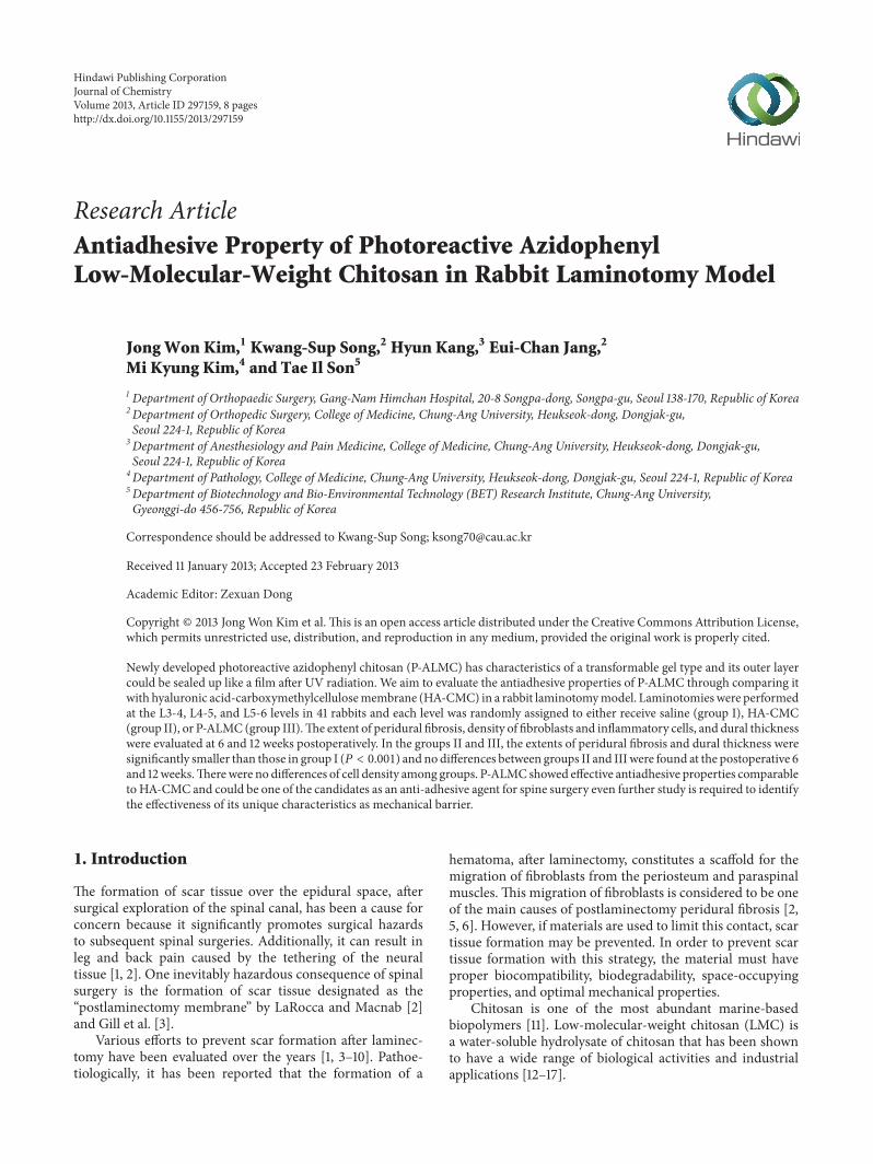
2.2. Synthesis and Photoreactivity Assay of Azidophenyl Chi-tosan Derivative. The photoreactivity of azidophenyl chi-tosan was determined using micropatterning. 30 𝜇L of20wt/wt % Azidophenyl chitosan solution was cast onto apolypropylene plate, and the cast azdidophenyl chitosan wasdried in the dark. Photomask was placed on the dried azi-dophenyl chitosan. With a photomask, azidophenyl chitosanwas irradiated with UV light for 3 minutes using a UV lamp(model: spot cure-9, Tokyo, Japan). During UV irradiation,the UV light lamp and sample were 5 cm apart, resulting inan intensity of 48mW/cm3. After UV light irradiation, thepolypropylene plate was washed three times with distilledwater for five minutes each time.
The micropattern illustrated the same pattern as that ofthe photomask (Figure 1). The azidophenyl chitosan reactedafter irradiation with UV light became insoluble in water andremained on plate, in contrast to the soluble nonirradiatedazidophenyl chitosan washed out.
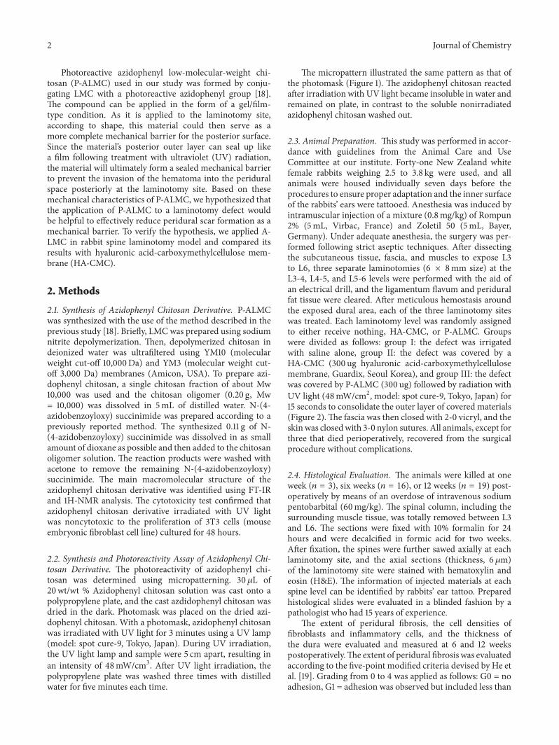
2.3. Animal Preparation. This study was performed in accor-dance with guidelines from the Animal Care and UseCommittee at our institute. Forty-one New Zealand whitefemale rabbits weighing 2.5 to 3.8 kg were used, and allanimals were housed individually seven days before theprocedures to ensure proper adaptation and the inner surfaceof the rabbits’ ears were tattooed. Anesthesia was induced byintramuscular injection of a mixture (0.8mg/kg) of Rompun2% (5mL, Virbac, France) and Zoletil 50 (5mL, Bayer,Germany). Under adequate anesthesia, the surgery was per-formed following strict aseptic techniques. After dissectingthe subcutaneous tissue, fascia, and muscles to expose L3to L6, three separate laminotomies (6 × 8mm size) at theL3-4, L4-5, and L5-6 levels were performed with the aid ofan electrical drill, and the ligamentum flavum and periduralfat tissue were cleared. After meticulous hemostasis aroundthe exposed dural area, each of the three laminotomy siteswas treated. Each laminotomy level was randomly assignedto either receive nothing, HA-CMC, or P-ALMC. Groupswere divided as follows: group I: the defect was irrigatedwith saline alone, group II: the defect was covered by aHA-CMC (300 ug hyaluronic acid-carboxymethylcellulosemembrane, Guardix, Seoul Korea), and group III: the defectwas covered by P-ALMC (300 ug) followed by radiation withUV light (48mW/cm2, model: spot cure-9, Tokyo, Japan) for15 seconds to consolidate the outer layer of covered materials(Figure 2).The fascia was then closed with 2-0 vicryl, and theskinwas closedwith 3-0 nylon sutures. All animals, except forthree that died perioperatively, recovered from the surgicalprocedure without complications.
2.4. Histological Evaluation. The animals were killed at oneweek (𝑛 = 3), six weeks (𝑛 = 16), or 12 weeks (𝑛 = 19) post-operatively by means of an overdose of intravenous sodiumpentobarbital (60mg/kg). The spinal column, including thesurrounding muscle tissue, was totally removed between L3and L6. The sections were fixed with 10% formalin for 24hours and were decalcified in formic acid for two weeks.After fixation, the spines were further sawed axially at eachlaminotomy site, and the axial sections (thickness, 6 𝜇m)of the laminotomy site were stained with hematoxylin andeosin (H&E). The information of injected materials at eachspine level can be identified by rabbits’ ear tattoo. Preparedhistological slides were evaluated in a blinded fashion by apathologist who had 15 years of experience.
The extent of peridural fibrosis, the cell densities offibroblasts and inflammatory cells, and the thickness ofthe dura were evaluated and measured at 6 and 12 weekspostoperatively.The extent of peridural fibrosis was evaluatedaccording to the five-point modified criteria devised by He etal. [19]. Grading from 0 to 4 was applied as follows: G0 = noadhesion, G1 = adhesion was observed but included less than
Journal of Chemistry 3
(a) (b)
Figure 1: Photomask (a) and micropatterned azidophenyl low-molecular-weight chitosan (P-ALMC) after UV irradiation (b).The pattern ofP-ALMC shows that P-ALMC reacted after irradiation with UV light and became insoluble in water, in contrast to the soluble nonirradiatedazidophenyl chitosan, is the same as the photomask.
HA-CMC
P-ALMC
L3-4
L4-5
L5-6
(a) (b) (c)
Figure 2: Operative fields in a rabbit model. (a) Pretreatment laminotomy windows. (b) After conditioning according to randomization, atL3-4 (control, group I), L4-5 (HA-CMC, group II), and L5-6 (P-ALMC, group III). (c) The consolidation at only outer surface of the coveredP-ALMC at L5-6 following UV radiation for 15 seconds was observed and its consistency was soft such like muscle.
one-third of the laminotomy defect, G2 = adhesion was morethan one-third and less than two-thirds, G3 = adhesion wasmore than two-thirds, and G4 = G3 and severely distorteddura due to severe scar tissue. If G0was observed, the shortestdistance from the dura to the posterior scar tissue within therange of the laminotomy site was measured.
Cell densities using fibroblast and inflammatory cellcounts were also evaluated, following the classification under400 times magnification by Hinton et al. [8] grade 1 for 100or fewer cells in every region; grade 2 for 100–150 cells inevery region; and grade 3 for 150 or more cells in everyregion. Fibroblasts and inflammatory cells were counted in
4 Journal of Chemistry
D
0.65 mm
(a)
D
0.65 mm
(b)
D
∗
0.65 mm
∗∗
∗
(c)
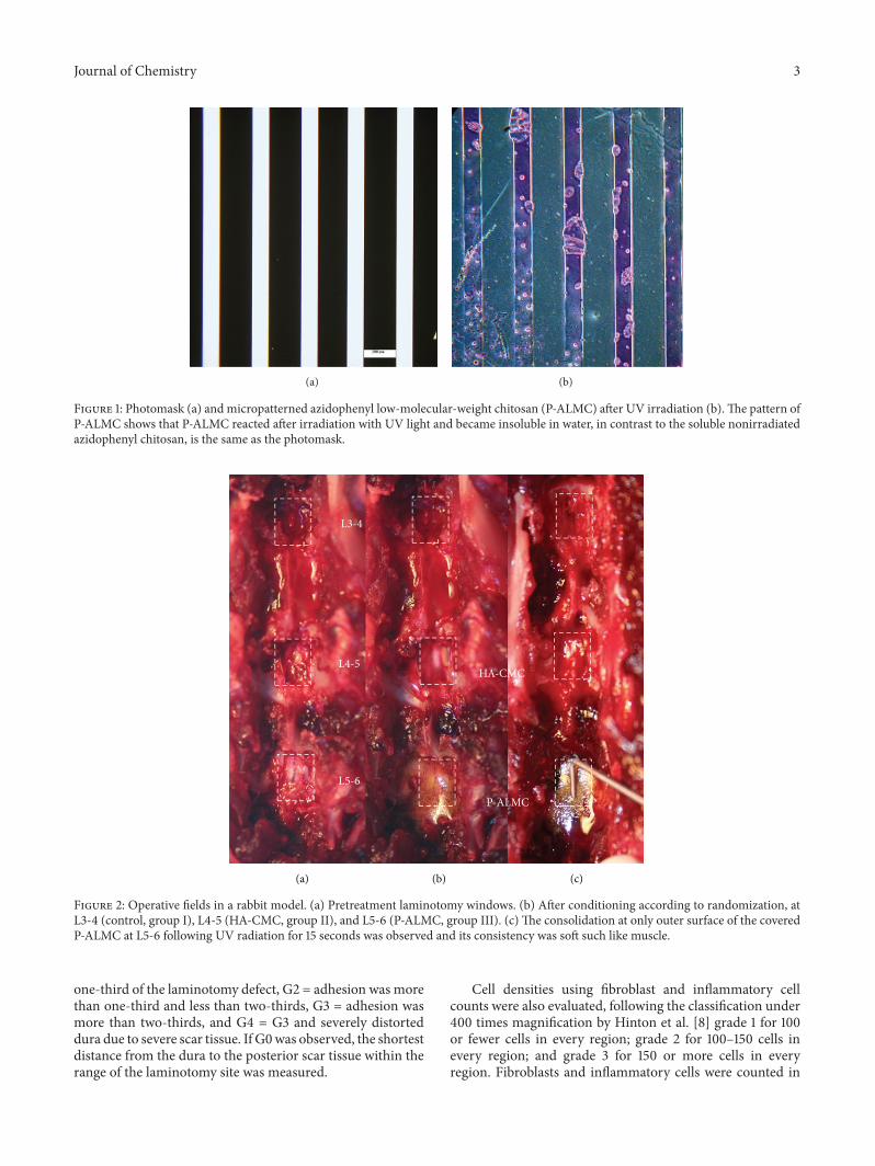
Figure 3: Histological sections of the laminotomy sites (between dotted lines) at one week ((a) control, group I; (b) HA-CMC, group II; (c)P-ALMC, group III). Group III showed that the clear space (asterisks) formed by the interpositional barrier of P-ALMC between the dura(D) and the posterior soft tissue is maintained in a comparison of groups I and II (H&E, ×1.25).
three fields located in the middle and at each margin of thelaminotomy.
Dural thicknesses were measured at three differentpoints, where the dura was the thickest within the marginsof the laminotomy area based on the judgment of thepathologist.
All evaluations were performed twice with 2-week inter-val by one pathologist. The above measurements of distancefrom dura to posterior scar tissue and dural thickness arepresented as average values.
2.4.1. Statistical Analysis. Adhesion, fibrosis score, and duralthickness were analyzed using the Kruskal-Wallis test fol-lowed by Boneferroni’s posttest. For categorical variable, Chi-squared test was used to correct for multiple comparisons.Data are reported as mean values, with variability expressedas standard deviations in the table and standard errorsin the figure. Interclass correlation coefficients (ICCs) andkappa reliability coefficient analyses were used to determinethe intraobserver agreements. Statistical analyses were per-formed with SPSS version 15.0 (SPSS Inc., Chicago, IL, USA),and significance was defined as 𝑃 < 0.05.
3. Results
Intraobserver agreements for grading of peridural fibrosisand the densities of cells were 𝑘 = 0.911 and 𝑘 = 0.874,respectively, and we considered evaluation at the second timeas the data. Intraobserver ICCs for measurement of distancefrom dura to scar tissue and dural thickness were 0.795 and0.784, respectively.
3.1. One Week Postoperatively. In three groups, hemorrhage,inflammatory cells, and some fibrous tissues were markedlyobserved on the posterior side of the dura. It was difficultto objectively compare those findings at the laminotomy siteamong the groups. However, there were tendency that the lessinfiltration of the hematoma into the spinal canal in groups IIand III were observed compared to group I. All three samplesin group III demonstrated the clear space considered as the
effects of mechanical barrier of P-ALMC between dura andposterior hematoma (Figure 3).
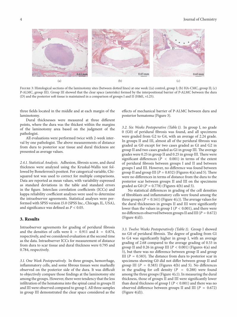
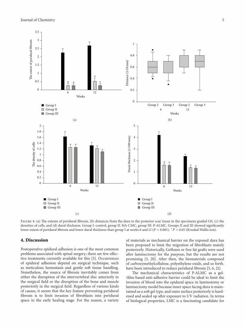
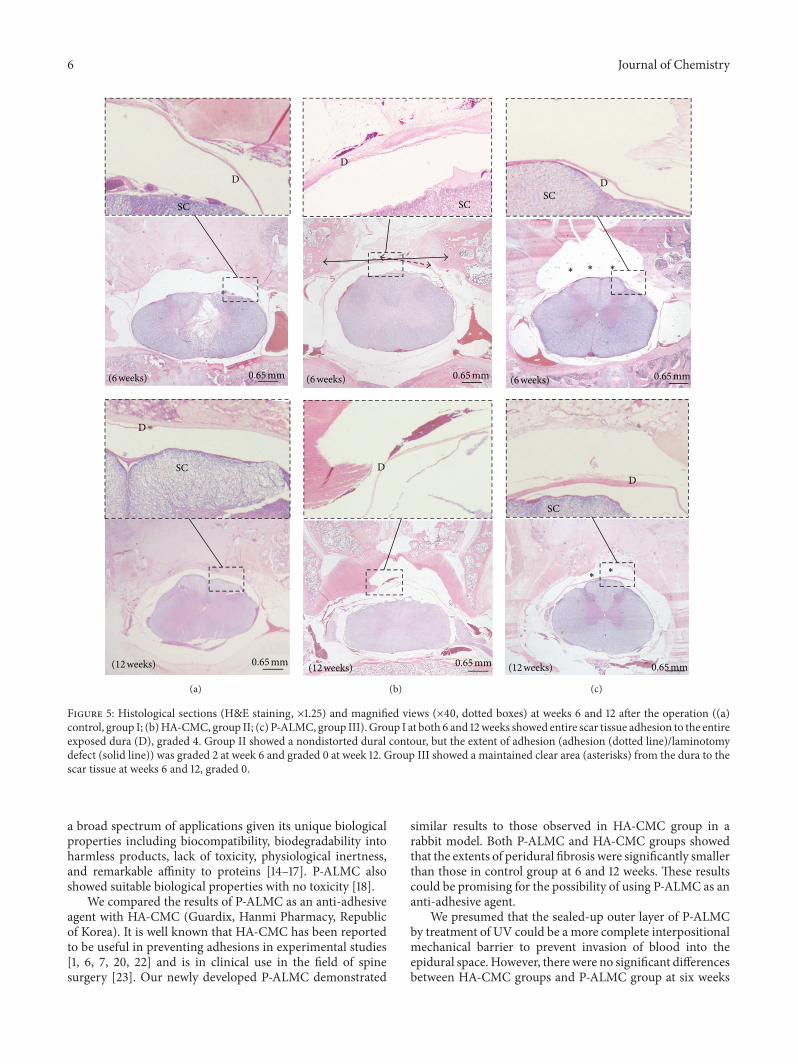
3.2. Six Weeks Postoperative (Table 1). In group I, no grade0 (G0) of peridural fibrosis was found, and all specimenswere graded from G2 to G4, with an average of 2.24 grade.In groups II and III, almost all of the peridural fibrosis wasgraded as G0 except for two cases graded as G1 and G2 ingroup II and two cases graded as G1 in group III. The averagegrades were 0.25 in group II and 0.25 in group III.There weresignificant differences (𝑃 < 0.001) in terms of the extentof peridural fibrosis between groups I and II and betweengroups I and III. However, no difference was found betweengroup II and group III (𝑃 = 0.832) (Figures 4(a) and 5).Therewere no differences in terms of distance from the dura to theposterior scar between groups II and III on the specimensgraded as G0 (𝑃 = 0.778) (Figures 4(b) and 5).
No statistical differences in grading of the cell densitiesof fibroblasts and inflammatory cells were found among thethree groups (𝑃 = 0.161) (Figure 4(c)).The average values forthe dural thicknesses in groups II and III were significantlylower than the values in group I (𝑃 < 0.001), and there wereno differences observed between groups II and III (𝑃 = 0.672)(Figure 4(d)).
3.3. Twelve Weeks Postoperatively (Table 1). Group I showedno G0 of peridural fibrosis. The degree of grading from G1to G4 was significantly higher in group I, with an averagegrading of 2.68 compared to the average grading of 0.53 ingroup II and 0.26 in group III (𝑃 < 0.001) (Figures 4(a) and5), but there was no difference between group II and groupIII (𝑃 = 0.583). The distance from dura to posterior scar inspecimens showing G0 did not differ between group II andgroup III (𝑃 = 0.583) (Figures 4(b) and 5). No differencesin the grading for cell density (𝑃 = 0.288) were foundamong the three groups (Figure 4(c)). Inmeasuring the duralthickness, those of groups II and III were significantly lowerthan dural thickness of group I (𝑃 < 0.001) and there was noobserved difference between groups II and III (𝑃 = 0.672)(Figure 4(d)).
Journal of Chemistry 5
3.5
3
2.5
2
1.5
1
0.5
06 12
Weeks
The e
xten
t of p
erid
ural
fibr
osis
Group IGroup IIGroup III
∗
∗
∗∗
(a)
1
0.8
0.6
0.4
0.2
0Group 2 Group 3
Dist
ance
(1/1
0 mm
)
Group 2 Group 36 12
Weeks
(b)
2
1.8
1.6
1.4
1.2
1
0.8
0.6
0.4
0.2
0
The d
ensit
y of
cells
6 12
Group IGroup IIGroup III
Weeks
(c)
5
4
3
2
1
0
Dur
al th
ickn
ess (
1/10
0 mm
)
6 12
Group IGroup IIGroup III
∗∗
∗∗
Weeks
(d)
Figure 4: (a) The extents of peridural fibrosis, (b) distances from the dura to the posterior scar tissue in the specimens graded G0, (c) thedensities of cells, and (d) dural thickness. Group I: control, group II: HA-CMC, group III: P-ALMC. Groups II and III showed significantlylower extent of peridural fibrosis and lower dural thickness than group I at weeks 6 and 12 (𝑃 < 0.001). ∗𝑃 < 0.05 (Kruskal-Wallis test).
4. Discussion
Postoperative epidural adhesion is one of the most commonproblems associated with spinal surgery; there are few effec-tive treatments currently available for this [5]. Occurrencesof epidural adhesion depend on surgical technique, suchas meticulous hemostasis and gentle soft tissue handling.Nonetheless, the source of fibrosis inevitably comes fromeither the disruption of the intervertebral disc anteriorly inthe surgical field or the disruption of the bone and muscleposteriorly in the surgical field. Regardless of various kindsof causes, it seems that the key feature preventing periduralfibrosis is to limit invasion of fibroblasts into periduralspace in the early healing stage. For the reason, a variety
of materials as mechanical barrier on the exposed dura hasbeen proposed to limit the migration of fibroblasts mainlyposteriorly. Historically, Gelfoam or free fat grafts were usedafter laminectomy for the purpose, but the results are notpromising [3, 20]. After then, the biomaterials composedof carboxymethylcellulose, polyethylene oxide, and so forth.have been introduced to reduce peridural fibrosis [5, 6, 21].
The mechanical characteristics of P-ALMC as a gel-/film-based anti-adhesive barrier could be ideal to limit theinvasion of blood into the epidural space in laminotomy orlaminectomymodel because inner space facing dura is main-tained as a soft gel type, and outer surface posteriorly is hard-ened and sealed up after exposure to UV radiation. In termsof biological properties, LMC is a fascinating candidate for
6 Journal of Chemistry
D
SC
D
SC
(12 weeks) 0.65 mm
(6 weeks) 0.65 mm
(a)
D
D
SC
(12 weeks) 0.65 mm
(6 weeks) 0.65 mm
(b)
SC
D
DSC
(12 weeks) 0.65 mm
(6 weeks) 0.65 mm
∗∗
∗∗∗
(c)
Figure 5: Histological sections (H&E staining, ×1.25) and magnified views (×40, dotted boxes) at weeks 6 and 12 after the operation ((a)control, group I; (b)HA-CMC, group II; (c) P-ALMC, group III). Group I at both 6 and 12weeks showed entire scar tissue adhesion to the entireexposed dura (D), graded 4. Group II showed a nondistorted dural contour, but the extent of adhesion (adhesion (dotted line)/laminotomydefect (solid line)) was graded 2 at week 6 and graded 0 at week 12. Group III showed a maintained clear area (asterisks) from the dura to thescar tissue at weeks 6 and 12, graded 0.
a broad spectrum of applications given its unique biologicalproperties including biocompatibility, biodegradability intoharmless products, lack of toxicity, physiological inertness,and remarkable affinity to proteins [14–17]. P-ALMC alsoshowed suitable biological properties with no toxicity [18].
We compared the results of P-ALMC as an anti-adhesiveagent with HA-CMC (Guardix, Hanmi Pharmacy, Republicof Korea). It is well known that HA-CMC has been reportedto be useful in preventing adhesions in experimental studies[1, 6, 7, 20, 22] and is in clinical use in the field of spinesurgery [23]. Our newly developed P-ALMC demonstrated
similar results to those observed in HA-CMC group in arabbit model. Both P-ALMC and HA-CMC groups showedthat the extents of peridural fibrosis were significantly smallerthan those in control group at 6 and 12 weeks. These resultscould be promising for the possibility of using P-ALMC as ananti-adhesive agent.
We presumed that the sealed-up outer layer of P-ALMCby treatment of UV could be a more complete interpositionalmechanical barrier to prevent invasion of blood into theepidural space. However, there were no significant differencesbetween HA-CMC groups and P-ALMC group at six weeks
Journal of Chemistry 7
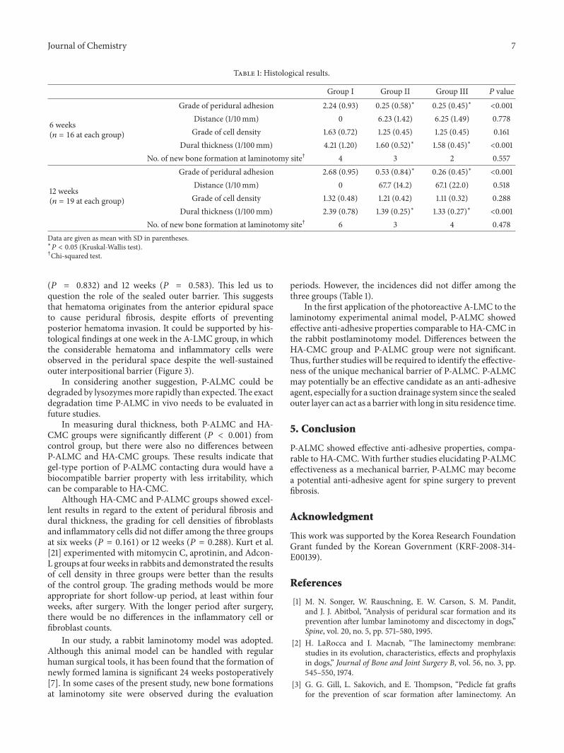
Table 1: Histological results.
Group I Group II Group III 𝑃 value
6 weeks(𝑛 = 16 at each group)
Grade of peridural adhesion 2.24 (0.93) 0.25 (0.58)∗ 0.25 (0.45)∗ <0.001Distance (1/10mm) 0 6.23 (1.42) 6.25 (1.49) 0.778Grade of cell density 1.63 (0.72) 1.25 (0.45) 1.25 (0.45) 0.161
Dural thickness (1/100mm) 4.21 (1.20) 1.60 (0.52)∗ 1.58 (0.45)∗ <0.001No. of new bone formation at laminotomy site† 4 3 2 0.557
12 weeks(𝑛 = 19 at each group)
Grade of peridural adhesion 2.68 (0.95) 0.53 (0.84)∗ 0.26 (0.45)∗ <0.001Distance (1/10mm) 0 67.7 (14.2) 67.1 (22.0) 0.518Grade of cell density 1.32 (0.48) 1.21 (0.42) 1.11 (0.32) 0.288
Dural thickness (1/100mm) 2.39 (0.78) 1.39 (0.25)∗ 1.33 (0.27)∗ <0.001No. of new bone formation at laminotomy site† 6 3 4 0.478
Data are given as mean with SD in parentheses.∗𝑃 < 0.05 (Kruskal-Wallis test).†Chi-squared test.
(𝑃 = 0.832) and 12 weeks (𝑃 = 0.583). This led us toquestion the role of the sealed outer barrier. This suggeststhat hematoma originates from the anterior epidural spaceto cause peridural fibrosis, despite efforts of preventingposterior hematoma invasion. It could be supported by his-tological findings at one week in the A-LMC group, in whichthe considerable hematoma and inflammatory cells wereobserved in the peridural space despite the well-sustainedouter interpositional barrier (Figure 3).
In considering another suggestion, P-ALMC could bedegraded by lysozymesmore rapidly than expected.The exactdegradation time P-ALMC in vivo needs to be evaluated infuture studies.
In measuring dural thickness, both P-ALMC and HA-CMC groups were significantly different (𝑃 < 0.001) fromcontrol group, but there were also no differences betweenP-ALMC and HA-CMC groups. These results indicate thatgel-type portion of P-ALMC contacting dura would have abiocompatible barrier property with less irritability, whichcan be comparable to HA-CMC.
Although HA-CMC and P-ALMC groups showed excel-lent results in regard to the extent of peridural fibrosis anddural thickness, the grading for cell densities of fibroblastsand inflammatory cells did not differ among the three groupsat six weeks (𝑃 = 0.161) or 12 weeks (𝑃 = 0.288). Kurt et al.[21] experimented with mitomycin C, aprotinin, and Adcon-L groups at fourweeks in rabbits and demonstrated the resultsof cell density in three groups were better than the resultsof the control group. The grading methods would be moreappropriate for short follow-up period, at least within fourweeks, after surgery. With the longer period after surgery,there would be no differences in the inflammatory cell orfibroblast counts.
In our study, a rabbit laminotomy model was adopted.Although this animal model can be handled with regularhuman surgical tools, it has been found that the formation ofnewly formed lamina is significant 24 weeks postoperatively[7]. In some cases of the present study, new bone formationsat laminotomy site were observed during the evaluation
periods. However, the incidences did not differ among thethree groups (Table 1).
In the first application of the photoreactive A-LMC to thelaminotomy experimental animal model, P-ALMC showedeffective anti-adhesive properties comparable to HA-CMC inthe rabbit postlaminotomy model. Differences between theHA-CMC group and P-ALMC group were not significant.Thus, further studies will be required to identify the effective-ness of the unique mechanical barrier of P-ALMC. P-ALMCmay potentially be an effective candidate as an anti-adhesiveagent, especially for a suction drainage system since the sealedouter layer can act as a barrierwith long in situ residence time.
5. Conclusion
P-ALMC showed effective anti-adhesive properties, compa-rable to HA-CMC.With further studies elucidating P-ALMCeffectiveness as a mechanical barrier, P-ALMC may becomea potential anti-adhesive agent for spine surgery to preventfibrosis.
Acknowledgment
This work was supported by the Korea Research FoundationGrant funded by the Korean Government (KRF-2008-314-E00139).
References
[1] M. N. Songer, W. Rauschning, E. W. Carson, S. M. Pandit,and J. J. Abitbol, “Analysis of peridural scar formation and itsprevention after lumbar laminotomy and discectomy in dogs,”Spine, vol. 20, no. 5, pp. 571–580, 1995.
[2] H. LaRocca and I. Macnab, “The laminectomy membrane:studies in its evolution, characteristics, effects and prophylaxisin dogs,” Journal of Bone and Joint Surgery B, vol. 56, no. 3, pp.545–550, 1974.
[3] G. G. Gill, L. Sakovich, and E. Thompson, “Pedicle fat graftsfor the prevention of scar formation after laminectomy. An
8 Journal of Chemistry
experimental study in dogs,” Spine, vol. 4, no. 2, pp. 176–186,1979.
[4] M. A. Sandoval and D. Hernandez-Vaquero, “Preventingperidural fibrosis with nonsteroidal anti-inflammatory drugs,”European Spine Journal, vol. 17, no. 3, pp. 451–455, 2008.
[5] G. S. DiZerega, S. Cortese, K. E. Rodgers et al., “A modernbiomaterial for adhesion prevention,” Journal of BiomedicalMaterials Research Part B, vol. 81, no. 1, pp. 239–250, 2007.
[6] M. O. Kasimcan, B. Bakar, S. Akta, A. Alhan, and M. Yilmaz,“Effectiveness of the biophysical barriers on the peridural fibro-sis of a postlaminectomy rat model: an experimental research,”Injury, vol. 42, no. 8, pp. 778–781, 2011.
[7] T. Kato, H. Haro, H. Komori, and K. Shinomiya, “Evaluationof hyaluronic acid sheet for the prevention of postlaminectomyadhesions,” Spine Journal, vol. 5, no. 5, pp. 479–488, 2005.
[8] J. L. Hinton Jr., D. J. Warejcka, Y. Mei et al., “Inhibition ofepidural scar formation after lumbar laminectomy in the rat,”Spine, vol. 20, no. 5, pp. 564–570, 1995.
[9] R. Henderson, B. Weir, L. Davis, B. Mielke, and M. Grace,“Attempted experimental modification of the postlaminec-tomy membrane by local instillation of recombinant tissue-plasminogen activator gel,” Spine, vol. 18, no. 10, pp. 1268–1272,1993.
[10] T. E. Kuivila, J. L. Berry, G. R. Bell, and A. D. Steffee,“Heparinized materials for control of the formation of thelaminectomy membrane in experimental laminectomies indogs,” Clinical Orthopaedics and Related Research, no. 236, pp.166–174, 1988.
[11] R. Muzzarelli, V. Baldassarre, F. Conti et al., “Biological activityof chitosan. Ultrastructural study,” Biomaterials, vol. 9, no. 3, pp.247–252, 1988.
[12] F. Shahidi and R. Abuzaytoun, “Chitin, chitosan, and co-products: chemistry, production, applications, and healtheffects,” Progress in Optics, vol. 49, pp. 93–135, 2005.
[13] T. Freier, H. S. Koh, K. Kazazian, and M. S. Shoichet, “Con-trolling cell adhesion and degradation of chitosan films by N-acetylation,” Biomaterials, vol. 26, no. 29, pp. 5872–5878, 2005.
[14] E. Khor and L. Y. Lim, “Implantable applications of chitin andchitosan,” Biomaterials, vol. 24, no. 13, pp. 2339–2349, 2003.
[15] A. K. Singla and M. Chawla, “Chitosan: some pharmaceuticaland biological aspects—an update,” Journal of Pharmacy andPharmacology, vol. 53, no. 8, pp. 1047–1067, 2001.
[16] S. Tokura, H. Tamura, and I. Azuma, “Immunological aspectsof chitin and chitin derivatives administered to animals,” Expe-rientia, vol. 87, pp. 279–292, 1999.
[17] T. Mori, M. Okumura, M. Matsuura et al., “Effects of chitin andits derivatives on the proliferation and cytokine production offibroblasts in vitro,” Biomaterials, vol. 18, no. 13, pp. 947–951,1997.
[18] K. I. Kim, J. W. Lee, Y. Ito et al., “Preparation of photo-reactiveazidophenyl chitosan derivative for immobilization of growthfactors,” Journal of Applied Polymer Science, vol. 117, no. 5, pp.3029–3037, 2010.
[19] Y. He, M. Revel, and B. Loty, “A quantitative model of post-laminectomy scar formation: effects of a nonsteroidal anti-inflammatory drug,” Spine, vol. 20, no. 5, pp. 557–563, 1995.
[20] J. J. Abitbol, T. L. Lincoln, B. I. Lind, D. Amiel, W. H. Akeson,and S. R. Garfin, “Preventing postlaminectomy adhesion: a newexperimental model,” Spine, vol. 19, no. 16, pp. 1809–1814, 1994.
[21] G. Kurt, M. H. Aytar, F. Dogulu et al., “A comparison of thelocal effectiveness of mitomycin C, aprotinin, and Adcon-L in
experimental peridural fibrosis,” Surgical Neurology, vol. 70, no.6, pp. 608–613, 2008.
[22] C.H. Yu, J. H. Lee,H. R. Baek, andH.Nam, “The effectiveness ofpoloxamer 407-based new anti-adhesive material in a laminec-tomy model in rats,” European Spine Journal, vol. 21, no. 5, pp.971–979, 2012.
[23] H. B. Lin, J. H. Dai, X. W. Wu et al., “Prevention of duraadherence in spinal canal after microendoscopic discectomyby different methods: a clinical study of 165 cases,” NationalMedical Journal of China, vol. 87, no. 43, pp. 3085–3087, 2007.
Submit your manuscripts athttp://www.hindawi.com
Hindawi Publishing Corporationhttp://www.hindawi.com Volume 2014
Inorganic ChemistryInternational Journal of
Hindawi Publishing Corporation http://www.hindawi.com Volume 2014
International Journal ofPhotoenergy
Hindawi Publishing Corporationhttp://www.hindawi.com Volume 2014
Carbohydrate Chemistry
International Journal of
Hindawi Publishing Corporationhttp://www.hindawi.com Volume 2014
Journal of
Chemistry
Hindawi Publishing Corporationhttp://www.hindawi.com Volume 2014
Advances in
Physical Chemistry
Hindawi Publishing Corporationhttp://www.hindawi.com
Analytical Methods in Chemistry
Journal of
Volume 2014
Bioinorganic Chemistry and ApplicationsHindawi Publishing Corporationhttp://www.hindawi.com Volume 2014
SpectroscopyInternational Journal of
Hindawi Publishing Corporationhttp://www.hindawi.com Volume 2014
The Scientific World JournalHindawi Publishing Corporation http://www.hindawi.com Volume 2014
Medicinal ChemistryInternational Journal of
Hindawi Publishing Corporationhttp://www.hindawi.com Volume 2014
Chromatography Research International
Hindawi Publishing Corporationhttp://www.hindawi.com Volume 2014
Applied ChemistryJournal of
Hindawi Publishing Corporationhttp://www.hindawi.com Volume 2014
Hindawi Publishing Corporationhttp://www.hindawi.com Volume 2014
Theoretical ChemistryJournal of
Hindawi Publishing Corporationhttp://www.hindawi.com Volume 2014
Journal of
Spectroscopy
Analytical ChemistryInternational Journal of
Hindawi Publishing Corporationhttp://www.hindawi.com Volume 2014
Journal of
Hindawi Publishing Corporationhttp://www.hindawi.com Volume 2014
Quantum Chemistry
Hindawi Publishing Corporationhttp://www.hindawi.com Volume 2014
Organic Chemistry International
ElectrochemistryInternational Journal of
Hindawi Publishing Corporation http://www.hindawi.com Volume 2014
Hindawi Publishing Corporationhttp://www.hindawi.com Volume 2014
CatalystsJournal of