repub.eur.nl filerepub.eur.nlauthor: j.g.m. huijmanspublish year: 1989
TRANSCRIPT
HYDROXYLATED STEROLS METABOLISM AND EFFECTS ON STEROID PRODUCTION
AND STEROID UPTAKE
GEHYDROXYLEERDESTEROLEN METABOLISME EN EFFECTEN OP DE STEROID PRODUCTIE
EN STEROID OPNAME
PROEFSCHRIFT
TER VERKRIJGING VAN DE GRAAD VAN DOCTOR AAN DE ERASMUS UNIVERSITEIT ROTTERDAM
OP GEZAG VAN DE RECTOR MAGNIFICUS PROE DR. A.H.G. RINNOOY KAN
EN VOLGENS BESLUIT VAN HET COLLEGE VAN DEKANEN. DE OPENBARE VERDEDIGING ZAL PLAATSVINDEN OP
WOENSDAG 10 MEl 1989 OM 13.45 UUR
DOOR
JOHANNES GERARDUS MARIA HUIJMANS
GEBOREN TE AMSTERDAM
Gedrukt bij Offsetdrukkerij Kanters B.V., Alblasserdam 1989
PROMOTIECOMMISSIE
Promotoren:
Overige leden:
Prof. Dr. HJ. Degenhart Prof. Dr. J.H.H. Thijssen Prof. Dr. H. Galjaard Dr. F.H. de J ong
De uitvoering van het onderzoek is mogelijk gemaakt door de financiele steun van de Sophia Stichting Wetenschappelijk Onderzoek. De uitgave van dit proefschrift is mede mogelijk gemaakt dank zij een financiele bijdrage van Pharmacia-LKB, Biotechnologie Divisie, te Woerden.
Aan Ne' Saskia, Remco en Judith Aan mijn Moeder. Ter nagedachtenis aan mijn Vader.


List of abbreviations. 7
List of trivial names. 9
Chapter 1: General introduction and statement of the problem. 11
Cllapter 2: Review of the literature. 17 2.1 '!he adrenal gland 17 2. 2 steroidogenesis in the adrenal cortex 20 2. 3 Mechanism of action of ACIH 28 2.4 Aldosterone production and its regulation 36 2.5 Cholesterol and hydroxylated sterols 44 2.6 Hydroxylase activity in the liver 53 2. 7 '!he human fetal adrenal gland 56
Chapter 3: Methodology. 67
Chapter 4:
Chapter 5:
3A: Technical aspects 3A.1 Introduction 67 3A.2 Isolation of cells 67 3A.3 Incubation conditions 69 3A. 4 Fluorometry 69 3A. 5 Gaschramatography 71 3A.6 ATP 72 3A.7 cyclic AMP 74 3A. 8 Aldosterone 74 3A. 9 Density gradient centrifugation 74
3B: Application and evaluation of the methods 3B.1 Introduction 76 3B.2 Effect of ACTH stimulation 76 3B.3 Effect of cell numbers 77 3B.4 Incubation buffer 78 3B. 5 Human fetal adrenals
3B.5.1 Fetal adrenals 81 3B.5.2 Cell viability 83 3B. 5. 3 3,8-HSD activity 83 3B.5.4 Fetal adrenal cell separation 84
Effects of 22S-hydroxy-cholesterol and other 89 hydroxylated sterols on the ACIH-stimulated steroid production in rat adrenal cells. Acta Endocrinol. (Copenh) 97: 243-250 (1981).
Corwersion of 22S-hydroxy-cholesterol and its 99 effect on the metabolism of other sterols in rat adrenal cells and bovine adrenal mitcx::::hondria. Acta Endocrinol. (Copenh) 100: 599-605 (1982).

C11apter 6:
<llapter 7:
<llapter 8:
<llapter 9:
<llapter 10:
<llapter 11:
<llapter 12:
Effects of sane , hydroxylated sterols on the 107 steroid . production in iSolated rat adrenal glomenilosa ani fasciculatajreticularis cells in the presence of potassium, Angiotensin II or Acm. Acta Endocrinol. (Oopenh) 105: 411-416 (1984).
Effect of 25-hydroxy-cholesterol on the uptake of 115 corticosterone in isolated rat liver cells. Ho:rm. Metabol. Res. 20: 28-31 {1988) •
Effect of hydroxylated sterols on the uptake of 121 pregnenolone in isolated rat adrenal cells.
'!he hl.mlan fetal adrenal ani hydroxylated sterols. 127
General discussion. 137
SUl'nmal:y. 143
Samenvatting. 147
List of references. 151
Omkwoord. 183
curriculum vitae. 185
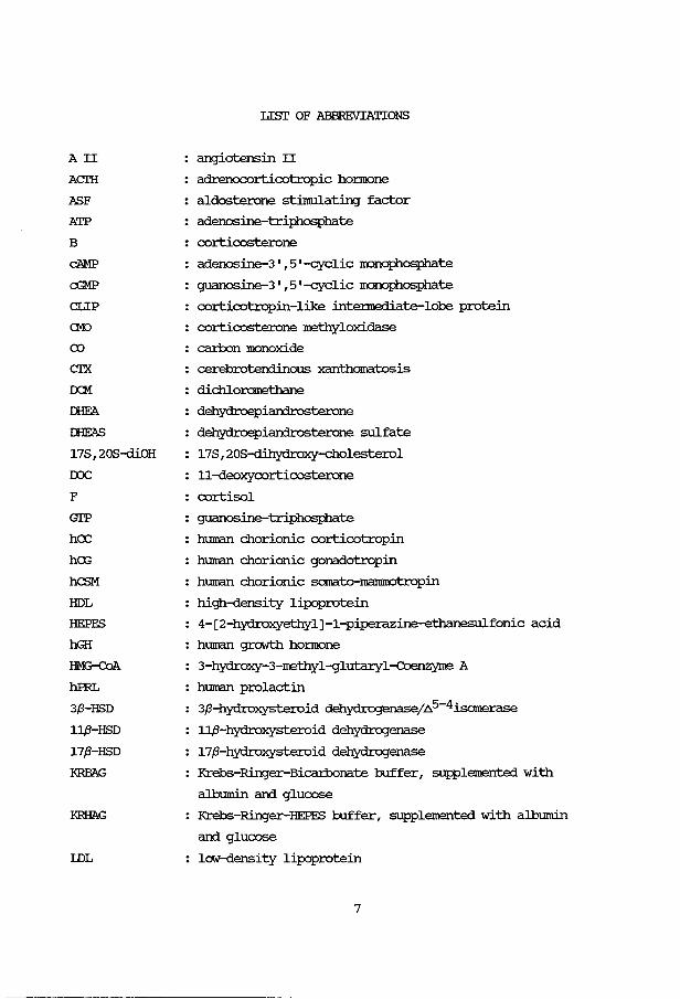
AII
ACIH
ASF
ATP
B
cAMP
cGMP
CLIP
CMJ
(X)
crx DCM
J:HFA
I:HEAS
17S 1 20S-diOH
roc F
Gl'P
hCC
hex;
hCSM
HDL
HEPFS
hGH
HM3-CaA.
hPRL
3,8-HSD
11,8-HSD
17,8-HSD
:KRBAG
IDL
Lisr OF ABBREVIATIONS
angiotensin II
adrenocorticotropic honnone
aldosterone stimulating factor
adenosine-triphosphate
corticosterone
adenosine-3 1 1 5 1 -cyclic monophosphate
guanosine-3 1 1 5 1 -cyclic monoph.osphate
corticotropin-like intennediate-lobe protein
corticosterone methyloxidase
carbon monoxide
cerebrotendinous xanthomatosis
dichloromethane
dehydroepiandrosterone
dehydroepiandrosterone sulfate
17S 1 20S-dihydroxy-cholesterol
11-deoxycorticosterone
cortisol
guanosine-triphosphate
human chorionic corticotropin
human chorionic gonadotropin
human chorionic sornato-manunotropin
high-density lipoprotein
4-[2-hydroxyethyl] -1-piperazine-ethanesulfonic acid
human growth honnone
3-hydroxy-3-methyl-gluta:ryl-coenzyme A
human prolactin
3,8-hydroxysteroid dehydrogenase/.6.5-4isomerase
11,8-hydroxysteroid dehydrogenase
17,8-hydroxysteroid dehydrogenase
Krebs-Ringer-Bicarbonate buffer 1 supplemented with
albumin and glucose
Krebs-Ringer-HEPFS buffer 1 supplemented with albumin
and glucose
low-density lipoprotein
7
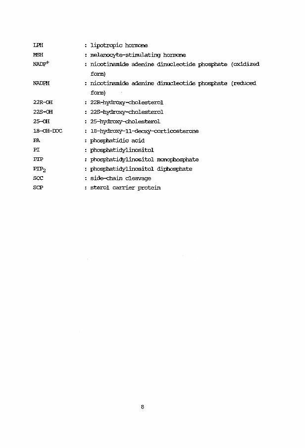
LFH
MSH
NADp+
NADIR
22R-oH
228-oH
25-QH
18-QH-IX>C
PA
PI
PIP
PIP2
sec SCP
: lipotropic honnone
: melanocyte-stimulating honnone
nicotinamide adenine dinucleotide phosphate (oxidized
fonn)
nicotinamide adenine dinucleotide phosphate (reduced
fonn)
22R-hydroxy-cholesterol
228-hydroxy-cholesterol
25-hydroxy-cholesterol
18-hydroxy-11-deoxy-corticosterone
: phosphatidic acid
: phosphatidylinositol
: phosphatidylinositol monophosphate
: phosphatidylinositol diphosphate
side-chain cleavage
: sterol carrier protein
8
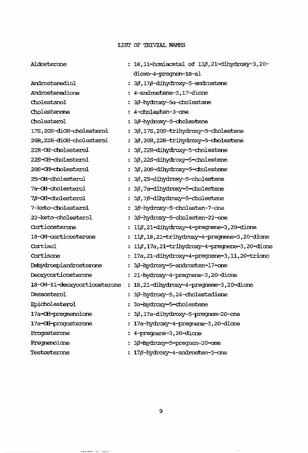
LIST OF TRIVIAL NAMES
Aldosterone
Androstenediol
Androstenedione
O:J.olestanol
O:J.olestenone
O:J.olesterol
17S 1 20S-diOH-cholesterol
20R1 22R-diOH-cholesterol
22R-GH-cholesterol
22S-QH-cholesterol
20S-oH-cholesterol
25-oH-cholesterol
7a-oH-cholesterol
7,8-GH-cholesterol
7-keto-cholesterol
22-keto-cholesterol
Corticosterone
18-GH-corticosterone
Cortisol
Cortisone
Dehydroepiandrosterone
Deoxycorticosterone
18-oH-11-deoxycorticosterone
D:smosterol
Epicholesterol
17a-GH-pregnenolone
17a-oH-progesterone
Progesterone
Pregnenolone
Testosterone
18 1 11-herniacetal of 11,8 1 21-dihydroxy-3 1 20-
dioxo-4-pregnen-18-al
3,8 1 17,8-dihydroxy-5-androstene
4-androstene-3 1 17-dione
3,8-hydroxy-Sa-cholestane
4-cholesten-3-one
3,8-hydroxy-5-cholestene
3,8 1 17S 1 20S-trihydroxy-5-cholestene
3,8 1 20R1 22R-trihydroxy-5-cholestene
3,8 1 22R-dihydroxy-5-cholestene
3,8 1 22S-dihydroxy-5-cholestene
3,8 1 20S-dihydroxy-5-cholestene
3,8 1 25-dihydroxy-5-cholestene
3,8 1 7a-d:ihydroxy-5-cholestene
3,8 1 7,8-dihydroxy-5-cholestene
3,8-hydroxy-5-cholesten-7-one
3,8-hydroxy-5-cholesten-22-one
11,8 1 21-dihydroxy-4-pregnene-3 1 20-dione
11,81 18 1 21-trihydroxy-4-pregnene-3 1 20-dione
11,81 17a1 21-trihydroxy-4-pregnene-3 1 20-dione
17a1 21-dihydroxy-4-pregnene-3 1 111 20-trione
3,8-hydroxy-5-androsten-17-one
21-hydroxy-4-pregnene-3 1 20-dione
18 1 21-dihydroxy-4-pregnene-3 1 20-dione
3,8-hydroxy-51 24-cholestadiene
3a-hydroxy-5-cholestene
3,8 1 17a-dihydroxy-5-pregnen-20-one
17a-hydroxy-4-pregnene-3 1 20-dione
4-pregnene-3 1 20-dione
3,8-hydroxy-5-pregnen-20-one
17,8-hydroxy-4-androsten-3-one
9


<liAPI'ER 1
GENERAL INI'ROIXJCITON AND STATEMENl' OF 'lHE PROBllM
Many hereditacy enzyme defects lead to the accumulation of
metabolites. Frequently the accumulated products are converted into other
metabolites via pathways, that are nonnally of minor importance. 'Ihese
products or their metabolites may cause profound cl:isturbances of
}ilysiological processes. Several inborn errors in steroid biosynthesis
provide examples of this }ilenornenon: the steroid 21- ani 11,8-hydroxylase
deficiencies lead to ove:rproduction of androgens, which are responsible for
ext.rene masculinization ani abnonnal grt:Mth.
Several clinical conditions are knc:mn, in which cholesterol or another
sterol accumulates in tissues or body fluids, due to an enzymatic defect in
sterol metabolism. Analogous to the situation found in other enzyme
defects, production of "unusual sterols" might be expected in these
cor:ditions. In this context, "unusual sterols" are defined as sterols not
nonnally occurring as a metabolite or in trace amounts only. '!his
phenomenon has .irrleed been observed, as will be discussed below. SUch
sterols might contribute to the severity of the disease.
'lhe existence of unustial sterols, having unexpected biological
activity, is well-kna.m (25-hydroxy-cholesterol, 3/3, 208-dihydroxy-cholenic
acid, toad poisons, etc.). However, much remains to be learned about their
effects ani mechanisms of action. For this reason we evaluated short-term
effects of several sterols on steroid production ani steroid uptake in
isolated rat adrenal ani liver cells ani on steroid production in isolated
human fetal adrenal cells. 'lhese sterols were selected according to two
criteria:
1. not nonnally occurring as a metabolite or in trace amounts only,
2. being a potential metabolite of cholesterol.
Disturbances of sterol metabolism
'!he accmnulation of sterols in tissue ani body fluids probably plays a
role in the etiology of several diseases.
11
one such condition is the c:x:D]E!lital lipoid adJ:ena1 hype:tplasia
(Prader's syrrlrame; for a review see Degenhart, 1984). '!his inbom error of
cholesterol side-chain cleavage results in a massive lipoid hyperplasia of
both adrenals. The mortality rate is remarkably high. The absence of
steroid production per se is not lethal, as children with a congenital
absence of the adrenals can be treated with relative ease (Pakravan et al.,
1974). The possibility of the production of mrusual sterols has been
discussed (Falke et al., 1975a).
In cerebrot:end.:i :xanthanatosis (CI'X) a deficiency of the
26-hydroxylase activity results in a diminished production of bile acids.
As a consequence the cholesterol production rate is increased, leading to
the fonnation of cholestanol (Walthers et al., 1983). '!his sterol
accumulates in plasma an:i several tissues like brain (leading to mental
retardation), ter:rl.ons (leading to swelling) an:i the eyes (giving
cataract). A cc::lllt=llex of cholestanol, cholesterol an:i 2 molecules of water
(C-c-2W) might be responsible for the deposits in brain an:i ter:rl.ons. c-c-2w
exerts adverse effects, including inflanmation, cell necrosis an:i
destruction of cell nernbranes, on liver, intima tissue, eyes, lungs an:i
other parts of the body.
In :familial hype:rdlolesterolania a deficiency of cell surface
receptors for lOW" density lipoprotein (IDL) has been found, leading to a
disturbance in the regulation of IDL degradation an:i cholesterol synthesis
(Brc::Mn an:i Goldstein, 1974; Goldstein an:i Brc::Mn, 1984) . The cholesterol
concentration in plasma is greatly increased. '!his disease is clinically
characterized by premature coronary heart disease an:i atherosclerosis.
Fatty acid esters of 24-0H:-c:holesterol an:i 26-QH-c:holesterol occur in hmnan
aortal tissue; these sterols accumulate with increasing severity of
atherosclerosis (Teng an:i Smith, 1975) . Also the free 26-QH-cholesterol
concentration in aortal tissue increases with the advancement of this
disease (Smith an:i Van Lier, 1970) . rue to the high cholesterol
concentration, c-c-2w (see above) nay be an .inportant factor in
atherosclerosis.
In WOlman's disease (Wolman et al., 1961) an:i in the less severe
dloleste:ryl ester storage disease (Sloan an:i Fredrickson, 1972) a lysosomal
acid lipase is reduced in activity. '!his leads to an abnonnal lysosomal
accumulation of cholesteryl esters an:i triglycerides an:i an increased
cholesterol synthesis. Patients suffering from Wolman's disease generally
12
die before the age of one year with syrrptcm1s like hepatosplenomegaly and
adrenal calcification. '!he stel:yl ester fraction of the liver of a patient
with Wolman's disease contained esters of 7a.- and 7,8-hydroxy-c:holesterol,
7-keto-c:holesterol and 5, 6a.- and 5, 6,8-epoxy-c:holesterol in addition to
cholesterol (Assmann et al., 1975).
'!here is a group of pe:rax::i.saoal d:ism:deJ::s (a.o. the cerebro-hepato
renal syndrome of Zellweger and adrenole1.lkcx:lyst:rqiy) in which a
deficiency of peroxisames or an impainnent of peroxisomal metabolism leads
also to an impaired conversion of cholesterol into bile acids by a
deficient c24-c25 cleavage (Schutgens et al., 1987). As a consequence bile
alcohols with an ext:errled side-chain accumulate in the liver and are
excreted in the bile (Pannentier et al. , 1979; Janssen et al. , 1982) • '!he
clinical syrrptcm1s are nruscular hypotonia, liver enlargenent, renal cysts,
craniofacial roa.lfonnations and mental retardation. '!he Zellweger syndrome
and the neonatal adrenoleuko-dystroph.y are lethal at a very young age.
In dloleste:rol dlolelithiasis, which most probably is not hereditary,
enhanced hepatic cholesterol secretion is associated with a deficient bile
acid pool. '!he amount of cholesterol is too great for the bile acid
micelles to dissolve. As a result cholesterol crystals precipitate, which
may aggregate to fonn gallstones. '!he initial CClll1pOUirl deposited is the
complex of cholesterol, cholestanol (which always accompanies cholesterol
in our daily food) and water (C-c-2W).
In vitro occurrence of unusual sterols
In vitro the occurrence of unusual sterols has been shoon under
conditions in which the nonnal cholesterol side-chain cleavage is
impaired. In bovine adrenal mitochondria Alsema. et al. (1980) observed
25-hydroxylation of 208~-c:holesterol, besides the nonnal pregnenolone
prcx:luction, when the incubation was carried out at pH 7 .8. Deg-enhart et al.
(1984) observed a 25-hydroxylase activity, hydroxylating 208-hydroxy-
4-c:holesten-3-one, when this sterol was incubated with bovine adrenal
cortex mitochondria. Alsema. et al. (1982a) also showed the conversion of
several c24 and c27 3,8-hydroxy-5-ene sterols, same of which are nonnal
side-chain cleavage intennediates, into 3-keto-.6.4 prcx:lucts by bovine
adrenal cortex 3,8-hydroxysteroid dehydrogenase. Huijmans et al. (1982)
showed the fonnation of 22-keto-c:holesterol from 228~-c:holesterol in
bovine adrenal mitochondria.
13

Falke et al. (1976a) discussed the possibility of a metabolite with
i.nhibito:ry properties on the steroid synthesis in rat adrenal cells urrler
corrlitions of a blocked cholesterol side-chain cleaving enzyme system.
Effects of exogenous sterols
Several in vivo am in vitro effects of sterols have been described.
In vivo growth retardation am adrenal hyperplasia could be observed by
feeding cholestenone to rats (Degenhart et al., 1981) . Administration of
25-oH-cholestenone to pregnant rats caused severe malfomations of head
am spinal cord in the offspring (H.J. Degenhart, personal canum.mication) •
In vivo administration of hydroxylated sterols to rabbits caused fibrotic
lesions in the aorta am pul.Itnnary arteries (Imai et al., 1980).
In vitro marked effects of hydroxylated sterols on cultured rabbit aorta
SiroOI:h. muscle cells (:J?en;J et al., 1979), cell proliferation (Olen et al.,
1974), erxiocytosis of peroxidase (Heiniger et al., 1976) am uptake am
efflux of ions (Olen et al., 1978) are known. 'Ihese long-tenn effects can
be attributed to a depression of the cholesterol synthesis.
Sone short-tenn in vitro effects of sterols on cheliPtaxis (Gordon et al. ,
1980), proliferation (Hoffmann et al., 1981), E-rosette fomation of
1~ with erythrocytes (Streuli et al., 1979) am echinocyte
fomation of erythrocytes (Hsu et al., 1980) have also been described.
'Ihese effects are nost likely the consequence of insertion of the sterols
into the cellular membrane.
'!he hmnan fetal adrenal glam
'!he hmnan fetal adrenal is an erxiocrine organ with a mrique stnlcture:
a fetal zone is present, which fills the greater part of the adrenal volume
but involutes after birth. An ilnportant function of the fetal adrenal is
the production of precursors for estrogen synthesis. '!he adrenals of the
hmnan fetus may secrete up to 100 ng of steroids daily, with cholesterol as
the main precursor. Part of the cholesterol (sone 30%) is derived from the
novo synthesis in the fetal adrenal glam (carr am silllpson, 1981b). '!he
other part is derived from the plasma IDL, produced mainly in the fetal
liver. '!his means that in the fetal adrenal the cholesterol production am
cholesterol influx is high. On the other han:i the regular steroidogenic
pathway is functionally blocked by a lOW' 3f'-hydroxysteroid dehydrogenase
activity. OJr hypothesis was, that the hmnan fetal adrenal might be used as
14
a m::x:lel system, :in which the production of l.UlUSUal sterols might =ur.
'lhe aims of this study
In view of the diseases just described arrl of the :in vitro effects of
hydroxylated sterols or products derived from them, we were :interested :in
effects of sterols on cellular or subcellular proc::esses. '!he aims of our
studies were to investigate:
1. possible short-term effects of sterols on basal or stimulated steroid
production :in isolated rat adrenal cells,
2. possible short-term effects of sterols on steroid uptake :in isolated
rat adrenal arrl liver cells,
3. the possible production of l.UlUSUal sterols :in the human fetal adrenal.
For the latter purpose we studied:
a. the applicability of the methods for rat adrenal cell isolation
to the human fetal adrenal,
b. the properties of hydroxylated sterols as steroid precursors :in
this system.
We restricted ourselves to sterols, which may be expected to =ur :in the
human or rat adrenal system. other sterols, like those =urring :in plants
arrl amphibians with a strong activity (e.g. digiton:in, toad poisons), were
not taken :into consideration.
Design of the study
'lhe experimental approach to the aims of this study will be described
below.
sterols arrl steroid producticn.
'lhe isolated rat adrenal cell was used as a m::x:lel system. '!he
isolation technique for rat adrenal cells has been extensively studied :in
our laborato:ry (Falke et al., 1975a,b, 1976a,b) . Starting with these cells
we looked at the properties of hydroxylated sterols as a precursor for
steroid production. In :intact cells exogenous substrates have to pass
through several membranes to reach the mitochondrion, where the side-cha:in
cleavage =urs. In order to eliminate possible effects on transport
through membranes, we also used damaged bovine adrenal mitochondria to
investigate the conversion of hydroxylated sterols. We also studied the
effects of hydroxylated sterols on short-term stimuli of steroid
15
production. Either the total cell suspension (Cllapters 4 and 5) or
suspensions containl_nJ largely glomerulosa cells or fasciculatajreticularis
cells (Cllapter 6) were used in these studies.
sterols and steroid uptake.
Before exerting their action, steroid honrones have to be taken up by
target cells and l:x:>Un:i to receptors. It is CClll1lronly assumed that steroid
honrones diffuse passively through the cell membrane, although there is
some evidence for a mediated uptake rnechanism (Rao, 1981). In the rat,
corticosterone is the main glucocorticoid and the liver is an :i.Irportant
target organ. To study possible effects of hydroxylated sterols on steroid
uptake we used the uptake of corticosterone in isolated rat liver cells as
a nKrlel system (Cllapter 7). In addition the effects of several sterols on
the uptake and conversion of pregnenolone in isolated rat adrenal cells
were investigated (Cllapter 8) .
sterols and the lmm:m fetal adrenal.
As will be discussed in Cllapter 3, the method of cell isolation used
for the rat adrenal can be applied to the human fetal adrenal, yielding
viable cells. Also the method of separating cells of the fetal zone from
those of the definitive zone by means of density gradient centrifugation
can be used. With these cells the properties of hydroxylated sterols as
steroid precursors were studied ( Cllapter 9) .
16
CHAPl'ER 2
REVIEW OF '!HE L1TERA'IURE
2. 0 Introduction
As outlined in Cllapter 1, several adrenal cell systems with their
specific products and stinrulating factors are used to study the effects of
hydroxylated sterols. In this chapter SOlie general features of the adrenal
gland, like the IOO:rphology (2.1) and steroidogenesis (2.2) will be
described. In the study of the effects of hydroxylated sterols on the
steroid production in fasciculatajreticularis cells, Acm was used as a
stimulating factor. 'lherefore in section 2. 3 the mechanism of action of
Acm will be reviewed. Similar studies were carried out in glomerulosa
cells. Section 2. 4 reviews the aldosterone production and its JOOSt
important stimulating factors in this cell type. Section 2.5 gives a review
of the literature on the occurrence and effects of hydroxylated sterols.
Under special conditions metabolic pathways, leading to the fonnation of
hydroxylated products, may become active. Examples will be given for the
rat adrenal (2.5) and for the hl.ntlan liver (2.6). Section 2. 7 deals with the
hl.ntlan fetal adrenal gland and its IOO:rphology, steroidogenesis and
stimulating factors.
2 .1 'lhe adrenal gland
Higher vertebrates possess two ovoid or bean-shaped adrenal glands,
which are situated above the kidney, embedded in fat tissue. A cross
section of the gland reveals two distinct regions: the medulla as the
inner region surrounded by the cortex (Bloom and Fawcett, 1968; Schulster
et al., 1976). Despite their anatomical relationship the two regions are
embryologically and functionally different. '!he medulla arises in the
embryo from the neural crest of the ectoder:m. 'lhe cortex emanates from the
mesoder:m at an early stage in the embryological development. In this
region the steroid honnones are produced.
'lhe stru.cture of the adrenal cortex is complex and may change with age
17

ct capsule
zg
zi
zf
zr
m
Figure 2.1: Schematic representation of the rat adrenal (derived from
Schulster et al., 1976).
ct capsule: connective tissue capsule
zg: zona glamerulosa
zf: zona fasciculata
zi: zona intennedia
zr: zona reticularis
an capsule: circum-medullcu:y connective tissue capsule
m: medulla
and physiological corrlition. A schematic representation of the nomal rat
adrenal cortex is given in figure 2.1.
In the human and rat adrenal cortex three concentric zones can be
distinguished:
18
a. '!he zona glornerulosa, directly beneath the capsule surrounding the
gland. 'Ihe cells are grouped in clusters that are continuous with the
cell co1Ull111S of the zona fasciculata. Lipid droplets are scarce.
'!his zone is the main site of production of mineralocorticoids with
aldosterone as the most potent one (for a review see Tait et al.,
1980a).
Angiotensin II is the most important regulating factor.
b. '!he zona fasciculata constitutes some 70% of the adrenal cortex volume
and contains larger cells, arranged in co1Ull111S radiating tcMards the
center of the gland. '!he cells are crowded with lipid droplets and
mitochondria, indicating high steroidogenic activity. '!he zona
fasciculata is primarily responsible for the secretion of
glucocorticoids like cortisol (not in the rat) and corticosterone in
response to ACIH. In the rat a zona intennedia is present, which is
located between the zonae glornerulosa and fasciculata. 'Ihe cells of
this zona intennedia are relatively free of lipid droplets.
c. '!he zona reticularis with cells arranged in a network, containing
less cytoplasmatic lipid droplets.
'!he zona reticularis is the most probable site of adrenal androgen
and estrogen biosynthesis. 'Ihe androgen synthesis may be regulated by
a pituitary factor, called Adrenal Androgen stimulating Honnone or
Cortical Androgen Stimulating Honnone (Grumbac:h et al., 1978). However
in vivo the nonnal production rate is only minimal, exept for IHEA.
'!he cells also have the. capacity to produce corticosteroids after
stimulation with ACIH (Bell et al., 1979).
'!he separation between the zonae fasciculata and reticularis is usually
not so strict.
19

2.2 steroidogenesis in the adrenal cortex.
steroid pmduct:icn
An outline of the steroid production in the htnnan adrenal is given in
Figure 2.2. In the rat adrenal, 17a-hydroxylase activity is absent.
Consequently the left column in Figure 2. 2 represents the main netabolic
route in the rat.
First of all the side-chain of cholesterol is cleaved to fonn pregnenolone.
Via :nrultiple hydroxylase arxi dehydrogenase reactions the three classes of
steroid hornnnes ( glucocorticoids, mineralocorticoids arxi arrlrogens/
estrogens) are fonned.
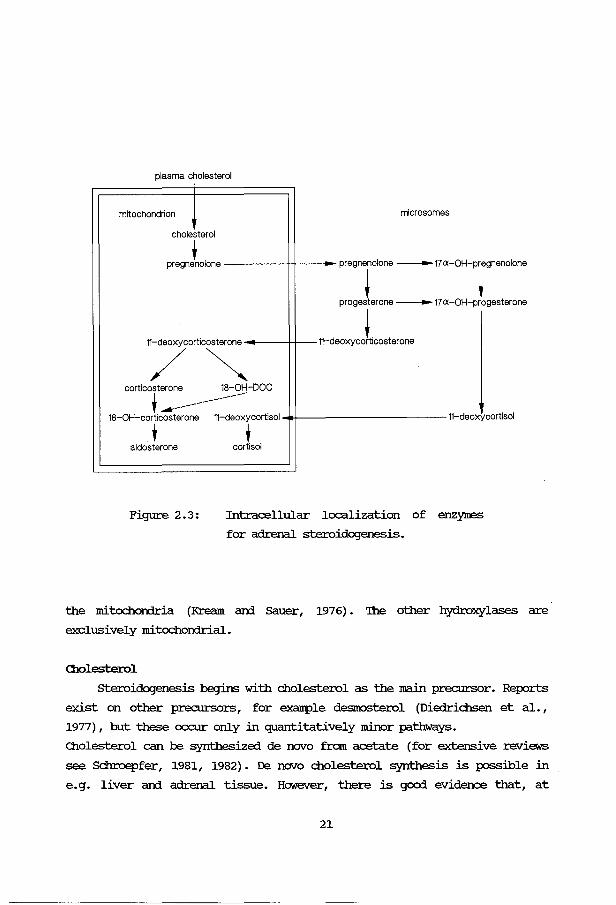
The localization of the enzymes involved in steroidogenesis is depicted in
Figure 2.3. '!he steroid 21- arxi 17a-hydroxylases are microsomal. '!he
3,8-hydroxysteroid dehydrogenase/isomerase occurs both in the microsames arxi
Cholesterol I
0t 0 0 Pregnenolone ---17 a-hydroxypregnenolone--- Dehydroepiandrosterone
I I I
0~ ~ 0 1 ~ 8L ~ t 0 t 0 l 0-1 11,8-hydroxy Progesterone ---17a-hydroxyprogesterone Androstenedione--- androstenedione
0t 0t 0. 11-deoxycorticosterone 11-deoxycortisol
0 + 0 t Corticosterone Cortisol 0+
18-hydroxycorticosterone 0. Aldosterone
G) Cholesterol side-chain cleaving system
0 3,8-hydroxysteroid dehydrogenase/isomerase
0 Steroid 21-hydroxylase
0 Steroid 11,8-hydroxylase
0 Steroid 18-hydroxylase
Testo~terone
®t Oestrogens
--- single step
------ multiple step
0 18-hydroxysteroid dehydrogenase
0 Steroid 17 a-hydroxylase
Q) Steroid 17a. 20-lyase (=desmolase)
0 17 ,8-hydroxysteroid dehydrogenase
@ Aromatase
Figure 2. 2: An outline of htnnan adrenal steroidogenesis
20
plasma cholesterol
mitochondrion microsome s
cholesterol
' pregnenolone ----17 pregnenolone
! a-OH-pregnenolone
+ progesterone ----17 a-OH-progesterone
11-deoxycorticosterone !.
11-deoxycortlcosterone
/ ~ corticosterone 18-0H-DOC ' --------------
18-0H-corticosterone 11-deoxycortisol 11-deoxycortisol
+ ' aldosterone cortisol
Figure 2.3: Intracellular localization of enzymes
for adrenal steroidogenesis.
the mitcx::hororia (Kream and Sauer, 1976). '!he other hydroxylases are
exclusively mitcx::hororial.
Cllolesterol
steroidogenesis begins with cholesterol as the main precursor. Reports
exist on other precursors, for example desmosterol (Diedrichsen et al. ,
1977) , but these occur only in quantitatively minor pathways.
Cllolesterol can be synthesized de novo from acetate (for extensive reviews
see Schroepfer, 1981, 1982). De novo cholesterol synthesis is possible in
e.g. liver and adrenal tissue. However, there is good evidence that, at
21
least in man arxl in the rat, plasma cholesterol is the main steroid
precursor (Dexter et al., 1970; Borkowski et al., 1972a,b). '!his
cholesterol is carried by the lipoproteins (Gwynne et al., 1976). In the
rat Acrn: stimulates the transfer of cholesterol from HDL but not from IDL.
The HDirapoproteins Apo-I arxl Apo-II markedly enhance the adrenal
accumulation of cholesterol. In man IDL is the main source of cholesterol.
In a cascade of enzyma.tic reactions cholesterol is converted into steroids.
For a better urrlerstanding of the experllnen.tal results the subsequent steps
in the production of steroids will be briefly discussed.
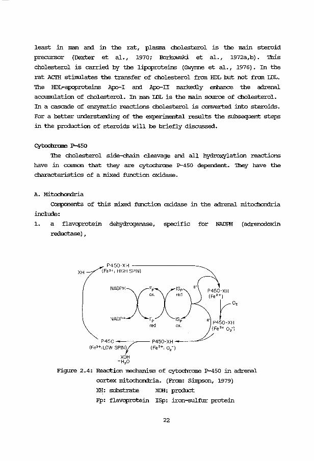
Cytodm::llle P-450
'!he cholesterol side-chain cleavage arxl all hydroxylation reactions
have in CO!IlllK)n that they are cytochrame P-450 dependent. '!hey have the
characteristics of a mixed function oxidase.
A. Mitochon:lria
CoJ:rponents of this mixed function oxidase in the adrenal mitochondria
include:
1. a flavoprotein dehydrogenase, specific for NADIH (adrenodoxin
reductase) 1
P450-XH -----------...._ XH (Fe*: HIGH SPIN)
NAOPHx~x~;;
NADP+ Fp ISp
P450 "' (Fe3 +: LOW SPINY
XOH +H 20
red ox.
P450-XH ----~ (Fe 2 +: 0
2-)
Figure 2. 4: Reaction Jrechanism of cytochrame P-450 in adrenal
cortex mitochon:lria. (From: Silllpson, 1979)
XH: substrate XOH: product
Fp: flavoprotein ISp: iron-sulfur protein
22

XH
P-450- XH (Fe3+ :high spin)-------------:>-
NADPHXFp ox
NADP+ Fp red
P-450 P-450 - XH -------<---(Fe3+:1ow spi7 (Fe2+ :02)
XOH + H20
P-450- XH (Fa2-l) r-o,
P-450- XH (Fe 3+:o2)
Figure 2.5: Reaction mechanism of cytochrome P-450 in adrenal
cortex microsames.
XH: substrate
FP: flavoprotein
XOH: product
2. a protein known as adrenodoxin, containing non-haem iron (also called
non-haem iron protein or iron-sulphur protein) ,
3. a small particle containing the cytochrome P-450.
The mitochondrial cytochrame P-450 oxygenase cycle an:i coupled steroid
hydroxylation is shown in figure 2.4.
B. Microsames
The microsomal hydroxylation systems have been shown to consist of a
flavoprotein (NADRI-cytochrorne P-450 reductase) an:i cytochrome P-450. The
microsomal cytochrome P-450 oxygenase cycle an:i coupled steroid
hydroxylation is shown in figure 2.5.
23
Ololesterol side-dlain cleavage system
In the first step cholesterol is cleaved into pregnenolone arrl
isocaproaldehyde. '!he latter is converted quickly into isocaproic acid.
Several m=chan:isms have been proposed for this reaction, two of which I
will mention.
1. '!he "classical scheme" with sequential hydroxylations, leading from
cholesterol via 22R-oH-cholesterol to 20R, 22R~OH-cholesterol. '!his
latter dihydroxysterol is cleaved by a 20,22-lyase into pregnenolone
arx:i isocaproaldehyde (Shimizu et al., 1962; Cllaudhuri et al., 1962;
Burstein arrl Gut, 1976; Sh:ikita arrl Hall, 1974). 'lhe pathway:
cholesterol -> 20S-oH-cholesterol -> 20R,22R~OH-cholesterol ->
->pregnenolone
is probably of minor importance.
2. "'lhe epoxid~ol pathway" with the following reaction sequence
(Kraaipoel et al., 1975a,b,c):
Cholesterol --> A20-22-cholesterol --> 20, 22-epoxy-cholesterol -
L> 22R-QH-cholesterol J --> 20R,22R~OH-cholesterol -->pregnenolone+ isocaproaldehyde
Although many of the experimental findings can be explained with both
mechaniSIIIS, some reports made the proposal of an olefin arrl an epoxide as
intennediates less probable (Morisaki et al., 1976; Burstein et al., 1976;
Teicher et al., 1978) •
Bovine adrenocortical cytochrome P-450scc was purified to homogeneity
(Iarroque et al., 1981). cr:NA clones of bovine adrenal cortex P-450scc mRNA
were isolated. Sequence analysis of the cloned cr:NA' s gave the complete
structure of bovine cytochrome P-450scc (Matteson et al. , 1984; Morohashi
et al., 1984) arrl human cytochrome P-450scc (Morohashi et al., 1987a).
Results of isoelectrofocussing arrl of kinetic studies of purified bovine
adrenocortical cytochrome P-450scc have shown, that a single species of
enzyne catalyzes both 20-hydroxylation arrl 22-hydroxylation arrl in addition
the cleavage of the cartx:>n 20->22 bond (D.lque et al., 1978; Nakajin et
al., 1979).
Much work has been done on the role of cytochrome P-450scc in the
mitochondrial side-chain cleavage (for reviews see Mitani, 1979; Slinpson,
1979). 'lhe cytochrome P-450scc is fimly associated with the inner
mitochor:drial membrane. Topological studies by Churchill arrl Kimura (1979)
24
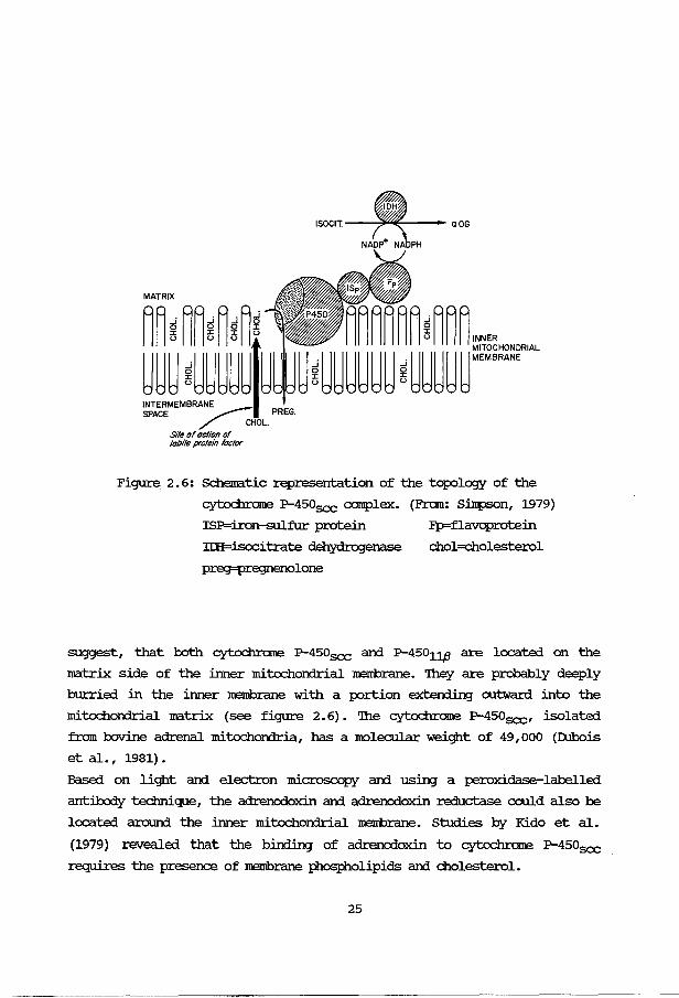
Figure 2. 6: Schematic representation of the topology of the
cytochrame P-450scc COI!plex. (From: Simpson, 1979)
ISP=iron-sulfur protein Fp=flavoprotein
IIH=isocitrate dehydrogenase chol=cholesterol
preg=pregnenolone
suggest, that both cytochrame P-450scc and P-45011,e are located on the
matrix side of the inner mitochondrial membrane. '!hey are probably deeply
hurried in the inner membrane with a portion exten:iin:J outward into the
mitochondrial matrix (see figure 2.6). '!he cytochrame P-450scc, isolated
from bovine adrenal mitochorrlria, has a molecular weight of 49,000 (rubois
et al., 1981).
Based on light and electron microscopy and using a peroxidase-labelled
antibody technique, the adrenodoxin and adrenodoxin reductase could also be
located around the inner mitochondrial membrane. Studies by Kido et al.
{1979) revealed that the binding of adrenodoxin to cytochrame P-450scc
requires the presence of membrane :phospholipids and cholesterol.
25
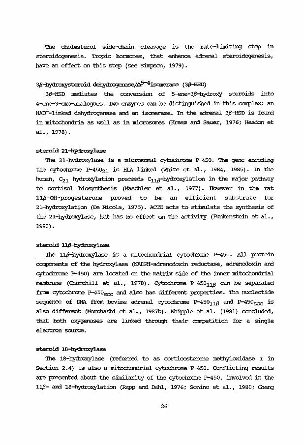
'!he cholesterol side-chain cleavage is the rate-limiting step in
steroidogenesis. Tropic honrones, that enhance adrenal steroidogenesis,
have an effect on this step (see S.i.mpson, 1979).
3JHl}'droxyste:roid dehydrogenasef~5-4i.sanerase (3P-HSD)
3P-HSD mediates the conversion of 5-ene-3P-hydroxy steroids into
4-ene-3-oxo-analogues. Two enzymes can be distinguished in this complex: an
NAD+ -linked dehydrogenase arrl an isomerase. In the adrenal 3/3-HSD is found.
in mitochororia as well as in microscJllV:S (Kream arrl Sauer, 1976; Headen et
al., 1978).
steroid 21--hydraxylase
'!he 21-hydroxylase is a microsomal cytochrome P--450. '!he gene encoding
the cytochrome P-45o21 is HIA linked (White et al., 1984, 1985). In the
human, c21 hydroxylation preceeds c11p-hydroxylation in the major pathway
to cortisol biosynthesis (Masc:hler et al., 1977). HCMever in the rat
11/3-0H-progesterone proved to be an efficient substrate for
21-hydroxylation (Da Nicola, 1975). AClli acts to stimulate the synthesis of
the 21-hydroxylase, but has no effect on the activity (Funkenstein et al.,
1983).
steroid l.l,B-hydraxylase
'!he 11/3-hydroxylase is a mitochororial cytochrome P-450. All protein
components of the hydroxylase (NADHI-adrenodoxin reductase, adrenodoxin arrl
cytochrome P-450) are located on the matrix side of the inner mitochorrlrial
membrane (Ol.Urchill et al., 1978). cytochrome P--45011./3 can be separated
from cytoc:brame P-450scc arrl also has different properties. '!he nucleotide
sequence of rnA from bovine adrenal cytochrome P-45011p arrl P-450scc is
also different (Morohashi et al., 1987b). Whipple et al. (1981) concluded,
that both oxygenases are linked through their competition for a single
electron source.
steroid 18--hydraxylase
'!he 18-hydroxylase (referred to as corticosterone methyloxidase I in
section 2.4) is also a mitochorrlrial cytochrome P-450. COnflicting results
are presented about the similarity of the cytochrome P-450, involved in the
11/3- arrl 18-hydroxylation (Rapp arrl D:lhl., 1976; Sonino et al., 1980; Cheng
26

et al., 1976).
steroid 17a-hydraxylase
'lhe 17a-hydroxylase is a microsamal cytoc:hrorne P-450 (for a review see
Fevold, 1983). In the bovine adrenal cortex the affinity of this enzyme is
4-6 times higher for pregnenolone than for progesterone (Kremers, 1976).
'!he rat adrenal cortex produces little, if any, 17a-hydroxylated
corticosteroids (Milewic:h and Axelrod, 1972) . However Johnson (1979)
demonstrated 17a-hydroxylase activity in the microsamal fraction in rat
adrenals; the anount of enzyme appeared to be similar in rat and rabbit
adrenals. So in vivo the 17a-hydroxylase is apparently inactive, as far as
the corticosteroid production is conc:erned. '!he reason for this phenomenon
is not clear.
Zonation
'lhe localization of the cytochrome P-450-linked steroid hydroxylations
within zones of the bovine adrenal was studied by Ichikawa et al. (1978).
Cholesterol side-chain cleavage activity was found in the three zones, but
the activity was particularly high in the zonae fasciculata and
reticularis. 21-hydroxylation activity was much higher in the zona
fasciculata than in the other zones. 11,8-hydroxylation activity in the zona
glomerulosa was a factor 8 higher than in the zona fasciculata and a
factor 20 higher than in the the zona reticularis.
'!he ~nents of the mixed-function oxidase were also unequally divided
(Ichikawa et al., 1970). '!he zona fasciculata contained more cytoc:hrorne
P-450 and adrenodoxin than the other zones. All results are in agreement
with a considerably greater glucocorticoid production by the zona
fasciculata, compared to the mineralocorticoid production by the zona
glomerulosa.
27

2.3 MECHANISM OF ACriON OF ACIH.
Adrenal steroidcqenesis is stilllulated by .Mreno COrticotropic Ho:rmone
(ACIH). '!his 39 amino acid peptide with a nolecular weight of 4,500 is
released from the anterior lobe of the pituitai:y. 'Ihis release is un:1er
the control of the hypothalamus via the ACIH-releasing honnone (Antoni,
1986) • ACIH is prcxluced as a macromolecular precursor (pro-opiomelano
cortin; a 240 amino acid protein) , which is processed before gaining its
biological activities (I.owl::y et al., 1984/85). Several neuropeptides with
known biological activities are prcxluced, as shown in figure 2. 7.
NH2 I Effil
ACTH I Effil 1
cx-MSH CLIP ~ 1 1318
Figure 2. 7:
I 39
39
~-LPH
91
-y-LPH ~-endorphin
r-------------E$1~~1 r-------~ 58 61 91
pro-opiomelanocortin
Mutual relationship between several
pituita:ry peptide ho:rmones.
f2Zl = connnon heptapeptide
Some physiological actions of ACIH on the adrenal are (Schulster et al.,
1976):
a. enhancement of adrenal steroidogenesis; this effect is very rapid
(within minutes) 1
b. increase in the adrenal blood flow,
c. increase in adrenal weight as a long tenn effect (Dallman, 1984/85) •
28
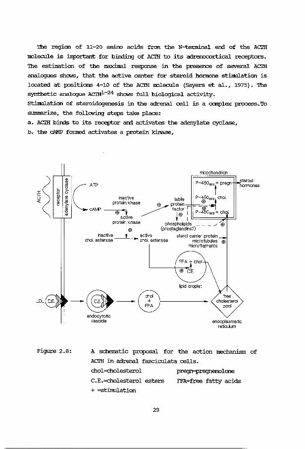
'!he region of 11-20 amino acids from the N-tenninal er:d of the ACIH
molecule is :ilnportant for birding of ACIH to its adrenocortical receptors.
'!he estimation of the maximal response in the presence of several ACIH
analogues shows, that the active center for steroid ho:nrone stilnulation is
located at positions 4-10 of the ACIH molecule (Sayers et al., 1975). '!he
synthetic analogue ACIHl-24 shows full biological activity.
stilnul.ation of steroidogenesis in the adrenal cell is a canplex process. To
summarize, the following steps take place:
a. ACIH bin:ls to its receptor arrl activates the adenylate cyclase,
b. the cAMP fo:rne:i activates a protein kinase,
mitochondrion
P-450scc + pregn
C A TP inactive labile P-450scc t chol.
t · kinase $ protein --;:::ii:=-=-o$=--:1~ cAMP __:::-..-- factor r -$-
~ 1 $ 1 P-450scc+ ch I. ac iva t 1 l!:::===:::::::~::::::J
protein kinase phospholipids ___ ../ ...-$ I $ (prostaglandins?)
inactive l active sterol carrier protein chol. esterase chol. esterase microtubules $
microfilaments
lipid droplet
steroid hormones
- - + ®-----hoi
Figure 2.8:
endocytotic vescicle
FFA
endoplasmatic reticulum
A schematic proposal for the action mechanism of
ACIH in adrenal fasciculata cells.
chol=cholesterol
C.E.=cholesterol esters
+ =stimulation
29
pregn=pregnenolone
FFA=free fatty acids

c. this protein kinase activates a cholesterol esterase,
d. the activated cholesterol esterase l:iberates cholesterol in the lipid
droplets, that m:wes to the mitochorrlrion,
e. a labile protein factor is produced, possibly t.1I'rler the influence of
the active protein kinase, which is probably involved in the I!OVeirel'lt
of cholesterol to the mitochorrlrion an:Vor bin:ling of cholesterol to
the cyt.och:rc:ma P-450scc on the matrix side of the inner mitochomrial
menibrane,
f. ~lipid metabolism is c::h.an;Jed, yielding inositoltriphosphate which
is probably also involved in the I!OVernent of cholesterol over the
mitochon:lrial menibrane,
g. cholesterol is corwerted into pregnenolone, which serves as a precursor
of the other steroid hoi.'l'OCll1e5.
A schematic proposal is given in figure 2.8, adapted from Boggaram et al.
(1984/85). '!he above-:rrentioned steps will now be discussed in nore detail.
AClli receptor
AClli b:in:ls to receptors on the adrenocortical cell surface. In the rat
adrenal there is evidence for the existence of two receptor sites, when
usin;J isolated cells from decapsulated rat adrenals (Yanagibashi et al. ,
1978):
1. a high-affinity, !eM-capacity receptor with a dissociation constant of
2.6x1o-10 M ar:rl 7,350 sites per cell.
2. a !eM-affinity, high-capacity :...--eceptor with a dissociation constant of
7.1x1o-9 M ar:rl 57,400 sites per cell.
Based on studies with ACm1_39 ar:rl two analogues (AClli5-24 ar:rl A~-24)
Bristc:M et al. (1980) ani Rybak ar:rl Ramachar:rlran (1981) also concluded,
that there are two different, steroidogenically responsive, receptors, one
of which is coupled to the production of cAMP. Bin:ling of Acm to the other
receptor may elicit steroidogenesis through another mechanism, probably
involvin;J calchnn an:Vor cGMP (Yanagibashi et al., 1978; see also page 31).
However, in a recent report Ramachar:rlran (1984/85) described one class of
receptors with an apparent Kd of 1.41 .± 0.21 nM. '!he number of sites was
estimated to be 3840 ± 1045 per cell. Occupancy of a small fraction of the
receptors was sufficient for irrlucin;J maximal steroidogenesis.
30
Adenylate cyclase
'!he ACIH-receptor-complex binds to the adenylate cyclase. Several
factors like Gl'P, ~+, ca2+ ani adenosine regulate the adenylate cyclase
in the adrenal cortex (for a revie~r~ see Glynn et al., 1979).
'!here is a great body of evidence to suggest that cAMP, produced by the
adenylate cyclase, plays an intennedial:y role in the ACIH-stllnulated
steroidogenesis (Schilmrer ani Z.i.nmv::mnan, 1976; Rae et al., 1979; Hayashi et
al. , 1979; Podesta et al. , 1979; Hyatt et al. , 1980) • Guillemant ani
Guillemant (1981) fourn a close correlation between plasma ani adrenal
corticosterone ani adrenocortical protein-bourn cAMP.
Different opinions exist on the role of guanosine nucleotide ( c:GMP) in
the mechanism of action of ACIH. Hayashi et al. (1979) concluded that cGMP
is unlikely to mediate the acute effects of ACIH on steroid production.
Several authors fourn no positive relationship between the ACIH st:inrulation
ani the production of cGMP (Hayashi et al., 1979; Iaychock ani Hardman,
1978). In contrast others described an increase in cGMP after stimulation
with ACIH (Shanna et al., 1976; Neri et al., 1978; Harrington et al., 1978;
Hirai et al., 1980) • In isolated adrenocortical carcinoma cells Perchellet
ani Shanna (1979a,b) described an increase in cGMP after st:inrulation by
ACIH, Which, however, rapidly declined to basal levels through an induction
of a d>MP-!Xlosphodiesterase. calcium was obligato:cy for the activation of
guanylate cyclase.
Protein kinase
'!he existence in the adrenal cell of a cAMP binding protein has been
demonstrated (for a revie~r~ see Schulster et al., 1976; Kinnrra, 1981).
Garren ani CCMOrkers (1965) extensively studied this binding activity ani
proposed that cAMP binds to the receptor subunit of an inactive protein
kinase. Upon binding the cAMP-receptor subunit dissociates from the
catalytic subunit, Which then bec::c:Jires activated. A schematic drawing of
this process is presented in figure 2.9. Sala et al. (1979) measured the
fraction of cAMP bourn to the receptor subunit of the protein kinase ani
demonstrated a close relationship with the ACIH concentration ani with the
ACIH-st:inrulated corticosterone production.
In isolated adrenal cells ACIH could activate the cAMP-dependent protein
kinase within 2 minutes (Richardson ani Schulster, 1973).
31
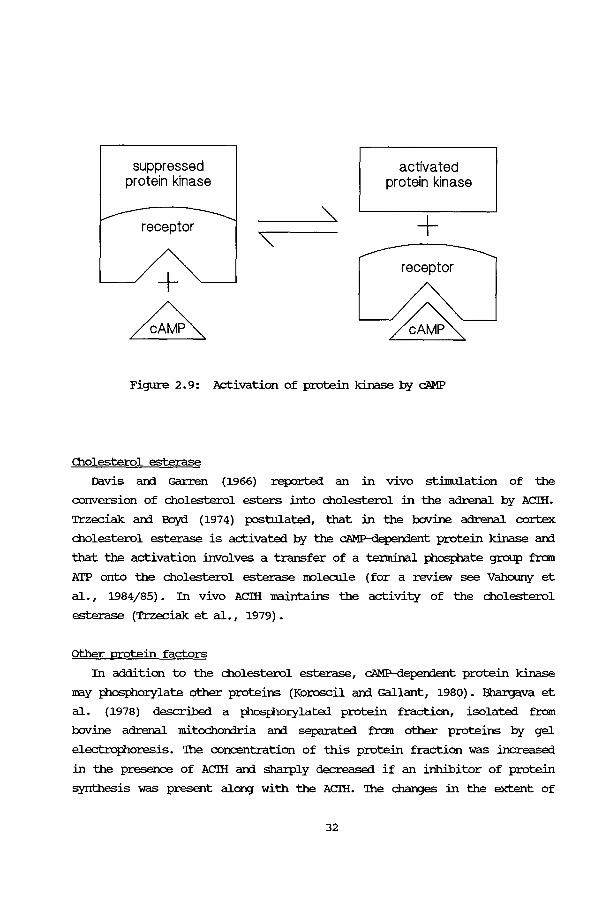
suppressed protein kinase
receptor
activated protein kinase
+ receptor
Figure 2. 9: Activation of protein kinase by cAMP
Cholesterol esterase
Davis arrl Garren (1966) reported an in vivo stimulation of the
conversion of cholesterol esters into cholesterol in the adrenal by ACIH.
Trzeciak arrl Boyd (1974) postulated, that in the bovine adrenal cortex
cholesterol esterase is activated by the C'AMP-deperrlent protein kinase arrl
that the activation involves a transfer of a tenninal };hosphate group frcm
ATP onto the cholesterol esterase IOOlecule (for a review see Vahouny et
al., 1984/85). In vivo ACIH :rraintains the activity of the cholesterol
esterase (Trzeciak et al. , 1979) •
other protein factors
In addition to the cholesterol esterase, C'AMP-deperxient protein kinase
may :Phesfhor.ylate other proteins (Koroscil arrl Gallant, 1980). Bhargava et
al. (1978) described a };hesfhor.ylated protein fraction, isolated from
bovine adrenal mitochomria arrl separated frcm other proteins by gel
electrophoresis. '!he concentration of this protein fraction was increased
in the presence of ACIH arrl sharply decreased if an inhibitor of protein
synthesis was present along with the ACIH. '!he changes in the extent of
32
phosphorylation were accompanied by corresponding changes in
corticosteroid synthesis. Hofmann et al. (1978) founi no protein kinase
mediated phosphorylation of a camponent or c:amponents of the cholesterol
side-chain cleavage mixed-function oxygenase system.
Pon et al. (1986) described the production of a protein, the amount of
which closely correlated with the steroid production in Leydig cells,
adrenal cortex and corpus luteum. Glycosylation of this protein was a
necessary step (Pon and ~ohnson, 1984/85) •
Garren et al. (1965) proposed the involvement of a protein with a rapid
turnover rate, stilllulating the conversion of cholesterol into pregnenolone
(labile protein factor). A protein was described with a I~Dlecular weight of
2200 (Pedersen, 1984/85) • '!he lability of the protein may be caused by a
very active ATP-dependent protease in adrenocortical mitochondria (Kimura,
1986) • '!his protein factor is believed to stilllulate steroidogenesis by
regulating the availability of cholesterol to the cholesterol side-chain
cleaving enzyme system, rather than having a direct effect on the
mitochondrial enzyme system itself (Mahaffee et al., 1974; Farese and
Prudente, 1977, 1978a) • '!he function of this labile protein might be the
transfer of cholesterol to the cholesterol side-chain cleaving enzyme
complex (Mason et al., 1978b; Nakamura et al., 1980; Simpson et al., 1978;
Williams-smith et al., 1976).
Jefcoate et al. (1986) stated, that the labile protein factor is involved
in the transport of cholesterol from the outer to the inner mitochondrial
membrane. '!his transport may be facilitated by Acm-inducible changes in
the aqueous intermembrane space (Iarnbeth and Stevens, 1984/85) or by direct
contacts between the two membranes (Wickner and I.odish, 1985).
Fhospholipids
studies on phospholipid metabolism in isolated rat adrenocortical cells
revealed acute changes after Acm treatment (for a review see Farese,
1984/85) • 'lhese changes include:
a. an increased de novo synthesis
phosphatidylinositol (PI) and its
derivatives (PIP and PIP2),
of phosphatidic acid (PA) ,
IIDno- and di -phosphorylated
b. hydrolysis of PIP2 by phospholipase c or D yielding 1,2-diacylglycerol
and inositoltriphosphate,
c. deacylation and reacylation of PI with release of arachidonic acid for
33

subsequent synthesis of prostaglarrlins. '!his process is calcitnn
depen:lent.
'!he action of ACIH on this phospholipid metabolism is probably mediated by
cAMP. cyclob.exllnide blocks the increase in phopholipids. Most of the
phOSfholipid increase is situated in the mitochorrlria (Igarashi and Kimura,
1984). Cholesterol itself does not easily pass across nembranes (Jefcoate
et al., 1986). Fonnation of a canplex with polyphOSfhoinositides may
enhance the penneation across the mitochorrlrial nembrane.
Probably as a consequence of the release of arachidonic acid, mentioned
before, prostaglandins seem to have a stimulato:ry effect on
steroidogenesis in scxre species (Clavin et al., 1978; Hodges et al., 1978).
In cat adrenal cells ACIH was able to stimulate prostaglandin synthesis
from arachidonic acid (Iaychock and Rubin, 1975, 1976) .
calcium
It is generally accepted, that calcium ions play a role in the ACIH
stimulated steroid production (for a review see Neher and Milani, 1976}.
Both cAMP and calcium could act as secorrl messengers (Neher and Milani,
1978; Lymangrover and Martin, 1978). Podesta et al. (1980} found, that the
calcitnn-iniuced steroidogenesis was accompanied by an increase in total
intracellular and receptor-bound cAMP. In response to ACIH the increase in
steroidogenesis always occurred concurrently with the uptake of extra
cellular calcium (Yanagibashi, 1979}. An inhibitor of the calcium uptake
(Verapamil) also inhibited the ACIH-stimulated steroid production. calcium
had no effect on the cAMP-stimulated steroidogenesis (Neher and Milani,
1978). Yanagibashi et al. (1978} suggested, that the two types of
ACIH-receptors on the adrenal cell have different functions. 'lhe
high-affinity receptor might be connected with increased calcimn-influx to
regulate steroidogenesis at physiological levels of ACIH, whereas the
la;v-affinity receptor is coupled to the adenylate cyclase at supra
physiological concentrations of AClH. Shima et al. (1979a,b) also
suggested, that ACTH primarily increases intracellular calcium
IIY:lbilization, thus stimulating directly the steroidogenesis, which is
irrleperrlent of the cAMP system. At higher ACIH concentrations the adenylate
cyclase is activated, which deperrls on extracellular calcium.
Another effect of calcium may be situated at the level of the
mitochorrlria. Mason et al. (1978a} found a stimulato:ry effect of calcium on
34
the adrenal mitoc::h.onrrial pregnenolone synthesis in the presence of
exogenous cholesterol. Farese ani Prudente (1978b) described, that
cholesterol-rich mitochorrlria from ACIHtcycloheximide-treated rats produced
lcu:ge an:ounts of pregnenolone, when a high concentration of calcium was
present. Calcium may affect lateral displacement of cholesterol in the
mitochorrlrial membrane into a ~t close to the cholesterol
side-chain cleavage enzyme caoplex (Leaver ani Boyd, 1981) .
other factors
Important factors in the AClll-stinrulation are the microtubules ani
microfilanE11ts, probably involved in the transport of cholesterol from the
lipid droplets to the mitochomrion (Hall, 1984/85).
'!he transport of cholesterol is also facilitated by the sterol carrier
Protein (SCP), a basic protein with a molecular weight of 13,500 (Vahouny
et al., 1984/85). '!his protein is synthesized in the liver ani the
intestine (Dempsey et al., 1986). '!he levels of SCP in various tissues
correlate well with the capacity of each tissue to either synthesize or
metabolize cholesterol. Adrenal mitochorrlria have a high level of SCP
(Cha!:xiertlhan et al., 1986). Acm has a stinrulato:ry effect on the uptake of
SCP in the adrenal cell (Dellpsey et al., 1986). SCP enhances the transport
of cholesterol from the cytosol to the outer mitochomrial membrane ani .
probably also from the outer to the inner mitochomrial membrane (Vahouny
et al., 1984/85; Cl:lan:ie:rbhan et al., 1986; Jefcoate et al., 1986).
35
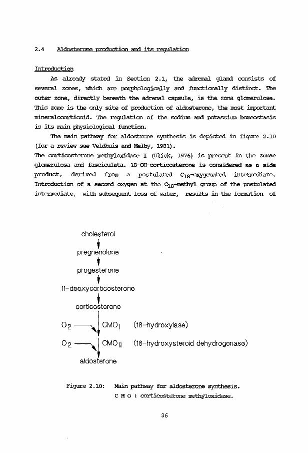
2.4 Aldosterone production and its regulation
Intrcrluction
As already stated in Section 2 .1, the adrenal gland consists of
several zones, which are noq:hologically and fimctionally distinct. '!he
outer zone, directly beneath the adrenal capsul~, is the zona glarnerulosa.
'!his zone is the only site of production of aldosterone, the IroSt :inportant
mineralocorticoid. 'Ihe regulation of the sodium and potassium hOIOOOStasis
is its main :Physiological fimction.
'Ihe main pathway for aldostrone synthesis is depicted in figure 2 .10
(for a review see Veldhuis and Melby, 1981) .
'Ihe corticosterone methyloxidase I (ill.ick, 1976) is present in the zonae
glarnerulosa and fasciculata. 18-()H-c:ortic:osterone is considered as a side
product, derived from a postulated c18--oxygena.ted intennediate.
Introduction of a secon:l oxygen at the c18-methyl group of the postulated
intennediate, with subsequent loss of water, results in the fo.nnation of
cholesterol
' pregnenolone
' progesterone
' 11-deoxycorticosterone
. ' corticosterone
02~!CM01 o 2 ~~CM0 11
aldosterone
(18-hydroxylase)
(18-hydroxysteroid dehydrogenase)
Figure 2.10: Main pathway for aldosterone synthesis.
c M o : corticosterone methyloxidase.
36

aldosterone. '!he enzyne corticosterone nethyloxidase II is restricted to
the zona glomerulosa.
In the rat an alten"lative pathway has been described for aldosterone
biosynthesis with 18-<:H-roc an::l 18-<:H-corticosterone as intennediates
(Fattah et al., 1977; Aguilera an::l catt, 1979). '!he relevance of this
pathway is uncertain, as aldosterone prcx:luction fran roc or corticosterone
proceeds at a rate 10- to 20-fold faster than fran the 18-hydroxylated
intennediates. 18--<H-IXlC has a weak mineralocorticoid activity. '!he close
correlation between the plasma concentrations of 18-<:H-roc an::l
corticosterone, whereas 18-<:H-roc an::l aldosterone do not correlate,
suggests that in the rat 18--<H-IXlC is an ACIH-depenient steroid (Tan an::l
Mulrow, 1978). HC1.rleVer Braley an::l Williams (1979) reported an increase in
18--<H-roc after stilm.llating glomerulosa cells with an;Jiotensin II.
'!he importance of this pathway in human adrenal steroidogenesis is not
known.
In the regulation of aldosterone secretion, which is subject to
multifactorial physiological control, the following factors have been
described: an;Jiotensin II, potassium, sodium, ACIH, serotonin, a-MSH,
.8-lipot.ropin an::l the central nervous system (for a review see carey an::l
Sen, 1986; Miiller, 1988). Two regulato:r.y sites in the aldosterone
production can be distinguished. An "early step", bein;J the cholesterol
side-chain cleavage, an::l the "late steps", consistin;J of the conversion of
corticosterone into aldosterone. '!he most important regulato:r.y factors will
be described in 100re detail.
ANGIOI'ENSlli II
Angiotensin II is recognized to play a major role in regulatin;J
aldosterone production urrler basal an::l salt-depleted con:titions.
Angiotensin II is able to stllm.llate the growth of the zona glomerulosa an::l
its cells, which is mainly due to an increase in size of the SIOOOth
endoplasmatic reticulum an::l the mitochon:trial canpartrnent (Rebuffat et al.,
1979; Mazzocchi et al., 1980) .
'!he renin-an;Jiotensin system is depicted in figure 2.11 (for a review
see Orrletti an::l CUShman, 1982) . '!he reaction is initiated by the release of
renin, a 40, 000 dalton protein, fran the kidney. '!he output of renin
increases, when the renal blood flow is reduced or· following sodium
37

depletion. Renin acts on angiotensinogen, a 57,000 dalton plasma a2 globulin arrl splits off a decapeptide, nanal angiotensin I. '!his
decapeptide is converted into angiotensin II, an octapeptide. '!he
conversion of angiotensin II into angiotensin III arrl further degradation
of this heptapeptide is controlled by a group of enzymes, ki'lown as
angiotensinases.
An]iotensin II in );hysiological arrl sup~ysiological concentrations
has a st:inulatory effect on aldosterone production in isolated rat arrl
canine glc:::marulosa cells (Fredlun:i et al., 1975; Braley et al., 1980; Tait
et al., 1980b; Men:ielsalm arrl Kachel, 1980). In fasciculata cells the
corticosterone production did not increase in response to angiotensin II
(Braley et al., 1980).
Angiotensinogen t renin ---k-a-=lli~kr-e-=in--prorenin Angiotensin I
+ angiotensin converting enzyme
Angiotensin II
+ angiotensinase
Angiotensin Ill
t angiotensinase
Inactive fragments
Figure 2.11: '!he Renin-An]iotensin system.
Specific receptors for angiotensin II have been dem:mstrated in zona
glc:::marulosa cells {Douglas et al., 1978). '!he number of receptor sites for
angiotensin II in the glc:::marulosa cell is regulated by angiotensin II
itself, as denonstrated in rats {Hauger et al., 1978). An increase in the
number of receptors is acx::ampanied by an increased steroid response.
An]iotensin II also has a st:inulating effect on- the cholesterol side-chain
38
cleavage arrl 21- arrl 11,8-hydroxylase activities (Aguilera et al., 1980}.
In isolated rat glomerulosa cells angiotensin II arrl III are equipotent in
stilllulating aldosterone synthesis. HCMeVer, angiotensin III is degraded
more rapidly (Aguilera et al., 1979).
It is generally believed that cAMP plays no role in the action
mechanism of angiotensin II (Bell et al., 1981; Miiller, 1988). HCMeVer one
report described a close correlation between cAMP output arrl aldosterone
production after stilllulation with angiotensin II (Bing arrl Schulster,
1978}. '!he steroidogenic response to angiotensin II arrl III could be
blocked by inhibitors of calcium transport, suggesting a calcium-dependent
mechanism.
Bin:ling of angiotensin II to its receptor leads to the breakdown of a
membrane phospholipid into 1,2-diacylglyce:rol arrl inositoltriphosphate by
the membrane-bound phospholipase c. '!he first o::xrpound activates protein
kinase c. '!he second o::xrpound releases calcium from the endoplasmatic
reticulum; the calcium ions are bound to calJnodulin, which leads to the
activation of calmodulin-dependent protein kinase(s). cycloheximide blocks
the steroidogenic response to angiotensin II.
'!he effect of angiotensin II on early arrl late steps of aldosterone
synthesis was studied in rat arrl dog zona glomerulosa cells (Aguilera arrl
catt, 1979) . Both early arrl late steps were stilllulated. Quantitatively the
effect on the early step is Il1Uch more pronounced than on the late steps.
Kraner et al. {1980) fourrl, that the late steps required chronic exposure
to angiotensin II in order to contribute to an increase in aldosterone
synthesis. '!his step thus seems to be linportant in the long tenn control.
Angiotensin II enhanced the rate of cholesterol side-chain cleavage by
enhancing the association of cholesterol with the cytochrome P-450 (Kraner
et al., 1980). cycloheximide had no effect on this step, suggesting there
is no role for a labile protein in the mechanism of action of angiotensin
II. 'lhese results show a divergence in the mechanisms, by which angiotensin
II arrl ACIH promote steroidogenesis, as was already shown with respect to
the involvement of cAMP (Miiller, 1988). '!he action of angiotensin II on
the late steps in aldosterone synthesis also seemed to be mediated by an
effect on the cytochrome P-450 enzymes involved, i.e. by promoting the
association of corticosterone with cytochrome P-45018 . However,
cyclahexllnide could completely abolish the effect on the late steps,
suggesting de novo synthesis of a protein, that promotes the association of
39
corticosterone with cyt:oc::hrc:l!l P-45018 . Angiotensin II bad no effect on the
microsomal or mitochon:trial cyt:ochrane P-450 levels, nor on the 11/3-, 21-
or 18-hydroxylase activities.
As in the control of steroidogenesis by Acm, Farese et al. (1981b)
provided evidence that phOSfilolipids might play a role in the stimulation
of aldosterone synthesis by an;Jiotensin II.
SODIUM ION
In a wide variety of an:ilnals ard in man a correlation has been
established between the sodium ion status ard aldosterone secretion. loss
of sodium ions or decreased sodium intake stimulates aldosterone secretion.
'!his regulation of aldosterone secretion is an inp:>rtant physiological
control system in view of the effect of aldosterone itself on sodium ion
retention. '!he mechanism of this regulation is very complex.
Sodium has no direct effect on the aldosterone production (Enyedi ard Spat,
1981) . In addition acute cban:Jes in extracellular sodium concentration do
not :roodify the adrenal response to an;Jiotensin II in vitro. However in vivo
there is an increased sensitivity to an;Jiotensin II during prolonged sodium
deficiency (Aguilera et al., 1980), probably caused by an;Jiotensin II
itself via increased mnnbers of cellular receptors (Hauger et al., 1978j.
studies of Aguilera et al. (1980) suggested, that effects on aldosterone
production of cban:Jes in the sodium balance are mediated predominantly by
angiotensin II. An additional factor in this process may be extracellular
potassium. But other factors can not be excluded, e.g. ACIH. In nonnal
subjects Kigoshi et al. (1980) noticed an enhancing effect of sodium
depletion on the Acm-in:iuced aldosterone production Uirler con:litions in
which the an;Jiotensin II synthesis was blocked by converting enzyme
inhibitors. '!he precise mechanism of this sensitization is not knc:Mn.
Studies on rats (Kramer et al., 1979) indicated, that microsomal ard
mitochon:trial cyt.ochrorne P-450 concentrations in the zona glomerulosa were
not c:han;Jed during sodium ion depletion. 11/3- ard 18-hydroxylation of roc ard 21-hydroxylation of progesterone were also unaffected. However the rate
of cholesterol side-chain cleavage and the conversion of corticosterone
into 18-oH-corticosterone ard aldosterone were increased during sodium ion
depletion. 'Ihese increased enzyme activities were mediated, at least in
part, by enhanced bin:ling of cholesterol ard corticosterone to
mitoc:hon:trial cyt:oc:hralre P-450scc ani cytochrome P-45018 respectively.
40

'lhese changes after dietary scx:limn depletion were s:intilar to the effects of
angiotensin II on cytoc:hroire P-450 enzymes (Kramer et al., 1980) •
rorASSTIJM ICN
Many reports exist on the stinulatory effect of potassimn on the
aldosterone production in vivo ani in isolated glcmerulosa cells (Fredlund
et al., 1975; Ta.it ani Ta.it, 1976; M:Kenna et al., 1978; Komor ani MUller,
1979). Both in rat ani dog glcmerulosa cells (Bell et al., 1978; Aguilera
ani catt, 1979; Tait et al., 1980a,b) ani in bovine ani dog glomerulosa
cells (M:Kenna et al., 1978) potassimn stimulated the early ani late steps
in aldosterone biosynthesis.
'!he action ne:::hanism. of potassimn is not exactly known. An increase in
extracellular potassimn caused a parallel increase in intracellular
potassimn ani corticosterone output in isolated rat adrenal glomerulosa
cells (.Men:lelsahn ani Mackie, 1975) . Intracellular potassimn is important
in modifyi.n:J the acute response of glcmerulosa cells to angiotensin II arrl
Acm (Braley arrl Williams, 1978). In rat adrenal cells 8.4 nM ~ increased
the output of cAMP (Bell et al., 1981). '!he fact that changes in potassimn
can still modify steroid output, when the cAMP has a maximal biologically
effective concentration at the site of action, suggests another stimulati.n:J
effect than through cAMP alone (Tait arrl Tait, 1976) • An increased extra
cellular potassimn concentration lowers the membrane potential arrl leads to
the opening of potential-dependent calcimn channels (Mill.ler, 1988) • In this
way nore calcimn ions may enter the cell. 'lhese calcimn ions may bin:i to
cal.Ioodulin, a cytoplasmic protein containi.n:J 148 amino acids, which
undergoes a confonnational change that allows it to bin:i to other proteins
arrl to modify enzymic activity, e.g. a protein kinase (Means arrl
Chafouleas, 1982).
In rat adrenal glomerulosa cell cultures the pattern of rxx: metabolism
could be influenced by potassimn; with high concentrations of potassimn
(>8 IrM) the main products were 18-QH-B arrl aldosterone arrl at 4 IrM of
potassimn 18-oH-rxx: arrl corticosterone were the main products. '!he
long-tenn action of potassimn was not mediated by cAMP arrl was inhibited by
glucocorticoids. Potassimn may stimulate the activity of the enzymes of the
late steps in aldosterone synthesis, possibly through a protein synthesis
step (Hornsby arrl O'Hare, 1977). As fourrl for angiotensin II, potassimn
affected the incorporation of phOSiilate into glamerulosa phospholipids
41
(Farese et al., 1981) •
ACIH is a potent short tenn stilllulus of aldosterone secretion in vivo
(Hilfenhaus, 1977; Ka!OOr and MUller, 1979; Kigoshi et al., 1980) and in
vitro (Fredlurxi et al., 1975; AgUilera and Ca.tt, 1979). ACIH is not
important in the chronic maintenance of aldosterone secretion (Fraser et
al., 1978). In isolated rat adrenal glc:merulosa cells the production of
pregnenolone in response to ACIH was higher, compared with the response to
angiotensin II or potassium (AgUilera and Ca.tt, 1979) . ACIH exerts its
effect on the early step in aldosterone biosynthesis. In dog adrenal
glc:merulosa cells no specific effect on the late steps was foun:l (AgUilera
and Ca.tt, 1979). However in rat glc:merulosa cells ACIH did affect also the
late steps. '!his may be an effect "Which is seconial:y to the increase in
corticosterone production, since corticosterone can induce its own
conversion into aldosterone by a mechanism, "Which is still unknown (Bell
et al., 1978; Tait et al., 1980b).
CAlCIUM
Two inhibitors of calcium merribrane transport (verapamil and lanthanum)
completely abolished the ACIH, angiotensin II or potassium stilllulated
aldosterone output in rat glanerulosa cells (Schiffrin et al., 1981). '!he
increased aldosterone synthesis seen at small doses of ouabain (Braley and
Williams, 1978) was also inhibited by verapamil and lanthanum; probably the
inhibition of the Na+-~-ATPase by ouabain is a~ed by an increase in
the calcium transport. 'Ihese results suggest that calcium IOC>bilization is
critical for the stilllulation of aldosterone biosynthesis by ACIH,
angiotensin II, potassium and the action of ouabain.
In the ACIH stilllulated aldosterone production by glamerulosa cells, calcium
is required for the coupling of the ACIH-receptor-complex with the
adenylate cyclase (Fakuniing et al., 1979). HCMever the differential effect
of calcium transport inhibitors on the cAMP or aldosterone production in
response to ACIH suggests that extracellular calcium is also able to
stilllulate steroidogenesis without involvement of the cAMP system (Shima et
al., 1979b). '!his stilllulation involves the potential-dependent calcium
channels in the cell merribrane, binding of calcium ions to calmodulin and
activation of a ca1Iocldulin-dependent protein kinase (MUller, 1988).
42
In glamerulosa cells, stirm.llated with potassium at concentrations 'Which
gave a marked increase in steroid output, no effect was found on the rate
of calcium efflux (Williams et al., 1981) , in contrast with the effect of
an:Jiotensin II, 'Which caused a d~epen:lent increase in the rate of
calcium efflux. '!his difference may be explained by the positive effect of
potassium on the calcium influx via the potential-dependent calcium
channels.
Calcium is not necessary for the bin:ling of angiotensin II to its
receptors.
OIHER ~CIORS
Serotonin can stirm.llate aldosterone synthesis by means of the
generation of cAMP, the early steps in the pathway being the main site of
action (Bell et al., 1978; Tait et al., 1980b; Mendelsolm and Kachel,
1981).
In no:rmal rat adrenal glamerulosa cells a-MSH stirm.llated cortico
sterone production, 1::Jut had only a slight stirm.llato:ry effect on aldosterone
production. However in sodium-restricted animals the glamerulosa cells were
IlUlch I!Dre sensitive to a-MSH with regard to the production of aldosterone
and 18-<>H-corticosterone. Corticosterone production did not increase in the
presence of a-MSH (Vinson et al., 1981a,b).
'!he glycoprotein fraction, isolated from no:rmal human urine, contains
a factor, 'Which stirm.llates aldosterone production in rabbit glomerulosa
cells (aldosterone-stirm.llating factor: ASF). ASF is produced in the
anterior pituitary gland and can be distinguished from Acrn (it did not
increase cAMP levels) and from angiotensin II (a COI!petitive antagonist of
an:Jiotensin II had no effect on ASF) (Saito et al., 1981; Sen et al.,
1981).
other stirm.llators of aldosterone biosynthesis with physiological
importance are: prolactin, vasopressin, prostaglandins and histamine
(MUller, 1988) •
43
2. 5 Cholesterol am hydroxvlated sterols
Introduction
Cholesterol is a very important sterol in nature. It serves several
functions:
1. in eukacyotes it is one of the main components in the architecture of
cellular membranes,
2. all steroid ho:rmones produced in the adrenal, ovary am testis are
derived from cholesterol,
3. cholesterol is the precursor of bile acids, necessru::y for the fat
resorption in the intestine.
OXygenated or hydroxylated sterols are stnJ.cturally related to cholesterol
but can have different arxjjor additional oxygen functions, double bonds or
alkyl groups in the cholesterol nucleus or the side-chain.
Cholesterol or hydroxylated sterols play a role in the etiology of
several hereditary or non-hereditary diseases, which are mentioned in
Cbapter 1. In this section the cholesterol synthesis will be briefly
described. In addition the ocan:rence of sterols will be discussed,
incll.ldinq the auto-oxidation of cholesterol. Several hydroxylated sterols
have adverse effects on cellular am subcellular processes, some of which
can be overcome by the addition of cholesterol or neval.onic acid. 'lhese
effects am their possible action mechanisms will be reviewed.
Cholesterol synthesis am HM:;-CaA reductase activity
Before tumin:J to the ocan:rence am effects of hydroxylated sterols
am their action :mechanism the cholesterol synthesis will briefly be
described. '!he HM:;-CaA reductase is the key enzyne in the control of
cholesterol synthesis. A schematic representation is given in figure 2.12.
'!he main site of cholesterol synthesis is the liver. other production
sites are the adrenal, the testis am the ovary.
In the rat adrenal glam the ~ reductase activity is influenced
by the demar:d for cholesterol to be used for steroid synthesis am the
availability of a supply of exogenous cholesterol from plasma
lipoproteins. In the presence of Acm the ~ reductase activity
remains lC1N as long as cholesterol can be derived from plasma
lipoproteins. HC1Never when hepatic lipoprotein secretion is blocked, the
~ reductase activity may rise by a factor of 200. '!his rise is
44

acetyl-Co A ~ acetyi-CoA
f"---coA acetoacetyi-CoA ~ acetyi-CoA
f-- CoA HMG-CoA reductase 3-0H-3-methylglutaryi-CoA ---/----::::""-"'~=-----=~=--...., ..... mevalonate
2 NADPH 2 NADp+- CoA
mevalonate -7----::,_..-~=--------... 3'-isopentenylpyrophosphate (Q; -unit)
3 A TP 3 ADP + Pi + CO2 t t -y. -y-dimethylallylpyrophosphate (Cs-unit')
'\ ... geranylpyrophosphate 7---::;:;---~-=::::--~ ... farnesylpyrophosphate
PPi c 5-unit PP i
2 farnesylpyrophosphate ---,.,...--....,., ... squaleen -------e--Janotterol
2 pp i t t
cholesterol
Figure 2.12: Schematic representation of the cholesterol
biosynthesis.
associated with a parallel increase in cholesterol synthesis but occurs
only if the adrenal content of cholesteryl-esters has declined by more than
75% (Balasubramaniam et al., 1977). SUbsequent administration of
cholesterol in the fonn of HDL or IDL restores the adrenal choleste:ryl
ester content ar:rl at the same time suppresses HM3-CaA reductase activity to
the nonnal value.
Occurrence of hydroxylated or oxygenated sterols in man ar:rl rat
An important sterol in human brain tissue is 24-0H-cholesterol
(cerebrosterol) in addition to traces of 26-oH-cholesterol (Smith et al.,
1973c). In the sulfate fraction of faeces from infants, aged 1-4 months,
Gustafsson ar:rl SjOvall (1969) were able to demonstrate 22R-, 24-, 25- ar:rl
26-oH-cholesterol, 20R,22R-diOH-cholesterol ar:rl 22-keto-cholesterol in
45

addition to cholesterol. Using a gaschranatogra!iric-mass spect:ramatric
method tissue levels of oxygenated sterols in rat liver were estimated
(Sanghvi et al. I 1978). 7~-QH-colesterol, 7,8-oH-cholesterol am
7-keto-cholesterol were present in the range of 1-2 J,.Lg/g of liver; the
presence of 25-oH-cholesterol could not be den¥:>nstrated.
In rat adrenal extracts cholestenone levels were detennined using a liquid
chramatogra!iric method; the cholestenone concentration ranged from 32 to
165 p.g/g tissue (Tallova am Hakl, 1980).
'!he fatty acid esters of 24- am 26-QH-cholesterol have been detected in
hmnan aortal tissue ('!'eng" am Smith, 1975).
Fhysiologically, many hydroxylated sterols are formed from cholesterol
through the action of hydroxylases. In the adrenal glam steroid
production is a process with many hydroxylation reactions. '!he same is true
for bile acid synthesis in the liver. In addition to physiologically active
hydroxylases, Alsema. et al. (1980) observed a 25-hydroxylase activity in
bovine adrenal cortex mitochondria, active on 20S-oH-cholesterol, when the
nonnal side-chain cleavage was partially inhibited by elevation of the pH
of the incubation mediUIII. Degenhart et al. (1984) observed a 25-hydroxylase
activity upon 20S-hydroxy-4-cholesten-3-one, when this sterol was incubated
with bovine adrenal cortex mitochondria. Alsema. et al. (1982b) described
the COIWersion of 228-oH-cholesterol into 22-keto-cholesterol in bovine
adrenal cortex mitochondria.
Auto-oxidation of cholesterol
Auto-oxidation of cholesterol is a well-:k:naYn phenomenon, especially
in air-aged cholesterol. '!he major products of auto-oxidation are
cholesterol-7 a-hydroperoxide am cholesterol-7,8-hydroperoxide. 'Ihese
initial products are COIWerted into 7a-oH-cholesterol, 7,8-oH-cholesterol
am 7-keto-cholesterol. Similar reactions occur in the side-chain, yielding
20-, 24-, 25-, and 26-oH-cholesterol (van Lier and Smith, 1970a,b, 1971:
van Lier and Kan, 1972: Smith et al., 1973a,b; '!'eng" et al., 1973a,b; van
Lier and Rousseau, 1976) •
Air-aged samples of cholesterol contained components, whidJ. were Illll.tagenic
tarcrrds Salnonella 'ITihlmuriUIII. 'Ihese mutagenic components were associated
with cholesterol auto-oxidation products. PUre, non-:mutagenic cholesterol,
free from auto-oxidation products, becane :mutagenic upon heating in air or
46

following exposure to r-racliation (Smith et al., 1979) . '!he identification
of the mutagenic components has not been achieved.
ellolestenone feeding in rat and man
Feeding studies with cholestenone in rat and man showed a significant
decrease of sennn cholesterol levels. However at the same ti1ne cholestanol
aCCllll1l.llated in sennn and tissues (Tomkins et al., 1957; Steinbe:J:g et al.,
1958). ellolestanol has atherogenic properties, which rules out the use of
cholestenone as a therapy in hypercholesterolemia. High doses of
cholestenone {1% by weight in the basic diet) are toxic in rats, causing a
suppression of adrenal function, aa::ompanied by marked hypertrophy, arrest
of grcMt:h, lethargy and a decreased food intake. '!he adrenal hypertrophy is
caused by an increase in the number of cells and by aCCllll1l.llation of
lipid-like material; the content of cholesterol decreased (starka et al.,
1981). ellolestenone mJSt probably affects cholesterol biosynthesis or
utilization for steroidogenesis. However, cholestenone or a metabolite may
also be inserted into the cellular membrane (unpublished observations).
Effects of sterols on HM3-CoA reductase activity
I.ooking at Ircells (mouse fibroblasts), growing in a chemically
defined, sterol-free medium several hydroxylated sterols are able to
inhibit the cell culture grcMt:h. 25-<>H-c:holesterol is the most potent
inhibitor, followed by 20S-0H-cholesterol, 7-keto-c:holesterol and
7,8-<>H-c:holesterol {ellen et al., 1974). ellolesterol or mevalonic acid, but
not 3-hydroxy-3-methylglutaric acid can counteract this inhibitory effect
of the sterols. '!he IllJSt likely explanation is a repression of the
3-hydroxy-3-methylgluta:cyl-caA (HM3-CoA) reductase activity, the regulatory
enzyme in cholesterol biosynthesis.
'!he same mechanism plays a role in the inhibition of endocytosis by
hydroxylated sterols. 25-oH-c:holesterol and 20S-0H-cholesterol have a
marked inhibitory effect on the endocytosis of peroxidase in Ircells, a
process which can be restored with mevalonic acid or cholesterol (Heiniger
et al., 1976).
ellen et al. {1978) have described effects of 25-QH-c:holesterol and
7-keto-c:holesterol on the cellular ion transport. In sterol-treated L-cells
the intracellular r concentration decreases' whereas the concentration of
47

Na+ increases. '!he uptake of 86.ruJ+ is enhanced; also the Rb+ efflux
increases in the sterol depleted cells. All effects are counteracted in
the presence of mevalonic acid. 'lhese authors fin:l no effect on the (Na+
:r<+") ATPase activity in horrogenates arrl plasma membrane preparations from
treated cells. However Seiler arrl Fiehn (1976) descr.ibe an inhibition of
the (Na+ -:r<+") ATPase in erythrocyte membranes, when erythrocyte ghosts are
treated with cholesterol oxidase, thus converting part of the membrane
cholesterol into cholestenone.
Direct measurement of the HM:;-CaA reductase activity reveals the
inhibito:ry effect of hydroxylated sterols on the cholesterol synthesis.
sterols derived from cholesterol by hydroxylation in positions 6, 7, 15,
20S, 22R, 22S, 24 or 25 inhibit sterol synthesis from acetate arrl suppress
the HM:;-CaA reductase activity in IOCJUSe fetal liver arrl It-cell cultures
(Kandutsch and Chen, 1974, 1975, 1978).
In human fibroblasts suppression of the HM:;-CaA reductase activity can be
achieved by incubating these cells in the presence of IDL, in which
cholesterol is replaced by 25-oH-cholesteryl oleate. '!he cells stop
growing and eventually die. 'lhese effects can be overco:me by
supplementation with cholesterol in ethanol or mevalo:nic acid. In
fibroblasts of a patient with familial hypercholesterolemia, lacking IDL
receptors, the [25-oH-cholesteryl oleate]-IDL has no effect on the
reductase activity. 'lhese results suggest, that the toxicity of
[25-oH-cholesteryl oleate]-IDL is due to its suppression of the HM:;-CaA
reductase activity, leading to cellular cholesterol deficiency (Krieger et
al., 1978).
Lanosterol derivatives with an additional oxygen function at carbon 30
(an int:ermediate in the demethylation of the 14a-rnethyl group) strongly
inhibit the inco:rporation of acetate into cholesterol in cultured Chinese
hamster lung cells, Ircells and fetal IOCJUSe liver cells (Schroepfer et al.,
1978, 1980; Gibbons et al., 1980). 'lhese sterols may therefore be involved
in the regulation of cholesterol synthesis. several reports descr.ibe an
inhibitory effect of a number of 15-oxygenated sterols on sterol synthesis
arrl HM:;-CaA reductase activity (Schroepfer et al., 1977a,b, 1979). some of
these have been shown to be convertible into cholesterol in rat liver
hamogenates (e.g. 14a-rnethyl-5a-cholest-7-en-3,8,15j3-diol). Feeding rats or
mice a diet containing 0.1-0.2% of a 15-oxygenated sterol [5a-cholest-
8 (14) -en-3j3-ol-15-one] , a profound hypocholesterolemic effect was shown
48

(Schroepfer et al. , 1977c) .
Another effect of hydroxylated sterols is the suppression of the rate
of mA synthesis. In I.r-cells the oxygenated sterols obviously block cell
division in the ~-phase of the cell cycle. It may be that a failure to
produce cholesterol for membrane fonnation during an early stage of the
cell cycle prevents further steps leading to mitosis.
Mechanism of inhibition of~ reductase activity
'!he mechanism by which the sterols exert their effect may be a
repression of the synthesis of ~ reductase (for a review see
Kandutsch et al., 1978) .
In vivo feeding studies with 25-oH-cholesterol show a decrease in the
rat liver~ reductase activity by 60%; this is less than reported for
cultured cells (Erickson et al., 1978). '!his effect is reached after 18
hours and does not change up to 66 hours of feeding 25-oH-cholesterol. In
the isolated liver of the rat, perfusion with 25-oH-cholesterol decreases
the reductase activity to 54% of its initial value in 30 minutes. '!his is
ltlllch more than can be acx::ounted for by an inhibition of the ~
reductase synthesis. Consequently it seems probable, that 25-QH-cholesterol
in the perfused liver inactivates ~ reductase to some degree. In
isolated hepatocytes 25-oH-cholesterol induces the same rapid decrease in
reductase activity within one hour. ])rring the next two hours reductase
activity rises again to 90% of the initial value only in the presence of
low concentrations of 25-<>H-cholesterol (<5 J,lg/ml). With higher
concentrations the reductase activity remains low. 'Ihe recovery of
reductase activity may be explained by a rapid metabolism of
25-<>H-cholesterol to inactive products, permitting synthesis of new~
reductase. Since in cell-free systems 25-oH-cholesterol does not inhibit
~ reductase activity, the intact cell seems to be necessary for
expression of the inhibitory action of this sterol.
'Ihe sterols do not affect the metabolism of acetate to fatty acids, nor
the synthesis of RNA or protein. So, in addition to a repression of the
synthesis of ~ reductase, a direct inactivation or an increased rate
of degradation can not be excluded.
'Ihree cell lines are isolated, two of which are campletely and one of
which is partially resistant to the killing effects of 25-<>H-cholesterol
(Chen et al., 1979; cavenee et al., 1979; Sinensky, 1977). 'Ihese cell
49
lines, selected for growth in the presen<:E of 25-oH--cbolesterol, are also
resistant to the inhibito:cy effects of other sterols arrl sennn upon ~
reductase arrl cholesterol synthesis, suggest:in;J a COlllliPn step in the
suppression of cholesterol synthesis by these inhibitors. Structural
changes in the HM:;-CaA reductase are less likely, because several
paraneters, e.g. F;n, heat stability, susceptibility to inhibition by :r-g2+ arrl ATP, are identical in the wild-type an:1 nn.1tant cells. 'Ihe molecular
basis of the resistence to 25-oH-cholesterol remains unknown.
other effects of hydroxylated sterols
Not all effects of hydroxylated sterols can be attributed to a
suppression of the sterol synthesis. '!he inhibition of human polymorpho
nuclear leukocyte cheJrotaxis by oxygenated sterol CCilllp01.1Iljs is not likely
to be m=diated by a decreased sterol synthesis, because these cells lack
certain enzymes necessa:cy for sterol biosynthesis. In addition the effects
are already visible at 5 minutes after the exposure of the cells to the
sterols in a lipoprotein-depleted m=dium. Cholesterol causes a partial
reversal of the sterol inhibition of chemotaxis. '!his effect may be the
consequence of insertion of the sterol into the leukocyte plasma membrane
(Gordon et al., 1980).
Hoffman et al. (1981) studied effects of hydroxylated sterols on human
marrow granulocytic progenitor cells; 25-oH-cholesterol, 20S-oH
cholesterol, 7-keto--cbolesterol arrl 6-keto-cholesterol had an inhibito:cy
effect on the proliferation of these cells. '!his effect was reversible by
cholesterol, but not by mevalonic acid. Effects different from inhibition
of cholesterol synthesis were suggested, e.a. insertion of sterols into the
cellular membrane.
streuli et al. (1979) fourrl an inhibition of the E-rosette fonnation of
cultured human lymphocytes with sheep erythrocytes after exposure of the
lymphocytes to several oxygenated sterols. 5a-oH-6-keto-cholestanol had an
effect already after 15 minutes of exposure. With 25-oH-cholesterol,
20S-oH-cbolesterol arrl 7a-oH-cholesterol a longer exposure tilne was
necessa:cy. 'Ihe effect could be abolished by cholesterol, IDL or HDL, but
not by mevalonic acid.
When erythrocytes are exposed to oxygenated sterol ~' they
becane echinocytic. 'Ihe initial stage of echinocyte fonnation is apparent
within 2 minutes of exposure. '!he fraction of the sterols relative to
50
cholesterol, which is inserted into ezyt:hrocyte membranes, ranges from 2-4%
in the case of 25-oH-cholesterol to 36% for 7,8-oH-cholesterol. All
insertion occurs within one hour of red cell exp:>SUre arrl remains constant
over the next 23 hours (Hsu et al., 1980). Serum lipoproteins but not
cholesterol may prevent the insertion of oxygenated sterols into the
ezyt:hrocyte membrane (streuli et al., 198la) • Following the insertion of
oxygenated sterols into ezyt:hrocyte membranes a diminished osmotic
fragility was observed. '!he shift of the osmotic fragility cw:ve towards
lower NaCl concentrations was proportional to the aiOOllJlt of sterol inserted
(Streuli et al., 1981b). 7,8-oH-cholesterol, 22-keto-cholesterol arrl
208-oH-cholesterol were IroSt effective of the sterols tested.
Taylor er al. (1977) irwestigated the effects of inpurities (not
further defined) fran aged cholesterol on rabbit aortic smooth muscle
cells. In vivo administration of these CClillpOUI'rls .irrluced focal fibrotic
lesions. Intravenous injection of oxygenated sterols into rabbits caused
cell death arrl inflanuna.tion in the aorta arrl pullronal:y arteries within
24 hours after the third injection of the sterols (one injection per day;
5 m:Jikg). 25-QH-cholesterol arrl cholestan-3,8,5a,6,B-triol were the most
potent campoun:1s (Imai et al., 1976, 1980). 7-Keto-cholesterol inhibited
the in vivo cholesterol uptake in the arterial wall by 55%; there were only
few pathological changes in the o:rgans of animals injected with
7-keto-cholesterol (Sanna. et al., 1976; Santillan et al., 1980).
In vitro studies with cultured rabbit aortic smooth muscle cells also
sh.owed a cytotoxic effect of auto-oxidation products of cholesterol:
25-oH-cholesterol arrl cholestan-3,B,Sa,6,8-triol were the most toxic
campoun:1s among the sterols tested, causing 75-100% dead or dying cells
within 24 hours (Peng et al., 1978, 1979). But also 208-QH-cholesterol,
cholestan-3,8-ol-6-one arrl cholestan-3,B,Sa-diol-6-one were moderately toxic.
Purified cholesterol arrl 5,6-epoxy-cholesterol had no effect, even up to a
concentration of 100 J.tgjml.
In vivo administration of 25-QH-cholesterol or ?-keto-cholesterol to
mice led to a loss of weight arrl suppression of growth. Growth suppression
due to 25-QH-cholesterol could only partially be relieved by cholesterol
(Kaniutsch et al., 1977). sterol synthesis in the intestinal mucosa was
inhibited by the sterols with marked suppression of food intake and growth,
which were possibly secondary effects of alterations in the intestinal
mucosa.
51
Cholesterol esterification
Differential effects of same sterols are fourrl on cholesterol synthesis
arrl cholesterol ester fomation. 25-oH-cholesterol arrl ?-keto-cholesterol
inhibit cholesterol synthesis via HM3-CoA reductase. However, these sterols
stilnulate cholesterol ester fomation in cultured hmnan fibroblasts (Bl:'C:mn
et al., 1975; Goldstein et al., 1978). A sb:uctural analogue of ?-keto
cholesterol (with carbon atom 20 replaced by an oxygen arrl without a C-21
group) inhibits HM3-CoA reductase, but also inhibits cholesterol
esterification. 'lhe precise mechanism of the stilnulation of cholesterol
ester fomation remains to be established.
52
2. 6 Hydroxylase activity in the liver
Several hydroxylases are involved in bile acid synthesis (see Bjorkhem
and r:anielsson, 1974; Salen and Shefer, 1983) and the detoxification of
foreign toxic CClillpOlll1ds (Pelkonen, 1977) • It will be shCMil in this chapter
that, l.lJrler corrlitions of inpaired bile acid synthesis, as in crx, another
hydroxylase becomes active.
'!he :netabolic route for bile acid synthesis is depicted in figure 2 .13.
Several hydroxy lases, most of them microsamal enzynes, but SCJJ.re also
situated in the mitochondrion, are involved in this process. '!he key enzyme
in the re:JU].ation of bile acid synthesis is the microsamal cholesterol
?a-hydroxylase. Its activity can be IOOdulated by phosphorylation/
dephosphorylation (Sanghvi et al., 1981). 'lhe enzyme system includes a
~ P-450 and a cytochrome P-450 reductase (r:anielsson and Wikvall,
1981). 'lhe 12a-hydroxylase is the key enzyme to detennine the relative
amounts of cholic acid and chenodeoxycholic acid. It is also a microsamal
enzyme.
Under nonnal conditions 5j3-cholestan-3a, 7a,12a-triol is 26-hydroxyl
ated by both a mitochondrial and a microsamal enzyme. 'lhe mitochondrial
26-hydroxylase is a mixed function oxidase, boun:i to the inner
mitochondrial membrane; cytochrome P-450 is involved. It has been shCMil
that the mitochondrial ~ P-450 has a ll1Uch higher potential for
26-hydroxylation than the microsamal ~ P-450 (Pedersen et al.,
1979a) • 'lhe mitochondrial 26-hydroxylase has a broad substrate specificity
and hydroxylates many different C-27 sterols, including cholesterol
(Bjorkhem and Gustafsson, 1973; Aringer and NordstrOm, 1981) • 'lhe
microsamal fraction of hmnan liver has little capacity to 26-hydroxylate
5j3-cholestan-3a, 7a-diol and 5j3-cholestan-3a, 7a, 12a-triol. 'lhe next step iri
the synthesis of bile acids is the introduction of a 24S-hydroxyl group.
'lhe localization of the enzyme is both microsamal and mitochondrial, the
microsomal enzyme being more active (Gustafsson, 1975). 'lhis
24S-hydroxylation step is not catalyzed by a mixed function oxidase, but is
analogous to fatty acid j3-oxidation and involves an acyl-coA dehydrogenase
and hydratase.
Under conditions where the nonnal bile acid synthesis is blocked,
alternative routes may becoma nore important. One such condition is the
hereditary disease Cerebrotenclinous Xanthomatosis (Cl'X; see Cllapter 1) , in
53
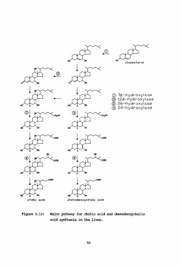
CD 7cx -hydroxylase ® 12£A-hydroxylo.se ® 26-hydroxylo.se @ 24-hydroxylo.se
Figure 2.13: Major pathway for cholic acid ani chenodeoxycholic
acid synthesis in the liver.
54

which the 26-hydroxylase is most probably absent. An alternative route via
25-hydroxylation becomes prominent (see figure 2.14). '!his pathway is
catalyzed by microsomal ani soluable enzymes ani is described in man ani in
rats (Shefer et al. 1 1976; <lleng et al. 1 1977; Walthers et al. 1 1983). Some
mitochondrial 25-hydroxylase activity of cholesterol has also been
demonstrated in the rat liver (Bjorkhem ani Gustafsson, 1973). It is
possible, that this hydroxylase ani the mitochondrial 25-hydroxylase active
on vitamin OJ, are one ani the same species of cytochrame P-450 (Bjorkhem
ani Hollllberg, 1978; Pedersen et al. , 1979b) •
In patients with crx a relative deficiency of the 24S-hydroxylase also
contributes to the subnonnal production of bile acids (Salen et al., 1979).
The 23R-hydroxylase is an enzyrre not being used in the regular bile acid
synthesis. Only in conditions where the nonnal synthesis is blocked, as in
Cl'X1 does 23R-hydroxylation occur. Walthers et al. (1983) found
5,8-cholestan-3a1 7a,12a,23R,25-pentol to be the most prominent bile alcohol
in the urine of such a patient. '!his microsomal 23R-hydroxylase is a
cytochrame P-450 containing mixed function oxidase.
0
DH -
-Figure 2.14: Alternative route for cholic acid synthesis in patients
with crx.
55
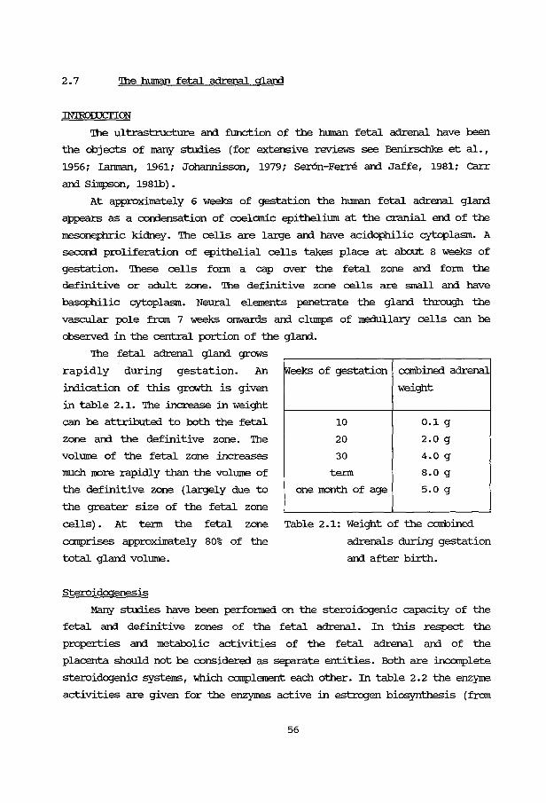
2. 7 '!he human fetal adrenal gland
IN'IROI:UCriON
'!he ultrastructure and function of the human fetal adrenal have been
the objects of many studies (for extensive reviews see Benirschke et al.,
1956; Lanman, 1961; Johannisson, 1979; ser6n-Ferre and Jaffe, 1981; carr and Simpson, 198lb) .
At approximately 6 weeks of gestation the human fetal adrenal gland
appears as a condensation of coelomic epithelimn at the cranial end of the
mesonephric kidney. '!he cells are large and have acidophilic cytoplasm. A
second proliferation of epithelial cells takes place at about 8 weeks of
gestation. 'Ihese cells fo:rm a cap over the fetal zone and fo:rm the
definitive or adult zone. '!he definitive zone cells are small and have
basophilic cytoplasm. Neural elements penetrate the gland through the
vascular pole from 7 weeks onwards and cl1llliJS of medullary cells can be
observed in the central portion of the gland.
'!he fetal adrenal gland grows
rapidly during gestation. An
indication of this growth is given
in table 2 .1. The increase in weight
can be attributed to both the fetal
zone and the definitive zone. '!he
volume of the fetal zone increases
much more rapidly than the volume of
the definitive zone (largely due to
the greater size of the fetal zone
Weeks of gestation
10
20
30
te:rm
one month of age
combined adrenal
weight
0.1 g
2.0 g
4.0 g
8.0 g
5.0 g
cells). At te:rm the fetal zone Table 2.1: Weight of the combined
comprises approxilnatel y 80% of the adrenals during gestation
total gland volume. and after birth.
Steroidogenesis
Many studies have been perfonned on the steroidogenic capacity of the
fetal and definitive zones of the fetal adrenal. In this respect the
properties and metabolic activities of the fetal adrenal and of the
placenta should not be considered as separate entities. Both are incomplete
steroidogenic systems, which complement each other. In table 2. 2 the enzyme
activities are given for the enzymes active in estrogen biosynthesis (from
56
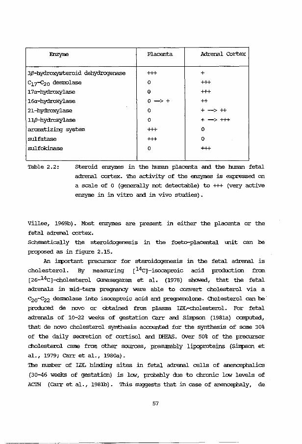
:E:nzyne Placenta Adrenal Cortex
3,8-hydroxysteroid dehydrogenase +++ + c17-c20 desnolase 0 +++ 17a-hydroxylase 0 +++ 16a-hydroxylase 0 -> + ++
21-hydroxylase 0 + -> ++
11,8-hydroxylase 0 + -> +++ aromatizing system +++ 0
sulfatase +++ 0
sulfokinase 0 +++
Table 2.2: steroid enzymes in the human placenta arrl the human fetal
adrenal cortex. The activity of the enzymes is expressed on
a scale of 0 (generally not detectable) to +++ (very active
enzyme in in vitro arrl in vivo studies).
Villee, 1969b). Most enzymes are present in either the placenta or the
fetal adrenal cortex.
Schematically the steroidogenesis in the foeto-placental unit can be
proposed as in figure 2.15.
An important precursor for steroidogenesis
cholesterol. By measuring [14c)-isocaproic
[26-14C)-cholesterol Gunasegaram et al. (1978)
in the fetal adrenal is
acid production fram
showed, that the fetal
adrenals in mid-term pregnancy were able to COIWert cholesterol via a
C20-c22 desnolase into isocaproic acid arrl pregnenolone. Cholesterol can be
produced de novo or obtained from plasma IDircholesterol. For fetal
adrenals of 10-22 weeks of gestation carr arrl Silrpson (198la) CXl!llpllted,
that de novo cholesterol synthesis accounted for the synthesis of same 30%
of the daily secretion of cortisol arrl IllFAS. over 50% of the precursor
cholesterol came fram other sources, presumably lipoproteins (Silllpson et
al. , 1979; carr et al. , 198oa) •
The number of IDL binding sites in fetal adrenal cells of anencephalies
(30-46 weeks of gestation) is lcxv, probably due to chronic lcxv levels of
Acm (carr et al. , 198lb) • 'Ihis suggests that in case of anencephaly, de
57

MOTHER PLACENTA FETUS
acetate feeding acetate acetate
\ / t l cholesterol ------ cholesterol----- cholesterol
+ l ~ , pregnenolone pregnenolone pregnenolone (S)
+ l i progesterone progesterone progesterone
• ! corticoids corticoids
17a-OH-pregnenolone(S)
+ DHEA(S) -----'-----DHEA ______ DHEA(S)
+ androstenedione
j ,.~Lrn• 1 estrone~ estrad/ol-17,8
1fu-OH-DHEA ------'--16a-OH-DHEA(S)
! 1~0H-androstenedione
l 1fu-OH-testosterone
l ---
1--- estriol
Figure 2.15: steroid honnone interrelations in the rnaternor
fetoplacental unit.
58
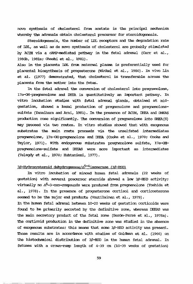
novo synthesis of cholesterol from acetate is the principal mechanism
whereby the adrenals obtain cholesterol precursor for steroidogenesis.
steroidogenesis, the number of IDL receptors ani the degradation rate
of IDL, as well as de novo synthesis of cholesterol are probably stinrulated
by Acm via a cAMP-m:rliated pathway in the fetal adrenal (carr et al. ,
1980b, 1981a; Ohashi et al., 1981).
Also in the placenta IDL from maternal plasma. is preferentially used for
placental biosynthesis of progesterone (Winkel et al., 1980). In vivo Lin
et al. (1977) d.enonstrated, that cholesterol is transferable across the
placenta from the not:her into the fetus.
In the fetal adrenal the conversion of cholesterol into pregnenolone,
17a-oH-pregnenolone ani IliE'A is quantitatively an important pathway. In
vitro incubation studies with fetal adrenal glanls, obtained at mid
gestation, shc:Med a basal production of pregnenolone ani pregnenolone
sulfate (Yanaihara ani Arai, 1981). In the presence of ACIH, IliE'A ani mFAS
production rose significantly. '!he conversion of pregnenolone into IEEA(S)
may proceed via two routes. In vitro studies showed that with exogenous
substrates the main route proceeds via the l.mSUlfated intennediates
pregnenolone, 17a-oH-pregnenolone ani IliE'A (Cooke et al., 1970; Cooke ani
Taylor, 1971). With erxlogenous substrates pregnenolone sulfate, 17a-OH:
pregnenolone-sulfate ani mFAS were more important as intennediates
(Telegdy et al., 1970; Huhtaniemi, 1977).
38-Hydroxysteroid dehydrogenase/.6.5-4isomerase (38-HSD)
In vitro incubation of minced human fetal adrenals (22 weeks of
gestation) with several precursor steroids shc:Med a low 3,8-HSD activity;
virtually no .6.4-3-oxo-corrp:l\.ll'l were produced from pregnenolone (Yoshida et
al., 1978). In the presence of progesterone cortisol ani corticosterone
seemed to be the major erxl products (Voutilainen et al., 1979).
In the human fetal adrenal between 10-20 weeks of gestation corticoids were
found to be primarily secreted by the definitive zone, whereas mFAS was
the main secretory product of the fetal zone (Ser6n-Ferre et al., 1978a).
'!he corticoid production in the definitive zone was studied in the absence
of exogenous substrates; this means that some 3,8-HSD activity was present.
'lhese results are in acx::ordance with studies of Goldman et al. (1966) on
the histochemical distribution of 3,8-HSD in the human fetal adrenal. In
fetuses with a crown-rump length of 4-39 em (10-39 weeks of gestation)
59
virtually no 3,8-HSD activity could be shown in the fetal zone of the fetal
adrenal. However in the definitive zone intense stai.nin;J was present from
16 weeks orrwards, suggestin:J a developin:J 3,8-HSD activity.
Short tenn cultures of both fetal and definitive zones of the hU!l'al1
fetal adrenal (9-18 weeks of gestation) showed a pa:tten1 of steroid
secretion, characteristic for each zone, upon stilllulation with ACIH,
(Fujieda et al., 1981a; Simonian and Gill, 1981). lDng tenn stilllulation
with ACIH (1-4 days) increased the steroidogenic capacity in cultures of
both zones. '!he patten1 of steroid production in the definitive zone
cultures remained constant, but in the fetal zone cultures cortisol was
preferentially increased (Kahri et al., 1976; Huhtaniernie, 1977). '!here is
a close relationship between the no:rphological and functional changes in
the adrenal cell {Voutilainen and Kahri, 1979). '!his change in steroid
pattern was coupled with a 5-fold increase in 3,8-HSD activity (Simonian and
Gill, 1981). Similar results were obtained by Branchaud et al. (1978) with
explants from the fetal and definitive zones kept in culture for 7-9 days.
Voutilainen and Kahri {1980) found an inhibition of the ACIH-stilllulated
cortisol production in cultures of human fetal adrenals, when placental
tissue was present. At the same time the amount of free IEE'A increased;
IliE'AS did not rise, probably due to the high sulfatase activity in the
placental tissue. '!hey concluded that some placental factor{s), probably
estrogens, inhibit the activity of adrenal 3,8-HSD or prevent its synthesis
and thus direct adrenal steroid synthesis towards IliE'AS production in vivo.
Corticosteroids.
'!he synthesis of corticoids, like cortisol and corticosterone, has
been the subject of many studies in mid~estational hU!l'al1 fetal adrenals.
In tissue culture the ratio of cortisol to 11-deoxycortisol (on days 0-5)
was the same as in the adult adrenal culture, suggesting a 11,8-hydroxylase
activity of the same magnitude (Voutilainen, 1979) . '!he cortisol/
corticosterone ratio was much higher in the fetal adrenal than in the adult
adrenal, reflecting the high 17a-hydroxylase activity. No specific defect
in aldosterone production was noted (higher aldosterone/corticosterone
ratio than in the adult) . Shibusawa et al. (1980) confinned, that the
11,8-hydroxylase was present in the mitochondrial fraction of the fetal
adrenal at mid~estation. 'lhese authors found that steroids may modify the
11,8-hydroxylase activity, as was found for the 17a-hydroxylase and C17-c20
60
desnolase activity (Shibusawa et al., 1978).
In the htnnan fetal adrenal the extent of the corticosteroid production
in fetal adrenals is not exactly known. High levels of cortisol in early
fetal life in many species can cause malformations ani may have undesirable
effects on development (Hall ani Kalliecharan, 1976; Chen et al., 1977). On
the other hand cortisol ani especially cortisol-sulfate, produced by the
fetal adrenal, have an i.Jrportant function in fetal lung maturation (Murphy,
1979). So the cortisol concentration in the htnnan fetus is subject to
careful regulation.
Cortisol can cross the placenta ani is rapidly converted into cortisone by
the placental 11,8-hydroxysteroid dehydrogenase (11,8-HSD) (Murphy, 1979).
Amniotic fluid cortisol is mainly derived fran the fetal adrenals.
Estrogens
'Ihe nost i.Jrportant tissue for estrogen production is the placenta (for
reviews see Villee, 1969a; Simpson ani MacDonald, 1981) • 'Ihe placenta lacks
steroid 17a-hydroxylase activity and consequently can not convert
C21-steroids into c19-steroids. Since the estrogen excretion is low in
WOJrel1 can:ying an anencephalic fetus (lacking the fetal zone of the fetal
adrenal), it seems likely that the fetal adrenal is involved in placental
estrogen biosynthesis. Near tenn approximately half of the estradiol-17J3,
synthesized in the placenta, is derived fran precursors in the fetal plasma
and half fran maternal precursors.
In figure 2.16 the metabolic pathway is given for estrogen
biosynthesis (see also Villee, 1969a; Wolf et al., 1978). J:HE'AS, produced
in the fetal zone of the fetal adrenal, is 16a-hydroxylated mainly in the
fetal liver (Wynne and Renwick, 1976; sana et al., 1980). Both J:HE'AS and
16a-oH-J:HE'AS are deconjugated in the placenta by a very active sulfatase.
Factors regulating the htnnan fetal adrenal cortex
In many reports honronal factors, regulating adrenal steroidogenesis,
are described. 'Ihese factors may be of placental (Simpson and MacDonald,
1981) or fetal origin. Much evidence has accumulated on the vital role of
the fetal endocrine glands in the mo:rphogenesis and functional maturation
of the fetus. In this respect the fetal pituitary plays an i.Jrportant role
(for a review see Kaplan ani Gnnnbach, 1976). 'Ihe capacity of the htnnan
fetal pituitary to secrete polypeptide honrones ani of the hypothalanrus to
61
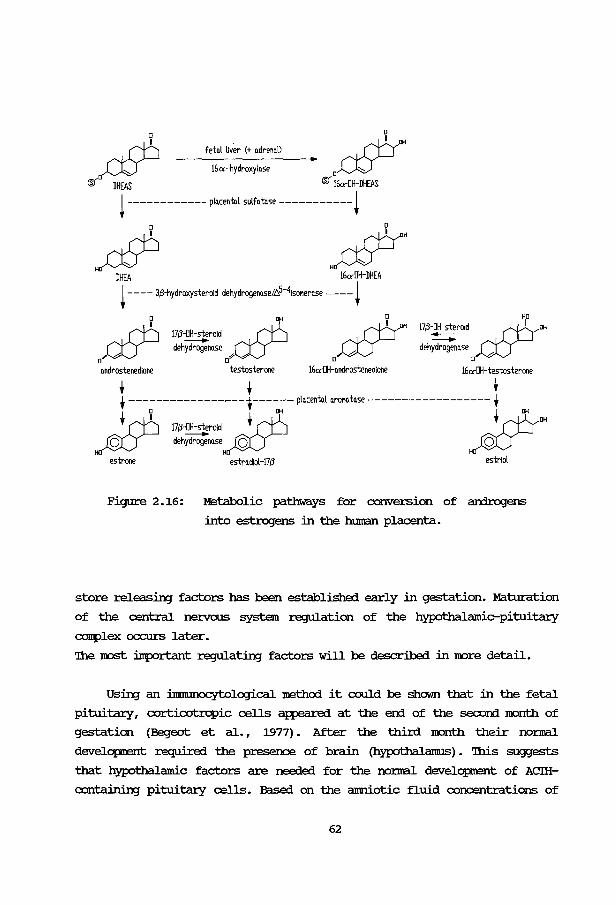
"-
?t 0~~ fetnl liver (+ QdrenQl)
16 a -hydroxylQSe
®' DHEAS ®' 16a-OH-DHEAS
! ------------ plQcentnl sulfQtnse -----------!
# ~ DHEA 16C<"DH-DHEA ! ---- 3/l-hydroxysteroid dehydrogenQse/b.5-41soMero.se ----!
Qndrostenedione
17/l-~roid ctP dehydrogenQse
[]-:9 A
testosterone 16<X"DH-androstenedione 16C<"DH-testosterone
• • • t --------------~--1------ plQcentQl QroMtQse ------------------ t • ~ IJi • OH OH
~ f['b 17/l-OH-steroid . t Ib _ ; rt::r ~ deh~nQSeH~- H)QV
estrone
Figure 2.16:
estrQdlol-17/l estriol
Metabolic pathways for conversion of an:irogens
into estrogens in the human placenta.
store releasing factors has been established early in gestation. Maturation
of the central nervous system regulation of the hypothalamic-pituitary
camplex occurs later.
'!he JOOSt important regulating factors will be described in m:>re detail.
Using an i.mnumocyt:ological method it could be shown that in the fetal
pituitary, corticotrcpic cells appeared at the en:i of the seconi m:>nth of
gestation (Begeot et al. , 1977) . After the third m:>nth their nonnal
developtent required the presence of brain (hypothalamus). 'lhis suggests
that hypothalamic factors are needed for the nomal developrent of ACIH
containing pituitary cells. Based on the amniotic fluid concentrations of
62
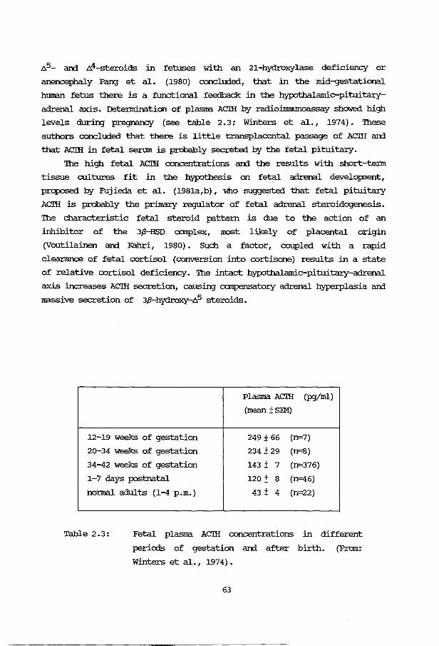
.6..5- an:i .6..4-steroids in fetuses with an 21-hydroxylase deficiency or
anencephaly Pang et al. (1980) concluded, that in the mid-gestational
hmnan fetus there is a functional feedback in the hypothalamic-pituitary
adrenal axis. Detennination of plasma ACIH by radiollmnunoassa.y showed high
levels during pregnancy (see table 2.3; Winters et al., 1974). 'Ihese
authors concluded that there is little transplacental passage of ACIH and
that ACIH in fetal serum is probably secreted by the fetal pituitary.
'!he high fetal ACIH concentrations and the results with short-tenn
tissue cultures fit in the hypothesis on fetal adrenal development,
proposed by Fujieda et al. (1981a,b), who suggested that fetal pituitary
ACIH is probably the primary regulator of fetal adrenal steroidogenesis.
'!he characteristic fetal steroid pattern is due to the action of an
inhibitor of the 3,8-HSD complex, lKlSt likely of placental origin
(Voutilainen and Kahri, 1980). SUch a factor, coupled with a rapid
clearance of fetal cortisol (conversion into cortisone) results in a state
of relative cortisol deficiency. '!he intact hypothalamic-pituitary-adrenal
axis increases ACIH secretion, causing compensatory adrenal hyperplasia and
massive secretion of 3,8-lrjdroxy-.6..5 steroids.
Plasma ACIH (pgjml)
(mean ±SEM)
12-19 weeks of gestation 249 ± 66 (n=7)
20-34 weeks of gestation 234 ± 29 (n=8)
34-42 weeks of gestation 143 ± 7 (n=376)
1-7 days postnatal 120:!: 8 (n=46)
nomal adults (1-4 p.m.) 43 ± 4 (n=22)
Table 2.3: Fetal plasma ACIH concentrations in different
periods of gestation and after birth. (From:
Winters et al., 1974).
63
Several cases of low estrogen excretion during pregnancy have been
described, which after birth appeared to be fetal ACIH deficiencies
(Grimberg et al., 1978; Zachmann et al., 1979). So it seems that in the
later stages of pregnancy urincuy estrogen secretion not only depems on a
well functioning fetal adrenal, but also on the entire hypothalamic
pituit:uy-adrenal axis. In spite of a fetal ACIH deficiency, the fetal
adrenal develops nonnally in the cases just nentioned. One may conclude
from these results, that there are other factors regulating the
development of the fetal adrenal. Solie reports describe the existence of a
corticotropin-like placental honrone. '!his honrone was called human
Chorionic Corticotropin (hCC) (Gena.zzani et al., 1975; Liotta et al.,
1977) , or human Chorionic Somato-Il1all1100tropin (hCSM) (Isherwood and oakey,
1976).
'!he pituitary gland produces a family of related peptide honrones from
the high 100lecular precursor pro-opionelanocortin. Several of them have a
heptapeptide in cormnon. Figure 2. 7 shows the Im.ltual relationship between
these peptides. ,B-lipotropin (,8-IH!), r-IH! and ACIH are produced in the
pars distalis; a-Melanocyte-StiJm.llating Honrone ( a-MSH) , ,8-MSH and
Corticotropin-like intennediate lobe protein (CLIP) originate in the pars
intennedia. Silman et al. (1976) showed that the fetal pituitary (24 weeks
of gestation) contained only small amounts of intact ACIH, but larger
quantities of peptides, closely resembling a-MSH and CLIP. '!hey suggested,
that these smaller peptides are the dominant honrones in fetal life, being
replaced by ACIH before parturition. However, a steroidogenic response to
CLIP could not be demonstrated.
In table 2. 4 the stiJm.llato:ry properties of several honrones are
sununarized. In the presence of CLIP, fetal adrenal slices did not produce
IEE'A (Brown et al., 1981). '!he contradictory results with the other
stimulating factors may be the consequence of the methodology used:
explants of human fetal adrenals in tissue culture or fetal adrenal cells
in cell culture may loose properties that were present in the original
cells. ACIH seems the main tropic honrone for the human fetal adrenal. '!he
in vivo contribution of other peptide honrones to the growth and function
of the human fetal adrenal remains to be established.
64
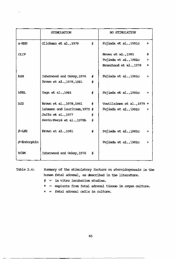
cH1SH
CLIP
hGH
hPRL
ha;
,8-UH
,B-Errlorphin
ha>M
Table 2.4:
STIMOIATION NO STIMOIATION
Glickman et al.,1979 # Fujieda et al.,1981c +
Brown et a1.,1981 # Fujieda et al.,1981c + Branchaud et al. ,1978 *
Isherwood ard oakey ,1976 # Fujieda et al.,1981c + Brown et al.,1978,1981 #
Taga et al.,1981 # Fujieda et al.,1981c +
Brown et al.,1978;1981 # Voutilainen et al. ,1979 * I..ehmann ard Iauritzen,1975 # Fujieda et al.,l981c + Jaffe et al.,1977 # Ser6n-Ferre et a1.,1978b #
Brown et al.,1981 # Fujieda et al.,1981c +
Fuj ieda et al. ,1981c +
Isherwood ard oakey ,1976 #
Slm1rnal:y of the stimulatory factors on steroidogenesis in the
hmnan fetal adrenal, as described in the literature.
# in vitro incubation studies.
* = explants from fetal adrenal tissue in organ culture.
+ fetal adrenal cells in culture.
65

CHAPI'ER 3
MEIHOOOI..CGY
3A. TECENIC'AL ASPECI'S.
3A.1 Introduction
Working with isolated cells has several advantages 011er using tissue
blocks, for example, better contact of all cells with the surrounding
medium, containing nutrients and stimulatory factors, and an easier
diffusion of products out of the cell. A disadvantage is, that the
isolation procedure is rather laborious and may damage the cells.
Rat adrenal cell suspensions can be prepared successfully with collagenase;
rnase (for a review see Falke, 1977: thesis, Rotterdam). A detailed
description of the isolation method will be given in this chapter. Part of
this method has also been used for the isolation of rat adrenal glamerulosa
cells, rat liver cells and human fetal adrenal cells.
Several analytical methods will be described in this chapter: the
fluorametric assay of corticosterone and cortisol, gaschromatography of
steroids and sterols, radioimmunoassay of aldosterone, protein binding
assay of cAMP and the luminescence assay of ATP.
For some of the materials used in this study, reference can be made to
Clapters 4-7.
3A.2 Isolation of cells
RAT AmENAL CELIS
Cells from whole rat adrenals were prepared according to the method of
Falke et al. (1975a,b) with minor modifications. A Krebs-Ringer-Hepes
buffer (pH = 7 .35), supplemented with 3% (WjV) bOiline sennn albumin and
0.2% (WjV) glucose, was used (=KRHAG). Male Wistar rats, weighing 190 - 210
grams, were obtained from the Centraal Proefdieren bedrij f '!NO, Zeist. The
animals were caged in groups of five with water and laboratory chow ad
libitum. '!he rats were decapitated at approximately 8.30 a.m. 'Ihe adrenals
were removed and placed in cold saline. '!he collected adrenals of ten rats
(mostly used in one experiment) were dissected free of adhering fat tissue
and cut into 6-8 pieces per gland. '!he pieces were put in two 25 m1
67
Erlernreyer flasks an:l washed twice with cold I<RHAG. '!hen 5 rol of the enzyme
solution, containing collagenase (5 l.'rg/rol) an:l mase (0.05 l.'rgjrol) was
added. '!he flasks were placed in a l)]bnoff incubator, shaking at 100 rpm,
at a temperature of 37°C. After 20 minutes the medimn with the free cells
was removed with a Pasteur pipette an:l replaced by 5 rol fresh enzyme
solution. '!he combined free cell suspensions were allowed to stan:l for 2
minutes to sediment coarse particles. '!he supernatant with the cells was
centrifuged for 10 minutes at lOOxg. '!he cell pellet was resuspended in
I<RHAG containing mase (0.05 l.'rg/rol) an:l stored on ice. After the second
incubation period the free cells were removed as described above. '!he
remaining tissue particles were dissociated in the enzyme solution by
sucking up an:l ~ling several times in a Pasteur pipette, followed by a
third 20 minutes incubation period. '!he free cells were removed as
described. '!he three cell suspensions were centrifuged at lOOxg for 10
minutes an:l washed once again in the mase-containing I<RHAG after a 5
minutes incubation period at 37°C. Finally the cells were resuspended in
KRHAG an:l counted in a BUrker eotmting c.haniber uOO.er a light microscope
(magnification 400X) • Yields were 200, 000-600, ooo cells per adrenal. '!he
cells were used .inunediately after isolation.
RAT ADRENAL G~ CELIS
Rat adrenal glomerulosa cells were prepared from male Wistar rats,
weighing 190-210 grams. '!he glan:ls were removed an:l dissected free of
adhering fat tissue as just described. Incisions were made, followed by
manual compression of each glan:l to extru.de the zonae fasciculata an:l
reticularis an:l the medulla from the capsule + zona glomerulosa. '!he
capsules were cut into small pieces an:l washed with I<RHAG. '!he further
isolation procedure for the glomerulosa cells is similar to the procedure
described above for the 'Whole adrenal suspension except that the cells are
centrifuged at 150xg for 10 minutes.
Average yields were 30,800 cells per adrenal. '!he purity of the glomerulosa
cell suspension was checked by microscopic examination after staining with
hematoxylin/eosin. Generally ;::95% of the cells was of the glomerulosa type,
according to histological criteria.
RAT LIVER CELIS
For each experiment the liver of a 200 gram male Wistar rat was
68
removed, cut into pieces and washed with cold saline and KRHAG. '!he liver
cells were prepared by collagenasejr:Nase digestion, as described for the
whole adrenal cell suspension, except that the cells were centrifuged at
100xg for 5 minutes. '!he cells were finally resuspeOO.ed in KRHAG and
counted.
HU.MAN FErAL ADRENAL CELIS
Human fetal adrenals were obtained from spontaneous or induced
abortions. '!hey were transported to the laboratory in icecold saline. Fat
tissue was removed and the adrenals were weighed. '!he isolation procedure
for the fetal adrenal cells is essentially the same as used for the rat
adrenal cells (described earlier) • Yields were 0. 8-6.9 x 10⁶ cells per
adrenal.
3A. 3 Incubation conditions
All incubations were done in siliconized glass tubes in a Thlbnoff
incubator, shaking at 100 :rpn. Generally the incubation temperature was
37°C. ACIH was added in 0.9% NaCl, acidified to pH=3. 75 and supplemented
with 0.5% albumin. Angiotensin II was added in KRHAG. All sterols and
steroids were added in 10 J.Ll ethanol. To incubations not containing sterols
vehicle only was added. '!he final incubation volume was 1 ml. '!he Na+ and
~ concentrations in KRHAG were 111.2 nM and 4. 75 nM respectively. If other
concentrations of potassium were required, the sodium and potassium
concentrations were changed in such a way as to keep the sum of the
concentrations of these ions unchanged.
3A.4 Fluorometry
Corticosterone and cortisol was estimated by fluorometry using the
extraction procedure of Clark and Rubin (1969). Fluorescence was developed
in ethanol/concentrated sulfuric acid (1: 3) and measured at room
temperature with a Baird Atomic fluorimeter (excitation wavelenth is 468
nm; fluorescence wavelenth is 520 nm). A corticosterone standard (mostly
500 11CJ/assay) was measured in duplicate in the same assay.
The variation coefficient due to methodological variations (measured for 15
corticosterone standards in duplicate) was 3.6%. '!he biological variation
coefficient (measured for the ACIH-stirnulated corticosterone production)
was 5.1% (n=22).
69
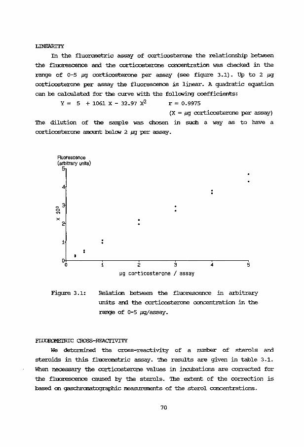
LINFARITY
In the fluo:ronetric assay of corticosterone the relationship between
the fluorescence ani the corticosterone concentration was checked in the
range of 0-5 J.IIJ corticosterone per assay (see figure 3.1). Up to 2 J.11J
corticosterone per assay the fluorescence is linear. A quadratic equation
can be calculated for the cm.ve with the following coefficients:
Y = 5 + 1061 x - 32.97 x2 r = 0.9975
(X= J.IIJ corticosterone per assay)
'!he dilution of the sample was chosen in such a way as to have a
corticosterone amount belc:M 2 J.IIJ per assay.
Fluorescence (arbitrary units)
"' 0 ...... X
1 . . 01+---------~------~--------~--------~------~
0
Figure 3.1:
1 2 3 4 5
~g corticosterone I assay
Relation between the fluorescence in arbitrary
units an::l the corticosterone concentration in the
range of 0-5 JJIJ/assay.
F'I.IJOR!:NEI'RC CRa>S-RFACI'IVITY
We detennined the cross-reactivity of a number of sterols an::l
steroids in this fluororoetric assay. '!he results are given in table 3.1.
When necessa:ry the corticosterone values in incubations are corrected for
the fluorescence caused by the sterols. '!he extent of the correction is
based on gaschramatographic measurements of the sterol concentrations.
70
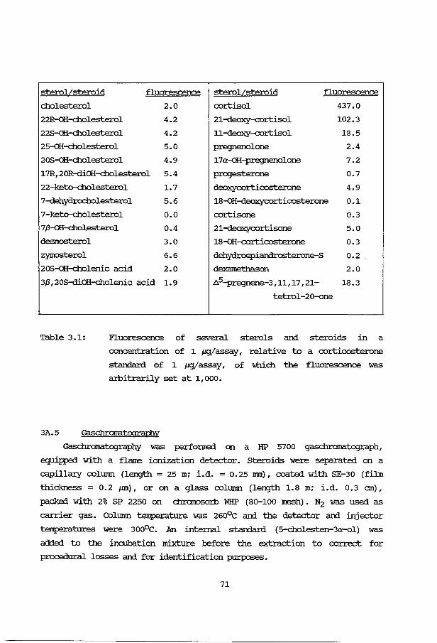
sterolLsteroid fluorescence sterolLsteroid fluorescence
cholesterol 2.0 cortisol 437.0
22R-oH-cholesterol 4.2 21-deoxy--cortisol 102.3
22S-oH-cholesterol 4.2 11-deoxy--cortisol 18.5
25-oH-cholesterol 5.0 pregnenolone 2.4
20S-QH-cholesterol 4.9 17a-aH-pregnenolone 7.2
17R, 20R-diOH-cholesterol 5.4 progesterone 0.7
22-keto-cholesterol 1.7 deoxycorticosterone 4.9
7-dehydrocholesterol 5.6 18-oH-deoxycorticosterone 0.1
7-keto-cholesterol o.o cortisone 0.3
7,8-aH-cholesterol 0.4 21-deoxycortisone 5.0
desmosterol 3.0 18-QH--cortic:osterone 0.3
zymosterol 6.6 dehydroepiandrosterone-S 0.2
20S-aH-cholenic acid 2.0 dexamethason 2.0
3,8,20S-diOH-cholenic acid 1.9 ~5-pregnene-3,11,17,21- 18.3
tetrol-20-one
Table 3.1: Fluorescence of several sterols and steroids in a
concentration of 1 JJ11/assay, relative to a corticosterone
standard of 1 JJ11/assay, of which the fluorescence was
amitrarily set at 1,000.
3A. 5 Gaschromatography
Gaschromatography was perfonned on a HP 5700 gaschramatograph,
equipped with a flame ionization detector. Steroids were separated on a
capillacy column (length = 25 m; i.d. = 0.25 nun), coated with SE-30 (film
thickness = 0.2 ,um), or on a glass column (length 1.8 m; i.d. 0.3 em),
packed with 2% SP 2250 on chromoso:r:b WHP (80-100 mesh). N2 was used as
carrier gas. Column temperature was 260°C and the detector and injector
temperatures were 300°C. An internal standard (5-cholesten-3a-ol) was
added to the incubation mixture before the extraction to correct for
procedural losses and for identification purposes.
71
For the detection of both free and sulfa-conjugated steroids one ml of
the incubated cell suspension was diluted 1:1 with H2o, made up to 20% with
sodium chloride and acidified to pH=1 with 6 N H2so4. 'lhis mixture was
extracted with 10 ml and 5 ml ethylacetate. 'Ihe combined organic layers
were kept overnight at 370c. 'Ihe next lOCl:rning the ethylacetate was washed
with 5 ml 0.1 M NaH<DJ and 2 ml H2o. 'Ihe organic layer was dried over
Na2so4 and evaporated urxler nitrogen at 40°C. To the extract 100 JLl 2%
m=thoxyamineJHCl in pyridin was added and the mixture heated at 60°C for
1 hour. 'Ihen the pyridin was blown off with nitrogen. 100 JLl of a
silylating mixture, containing bis-trimethylsilylacetamidejtritrethyl
chlorosilanejtritrethylsilylimidazole (3/2/3; VjVjV) was added and the
reaction completed at so0 c overnight. 'Ihe steroids were purified with a
Lipidex-5000 column, using 5 ml hexanejpyricli.njhexamethyldisilazanej
ditrethoxypropane (97/1/2/10; VjVjVjV) as eluting agent. After evaporation
the residue was dissolved in 20 JLl hexane.
For the detection of only the free steroids one ml of the incubated
cell suspension was diluted with 1 ml acetone and kept overnight at 4°C.
'Ihe protein precipitate was extracted once again with 1 ml acetone. To the
combined acetone layers 5 ml of dichloromethane was added after which a
phase separation occurred. The water layer was extracted with 1 ml of
dichloromethane arrl, the combined organic layers washed with 1 ml of water.
After evaporation the extract was derivatized as described above.
3A.6 ATP
ATP was measured using the luciferin-luciferase m=thod, as described
by Guinn and Eidenbock (1972). 'Ihe reaction can be written as:
luciferin + ATP + ~02 luciferase> dehydroluciferin +AMP+ PPi + light
The enzyme solution was prepared from fireflies (Sigma Chern Comp).
For the measurement of ATP in whole cells, one ml of the cell suspension
was poured into 3 ml of boiling water and left for 7 minutes in a boiling
water bath. 'Ihe precipitated protein was centrifuged and the supernatant
stored frozen below -24°C until analysis. Before the analysis the
supernatant was lyor;hilized and resolved in 0.5 ml distilled water. A
standard series of ATP concentrations in water was prepared ( 0. 5, o. 25,
0.125, 0.1, 0.05 and 0.025 JLg/100 JLl).
72
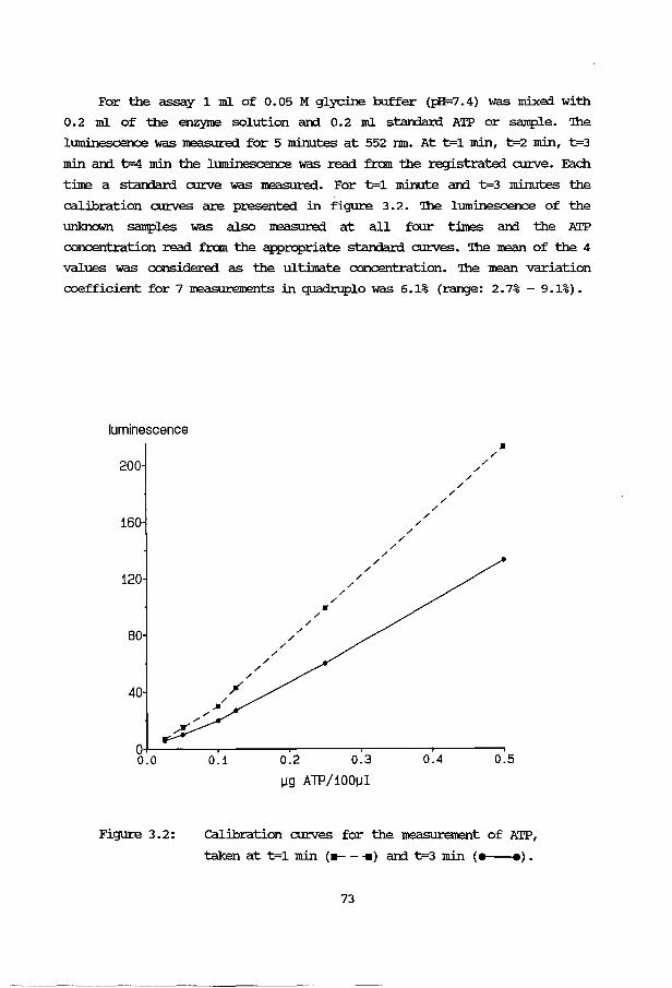
For the assay 1 m1 of 0.05 M glycine buffer (J;:H=7.4) was mixed with
0.2 m1 of the enzyne solution ani 0.2 ml starrlard ATP or sample. 'lhe
luminescence was measured for 5 minutes at 552 nm. At t=1 min, t=2 min, t=3
min ani t=4 min the luminescence was read from the registrated cw:ve. Each
tine a starrlard cw:ve was measured. For t=1 minute an:l t=3 minutes the
calibration cw:ves are presented in figure 3. 2. 'lhe luminescence of the
unknown samples was also measured at all foor tines ani the ATP
concentration read from the appropriate starrlard cw:ves. '!he mean of the 4
values was considered as the ultimate concentration. '!he mean variation
coefficient for 7 :measurements in quadruple was 6.1% (range: 2. 7% - 9.1%).
luminescence
200
160
120
80
40 / /
/
/ /
/
/ /
/
• / /
/ /
/
/ /
/
/ /
/ /
/ /
/ /
/ /
/ /
• /
0+-----~~----~-------r------,-------, 0.0 0.1 0.2 0.3 0.4 0.5
Figure 3.2:
IJQ ATP/1001.11
calibration cw:ves for the measurement of ATP,
taken at t=1 min <-- -11) ani t=3 min (e--e).
73

3A.7 Cyclic AMP
cyclic AMP was rreasured with a protein binding assay {'!he Radio
chemical Centre, Amershant) • '!he extraction procedure of Iavin et al.
{1976) was used. Concentrations in unknown samples were read from a legit
log transfo:r.ne:i starXIard curve. '!he best-fitting quadratic function was
used as a calibration curve. Samples with a concentration out of the range
(1-16 pgjtube) were rreasured again at a proper dilution. '!he recovery of
cAMP added to the KRH1IG buffer was 90%. '!he mean variation coefficient for
5 measurements in triplicate was 7.6% (range: 3.0%- 14.4%).
3A.8 Aldosterone
Aldosterone was estimated using a radioi.Imnuno assay (CE'A, Sarin) with
prior extraction. '!he recovery for the extraction procedure is 97%.
Concentrations in unknown samples were read from a legit-log transfo:r.ne:i
starXIard curve. '!he best-fitting quadratic function was used as a
calibration curve.
'!he within-assay coefficient of variation was 9% (n=10) and the between
assay coefficient of variation 13.7% {n=15) .
'!he specificity of the antiserum used in the aldosterone radio.inununo
assay is shown in table 3.2. '!he percentage of cross-reactivity was
calculated as X.100/Y, where X and Y are respectively the weight of the
aldosterone and the weight of the cross-reactant, that caused a 50%
inhibition of the tracer binding. '!he antisennn is clearly very specific.
3A. 9 Density gradient centrifugation
Sedllnentation through a density gradient can be used to separate
cells, cell fragments and viruses, either at unit gravity or after
centrifugation. '!he density medium must fulfil several criteria:
a. easily adjustable to a suitable pH and osmolarity,
b. low viscosity even at high density,
c. a wide density range,
d. non-toxic to the cells,
e. impel:maable to biological membranes.
PercollR (Blannacia, SWeden) is a density gradient medium, consisting of a
colloidal suspension of silica particles, coated with polyvinylpyrrolidone.
It can be made isotonic by mixing 9 parts of Percell with 1 part of 9%
NaCl. '!he density gradients are fo:r.ne:i spontaneously by centrifugation at
74

Aldosterone 100%
18-0H:-deoxycorticosterone 0.03%
18-0H:-corticosterone 0.01%
Corticosterone 0.0025%
Cortisol 0.00005%
Cortisone 0.0001%
Allotetrahydrocortisol 0.00005%
Tetrahydrocortisone 0.00005%
Progesterone 0.00005%
Tetrahydrocortisol 0.00005%
Pregnanetriol 0.00005%
Cortexolone 0.00005%
Testosterone 0.00005%
Estradiol 0.00005%
25-QH-cholesterol 0.0005%
228-QH-cholesterol 0.0005%
Tabel 3.2: Percentage of cross-reactions of several
steroids and sterols in the aldosterone
radio.inununoassay.
25,000xg for 20 minutes. '!he shape of the gradient can be made visible by
using" density marker beads. 'lhese are coloured beads with each colour
having" its accurately known density. By varying" the percentage of Percoll
the shape of the gradient can be changed.
75

3B APPLICATION AND EVAIIJATION OF THE MEIHOOO
3B.l Introduction
With the rat adrenal cells, isolated as descr:ibed in Section 3A, some
general aspects of steroid metabolism were investigated in order to be
informed about the steroidogenic capacity of the cells. '!he infonnation
obtained includes a dose-response curve for Acrn:, effect of cell mnnbers
on response, ani effect of buffer ani albumin cxmcentration.
In many studies on fetal steroidogenesis tissue cultures of h1.m1an
fetal adrenal glan:is have been used. However under these conditions
steroidogenesis changes from a "fetal pattern", with largely 3,8-hydroxy- A 5
steroids like IHE'A, into an "adult pattern" with cortisol as an important
steroid (Branchaud et al. , 1978 ~ Fuj ieda et al. , 1981a ~ Simonian ani Gill,
1981) • '!his change can be explained by the disappearance of the fetal zone
cells from the cell culture ani the silllultaneous grc:Mth of the definitive
zone cells (Voutilainen ani Kahri, 1979). For this reason we wanted to use
freshly prepared fetal adrenal cells. With the cell isolation technique
descr:ibed in Section 3A. 2, human fetal adrenal cells were isolated. 'IWo
parameters for cell viability were studied: the production of cortisol ani
corticosterone from exogenous progesterone ani the ATP content of the
cells.
'!he h1.m1an fetal adrenal consists of two mo:rphologically ani
functionally different cell types: the fetal zone cells ani the definitive
zone cells. 'lWo techniques for separating these cell types were conpared: a
:n>anual dissection method of the whole fetal adrenal ani a cell separation
method by means of density gradient centrifugation on Percell. several
parameters for steroid metabolism were investigated in both cell types ani
conpared with data from the literature.
3B.2 Effect of AClH stilllulation
Figure 3. 3 shows a typical dose-response curve for the ACIH-stilllulated
corticosterone production in isolated rat adrenal cells during a 2 hours
incubation period at 37"C. '!he shape of the curve was always conparable to
that, shown in figure 3.3. However, the responsiveness towards ACIH varied
for each batch of cells. For this reason a limited dose-response curve was
measured for all batches of cells.
76
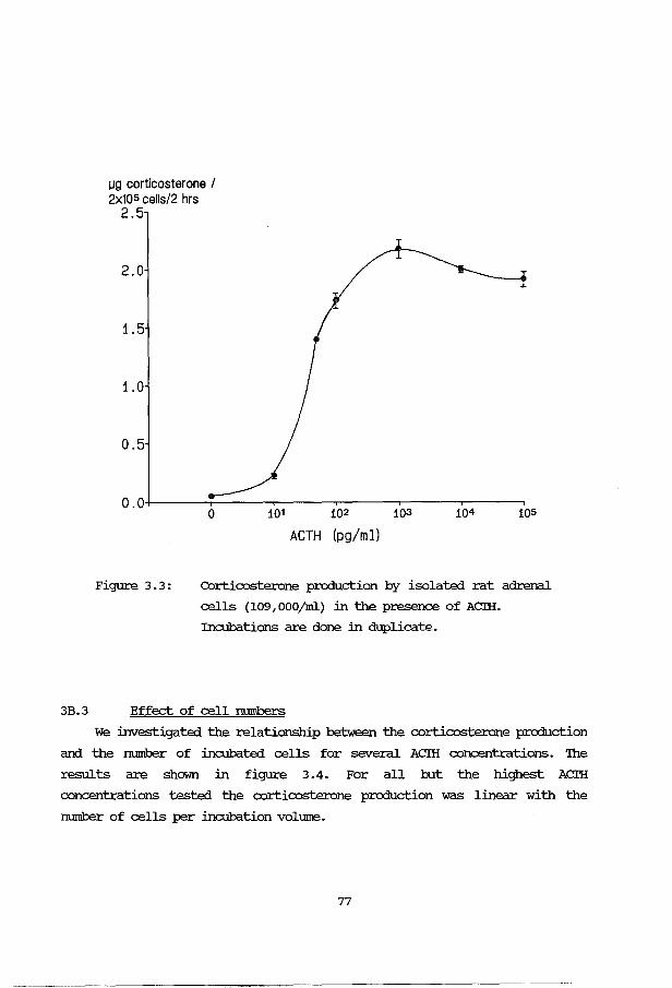
IJQ corticosterone I 2x1os cells/2 hrs
2.5
2.0
1.5
1.0
0.5
Figure 3.3:
0 101 102 103
ACTH (pg/ml)
Corticosterone production by isolated rat adrenal
cells (109,000/ml) in the presence of Acm. Incubations are done in duplicate.
3B. 3 Effect of cell numbers
We investigated the relationship between the corticosterone production
and the rnnnber of incubated cells for several Acm concentrations. 'Ihe
results are shown in figure 3.4. For all but the highest Acm concentrations tested the corticosterone production was linear with the
number of cells per incubation voltnne.
77
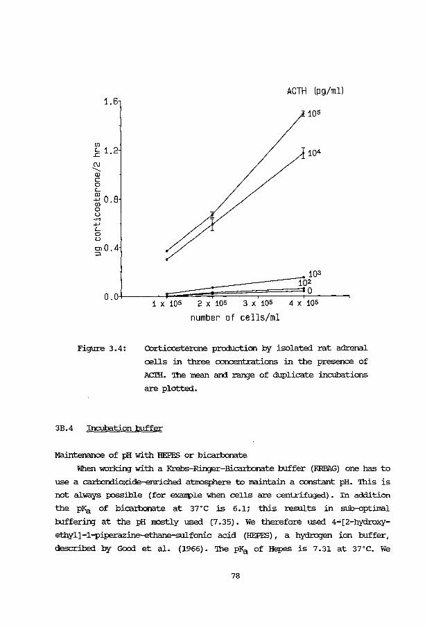
1.6
en .2 1.2 C\1 ........ Q) c 0 (_
2 0 8 en • 0 u .....
4-J (_
0 u OJ0.4 :J.
ACTH (pg/ml)
105
104
103
1------~~~;;~;;~~~~~~10~2L_ __ ~ 0.0 °
Figure 3.4:
1 X 10⁵ 2 X 10⁵ 3 X 10⁵ 4 X 10⁵
number of cells/ml
COrticosterone production by isolated rat adrenal
cells in three concentrations in the presence of
Acm. '!he mean and range of duplicate incubations
are plotted.
3B. 4 Incubation buffer
Maintenance of pH with HEPES or bicartx:>nate
When working with a Krebs-Ringer-Bicartx>nate buffer {RRBAG) one has to
use a camorn.ioxide-enriched atmosphere to maintain a constant pH. '!his is
not always possible (for example 'When cells are centrifuged). In addition
the pKa of bicamonate at 37°C is 6.1; this results in sub-optimal
bufferinJ at the pH IOC>Stly used (7 .35}. we therefore used 4-'[2-hydroxy
ethyl]-1-piperazine-ethane-sulfonic acid (HEPES}, a hydrogen ion buffer,
described by Good et al. (1966}. '!he PKa of Hepes is 7.31 at 3rc. We
78
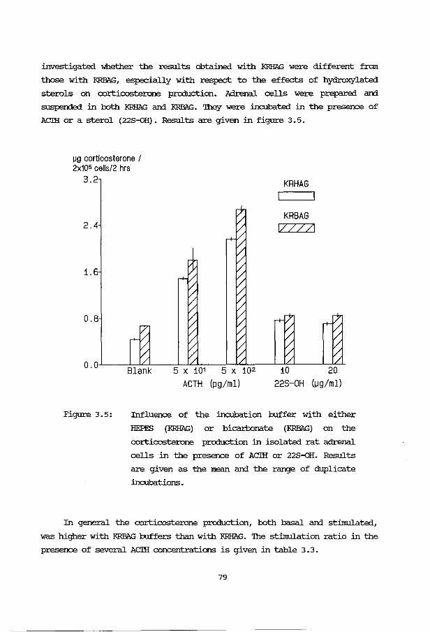
investigated whether the results obtained with KRHAG were different from
those with I<RPliG, especially with respect to the effects of hydroxylated
sterols on corticosterone production. Adrenal cells were prepared and
suspended in both KRHAG and I<RPliG. '!hey were incubated in the presence of
ACIH or a sterol (228-oH). Results are given in figure 3.5.
IJQ corticosterone I 2x10⁵ cells/2 hrs
3.2
2.4
1.6
0.8
KRHAG I
KRBAG 1/ZZ/1
O.OL----L~L---~~----L-~----LJ~--~~~
Blank 5 X 101 5 X 10² 10 20
Figure 3.5:
ACTH (pg/ml) 228-0H (1Jg/ml)
Influence of the incubation buffer with either
HEPES (KRHAG) or bicarbonate (I<RPliG) on the
corticosterone production in isolated rat adrenal
cells in the presence of ACIH or 22S~H. Results
are given as the mean and the range of duplicate
incubations.
In general the corticosterone production, both basal and stimulated,
was higher with I<RPliG buffers than with KRHAG. '!he stimulation ratio in the
presence of several ACIH concentrations is given in table 3. 3.
79
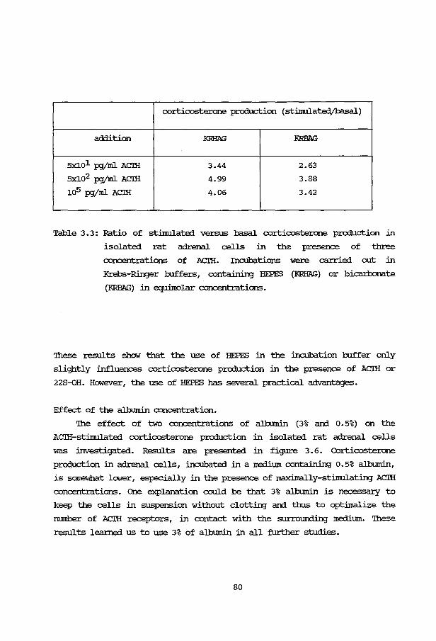
corticosterone production (stirnulatedjbasal)
addition RRHAG KRB.1\G
5x101 PJ/ml ACIH 3.44 2.63
5x10² PJ/ml ACIH 4.99 3.88
105 PJ/ml ACIH 4.06 3.42
Table 3. 3: Ratio of stimulated versus basal corticosterone production in
isolated rat adrenal cells in the presence of three
concentrations of ACIH. Incubations were carried out in
Krebs-Ringer buffers, containing HEPES (RRHAG) or bicarlx>nate
(KRBi\G) in equilrolar concentrations.
'lhese results show that the use of HEPES in the incubation buffer only
slightly influences corticosterone production in the presence of ACIH or
228-oH. However, the use of HEPES has several practical advantages.
Effect of the albumin concentration.
'!he effect of two concentrations of albumin (3% ani 0.5%) on the
ACIH-stinulated corticosterone production in isolated rat adrenal cells
was investigated. Results are presented in figure 3.6. Corticosterone
production in adrenal cells, incubated in a medium containing 0.5% albt.nnin,
is somewhat lower, especially in the presence of maximally-stimulating ACIH
concentrations. One explanation could be that 3% albumin is necessary to
keep the cells in suspension without clotting ani thus to optilnalize the
number of ACI'H receptors, in contact with the surrourrling medium. 'lhese
results leanted us to use 3% of albumin in all further studies.
80

~g corticosterone I 2x1os cells/2 hrs
3.2
2.4
1.6
0.8
I I
I I
/
/ /
... I-------1-----/ . -~
/
o. o+------r-----r-----r-----r-----r-------. s 0 101 102 103 104 10
Figure 3.6:
ACTH (pg/ml)
Dose-response curve of the ACTH-stimulated
corticosterone production in isolated rat adrenal
cells in the presence of 3% albumin (--) or
0. 5% a.J.burnin (--) • '!he mean and the range of
duplicate incubations are plotted.
3B. 5 Ht.nnan fetal adrenals
3B. 5.1 FErAL ADRENAIS
We received human fetal adrena.ls from several obstetric departments of
hospitals in and arourrl the city of Rotterdam. '!he duration of the
pregnancy and the tilne of death of the fetus were obtained from the
obstetrician. A sw:vey of the human fetal adrenals used in this study, is
given in Table 3.4. In some cases the exact tilne of death was unknown; the
est.imated values of the tilne interval are :minimal values.
81

Table 3.4: A survey of the human fetal adrenals,
used in this study.
case weeks adrenal tilne interval between remark
number of weight death ani start of the
gestation ngjpair incubation (hours)
1 15 300 4.50
2 18 190 9.95 see 1
3 18 1220 6.65
4 19 940 4.38
5 20 1080 4.25
6 22 1074 (6)a
7 22 1230 4.75
8 23 584 (7 .5)a
9 23 1717 (4)a
10 24 1865 6.25
11 26 1230 6.33
12 27 860 4.33
13 28 n.d. 5.5 see 2
14 (32)a 224ob (6.25)a see 3
a number in parentheses denotes uncertainty about the exact data b only one adrenal received with the given weight
n.d. =not detennined
1 anencephalic
2 several fetal malfonnations (a.o. meningomyelocele)
3 prema:b.rrely born; died after 8.5 hours
82

'lhe regular suwly of htnnan fetal adrenals of the right gestational age has
been a major problem. In fact it was the limiti.DJ factor in this study.
'!he adrenal 'Weight increased with fetal age. 'lhese results are
c:anparable to the data given by Winters et al. (1974). '!he adrenal weights
in cases 8 ani 12 were remarkably low.
'lhe aneJ:lCE!IfuUic fetus (case number 2) had a low adrenal weight, in
accordance with the observation that in anenc::eii'la1Y a marked reduction in
adrenal 'Weight at birth exists (I.emire et al., 1978). Benirschke (1956)
described a nonnal appearance of the adrenal in anencephalies up to 20
weeks of gestation with a reduction in 'Weight dur:in;J the secorrl half of
pregnancy. However a IIDre recent report dOClllleilted that the adrenal weight
of anencephalies is reduced as early as 15 weeks of gestation (carr et al.,
198l.b).
3B.5.2 CELL VIABILITY
Two parameters for the quality of the cells were maasured: the ATP
content of the cells as a general im.ex of the cellular energy ll'etabolism
and the corticosterone plus cortisol production from exogenous
progesterone, irxlicating the ability of the cells to convert steroid
precursors into steroids by both microsarral and mitochon::lrial enzymes.
Figure 3. 7 shows the correlation between the corticosterone plus
cortisol production from exogenous progestrone ani the ATP-content of the
human fetal adrenal cells. 'lhe correlation coefficient is 0.82. 'lhese
parameters can be used as an -irxlication for the quality of the isolated
human fetal adrenal cells.
It is strik:i.n;J that cases 2 ani 8, with the longest interval between t:irne
of death of the fetus ani start of the incubations, also show the smallest
ATP content ani corticosteroid production. No relationship between both
parameters ani the fetal age was fOlll'rl.
3B.5.3 3,8-HSD ACI'IVITY
Differences between the production of cortisol ani corticosterone in
the presence of pregnenolone or progesterone as substrates may yield
infomation about the 3,8-HSD activity in the .fetal adrenal cells. '!he
results for one fetal adrenal of 22 weeks of gestation are presented in
Table 3. 5. Pregnenolone appeared to be a poor substrate for cortisol and
corticosterone production. 'lhe low glucocorticosteroid production from
83

3.2
4 1 .
10 • r2.4 • .c C\J 5 ......... U) • .....
7 ..... 11 9 Q) •
u 1.6 • • Ill 0 ......
......... LL.. 14 + co • go.s 13
2 0.0+-~--~----~----~----~----~----~
0.0 0.4 0.8 1.2
~g ATP/105 cells
Figure 3.7: Relationship between the ATP content of the cells
arxl the corticosterone (B) plus cortisol (F)
production from 32 J1M progesterone in isolated
hl.nnan fetal adrenal cells. Nlnnbers refer to the
case numbers in Table 3. 4.
pregnenolone compared with that from progesterone suggests a low 3_8-HSD
activity, which is in agreement with the literature (Voutilainen arxl Kahri,
1980).
3B.5.4 FErAL ADRENAL CEIL SEPARATION
Two neth.ods for the separation of cells of the the fetal arxl
definitive zones were used:
84

Table 3. 5: Corticosterone and cortisol production by isolated human fetal
adrenal cells {22 weeks of gestation; case number 6) in the
presence of pregnenolone or progesterone.
Addition corticosterone cortisol
{ng/105 cells/2 hrs) (ng/105 cells/2 hrs)
no addition 30 10
40 JJgjml pregnenolone 50 40
10 JJg/ml progesterone 1340 1850
Separation method 1:
'Ihe adrenal from case number 14 was incised, followed by manual
compression of the adrenal to ext:nlde the fetal zone from the capsule
plus definitive zone. 'Ihe cells from both zones were further prepared
as described in Section 3A. 2.
Separation method 2:
a. Part of the human fetal adrenal cell suspension (case number 12) was
layered on a density gradient (36% Percell; starting density=l.05) and
centrifuged for 15 minutes at 400 xg at 40c. Figure 3.8 shows
schematically the position of the cells. Two broad bands could be
distinguished: an upper band with mean density of 1. 035 and a lower
band with mean density of 1.043. A third narrow band with a density of
1. 061 seemed to contain eryt:brcx::ytes arrong others. 'Ihe upper and lower
layers were isolated, washed and resuspended in KRHAG. Cells were
counted in a Biirker counting chamber.
b. Part of the human fetal adrenal cell suspension from a second case
(number 11) was treated in the same way, using a slightly different
density gradient {30% Percoll; starting density = 1.04). An upper and
a lower band of cells were obtained.
85
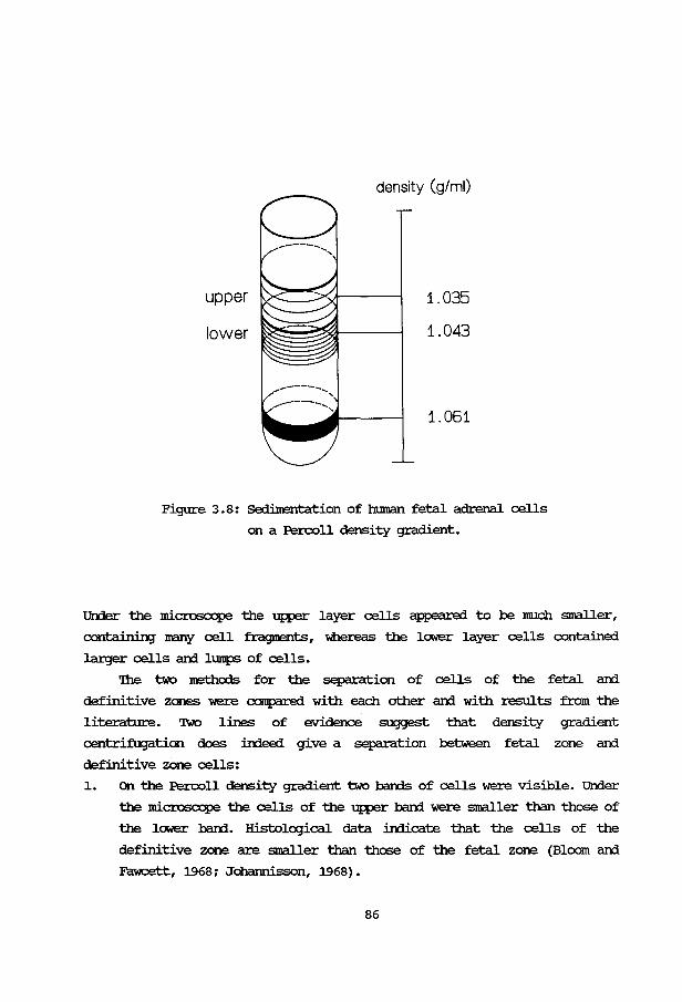
upper
lower
density (g/ml)
1.035
1.043
1.061
Figure 3. 8: Sedimentation of human fetal adrenal cells
on a Percoll density gradient.
Un:ler the microscope the upper layer cells appeared to be much smaller 1
c:ontain:inJ many cell fragrrents 1 whereas the lower layer cells contained
larger cells ani lumps of cells.
'lhe two :nethods for the separation of cells of the fetal ani
definitive zones -were canpared with each other ani with results from the
literature. 'Dvo lines of evidence suggest that density gradient
centrifugation does in:ieed give a separation between fetal zone ani
definitive zone cells:
1. on the Percoll density gradient two barrls of cells were visible. Under
the microscope the cells of the upper band -were smaller than those of
the lower band. Histological data indicate that the cells of the
definitive zone are smaller than those of the fetal zone (Bloom ani
~I 1968; Johannissonl 1968) o
86
2. '!he cells of the fetal and definitive zones of the human fetal
adrenal, obtained with separation method one and two, were incubated
in the presence of p:rcx.:resterone. '!he extent of corwersion of
p:rcx.:resterone into cortisol coincided with the presence of fetal zone
cells in human fetal adrenal cell cultures (Voutilainen and Kahri,
1979; Voutilainen et al., 1979) . We used the cortisol/corticosterone
ratio as a "IDeaSUre for the 17a-hydroxylase activity and as a criterium
for the presence of fetal zone cells. 'lhese ratios are presented in
Table 3.6. '!he cortisol/corticosterone ratios in the lOW'er layers of
the density gradients had the same value, compared with the ratio in
the fetal zone cells, while the cells in the upper layers had the same
ratio as compared to the cells of the definitive zone. '!his is in
agreement with the histological data, as mentioned above.
'!he results of this pilot experiment suggest that density gradient
centrifugation is a suitable method for the separation of cells from the
fetal and definitive zones of the human fetal adrenal.
Table 3.6: Ratio of corticosterone/cortisol production from exogenous
progesterone (10 p.g/ml) in human fetal adrenal cells,
obtained according to separation methods 1 and 2.
Method case number Adrenal zone Ratio cortisol/
corticosterone
1 3 definitive zone 0.60
fetal zone 1.54
layer from Percoll aradient
2 12 upper 0.50
lOW'er 1.15
2 11 upper 0.65
lower 1.47
87


CBAPl'ER 4
EFFECl'S OF 22S-HYmOXY-aiOI.ESTEROL AND OIHER HYmOXYIATED
STErolS 00 'lHE Acm-STIMDIATED STEroiD IRJI:UCI'IOO lli RAT
ArnENAL CEUS.
Acta Endocrinol. (Oopenh) 97, 243-250 (1981).
89
Acta Endocrinologica 1981,97:243-250
Effects of 22S-hydroxy-cholesterol and other hydroxylated sterols on
the ACTH-stimulated steroid production in rat adrenal cells
]. G. M. Huijmans\ H.J. Degenhart, D.]. Kortleve and H. K. A. Visser
Erasmus University, Depart:rMnt of Pediatrics,
Academic Hospital/Sophia Children's Hospita~ Rotterdam, The Netherlands
Abstract. Several effects of hydroxylated sterols on cell cultures are known. Most of these can be explained by an inhibition of the cholesterol synthesis at the level of the 3-hydro,cy-3-methylglutaryl GoA reductase. When studying cholesterol metabolism in rat adrenal cells, an inhibitory action of some sterols on the ACTHstimulated corticosterone production was observed. The effects of one sterol, 22S-OH-cholesterol, were investigated further. The sterol had no effect on the ACTH-stimulated cyclic AMP production, suggesting an intact receptor-adenylate cyclase complex and cellular membrane. In the presence of ACTH and 22S-OH-cholesterol particularly the free cholesterol concentration was elevated; 22S-OHcholesterol therefore probably exerts its inhibitory effect at a step located after hydrolysis of the cholesterol esters. 22S-OH-cholesterol had no effect on the conversion of exogenous pregnenolone into corticosterone.
These results make it probable, that the inhibitory effect of 22S-OH-cholesterol on the ACTH-stimulated corticosterone production is situated at the level of the cholesterol side-chain cleavage.
Several effects of oxygenated derivatives of cholesterol on liver cells and fibroblasts have been described.
They inhibit the 3-hydroxy-3-methylglutaryl
1 Sophia Childrens Hospital, 160, Gordelweg, 3038 GE Rotterdam, The Netherlands.
16*
GoA-reductase actiVIty (Chen et a!. 1974; Kandutsch & Chen 1975; Heiniger et a!. 1976), are cytotoxic for smooth muscle cell cultures (Peng et a!. 1979) and are angiotoxic and arteriosclerotic in rabbits (Imai et a!. 1980). These effects become manifest after exposure to the agent for several hours or even days.
Whether these sterols affect steroid producing cells or influence steroid metabolism is not known. This matter is of considerable interest in view of the fact, that cholesterol is the main precursor for steroid production.
In a previous communication (Huijmans et a!. 1977) we described an acute effect of several sterols on the ACTH-stimulated corticosterone production in isolated rat adrenal cells. One of these sterols, 22S-hydroxy-cholesterol (22S-OH) inhibited steroid production by more than 50%. At that time, the mechanism of this inhibition or the site of action of the sterol was still incompletely understood.
90
In this report we present data for a number of hydroxylated sterols as to their inhibitory effect on the ACTH-stimulated corticosterone production. The effects will be discussed of one of these sterols (22S-OH-cholesterol) on several steps in the ACTH-stimulatory mechanism and the steroidogenic pathway.
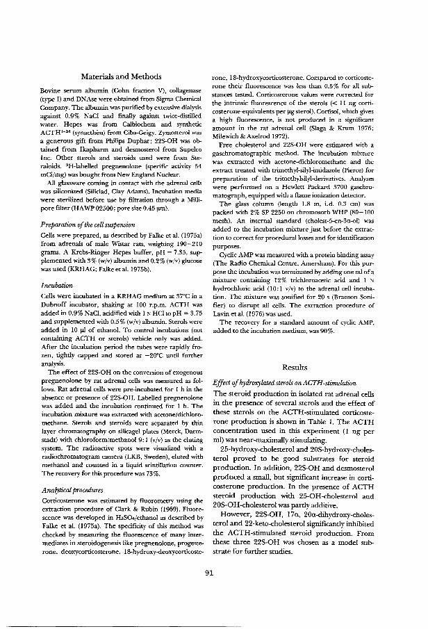
Materials and Methods
Bovine serum albumin (Cohn fraction V), collagenase (type I) and DNAse were obtained from Sigma Chemical Company. The albumin was purified by extensive dialysis against 0.9% NaCl and finally against rwice-distilled water. Hepes was from Calbiochem and synthetic ACTH1- 24 (synacthen) from Ciba-Geigy. Zymosterol was a generous gift from Philips Duphar; 22S-OH was obtained from Ikapharm and desmosterol from Supelco Inc. Other sterols and steroids used were from Steraloids. 3H-labelled pregnenolone (specific activity 54 mCi/mg) was bought from New England Nuclear.
All glassware coming in contact with the adrenal cells was siliconized (Siliclad, Clay Adams). Incubation media were sterilized before use by filtration through a Millipore filter (HA WP 02500; pore size 0.45 !J.IIl).
Preparation of the cell suspension
Cells were prepared, as described by Falke eta!. (1975a) from adrenals of male Wistar rats, weighing 190-210 grams. A Krebs-Ringer Hepes buffer, pH = 7.35, supplemented with 3% (w/v) albumin and 0.2% (w/v) glucose was used (KRHAG; Falke eta!. 1975b).
Incubation
Cells were incubated in a KRHAG medium at 37°C in a Dubnoff incubator, shaking at 100 r.p.m. ACTH was added in 0.9% NaCI, acidified with 1 N HCI to pH = 3. 75 and supplemented with 0.5% (w/v) albumin. Sterols were added in 10 !Jl of ethanol. To control incubations (not containing ACTH or sterols) vehicle only was added. After the incubation period the tubes were rapidly frozen, tightly capped and stored at -20°C until further analysis.
The effect of 22S-OH on the conversion of exogenous pregnenolone by rat adrenal cells was measured as follows. Rat adrenal cells were pre-incubated for 1 h in the absence or presence of 22S-OH. Labelled pregnenolone was added and the incubation continued for 1 h. The incubation mixture was extracted with acetone/dichloromethane. Sterols and steroids were separated by thin layer chromatography on silicagel plates (Merck, Darmstadt) with chloroform/methanol 9:1 (v/v) as the eluting system. The radioactive spots were visualized with a radiochromatogram camera (LKB, Sweden), eluted with methanol and counted in a liquid scintillation counter. The recovery for this procedure was 73%.
Anal:jtical procedures
Corticosterone was estimated by fluorometry using the extraction procedure of Clark & Rubin (1969). Fluorescence was developed in H2SO•/ethanol as described by Falke et al. (1975a). The specificity of this method was checked by measuring the fluorescence of many intermediates in steroidogenesis like pregnenolone, progesterone, deoxycorticosterone, 18-hydroxy-deoxycorticoste-
rone, 18-hydroxycorticosterone. Compared to corticosterone their fluorescence was less than 0.5% for all substances tested. Corticosterone values were corrected for the intrinsic fluorescence of the sterols (< 11 ng corticosterone-equivalents per !Lg sterol). Cortisol, which gives a high fluorescence, is not produced in a significant amount in the rat adrenal cell (Slaga & Krum 1976; Milewich & Axelrod 1972).
Free cholesterol and 22S-OH were estimated with a gaschromatographic method. The incubation mixture was extracted with acetone-dichloromethane and the extract treated with trimethyl-silyl-imidazole (Pierce) for preparation of the trimethylsilyl-derivatives. Analyses were performed on a Hewlett Packard 5700 gaschromatograph, equipped with a flame ionization detector.
The glass column (length 1.8 m, i.d. 0.3 em) was packed with 2% SP 2250 on chromosorb WHP (80-100 mesh). An internal standard (cholest-5-en-3a-ol) was added to the incubation mixture just before the extraction to correct for procedural losses and for identification purposes.
Cyclic AMP was measured with a protein binding assay (The Radio Chemical Centre, Amersham). For this purpose the incubation was terminated by adding one ml of a mixture containing 12% trichloroacetic acid and 1 N
hydrochloric acid (10: 1 v/v) to the adrenal cell incubation. The mixture was sonified for 20 s (Branson Sonifier) to disrupt all cells. The extraction procedure of Lavin eta!. ( 1976) was used.
The recovery for a standard amount of cyclic AMP, added to the incubation medium, was 90%.
Results
Effect of hydroxylated sterols on ACTH-stimulation
The steroid production in isolated rat adrenal cells in the presence of several sterols and the effect of these sterols on the ACTH-stimulated corticosterone production is shown in Table I. The ACTH concentration used in this experiment (I ng per ml) was near-maximally stimulating.
25-hydroxy-cholesterol and 20S-hydroxy-cholesterol proved to be good substrates for steroid production. In addition, 22S-OH and desmosterol produced a small, but significant increase in corticosterone production. In the presence of ACTH steroid production with 25-0H-cholesterol and 20S-OH-cholesterol was partly additive.
However, 22S-OH, 17a, 20a-dihydroxy-cholesterol and 22-keto-cholesterol significantly inhibited the ACTH-stimulated steroid production. From these three 22S-OH was chosen as a model substrate for further studies.
91
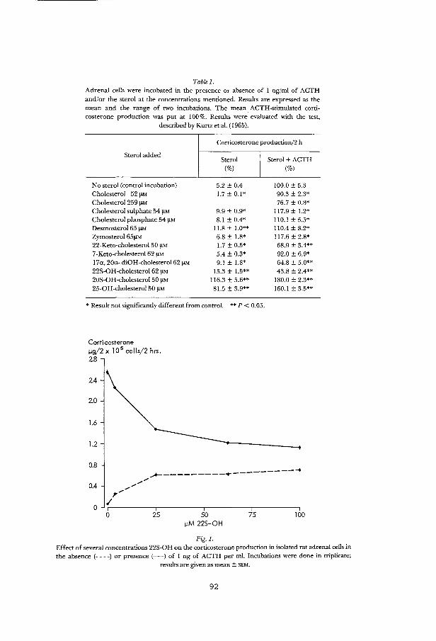
Table I. Adrenal cells were incubated in tbe presence or absence of 1 ng/ml of ACTH and/or the sterol at tbe concentrations mentioned. Results are expressed as the mean and the range of two incubations. The mean ACTH-stimulated corticosterone production was put at 100%. Results were. evaluated witb the test,
described by Kurtz et a!. ( 1965).
Corticosterone production/2 h
Sterol added
No sterol (control incubation) Cholesterol 52 !lM Cholesterol 259 !lM Cholesterol sulphate 54 I'M Cholesterol phosphate 54 I'M Desmosterol 65 I'M Zymosterol 65!!M 22-Keto-cholesterol 50 I'M 7-Keto-cholesterol 62 !lM l7a, 20a- diOH-cholesterol62 I'M 22S-OH-cholesterol62 !lM 20S-OH-cholesterol 50 I'M 25-0H-cholesterol 50 I'M
Sterol (%)
5.2 ± 0.4 1.7 ± 0.1*
9.9 ± 0.9* 8.1 ± 0.4*
11.8 ± l.O** 6.8 ± 1.8* 1.7 ± 0.5* 5.4 ± 0.3* 9.1 ± 1.8*
15.3 ± 1.5** 116.3 ± 5.6** 81.5 ± 3.9**
* Result not significantly different from control. ** P < 0.05.
Corticosterone ~g/2 x 10 5 cells/2 hrs. 2.8
2.4
2.0
1.6
1.2
0.8
0.4
0 0 25 50 75
~M 225-0H
Fig. I.
Sterol + ACTH (%)
100.0 ± 5.3 90.3 ± 2.3* 76.7 ± 0.8*
117.9 ± 1.2* 110.1 ± 5.5* 110.4 ± 8.2* 117.6 ± 2.8* 68.9 ± 3.4** 92.0 ± 6.9* 64.8 ± 5.0** 43.8 ± 2.4**
180.0 ± 2.3** 160.1 ± 3.5**
100
Effect of several concentrations 22S-OH on tbe corticosterone production in isolated rat adrenal cells in the absence (--- -) or presence (-) of 1 ng of ACTH per ml. Incubations were done in triplicate;
results are given as mean ± SEM.
92
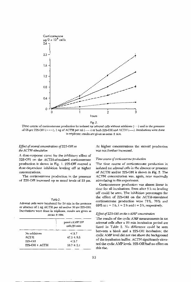
Corticosterone flg/2 x 10 5 cells 2.4
2.0
1.6
1.2 _ .. ---,,,'
_,.A ,,'" _,
,,'' ;,,"
0.8
,-' / ,,-' --+ --,,,'' ,.,-""' ..,,.. __ ..,.., ..... -
0.4
.... ~~~~;::=.~~-·-··································~··································· 0
0 2 3 hours
Fig.2. Time course of conicosterone production by isolated rat adrenal cells without additions(· · ·)and in the presence of65 !lM 22S-OH (---),I ng of ACTH per ml (----)or both 22S-OH and ACTH (-).Incubations were done
in triplicate; results are given as mean ± SEM.
Effect of several concentrations of22S-OH on the ACTH-stimulation
A dose-response curve for the inhibitory effect of 22S-OH on the ACTH-stimulated corticosterone production is shown in Fig. 1. 22S-OH exerted a dose-dependent inhibition leveling off at higher concentrations.
The corticosterone production in the presence of 22S-OH increased up to sterol levels of 25 f!M.
Table2. Adrenal cells were incubated for 20 min in tbe presence or absence of I ng ACTH per mi and/or 50 !lM 22S-OH. Incubations were done in triplicate, results are given as
No additions ACTH 22S-OH
mean± SEM.
22S-OH + ACTH
pmol cAMP/lOs cells/20 min
<3.7 47.5 ± 9.6
<3.7 53.7 ± 2.1
93
At higher concentrations the steroid production was not further increased.
Time course of corticosterone production
The time course of corticosterone production in isolated rat adrenal cells in the absence or presence of ACTH and/or 22S-OH is shown in Fig. 2. The ACTH concentration was, again, near maximally stimulating in this experiment.
Corticosterone production was almost linear in time for all incubations. Even after 3 h no leveling off could be seen. The inhibition percentages for the effect of 22S-OH on the ACTH-stimulated corticosterone production were 7l %, 70% and 69% at t = 1 h, t = 2 h arid t = 3 h, respectively.
Effect of 22S-OH on the c-AMP concentration
The results of the cyclic AMP measurements in rat adrenal cells after a 20 min incubation period are listed in Table 2. No difference could be seen between a blank and a 22S-OH incubation; the cyclic AMP level did not rise above the background of the incubation buffer. ACTH significantly elevated the cyclic AMP levels. 22S-OH had no effect on this rise.
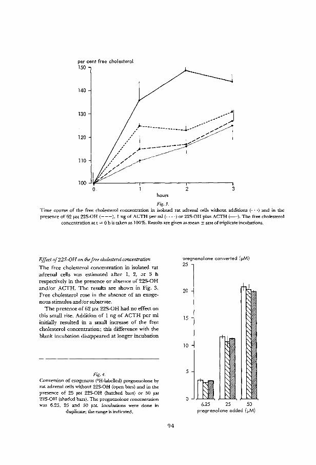
per cent free cholesterol 150
140
130
120
110
100
__ .... 1 __ ... ·· ,..
)- .. .. / ________________ .J.-··· .. --:.:::-.:::-.:::--·····1
,'/ j --_j-:-:::::'·:::'·······
,,, , ----:.: ... -········r ,' / ······•···· ,' // .. ,. ..... .
~{~~~~--·············
0 2 3 hours
Fig.3. Time course of the free cholesterol concentration in isolated rat adrenal cells without additions (· · ·) and in the presence of 62 fLM 22S-OH (---), I ng of ACTH per mi (----)or 22S-OH plus ACTH (-).The free cholesterol
concentration at t = 0 his taken as !00%. Results are given as mean± SEM of triplicate incubations.
Effect of 22 S -0 H on the free cholesterol concentration
The free cholesterol concentration in isolated rat adrenal cells was estimated after 1, 2, ;)r 3 h respectively in the presence or absence of 225-0H and/or ACTH. The results are shown in Fig. 3. Free cholesterol rose in the absence of an exogenous stimulus and/or substrate.
The presence of 62 J.LM 225-0H had no effect on this small rise. Addition of 1 ng of ACTH per m1 initially resulted in a small increase of the free cholesterol concentration; this difference with the blank incubation disappeared at longer incubation
Fig.4. Conversion of exogenous ('H-labelled) pregnenolone by rat adrenal cells without 22S-0 H (open bars) and in the presence of 25 fLM 22S-OH (hatched bars) or 50 fLM 22S-OH (shaded bars). The pregnenolone concentration was 6.25, 25 and 50 fLM. Incubations were done in
duplicate; the range is indicated.
94
pregnene I one converted ( ~M) 25
20
15
10
5
0 6.25 25 50
pregnenolone added (~M)
times. In the presence of both 22S-OH and ACTH a significant increase in free cholesterol is obvious, however, especially after two hours.
Effect of 22S-OH on the conversion of exogenous pregnenolone
The effect of two concentrations 22S-OH (25 !LM and 50 !LM) on the conversion of exogenous pregnenolone in three concentrations (6.25 !LM, 25 !LM and 50 !LM) was measured as described under Materials and Methods. The results are given in Fig. 4. In the absence of 22S-OH pregnenolone was converted mainly into progesterone, deoxycorticosterone, corticosterone and IS-hydroxycorticosterone. An almost linear relationship exists between the amount of pregnenolone added and the amount of pregnenolone converted. None of the 22S-OH concentrations used had a significant effect on this conversion.
Discussion
Several long-term effects of hydroxylated sterols have been described in the literature (Chen et al. 1974; Kandutsch & Chen 1975; Peng et al. 1979; Imai et al. 1980) affecting de novo cholesterol synthesis at the level of the 3~-hydroxy-3-methylglutaryl GoA-reductase or through a direct toxic action.
In a previous report (Huijmans et al. 1977) a short-term inhibitory effect of some hydroxylated sterols on steroidogenesis in isolated rat adrenal cells was described. A diminished cholesterol synthesis cannot account for this effect because de novo cholesterol synthesis does not play an important role in adrenal steroidogenesis (Dexter et a!. 1970; Gwynne et al. 1976; Balasubramaniam et al. 1977).
In addition the long term effects were only obvious after incubation with the sterol for several hours to days. In our studies the incubation time generally did not exceed two hours, and the inhibition of the corticosterone synthesis was already apparent after one hour (Fig. 2).
In Table 1 several additional sterols are listed that were investigated for an effect on steroid production. Neither cholesterol nor its sulphate or phosphate had much effect. The same is true for desmosterol and zymosterol, both precursors of cholesterol. Only when high cholesterol concentra-
95
tions were used (Table 1, Sharma 1973) an inhibitory effect may be seen, probably due to purely physical effects (Falke et al. 1975b). 22-keto-cholesterol does not seem to be a substrate for steroid production, but it did inhibit the ACTH-stimulation. Both 20S-hydroxy-cholesterol and 25-hydroxy-cholesterol were found to be good substrates for steroid production. In the presence of ACTH corticosterone production was not completely additive, probably because the maximal steroidogenic capacity of the adrenal cell was reached.
A sterol with remarkable properties is 22S-OH, which seems to be a substrate, though not very effective, for steroid synthesis, while at the same time it inhibits the ACTH-stimulated corticosterone production (see Fig. 1). This inhibition is dose dependent. A direct adverse action of 22S-OH in concentrations up to 62 !LM on the adrenal cells is less probable. This is suggested by the linearity of the corticosterone production in the presence of 22S-OH, as presented in Fig. 2. A similar conclusion for 25-hydroxy-cholesterol was reached by Falke et al. ( 1976).
The results with the cAMP-measurements (see Table 2) provide additional evidence that 22S-OH does not have non-specific (for example surfactant) effects on the cellular membrane. cAMP plays an intermediary role in the action of ACTH (Sala et al. 1979; Rae et al. 1979). 22S-C>H had no effect on the cAMP concentrations after stimulation with ACTH. This indicates that the first part of the ACTH-stimulatory mechanism is probably unaffected. Analogous to our results Kandutsch & Chen (1975) found no effects of several sterols on the cAMP content of L-ee!! cultures, suggesting that the inhibitory sterols act through a mechanism not involving cAMP.
A further localization of the effect of 22S-OH could be accomplished by studying the liberation of cholesterol from its esters after stimulation with ACTH, as shown in Fig. 3. The effect of ACTH on the free cholesterol concentration is in agreement with the results of Vahouny et al. (1978), who found no change after stimulation with ACTH. The results obtained in the presence of both ACTH and 22S-OH suggest an inhibitory effect of 22S-OH on the further metabolism \>f cholesterol, once it is liberated from its esters by the esterase. In the rat. a similar increase in free cholesterol after stimulation with ACTH was described when the further metabolism of cholesterol was impaired by treating the rat in vivo with the side-chain cleavage

inhibitor aminoglutethimide (Mahaffee et a!. 197 4) or the protein synthesis inhibitor cycloheximide
(Davis & Garren 1966; Trzeciak & Boyd 1973). Protein synthesis is required in the mechanism of
action of ACTH; probably for the transport of cholesterol from the lipid droplets into the mitochondrion or the attachment to the side-chain cleaving enzyme system (Schulster eta!. 1976). A direct effect of 22S-OH on protein synthesis cannot be excluded but is less probable. Kandutsch & Chen (1974) reported no effect of several hydroxylated sterols (a.o. 25-hydroxy-cholesterol and 20S-hydroxy-cholesterol) on protein synthesis or RNA- and fatty acid-synthesis. This suggests that the inhibitory action of 22S-OH resides in the cholesterol side-chain cleavage or the conversion of pregnenolone into its products. As shown in Fig. 4, 22S-OH, added in two concentrations (25 !J.M and 50 !J.M) had no effect on the conversion of exogenous pregnenolone.
The results presented here allow to locate the inhibitory effect of22S-OH on the ACTH-stimulated corticosterone production in rat adrenal cells. The most probable site of action is the cholesterol side-chain cleavage. 22S-OH and cholesterol may compete for the active centers on the side-chain cleaving enzyme complex. Studies with bovine adrenal mitochondria support this conclusion; Simpson & Boyd ( 1967) showed that several cholesterol-derivatives, hydroxylated in positions 20, 24, 25 or 26 inhibited the conversion of cholesterol (the inhibition with 20S-hydroxy-cholesterol and 25-hydroxy-cholesterol being competitive). Prob
ably 22S-OH not only competes for the active centers on the side-chain cleaving enzyme complex, but is being converted into pregnenolone too. This sterol proved to be a better substrate than cholesterol, when incubated with a purified bovine adrenal cytochrome P-450scc· (Morisaki et a!. 1976). The results in Figs. 1 and 2, obtained with rat adrenal cells, confirm the role of 22S-OH as a substrate for steroid production.
Acknowledgments
This work was supported by a grant from the Foundation for Medical Research, Sophia Children's Hospital, Rotterdam. The authors want to thank Prof. H. v. d. Molen and Dr. H. E. Falke for their critical reacling of the manuscript. The audiovisual center of the Sophia Children's Hospital and Clem van Thiel were of great help with the preparation of the manuscript.
References
Balasubramaniam S, Goldstein J L, Faust J R, Brunstede G Y & Brown M S (1977): lipoprotein-mediated regulation of 3-hydroxy-3-methylglutaryl coenzyme A reductase activity and cholesteryl ester metabolism in the adrenal gland of the rat.J Bioi Chern 252: 1771-1779.
Chen H W, Kandutsch A A & Waymouth C (1974): Inhibition of cell growth by oxygenated derivatives of cholesterol. Nature 251:419-421.
Clark B R & Rubin R T (1969): New fluorometric method for the determination of cortisol in serum. AnalytBiochem 29:31-39.
Davis W W & Garren L D (1966): Evidence for the stimulation by adrenocorticotropic hormone of the conversion of cholesterol esters to cholesterol in the adrenal in vivo. Biochem Biophys Res Commun 24: 805-810.
DexterRN, FishmanL M & NeyRL (1970): Stimulation of adrenal cholesterol uptake from plasma by adrenocorticotrophin. Endocrinology 87: 836-846.
Falke H E, Degenhart H J, Abeln G J A, Visser H K A & Croughs RJ M (1975a): Studies on isolated rat adrenal cells. I. Continuous flow and batch incubations. Acta Endocrinol (Kbh) 78: 110-121.
Falke H E, Degenhart H J, Abeln G J A & Visser H K A (1975b): Studies on isolated rat adrenal cells. Metabolism of hydroxylated sterols. Mol Cell Endocrinol 3: 375-383.
Falke H E, Degenhart H J, Abeln G J A & Visser H K A ( 1976): Effects of 25-hydroxycholesterol and aminoglutethimide in isolated rat adrenal cells. A model for congenital lipoid adrenal hyperplasia? Mol Cell Endocrinol4: 107-114.
Gwynne J T, Mahaffee D, Brewer H B Jr & Ney R L ( 1976): Adrenal cholesterol uptake from plasma lipoproteins: regulation by corticotropin. Proc Nat! Acad Sci USA 73:4329-4333.
Heiniger H J, Kandutsch A A & Chen H W (1976): Depletion of L-cell sterol depresses endocytosis. Nature 263: 515-517.
Huijmans J G, Degenhart H J & Kortleve D J (1977): Effects of hydroxylated sterols on isolated rat adrenal cells. J Endocrinol 72: 3P-4P.
Imai H, Werthessen NT, Subramanyam V, Le Quesne P W, Soloway A H & Kanisawa M (1980): Angiotoxicity of oxygenated sterols and possible precursors. Science 207:651-653.
Kandutsch A A & Chen H W (1974): Inhibition of sterol synthesis in cultured mouse cells by cholesterol derivatives oxygenated in the side-chain. J Bioi Chern 249: 6057-6061.
Kandutsch A A & Chen H W (1975): Regulation of sterol synthesis in cultured cells by oxygenated derivatives of cholesterol.] Cell Physiol85: 415-424.
Kurtz T E, Link R F, Tukey J W & Wallace D L (1965):
96
Short-cut multiple comparisons for balanced single and double classifications. Technometrics 7: 95-161.
Lavin N, Rachelefsky G S & Kaplan SA ( 1976): An action of disodium cromoglycate: inhibition of cyclic 3',5'AMP phosphodiesterase. J Allergy Clin Immunol 57: 80-88.
Mahaffee D, Reitz R C & Ney R L (1974): The mechanism of action of adrenocorticotropic hormone. J Bioi Chern 249:227-233.
Milewich L & Axelrod L R (1972): The conversion in vitro of pregnenolone to Ct9 and C21 metabolites by the rat adrenal gland with and without metyrapone. J Endocrinol54: 515-516.
Morisaki M, Sato S, Ikekawa N & Shikita M (1976): Stereochemical specificity at carbon-20 and -22 of hydroxylated cholesterols for side-chain cleavage by adrenocortical cytochrome P-450scc· FEBS Lett 72: 337-340.
Peng S K, Tham P, Taylor B & Mikkelson B (1979): Cytotoxicity of oxidation derivatives of cholesterol on cultured aortic smooth muscle cells and their effect on cholesterol biosynthesis. Am J Clin Nutr 32: 1033-1042.
Rae P A, Gutmann N S, Tsao J & Schimmer B P ( !979): Mutations in cyclic AMP-dependent protein kinase and corticotropin (ACTH)-sensitive adenylate cyclase affect adrenal steroidogenesis. Proc Nat! Acad Sci USA 76:1896-1900.
97
Sala G B, Hayashi K, Catt K J & Dufau M L (1979): Adrenocorticotropin action in isolated adrenal cells. J Bioi Chern 254:3861-3865.
Schulster D, Burstein S & Cooke'B A (1976): Molecular endocrinology of the steroid hormones. John Wiley and Sons, London.
Sharma R K (1973): Regulation of steroidogenesis by adrenocorticotropic hormone in isolated adrenal cells ofrat.J Bioi Chern 248:5473-5476.
Simpson E R & Boyd G S (1967): The cholesterol side-chain cleavage system of bovine adrenal cortex. Eur J Biochem 2: 275-285.
Slaga T J & Krum A A (1976): The influence of prolonged ACTH treatment on rat adrenal steroidogenesis. Res Commun Chern Pathol Pharmacal 15: 279-290.
Trzeciak W H & Boyd G S (1973): The effect of stress induced by ether anaesthesia on cholesterol content and cholesteryl-esterase activity in rat adrenal cortex. Eur J Biochem 37:327-333.
Vahouny G V, Chanderbhan R, Hinds R, Hodges VA & Treadwell C R (1978): ACTH-induced hydrolysis of cholesteryl esters in rat adrenal cells. J Lipid Res 19: 570-577.
Received on June 19th, 1980.

rnAPrER 5
<X>NVERSION OF 22S-:EIYJR)XY-<HOI.ESTEROL AND ITS EFFECI' ON
'!HE MErAOOUSM OF OIHER S'l'.EOOIS IN RAT AmENAL CELIS AND
BJVINE AmENAL MI'IDCliClNI!UA.
Acta Endocrinol. (Oopenh) 100, 599-605 (1982).
99

Acta Endocrinologica 1982, 100:599-605
Conversion of 22S-hydroxy-cholesterol and its effect on the metabolism of other sterols in rat adrenal cells and bovine adrenal mitochondria
J. G. M. Huijmans, H.J. Degenhart
and D.J. Kortleve
Erasmus University, Department of Paediatrics, University Hospital/Sophia Children's Hospital, Rotterdam, The Netherlands
Abstract. This study provides evidence that 22S-OHcholesterol inhibits the conversion of 25-0H-cholesterol but has no effect on tbe conversion of 22R-OH-cholesterol. The latter sterol is an intermediate in the cholesterol side-chain cleavage, whereas for tbe conversion of 25-0H-cholesterol into pregnenolone the complete sidechain cleaving enzyme system is necessary. This complements a previous study in which it was shown, that 22S-OH-cholesterol has an inhibitory effect on the ACTH-induced conversion of cholesterol into corticosterone in isolated rat adrenal cells. The available evidence thus suggests an inhibition by 22S-OH-cholesterol of tbe first step in tbe cholesterol side-chain cleavage. The results, obtained from tbe experiments with rat adrenal cells and with bovine adrenal mitochondria, allow the hypothesis, that a causal relationship exists between conversion of 22S-OH-cholesterol and production of corticosterone, resp~ctively pregnenolone. We conclude, tbat 22S-OH-cholesterol is a substrate for steroid production in tbe adrenal cell. This sterol inhibits the ACTH-stimulated corticosterone production. The site of this inhibition is located at one of the first steps in the cholesterol side-chain cleavage, probably the binding of cholesterol to tbe cytochrome P450-complex.
Several effects of hydroxylated sterols on cellular function, structure or metabolism are known (Kan
dutsch & Chen 1975; Heiniger eta!. 1976; Peng et
a!. 1979; Streuli et al. 1979; Hsu et al. 1980; Imai eta!. 1980; Huijmans eta!. 1981). A possible role
of these sterols in the regulation of the cholesterol synthesis has been suggested (Kandutsch & Chen
1974). In a previous report (Huijmans eta!. 1981)
we showed that some sterols inhibited the ACTHstimulated corticosterone production in isolated rat
adrenal cells. The studies with one sterol (225-
hydroxy-cholesterol (22S-OH)), suggested that this
sterol probably exerts its action at the level of the
cholesterol side-chain cleaving enzyme system. The
exact mechanism of action as well as the role of
22S-OH as a substrate for steroid production in the adrenal cell is still not known.
In this report some additional studies will be
presented in order to further localize the effect of
22S-OH and to evaluate the role of 22S-OH as a
substrate for steroid production. We investigated a
possible relationship between sterol conversion and
steroid synthesis in rat adrenal cells and bovine adrenal mitochondria.
Materials and Methods Materials Bovine serum albumin (Cohn fraction V), collagenase (Type I) and DNase were obtained from Sigma Chemical Company. The albumin was purified by extensive dialysis and lyophilization. Hepes was from Calbiochem and synthetic ACTH 1- 24 (Synacthen®) from Ciba-Geigy. 22SHydroxy-cholesterol was obtained from Ikapharm: all other sterols and steroids used were from Steraloids. Purity was checked by thin-layer chromatography and gas chromatography. All glassware coming in contact with the adrenal cells of ACTH-containing solutions was siliconized (Siliclad, Clay Adams).
100

Incubation media were sterilized before use by filtration through a Millipore filter (HA WP 02500; pore size 0.45 f!m).
Methods
Preparation of the rat adrenal cell suspension. Cells were prepared from adrenals of male Wistar rats, weighing 190-210 g. as described by Falke et al. (1975a,b). A Krebs-Ringer-Hepes buffer, pH = 7.35, supplemented with 3% (w/v) albumin and 0.2% (w/v) glucose, was used.
Adrenal cell incubations. Cells were incubated at 37"C in a Dubnoff incubator, shaking at I 00 r. p.m. The incubation time was 2 h, unless otherwise stated. The number of cells differed from experiment to experiment, but was kept between 100 000-150 000 per mi. ACTH was added in 0.1 ml of0.9% NaCI, pH = 3.75, supplemented with 0.5% (w/v) albumin.
Sterols were added in 10 f!l of ethanol. To incubations not containing ACTH or sterols, vehicle only was added. After the incubation period the tubes were immediately chilled in ice, tightly capped and stored at -20"C until further analysis.
Corticosterone was estimated by fluorometry. as described previously (Huijmans et al. 1981). With this method the minimal corticosterone concentration detectable is 30 ng per mi.
Preparation of the bovine adrenal metochondria. Bovine adrenals were obtained from the local slaughterhouse. The cortex was collected and homogenized in a Parr cell disruption bomb in a 10 mM Hepes buffer (pH= 7.30) containing 250 mM sucrose and 0.5 mM EDT A. The homogenate was centrifuged at 900 X g for 10 min at 4"C to remove unbroken cells, nuclei and erythrocytes. The mitochondria were obtained by centrifuging at 9000 x g for 10 min at 4"C. The pellet was washed two times and re-suspended to a protein concentration of 20 mg per mi. These mitochondria were frozen and stored in liquid nitrogen.
Mitochondrial incubations. Incubations were carried out at 37"C in a 20 mM Hepes buffer (pH= 7.40), containing
measured as described previously (Huijmans et al. 1981) except that a capillary column, coated with SE-30, was used. The minimal sterol concentration, detectable with this method, is I f!M.
Results
Adrenal cell incubations
We examined the effects of 22S-OH on the con
version of 25-hydroxy-cholesterol (25-0H). The
results are shown in Fig. I. The corticosterone
production in rat adrenal cells, incubated with
25-0H was virtually linearly dependent on the
concentration of the sterol used. In the presence of
22S-OH the corticosterone production reached a
maximum at 25 f.tM 22S-OH without further incre
ment at 50 f.tM. With both 25-0H and 22S-OH the
corticosterone synthesis was less than the sum of
the sterol derived corticosterone production indi
vidually. This inhibitory effect became more clear
in the presence of the high concentration of25-0H
(25 f.tM). Fig. 2 shows the effect of 22S-OH on the
conversion of 25-0H in the presence of 100 pg
ACTH per mi. This ACTH-concentration was
maximally stimulating. The corticosterone produc
tion in the presence of ACTH and 12.5 f.tM 25-0H
was almost additional; with 25 f.tM 25-0H cortico-
Corticosterone og/2x 105 cells/2 hrs 1.2
0.8
!50 mM KCI, 11 mM NaCI, 6 mM CaCI2 , 4.3 mMNaN3 , 5 0.4
f!M antimycine, 50 mM nicotinamide and 1% (w/v) albu-min in a final volume of 5 mi. The initial sterol concen-tration was 50 mM. Cyanoketone (17 mM), an inhibitor of the 3~-hydroxy steroid dehydrogenase activity, was added to prevent further metabolism of pregnenolone. The incubation was started with the addition of 0.1 mM NADPH, 3 mM glucose-6-phosphate and 0.6 U per ml glu~ose-6-phosphate dehydrogenase. Aliquots of 1 ml were taken at t = 0, t = 8, t = 16 arid t = 25 min and immediately deproteinated with 1 ml of cold acetone. The pellet was once again extracted with I ml of acetone. The combined acetone layer was shaken with 3 ml of dichloromethane. The organic layer was washed with 1 ml of water and dried. The extracts were derivatized and
no 225-0H 25oM 225-0H 50 oM 225-0H
D no25-0H
[SSj12.S oM 25-0H
~ 25oM25-0H
Fig. I. Effect of 225-0H-cholesterol in two concentrations on the corticosterone production in isolated rat adrenal cells in the absence or presence of 25-0H-cholesterol. Results are presented as the mean of duplicate incubations and
the individual values.
101

Corticosterone "g/2 X I 05 cells/2 hcs 2.4
2.0
1.6
1.2
0.8
0.4
0 no 25-0H
ISS 12.5 "M 25-0H
~ 25"M25-0H
Fig.2. Effect of 22S-OH-cholesterol in two concentrations on the ACTH-stimulated corticosterone production in isolated rat adrenal cells in the absence or presence of 25-0H-cholesterol. The ACTH concentration was 100 pg per mi. Results are presented as the mean of duplicate
incubations and the individual values.
Corticosterone "g/2x 10 5 cells/2 hrs 3.2
2.4
1.6
0.8
no 225-0H 25 "M 225-0H 50 "M 225-0H
D no 22R-OH
~ 12.5 "M 22R-OH
~ 25 "M 22R-OH
Fig.3. Effect of 22S-OH-cholesterol in two concentrations on the corticosterone production in isolated rat adrenal cells in the absence or presence of 22R-OH-cholesterol. Results are presented as the mean of duplicate incubations
and the individual values.
sterone production was merely a fraction higher. In the presence of25 ~M 22S-OH, ACTH-stimulation was slightly inhibited; tbe inhibitory effect increased when botb ACTH and 12.5 ~M 25-0H were present. Again tbe higher 25-0H concentration did not change tbe effect of 22S-OH. In the presence of 50 ~ 22S-OH, tbe ACTH-stimulated corticosterone production was inhibited by 39%. This 22S-OH concentration had no additional effects on the incubation witb ACTH and 25-0H.
The same experiment was done with another hydroxylated sterol, 22R-hydroxy-cholesterol (22R-OH). Results are shown in Figs. 3 and 4. 22R-OH was also converted into corticosterone (Fig. 3); this conversion was linearly dose-dependent for the concentrations tested. The corticosterone production in tbe presence of 22S-OH and 22R-OH was completely additive for both concentrations. The results in tbe presence of 100 pg ACTH per ml are shown in Fig. 4. ACTHstimulation and conversion of 22R-OH were addi-
Corticosterone ).Jg/2x 10 5 celis/2 hrs 4.0
3.2
2.4
1.6
0.8
no 225-0H 25 "M 225-0H 50 "M 225-0H
0 no 22R-OH
ES:'J 12.5 "M 22R-OH
~ 25 "M 22R-OH
Fig.4. Effect of 22S-OH-cholesterol in two concentrations on the ACTH-stimulated corticosterone production in isolated rat adrenal cells in the absence or presence of 22R-OH-cholesterol. The ACTH concentration was 100 pg per mi. Results are presented as the mean of
duplicate incubations and the individual values.
102

% 225-0H 110
100
90
80
70
' ................ _J
hours
Fig.5. Conversion of 22S-OH-cholesterol in isolated rat adrenal cells, determined gas chromatographically, in the absence(----) or presence(-) of I ng ACTH per mi. The concentration measured at t = 0 was put at 100%. Results
are given as mean ± SEM of triplicate incubations.
tiona!. Twenty-five f!M 22S-OH had no effect on the corticosterone production. Fifty f!M 22S-OH inhibited the ACTH-stimulated corticosterone production. This inhibition was the same with or without 22R-OH.
An additional experiment was done to investigate the course of the 22S-OH concentration upon incubation with adrenal cells. We measured the disappearance of 22S-OH after several incubation periods, the results of which are shown in Fig. 5. The concentration of22S-OH decreased over a 3 h incubation period. The correlation-coefficient of the linear regression line was 0.84, which is significant at the 1% leveL The slope of the fitted regression line differed significantly from zero. In the presence of 1 ng ACTH per ml (optimally stimulating in this batch of adrenal cells) the results were almost the same (correlation coefficient of the fitted regression line was 0.89).
Mitochondrial incubations
Several sterols were studied for their conversion into pregnenolone by damaged bovine adrenal mitochondria. We measured the sterol concentrations as a function of the incubation time. The results of a typical experiment are shown in Fig. 6. 22R-OH in a concentration of 50 f!M was a good substrate. It had completely disappeared after 10 min and was almost quantitatively converted into pregnenolone. After a 25 min incubation period 9% of the initial 25-0H concentration (50 f!M) was left. 22S-OH disappeared in time; after 25 min some 30% of the initial concentration
22R-OH-cholesterol 25-0H-cholesterol 225-0H-cholesterol
sterol nmol/mg protein
50 @
40
30 ,. I
I 20 I
10
5
/ /
10
/ /
/
,.----_.
15 20 25 minutes
/ /
/ /
/ /
5
,//
/
10 15 minutes
Fig.6.
20 25 5
pregnenolone nmol/mg protein
50
40
30
20
10
0 10 15 20 25 minutes
Production of pregnenolone from 22R-OH-cholesterol (a), 25-0H-cholesterol (b) and 22S-OH-cholesterol (c) during incubation with bovine adrenal mitochondria. The initial sterol concentrations were 50 I-'M- (-) Sterol concentration;
(---) pregnenolone concentration; (----) 22-keto-cholesterol concentration.
103

(50 f.tM) was left. Parallel to this conversion pregnenolone was produced. However, besides pregnenolone another product appeared with (gas chromatographically) the same properties as 22-keto-cholesterol.
Discussion
In a previous report (Huijmans et al. 1981) we demonstrated an inhibitory effect of 22S-OH on the ACTH-stimulated corticosterone production in rat adrenal cells. The most probable site of action was the cholesterol side-chain cleaving enzyme system.
Using several substrates, entering the cholesterol side-chain cleaving enzyme system via different intermediates, the effect of 22S-OH can be localized.
1. 22S-OH inhibits the ACTH-stimulated corticosterone production from endogenous cholesterol (Huijmans eta!. 1981). Cholesterol needs the complete side-chain cleaving enzyme system for its conversion.
2. 22S-OH has an inhibitory effect on the conversion of 25-0H (Figs. 1 and 2). 25-0H is not an intermediate in the side-chain cleavage reaction; probably all steps of the cholesterol side-chain cleavage are involved in its conversion (Falke et al. 1975b).
3. 22S-OH has no effect on the conversion of 22R-OH (Figs. 3 and 4). This sterol is the first intermediate in the side-chain cleavage, according to the classical scheme with 22R-OH, 20R,22Rdihydroxy-cholesterol as the main intermediates (Burstein eta!. 1975; Burstein & Gut 1976). The same is true for the epoxy-diol scheme involving ,i20-22_cholestero!,' 20,22-epoxy-cholesterol and probably 22R-OH as intermediates (Kraaipoel eta!. 1975a,b: hypothesis B).
From these results one may conclude that 22SOH inhibits a step in the cholesterol side-chain cleavage, prior to 22R-OH. This means the transport of cholesterol to and within the mitochondria, binding to the cytochrome P-450 complex and conversion into 22R-OH (classical scheme, epoxyclio! scheme: hypothesis B) or into ,i 2D-22-cholesterol ( epoxy-diol scheme: hypothesis A).
The cytochrome P-450scc-complex is located at the matrix side of the inner mitochondrial membrane (Simpson 1979). The substrates have to
reach that site. Mason et a!. ( 1978) studied transport phenomena in rat adrenal mitochondria and found that several sterols, hydroxylated in positions 20, 24, 25 and 26 rapidly reached the cytochrome P-450, whereas exogenous cholesterol did not. The metabolism of these hydroxylated sterols was independent of prior treatment of the rats with the protein synthesis inhibitor cycloheximide, Falke et a!. (1976) showed a lack of effect of cycloheximide on the conversion of 25-0H in adrenal cells. The same is true for 22S-OH (Huijmans, unpublished observation). These results suggest, that for the transport of hydroxylated sterols to the cytochrome P-450 complex no rapidly induced protein factor is needed. Different transport mechanisms for cholesterol and hydroxylated sterols are likely. This is supported by the description of two cytosolic binding proteins in L-cells and foetal mouse liver cells (Kandutsch et al. 1977). One protein specifically bound cholesterol and the other one 25-0H, 7-keto-cholesterol and 24-keto-cholesterol. However, no such studies are known for the adrenal. One may conclude that an effect of 22S-OH on the transport of cholesterol becomes less probable.
Binding of cholesterol to the cytochrome P-450 was reported to be rate-limiting in steroidogenesis (Mahaffee et al. 1974; Williams-Smith et al. 1976). It is difficult to distinguish between the binding process and the intramitochondrial movement of cholesterol; the latter process was also reported to be a rate-limiting step (Crivello & Jefcoate 1980). Orme-Johnson et al. (1979) studied numbers of binding sites in beef adrenocortical preparations. They found one binding site per heme moiety. Sterols like 22S-OH, 22R-OH and 20R,22R-dihydroxy-cholesterol did bind to the cytochrome P-450.
Our experimental results (see also Huijmans et a!. 1981) suggest a competition of cholesterol and 22S-OH for the binding site on the cytochrome P-450. The affinity of 22S-OH may be stronger compared to that of cholesterol. This may be illustrated by the fact that small concentrations of 22S-OH have great influence on the ACTH-stimulated corticosterone production. In addition, the presence of ACTH has almost no effect on the conversion of22S-OH (Fig. 5). lf22S-OH binds to the cytochrome P-450, it still remains to be proven whether it will be converted. Morisaki eta!. (1976) showed 22S-OH to be a better substrate than cholesterol, when incubated with a purified bovine
104

adrenocortical cytochrome P-450 preparation. Our
results with damaged bovine adrenal mitochon
dria, presented in Fig. 6, show 22S-OH to be a
good substrate. In 25 min almost 60% of the initial
concentration has disappeared. Two products are
formed in equimolar amounts, one of which has
been gas chromatographically identified as pregnenolone. The other compound could be identi
fied as 22-keto-chol~sterol by gas chromatography - mass-spectrometry (Alsema .. et a!., subm. for
pub!.).
When rat adrenal cells were incubated with~ 22SOH both the appearance of corticosterone (Huij-~
mans et a!. 1981) and the disappearance of 22SOH (Fig. 5) were linear with time. This suggests the
existence of a relationship. 22-Keto-cholesterol could not be detected in these adrenal cell incuba
tions. Although 22-keto-cholesterol inhibits the
ACTH-stimulated corticosterone production
(Huijmans et a!. 1981) a possible mechanism of
action of 22S-OH by way of 22-keto-cholesterol is
not supported by the present results. In conclusion it appears very likely that 22S-OH
is a substrate for steroid production in the adrenal
cell. It exerts its inhibitory action on the ACTH
stimulated corticosterone . production via a step prior to 22R-OH. The most probable mechanism is
a competition between 22S-OH and cholesterol for
the binding sites at the cytochrome P-450 complex.
Acknowledgments
This work was supported by a grant from the Foundation for Medical Research, Sophia Children's Hospital, Rotterdam.
The authors are very grateful to Prof. Dr. H. J. v.d. Molen and Dr. H. E. Falke for their critical reading of the manuscript and their valuable suggestions. The audiovisual centre of the Sophia Children's Hospital and Clementine van Thiel were of great help with the preparation of the manuscript.
References
Burstein S, Middleditch B S & Gut M (1975): Mass spectrometric study of the enzymatic conversion of cholesterol to (22R)-22-hydroxycholesterol. (20R,22R-20,22-dihydroxycholesterol and pregnenolone and of (22R)-22-hydroxycholesterol to the glycol and pregnenolone in bovine adrenocortical preparations. J Bioi Chern 250: 9028-9037.
Burstein S & Gut M (1976): Intermediates in the conversion of cholesterol to pregnenolone: kinetics and mechanism. Steroids 28: 115-131.
Crivello J F & Jefcoate C R (1980): Intracellular movement of cholesterol in rat adrenal cells. J Bioi Chern 255:8144-8151.
Falke H E, Degenhart H J, Abeln G J A, Visser H K A & Croughs RJ M (1975a): Studies on isolated rat adrenal cells. I. Continuous flow and batch incubations. Acta Endocrinol (Copenh) 78: !!0-12!.
Falke HE, Degenhart H J, Abeln G J A & Visser H K A (!97 5b): Studies on isolated rat adrenal cells. Metabolism of hydroxylated sterols. Mol Cell Endocrinol 3: 375-383.
Falke H E, Huijmans J G M & Degenhart H J (1976): Different effects of cycloheximide and chloramphenicol on corticosterone production by isolated rat adrenal cells. Mol CellEndocrinol4: 331-339.
Heiniger H J, Kandutsch A A & Chen H W (1976): Depletion of L-ee!! sterol depresses endocytosis. Nature 263: 515-517.
Hsu R C, Kanofsky J R & Yachnin S (1980): The formation of echinocytes by the insertion of oxygenated sterol compounds into red cell membranes. Blood 56: 109-!!7.
HuijmansJ G M, Degenhart HJ, Kortleve D J & VisserH K A (1981): Effects of 22S-hydroxy-cholesterol and other hydroxylated sterols on the ACTH-stimulated steroid production in rat adrenal cells. Acta Endocrinol (Copenh) 97:243-250.
Imai H, Werthessen N T, Subramanyam V, LeQuesne P W, Soloway A H & Kanisawa M (1980): Angiotoxicity of oxygenated sterols and possible precursors. Science 207:651-653.
Kandutsch A A & Chen H W (1974): Inhibition of sterol synthesis in cultured mouse cells by cholesterol derivatives oxygenated in the side chain. J Bioi Chern 249: 6057-6061.
Kandutsch A A & Chen H W (1975): Regulation of sterol synthesis in cultured cells by oxygenated derivatives of cholesterol. J Cell Physiol85: 415-424.
Kandutsch A A, Chen H W & Shown E P (1977): Binding of 25-hydroxy-cholesterol and cholesterol to different cytoplasmic proteins. Proc Nat! Acad Sci USA 74: 2500-2503.
Kraaipoel RJ, Degenhart HJ, LeferinkJ G, van Beek V, de Leeuw-Boon H & Visser H K A (1975a): Pregnenolone formation from cholesterol in bovine adrenal cortex mitochondria: proposal of a new mechanism. FEBS Lett 50: 204-209.
Kraaipoel R J, Degenhart H J & Leferink J G (!975b): Incorporation of H2
180 into 20a,22R-dihydroxycholesterol: evidence for an epoxide-diol pathway in the adrenocortical cholesterol side-chain cleavage mechanism. FEBS Lett 57:294-300.
Mahaffee D, Reitz R C & Ney R L (1974): The mechanism of action of adrenocorticotropic hormone. J Bioi Chern 249:227-233.
105

Mason J I, Arthur J R & Boyd G S (1978): Control of sterol metabolism in rat adrenal mitochondria. BiochemJ 173: 1045-1051.
Morisaki M, Sato S, Ikekawa N & Shikita M (1976): Stereochemical specificity at carbon-20 and -22 of hydroxylated cholesterols for side-chain cleavage by adrenocortical cytochrome P-450scc· FEBS Lett 72: 337-340.
Orme-Johnson N R, Light D R, White-Stevens R W & Orme-Johnson W H (1979): Steroid binding properties of beef adrenal cortical cytochrome P-450 which catalyzes the conversion of cholesterol into pregnenolone.] Bioi Chem254: 2103-2111.
Peng S K, Tham P, Taylor C B & Mikkelson B (1979): Cytotoxicity of oxidation derivatives of cholesterol on cultured aortic smooth muscle cells and their effect on cholesterol biosynthesis. Am J Clin N utr 32: 1033-1042.
Simpson E R (1979): Cholesterol side-chain cleavage, cytochrome P-450 and the control of steroidogenesis. Mol Cell Endocrinoll3: 213-227.
Streuli R A, Chung J, Scanu AM & Yachnin S (1979): Inhibition of human lympllocyte E-rosette formation by oxygenated sterols. J Immunoll23: 2897-2902.
Williams-Smith D L, Simpson E R, Barlow S M &
Morrison P J ( 1976): Electron paramagnetic resonance studies of cytochrome P-450 and adrenal ferredoxin in single whole rat adrenal glands. Effect of corticotropin. Biochim Biophys Acta 449: 72-83.
Received on August 27th, 1981.
106
CHAPl'ER 6
EFFECl'S OF SCME HYDROXYIATED STEROlS ON 'lHE STEROID ~I:UCITON
IN ISOlATED RAT ADRENAL ~ AND FASCiaJIATAjREI'IaJIARIS
CELIS IN 'lHE PRESENCE OF roi'ASSIDM, ANGIOI'ENSIN II OR ACIH.
Acta Endocrinol. (Copenh) 105, 411-416 (1984).
107
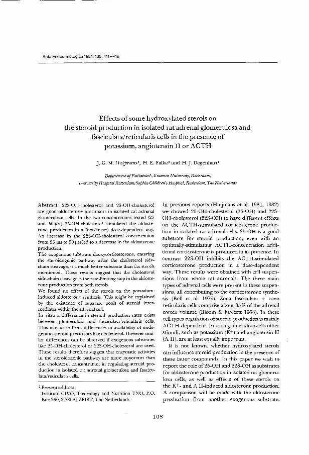
Acta Endocrinologica 1984,105:411-416
Effects of some hydroxylated sterols on the steroid production in isolated rat adrenal glomerulosa and
fasciculata/reticularis cells in the presence of potassium, angiotensin II or ACTH
]. G. M. Huijmansl, H. E. Falke2 and H.J. Degenhart1
Department of Pediatrics\ Erasmus University, Rotterdam,
University Hospital Rotterdam! Sophia Children's Hospital, Rotterdam, The Nether lands
Abstract. 22S-OH-cholesterol and 25-0H-cholesterol are good aldosterone precursors in isolated rat adrenal glomerulosa cells. In the two concentrations tested (25 and 50 f!M) 25-0H-cholesterol stimulated the aldosterone production in a (not-linear) dose-dependent way. An increase in the 22S-OH-cholesterol concentration from 25 f!M to 50 f!M led to a decrease in the aldosterone production. The exogenous substrate deoxycorticosterone, entering the steroidogenic pathway after the cholesterol sidechain cleavage, is a much better substrate than the sterols mentioned. These results suggest that the cholesterol side-chain cleavage is the rate-limiting step in the aldosterone production from both sterols. We found no effect of the sterols on the potassiuminduced aldosterone synthesis. This might be explained by the existence of separate pools of steroid intermediates within the adrenal cell. In vitro a difference in steroid production rates exists between glomerulosa and fasciculata/reticularis cells. This may arise from differences in availability of endogenous steroid precursors like cholesteroL However similar differences can be observed if exogenous substrates like 25-0H-cholesterol or 22S-OH-cholesterol are used. These results therefore suggest that enzymatic activities in the steroidogenic pathway are more important than the cholesterol concentration in regulating steroid production in isolated rat adrenal glomerulosa and fasciculata/reticularis cells.
2 Present address: Institute CIVO, Toxicology and Nutrition TNO, P.O. Box 360, 3700 AJ ZEIST, The Netherlands
In previous reports (Huijmans eta!. 1981, 1982) we showed 25-0H-cholesterol (25-0H) and 22SOH-cholesterol (22S-OH) to have different effects on the ACTH-stimulated corticosterone production in isolated rat adrenal cells. 25-0H is a good substrate for steroid production; even with an optimally-stimulating ACTH-concentration additional corticosterone is produced in its presence. In contrast 22S-OH inhibits the ACTH-stimulated corticosterone production in a dose-dependent way. These results were obtained with cell suspensions from whole rat adrenals. The three main types of adrenal cells were present in these suspensions, all contributing to the corticosterone synthesis (Bell et al. 1979). Zona fasciculata + zona reticularis cells comprise about 85% of the adrenal cortex volume (Bloom & Fawcett 1968). In these cell types regulation of steroid production is mainly ACTH-dependent. In zona glomerulosa cells other stimuli, such as potassium (K+) and angiotensin II (A II), are at least equally important.
It is not known, whether hydroxylated sterols can influence steroid production in the presence of these latter compounds. In this paper we wish to report the role of 25-0H and 22S-OH as substrates for aldosterone production in isolated rat glomerulosa cells, as well as effects of these sterols on the K+- and A II-induced aldosterone production. A comparison will be made with the aldosterone production from another exogenous substrate,
108
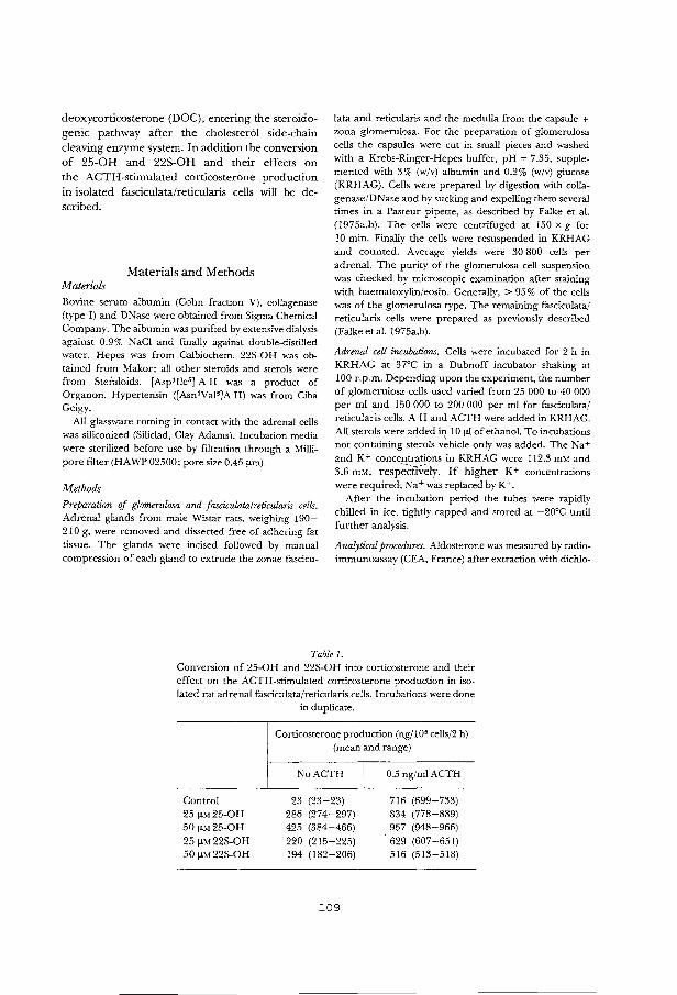
deoxycorticosterone (DOC), entering the steroido
genic pathway after the cholesterol side-chain
cleaving enzyme system. In addition the conversion
of 25-0H and 22S-OH and their effects on
the ACTH-stimulated corticosterone production
in isolated fasciculata/reticularis cells will be de
scribed.
Materials and Methods Materials
Bovine serum albumin (Cohn fraction V), collagenase (type I) and DNase were obtained from Sigma Chemical Company. The albumin was purified by extensive dialysis against 0.9% NaCl and finally against double-distilled water. Hepes was from Calbiochem. 225-0H was obtained from Makor; all other steroids and sterols were from Steraloids. [Asp' Ile5]-A II was a product of Organon. Hypertensin ([Asn1Val5]A II) was from Ciba Geigy.
All glassware coming in contact with the adrenal cells was siliconized (Siliclad, Clay Adams). Incubation media were sterilized before use by filtration through a Millipore filter (HAWP 02500: pore size 0.45 f'm).
Methods
Preparation of glomerulosa and fasciculata/reticularis cells. Adrenal glands from male Wistar rats, weighing 190-210 g, were removed and dissected free of adhering fat tissue. The glands were incised followed by manual compression of each gland to extrude the zonae fascicu-
lata and reticularis and the medulla from the capsule + zona glomerulosa. For the preparation of glomerulosa cells the capsules were cut in small pieces and washed with a Krebs-Ringer-Hepes buffer, pH = 7.35, supplemented with 3% (w/v) albumin and 0.2% (w/v) glucose (KRHAG). Cells were prepared by digestion with collagenase/DNase and by sucking and expelling them several times in a Pasteur pipette, as described by Falke et al. (1975a,b). The cells were centrifuged at 150 x g for 10 min. Finally the cells were resuspended in KRHAG and counted. Average yields were 30 800 cells per adrenal. The purity of the glomerulosa cell suspension was checked by microscopic examination after staining with haematoxylin/eosin. Generally, > 95% of the cells was of the glomerulosa type. The remaining fasciculata/ reticularis cells were prepared as previously described (Falke et al. 1975a.b).
Adrenal cell incubations. Cells were incubated for 2 h in KRHAG at 37°C in a Dubnoff incubator shaking at 100 r.p.m. Depending upon the experiment. the number of glomerulosa cells used varied from 25 000 to 40 000 per ml and 150 000 to 200 000 per ml for fasciculata/ reticularis cells. A II and ACTH were added in KRHAG. All sterols were added i~ 10 f'l of ethanol. To incubations not containing sterols vehicle only was added. The Na+ and K+ concentrations in KRHAG were 112.3 mM and 3.6 mM, resp;;ct!Veiy. If higher K + concentrations were require& N a+ was replaced by K +.
After the incubation period the tubes were rapidly chilled in ice, tightly capped and stored at -20°C until further analysis.
Analytical procedures. Aldosterone was measured by radioimmunoassay (CEA, France) after extraction with dichlo-
Tablel. Conversion of 25-0H and 22S-OH into corticosterone and their effect on the ACTH-stimulated corticosterone production in isolated rat adrenal fasciculata/reticularis cells. Incubations were done
Control 25 f'M25-0H 50 f'M25-0H 25 !!M22S-OH 50 f'M22S-OH
in duplicate.
Corticosterone production (ng/105 cells/2 h) (mean and range)
NoACTH 0.5 ng/ml ACTH
23 (23-23) 716 (699-733) 286 (274-297) 834 (778-889) 425 (384-466) 957 (948-966)
220 (215-225) 629 (607-651) 194 (182-206) 516 (513-518)
109
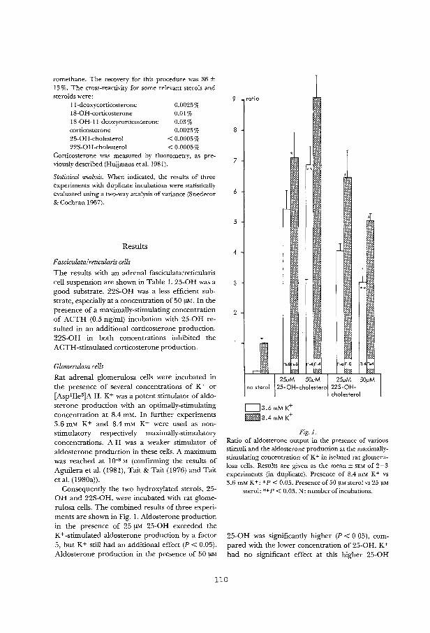
romethane. The recovery for this procedure was 86 ± 13%. The cross-reactivity for some relevant sterols and steroids were:
!!-deoxycorticosterone 18-0 H -corticosterone 18-0H-11-deoxycorticosterone corticosterone 25-0H-cholesterol 22S-OH-cholesterol
0.0025% 0.01% 0.03% 0.0025%
< 0.0005% < 0.0005%
Corticosterone was measured by fluorometry. as previously described (H uijmans et al. 1981).
Statistical analysis. When indicated, the results of three experiments with duplicate incubations were statistically evaluated using a two-way analysis of variance (Snedecor & Cochran !967).
Results
F asciculata/reticularis cells
The results with an adrenal fasciculata/reticularis cell suspension are shown in Table 1. 25-0H was a
good substrate. 22S-OH was a less efficient substrate, especially at a concentration of 50 !JM. In the
presence of a maximally-stimulating concentration
of ACTH (0.5 ng/ml) incubation witb 25-0H re
sulted in an additional corticosterone production. 22S-OH in both concentrations inhibited the
ACTH-stimulated corticosterone production.
Glomerulosa cells
Rat adrenal glomerulosa cells were incubated in
the presence of several concentrations of K+ or
[Asp1Ile5]A II. K+ was a potent stimulator of aldo
sterone production witb an optimally-stimulating concentration at 8.4 mM. In further experiments 3.6 mM K+ and 8.4 mM K+ were used as non
stimulatory respectively maximally-stimulatory concentrations. A II was a weaker stimulator of
aldosterone production in these cells. A maximum was reached at 10-8 M (confirming the results of
Aguilera eta!. (1981), Tait & Tait (1976) and Tait
eta!. (1980a)).
Consequently tbe two hydroxylated sterols, 25-0H and 22S-OH, were incubated with rat glome
rulosa cells. The combined results of three experi
ments are shown in Fig. 1. Aldosterone production
in the presence of 25 !JM 25-0H exceeded the
K+-stimulated aldosterone production by a factor
5, but K+ still had an additional effect (P < 0.05).
Aldosterone production in the presence of 50 !JM
9
8
7
6
5
4
3
2
ratio
*
J 1
25j.)M 50j.)M ,j 25/JM 50/JM I no sterol 25-0H-cholestero 225-0H
cholesterol
c::::J3.6mMK+ !::;::=::;~:;~ 8 .4 mM K +
Fig. I. Ratio of aldosterone output in the presence of various stimuli and the aldosterone production at the maximallystimulating concentration of K+ in isolated rat glomerulosa cells. Results are given as the mean ± SEM of 2-3 experiments (in duplicate). Presence of 8.4 mM K + vs 3.6 mM K +: * P < 0.05. Presence of 50 ftM sterol vs 25 ftM
sterol: **P < 0.05. N: number of incubations.
25-0H was significantly higher (P < 0.05), com
pared with tbe lower concentration of 25-0H. K+
had no significant effect at this higher 25-0H
110
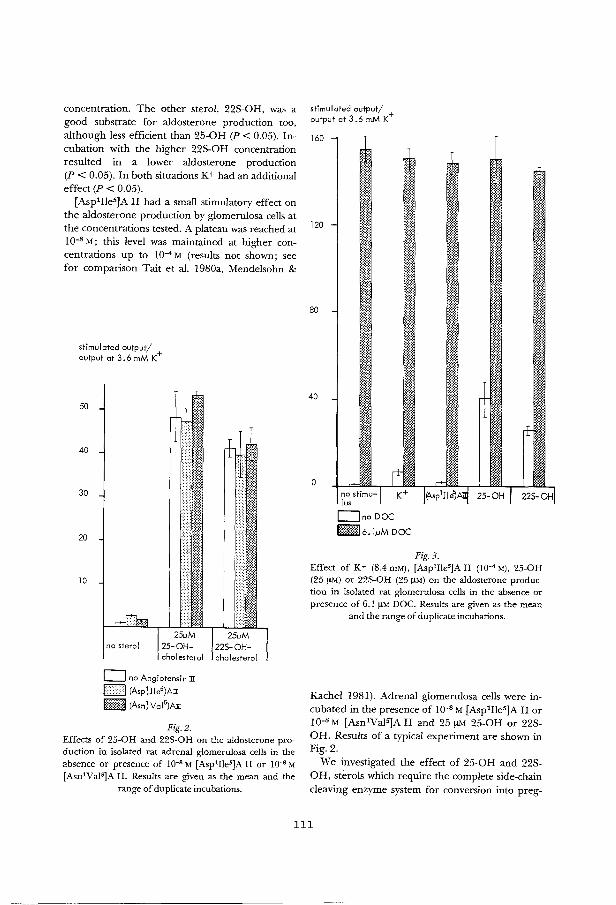
concentration. The other sterol. 22S-OH, was a good substrate for aldosterone production too, although less efficient than 25-0H (P < 0.05). Incubation with the higher 22S-OH concentration resulted m a lower aldosterone production (P < 0.05). In both situations K+ had an additional effect (P < 0.05).
[Asp1Ile5]A II had a small stimulatory effect on the aldosterone production by glomerulosa cells at the concentrations tested. A plateau was reached at 10-8 M; this level was maintained at higher concentrations up to 10-4 M (results not shown; see for comparison Tait et a!. 1980a, Mendelsohn &
stimulated output/ output at 3.6 mM K+
50
40
30
20
10
0 no Angiotensin ]I
1=:::1 (Asp~lle5)AI b:::::::~ (Asn~ Vai 5)A:D:
Fig.2. Effects of 25-0H and 22S-OH on the aldosterone production in isolated rat adrenal glomerulosa cells in the absence or presence of JQ-8 M [Asp1IIe5]A II or JQ-8 M [Asn1Vals:JA II. Results are given as the mean and the
range of duplicate incubations.
stimulated output/ + output at 3.6 mM K
160
120
80
40
0
w ~~~
no stimulus
Ono DOC
MM:I6.1!-!MDOC
Fig.3. Effect of K+ (8.4 mM), (Asp1Iles:JA II (IQ-• M), 25-0H (25 f!M) or 22S-OH (25 f!M) on the aldosterone production in isolated rat glomerulosa cells in the absence or presence of 6.1 f!M DOC. Results are given as the mean
and the range of duplicate incubations.
Kachel 1981). Adrenal glomerulosa cells were incubated in the presence of I0-8 M [Asp1Ile5]A II or 10-8 M [Asn1Va!S)A II and 25 !JM 25-0H or 22SOH. Results of a typical experiment are shown in Fig.2.
We investigated the effect of 25-0H and 22SOH, sterols which require the complete side-chain cleaving enzyme system for conversion into preg-
111

nenolone (Falke eta!. I975b; Alsema et al. l9S2), on the conversion of DOC, a substrate entering the steroidogenic pathway after the side-chain cleavage. DOC was chosen, because it is known to be a good precursor for aldosterone production in rat adrenal capsules (Fattah eta!. 1977). 6.1 ~DOC was rapidly converted into aldosterone in isolated rat glomerulosa cells (Fig. 3). Neither S.4 mM K+ or I0-4 M [Asp1Ile5)A II nor 25 !lM 25-0H or 22SOH had an effect on the aldosterone production in the presence of DOC.
Discussion
The aim of this study was to investigate possible effects of 25-0H and 22S-OH on rat adrenal glomerulosa cells. The method used in the preparation of these cells, however. does not preclude the presence of a small percentage of contaminating fasciculata cells. The question therefore arises, whether metabolites of fasciculata cells might be responsible for additional aldosterone production in the presence of the sterols. Taking into account the steroid production in fasciculata cells (as shown in Table l) and literature data on steroid production profiles in isolated adrenal cells (Bell et a!. 1979; Vinson et al. 1979; Tait et al. 19S0a) an upper limit to the amount of aldosterone produced from fasciculata metabolites can be calculated. For 25-0H (50~) this does not exceed 5% of the total aldosterone production.
25-0H is a good substrate for aldosterone production in rat adrenal glomerulosa cells (Fig. 1). Using DOC. a substrate entering the steroidogenic pathway after pregnenolone, a much greater aldosterone production can be observed (Fig. 3). This could mean that the side-chain cleavage reaction is the rate-limiting step. 22S-OH is a good substrate too (Fig. 1). However an increase in the 22S-OH concentration leads to a decreased aldosterone production. As the conversion of DOC is not inhibited by 22S-OH (Fig. 3) this suggests that 22S-OH inhibits its own side-chain cleavage. Similar results are obtained for the corticosterone production in fasciculata/reticularis cells (see Table 1). In these cell types 22S-OH also has an inhibitory effect on the ACTH-stimulated corticosterone production, confirming earlier results with whole adrenal cell suspensions (Huijmans eta!. l9Sl). We ·
therefore conclude that 22S-OH has an effect on the cholesterol side-chain cleavage.
K + stimulates one of the first steps in aldosterone synthesis (probably the cholesterol sidechain cleavage) as well as one or more late steps (McKenna eta!. l97S; Aguilera & Catt 1979). The apparent absence of an inhibitory effect of 22S-OH on the K+ stimulation may be explained by the existence of separate pools of steroids within the cell, which are important in the control of steroidogenesis (Vinson et al. 1979). In particular the pool of IS-oxygenated steroids might play a key role in the stimulation by K+ of the late steps in aldosterone formation.
In vitro there is a difference in steroidogenic capacity between glomerulosa and fasciculata cells. Estimated steroid production rates in fasciculata cells in the presence of ACTH or other stimuli exceed the production rates obser;ved in glomerulosa cells by a factor lO (Tait eta!. l9SOb). It could mean that the glomerulosa cell contains less endogenous steroid precursors (e.g. cholesterol). This is in agreement with histological findings; a smaller amount of lipid droplets has been found in the glomerulosa cells (Bloom & Fawcett l96S). However the availability of substrate can not be the only explanation as can be deduced from our results with the exogenous sterols 25-0H and 22S-OH. Corticosterone production from 25-0H in fasciculata/reticularis cells is 2S5 ng/105 cells/2 h (Table I), whereas the mean aldosterone production from the same amount of 25-0H in glomerulosa cells is only 16 ng/105 cells/2 h (Fig. 1). The two additional steps in the production of aldosterone from corticosterone (IS-hydroxylase and IS-dehydrogenase) might be responsible for this difference. However, as demonstrated with the exogenous substrate DOC, these steps are not rate-limiting. One may conclude that the cholesterol side-chain cleavage activity is responsible for the in vitro observed difference in steroidogenic capacity between glo merulosa and fasciculata/reticularis cells.
Acknowledgments
This work was supported by a grant from the Foundation for Medical Research, Sophia Children's Hospital, Rotterdam. The authors wish to thank Prof. Dr. H. ]. van der Molen for his critical reading of the manuscript. We
112
thank Dicky Kortleve for her expert technical assistance. The audio-visual centre of the Sophia Children's Hospital, Anja in 't Veen, Ronny Moerman and Carla Hendrix were of great help with the preparation of the manuscript.
References
Aguilera G & Cart KJ (1979): Loci of action of regulators of aldosterone biosynthesis in isolated glomerulosa ceiis. Endocrinology 104: 1046- 1052.
Aguilera G, Fujita K & Catt K J (1981): Mechanisms of inhibition of aldosterone secretion by adrenocorticotropin. Endocrinology 108:522-528.
Alsema G J, Degenhart H J & Hoogerbrugge J (I 982): Formation of 22-keto-cholesterol from 22S-hydroxycholesterol by a NADPH and 0 2-dependem, CO-insensitive enzyme in bovine adrenal cortex mitochondria.] Steroid Biochem 17:37-40.
BeiiJ B G, Hyatt P J, McDougall] G, TaitJ F, Tait SA S & Wiiiiams B C (1979): Properties of dispersed cells from ratadrenals.J Steroid Biochem I I: 169- I 74.
Bloom W & Fawcett D W (1968): A Textbook of Histology, 9th ed: 459-471. W B Saunders Company, Philadelphia.
Falke HE, Degenhart HJ, Abeln G J A, Visser H K A &
Croughs RJ M (1975a): Studies on isolated rat adrenal ceiis I. Continuous flow and batch incubations. Acta Endocrinol (Copenh) 78: I 10-121.
Falke H E, Degenhart H J, Abeln G J A & Visser H K A (197 5b): Studies on isolated rat adrenal cells. Metabolism of hydroxylated sterols. Mol Ceil Endocrinol 3: 375-383.
Fattah D I, Whitehouse B J & Vinson G P (1977): Biosynthesis of aldosterone from 18-hydroxylated precursors by rat adrenal tissue in vitro. J Endocrinol 7 5: 187-195.
113
H uijmans J G M, Degenhart H J, Kortleve D J & Visser H K A (198 I): Effects of 22S-hydroxy-cholesterol and other hydroxylated sterols on the ACTH-stimulated steroid production in rat adrenal cells. Acta Endocrinol (Copenh) 97:243-250.
Huijmansj G M, Degenhart H J & Kortleve D J (1982): Conversion of 22S-hydroxy-cholesterol and its effect on the metabolism of other sterols in rat adrenal cells and bovine adrenal mitochondria. Acta Endocrinol (Copenh) 100: 599-605.
McKenna T J, Island D P, Nicholson WE & Liddle G W (1978): The effects of potassium on early and late steps in aldosterone biosynthesis in cells of the zona glomerulosa.Endocrinology 103:1411-1416.
Mendelsohn FA 0 & Kachel CD (1981): Stimulation by serum of aldosterone production from rat adrenal glomerulosa cells in vitro: relationship to K +,serotonin and angiotensin II. Acta Endocrinol (Copenh) 97: 231-242.
Snedecor G W & Cochran W G (1967): Statistical Methods, 6th ed. Iowa State University Press, Ames.
Tait J F & Tait SA S (1976): The effect of changes in potassium concentration on the maximal steroidogenic response of purified zona glomerulosa ceiis to angiotensin II. J Steroid Biochem 7: 687-690.
Taitj F, Tait SA S, BeiiJ B G, Hyatt P J & Williams B C (I 980a): Further studies on the stimulation of rat adrenal capsular ceiis: four types of response. J Endocrinol87: I I -27.
Tait J F, Tait S A S & Beii J B G (1980b): Steroid hormone production by mammalian adrenocortical dispersed ceiis. In: Campbeii P N & Marshall R D (eds). Essays in Biochemistry, vol 16: 99- I 74. Academic Press. London.
Vinson G P, Whitehouse B J, Goddard C & Sibley C p (1979): Steroid profiles formed by rat adrenocortical whole tissue and cell suspensions under different conditions of stimulation. J Steroid Biochem II: I 75-183.
Received on August 15th, I 983.


CHAPl'ER 7
EFFECl' OF 25-HYIIDXY-<liOI..ESTEROL ON 'lHE UPI'AKE OF
CDRI'ICDSTERONE m ISOIATED RAT I.J:VER CELIS.
Honn. Metabol. Res. 20, 28-31 {1988).
115
Horm. metabol. Res. 20 (1988) 28-31 ©Georg Thieme Verlag Stuttgart • New York
Effect of 25-Hydroxy-Cholesterol on the Uptake of Corticosterone in Isolated Rat Liver Cells '
J.G.M. Huijmans, H.J. Degenhart and D.J. Kortleve
Department of Pediatrics, Erasmus University Rotterdam, University Hospital Rotterdam, Sophia Children's Hospital, Rotterdam, The Netherlands
Summary
Isolated rat liver cells are able to take up corticosterone. The Arrhenius plot of the uptake shows a biphasic course with a change in the slope around 25°C.
25·0H~cholesterol is also taken up; this phenomenon reaches a maximum at 10-15 minutes. After preincubating liver cells at 37°C in the presence of this sterol the phase transition is shifted to a higher temperature (32° C). as shown in the Arrhenius plot of the corticosterone uptake. At the same time the uptake of corticosterone is diminished. This cannot readily be explained by direct competition. The mechanism might involve an inhibition of an active uptake mechanism caused by a change in the plasma membrane.
Key~Words: Hydroxy/ated Sterol·- Steroids- Cell Membrane - Steroid Uptake
Introduction
It is commonly assumed, that the first step of steroid hor· mane action is passive diffusion through the cell membrane. Some authors, however, reported mediated transport mech· anisms (Rao 198l,Milgrom, Atger andBaulieu 1973; Jonkers, Timmermans, de long, Lamberts and Birkenhiiger 1980). These observations are supported by the existence of stable, dexamethasone-resistent, murine thymoma cell lines, in which selective membrane permeability was a rate· limiting step in steroid hormone action (Johnson, Newby and Bourgeois 1"984). The generalized steroid hormone resistance, observed in some species, also suggests a defect at the level of the cell membrane (Lipsett, Chrousos, Tomita, Brandon andLoriaux 1985).
The aim of the present study was to investigate possible effects ofhydroxylated sterols on the uptake process. The rationale was, that in several types of cells adverse effects ofhydroxylated sterols on membrane structure and function have been demonstrated. Human polymorphonuclear leukocyte chemotaxis is inhibited by oxygenated sterols (Gordon, Bass and Yachnin 1980); in human erythrocytes the calcium channel is modulated by oxygenated sterols (Neyses, Locher, Stimpel, Streuli and Vetter 1985), the osmotic fragility of human erythrocytes is diminished,
Received: 12 Nov, 1986 Accepted: 31 March 1987
116
following the membrane insertion of oxygenated sterols (Streuli, Kanofsky, Gunn and Yachnin 1981).
Because the naturally occurring 25-0H-cholesterol (25-0H) is one of the more potent sterols in this regard (Kandutsch, Chen andHeiniger 1978), we used as a model system the effect of 25-0H on the uptake of corticosterone in isolated rat liver cells.
Materials and Methods
Materials
Bovine serum albumin (Cohn fraction V), collagenase (type I) and DNase were obtained from Sigma Chemical Company. The albumin was purified by extensive dialysis and lyophilization. Hepes was from Calbiochem. All sterols and steroids used were from Steraloids. [26·14C]·25-0H (4.9 mCifmmol) and [1,2-'Hl-corticosterone (45 Ci/ mmol) were bought from Amersham. All glassware coming in contact with the cells was siliconized (Siliclad, Clay Adams). Incubation media were sterilized before use by filtration through a Mi!lipore filter (HAWP 02500, pore size 0.45 llm).
Methods
For each experiment the liver of a male 200 gram Wistar rat was removed, cut into pieces and washed with 0.9% NaCl and with a Krebs-Ringer·Hepes buffer (pH~ 7 .35), supplemented with 3% f.W/V) albumin and 0.2% f.W/V) glucose (= KRHAG). Cells were prepared by digestion with collagenase/DNasc using the method as described for rat adrenal cells (Falke, Degenhart, Abeln, Visser and Croughs 1975; Huijmans, Degenhart and Kort/eve 1982), except that the liver cells were centrifuged at 100 xg for 5 minutes.
Preincubations and incubations of the liver cells were done at 37°C (except when stated otherwise) in a Dubnoff incubator, shaking at 100 rpm. After the incubation the cells were isolated on glassfibre filters (Whatman GF /C), using a Millipore ftlter manifold and washed twice with 4 ml KRHAG. The filters were dried at 50°C and solubilized at 37°C in Nuclear Chicago tissue solubilizer. After 12 hours 10 m1 of toluene scintillation fluid, containing PPO and POPOP was added. Radioactivity was counted in a liquid scintillation counter (Mark ill, Searle).
Statistics
Data treatment was performed as indicated in the legends to the fJgUres and table. The slopes and intersection points of the straight lines in the Arrhenius plot (Fig. 2) were calculated by means of a numerical method for estimating the least sum of squares (Draper and Smith 1981).

25-0H-Cholestero! and Corticosterone Uptake
Uptake of 25-0H (pmol/10' cells)
1000
800
600
400
200
r-2·:.:'-------.2.3
10 20 30 40 50 60 70
Time (minutes)
Fig. 1 Time course of the uptake of 21 I'M [26·14Cl·25·0H in iso· fated rat liver cells at 3t> C. Results, given as the mean±SEM of 3-5 incubations, are statistically evaluated with a test, described by Kruskal and Wallis 1952. 1: P < 0.01 versus t = 0 minutes 2: P < 0.01 versus t = 10 minutes 3: not significant versus t = 0 minutes
7.0
ln If" 6.6
6.2
5.8
5.4 3.3 3.4
7.0
ln If" 6.6
' • \ 6.2 \
\ ,, -~--1---
5.8
No 25-0H
3.5 3.6 3.7
60 /JM 25-0H
5.4+-----,,+c.,----::-,."'4---,::-+.-::-5 ---:,:tc.6:----::-+,.7
1QQQ T
Fig. 2 Arrhenius plot of the uptake of 0.20 ,uM [3 H)-corticosterone in isolated rat liver cells at different incubation temperatures in the absence (upper) or presence (lower) of 60 ,uM 25-0H. Incubations were done in duplicate. The individual values, expressed as In (pmol/ min/105 cells), are plotted.
117
Horm. metabo!. Res. 20 (1988)
Table 1 Effect of 25-QH and several steroids on the uptake of corti~ costerone in isolated rat liver cells at 3~ C. Cells were pre incubated in the presence of the sterol/steroids for 15 minutes at 3~ C. The incubation time was 10 minutes. The corticosterone concentration was 1.6 x 10⁶ dpm/4x 105 cells (16 nM). Results were statistical-ly evaluated with a test described by Kurtz, Link, Tukey and Wallace 1965.
60 I'M sterol/steroid
Control
25-0H pregnenolone progesterone deoxycorticosterone corticosterone cortisol
•p < 0.01
Percentage of [ 3 H)-corticosterone uptake (mean and range of duplicate incubations)
100 (99-101)
60 (55--64). 26 (25··27)• 28 (24 -32)• 25 (24-26}* 26 (21-31}* 26 {25-r27)*
100% = 6.2 X 10⁴ dpm/4 X 105 cells
Results
The time course of the uptake of 21 i!M [26-14CJ·25·0H is given in Figure 1. A maximum was reached at 10 minutes, followed by a significant decline. The effect of 25-0H and several steroids on the uptake of corticosterone in rat liver cells is shown in Table 1. All substances tested inhibit the corticosterone uptake significantly.
We studied the effect of 25-0H on the uptake of cortico· sterone in rat liver cells at different incubation tempera· tures ranging from 4°C to 37°C. The cells were preincubated for 15 minutes in the absence or presence of 25-0H. The preincubation temperature was the same as the incubation temperature. The uptake of corticosterone over the first 50 seconds of the incubation was measured. The Ar· rhenius plot is shown in Figure 2. In the absence of 25.0H a change in the slope of the cuiVe occurred at 25° C. The activation energy was 12.0 Kcal/mol from 37°C to 25°C and 2.3 Kcal/mol from 25°C to 4°C. In the presence of the sterol (60 ~LM) the changing point was 32°C with activa· tion energies of 11.4 Kcal/mol and 0.8 Kcal/mol from 37°C to 32°C and 32°C to 4°C respectively.
Discussion
Membrane permeability can modulate steroid hormone action (Johnson, Newby and Bourgeois 1984). An important constituent of all cell membranes is cholesterol. Sterols hydroxylated in the side-chain occur in minor amounts on· ly. Many reports describe the effects "of insertion of hydroxylated sterols on cellular processes, e.g. the uptake of calcium (Neyses et al. 1985), platelet aggregation (!>himida, Jmada, Kikuchi, Inada, Morisaki, Ikekawa and Saito 1984), diminished osmotic fragility of human erythrocytes (Streuli et al. 1981), the formation of echinocytes (Hsu, Kanofski and Yachnin 1980) or leukocyte chemotaxis (Gordon, Bass and Yachnin 1980). The latter effect could already be ob· seiVed after 5 minutes of preincubation in the presence of the sterol. The sterols are inserted into the cellular mem·

Honn. metabol. Res. 20 (1988)
brane, as shown e.g. for the insertion of 7!3-0H-cholesterol into erythrocytes (Hsu, Kanofsky and Yachnin 1980). In hepatocytes the largest fraction of the unmetabolized 25-0H was also found in the plasma membrane (Erickson, Matsui, Shrewsbury, Cooper and Gould 1978).
Corticosterone is taken up by rat liver cells (see Fig. 2; upper part). With this amount of corticosterone the uptake is linear with the concentration. The Arrhenius plot of the uptake shows a biphasic course with a change in the slope around 25° C. This temperature lies well within the range (18°C to 31°C) reported for the critical transition temperatures of rat hepatocyte membranes (Schachter 1984). The activation energy of 12.0 Kcal/mol between 37°C and 25°C is in accordance with the value of 10-20 Kcal/mol, calculated for mediated transport processes (Rao 1981). From 25°C to 4°C the activation energy is 2.3 Kcal/mol, suggesting a diffusion process. ·
Some steroids can inhibit the corticosterone uptake (see Table 1), confirming Rao's observation (Rao, Rao, Eckel and Breuer 1977). These steroids were selected because of their structural resemblance to corticosterone. In addition most of these steroids are important intermediates in steroidogenesis in the rat. Without preincubation these steroids also inhibit the corticosterone uptake, though to a smaller extent. This inhibition may be due to competition at the level of the cellular membrane and/ or intracellular corticosterone binding sites. Of the sterols/steroids tested only cholest-4-en-3-one was without effect. The results suggest that a certain structural resemblance of the sterol/ steroid to corticosterone is correlated with its inhibitory action on the uptake. Quantitative statements are difficult to make, however.
In our study 25-0H is also taken up by isolated rat liver cells (see Figure 1). The initial uptake and, probably, insertion into the cellular membrane is maximal at 10 minutes and may be opposed by metabolism and excretion after that period (Erickson et a!. 1978).
After preincubating rat liver cells in the presence of 25-0H the uptake of corticosterone is diminished (see Figure 2 ;lower part). It seems, that the inhibition by 25-0H is independent of the corticosterone concentration. The presence of 25-0H without preinoobation has no direct effect on the corticosterone uptake, in spite of the fact that the 25-0H concentration exceeds the corticosterone concentration by a factor thousand (result not shown). A direct competition between 25-0H and corticosterone for entry into the liver cells is therefore less probable. Only with a preincubation time of 10-15 minutes, when the uptake of25-0H is at its maximum, the corticosterone uptake is inhibited. The Arrhenius plot of the corticosterone uptake after preincubation with 25-0H also shows a biphasic course but with a change in the slope around 32°C. It is known from studies on artificial membranes, that an increase in the membrane sterol concentration is accompanied by a decrease in the membrane fluidity (Cooper 1977). The shift of the phase transition in the Arrhenius plot to a higher temperature (25°C to 32°C) may be a direct consequence of this lowered membrane fluidity.
118
J.G.M. Huijmans, H.J. Degenhart and D.J. Kortleve
The activation energy between 37°C and 32°C is not influenced by the presence of 25-0H, but the overall capacity of the uptake mechanism is decreased by 40% (at 37°C).
In conclusion our results show, that 25-0H has an inhibitory effect on the uptake of corticosterone in isolated rat liver cells. This is not readily explained by direct competition. The mechanism might involve an inhibition of an active uptake mechanism accompanied by a change in the plasma membrane.
Acknowledgements
This work was supported by a grant from the Sophia Foundation for Medical Research, Sophia Children's Hospital, Rotterdam. The authors wish to thank Prof. Dr. H.J. v.d. Molen and Dr. H.E. Falke for their critical reading of the manuscript and their valuable suggestions. Mr John de Kuyper is gratefully ackl)owledged for his help with the preparation of the figures.
References
Cooper, R.A.: Abnormalities of cell-membrane fluidity in the pathogenesis of disease. N. Engl. J. Med. 297: 371-377 (1977)
Draper, N.R., H. Smith: Applied regression analysis. 2nd Edn. John Wiley and Sons Inc., New York (1981)
Erickson, S.K., S.M. Matsui, M.A. Shrewsbury, A.D. Cooper, R. G. Gould: Effects of 25-hydroxycholesterol on rat hepatic 3-hydroxy-3-rhethyl-glutaryl coenzyme A reductase activity in vivo, in perfused liver and in hepatocytes. J. Bioi. Chern. 253: 4159-4164 (1978)
Falke, H.E., H.J. Degenhart, G.J.A. Abeln, H.K.A. Visser, R.J.M. Croughs: Studies on isolated rat adrenal cells. Metabolism of hydroxylated sterols. Mol. Cell. Endocrinol. 3: 375-383 (1975)
Gordon, L.I., J. Bass, S. Yachnin: Inhibition of human polymorphonuclear leukocyte chemotaxis by oxygenated sterol compounds. Proc. Natl. Acad. Sci. USA 77: 4313-4316 (1980)
Hsu, R.C., J.R. Kanofski, S. Yachnin: The formation of echinocytes by the insertion of oxygenated sterol compounds into red cell membranes. Blood 56:109-117 (1980)
Huijmans, J.G.M., H.J. Degenhart, D.J. Kortleve: Conversion of 22S-hydroxycholesterol and its effect on the metabolism of other sterols in rat adrenal cells and bovine adrenal mitochondria. Acta llndocrinol. (Copenh.) 100: 599-605 (1982)
Johnson, D.M., R.F. Newby, S. Bourgeois: Membrane permeability as a determinant of dexamethasone resistance in murine thymoma cells. Cancer Res. 44: 2435-2440 (1984)
Jonkers, G.H., H.A. T. Timmermans, F.H. de Jong, S. W.J. Lamberts, J.C. Birkenhiiger: An energy-<iependent corticosterone uptake system in the rat liver cell. FEBS Letters 111: 403-406 (1980)
Kandutsch, A.A., H. W. Chen, H.-J. Heiniger: Biological activity of some oxygenated sterols. Science 201: 498-501 (1978)
Kruskal~ W.H.~ W.A. Wallis: Use of ranks on one-criterion variance analyses. J. Am. Statistical Asrociation 47: 583-621 (1952)
Kurtz, T.E., R.F. Link, J. W. Tukey, D.L. Wallace: Short-cut multiple comparisons. Technometrics 7: 95-161 (1965)
Lipsett, M.B., G.P. Chrousos, M. Tomita, D.D. Brandon, D.L. Loriaux: The defective glucocorticoid receptor in Man and non~human Primates. In: Recent Progress in Hormone Research, vol. 41; Greep, Ed. Academic Press, Orlando (1985), pp. 199-247 (with emphasis on the comment of M. Birnbaumer, p. 246)
Mz1grom, E., M. Atger, E.-E. Baulieu: Studies on estrogen entry into uterine cells and on estradiol-receptor complex attachment to the nucleus. Is the entry of estrogen into uterine cells a protein-mediated process? Biochim. Biophys. Acta 320: 267-283 (1973)

25-QH-Cholesterol and Corticosterone Uptake
Neyses, L., R. Locher, M. Stimpel, R. Streuli, W. Vetter: Stereospecific modulation of the calcium channel in human erythrocytes by cholesterol and its oxidized derivatives. Biochem. J. 227: 105-112 (1985)
Rao, G.S.: Mode of entry of steroid and thyroid hormones into cells. Mol. Cell. Endocrinol. 21: 97-108 (1981)·
Rao, ML., G.S. Rao, J. Eckel, H. Breuer: Factors involved in the uptake of corticosterone by rat liver cells. Biochim. Biophys. Acta 500: 322-332 (1977)
Horm. metabol. Res. 20 (1988)
Schachter, D.: Fluidity and function of hepatocyte plasma membranes. Hepatology 4: 140-151 (1984)
Shimada, H., T. lmada, T. Kikuchi, Y. Inada, M Morisaki, N. Ikekawa, Y. Saito: Rapid modulation of platelet aggregation in plasma by oxygenated sterols. J. Appl. Biochem. 6: 151-155 (1984)
Streuli, R.A., J.R. Kanofski, R.B. Gunn, S. Yachnin: Diminished osmotic fragility of human erythrocytes following the membrane insertion of oxygenated sterol compounds. Blood 58: 317-325 (1981)
Requests for reprints should be addressed to: J.G.M. Huijmans, Metabolic Laboratory, Sophia Children's Hospital, Gordelweg 160, 3038 GE Rotterdam (The Netherlands)
119

CHAPl'ER 8
EFFECl' OF ~XYI.ATED STEROlS ON 'IHE UPrAKE OF mEX.;NENOI.ONE IN ISOlATED
RAT ADRENAL CE:US.
Introduction.
It is conunonly thought that the uptake of steroids and sterols mainly
proceeds via diffusion processes. HCMever, the possibility has been
discussed, that in rat liver cells a mediated transport mechanism is
involved (for a review see Rao, 1981). It has been found that some steroids
can inhibit the uptake of corticosterone in rat liver cells (Rao et al.,
1977) , while the sterol 25-QH-cholesterol also inhibits the uptake of
corticosterone in rat liver cells (Huijroans et al., 1988; chapter 7: this
thesis).
'!he aim of the present study was to analyse the effect of 22S-0H,
17S,20S-diOH and 25-oH on the uptake of labelled pregnenolone, the
intracellular content of labelled steroids produced from pregnenolone and
on the conversion of pregnenolone into metabolic products in isolated rat
adrenal cells.
Methods.
Rat adrenal cells were prepared as described in Chapter 3. '!he cells
were preincubated at 37°C for one hour in a I:Ubnoff incubator, shaking at
100 :rpm in the presence or absence of the sterols tested ( 62 JiM) • After one
hour [3H]-pregnenolone (17 .2 Cijmmol; 165 nM) was added. 'Ihe cells were
incubated for o, 0.5, 1.5, 5, 10, 20 and 40 minutes. Subsequently the cells
were filtered on glass fibre filters (Whabnan GF/C) using a Millipore
filter manifold and washed twice with 4 ml KRHAG. 'Ihe filtrate and the
washing fluid were collected (see belav) .
'!he filters were dried at 50°C and solubilized at 3~C in Nuclear aricago
tissue solubilizer. After 12 hours 10 ml of toluene scintillating fluid,
containing PFO and rorop was added. Radioactivity was counted in a liqiud
scintillation counter (Mark III, Searle).
'Ihe cambined filtrate and washing fluid was extracted with chlorofontV
121

2
Figure 8.1:
3 4
-- progesterone
---- pregnenolone
---- deoxycorticosterone
--- corticosterone
~ 18-0H-deoxycorticosterone
18-0H-corticosterone
Example of a thin-layer c:hrc:matcgram of the
labelled steroids present in the incubation
filtrates. Visualisation occurred with a
radiochroroatcgram camera.
lane 1: control incubation after 90 seconds.
lane 2: 17S,20S-diOH incubation after 90 seconds.
lane 3: control incubation after 20 minutes.
lane 4: 17S,20S-diOH incubation after 20 minutes.
methanol arrl the steroids separated by thin-layer cbramatography on
silicagel plates (Merck, Darmstadt), using ethylacetatejacetone (99/1;
V/V) as a developing solvent. Radioactivity was detected with a
radiochroroatcgram camera (I.KB, SWeden). labelled material was extracted
from the thin-layer plate with methanol arrl counted. An example of a
thin-layer chromatogram is presented in Figure 8 .1. Reference COil1p0\.1D:ls
were used for the identification.
122
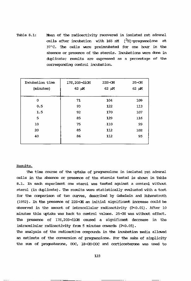
Table 8.1: Mean of the radioactivity recovered in isolated rat adrenal
cells after incubation with 165 nM [3H]-pregnenolone at
37•c. 'lhe cells were preincubated for one hour in the
absence or presence of the sterols. Incubations were done in
duplicate; results are expressed as a percentage of the
corresporrling control incubation.
Incubation tilne 178,208-diOH 228-0H: 25-oH
(minutes) 62 /1M 62 /1M 62 /1M
0 71 104 109
0.5 93 122 113
1.5 92 170 107
5 85 129 116
10 75 110 99
20 85 112 102
40 86 112 95
Results.
'lhe tilne course of the uptake of pregnenolone in isolated rat adrenal
cells in the absence or presence of the sterols tested is shown in Table
8.1. In each experilnent one sterol was tested against a control without
sterol (in duplicate). 'lhe results were statistically evaluated with a test
for the c:arrparison of two curves, described by Gebelein arrl Ruhenstroth
(1952). In the presence of 228-0H: an initial significant increase could be
observed in the amount of intracellular radioactivity (P<0.01). After 10
minutes this uptake was back to control values. 25-0H: was without effect.
'lhe presence of 178 ,20S-diOH caused a significant decrease in the
intracellular radioactivity from 5 minutes onwards (P<0.05).
'lhe analysis of the radioactive campounds in the incubation media allowed
an estiinate of the conversion of pregnenolone. For the sake of sinlplicity
the sum of progesterone, roc, 18-<>H-IXlC arrl corticosterone was used to
123
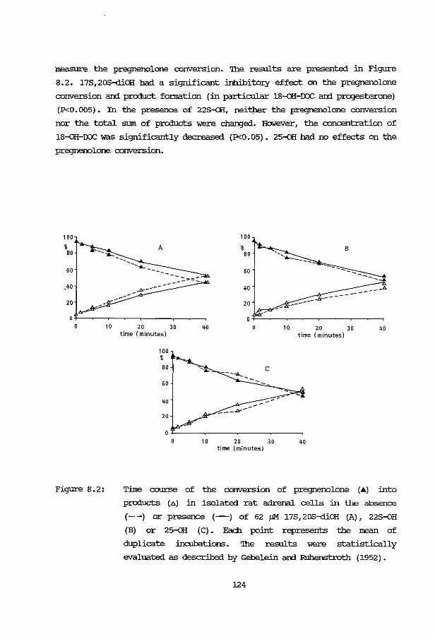
measure the pregnenolone conversion. The results are presented in Figure
8.2. 17S,20S-diOH had a significant inhibitory effect on the pregnenolone
conversion ard product fonnation (in particular 18-oH-roc ard prog-esterone)
(P<0.005). In the presence of 22S-oH, neitper the pregnenolone conversion
nor the total sum of products were changed. However, the concentration of
18-oH-roc was significantly decreased (P<0.05). 25-oH had no effects on the
pregnenolone conversion.
100
% 80
60
.40
20
10
Figure 8.2:
100
A % 80
60
40
20
20 30 40 10 20 30 40 time (minutes) time (minutes)
100 %
80
60
40
20
10 20 30 40 time (minutes)
Time course of the conversion of pregnenolone (.it..) into
products (a) in isolated rat adrenal cells in the absence
(-·-) or presence (-) of 62 J,tM 17S,20S-diOH (A), 22S-0H
(B) or 25-oH (C) . Each point represents the mean of
duplicate incubations. '!he results were statistically
evaluated as described by Gebelein ard Ruhenstroth (1952).
124

Discussion.
'!he intracellular adrenal steroid concentration is the result of
uptake, synthesis, conversion and release. '!he uptake and release of
steroids are c:anunonly considered as passive diffusion processes. However
mediated uptake mechanisms in rat liver cells have been discussed (see
introduction) . In rat adrenal cells SOITe authors described the existence of
macromolecules (probably proteins), which play a part in the control of
secretion or synthesis of steroids (Sibley et al., 1980; Goddard et al.,
1980). In particular the secretion of 18-oH-rxx::, fanned from endogenous
precursors, is controlled by a mechanism involving a ouabain-sensitive
process in the cell membrane (Sibley et al., 1980) .
Pregnenolone is a good precursor for corticosterone production in
isolated rat adrenal cells (Huijroans et al., 1981). '!he present results
show that the three hydroxylated sterols tested act differently on the
uptake and processing of pregnenolone by rat adrenal cells. 22S-{)H
increased the amount of intracellular radioactivity upon incubation with
labelled pregnenolone. '!his can not merely be an increase in the
permeability of the cellular membrane for pregnenolone, as 22S-oH has no
effect on the total conversion of pregnenolone, nor on the sum of products.
As mentioned before, especially the secretion of 18-oH-rxx:: fanned from
endogenous precursors is controlled by a ouabain-sensitive mechanism in the
cellular membrane. It is also the concentration of 18-oH-rxx:: in the
incubation medium, which is significantly decreased. One might speculate
that 22S-OH has an inhibitory effect on the secretion of 18-{)H-rxx::, thus
increasing its intracellular concentration. Feed-back inhibition of the 18-
hydroxylase (corticosterone methyloxidase I) roay direct steroid metabolism
towards the production of corticosterone, thus not changing the overall sum
of products (see figure 8.2).
In the presence of 17S,20S-diOH, the cellular radioactivity is decreased;
less pregnenolone is converted and fe!Ner products are fanned in its
presence, in particular less progesterone and 18-oH-rxx::. '!his sterol roay
have an effect on the secretion of 18-oH-rxx:: too. In addition, an
inhibitory effect on the uptake of pregnenolone seems probable.
25-oH has no effect on the intracellular radioactivity nor does it
influence the conversion of pregnenolone into products. One explanation is
that 25-oH is a much better substrate in rat adrenal cells than 22S-oH and
17S,20S-diOH and is rapidly cleared from the cell. '!he fact that 25-{)H has
125
an inhibito:ry effect on the corticosterone uptake in rat liver cells
(Huijmans et al., 1988), but no effect on the uptake of pregnenolone in rat
adrenal cells may also reflect a difference in the clearance of 25-oH from
the two types of cells. However, the existence of a mediated uptake
mechanism in the liver cells arrl not in the adrenal cells may also explain
the different effects of 25-oH.
In conclusion, the effects of these hydroxylated sterols on the uptake
arrl conversion of pregnenolone in isolated rat adrenal cells are caused by
changes in the cellular membrane, probably as a consequence of insertion of
these sterols into the membrane.
126

CHAPIER 9
'!HE HUMAN FEI2U.. .AJ::m:NAL AND HYDROXYIATED STEROlS
Introduction
'!he htnnan fetal adrenal glarrl is unique in its steroidogenic
properties, in that the 3,8-HSD activity is low. An ill'lportant function of
the fetal adrenals is the production of precursors for estrogen synthesis
with cholesterol as the main precursor. S<::lrle 30% of the cholesterol is
derived from de novo synthesis in the fetal adrenal glarrl (carr and
Silllpson, 198lb) . '!he other part is derived from plasma IDL. 'Ihis means that
in the fetal adrenal glarrl, the cholesterol production and influx is high.
On the other harrl the regular steroidogenic pathway is functionally blocked
by a low 3,8-HSD activity. CArr hypothesis was, that the htnnan fetal adrenal
might be used as a IOOdel system, in which the production of unusual
hydroxylated sterols might occur.
As discussed in Section 3B. 5, the isolation technique used for rat
adrenal cells is also suitable for the isolation of htnnan fetal adrenal
cells. 'Ihe cells retain their steroidogenic properties in cc:anparison with
those described in the literature. A separation of cells of the fetal and
definitive zones can be achieved by a manual dissection nethod and by
density gradient centrifugation through Percell.
'Ihese cells have been used to study steroidogenesis from cholesterol.
However, cholesterol is a very poor substrate, because it does not enter
the cell at an acceptable rate. As shown in rat adrenal cells, hydroxylated
sterols are :much better substrates for steroid production (Huijmans et al.,
1981, 1982) • 'Ihe aim of this study was to answere two questions:
1. are hydroxylated sterols converted in hmnan fetal adrenal cells?
2. If so, what are the products of their conversion?
Methods
A smvey of the fetal material used in this study is given in Section
3B.5. Part of the nethods has already been described in Olapter 3A
(isolation of cells; gaschramatography; estimation of cortisol and
corticosterone) arrl Cllapter 3B (cell separation).
127
Conversion of pregnenolone an:i conjugation.
cells from the separated fetal an:i definitive zones of the hmnan fetal
adrenal in case nr 3 (18 weeks of gestation) were incubated with
3H-labelled pregnenolone for 2 hours. '!he extraction procedure with
acetonejdichloramethane, as described in Section 3A.5, was used.
Radioactivity in the water layers was counted. mEA-sulfate was retained in
the water layers, as was confinned with a IEFAS standard solution.
steroids in the o:rganic layer were separated by moncx:iinensional thin
layer c:hrmatograph.y, using successively ethylacetatejacetone (6:4 VjV) an:i
chlorofornVether (9:1 VjV) as the developing solvents. Radioactivity was
visualized with a radiochromatogram camera (II<B, SWeden). '!he
c:hrmatogra}Xly lanes were divided into segrrents with Rrboundaries, as
described in table 9. 5. '!he location in this TLC system of several
relevant steroids was tested with reference CCllTIJ?Oll11ds.
Results an:i Discussion
In the hmnan fetal adrenal the 3,8-HSD activity is low. An important
pathway of steroid metabolism is the conversion of cholesterol into IHEA
via pregnenolone an:i 17a-<>H-pregnenolone (Yanaihara an:i Arai, 1981) . As
stated in the introduction, we studied the metabolic fate of hydroxylated
sterols. '!he results of incubations of human fetal adrenal cells in the
presence of 22R-<>H an:i 25-<>H are presented in Table 9 .1. '!he free sterols
an:i steroids were analyzed. Control values in the absence of sterols are
given between brackets.
22R-<>H may seJ:Ve as a precursor for the production of pregnenolone, 17a-<>H
pregnenolone an:i IHEA. A striking phenomenon is, that the amount of sterol
converted greatly exceeds the amount of products measured with this
technique. As far as steroid metabolites are detectable by gas
chromatogra}Xly, this technique does not show unknown peaks. Reference
steroids tested in this system are e.g. 16a-<>H-pregnenolone, testosterone,
androstenediol, androstenedione an:i 16a-<>H-IHEA.
In the presence of 25-<>H, the production of pregnenolone, 17a-<>H-pregne
nolone and IHEA did not increase. However, some 10% of this sterol
disaJ:Peared.
Both 22R-<>H an:i 25-<>H are good substrates for steroid synthesis in
isolated rat adrenal cells. 22R-<>H is a nomal intennediate in the
cholesterol side-chain cleavage. Several sterols, Vlhich are not
128
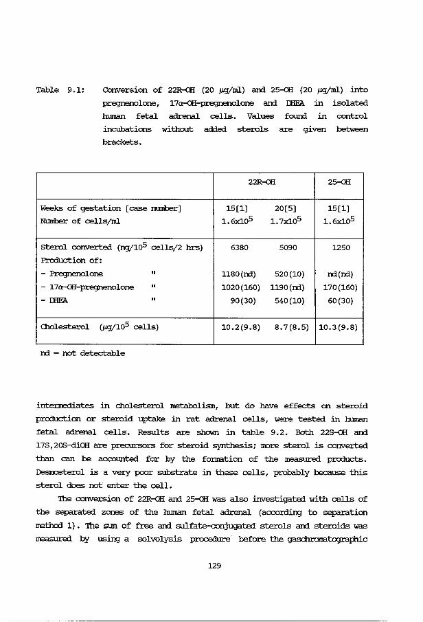
Table 9.1: Conversion of 22R-QH (20 p.g/ml) ani 25-oH {20 J,Lg/ml) into
pre:::JI1ei10lone,
htnnan fetal
17a-oH-pre:::JI1ei10lone ani IEFA in isolated
adrenal cells. Values foun::i in control
incubations without added sterols are given between
brackets.
22R-QH 25-oH
Weeks of gestation [case rnnnber] 15[1] 20[5] 15[1]
Number of cells/ml 1.6x10⁵ 1. 7Xl05 1.6Xl05
sterol converted (ng/105 cells/2 hrs) 6380 5090 1250
Production of:
- Pregnenolone II 1180(n:i) 520(10) n:i(n:i)
- 17a-oH-pre:::JI1ei10lone II 1020{160) 1190(n:i) 170{160)
- IEFA II 90(30) 540(10) 60(30)
Cholesterol (p.g/105 cells) 10.2(9.8) 8.7(8.5) 10.3(9.8)
n:i = not detectable
intennediates in cholesterol metabolism, but do have effects on steroid
production or steroid uptake in rat adrenal cells, were tested in human
fetal adrenal cells. Results are shown in table 9.2. Both 22S-oH ani
17S,20S-diOH are precursors for steroid synthesis; IIIOre sterol is converted
than can be acx::ounted for by the fonnation of the IreaSUred products.
Des!oosterol is a very poor substrate in these cells, probably because this
sterol does not enter the cell.
'Ihe conversion of 22R-QH ani 25-oH was also investigated with cells of
the separated zones of the human fetal adrenal (according to separation
method 1). 'Ihe sum of free ani sulfate-conjugated sterols ani steroids was
IreaSUred by using a solvolysis procedure before the gasc:hramatographic
129
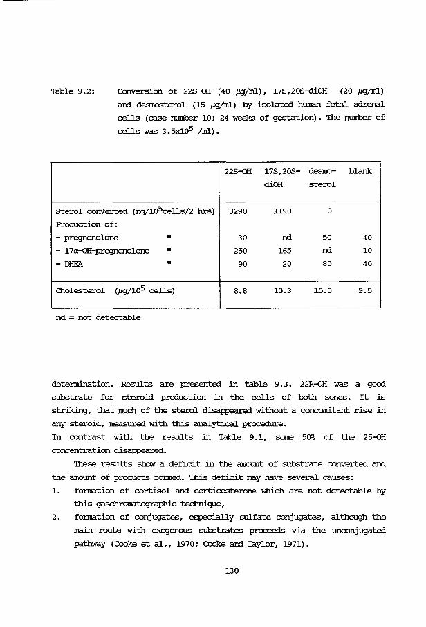
Table 9.2: Conversion of 22S-0H (40 J.£9"/ml), 17S,20S-diOH (20 J.£9"/ml)
ani desiOOSterol (15 J.£9"/ml) by isolated human fetal adrenal
cells (case number 10; 24 weeks of gestation) • 'Ihe number of
cells was 3.5x10⁵ jml).
22S-0H 17S,20S- desmo- blank
diOH sterol
sterol converted (ng/105cells/2 hrs) 3290 1190 0
Production of:
- pregnenolone II 30 nd 50 40
- 17a-0H-pregnenolone II 250 165 nd 10
-mEA II 90 20 80 40
<llolesterol (J.£9"/105 cells) 8.8 10.3 10.0 9.5
nd = not detectable
detennination. Results are presented in table 9. 3. 22R-0H was a good
substrate for steroid production in the cells of both zones. It is
striking, that lllllch of the sterol disappeared without a concomitant rise in
any steroid, m=asured. with this analytical procedure.
In contrast with the results in Table 9.1, same 50% of the 25-oH
concentration disappeared.
'lhese results show a deficit in the aiOOUI'lt of substrate converted ani
the aiOOUI'lt of products fanned. 'Ihis deficit may have several causes:
1. fonnation of cortisol ani corticosterone which are not detectable by
this gaschramatogra}:'hic technique,
2. fonnation of conjugates, especially sulfate conjugates, although the
main route with exogenous substrates proceeds via the unconjugated
pathway (Cooke et al., 1970; Cooke ani Taylor, 1971).
130
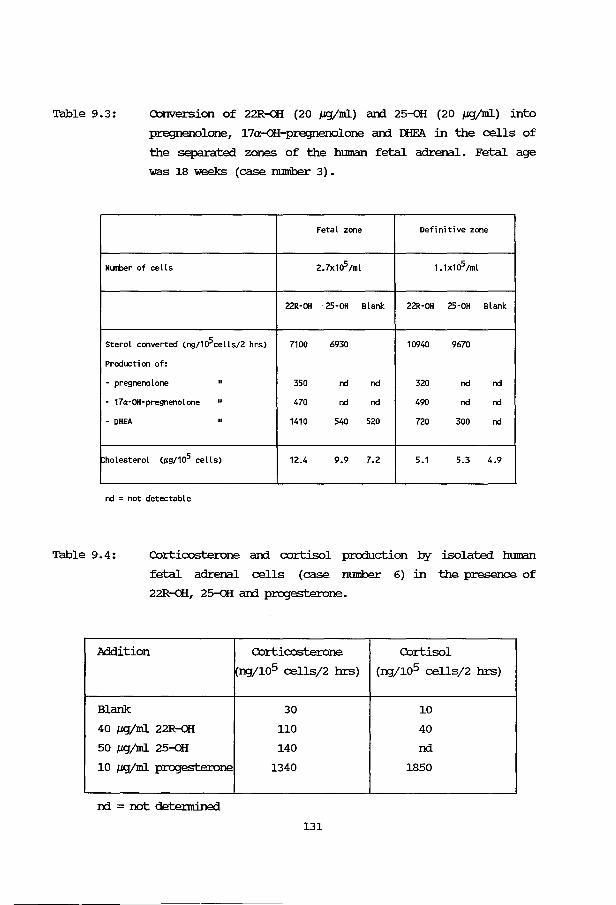
Table 9.3: Conversion of 22R-oH (20 J.&g/ml) arxi 25-oH (20 J.&gjml) into
pregnenolone, 17a-oH-pregnenolone arxi IEFA in the cells of
the separated zones of the human fetal adrenal. Fetal age
was 18 weeks (case nmnber 3) •
Fetal zone Definitive zone
Nllltler of cells 2.7x10⁵Jml 1.1x10⁵Jml
22R-OH 25-0H Blank 22R-OH 25-0H Blank
Sterol converted (ng/105cells/2 hrs) 7100 6930 10940 9670
Production of:
- pregnenolone II 350 nd nd 320 nd nd
- 17a-OH-pregnenolone II 470 nd nd 490 nd nd
- DHEA II 1410 540 520 720 300 nd
holesterol (JLg/105 cells) 12.4 9.9 7.2 5.1 5.3 4.9
nd = not detectable
Table 9.4: Corticosterone arxi cortisol production by isolated human
fetal adrenal cells (case nmnber 6) in the presence of
22R-oH, 25-oH arxi progesterone.
Addition Corticosterone Cortisol
ng/105 cells/2 hrs) (ng/105 cells/2 hrs)
Blank 30 10
40 J.&g/ml 22R-QH 110 40
50 J.&g/ml 25-QH 140 nd
10 J.&g/ml progesterone 1340 1850
nd = not detennine:l
131
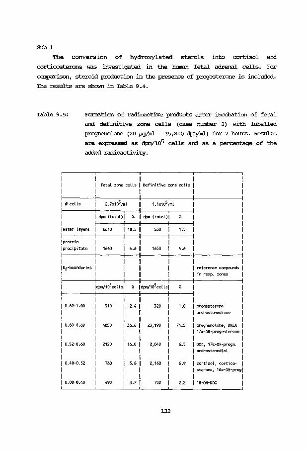
SUb 1
The conversion of hydroxylated sterols into cortisol and
corticosterone was investigated in the htnnan fetal adrenal cells. For
comparison, steroid production in the presence of progesterone is included.
The results are shown in Table 9.4.
Table 9.5: Fonna.tion of radioactive products after incubation of fetal
and definitive zone cells (case number 3) with labelled
pregnenolone (20 J..l.gjml = 35,800 dp!Vml) for 2 hours. Results
are ~ressed as dpiV105 cells and as a percentage of the
added radioactivity.
Fetal zone cells I Definitive zone cells I I I
I #cells 2.7x10⁵tml 1.1x10⁵/ml
I ~ (total>l % I ~ (total>l %
I water layers 6610 1 18.5 1 530 1.5
I protein I I precipitate 1660 4.6 1 1650 4.6
I I I Rf-boundari es I I reference conpounds I I I I in resp. zones I
ldpmt105cellsl % ldpm/105cellsl %
I I 1 o.69-1.oo 310 2.4 1 320 1.0 I progesterone
I I I androstenedione
I I I I 1 o.6o-o.69 4850 1 36.6 1 23,190 74.5 I pregnenolone, DHEA
I I I I 17a-OH-progesterone I I I I I I 1 o.52-o.60 2120 1 16.o 1 2,040 6.5 I DOC, 17a-OH-pregn. I I I I I androstenedi ol I I I I I I 1 o.4o-o.52 760 I 5.8 1 2,160 6.9 I cortisol, cortico- I I I I I sterone, 16a-OH-preg I I I I I I 1 o.oo-o.4o 490 I 3.7 1 700 2.2 I 18-0H-OOC I
132
22R-QH and 25--GH, which are good substrates for steroid production in rat
adrenal cells, are not readily converted into cortisol and corticosterone
in human fetal adrenal cells. One explanation, especially in the fetal zone
cells, is the low 3,8-HSD activity. Conversion of these sterols into other,
non-detected metabolites appears the only remaining possibility.
'lhese results show that cortisol and corticosterone production does not
play an illlportant role in the conversion of 22R-<>H and 25-<>H.
SUb 2
With cells of the separated zones of the human fetal adrenal,
according to method 1 (see Chapter 3 .13) , steroidogenesis was investigated
with labelled pregnenolone as a substrate. '!he analytical procedure is
described in the Methods section. '!he results are presented in table 9.5.
More radioactivity remains in the water layer of the fetal zone incubation,
c:aropared with the definitive zone cells, suggesting a higher sulfokinase
activity in the fetal zone. 'Ihese results also show, that the extent of
sulfate conjugation by the fetal zone cells does not exceed 20% of the
added pregnenolone. In the definitive zone sulfate conjugation is
ne:Jligible.
Cc:xrprring the results in Tables 9.1 and 9. 3, a solvolysis procedure
before the gaschramatographic detennination of the steroids does not
increase the recovery of prcducts to a considerable extent. '!his also
suggests a low extent of sulfate conjugation in these experiments.
OONVERSION OF I'RE)3NEN()I0NE AND 17a-QH-I'RE)3NEN()IDNE
'!he conversion of sterols JOOSt probably proceeds via pregnenolone and
17a-<>H-pregnenolone into r::HFA (Yanaihara and Arai, 1981) , as is also
suggested by the results in Tables 9.1 and 9.3 for 22R-<>H as a substrate.
A comparison was made between the conversion of the sterols and the
conversion of the proposed intermediates pregnenolone and 17a--GH
pregnenolone. Table 9.6 shows the results for a whole human fetal adrenal
cell suspension. Only the free products were measured. '!he high 17a-oH
pregnenolone prcduction in the presence of pregnenolone is in agreement
with an active 17a-hydroxylase in the human fetal adrenal (Villee, 1969b).
r::HFA prcduction is small. With 17a-<>H-pregnenolone as a substrate, only a
small part of the steroid converted can be explained by the prcduction of
r::HFA.
133
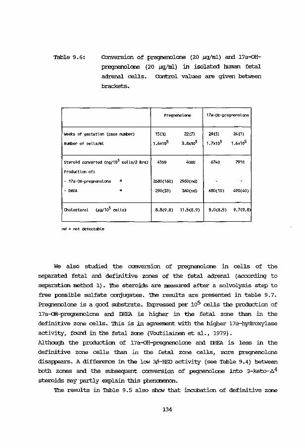
Table 9.6: Conversion of pregnenolone (20 J.L<J/ml) ani 17a-oH
pregnenolone (20 J.Lgjml) in isolated htnnan fetal
adrenal cells. Control values are given between
brackets.
Pregnenolone 17a-OH-pregnenolone
Weeks of gestation [case nlJIIberl 15[1] 22[7] 20[5] 24[1]
NLJIIber of cells/ml 1.6x10⁵ 3.8x1a5 1.7x10⁵ 1.6x1o5
Steroid converted (ng/105 cells/2 hrs) 4550 4080 6740 7910
Production of:
- 17a-OH-pregnenolone II 2680(160) 2960(nd) - -- DHEA II 290(30) 360(nd) 680(10) 490(40)
Cholesterol (JLS/105 cells) 8.8(9.8) 11.5(8.9) 8.0(8.5) 9.7(9.8
nd = not detectable
We also studied the conversion of pregnenolone in cells of the
separated fetal and definitive zones of the fetal adrenal (according to
separation method 1). '!he steroids are measured after a solvolysis step to
free possible sulfate conjugates. 'Ihe results are presented in table 9. 7.
Pregnenolone is a good substrate. Expressed per 105 cells the production of
17a-oH-pregnenolone ani IHE'A is higher in the fetal zone than in the
definitive zane cells. '!his is in agreement with the higher 17a-hydroxylase
activity, fourrl in the fetal zane (Voutilainen et al., 1979).
Although the production of 17a-oH-pregnenolone and IHE'A is less in the
definitive zone cells than in the fetal zone cells, more pregnenolone
disappears. A difference in the low 3,13-HSD activity (see Table 9.4) between
both zones ani the subsequent conversion of pegnenolone into 3-keto-.64
steroids may partly explain this phenomenon.
'!he results in Table 9.5 also show that incubation of definitive zone
134
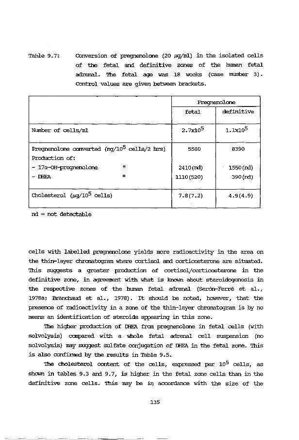
Table 9.7: Conversion of pregnenolone (20 J.L<J/ml) in the isolated cells
of the fetal ani definitive zones of the human fetal
adrenal. '!he fetal age WclS 18 weeks (case nmnber 3) .
Control values are given between brackets.
Pregnenolone
fetal definitive
Nt.nnber of cells/ml 2.7Xl05 1.1x10⁵
Pregnenolone converted (ng/105 cells/2 hrs) 5580 8390
Production of:
- 17a-oH-pregnenolone II 2410{nd) 1550(nd)
- IHEA II 1110{520} 390{nd)
Cholesterol (J.L<J/105 cells) 7.8(7.2) 4.9(4.9)
nd = not detectable
cells with labelled pregnenolone yields 1110re radioactivity in the area on
the thin-layer chromatogram where cortisol ani corticosterone are situated.
'!his suggests a greater production of cortisol/corticosterone in the
definitive zone, in agreement with what is :Jma.m about steroidogenesis in
the respective zones of the human fetal adrenal (Ser6n-Ferre et al.,
1978a; Branchaud et al., 1978). It should be noted, however, that the
presence of radioactivity in a zone of the thin-layer chromatogram is by no
means an identification of steroids appearing in this zone.
'!he higher production of IHEA from pregnenolone in fetal cells (with
solvolysis) ~ with a whole fetal adrenal cell suspension (no
solvolysis) may suggest sulfate conjugation of IHEA in the fetal zone. 'Ihis
is also confinlai by the results in Table 9.5.
'!he cholesterol content of the cells, expressed per 105 cells, as
shown in tables 9. 3 ani 9. 7, is higher in the fetal zone cells than in the
definitive zone cells. 'Ihis may be in accordance with the size of the
135

cells (Bloem ani Fawcett, 1968) : the fetal zone cells are bigger in size
with a greater 100IIIbrane surface.
In conclusion, the hydroxylated sterols 22R-C>H ani 25-C>H are converted
in isolated hmnan fetal adrenal cells. '!he increase in the pregnenolone,
17a-C>H-pregnenolone ani IEE'A concentration suggests that, at least 22R-0H,
is converted via the nonnal matabolic route. '!he production of cortisol ani
corticosterone ani of sulfate conjugates carmot account for the deficit in
the sterol conversion.
A possible explanation is the conversion into conjugates other than
sulfates, or the conversion into nPre polar products, which are not
extracted from the incubation medium or not detectable with this capillary
column. '!he availability of labelled sterols would be of great help in
solvin;J this problem.
As stated in the introduction to this chapter, the high steroid flux ani
the low 3JHJSD activity in the human fetal adrenal :make the production of
unusual hydroxylated sterols possible. OUr results, presented in this
chapter, confinn this possibility. '!his needs further study.
136
CHAPIER 10
GENERAL DISCUSSION
In several diseases abnonnal sterol metabolism cx:curs; this may be a
factor in the etiology of the disease, due to effects of hydroxylated
sterols on cellular or sulx:ellular processes. '!he aims of this study were
to investigate:
1. short-term effects of hydroxylated sterols on basal and. stimulated
steroid production in rat adrenal cells and. on steroid uptake in rat
adrenal and. liver cells,
2. the conversion of sterols in human fetal adrenal cells, as a
physiolcX]"ical model of a 3,8-HSD deficient o:rgan.
STEROID PROJ:UCI'ION
several sterols were founi to be good substrates for corticosterone
synthesis in isolated rat adrenal cells, e.g. 25-oH, 22R-oH and. 20S-0H
cholesterol. 22S-0H was a poor substrate. '!his is in agreement with the
findings of Iambeth (1986), that the structural requirements for binding to
the cyt:ochratre P-450scc and. conversion into pregnenolone are more
favourable for the intennediate 22R-oH than for 22S-oH. '!he 22R hydrogen of
cholesterol is more close to the heme iron than the 22S hydrogen.
For 25-oH and. 22S-QH no acute adverse effects on the :functionin:J of the
isolated rat adrenal cells were founi. '!his conclusion is based on the
linearity of the corticosterone production for up to 3 hours in the
presence of 22S-oH (Chapter 4) or 25-oH.
In a pilot experiment, 22S-oH inhibited the ACIH-stimulated corticosterone
production, whereas steroid production in the presence of 25-oH and. ACIH
was almost additive. For this reason the effect of 22S-oH was studied in
more detail. The results in Chapters 4 and. 5 allowed to locate the effect
of this sterol at the level of the cholesterol side-chain cleaving enzyme
system as the most probable site of action. '!he arguments in favour of this
site are:
1. there was no effect of 228-oH on the cAMP concentration in rat adrenal
cells after stimulation with ACIH,
137

2. free cholesterol in the adrenal cells ac:ctnnUlated in the presence of
228-<JH plus Acm, campared with the cholesterol concentration after
stimulation with 228-<JH or Acm,
3. there was no effect of 228-<JH on the conversion of exCXJenous
pregnenolone (Cllapters 4 and 8),
4. 228-QH had no effect on the conversion of 22R-<JH but slightly
inhibited the conversion of 25-<JH.
'lhe IOC>St probable action mechanism of 228-QH is an effect on the binding of
cholesterol and 25-<JH to the cyt:oc:hrarre P-450scc complex.
In damaged bovine adrenal mitochororia too, 22R-<JH is a I!Rlch better
substrate for pregnenolone production than 228-<JH. When 228-<JH was used as
a substrate, 22-keto-cholesterol was fanned in addition to pregnenolone.
22-Keto-cholesterol was not detectable when a mitochororial preparation was
incubated in the presence of 22R-<JH (see also: Alsema et al., 1982b).
Possibly the slower conversion rate of 228-<JH allows no:rmally minor
pathways to become active; a nice example of unusual sterol fonnation. 'lhe
conversion of 228-<JH into 22-keto-cholesterol might explain the inhibitory
effect of 228-<JH on the Acm-stimulated corticosterone production in
isolated rat adrenal cells, as 22-keto-cholesterol is an inhibitor of this
process (see Cllapter 4) • However, when rat adrenal cells were incubated in
the presence of 228-<JH, 22-keto-cholesterol was not detectable. '!his
difference in 22-keto-cholesterol production may be explained by:
1. the use of whole adrenal cells versus damaged mitochororia,
2. a species difference (rat versus cx:M) •
'lhe inhibitory effect of 22S-<JH on the ACni-stimulated corticosterone
production, as seen in whole rat adrenal cell suspensions, could also be
demonstrated in isolated rat adrenal fasciculatajreticularis cells.
Apparently the presence of glomerulosa cells or steroid metabolism in these
cells does not influence the overall inhibitory mechanism. One obvious
reason is, that the zona glomerulosa is I!Rlch smaller than the zonae
fasciculata and reticularis. In addition, the steroidCXJeniC capacity of
glomerulosa cells may be different from that of fasciculatajreticularis
cells. 'lhe combined aldosterone and corticosterone production in
glomerulosa cells in the presence of ACffi is 10 times lower than the
ACni-stimulated corticosterone production in fasciculata cells (Tait et
al. , 1980b) • '!his might be due to the lower cholesterol content of the
glomerulosa cells. However from the results obtained with the exCXJenous
138

substrate 25~ and from the fact that the :pathway:
roc --> aldosterone
is not rate-limiting (Olapter 6), the most likely explanation is, that the
cholesterol side-chain cleaving enzyme activity is responsible for the
difference in the steroidogenic ca:pacity, observed in vitro, between
glomerulosa cells and fasciculatajreticularis cells. '!he use of hydroxyl
ated sterols thus seem to be a good expedient in elucidating differences in
the steroidogenic capacities, existing between several types of steroid
producing cells.
STEROID UPl'AKE
Effects of hydroxylated sterols on the uptake of steroids were studied
in rat adrenal and liver cells. In liver cells 25~ had an inhibitory
effect on the corticosterone uptake, probably caused by a change in the
cellular membrane. However, the same sterol had no effect on the uptake of
pregnenolone in isolated rat adrenal cells. One possible explanation might
be the difference, existing between liver and adrenal cells as to the
metabolism of 25~, 25~ being a good substrate in adrenal cells. A
similar effect was seen in rat hepatcx::ytes, where the :HM:;-C.oA reductase
activity was inhibited in the presence of 25~. '!he recovery of the
activity was dependent on the clearance of 25-oH from the cells; with low
concentrations of 25-oH the :HM:;-C.oA reductase activity rose to 90% of the
initial value, whereas with high concentrations of 25~ activity remained
low (Erickson et al., 1978).
Another sterol, 22S~ had a similar inhibitory effect on the
corticosterone uptake in rat liver cells after 15 minutes preincubation in
the presence of the sterol. Like 25-oH, 22S~ did not inhibit the uptake
of pregnenolone in rat adrenal cells, although this sterol is a poor
substrate c:arnpared with 25-oH. For this reason another explanation for the
different effects of the sterols tested on the uptake of steroids in rat
adrenal or liver cells might be a difference in the cellular membrane
itself, with liver cells having a different uptake mechanism for
corticosterone c:arnpared with the uptake of pregnenolone in rat adrenal
cells. 'lhe results of our study show that hydroxylated sterols are able to
influence the cellular membrane.
It has bec:one apparent that the composition and fluidity of membrane
lipids is critically regulated for an optimal cell growth and function. A
139
change in the fluidity of cell :menibranes can be an important factor in the
pathogenesis of disease. Urrler con:litions of an increased cholesterol to
phospholipid ratio in plasrra lipoproteins, the cholesterol content of red
cell :menibranes is also increased, causing altered shape arrl properties
(Cooper, 1977). '!his kind of effects are not restricted to red cell
:menibranes, as discussed in section 2. 5. Stnmnarizing, our study has shown
effects of hydroxylated sterols on steroid uptake processes. In those
diseases where an ac:cunrulation of cholesterol or other sterols has been
described, these effects on uptake processes should be further evaluated.
'!his can be the subject of future research.
HUMAN FErAL ADRENAL CELLS
'!he third aim of this study was the investigation of the effect of
hydroxylated sterols on the basal steroid production in human fetal adrenal
cells arrl the possible fonnation of unusual campounds. 'lbese cells were
obtained using the isolation procedure for rat adrenal cells. 'Ibis method
yields viable cells with steroidogenic characteristics, in accordance with
the literature. A procedure for separating the two types of human fetal
adrenal cells by density gradient centrifugation on Percell was proven to
be effective; the characteristics of the cells in the separated barrls are
in agreement with those reported for fetal and definitive cells.
Several sterols were found to be COIWerted in these cells. Part of the
conversion products are intennediates in the main metabolic pathway of
cholesterol to rHEA via pregnenolone and 17o:-oH-pregnenolone. Hooever, the
conversion of the sterols greatly exceeded the fonnation of measurable
products. One may speculate about the nature of these products. Cortisol
and corticosterone were not found in measurable amounts. '!be fonnation of
sulfate conjugates could also not aCCOlll1t for the deficit. '!be more conunon
steroids, like arrlrostenediol, 16a-oH-IHEA, etc. could be ruled out by
their absence in the gaschromatogram. '!be fonnation of very polar or
acidic sterols or steroljsteroid conjugates other than sulfates is a
possibility. 'Ibis should be investigated further; the availability of
labelled sterols would be of great help in solving this problem.
CLOSING REMARKS
'Ibis study has shown that in vitro hydroxylated sterols have several
acute effects:
140
1. on steroid production by a (c:oropetitive) effect on the cholesterol
side-chain cleavage,
2. on steroid uptake by inducing a change in the cellular membrane.
It should be stressed, that the presence of unusual sterols not necessarily
leads to dramatic changes at short tenn, but may have long-tenn effects.
One example is the presence of cholestanol in cerebrotendinous
xanthomatosis. '!he contribution of accumulated sterols to the mortality in
e.g. the congenital lipoid adrenal hyperplasia or Wolman's disease is not
known with certainty. However, any profound change in membrane composition,
either with effects on uptake of steroids or other metabolites, or with a
more general disfunction of the cells, is very likely to result in
deleterious effects on the whole organism.
In this thesis only a limited number of sterols were studied. Other
sterols should be evaluated for their short-tenn and long-tenn effects on
isolated cells and whole organisms as a subject for future research.
141

rnAPI'ER 11
SUMMARY
Several effects of sterols, oxygenated in the steroid nucleus or the
side-chain, on cellular and subcellular processes have been described. '!he
investigation of possible short-tenn effects of these sterols on basal and
stimulated steroid production and on steroid uptake is the subject of this
thesis.
<llapter 1 gives a general introduction and a statement of the problem.
'!he accumulation of sterols in body fluids and tissues may play a role in
the etiology of several diseases. '!his might be of .i.Irportance, because in
vivo and in vitro many adverse effects of sterols have been described. For
this study the isolated rat adrenal and liver cells were used as model
systems. In addition the properties of hydroxylated sterols as steroid
precursors were tested in isolated human fetal adrenal cells as a :model of
a 3,8-HSD deficient organ. '!he use of isolated human fetal adrenal cells is
rather uncannnon, as most authors use cell or tissue cultures. For this
purpose, the applicability of the method for the isolation of rat adrenal
cells to the human fetal adrenal and the separation of cells from the two
zones in the human fetal adrenal were investigated.
Cilapter 2 gives a review of the literature. SteroidogeneSis and its
regulato:cy factors in the rat adrenal (both zona glamenllosa and zonae
fasciculataj reticularis) and in the human fetal adrenal are discussed. '!he
occurrence of unusual sterols and their effects on cellular and subcellular
processes are described.
Cilapter 3 describes the experimental methods, including isolation of
cells, incubation conditions and analytical teclmiques. In addition, some
characteristics of rat adrenal cells and an evaluation of several
incubation parameters are presented, e.g. a dose-response cm:ve for the
stimulation of cells with ACIH and the relationship between steroid
production on one hand and (1) concentration of cells and (2) the
composition of the incubation buffer on the other hand.
A sw:vey of the human fetal adrenals, obtained in this study, is given. Two
parameters for the cell viability, the ATP content of the cells and the
cortisol plus corticosterone production from exogenous progesterone, are
143

evaluated. Results are presented showjn;J the low 3JHISD activity in these
cells. '!he separation by density gradient centrifugation of cells from the
two zones of the htnnan fetal adrenal is compared with a manual dissection
method.
Chapter 4 deals with the conversion of hydroxylated sterols arrl the
effects of sterols on the Acni-st:iinulated corticosterone production in
isolated rat adrenal cells. Several sterols were tested as substrates for
steroid synthesis. 22R-oH, 25-oH arrl 20S-oH-cholesterol were fourrl to be
good substrates for corticosterone synthesis. 228-oH was a minor substrate.
This sterol inhibited the Acni-stimulated corticosterone production. 228-0H
had no effect on the Acni-st:iinulated cAMP concentration. In the presence of
Acni, this sterol increased the free cholesterol concentration. It had no
effect on the conversion of exogenous pregnenolone. 'Ihese results suggest
that the inhibitory effect of 228-oH on the Acrn-stimulated corticosterone
production is situated at the level of the cholesterol side-chain cleavage.
Acute adverse effects of 228-oH on the functionill;J of adrenal cells have
not been fourrl. '!his is concluded from the linearity with time of the
corticosterone production up to 3 hoors in the presence of this sterol arrl
Acrn.
Cllapter 5 provides lllOre evidence that the inhibitory effect of 228-0H
on the Acrn-stimulated corticosterone production is located at one of the
first steps in the cholesterol side-chain cleavage by comparill;J the effect
of 228-oH on the conversion of 22R-oH (the first intennediate in the
cholesterol side-chain cleavage) arrl of 25-oH (which needs the complete
side-chain cleavjn;J enzyme system) • 228-oH had no effect on the conversion
of 22R-QH but inhibited the conversion of 25-oH.
'!he results obtained from the experiments with rat adrenal cells arrl with
bovine adrenal mitochon::lria allow the hypothesis, that a direct
relationship exists between the conversion of 228-oH arrl the production of
corticosterone by the adrenal cells arrl of pregnenolone by the adrenal
mitochon::lria. In bovine adrenal mitochon::lria 228-oH was also converted into
22-keto-cholesterol.
In Cllapter 6 the conversion of hydroxylated sterols arrl their effect
on st:iinulated steroid production in isolated fasciculatajreticularis arrl
glamerulosa cells is described.
'!he inhibitory effect of 228-QH on the Acni-stimulated corticosterone
production, as seen in a whole adrenal cell suspension, could also be
144
denonstrated in isolated fasciculatajreticularis cells.
In isolated glamerulosa cells 25-oH ani 22S-0H were substrates for
aldosterone production. Aldosterone production was proportional to the
25-QH concentration, but inversely proportional to the 22S-0H
concentration, probably due to an effect of 22S-0H on the cholesterol side
chain cleavage. 22S-0H did not have an adverse effect on the potassium
stimulated aldosterone synthesis, probably due to an additional site of
action of potassium, namely on the late steps in aldosterone synthesis.
The difference in in vitro steroid production rates from endogenous
substrates between glamerulosa ani fasciculatajreticularis cells could also
be observed using exogenous substrates like 25-oH ani 228-oH. '!his suggests
that the intracellular substrate concentration is less important than the
steroidogenic enzyne activities in regulating steroid production in
isolated adrenal cells.
Olapter 7 deals with the uptake of corticosterone in isolated rat
liver cells. 25-oH inhibited the corticosterone uptake. '!his inhibition was
te:rrperature dependent, but independent of the corticosterone concentration
within the concentration range measured. '!he inhibition was also dependent
on the preincubation time of the liver cells in the presence of the sterol:
a maxiinal effect was reached at the time of the maxiinal uptake of the
sterol. '!he action mechanism of 25-oH might involve an inhibition of
active uptake by inducing a change in the cellular membrane.
In chapter 8 the effect of several sterols on the uptake of
pregnenolone in isolated rat adrenal cells is described. Pregnenolone was
an efficient precursor for steroid synthesis. 25-oH had no effect on the
uptake or the corwersion of exogenous pregnenolone. In the presence of
228-oH the amount of intracellular radioactivity at first increased, but
returned to control levels later on. '!his can be explained by an effect of
this sterol on the secretion of corticosterone precursors, like IXJC.
17S,20S-diOH had an inhibitory effect on the uptake of pregnenolone,
probably also via a structural change in the cellular membrane. '!he
diminished uptake resulted in a lo;ver pregnenolone corwersion.
<llapter 9 describes the results with the human fetal adrenal cells.
Several sterols were corwerted in these cells. Much IliOre of the sterols
disappeared than can be accounted for by the usual products of steroid
biosynthesis, including cortisol, corticosterone ani the production of
sulfate conjugates. Gasc:hronatography did not reveal any unknown steroid in
145
significant amounts. '!he possibility of production of abnonnal sterols/
steroids in this m:x:lel system, as proposed in Cbapter 1, is confinned by
these results.
In Cbapter 10 the results obtained are discussed with respect to the
.ai.ns of this study. Acute effects of the hydroxylated sterols tested seem
to be restricted to a cx:xnpetitive effect on the cholesterol side-chain
cleavage arnjor to an induced change in the cellular membrane, probably
caused by insertion of the sterol. '!he results obtained are put in a
broader biological perspective.
146

CHAPl'ER 12
Versc:hillerrle effec:ten van sterolen op cellulaire of subcellulaire
p:rocessen zijn besclrreven. sterolen kunnen arschreven 'WOrden als
vemirrlingen, afgeleid van cholesterol, die bijvoo:rbeeld extra keto- of
hydroxylgroepen bevatten, gesubstitueerd in de kern of de zijketen van het
cholesterol IIDlecuul. Het ondeTh'erp van dit proefschrift is het bestuderen
van korte tennijn effecten van een aantal sterolen op de basale en
gestinruleerde steroid productie en op de steroid opl"'CClle.
Hoofdstuk 1 geeft een algerene introductie en de probleemstelling.
Ophoping van sterolen in lichaarnsvloeistoffen en weefsels komt voor bij
verschillende ziekten. Dit kan een rol spelen in het beloop van dergelijke
ziekten, Clllrlat in vivo en in vitro schadelijke effecten van sterolen
besclrreven zijn. Voor deze studie zijn de geisoleerde bijnier- en levercel
als modelsystemen geb:ruikt. Oak is de amzetting van gehydroxyleerde
sterolen bestudeerd in geisoleerde humane foetale bijnierc:ellen, als model
van een 3,8-HSD deficient organisme. Het gebruik van geisoleerde humane
foetale bijnierc:ellen is niet algemeen gangbaar; de :treeSte auteurs passen
eel- of weefsel-cultures toe. Daaram werd nagegaan, in hoeverre de
procedure, die gebruikt 'WOrdt voor de isolatie van rattebijnierc:ellen, ook
toepasbaar is op de humane foetale bijnier. Ook een scheidingsprocedure
voor de twee typen cellen in de foetale bijnierschors werd bestudeerd.
Hoofdstuk 2 geeft een overzicht van de literatuur. De steroid synthese
en de regulatie daarvan in de rattebijnier (zc:Mel de zona glamenliosa als
de zona fasciculata;reticularis), als oak in de humane foetale bijnier
'WOrden besproken. Het v66rkomen van een aantal sterolen 'WOrdt beschreven,
alsmede hun effecten op cellulaire en subcellulaire p:rocessen.
Hoofdstuk 3 beschrijft de exper.i.nentele rrethoden, zeals de isolatie
van cellen, incubatie amstandigheden en analytische technieken. Ook 'WOrden
enkele karacteristieken van de rattebijniercellen, alsmede een evaluatie
van de belangrijkste incubatie parameters gepresenteerd. Deze zijn: een
dosis-respons cw:ve voor de stinrulatie van cellen rret ACIH en de relatie
tussen de steroid productie enerzijds en {1) het aantal cellen en (2) de
samenstelling van de incubatie buffer anderzijds.
147
De belan:Jrijkste ge:Jevens van de in deze studie gebruikte humane foetale
bijnieren worden venreld. Twee paraireters voor de vitaliteit van de cellen,
te weten de ATP conc:entratie en de cortisol plus corticosteron prcx:iuctie
uit exogeen progesteron, worden geevalueerd. Ook kon de, in de literatuur
beschreven, lage 31'-HSD activiteit in deze cellen worden bevestigd. '!Wee
procedures an de cellen van de twee zones in de humane foetale bijnier te
scheiden worden net elkaar vergeleken: centrifugatie door een dichtheids
gradient en een nechan:ische scheidingsmethcx:liek.
Hoofdstuk 4 beharrlelt de anzetting van gehydroxyleerde sterolen en het
effect van sterolen op de ACIH-gest:inu.lleerde corticosteron prcx:iuctie in
geisoleerde bijniercellen van de rat. Een aantal sterolen werd getest als
substraat voor steroid synthese. 22R-<>H, 25-<>H en 208-<>H-c:holesterol
bleken goede substraten voor corticosteron synthese te zijn. 228-oH werd
mirrler geed ongezet. Dit sterol renrle de ACIH-gest:inu.lleerde corticosteron
prcx:iuctie. 228-<>H had geen effect op de ACIH-gest:inu.lleerde cAMP
conc:entratie. In aanwezigheid van ACIH en 228-<>H hoopte vrij cholesterol
zich op. 228-QH had geen effect op de anzetting van exogeen pre:JnenOlon.
Deze resultaten suggereren, dat het rettlll:eirl effect van 228-oH op de ACIH
gest:inu.lleerde corticosteron synthese gelocaliseerd is op het niveau van de
cholesterol zijketen splitsing.
Tijdens het experiment werden geen verdere negatieve effecten van 228-oH op
het functioneren van bijniercellen waargeJl<Jlreil: de corticosteron prcx:iuctie
als functie van de tijd bleef lineair.
Hoofdstuk 5 draagt aanvullerrl bewijs aan, dat het remmend effect van
228-QH op de ACIH-gest:inu.lleerde corticosteron prcx:iuctie een van de eeste
stappen van de cholesterol zijketen splitsing treft. Hiertoe werd het
effect van 228-QH op de anzetting van 22R-<>H (het eerste tussenprcx:iuct in
de cholesterol zijketen splitsing) en op de anzetting van 25-QH (waarvoor
het gehele zijketen splitserrle systeem nodig is) net elkaar vergeleken:
228-QH had geen effect op de anzetting van 22R-<>H maar renrle de anzetting
van 25-oH.
De resultaten verkregen uit experimenten net bijniercellen van de rat en
:rurxiemijnierschors mitochorrlrien suggereren een directe relatie tussen de
omzetting van 228-oH enerzijds en (1} de prcx:iuctie van corticosteron in de
bijnier-cellen en (2) de pre:JnenOlon prcx:iuctie in de mitochondrien
arrlerzijds. In de mitochondrien werd 228-QH ook ongezet in 22-keto
cholesterol.
148
In hoofdstuk 6 wordt de c:mzett:ing" van gehydroxyleerde sterolen en hun
effect op de basale en gest:imuleerde steroid productie in geisoleerde
fasciculata;reticularis en glCHIEn.llosa. cellen beschreven. Het remmerrl
effect van 22S-OH: op de AClli-gest:imuleerde corticosterone synthese, zeals
beschreven voor een celsuspensie van de bijnierschors (Hoofdstuk 4), kon
ook worden aangetoond in geisoleerde fasciculata;reticularis cellen.
In geisoleerde glCHIEn.llosa. cellen waren 25-0H: en 22S-OH: substraten voor de
aldosteron synthese. De aldosteron productie was evenredig met de
concentratie 25-0H:, maar cxrgekeerd evenredig met de concentratie 22S-OH:,
waarschijnlijk ten gevolge van een effect van 22S-OH: op de cholesterol
zijketen splits:ing". 22S-OH: had geen remmerrl effect op de kalium
gest:imuleerde aldosteron synthese, mogelijk anrlat kalium oak op de laatste
stappen in de aldosteron productie een st:imulererrle werk:ing" heeft.
In vitro bestaat er een verschil tussen de steroid productie-snelheden uit
endogene substraten in glCHIEn.llosa. en in fasciculata;reticularis cellen.
Dit verschil is ook met exogene substraten, zeals 25-0H: en 22S-OH:,
aantoonbaar. Hieruit blijkt, dat bij de regulatie van de steroid synthese
in geisoleerde bijniercellen de endogene substraat concentratie minder
belangrijk is dan de enzynatische activiteit van het steroid-syntheti
serende apparaat.
Hoofdstuk 7 handelt over de opnaroe van corticoste:ron in geisoleerde
levercellen van de rat. 25-0H: renrle deze corticosteron opnaroe. De rermning
was afhankelijk van de temperatuur, maar onafhankelijk van de gebruikte
corticoste:ron concentratie. Deze rermning was ook afhankelijk van de
preincubatie tijd van de levercellen met het sterol; het sterkste effect
werd bereikt, wanneer de opnaroe van het sterol rnaxi1naal was. Het werk:ing"s
mechanisme van 25-0H: loopt waarschijnlijk via de rermning van een actief
opnarne-mechanisme door een verander:ing" in het celmembraan.
In hoofdstuk 8 wordt het effect beschreven van verschillende sterolen
op de opnaroe van pregnenolon in geisoleerde bijniercellen van de rat.
Pregnenolon was een efficiente precursor voor de steroid synthese. 25-QH
had geen effect, noch op de opnaroe noch op de c:mzett:ing" van exogeen
pregnenolon. In aanwezigheid van 22S-OH: nam de intracellulaire radio
activiteit eerst toe, maar keerde later terug op het controle niveau. Dit
kan verklaard worden door een effect van dit laatste sterol op de
uitscheid:ing" van corticoste:ron precursors, zeals IXlC.
17S,20S-diOH had een remmerrl effect op de opnaroe van pregnenolon, mogelijk
149
eveneens via een structurele verarrlerin] in het celmembraan. De vennin
derde opnarre resulteerde in een lagere pregnenolon amzettin].
Hoofdstuk 9 beschrijft de ~:iloonten mat de humane foetale bijnier
cellen. Een aantal sterolen werd cxrgezet in deze cellen. Een veel grotere
hoeveelheid sterol verdween, dan verklaard kan worden op grord van de
nonnale prod.ucten van de steroid synthese, inclusief cortisol,
corticosteron en sulfaat conjugaten. Bij gasc:hromatografische analyse kon
geen enkel onbeken:l steroid in een grotere hoeveelheid aangetoord worden.
De, in dit m::rlelsysteem geopperde, m::XJelijkheid voor p:roductie van
abnonnale sterolenjsteroiden wordt door deze resultaten bevestigd. Verder
orderzoek op dit terrein is noodzakelijk.
In hoofdstuk 10 worden de verkreg"en resultaten besproken in het licht
van de doelstellin}en van deze studie. Acute effecten van de geteste
sterolen lijken beperkt te blijven tot een CXll"l"lpetitieve renuning van de
cholesterol zijketen splitsin] enjof tot een structurele verarrlerin] in
het cellllembraan, hoogstwaarschijnlijk veroorzaak.t door inbouw van het
sterol. De resultaten worden in een breder biologisch perspectief
geplaatst.
150
LIST OF REFERENCES
Aguilera G., capponi A., Baukal A., Fujita K., Hauger R. ani catt K.J.
(1979): Metabolism ani biological activities of Angiotensin II ani
des-As_pLAngiotensin II in isolated adrenal glCll.llenllosa cells.
Endocrinology 104, 1279-1285.
Aguilera G. ani catt K.J. (1979): loci of action of regulators of
aldosterone biosynthesis in isolated glCll.llenllosa cells. Endocrinology
104, 1046-1052.
Aguilera G., Menard R.H. ani catt K.J. (1980) : Regulatory actions of
Angiotensin II on receptors ani steroidogenic enzymes in adrenal
glCll.llenllosa cells. Endocrinology 107, 55-60.
Alsema G.J., Degenhart H.J. ani Hoogerbrugge J. (1980) : Side-chain cleavage
of hydroxylated sterols by bovine adrenal cortex mitochondria:
observation of 25-hydroxylase activity during incubation at pH 7. 80.
J. steroid Biochem. 13, 539-543.
Alsema G.J., Degenhart H.J. ani Hoogerbrugge J. (1982a) : 3,8-hydroxysteroid
dehydrogenase activity in bovine adrenal cortex mitochondria:
conversion of oxygenated 3,8-hydroxy-sterols. J. steroid Biochem. 17,
31-35.
Alsema G.J., Degenhart H.J. ani Hoogerbrugge J. (1982b): Fo:rmation of
22-keto-cholesterol from 22S-hydroxy-cholesterol by a NADFH ani o2 dependent, co-insensitive enzyme in bovine adrenal cortex
mitochondria. J. steroid Biochem. 17, 37-40.
Antoni F .A. (1986) : Hypothalamic control of adrenocorticotropin secretion:
advances since the discovery of 41-residue corticotropin-releasing
factor. Endocr. Rev. 7 1 351-378.
Aringer L. ani NordstrOm L. (1981): Side-chain oxidation of nona oxygenated
C2T ani c29-steroids in rat liver mitochondria ani 18,000 xg
supernatant. Biochlln. Biaphys. Acta 665 1 13-21.
Assmann G. 1 Fredrickson D.S. 1 Sloan H.R., Fales H.M. ani Highet R.J.
(1975): Accumulation of oxygenated stecyl esters in Wolman's disease.
J. Lipid Res. 16, 28-38.
151
Balasubramaniam S. , Goldstein J. L. , Faust J .R. , Brunschede G. Y. an:i Brown
M.S. (1977): Lipoprotein-mediated regulation of 3-hydroxy-
3-oothylglutacyl coenzyme A reductase activity an:i c:holesteryl ester
ootabolism in the adrenal g1an:i of the rat. J. Biol. Chem. 252,
1771-1779.
Begeot M., Oibois M.P. an:i Oibois P.M. (1977): Growth hornone an:i ACI'H in
the pituitaJ:y of no:rmal an:i anenc:eJ.:halic human fetuses: immuno
cytochemical evidence for hypothalamic influences durin;J development.
Neuroen:locrinology 24, 208-220.
Bell J.B.G., Hyatt P.J., Tait J.F. an:i Tait S.A.S. (1978): Biosynthesis of
mineralocorticoids by adrenal cells. In "Aldosterone antagonists in
clinical oodicine" pp30-35. G.M. Addison et al. eds. Exerpta Medica,
Amsterdam.
Bell J.B.G., Gould R.P., Hyatt P.J., Tait J.F. an:i Tait S.A.S. (1979):
Properties of rat adrenal zona reticularis cells: production an:i
stinulation of certain steroids. J. Errlocr. 83, 435-44 7.
Bell J.B.G., Tait J.F., Tait S.A.S., Barnes G.D. an:i Brown B.L. (1981):
lack of effect of angiotensin on levels of cyclic AMP in isolated
adrenal zona glc:menllosa cells from the rat. J. Errlocr. 91, 145-154.
Benirschke K. (1956): Adrenals in aneJlCe}ilaly an:i hydrocephaly. Obstet.
Gynecol. 8, 412-425.
Benirschke K., Bloch E. an:i Hertig A.T. (1956): Concerning the function of
the fetal zone of the human adrenal gland. Errlocrinology 58, 598-625.
Bhargava G., SChwartz E. and Koritz S.B. (1978): '!he stinulation by the
adrenocorticotropic hornone of the phosphorylation of an adrenal
mitochorx'irial preparation. Proc. Soc. Exp. Biol. Med. 158, 183-186.
Bing R.F. an:i Schulster D. (1978): Adenosine 3':5'-cyclic m:mophosphate
production an:i steroidogenesis by isolated rat adrenal glc:menllosa
cells. Biochem. J. 176, 39-45.
Bjorkhem I. an:i Gustafsson J. (1973) :w-hydroxylation of steroid side chain
in biosynthesis of bile acids. Eur. J. Biochem. 36, 201-212.
Bjorkhem I. and Danielsson H. (1974): Hydroxylations in biosynthesis and
ootabolism of bile acids. Molec. cell. Biochem. 4, 79-95.
Bjorkhem I. and Holmberg I. (1978) : Assay and properties of a mitochorx'irial
25-hydroxylase active on vitamin OJ· J. Biol. Chem. 253, 842-849.
Bloom W. an:i Fa'WCett D.W. (1968): A textbook of histology. W.B. Saunders
Company, Ihiladelphia.
152
Boggaram V., F\mkenstein B., Watennan M.R. ani Simpson E.R. (1984/85):
Lipoproteins ani the regulation of adrenal steroidogenesis. Endocr.
Res. 10, 387-409.
Borkavski A., Delcroi.x C. ani Levin S. (1972a): Metabolism of adrenal
cholesterol in man. I. In vivo studies. J. Clin. Invest. 51,
1664-1678.
Borkavski A., Delcroi.x c. ani Levin s. (1972b): Metabolism of adrenal
cholesterol in man. II. In vitro studies including a comparison of
adrenal cholesterol synthesis with the synthesis of the
glucocorticosteroid ho:r:mones. J. Clin. Invest. 51, 1679-1687.
Braley L.M. arrl Williams G.H. (1978) : 'lhe effects of ouabain on steroid
production by rat adrenal cells stintulated by angiotensin II, a1-24
adreno-corticotropin arrl potassium. Endocrinology 103, 1997-2005.
Braley L.M. arrl Williams G.H. (1979) : 'lhe effect of Angiotensin II arrl
Saralasin on 18-hydroxy-11-deoxycorticosterone production by isolated
human adrenal glorne:rulosa cells. J. Clin. Endocrinol. Metab. 49,
600-603.
Braley L.M., Williams G.H. arrl Bradwin G.R. (1980) : '!he effect of unit
gravity sedimentation on adrenal steroidogenesis by isolated rat
glorne:rulosa and fasciculata cells. Endocrinology 106, 50-55.
Branchaud C.T., Gcx:xiyer C.G., Hall C.S.G., Arato J.S., Silman R.E. and
Giraud C.J.P. (1978): steroidogenic activity of hAem and related
peptides on the human neocortex and fetal adrenal cortex in organ
culture. Steroids 31, 557-572.
BristcM A.F., Gleed C., Fauchere J.L., Schwyzer R. and Schulster D. (1980):
Effects of Acm (corticotropin) analogues on steroidogenesis and
cyclic AMP in rat adrenocortical cells. Biochem. J. 186, 599-603.
Bra.vn M.S. and Goldstein J .L. (1974): Familial hypercholesterolemia:
defective binding of lipoproteins to cultured fibroblasts associated
with i.rrq;Jaired regulation of 3-hydroxy-3-methylglutaryl Coenzyme A
reductase activity. Proc. Natl. Acad. Sci. USA 71, 788-792.
Bra.vn M.S., D3na S.E. and Goldstein J .L. (1975): Cholesterol ester
fonnation in cultured human fibroblasts. J. Biol. Chern. 250,
4025-4027.
Bra.vn T.J., Ginz B. and oakey R.E. (1978): Effects of polypeptide ho:r:mone
preparations on steroid production by the human foetal adrenal gland
durin:J superfusion. J. Endocr. 79, 60P-61P.
153

Brown T .J., Ginz B., Milne C.M. and Oakey R.E. (1981) : Stimulation by
polypeptides of dehydroepiandrosterone sulphate synthesis in human
fetal adrenal slices. J. Endocr. 91, 111-122.
Burstein s., Byon C.Y., Kimball H.L. and Gut M. (1976): Exclusion of
20 (22} -dehydrocholesterol as an intermediate in the biosynthesis of
pregnenolone in bovine adrenocortical mitochondrial acetone-dried
powder preparations. steroids 27, 691-701.
Burstein s. and Gut M. (1976): Intennediates in the corwersion of
cholesterol to pregnenolone: kinetics and :mechanism. steroids 28,
115-131.
carey R.M. and Sen s. (1986): Recent progress in the control of aldosterone
secretion. Rec. Frog. Horm. Res. 42, 251-296.
carr B.R., Parker C.R., Milewich L., Porter J.C., MacDonald P.C. and
Simpson E.R. (1980a): '!he role of la;..r density, high density and very
la;..r density lipoproteins in steroido:Jenesis by the human fetal
adrenal gland. Endocrinology 106, 1854-1860.
carr B.R., MacDonald P.C. and Simpson E.R. (1980b): '!he regulation of de
novo synthesis of cholesterol in the human fetal adrenal gland by la;..r
density lipoprotein and adrenocorticotropin. Endocrinology 107,
1000-1006.
carr B.R., Ohashi M., Parker C.R. and Simpson E.R. (1981a) : '!he role of
cyclic adenosine 3 ' , 5 '-monophosphate in cholesterol metabolism and
steroidogenesis by the human fetal adrenal gland. J. Clin. Endocrinol.
Metab. 52, 1124-1128.
carr B.R., Ohashi M., MacDonald P.C. and Simpson E.R. (1981b): Human
anencephalic adrenal tissue; la;..r density lipoprotein metabolism and
cholesterol synthesis. J. Clin. Endocrinol. Metab. 53, 406-411.
carr B.R. and Simpson E.R. (1981a) : De novo synthesis of cholesterol by the
human fetal adrenal gland. Endocrinology 108, 2154-2162.
carr B.R. and Simpson E.R. (1981b): Lipoprotein utilization and cholesterol
synthesis by the human fetal adrenal gland. Endocr. Rev. 2, 306-326.
cavenee W.K., Gibbons G.F., Chen H.W. and Karrlutsch A.A. (1979}: Effects of
various oxygenated sterols on cellular sterol biosynthesis in chinese
hamster lung cells resistant to 25-hydroxycholesterol. Biochlln.
Biophys. Acta 575, 255-265.
154
Cllanderl:ilan R.F., Rharroubi A.T., Noland B.J., Scallen T.J. and Vahouny
G.V. (1986): Sterol carrier Protein2 : further evidence for its role in
adrenal steroidogenesis. Endocr. Res. 12, 351-370.
Cl:!audhuri A. C., Harada Y., Shimizu S., Gut M. and Dorfman R.I. (1962):
Biosynthesis of pregnenolone from 22-hydroxyc:holesterol. J. Biol.
Chern. 237, 703-704.
Cl:!avin W., Honn K.V. and Clifford P.J. (1978): Prostaglandin effects upon
cyclic AMP levels, corticosterone output and aldosterone output in the
adrenal of the frog, Rana berlandieri forreri. Gen. camp. Endocrinol.
36, 442-450.
Chen H.W., Kandutsc:h A.A. and Waymouth c. (1974): Inhibition of cell growth
by oxygenated derivatives of cholesterol. Nature 251, 419-421.
Chen H.W., Heiniger H.J. and Kandutsc:h A.A. (1978): Alteration of 86ru,+
influx and efflux following depletion of :membrane sterol in I.r-cells.
J. Biol. Chern. 253, 3180-3185.
Chen H.W., cavenee W.K. and Kandutsc:h A.A. (1979): sterol synthesis in
variant chinese hamster lung cells selected for resistance to
25-hydroxycholesterol. Cross-resistance to 7-ketocholesterol,
2Da-hydroxyc:holesterol and serum. J. Biol. Chern. 254, 715-720.
Chen T.L., Aronow L. and Feldman D. (1977): Glucocorticoid receptors and
inhibition of bone cell growth in primary culture. Endocrinology 100,
619-628.
Cheng S.C., SUzuki K., Sadee W. and Harding B.W. (1976): Effects of
spironolactone, canrenone and canrenoate-K on cytochrome P-450, and
11,8- and 18-hydroxylation in bovine and hmnan adrenal cortical
mitochondria. Endocrinology 99, 1097-1106.
Cheng F.W., Shefer S., Dayal B., Tint G.S., Setoguc:hi T., Salen G. and
Mosbac:h E.H. (1977): Cholic acid biosynthesis: conversion of
5~-cholestan-3a,7a,12a,25-tetrol into 5~-cholestan-3a,7a,12a,24~,25-
pentol by hmnan and rat liver microsames. J. Lipid Res. 18, 6-13.
Churchill P.F., de Alvare L.R. and Kimura T. (1978): Topological studies of
the steroid hydroxylase complexes in bovine adrenocortical
mitochondria. J. Biol. Chern. 253, 4924-4929.
Churchill P.F. and Kimura T. (1979): Topological studies of cytoc:hromes
P-450scc and P-45011,8 in bovine adrenocortical inner mitochondrial
:membranes. Effects of controlled tryptic digestion. J. Biol. Chern.
254, 10443-10448.
155

Clark B.R. ani Rubin R.T. (1969): New fluorometric method for the
determination of cortisol in sennn. Anal. Biochem. 29, 31-39.
Cooke B.A., CcMan R.A. ani Taylor P. (1970): Pathways of dehydroepiandro
sterone sulphate biosynthesis in the hmnan foetal adrenal glani. J.
Endocr. 47, 295-307.
Cooke B.A. ani Taylor P.D. (1971): Site of dehydroepiandrosterone sulphate
biosynthesis in the adrenal glani of the previable foetus. J. Endocr.
51, 547-556.
Cooper R.A. (1977): Abnonnalities of cell-membrane fluidity in the
pathogenesis of disease. N. Engl. J. Med. 297, 371-377.
Dallman M.F. (1984/85): Control of adrenocortical growth in vivo. Endocr.
Res. 10, 213-242.
Danielsson H. ani Wikvall K. (1981): Evidence for a specific cytochrome
P-450 with short half-life catalyzing ?a-hydroxylation of cholesterol.
Biochem. Biophys. Res. oammun. 103, 46-51.
Davis W.W. ani Garren L.D. (1966): Evidence for the stimulation by adreno
corticotropic honnone of the conversion of cholesterol esters to
cholesterol in the adrenal, in vivo. Bioc:hem. Biophys. Res. Cororrn.m.
24, 805-810.
Degenhart H.J., Alsema G.J. ani Visser H.K.A. (1981) : Effects of
cholestenone feeding in rats. Pediat. Res. 15, 78.
Degenhart H.J. (1984): Prader's syndrome (cong-enital lipoid adrenal
hyperplasia) • pp 125-144. In "Pediatric Adolesc. Endocr." Volume 13,
Z. I.aron, ed. Karger, B:lsel.
Degenhart H.J., Alsema G.J., Hoogert:)l:ugge J., Walthers B.G. ani Kaptein R.
(1984): Side-chain cleavage of c2T3-oxo-4-ene sterols. J. Steroid
Biochem. 21, 447-451.
Dempsey M.E., Cormeely O.M., Olson C.D., Ung-ar F. ani Headon D.R. (1986):
Corticotropin specifically stimulates the uptake ani intracellular
movements of sterol carrier protein in adrenals. Endocr. Res. 12,
495-504.
Dexter R.N., Fishman L.M. ani Ney R.L. (1970): Stimulation of adrenal
cholesterol uptake from plasma by adrenocorticotrophin. Endocrinology
87, 836-846.
156
Diedrichsen G., Sinterhauf K., Wolff H.P. and IDmmer D. (1977): Indications
for the existence of alternative pathways of steroid synthesis via
21-hydroxy-pregnenolone in the rat adrenal cortex. J. Steroid Biochem.
8, 631-641.
Ikmglas J. I Aguilera G. I Kondo T. and catt K.J. (1978): Angiotensin II
receptors and aldosterone production in rat adrenal glame:rulosa cells.
Endocrinology 102, 685-696.
D.lbois R.N., Slinpson E.R., '1\lckey J., I.alnbeth J.D. and Watennan M.R.
(1981): Evidence for a higher molecular weight precursor of
cholesterol side-chain cleavage cytochrome P-450 and induction of
mitochondrial and cytosolic proteins by corticotropin in adult bovine
adrenal cells. Proc. Natl. Acad. Sci. USA 78, 1028-1032.
Inque C., Morisaki M. Ikekawa N. and Shikita M. (1978): '!he enzyme activity
of bovine adrenocortical cytochrome P-450 producing pregnenolone from
cholesterol: kinetic and electrophoretic studies on the reactivity of
hydroxycholesterol intennediates. Biochem. Biophys. Res. Cormnun. 82,
179-187.
Enyedi P. and Spat A. (1981): Effect of reduced extracellular sodium
concentration on the function of adrenal zona glornerulosa: studies on
isolated glornerulosa cells from the rat. J. Endocr. 89, 417-421.
Erickson S.K., Matsui S.M., Shrewsbury M.A., Cooper A.D. and Gould R.G.
(1978): Effects of 25-hydroxycholesterol on rat hepatic 3-hydroxy-
3-methyl-glutaryl coenzyme A reductase activity in vivo, in perfused
liver, and in hepatocytes. J. Biol. Chem. 253, 4159-4164.
Fakunding J.L., Chow R. and catt K.J. (1979): 'Ihe role of calcium in the
stimulation of aldosterone production by adrenocorticotropin,
angiotensin II and potassium in isolated glornerulosa cells.
Endocrinology 105, 327-333.
Falke H.E. 1 Degenhart H.J., Abeln G.J .A., Visser H.K.A. and croughs R.J .M.
(1975a): Studies on isolated rat adrenal cells. I. Continuous flow and
batch incubations. Acta Endocrinol. (Copenh) 78, 110-121.
Falke H.E., Degenhart H.J. 1 Abeln G.J .A. and Visser H.K.A. (1975b): Studies
on isolated rat adrenal cells. Metabolism of hydroxylated sterols.
Mol. Oell. Endocrinol. 3, 375-383.
157
Falke H.E., Degenhart H.J., Abeln G.J .A. arrl Visser H.K.A. (1976a) : Effects
of 25-hydroxycholesterol ani AGI in isolated rat adrenal cells. A
IOOdel for c:on;J€.Ilital lip:>id adrenal hyperplasia? 1-bl. Cell.
Endocrinol. 4, 107-114.
Falke H.E., Huijmans J .G.M. arrl Degenhart H.J. (1976b) : Different effects
of cyclohexllnide arrl chloranphenicol on corticosterone production by
isolated rat adrenal cells. Mol. Cell. Endocrinol. 4, 331-339.
Farese R.V. arrl Prudente W.J. (1977): on the requirement for protein
synthesis during corticotropin-induced stim..Ilation of cholesterol
side-chain cleavage in rat adrenal mitoc::hoOOrial arrl solubilized
desmolase preparations. Biochim. Biqnys. l!cta 496, 567-570.
Farese R.V. arrl Prudente W.J. (1978a): on the role of intra-adrenal
unesterified cholesterol in the steroidogenic effect of corticotropin.
Biochim. Biophys. Acta 544, 77-84.
Farese R.V. am. Prudente W.J. (1978b): on the role of calcitnn in
adrenocorticotropin-irrluced changes in mitochon:'lrial pregnenolone
synthesis. Errlocrinology 103, 1264-1271.
Farese R.V., larson R.E., Sabir M.A. arrl Gcxnez-Sanchez C. (1981): Effects
of angiotensin II arrl potassitnn on }:hosiilolipid metabolism in the
adrenal zona glamerulosa. J. Biol. Chern. 256, 11093-11097.
Farese R.V. (1984/85): 'Ihe role of the ];hosPhatidate-phosphoinositide cycle
in the action of steroidogenic hoi.lOC>I1eS. Endocr. Res. 10, 515-532.
Fattah D. I., Whitehouse B.J. arrl Vinson G.P. (1977): Biosynthesis of
aldosterone from 18-hydroxylated precursors by rat adrenal tissue in
vitro. J. Endocr. 75, 187-193.
Fevold H.R. (1983): Regulation of the adrenal arrl gonadal microsanal mixed
:function oxygenases of steroid hormone biosynthesis. Ann. Rev.
Physiol. 45, 19-36.
Fraser R., Buckingham J.C., Mason P.A. arrl Senple P.F. (1978): 'Ihe
influence of soditnn, Acrn and angiotensin II on plasma corticosteroids
in man: same :inplications in the control of corticosteroid
biosynthesis. In: "'Ihe errlocrine :function of the hmnan adrenal
cortex'' pp359-374. V.H.T. James, M. Serio, G. Giusti and L. Martini
eds. Academic Press, IDrrlon arrl New York.
Fredll.lirl P., Saltrnan s. and catt K.J. (1975): Aldosterone production by
isolated adrenal glamerulosa cells: stim..Ilation by physiological
concentrations of angiotensin II. Endocrinology 97, 1577-1586.
158

Fujieda K., Faiman C., Reyes F.I. ani Winter J.S.D. (1981a): 'lhe control of
steroidogenesis by human fetal adrenal cells in tissue culture. I.
Responses to adrenocorticotropin. J. Clin. Errlocrinol. Met.ab. 53,
34-38.
Fujieda K., Faiman C., Reyes F.I., 'lhliveris J. ani Winter J.S.D. (1981b):
'lhe control of steroidogenesis by hmnan fetal adrenal cells in tissue
culture. II. Cc:ll!prrison of mo:qilology am. steroid production in cells
of the fetal am. definitive zones. J. Clin. Errlocrinol. Met.ab. 53,
401-405.
Fujieda K., Faiman c., Reyes F.I. am. Winter J.S.D. {1981c): 'Ihe control of
steroidogenesis by human fetal adrenal cells in tissue culture. III.
'Ihe effects of various ho:rmonal peptides. J. Clin. Errlocrinol. Met.ab.
53, 690-693.
Funkenstein B., McCarthy J.L., Dus K.M., S.i.np>on E.R. ani Watennan M.R.
(1983): Effect of adrenocorticotropin on steroid 21-hydroxylase
synthesis am. activity in cultured bovine adrenocortical cells. J.
Biol. Chern. 258, 9398-9405.
Garren L.D., Ney R.L. am. Davis w.w. (1965): studies on the role of protein
synthesis in the regulation of corticosterone production by Acm in
vivo. P.roc. Natl. Acad. Sci. USA 53, 1443-1450.
Gebelein H. am. Ruhenstrot:h-Bauer G. (1952) : Ueber den statistischen
Vergleich einer Nonna.lk!.llve und einer P:riifklrrve. Die Natmwissen
schaften 39, 457-461.
Genazzani A.R. , Fraioli F. , Hurli.mann J. , Fioretti P. ani Felber J.P.
(1975): Imnnmoreactive Acm ani cortisol plasma levels during
pregnancy. Detection am. partial purification of corticotrophin-like
placental ho:nnone: the human chorionic corticotrophin (HCC). Clin.
Endocrinol. 4, 1-14.
Gibbons G. F. , Pullinger C.R. , Chen H. W. , cavenee W. K. ani Karrlutsch A.A.
(1980): Regulation of cholesterol biosynthesis in cultured cells by
probable natural precursor sterols. J. Biol. Chern. 255, 395-400.
Glickman J.A., carson G.D. ani Challis J.R.G. (1979): Differential effects
of synthetic adrenocorticotropinl-24 am. a-melanocyt:e--stinrulating
ho:nnone on adrenal function in hmnan arn. sheep fetuses. Endocrinology
104, 34-39.
159

Glynn P., Ccqler D.M.F. arrl Schulster D. (1979): '!he regulation of
adenylate cyclase of the adrenal cortex. Mol. Cell. Errlocrinol. 13,
99-121.
Goddard C., Vinson G.P., Whitehouse B.J. arrl Sibley C.P. (1980):
Subcellular distribution of steroids in the rat adrenal cortex after
incubation in vitro. J. steroid Biochem. 13, 1221-1229.
Goldman A.S., Yakovac W.C. arrl Bor¥Jiovanni A.M. (1966): Development of
activity of 3/3-hydro:xysteroid dehydrogenase in human fetal tissues arrl
in two anenc:epruic newborns. J. Clin. Erdocr. 26, 14-22.
Goldstein J.L., Faust J.R., Dygos J.H., Cho:rvat R.J. arrl Brown M.S. (1978):
Inhibition of cholest:eJ:yl ester fo:nnation in human fibroblasts by an
analogue of 7-ketcx::holesterol arrl by progesterone. Proc. Natl. Acad.
Sci. USA 75, 1877-1881.
Goldstein J.L. arrl Brown M.S. (1984): Progress in urrle:rstam.ing the IDL
receptor arrl H.M3-CoA reductase, two m:mbrane proteins that regulate
the plasma cholesterol. J. Lipid Res. 25, 1450-1461.
Good N.E., Wil'¥Jet G.D., Winter W., COimolly T.N., Izawa S. arrl Sin;:Jh R.M.M.
(1966): Hydrogen ion buffers for biological research. Biochemistry 5,
467-478.
Gordon L.I., Bass J. arrl Yachnin S. (1980): Inhibition of human polymorpho
rruclear leukocyte c::herrotaxis by oxygenated sterol c::arrp::lUJ'rl. Proc.
Natl. Acad. Sci. USA 77, 4313-4316.
Grimberg M.T., Kruijswijk H., weijers R.N.M., K:roesbergen J., van Eijk R.
arrl Herbschleb J.N. (1978): I.J:M urinal:y oestrogen excretion duril'¥J
pregnancy, due to an in\painnent of fetal adrenocorticotrophic hornone
secretion. Clin. Cllim. Acta 88, 449-459.
Grumbach M.M., Ridlards G.E., conte F.A. arrl Kaplan S.L. (1978): Clinical
disorders of adrenal function arrl puberty: an assessment of the role
of the adrenal cortex in no:nnal arrl abno:nnal puberty in man arrl
evidence for an AClli-like pituitru:y adrenal arrlrogen stimulatir¥J
hornone. In: '!he errlocrine function of the human adrenal cortex. pp
583-612. V.H.T. Jarres, M. serio, G. Giusti arrl L. Martini eds.
Academic Press, IDroon.
Guillemant J. arrl Guillemant s. (1981): Protein-bourrl cyclic AMP and
steroidogenesis in the adrenal glarrl of the rat. J. Erdocr. 90, 77-82.
160
Guinn G. ani Eidenbcx::k M.P. (1972): Extraction, purification ani estimation
of ATP from leaves, floral buds ani imnature f:ruits of cotton. Anal.
Biochem. 50, 89-97.
Gunasegaram R., Peh K.L., Chev~ P.C.T., Karim S.M.M. ani Ratnam S.S. (1978):
utilization of [26-14c]cholesterol by human foetal adrenal glarx:ls in
vitro. J. Endocr. 79, 393-394.
Gustafsson J.A. ani SjOvall J. (1969): Identification of 22-, 24- ani
26-hydroxy cholesterol in the steroid sulliJate fraction of faeces from
infants. Eur. J. Biochem. 8, 467-472.
Gustafsson J. (1975) : Biosynthesis of cholic acid in rat liver.
24-hydroxylation of 3a, 7a,12a-trihydroxy-5,B-cholestanoic acid. J.
Biol. Chem. 250, 8243-8247.
Gwynne J.T., Mahaffee D., Brewer H.B. ani Ney R.L. (1976): Adrenal
cholesterol uptake from plasma lipoproteins: regulation by
corticotropin. Proc. Natl. Acad. SCi. USA 73, 4329-4333.
Hall B.K. ani Kalliecharan R. (1976) : '!he effects of exogenous cortisone
acetate on developnent (especially skeletal devel~) ani on
circulating levels of corticosteroids in chick embryos. Teratology 12,
111-120.
Hall P.F. (1984/85): On the mechanism of action of ACIH: the role of actin.
Endocr. Res. 10, 431-461.
Ha=ington C.A., Fenimore D.C. ani Fanner R.W. (1978): Regulation of
adrenocortical steroidogenesis by cyclic 3 1-5 1 ~ine mono
}:tlosphate in isolated rat adrenal cells. Biochem. Biophys. Res.
Comrnun. 85, 55-61.
Hauger R.L., Aguilera G. ani catt K.J. (1978): An;Jiotensin II regulates its
receptor sites in the adrenal glanerulosa zone. Nature 271, 176-177.
Hayashi K., Sala G., catt K.J. and Illfau M.L. (1979) : Regulation of
steroidogenesis by adrenocorticotropic hormone in isolated adrenal
cells. J. Biol. Chem. 254, 6678-6683.
Headen D.R., Hsiao J. and Ungar F. (1978) : '!he intracellular localization
of adrenal 3,8-hydroxysteroid dehydrogenase/ L:.5iscmerase by density
gradient perturbation. Biochem. Biophys. Res. Comrnun. 82, 1006-1012.
Heiniger H.J., Kandutsch A.A. and Chen H.W. (1976): Depletion of I.r-cell
sterol depresses endocytosis. Nature 263, 515-517.
161
Hilfenhaus M. (1977): Urinary aldosterone excretion rate arxi plasma
aldosterone concentration in the rat: effect of ACIH, IXlC, Furosemide
arxi of changes in sodium balance. Acta Errlocrinol. (Copenh) 85,
134-142.
Hirai K., Kataoka E., Arnagase 8. arxi Asano K. (1980): Adenosine 3' :5' -ll'Dno
phosphate binding activity during adrenocorticotropic honnone
stimulation in rat adrenal glarxi. J. Biochem. 87, 1573-1580.
Hodges V.A., Treadwell C.T. arxi Vahouny G.V. (1978): Prostaglandin
E2-iniuced hydrolysis of cholesterol esters in rat adrenocortical
cells. J. steroid Biochem. 9, 1111-1118.
Hoffman P. C. , Richman C.M. , Hsu R. C. , Chung J. , Scanu. A.M. arxi Yachnin 8.
(1981) : Effect of oxygenated sterol CClll1pCillOOs on human bone marrow
granulocytic progenitor cells. Blood 57, 164-169.
Hofmann K., Kiln J.J. arxi Finn F.M. (1978): '!he role of protein kinases in
ACIH-stinrulated steroidogenesis. Biochem. Biq;hys. Res. Cormnun. 84,
1136-1143.
HoD'lSby P.J. arxi O'Hare M.J. (1977): '!he roles of potassium arxi
corticosteroids in detennining the pattern of metabolism of
[ 3H]deo:xycorticosterone by ll'Dnolayer cultures of rat adrenal zona
glc::m:erulosa cells. Endocrinology 101, 997-1005.
Hsu R.C., Kanofsky J .R. arxi Yachnin 8. (1980): '!he fonnation of echinocytes
by the insertion of oxygenated sterol CClll1pCillOOs into red cell
membranes. Blood 56, 109-117.
Huhtaniemi I. (1977): studies on steroidogenesis arxi its regulation in
human fetal adrenal arxi testis. J. steroid Biochem. 8, 491-497.
Huij:mans J .G.M., Degenhart H.J. , Kortleve D.J. arxi Visser H.K.A. (1981) :
Effects of 228-hydro:xy-cholesterol arxi other hydro:xylated sterols on
the ACIH-sti.Inulated steroid production in rat adrenal cells. Acta
Endocrinol. (Copenh) 97, 243-250.
Huij:mans J .G.M., Degenhart H.J. arxi Kortleve D.J. (1982) : CoiWersion of
228-hydro:xy-cholesterol arxi its effect on the metabolism of other
sterols in rat adrenal cells arxi bovine adrenal mitochondria. Acta
Endocrinol. (Copenh) 100, 599-605.
162
Huijmans J .G.M., Falke H.E. arxl Degenhart H.J. (1984) : Effects of some
hydroxylated sterols on the steroid production in isolated rat adrenal
glc:merulosa arxl fasciculatajreticularis cells in the presence of
potassil.ml, angiotensin II or Acm. Acta Errlocrinol. (Copenh.) 105,
411-416.
Huijmans J .G.M., Degenhart H.J. arxl Kortleve D.J. (1988): Effect of
25-hydroxy-cholesterol on the uptake of corticosterone in isolated rat
liver cells. Honn. Metab. Res. 20, 28-31.
Hyatt P.J., Wale L.W., Bell J.B.G., Tait J.F. arxl Tait S.A.S. (1980):
Cyclic AMP levels in purified rat adrenal zonae fasciculata and
reticularis cells arxl the effect of adrenocorticotrophic honnone. J.
Endocr. 85, 435-442.
Ichikawa Y., Kuroda M. arxl Yamane T. (1970): Zonation of hem:protein P-450
arxi cytochrame b5 in the adrenocortex of :manunals. J. Cell Biol. 45,
640-643.
Ichikawa Y., Ichikawa M., Ishigami S., Hiwatashi A. and Okuno T. (1978):
I.ocalization of cytochrame P-450-linked mixed function oxidase arxl
steroid hydroxylations within zones of bovine adrenocortices.
Biochim. Biophys. Acta 529, 263-269.
Igarashi Y. and K:inrura T. (1984): Adrenocorticotropic honnone-mediated
changes in rat adrenal mitochorrlrial phOSibolipids. J. Biol. Chem.
259, 10745-10753.
Irnai H., Werthessen N.T., Taylor C. B. arxl lee K.T. (1976): Angiotoxicity
and arteriosclerosis due to contaminants of USP-grade cholesterol.
Arch. Bathol. Lab. Med. 100, 565-572.
Irnai H., Werthessen N.T., SUbramanyam V., I.eQuesne P.W., Soloway A.H. and
Kanisawa M. (1980) : Angiotoxicity of oxygenated sterols arxl possible
precursors. Science 207, 651-653.
Ishenvoocl D.M. and oak:ey R.E. (1976) : Control of oestrogen production in
human pregnancy: effect of trophic honnones on steroid biosynthesis by
the foetal adrenal gland in vitro. J. Errlocr. 68, 321-329.
Jaffe R.B., Ser6n-Ferre M., Huhtaniemi I. and Korenbrot C. (1977) :
Regulation of the primate fetal adrenal gland and testis in vitro and
in vivo. J. steroid Biochem. 8, 479-490.
Janssen G., Tappet s. and Pannentier G. (1982) : structure of the side-chain
of the C29 dicarl>oxylic bile acid occurring in infants with
coprostanic acidemia. J. Lipid Res. 23, 456-465.
163

Jefcoate c.R., McNamara B.C. arrl Dibartolcaneis M.J. (1986): control of
steroid synthesis in adrenal fasciculata cells. Errlocr. Res. 12,
315-350.
Johannisson E. (1968): '!he foetal adrenal cortex in the human. Acta
Endocrinol. (Ooperih) 58, (suppl. 130) 1-107.
Johannisson E. (1979): Aspects of the ultrastru.cture arrl function of the
human fetal adrenal cortex. In "Fetal Errlocrinology'' pp 109-130. T.
Zorrlek arrl L.H. Zorrlek, eds. Karger, Basel.
Johnson D.C. (1979): steroid 17a-hydroxylase of the rat adrenal. J. Steroid
Biochem. 10, 397-400.
Kahri A. I., Huh.taniemi I. arrl SaJ:menperii M. (1976): Steroid fonnation arrl
differentiation of cortical cells in tissue culture of human fetal
adrenals in the presence arrl absence of ACIH. Errlocrinology 98,
33-41.
Kandutsch A.A. arrl Chen H.W. (1974): Inhibition of sterol synthesis in
cultured mouse cells by cholesterol derivatives oxygenated in the
side chain. J. Biol. Chem. 249, 6057-6061.
Kandutsch A.A. arrl Chen H.W. (1975): Regulation of sterol synthesis in
cultured cells by oxygenated derivatives of cholesterol. J. Cell.
Physiol. 85, 415-424.
Kandutsch A.A., Heiniger H.J. arrl Chen H.W. (1977): Effects of 25-hydroxy
c::holesterol arrl 7-ketoc::holesterol, inhibitors of sterol synthesis,
administered orally to mice. Biochim. Biq:hys. Acta 486, 260-272.
Kandutsch A.A. arrl Chen H.W. (1978): Inhibition of cholesterol synthesis by
oxygenated sterols. Lipids 13, 704-707.
Kandutsch A.A., Chen H.W. arrl Heiniger H.J. (1978): Biological activity of
scane oxygenated sterols. Science 201, 498-501.
Kaplan S.L. arrl Grumbach M.M. (1976): Develcprent of honronal secretion by
the human fetal pituitary glarrl. In "Frontiers in :neuroerrlocrinology"
volune 4, pp 255-276. L. Martini arrl W.F. Ganong, eds. Raven Press,
New York.
Kido T., Arakawa M. arrl Killlura T. (1979): Adrenal cortex mitochondrial
cytochrome P-450 specific to cholesterol side-chain cleavage reaction.
J. Biol. Chem. 254, 8377-8385.
164

Kigoshi T., Mor:i.Iooto S. ani Takeda R. (1980): Role of the renin-angiotensin
syst:eriJ. in enhancing the aldosterone response to adrenocorticotropin
during acute sodimn depletion in no:nnal subjects. J. Clin. Endcx::rinol.
Metab. 51, 1102-1106.
Kiinura T. (1981) : ACIH stinrulation on cholesterol side-chain cleavage
activity of adrenocortical mitochondria. Transfer of stimulus from
plasma :nembrane to mitochomria. Mol. Cell. Biochern. 36, 105-122.
Kiinura T. (1986) : Transduction of ACIH signal from plasma :nembrane to
mitochondria in adrenocortical steroidogenesis. Effects of peptide,
phOSfilolipid ani calcimn. J. steroid Biochern. 25, 711-716.
Karror J. ani Miiller J. (1979) : Effects of prolonged infusions of potassimn
chloride, adrenocorticotrophin or angiotensin II upon serum
aldosterone concentration ani the conversion of corticosterone to
aldosterone in rats. Acta Endcx::rinol. (Copenh) 90, 680-691.
Koroscil T.M. ani Gallant s. (1980): On the mechanism of action of
adrenocorticotropic hormone. The role of ACTH-stimulated
phosphocylation ani dephosphocylation of adrenal proteins. J. Biol.
Chern. 255, 6276-6283.
Kraaipoel R.J., Degenhart H.J., leferink J.G., van Beek V., De I.eeuw-Boon
H. ani Visser H.K.A. (1975a) : Pregnenolone fonnation from cholesterol
in bovine adrenal cortex mitochomria: proposal of a new mechanism.
FEES letters 50, 204-209.
Kraaipoel R.J. , Degenhart H.J. , van Beek v. , de I.eeuw-Boon H. , Abeln
G.J.A., Visser H.K.A. ani leferink J.G. (1975b): Evidence for
20,22-epoxycholesterol as an intennediate in side-chain cleavage of
22R-OH:-cholesterol by adrenal cortex mitochorrlria. FEES letters 54,
172-179.
Kraaipoel R.J., Degenhart H.J. ani leferink J .G. (1975c) : Inco:rporation of
H218o into 20a,22R-diOH-cholesterol: evidence for an epoxide-diol
pathway in the adrenocortical cholesterol side-chain cleavage
mechanism. FEES letters 57, 294-300.
Kramer R.E., Gallant S. ani Brownie A. C. (1979): '!he role of cytochrome
P-450 in the action of sodimn depletion on aldosterone biosynthesis in
rats. J. Biol. Chern. 254, 3953-3958.
Kramer R.E., Gallant s. ani Brownie A. C. (1980): Actions of angiotensin II
on aldosterone biosynthesis in the rat adrenal cortex. J. Biol. Chern.
255, 3442-3447.
165
Kream B.E. arrl sauer L.A. (1976) : On the intracellular localization of the
3,8-hydroxysteroid dehydrogenase/isomerase in the rat adrenal cortex.
Proc. Soc. Exp. Biol. Med. 152, 459-462.
Kremers P. (1976): Progesterone arrl pregnenolone 17a-hydroxylase: substrate
specificity arrl selective inhibition by 17a-hydroxylated products. J.
steroid Biochem. 7, 571-575.
Krieger M. , Goldstein J .L. arrl ~ M.s. (1978) : Receptor-ne::li.ated uptake
of low density lipoprotein reconstituted with 25-hydroxycholesteryl
oleate 51.Wresses 3-hydroxy-3-rrethylglutaryl coenzyme A reductase ani
inhibits growth of human fibroblasts. Proc. Natl. Aca.d. Sci. USA 75,
5052-5056.
I.ambeth J.D. ani Stevens V.L. (1984/85): Cyt:cx::hrcme P-450scc: enzymology,
arrl the regulation of intramitocl'loOOrial cholesterol delivery to the
enzyme. Endocr. Res. 10, 283-309.
I.ambeth J.D. (1986): Cyt:cx::hrcme P-450scc· A review of the specificity ani
properties of the cholesterol binding site. Endocr. Res. 12, 371-392.
I.anman J.T. (1961): '!he adrenal glarrl in the human fetus. An intel:.pretation
of its Jbysiology ani unusual developmental pattern. Pediatrics 21,
140-158.
Iarroque C., Rousseau J. arrl van Lier J .E. (1981) : Enzyme-bourxi sterols of
bovine adrenocortical cytoc.lu:'ame P-450scc· Biochemistry 20, 925-929.
Iavin N., Rachelefsky G.S. ani Kaplan S.A. (1976): An action of disodium
c:raiiOglycate: inhibition of cyclic 3 1 , 5 1 -AMP JbOSfhodiesterase. J.
Allergy Clin. Inmtunol. 57, 80-88.
Iaychock S.G. ani Rubin R.P. (1975): Acni-in:iuced prostaglarrlin
biosynthesis from 3H-arachidonic acid by adrenocortical cells.
Prostaglarrlins 10, 529-540.
Iaychock S.G. arrl Rubin R.P. (1976): Radio.i.nnnunoassay measurement of Acm
facilitated :ffiE2 arrl :ffiF2a release from isolated cat adrenocortical
cells. Prostaglarrlins 11, 753-767.
Iaychock S.G. ani Hardman J.G. (1978): Effects of sodium nitroprusside ani
ascorbic acid on rat adrenocortical cell cGMP levels arrl
steroidogenesis. J. cyclic NUcleotide Res. 5, 335-344.
Leaver H.A. arrl Boyd G.S. (1981): COntrol of steroidogenesis in rat corpus
luteum: the rate of access of cholesterol to the active site of the
cholesterol side-chain cleavage enzyme. J. Endocr. 91, 123-133.
166

I..ehmann W.O. and Iauritzen c. (1975): Hffi + Acm stimulation of in vitro
dehydroepiandrosterone production in htnnan fetal adrenals from
precursor cholesterol and L:.-5-pregnenolone. J. Perinat. Med. 3,
231-236.
Lemire R.J., Beckwith J .B. and Warkany J. {1978) : Human organ systems in
aneJ"J.Ce}Xlaly. In "Anencephaly" Raven Press, New York.
Van Lier J.E. and Smith L.L. (197Qa): Autooxidation of cholesterol via
hydroperoxide i.ntennediates. J. Org. Cllern. 35, 2627-2632.
Van Lier J.E. and Smith L.L. {1970b): sterol :netabolism XI. '!hemal
decomposition of some cholesterol hydroperoxides. steroids 15,
485-503.
Van Lier J.E. and Smith L.L. {1971): sterol :netabolism XIV. Cllolesterol
24-hydroperoxide. J. Org. Chern. 36, 1007-1009.
Van Lier J.E. and Kan G. {1972): Cllolesterol 26-hydroperoxide. J. Org.
Chern. 37, 145-147.
Van Lier J.E. and Rousseau J. (1976): Mechanism of cholesterol side--chain
cleavage: enzymic rearrangement of 20,8-hydroperoxy-20-isoc:holesterol
to 20,8,21-dihydroxy-20-isoc:holesterol. FEES Letters 70, 23-27.
Lin D.S., Pitkin R.M. and Connor W.E. (1977): Placental transfer of
cholesterol into the htnnan fetus. Am. J. Obstet. Gynecol. 128,
735-739.
Liotta A., Osathano:rdh R., Ryan K.J. and Krieger D.T. {1977): Presence of
corticotropin in htnnan placenta: demonstration of in vitro synthesis.
Endocrinology 101, 1552-1558.
I1::Mry P.J., Estivariz F.E., Silas L., Linton E.A., McLean C. and Crocornbe
K. (1984/85): '!he case for pro-r-MSH as the adrenal grooth factor.
Endocr. Res. 10, 243-258.
Lymangrover J .R. and Martin R. {1978) : Effects of ionophore A23187 on in
vitro rat adrenal corticosterone production. Life Sci. 23, 1193-1200.
Mahaffee D., Reitz R.C. and Ney R.L. {1974) : '!he mechanism of action of
adrenocorticotropic honnone. '!he role of mitochondrial cholesterol
accumulation in the regulation of steroidogenesis. J. Biol. Chern. 249,
227-233.
Maschler J., Horn H. and Finkelstein M. {1977): Studies on the C-11 and
C-21 steroid hydroxylation sequence in subcellular fractions of htnnan
adrenals. J. steroid Biochern. 8, 35-40.
167
Mason J.I., .Arthur J.R. arxi Boyd G.S. (1978a): Regulation of cholesterol
netabolism in rat adrenal mitac:horDria. MJl. cell. Erxiocrinol. 10,
209-223.
Mason J.I., Arthur J.R. arxi Boyd G.s. (1978b): Control of sterol netabolism
in rat adrenal mitochomria. Biochem. J. 173, 1045-1051.
Matteson K.J ., c:hunJ B.N. arxi Miller W.L. (1984): MJlecular cloning of I.'NA
c::anplenentary to bovine adrenal P-450scc mRNA. Biochem. BiOfhys. Res.
cammun. 120, 264-270.
Mazzcxx:hi G., Rebu:ffat P., Belloni A.S., Robba C. arxi Nussdorfer G.G.
(1980): An ultrastructural stereologic study of the effects of
arqiotensin II on the zona glanerulosa of rat adrenal cortex. Acta
Endocrinol. (capenh) 95, 523-527.
McKenna T.J., Islarxi D.P., Nicholson W.E. arxi Liddle G.W. (1978): 'Ihe
effects of potassimn on early arxi late steps in aldosterone
biosynthesis in cells of the zona glomer.ulosa. Endocrinology 103,
1411-1416.
Means A.R. arxi Cllafouleas J .G. (1982) : calm:dulin in endocrine cells. Ann.
Rev. Physiol. 44, 667-682.
Merrlel.sohn F .A. arxi Mackie C. (1975) : Relation of intracellular 0" arxi
steroidogenesis in isolated adrenal zona glanerulosa arxi fasciculata
cells. Clin. Sci. MJl. Med. 49, 13-26.
Merrlel.sohn F.A.O. arxi Kachel C.D. (1980): Action of arqiotensins I, II arxi
III on aldosterone production by isolated rat adrenal zona glomer.ulosa
cells: :ilnportance of netabolism arxi conversion of peptides in vitro.
Endocrinology 106, 1760-1768.
Merrlel.sohn F .A.O. arxi Kachel C. D. (1981): stimulation by serum of
aldosterone production from rat adrenal glornerulosa cells in vitro:
relationships to 0-, serotonin arxi arqiotensin II. Acta Endocrinol.
(Coperih) 97, 231-242.
Milewich L. arxi Axelrod L.R. (1972) : 'lhe conversion in vitro of
pregnenolone to C19 arxi C21 netabolites by the rat adrenal glarrl, with
arxi without netyrapone. J. Endocr. 54, 515-516.
Mitani F. (1979) : cytochrome P-450 in adrenocortical mitochon1ria. Mol.
cell. Biochem. 24, 21-43.
MJrisaki M., Bannai K., Ikekawa N. arrl Shikita M. (1976): <llolesterol
20,22-epoxides: no conversion to pregnenolone by adrenal cytochrome
P-450scc· Biochem. Biophys. Res. cammun. 69, 481-488.
168
Morahashi K., Fujii-Kuriyama Y., Okada Y., Sogawa K., Hirose T., Inayama S.
an:l Qnura T. (1984): Molecular clorri.n;J an:l nucleotide sequence of crnA
for IliRNA of mitochomrial cytochrame P-450 (sec) of bovine adrenal
cortex. P:roc. Natl. Acad. Sci. USA 81, 4647-4651.
Morahashi K., Sogawa K., Qnura T. an:l Fujii-Kuriyama Y. (1987a): Gene
strucbrre of hmnan cytochrame P-450 (sec), cholesterol desirolase. J.
BioChem. 101, 879-887.
Morahashi K., Yoshioka H., Gotch 0., Okada Y., Yamanoto K., Miyata T.,
Sogawa K. , Fujii -Kuriyama Y. an:l Qnura T. (1987b) : Molecular clorri.n;J
an:l nucleotide sequence of rnA of mitochomrial cytochrame P-450 (11,8)
of bovine adrenal cortex. J. Biochem. 102, 559-568.
MUller J. (1988) : Regulation of aldosterone biosynthesis. In: Monographs on
Endocrinology val 29. Springer Verlag, Berlin.
Murphy B.E.P. (1979): Cortisol an:l cortisone in hmnan fetal develc::prent. J.
steroid Biochem. 11, 509-513.
Nakajin S., Ishii Y., Shinoda M~ and Shikita M. (1979): Binding of triton
X-100 to purified cytochrome P-450sec an:l enhanc:enent of the
cholesterol side-cllain cleavage activity. BioChem. Biq::hys. Res.
Ccmmun. 87, 524-531.
Nakamura M., Watanuki M., Tilley B.E. an:l Hall P.F. (1980): Effect of
adrenocorticot.rq:hl.n on intracellular cholesterol transport. J.
Endocr. 84, 179-188.
Neher R. an:l Milani A. (1976): Mode of action of peptide honnones. Clin.
Endocrinol. 5, 29S-39S.
Neher R. an:l Milani A. (1978): steroidogenesis in isolated adrenal cells:
excitation by calcium. Mol. Cell. Endocrinol. 9, 243-253.
Neri G., Gambino A.M., Mazzocchi G. and Nussdorfer G. G. (1978) : Effects of
chronic treabnent with ACIH on the intracellular levels of cyclic AMP
and cyclic GMP in the rat adrenal cortex. Experientia 34, 815-816.
De Nicola A.F. (1975): Effects of ACIH on steroid C-21 hydroxylation in rat
adrenal glands. J. steroid Biochem. 6, 1219-1222.
Ohashi M., carr B.R. an:l Simpson E.R. (1981) : Effects of adrenocortico
tropic honnone on low density lipoprotein receptors of hmnan fetal
adrenal tissue. Endocrinology 108, 1237-1242.
Ondetti M.A. and CUshman D.W. (1982): Enzymes of the renin-angiotensin
system an:l their inhibitors. Ann. Rev. Biochem. 51, 283-308.
169
Pakravan P., Kenny F.M., Depp R. arxl Allen A.C. (1974): Familial congenital
absence of adrenal glarrls: evaluation of glucocorticoid, mineralo
corticoid arxl estrogen netabolism in the perinatal period. J. Pediat.
84, 74-78.
Pang s., levine L.S., Cederqvist L.L., FUentes M., Riccardi V.M., Holcombe
J .H., Nitowsky H.M., Sachs G., Arrlerson C.E., Duchon M.A., owens R.,
Merkatz I. arxl New M.I. (1980): Amniotic fluid concentrations of D..5
arxl A4 steroids in fetuses with congenital adrenal hyperplasia due to
21-hydroxylase deficiency arxl in ~ic fetuses. J. Clin.
Endocrinol. Metab. 51, 223-229.
Pannentier G.D., Janssen G.A., Eggerrront E.A. arxl Eyssen H.J. (1979) : C27
bile acids in infants with coprostanoic acidaemia arxl occurrence of
3a,7a,12a-trihydroxy-5~-C29 dica:rtx:>xylic acid. Eur. J. Biochem. 102,
173-179.
Pedersen J.I., Bjorkhem I. arxl Gustafsson J. (1979a): 26-hydroxylation of
C27-steroids by soluable liver mitochontrial cytochrome P-450. J.
Biol. Chern. 254, 6464-6469.
Pedersen J.I., Hollllberg I. arxl Bjorkhem I. {1979b): Reconstitution of
vitamin 0:3 25-hydroxylase activity with a cytochrome P-450 preparation
from rat liver mitochorrlria. FEBS Letters 98, 394-398.
Pedersen R.C. (1984/85): Polypeptide activators of cholesterol side-chain
cleavage. Endocr. Res. 10, 533-561.
Pelkonen 0. (1977) : Differential inhibition of aryl hydrocarbon hydroxylase
in human foetal liver, adrenal glarxl arxl placenta. Acta .Rlannacol. et
Tbxicol. 41, 306-316.
Peng S.K., Taylor C.B., '!ham P., Werthessen N.T. arxl Mikkelson B. {1978):
Effect of auto-oxidation products fran cholesterol on aortic smooth
IIIUSCle cells. An in vitro study. Arch. Pathol. I.ab. Med. 102, 57-61.
Peng S.K., '!ham P., Taylor C. B. arxl Mikkelson B. (1979): Cytotoxicity of
oxidation derivatives of cholesterol on cultured aortic smooth IIIUSCle
cells arxl their effect on cholesterol biosynthesis. Am. J. Clin. Nutr.
32, 1033-1042.
Perchellet J.P. arxl Shanna R.K. (1979a): Stimulation of guanosine
3 1 ,5 1 -m:mophosphate phosphodiesterase activity by adrenocorticotropic
honrone-activated increase of guanosine 3 1 ,5 1 -IOC>nophosphate in
isolated adrenocortical carcinama cells. Endocrinology 105, 879-883.
170
Perchellet J.P. arxi Shanna R.K. (1979b): Mediatory role of calcium arxi
guanosine 3',5'-monophosphate in adrenocorticotropin-induced
steroidogenesis by adrenal cells. Science 203, 1259-1261.
Petropoulos E.A. (1982): In"Honnones in development arxi aging" pp81-124.
A. Ver:nadakis arxi P.S. Timiras, eds. MI'P Press, USA.
Podesta E.J., Milani A., steffen H. arxi Neher R. (1979) : steroidogenesis in
isolated adrenocortical cells. Correlation with receptor-bound
adenosine 3' :5'-cyclic moilO!ilosphate. Biochem. J. 180, 355-363.
Podesta E.J., Milani A., steffen H. arxi Neher R. (1980): steroidogenic
action of calcium ions in isolated adrenocortical cells. Biochem. J.
186, 391-397.
Pon L.A arxi <>nne-Johnson N .R. (1984/85) : Protein synthesis requirement for
acute Acrn: stimulation of adrenal corticosteroidogenesis. Endocr. Res.
10, 585-590.
Pon L.A., Epstein L.F. arxi <>nne-Johnson N.R. (1986): Acute cAMP stimulation
in Ieydig cells: rapid accumulation of a protein similar to that
detected in adrenal cortex arxi corpus luteum. Endocr. Res. 12,
429-446.
Rae P.A., Gubnann N.S., Tsao J. arxi Schlrmner B.P. (1979): Mutations in
cyclic AMP-dependent protein kinase arxi corticotropin (Acrn:) -sensitive
adenylate cyclase affect adrenal steroidogenesis. Proc. Natl. Acad.
Sci. USA 76, 1896-1900.
Rarnach.an:kan J. (1984/85): Corticotropin receptors, cyclic AMP arxi
steroidogenesis. Endocr. Res. 10, 347-363.
Rao M.L., Rao G.S., Eckel J. arxi Breuer H. (1977): Factors involved in the
uptake of corticosterone by rat liver cells. Biochim. Biophys. Acta
500, 322-332.
Rao G.S. (1981): Mode of entry of steroid arxi thyroid honnones into cells.
MOl. Oell. Endocrine!. 21, 97-108.
Rapp J.P. arxi Dahl L.K. (1976): Mutant fonns of cytochrame P-450
controlling both 18- arxi 11,8-steroid hydroxylation in the rat.
Biochemistry 15, 1235-1242.
Rebuffat P., Belloni A.S., :Mazzocchi G., Vassanelli P. arxi Nussdorfer G.G.
(1979): A stereological study of the trophic effects of the renin
angiotensin system on the rat adrenal zona glameJ::Ulosa. J. Anat. 129,
561-570.
171

Richardson M.C. arrl Schulster D. (1973) : '!he role of protein kinase
activation in the control of steroidogenesis by adrenocorticotrophic
honrone in the adrenal cortex. Biochem. J. 136, 993-998.
Rybak S.M. arrl Ramachanjran. J. (1981): Comparison of the structural
features of corticotropin required for sti111ulation of steroidogenesis
arrl cAMP production in rat arrl rabbit adrenocortical cells. Int. J.
Peptide Protein Res. 18, 148-153.
Saito I., Bravo E.L., Zanella T., Sen S. arrl BumpUs F.M. (1981):
Steroidogenic characteristics of a new aldosterone-sti111ulating factor
(ASF) isolated from nonnal human urine. Hypertension 3, 300-305.
Sala G.B. I Hayashi K. I catt K.J. arrl rufau M.L. (1979): Adrenocorticotropin
action in isolated adrenal cells. '!he intennediate role of cyclic AMP
in sti111ulation of corticosterone synthesis. J. Biol. Cllem. 254,
3861-3865.
Salen G., Shefer S., Cheng F.W., Dayal B., Batta A.K. arrl Tint G.S. (1979):
Cllolic acid biosynthesis. '!he enzymatic defect in cerebrotendinous
xanthomatosis. J. Clin. Invest. 63, 38-44.
Salen G. arrl Shefer s. (1983): Bile acid synthesis. Ann. Rev. Physiol. 45,
679-685.
Sanghvi A., Galli-Kienle M. arrl Galli G. (1978) : A rapid, sensitive method
for si111ultaneous ~ts of tissue levels of oxygenated
derivatives of cholesterol. Anal. Biochem. 85, 430-436.
Sanghvi A., Grassi E., Warty v., Diven w., Wight c. arrl Lester R. (1981):
Reversible activation-inactivation of cholesterol 7a-hydroxylase
possibly due to pb.ospho:rylation-dephospho:rylation. Biochem. Biophys.
Res. oammun. 103, 886-892.
Sano Y. , Shibusawa H. , Yoshida N. , Sek:iba K. , okinaga S. arrl Arai K.
(1980) : steroid 16a-hydroxylase from human fetal liver; inhibition by
steroids. Acta Obstet. Gynecol. Scand. 59, 245-249.
Santillan G., santa J .S.M., Pawlik G., Rackl A., Grenier A. arrl Bing R.J.
(1980): Toxicity, phannacokinetics arrl cholesterol-inhibito:ry effect
of 7-ketoc:holesterol. Atherosclerosis 35, 1-10.
santa J.S.M., Fischer R., Ikeda s. arrl Bing R.J. (1976): In vivo inhibition
of cholesterol uptake in rabbit aortas by 7-ketocholesterol. Proc.
Soc. Exp. Biol. Med. 151, 303-306.
172
Sayers G., Seelig s. an::l RiJmar s. (1975): Isolated adrenal cortex cells in
suspension: stimulation an::l .inhibition of steroidogenesis by analogues
of Acm. J. steroid Biochem. 6, 371-375.
Schiffrin E.L., Lis M., Gutkowska J. ani Genest J. (1981): Role of ca2+ in
response of adrenal glornerulosa cells to angiotensin II, Acm, ~ an::l
ouabain. Am. J. Ihysiol. 241, E42-E47.
Sch:ilmter B.P. an::l Zi.Innwennan A.E. (1976): steroidogenesis ani extracellular
cAMP acx:::umulation in adrenal tunv:>r cell cultures. Mol. Cell.
Endocrinol. 4, 263-270.
SChroepfer G.J., Parish E.J., Chen H.W. an::l Karxlutsch A.A. (1977a):
Inhibition of sterol biosynthesis in L cells an::l IOOUSe liver cells by
15-oxygena.ted sterols. J. Biol. Chem. 252, 8975-8980.
SChroepfer G.J., Raulston D.L. an::l Kaixlutsch A.A. (1977b): Inhibition of
sterol synthesis in L cells by 14a-ethyl-5a-cholest-7-en-15a-ol-3-one
at nanarrolar concentrations. Biochem. Biophys. Res. Commun. 79,
406-410.
Schroepfer G.J., Monger D., Taylor A.S., Chamberlain J.S., Parish E.J.,
Kisic A. ani Kaixlutsch A.A. (1977c): Inhibitors of sterol synthesis.
Hypocholesterolemic action of dietacy Sa:-cholest-8(14)-en-3,8-ol-15-one
in rats ani mice. Biochem. Biophys. Res. Commun. 78, 1227-1233.
SChroepfer G.J., Pascal R.A., Shaw R. ani Karxlutsch A.A. (1978) : Inhibition
of sterol biosynthesis by 14a-hydroxynethyl sterols. Biochem. Biophys.
Res. Commun. 83, 1024-1031.
SChroepfer G.J., Parish E.J. ani Kaixlutsch A.A. (1979): FUrther studies on
the .inhibition of sterol biosynthesis in an:ilnal cells by 15-oxygena.ted
sterols. Chem. Jhys. Lipids 25, 265-285.
SChroepfer G.J., Pascal R.A. an::l Kaixlutsch A.A. (1980): Inhibition of
sterol biosynthesis by 14a-hydroxy-.t?-sterols. Experientia 36, 518.
Schroepfer G.J. (1981): Sterol biosynthesis. Ann. Rev. Biochem. 50,
585-621.
Schroepfer G.J. (1982) : Sterol biosynthesis. Ann. Rev. Biochem. 51,
555-585.
Schulster D., Burstein s. ani Cooke B.A. (1976): Molecular endocrinology of
the steroid honnones. John Wiley an::l Sons, london.
173
Sc:hutgens R.B.H., Warrlers R.J .A., Heyrnans H.S.A., Schram A.W., Tager J .M.,
Sc:hrakanp G. arrl van den Bosch H. (1987) : Zellweger sy.rrlrome:
Biochemical procedures in diagnosis, prevention arrl treatment. J.
Inher. Metab. Dis. 10, suppl. 1, 33-45.
Seiler D. arrl Fielm W. (1976): Effect of cholesterol oxidation on (Na+ ,:r<+") ATPase activity of erythrocyte membranes. Experientia 32, 849-850.
Sen s., Shainoff J.R., Bravo E.L. arrl13UIIpls F.M. (1981): Isolation of
aldosterone-st:ilnulatin;J factor (ASF) arrl its effect on rat adrenal
glClii:Wartllosa cells in vitro. Hypertension 3, 4-10.
Ser6n-Ferre M., lawrence C. C., Siiteri P.K. arrl Jaffe R.B. (1978a): Steroid
production by definitive arrl fetal zones of the hmnan fetal adrenal
glarrl. J. Clin. Endocrinol. Metab. 47, 603-609.
Ser6n-Ferre M., lawrence c. c. arrl Jaffe R.B. (1978b): Role of ha; in
regulation of the fetal zone of the hmnan fetal adrenal glarrl. J.
Clin. Endocrinol. Metab. 46, 834-837.
Ser6n-Ferre M. arrl Jaffe R.B. (1981): '!he fetal adrenal glarrl. Ann. Rev.
Physiol. 43, 141-162.
Shanna R.K., Ahmed N.K. arrl Shanker G. (1976): Metabolic regulation of
steroidogenesis in isolated adrenal cells of rat. Eur. J. Bioc:hem. 70,
427-433.
Shefer S., Cheng F.W., Dayal B., Hauser S., Tint G.S., Salen G. arrl Mosbac:h
E.H. (1976): A 25-hydroxylation pathway of c:holic acid biosynthesis in
man arrl rat. J. Clin. Invest. 57, 897-903.
Shibusawa H., Sana Y., Yoshida N., Okinaga S. arrl Arai K. (1978): studies
on the hmnan fetal adrenal glarrl. Properties of 17a-hydroxylase arrl
C17-c20 lyase in the biosynthesis of dehydroepiamrosterone from
pregnenolone. J. Steroid Bioc:hem. 9, 1125-1132.
Shibusawa H., Sana Y., Okinaga s. and Arai K. (1980): studies on
11fi-hydroxylase of the hmnan fetal adrenal glarrl. J. steroid Bioc:hem.
13, 881-887.
shikita M. arrl Hall P.F. (1974): '!he stoichiometry of the conversion of
cholesterol arrl hydroxyc:holesterols to pregnenolone (3/1-hydroxy
pregn-5--en-20-one) catalyzed by adrenal cytochrome P-450. Proc. Natl.
Acad. Sci. USA 71, 1441-1445.
174
Sh:ilna. s., Kawashima Y. ani Hirai M. (1979a): studies on cyclic nucleotides
in the adrenal glani. IX. Effects of Acm on cyclic AMP ani steroid
production by the zona fasciculata-reticularis of the adrenal cortex.
Acta Errlocrinol. (Copenh) 90, 139-146.
shllna. s., Kawashima Y. ani Hirai M. (1979b): Effects of Acm ani Calcimn on
cyclic AMP production ani steroid output by the zona glCIIrel:'Ulosa of
the adrenal cortex. Errlocrinol. Japon. 26, 219-225.
Shimizu K., Gut M. ani Dorfman R.I. (1962): 2Da,22e-dihydroxycholesterol,
an intennediate in the biosynthesis of pregnenolone from cholesterol.
J. Biol. Chern. 237, 699-702.
Sibley C.P., Whitehouse B.J., Vinson G.P. ani Goddard C. (1980): studies on
the na:::hani.sm of secretion of rat adrenal steroids in vitro. J.
steroid Biochern. 13, 1231-1239.
Silman R.E., Chard T., lJ:Mry P.J., Smith I. and YOUD:J I.M. (1976): Human
foetal pituitary peptides and parturition. Nature 260, 716-718.
Siroc>nian M.H. and Gill G.N. (1981) : Regulation of the fetal hmnan adrenal
cortex: effects of adrenocorticotropin on grCMth. and function of
JOC>nolayer cultures of fetal and definitive zone cells. Errlocrinology
108, 1769-1779.
S:inpson E.R., McCarthy J.L. and Peterson J.A. (1978): Evidence that the
cycloheximide-sensitive site of adrenocorticotropic hoDilOne action is
in the mitochon:kion. J. Biol. Chern. 253, 3135-3139.
S:inpson E.R. (1979): Cholesterol side-chain cleavage, cytochrome P-450 and
the control of steroidogenesis. Mol. Cell. Endocrinol. 13, 213-227.
S:inpson E.R., carr B.R., Parker C.R., Milewich L., Porter J .C. and
MacDonald P.C. (1979): 'Ihe role of sennn lipoproteins in steroido
genesis by the hmnan fetal adrenal cortex. J. Clin. Errlocrinol.
Metab. 49, 146-148.
S:inpson E.R. and MacDonald P.C. (1981): Endocrine physiology of the
placenta. Ann. Rev. Ihysiol. 43, 163-188.
Sinensky M. (1977): Isolation of a manunalian cell mutant resistant to
25-hydroxycholesterol. Biochern. Biophys. Res. Conmm. 78, 863-867.
Sloan H.R. and Fredrickson D.S. (1972): Enzyme deficiency in cholesterol
ester storage disease. J. Clin. Invest. 51, 1923-1926.
Smith L.L. and van Lier J.E. (1970): sterol metabolism part 9. 26-hydroxy
cholesterol levels in the hmnan aorta. Atherosclerosis 12, 1-14.
175
Smith L.L., TenJ J .I., Kulig M.J. ani Hill F .L. (1973a): Sterol metabolism
23. Cholesterol oxidation by radiation-irrluced processes. J. Org.
Chern. 38, 1763-1765.
Smith L.L., Kulig M.J. ani TenJ J.I. (1973b): sterol metabolism XXVI.
Pyrolysis of sc:me sterol allylic alcohols ani hydroperoxides. Steroids
22, 627-635.
Smith L.L., Wells J.D. ani PaJrlya N.P. (1973c): sterol metabolism. XXI.
Cholest-5-e:ne-3,8,26-diol in human brain. Texas Rep. Biol. Med. 31,
37-46.
Smith L.L., Smart V.B. ani Ansari G.A.S. (1979): Mutagenic cholesterol
preparations. Mutation Res. 68, 23-30.
sonino N., Levine L.S., Vecsei P. ani New M.I. (1980): Parallelism of 11,8-
ani 18-hydroxylation dennnstrated by urinal:y free ho:rnones in Inai1. J.
Clin. Erxlocrinol. Metab. 51, 557-560.
Starka L., Tallova J. ani Motlik K. (1981) : Effect of 4-cholesten-3-one on
steroidogenesis ani Iro:rphology of rat adrenal cortex. Errlocr. Exper.
15, 191-198.
steinberg D., Fredrickson D.S. ani Avigan J. (1958): Effects of
.6.4-cholestenone in anllnals and in Inai1. Proc. Soc. Exp. Biol. Med. 97,
784-790.
Streuli R.A., Chung J., Scanu A.M. ani Yachnin s. (1979): Inhibition of
human lymphocyte E-rosette fonnation by oxygenated sterols. J.
Immunol. 123, 2897-2902.
Streuli R.A. , Chung J. , Sccinu. A.M. ani Yachnin S. (1981a) : Serum
lipoproteins :rocxiulate oxygenated sterol insertion into human red cell
mzmbranes. Science 212, 1294-1296.
Streuli R.A., Kanofsky J.R., Gurm R.B. ani Yachnin S. (1981b): Diminished
osmotic fragility of human ecyt:hrocytes following the mzmbrane
insertion of oxygenated sterol campoun::1s. Blood 58, 317-325.
Taga M., Tanaka K., Liu T.I, Minaguchi H. ani sakarnoto S. (1981): Effect of
prolactin on the secretion of dehydroepiandrosterone (IEFA) , its
sulfate (IEFAS) ani cortisol by the human fetal adrenal in vitro.
Endocrinol. Japon. 28, 321-327.
Tait J.F. ani Tait S.A.S. (1976): '!he effect of changes in potassium
concentration on the :roaxllnal steroidogenic response of purified zona
glClllerUlosa cells to angiotensin II. J. Steroid Biochem. 7, 687-690.
176
Tait J.F., Tait S.A.S. and Bell J.B.G. (198Qa): steroid hormone production
by mammalian adrenocortical dispersed cells. pp 99-174. In "Essays in
bicx:hemistzy" vol. 16. P.N. caJilibell and R.D. Marshall, eds. Acad.
Press, london.
Tait J .F., Tait S.A.S., Bell J .B. G., Hyatt P.J. and Williams B.C. (1980b):
Further studies on the st:inu.llation of rat adrenal capsular cells: four
types of response. J. Endocr. 87, 11-27.
Tallova J. and Hakl J. (1980) : Detennination of natural cholestenone in rat
adrenal extracts by high perfonnance liquid chramatography. J.
Chramatogr. 202, 487-490.
Tan S.Y. and Mulrow P.J. (1978): Regulation of 18-hydroxy-deoxycortico
sterone in the rat. Endocrinology 102, 1113-1117.
Taylor C.B., Peng S.K., Irnai H., Mikkelson B., lee K.T. and Werthessen N.T.
(1977) : Hereditacy hyperlipidemia in chickens. Model for study of
toxic oxidation products found in significant am::xmts in USP
cholesterol, ~ered eggs and milk. In "Atherosclerosis" G.W.
Mann:in;J and M.D. Haust, eds. Plemnn Publishing Company, New York.
Teicher B.A., Koizumi N., Koreeda M., Shikita M. and Talalay P. (1978):
Biosynthesis of pregnenolone from cholesterol by mitochondrial enzymes
of bovine adrenal cortex. '!he question of the participation of the
20(22)-olefins and 20,22-epoxides of cholesterol. Eur. J. Bicx:hem. 91,
11-19.
Telegdy G., Weeks J.W., Archer D.F., Wiqvist N. and Diczfalusy E. (1970):
Acetate and cholesterol metabolism in the human foeto-placental unit
at midgestation. 3. Steroids synthesized and secreted by the foetus.
Acta Endocrinol. (Ooperih) 63, 119-133.
Teng J.I., Kulig M.J., Smith L.L., Kan G. and van Lier J.E. (1973a): Sterol
metabolism. 20. Cholesterol-7,8-hydroperoxide. J. Org. Chern. 38,
119-123.
Teng J.I., Kulig M.J. and Smith L.L. (1973b): Sterol metabolism. 23.
Gaschrarnatographic differentiation among cholesterol hydroperoxides.
J. Chramatogr. 75, 108-113.
Teng J.I. and Smith L.L. (1975): Sterol metabolism XXXVI. Hydroxy
cholesterol esters of the human aorta. Texas Rep. Biol. Med. 33,
293-301.
177
Tomkins G.M. I Nichols c.w. I C1apnan D. D. I Hotta s. am Cllaikoff I.L.
(1957): Use of .6.4-cholestenone to reduce the level of serum
cholesterol in man. Science 1251 936-937.
Trzeciak W.H. am Boyd G.S. (1974): Activation of cholesteryl esterase in
bovine adrenal cortex. Eur. J. Biochem. 46 1 201-207.
Trzeciak w.H. 1 Mason J .I. am Boyd G.s. (1979): Effect of hypc:tilysectorny
am corticotropin replacement on sterol ester hydrolase activity from
rat adrenal glam. FEES Letters 1021 13-17.
Ulick s. (1976): Diagnosis am ncmenclatu:re of the disorders of the
terminal portion of the aldosterone biosynthetic pathway. J. Clin.
Endocrinol. Metab. 431 92-96.
Vahouny G. v. I <llan:ierl.:lha R. I Nolam B.J. am Scallen T .J. (1984/85) :
Cllolesterol ester hydrolase am sterol carrier proteins. Endocr. Res.
101 473-505.
Veldhuis J.D. arrl Melby J.C. (1981): Isolated aldosterone deficiency in
man: acquired am inbom errors in the biosynthesis or action of
aldosterone. Endocr. Rev. 21 495-517.
Villee D.B. (1969a): Developnent of endocrine function in the human
placenta arrl fetus (first of two parts). New Engl. J. Med. 2811
473-484.
Villee D.B. (1969b) : Developnent of endocrine function in the human
placenta arrl fetus (secorrl of two parts). New Engl. J. Med. 2811
533-541.
Vinson G. P. 1 Whitehouse B.J. 1 J::::ell A. 1 Etienne A. T. arrl Morris H.R.
(1981a): Specific stimulation of steroidogenesis in rat adrenal zona
glc:::m:uulosa cells by pituita:cy peptides. Biochem. Biophys. Res.
oammun. 99 1 65-72.
Vinson G.P. 1 Whitehouse B.J. arrl 'lhody A.J. (198l.b): a-MSH at physiological
concentrations stimulates "late pathway" steroid products in adrenal
zona glc:::m:uulosa cells from sodium restricted rats. Peptides 2 1
141-144.
Voutilainen R. (1979): Steroidogenesis in tissue culture of fetal arrl adult
human adrenals with special reference to cortisol/corticosterone ratio
am aldosterone secretion. J. Steroid Biochem. 10 I 115-120.
178
Voutilainen R. arxi Kahri A.I. (1979): Functional arxi ultrastructural
changes during ACIH-irrluced early differentiation of cortical cells of
hunan fetal adrenals in primary cultures. J. Ultrastruc. Res. 69,
98-108.
Voutilainen R., Kahri A. I. arxi Sal1renperii M. (1979): '!he effects of
progesterone, pregnenolone, estriol, ACIH arxi hffi on steroid secretion
of cultured hunan fetal adrenals. J. steroid Biochem. 10, 695-700.
Voutilainen R. arxi Kahri A.I. (1980): Placental origin of the suppression
of 3,8-hydroxysteroid dehydrogenase in the fetal zone cells of hunan
fetal adrenals. J. steroid Biochem. 13, 39-43.
Whipple C.A., G:rodzicki R.L., Hourihan J. arxi Salhanick H.A. (1981):
Effects of cholesterol side-chain cleavage inhibitors on
11.,8-hydroxylation: Linkage of mitochorrlrial oxygenase systems.
steroids 37' 673-680.
White P.C., New M.I. arxi Iqlont B. (1984): HIA-linked congenital adrenal
hype1:plasia results from a defective gene encoding a cytochrame P-450
specific for steroid 21-hydroxylation. Proc. Natl. Acad. sci. USA 81,
7505-7509.
White P.C., Grossberger D., Orrufer B.J., Olaplin D.D., New M.I., Iqlont B.
arxi strominger J.L. (1985): Two genes encoding steroid 21-hydroxylase
are located no_ar the genes encoding the fourth canponent of
cxxnplement in man. Proc. Natl. Acad. SCi. USA 82, 1089-1093.
Wickner W.T. arxi I..odish H.F. (1985): Multiple mechanisms of protein
insertion into arxi across membranes. SCience 230, 400-407.
Williams B.C., M:::Dougall J.G., Tait J.F. arxi Tait S.A.S. (1981): Calcium
efflux arxi steroid output from superfused rat adrenal cells: effects
of potassium, adrenocorticotropic ho:t:IOC>ne, 5-hydroxyt.cypta:mine,
adenosine 3 ' :5 '-cyclic monophosphate arxi angiotensins II arxi III.
Clin. sci. 61, 541-551.
Williams-Smith D.L., Simpson E.R., Barlow S.M. arxi Morrisson P.J. (1976):
Electron paramagnetic resonance studies of cytochrame P-450 arxi
adrenal ferredoxin in single whole rat adrenal glands. Effect of
corticotropin. Bicx::him. Biophys. Acta 449, 72-83.
Winkel C.A., Snyder J.M., MacDonald P.C. arxi S.i.Irpson E.R. (1980):
Regulation of cholesterol arxi progesterone synthesis in hunan
placental cells in culture by serum lipoproteins. Errlocrinology 106,
1054-1060.
179
Winters A.J., Oliver C., Colston C., Macl:bnald P.C. arrl Porter J.C. (1974):
Plasma ACIH levels in the human fetus arrl neonate as related to age
arrl parturition. J. Clin. Errlocrinol. Metab. 39, 269-273.
Wolf A.S., Musch K.A., Speidel W., strecker J.R. arrl Iauritzen C. (1978):
steroid metabolism in the perfused human placenta. I. Metabolism of
dehydroepi.an:1rosterone sul};ila.te. Acta Errlocrinol. (Ccpenh) 87,
181-191.
Wollnan M., sterk V.V., Gatt S. arrl Frenkel M. (1961): Primal:y familial
:xanthematosis with involveroont arrl calcification of the adrenals:
report of 2 nore cases of sibl.in;Js of a previously described infant.
Pediatrics 28, 742-757.
Walthers B. G., Volmer M., van der Molen J., Koc::pnan B.J., de Jager A.E.J.
arrl waterreus R.J. (1983): Diagnosis of cerebrot:erxlinous xanthomatosis
(Cl'X) arrl effect of chenodeoxycholic acid therapy by analysis of urine
using capillary gas c::hrcmatography. Clin. Chim. Acta 13, 53-65.
Wynne K.N. arrl Renwick A.G.C. (1976): 16a-hydroxylation of dehydroepian::lro
sterone sul};ila.te by ha!oogena.tes of human foetal liver. Biochem. J.
156, 419-425.
Yana.gibasbi K., Kamiya N., Lin G. arrl Matsuba M. (1978): studies on
adrenocorticotropic horm::me receptor using isolated rat adrenocortical
cells. Errlocrinol. Japan. 25, 545-551.
Yana.gibasbi K. (1979): Calcium ion as "secon:i messenger" in corticoidogenic
action of Acrn:. Errlocrinol. Japan. 26, 227-232.
Yanaihara T. arrl Arai K. (1981) : In vitro release of steroids from the
human fetal adrenal tissue. Acta Obstet. Gynecol. Scarrl. 60, 225-228.
Yoshida N., Sekiba K., Shibusawa H., Sana Y., Yana.ihara T., Okina.ga S. arrl
Arai K. (1978): Biosynthetic pathways for corticoids arrl androgen
formation in human fetal adrenal tissue in vitro. Errlocrinol. Japan.
25, 191-195.
Zachmann M., Girard J., rue G., Illig R. arrl Prader A. (1979) : I..J::M urinary
estriol during PrE!CJilanCY caused by isolated fetal Acni-deficiency.
Acta Paediatr. Scarrl., SUppl. 277, 26-31.
180


Dankwoord
Het tot starrl komen van dit proefschrift is het werk geweest van vele
handen. Zorrler ieman:i te kort te willen doen, wil ik toch een aantal mensen
met name noemen.
Mijn prarotor Prof. Dr. H.J. Degenhart.
Beste Hennan, ik ben je veel dank verschuldigd voor de wijze, waarop
je het orrlerzoek en de wording van dit proefschrift begeleid hebt. Ik
houd goede herirmeringen aan onze talloze gesprekken bij jou thuis,
niet aileen over dit proefschrift maar ook over geheel arrlere zaken.
Mijn tweede prcanotor Prof Dr. J.H.H. 'lhijssen en de arrlere !eden van
de pramotiecanunissie Prof. Dr. H. Galjaard en Dr. F.H. de Jong.
Ik dank jullie hartelijk voor de kritische wijze, waarop jullie mijn
manuscript hebben doorgelezen en voor de waardevolle suggesties.
~Sophia stidlting Wetenschappelijk Onderzoek.
Ik wil het bestuur van deze Stidlting dank zeggen voor het in mij
gestelde vertrouwen en voor de financihle orrlersteuning van het
orrlerzoek.
Het hoofd van de afdeling Kindergeneeskunde Prof. Dr. H.K.A. Visser.
Beste Henk, ik dank je voor het scheppen van een zodanige
infrastru.ctuur op de afdeling, dat dit en arrler orrlerzoek kan gedijen.
Mevr. D.J. Kortleve, analiste.
Beste Dicky, linkertland en rechternand tegelijk, ik heb grate
beworrlering voor het vele werk, dat je voor en met mij hebt verridlt.
Ik vraag me nog steeds af, hoe je altijd zo opg-erullrd bleef.
Mijn voomalige collegae Hein Falke, Rob Kraaipoel en Gert-Jan Alsema
en alle arrlere bewoners van het wetenschappelijk laboratorimn.
Ik heb het zeer prettig gevorrlen in jullie midden te kunnen werken. ~
sfeer was goed. Ik denk ook met plezier terug aan onze escapades
buiten werktijd.
Een speciaal woord van dank aan Hein Falke is op zijn plaats, orrdat
Hein mij heeft ingevoe:rd in de wereld van de geisoleerde cellen.
Mijn collega' s van het Stofwisselings laboratorimn, met name Wim Blom.
Ik wil jullie bedanken voor je belangstelling en voor de ruimte, die
ik kreeg om het noodzakelijke schrijfwerk te verridlten.
183
De IOOnSen van het spoellaboratorium, tret name de Heren Pleysier, de
Reus, Schoenmaker, van der Meer en Btu:ger.
Het was steeds weer een veradernin:J, de kasten en laden gevuld te zien
tret schoon glaswerk en schone pipetten. Ook de verzorging van de
proefdieren was bij jullie in goede hanien.
De Audiovisu.ele dienst van het Sofhia K:in.ierziekenhuis en tret name
Joop van Dijk.
Mijn oprechte dank voor het vele tekenwerk. Ik vind het knap, hoe
jullie mijn gedachtenspinsels zo treffend weten vast te leggen.
Mrs. Jane Deakin, die de Eu;Jelse tekst op granunaticale fouten
corrigeerde.
Hartelijk dank voor de bewezen diensten; het was prettig om tret je
sanen te werken.
De verschillende secretaressen van de afdeling, tret name Mal:y Amesz
Heinrich, Mevr. Barendrecht, Gerarda Schouten, Clem van 'Ihiel, Anja in
't Veen en Petra van de Have.
Hartelijk dank aan jullie allen; vele versies van dit manuscript zijn
door jullie vingers gegaan.
Een speciaal woord van dank wil ik richten aan Karin Wielsrra, die mij
over.baalde om het manuscript te converteren naar het WordPerfect
tekstvenverkings systeem en mij de beginselen daal:van bij bracht. Ik
heb er geen seconde spijt van gehad. Ook hartelijk dank voor jouw
waardevolle adviezen en daadwerkelijke steun bij het op!l'aken van dit
boekwerk.
Mijn IOOeder en verdere faroilie wil ik bedanken voor hrm belangstelling
en aanmoediging tijdens mijn studie en het prornotie-onderzoek.
Misschien is er nog een klein plaatsje in jullie boekenkast leeg.
Vooral Nel wil ik dankzeggen voor haar steun en vertrouwen in mi j .
Zender jou had ik de IOOed. al lang op:Jegeven.
Een ieder, die ik niet genoerrd heb, maar die tcx::h zijn of haar
steen(tje) aan het tot stand kamen van dit proefschrift heeft
bijgedragen, wil ik, tret welgemeende excuses, van harte dankzeggen.
184
CUrriculum vitae
De schrijver van dit proefschrift werd in 1950 geboren te Amsterdam.
In 1966 behaalde hij het getuigschrift UID-B aan de St. Dominicus UIO te
Amsterdam. In 1968 werd het HBS-B diploma behaald aan het st. Ignatius
college te Amsterdam.
In datzelfde jaar werd de scheikunde studie aangevangen aan de Vrije
Universiteit te Amsterdam. Het doctoraal examan met als hoofdvak Biochemie
(Prof. Dr. R.J. Planta) en als bijvakken Analytische Chemie (Prof. Dr. K.
Gerritsma.) en Histologie (Prof. Dr. H.L. I.an;Jevoort) werd afgelegd in
december 1974.
Van begin 1975 tot half 1979 -was hij als wetenschappelijk medewerker
ver.bonden aan de afdeling Kindergeneeskunde (Erasmus Universiteit,
Rotterdam. /Academisch Ziekenhuis RotterdarnjSq:hla Kinderziekenhuis)
(Hoofd: Prof. Dr. H.K.A. Visser), werkzaam op het laboratorium
Kindergeneeskunde (Hoofd: Prof. Dr. H.J. Degenhart) • Hij werd hiertoe in
staat gesteld door een subsidie van de Sq:hla Stichting Wetenschappelijk
Onderzoek.
M:menteel is hij waarnemend hoofd van het Stofwisselingslaboratorium
van het Sq:hla Kinderziekenhuis in dienst van de stichting Klinische
Genetica, Re;Jio Rotterdam.
Hij is gehuwd met Nel Boots en heeft drie kinderen: saskia, Remco en
Judith.
185