reprogramming a broken heart
TRANSCRIPT
Cell Stem Cell
Previews
Reprogramming a Broken Heart
Emil M. Hansson1,2 and Kenneth R. Chien1,2,*1Department of Stem Cell and Regenerative Biology, Harvard University, Cambridge, MA 01238, USA2MGH Cardiovascular Research Center, Boston, MA 02114, USA*Correspondence: [email protected]://dx.doi.org/10.1016/j.stem.2012.06.014
Fibrosis resulting from cardiac injury presents a major challenge to restoring heart function after myocardialinfarction. Two recent papers inNature report successful in vivo reprogramming of fibroblasts to cardiomyo-cytes in injured mouse hearts (Qian et al., 2012; Song et al., 2012), resulting in improved cardiac function andreduced scar formation.
In the beginning, there was MyoD. The
discovery of this master muscle gene,
which has the capacity to convert fibro-
blasts into muscle, over 25 years ago
(Davis et al., 1987) transformed the field
of developmental biology and formed a
scientific foundation for modern day cell
reprogramming. The many iterations of
this technology now include reprogram-
ming into iPSCs (Takahashi and Yama-
naka, 2006), direct reprogramming of
hematopoietic lineages and exocrine
pancreatic cells to pancreatic beta islet
cells, and direct conversion of fibroblasts
to distinct differentiated cell types, in-
cluding neurons (see Graf, 2011 for a
recent review).
Despite intense efforts and the relative
ease with which fibroblasts can be re-
programmed into skeletal myocytes,
reprogramming fibroblasts to cardiomyo-
cytes remained elusive for many years. To
date, none of the cardiac transcription
factors by themselves have been shown
to dominantly activate a cardiac cell
phenotype in nonmuscle cells, and early
fibroblast-cardiomyocyte heterokaryon
studies had suggested that such a single
master cardiac gene may not exist (Evans
et al., 1994). Recent progress in reprog-
ramming into the cardiac lineage was
reported by Ieda et al. (2010). In this study,
cultures of mouse fibroblasts established
from the heart or skin were transduced
with retroviruses encoding Gata4, Mef2C,
and Tbx5 (GMT). This resulted in a subset
of the transduced fibroblasts acquiring
features of cardiomyocyte-like cells, in-
cluding activation of the cardiomyocyte-
specific transgenic promoter a-MHC, ex-
pression of structural proteins specific
for cardiomyocyte sarcomeres, and, with
low efficiency, spontaneous beating. Now,
a duo of studies (Qian et al., 2012; Song
et al., 2012) extend these findings by re-
porting the conversion of fibroblasts into
cardiomyocyte lineages with viral vectors
for three or four cardiac transcription fac-
tors after in vivo cardiac injury, which has
clear, important implications for cardiac
developmental biology, physiology, and
regenerative therapeutics (see Figure 1).
In these studies, the laboratories of
Deepak Srivastava (Qian et al., 2012) and
Eric Olson (Song et al., 2012) transduced
cardiac cells with retroviruses encoding
GMT (Qian et al., 2012) or GMT supple-
mented with Hand2 (GHMT; Song et al.,
2012) in vivo following experimentally
induced myocardial infarction with coro-
nary artery ligation. Following the infarct,
cardiac fibroblasts become activated,
migrate to the site of injury, andproliferate,
thus rendering these cells susceptible
to retroviral infection and subsequent
GMT/GHMT expression. The transduced
hearts were examined 3 to 4 weeks later,
and both groups report a conversion
of transduced fibroblasts, identified by
genetic lineage tracing of noncardiomyo-
cyte cells in the heart, to an ‘‘induced
cardiomyocyte-like (iCM)’’ (Qian et al.,
2012) or ‘‘induced cardiac-like myocyte
(iCLM)’’ (Song et al., 2012) state. Interest-
ingly, both studies show that reprogram-
ming is significantly more efficient in situ
than under tissue culture conditions, with
a reprogramming efficiency of up to
12% in the heart (Qian et al., 2012), and a
substantial number of cardiomyocytes
in the border region of the myocardial
infarct,where the retroviruswasdelivered,
were iCMs/iCLMs (Figure 1). The vast
majority of iCMs/iCLMs were derived
from cardiac fibroblasts. Genetic labeling
of cardiomyocytes prior to virus ex-
posure showed that the labeled cells
were diluted by newly reprogrammed
Cell Stem
cells, which ruled out the possibility that
cell fusions between transduced cardiac
fibroblasts and cardiomyocytes were
erroneously regarded as reprogramming
events. Importantly, the reprogramming
procedure resulted in reduced scar size
and an increase in the ejection fraction
of the left ventricle, a functional readout
of the contractile capacity of the heart,
and is of immediate interest from a
translational perspective.
The physiology of in vivo myocardial
repair appears to be a critical element
promoting the conversion of cardiac non-
myocytes to functional cardiomyocytes.
In vitro, the same combination of tran-
scription factors exhibit low efficiency of
cardiac reprogramming when assessed
by spontaneous beating of transduced
fibroblasts in culture (Ieda et al., 2010;
Song et al., 2012), and in certain cases
the GMT combination does not induce a
complete cardiac muscle cell phenotype
(Chen et al., 2012; Protze et al., 2012).
This suggests that the endogenousmilieu,
or more likely paracrine factors that
accompany the mobilization and expan-
sion of cardiac fibroblasts after cardiac
injury, represent a critical checkpoint for
the reprogramming event, and might
markedly enhance the efficiency of con-
version (Figure 1). Notably, Zhou et al.
recently reported that epicardial cells are
activated upon cardiac injury and secrete
paracrine factors that may also contribute
to the enhanced reprogramming in vivo
(Zhou et al., 2011). The improvement in
cardiac function may be due, in part, to
a decrease in fibrosis or improved func-
tion of preexisting cardiomyocytes, in
addition to an augmentation in functional
cardiomyocyte mass after injury. On the
developmental front, these studies define
a combinatorial pathway for cardiogenesis
Cell 11, July 6, 2012 ª2012 Elsevier Inc. 3
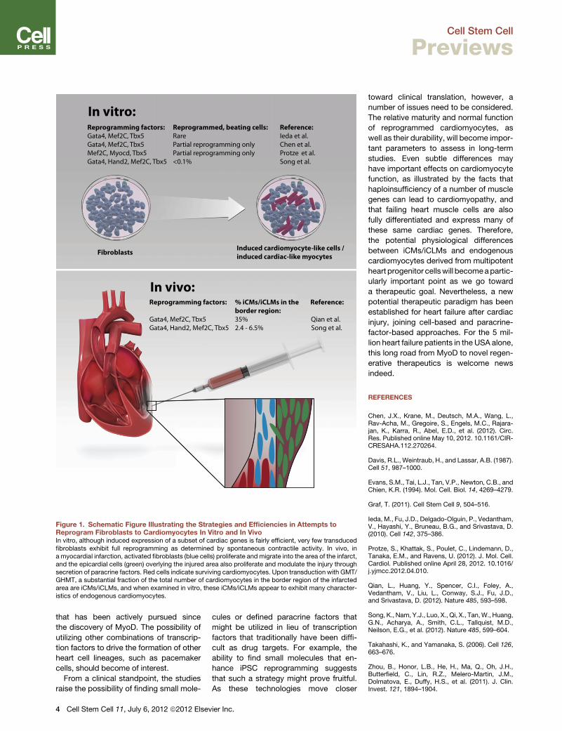
Figure 1. Schematic Figure Illustrating the Strategies and Efficiencies in Attempts toReprogram Fibroblasts to Cardiomyocytes In Vitro and In VivoIn vitro, although induced expression of a subset of cardiac genes is fairly efficient, very few transducedfibroblasts exhibit full reprogramming as determined by spontaneous contractile activity. In vivo, ina myocardial infarction, activated fibroblasts (blue cells) proliferate and migrate into the area of the infarct,and the epicardial cells (green) overlying the injured area also proliferate and modulate the injury throughsecretion of paracrine factors. Red cells indicate surviving cardiomyocytes. Upon transduction with GMT/GHMT, a substantial fraction of the total number of cardiomyocytes in the border region of the infarctedarea are iCMs/iCLMs, and when examined in vitro, these iCMs/iCLMs appear to exhibit many character-istics of endogenous cardiomyocytes.
Cell Stem Cell
Previews
that has been actively pursued since
the discovery of MyoD. The possibility of
utilizing other combinations of transcrip-
tion factors to drive the formation of other
heart cell lineages, such as pacemaker
cells, should become of interest.
From a clinical standpoint, the studies
raise the possibility of finding small mole-
4 Cell Stem Cell 11, July 6, 2012 ª2012 Elsev
cules or defined paracrine factors that
might be utilized in lieu of transcription
factors that traditionally have been diffi-
cult as drug targets. For example, the
ability to find small molecules that en-
hance iPSC reprogramming suggests
that such a strategy might prove fruitful.
As these technologies move closer
ier Inc.
toward clinical translation, however, a
number of issues need to be considered.
The relative maturity and normal function
of reprogrammed cardiomyocytes, as
well as their durability, will become impor-
tant parameters to assess in long-term
studies. Even subtle differences may
have important effects on cardiomyocyte
function, as illustrated by the facts that
haploinsufficiency of a number of muscle
genes can lead to cardiomyopathy, and
that failing heart muscle cells are also
fully differentiated and express many of
these same cardiac genes. Therefore,
the potential physiological differences
between iCMs/iCLMs and endogenous
cardiomyocytes derived from multipotent
heart progenitor cellswill becomeapartic-
ularly important point as we go toward
a therapeutic goal. Nevertheless, a new
potential therapeutic paradigm has been
established for heart failure after cardiac
injury, joining cell-based and paracrine-
factor-based approaches. For the 5 mil-
lion heart failure patients in the USA alone,
this long road from MyoD to novel regen-
erative therapeutics is welcome news
indeed.
REFERENCES
Chen, J.X., Krane, M., Deutsch, M.A., Wang, L.,Rav-Acha, M., Gregoire, S., Engels, M.C., Rajara-jan, K., Karra, R., Abel, E.D., et al. (2012). Circ.Res. Published online May 10, 2012. 10.1161/CIR-CRESAHA.112.270264.
Davis, R.L., Weintraub, H., and Lassar, A.B. (1987).Cell 51, 987–1000.
Evans, S.M., Tai, L.J., Tan, V.P., Newton, C.B., andChien, K.R. (1994). Mol. Cell. Biol. 14, 4269–4279.
Graf, T. (2011). Cell Stem Cell 9, 504–516.
Ieda, M., Fu, J.D., Delgado-Olguin, P., Vedantham,V., Hayashi, Y., Bruneau, B.G., and Srivastava, D.(2010). Cell 142, 375–386.
Protze, S., Khattak, S., Poulet, C., Lindemann, D.,Tanaka, E.M., and Ravens, U. (2012). J. Mol. Cell.Cardiol. Published online April 28, 2012. 10.1016/j.yjmcc.2012.04.010.
Qian, L., Huang, Y., Spencer, C.I., Foley, A.,Vedantham, V., Liu, L., Conway, S.J., Fu, J.D.,and Srivastava, D. (2012). Nature 485, 593–598.
Song, K., Nam, Y.J., Luo, X., Qi, X., Tan,W., Huang,G.N., Acharya, A., Smith, C.L., Tallquist, M.D.,Neilson, E.G., et al. (2012). Nature 485, 599–604.
Takahashi, K., and Yamanaka, S. (2006). Cell 126,663–676.
Zhou, B., Honor, L.B., He, H., Ma, Q., Oh, J.H.,Butterfield, C., Lin, R.Z., Melero-Martin, J.M.,Dolmatova, E., Duffy, H.S., et al. (2011). J. Clin.Invest. 121, 1894–1904.