reproduction introduction sexual determination, differentiation and development hormones and male...
TRANSCRIPT
Reproduction
• Introduction• sexual determination, differentiation and
development• Hormones and male reproduction• Hormones and female reproduction• Hormones of pregnancy, parturition and
lactation
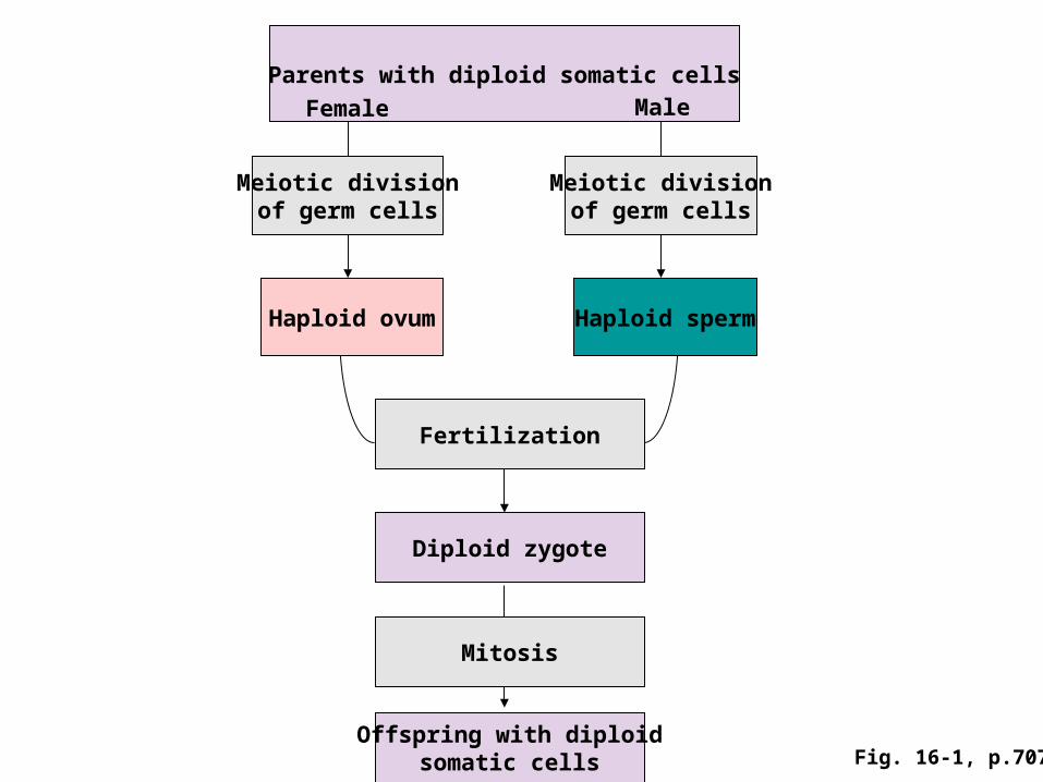
Parents with diploid somatic cells
Meiotic divisionof germ cells
Fertilization
Meiotic divisionof germ cells
Haploid ovum Haploid sperm
Diploid zygote
Mitosis
Offspring with diploidsomatic cells
MaleFemale
Fig. 16-1, p.707
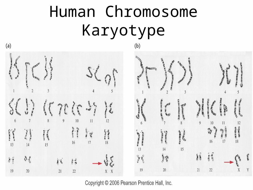
Human Chromosome Karyotype
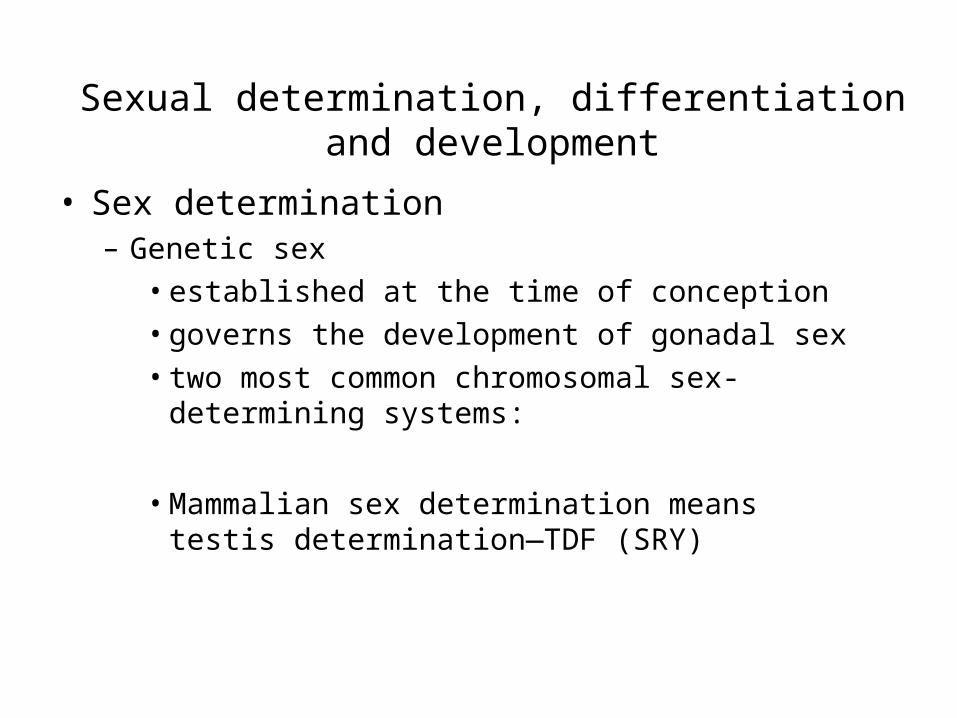
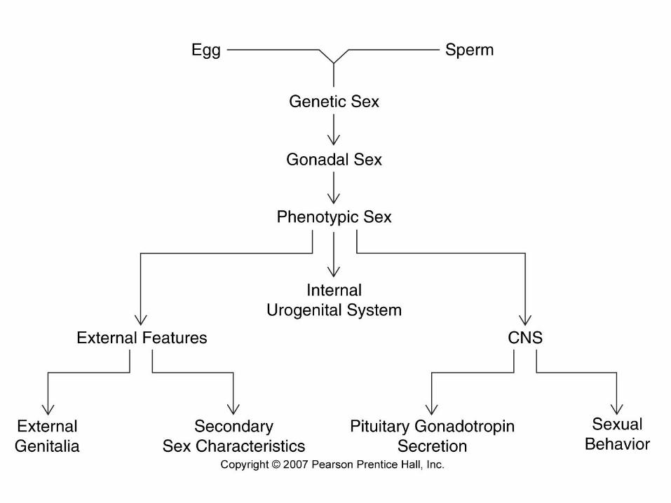
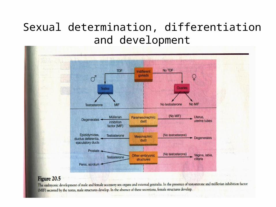
Sexual determination, differentiation and development
• Sex determination– Genetic sex
• established at the time of conception• governs the development of gonadal sex• two most common chromosomal sex-
determining systems:
• Mammalian sex determination means testis determination—TDF (SRY)
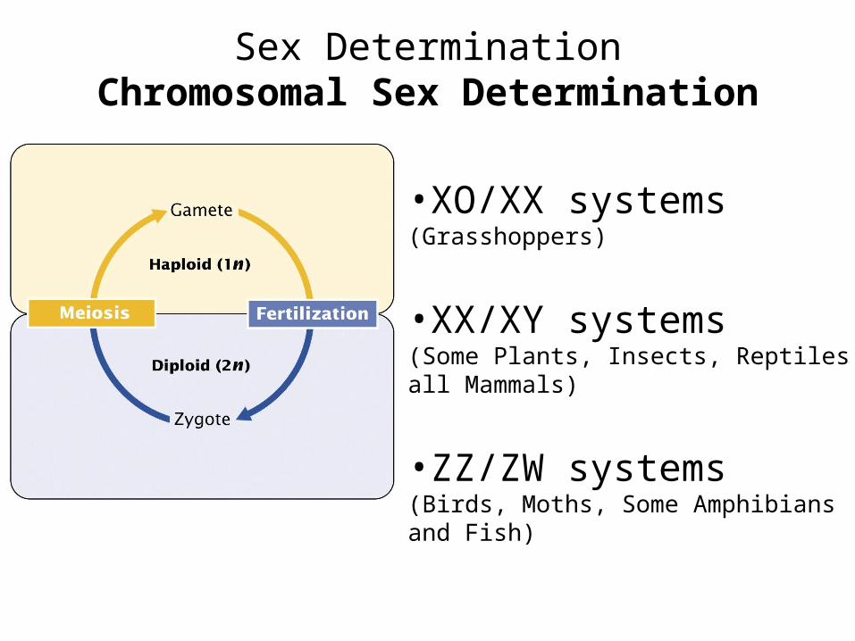
Sex DeterminationChromosomal Sex Determination
•XO/XX systems(Grasshoppers)
•XX/XY systems(Some Plants, Insects, Reptiles, all Mammals)
•ZZ/ZW systems(Birds, Moths, Some Amphibians and Fish)
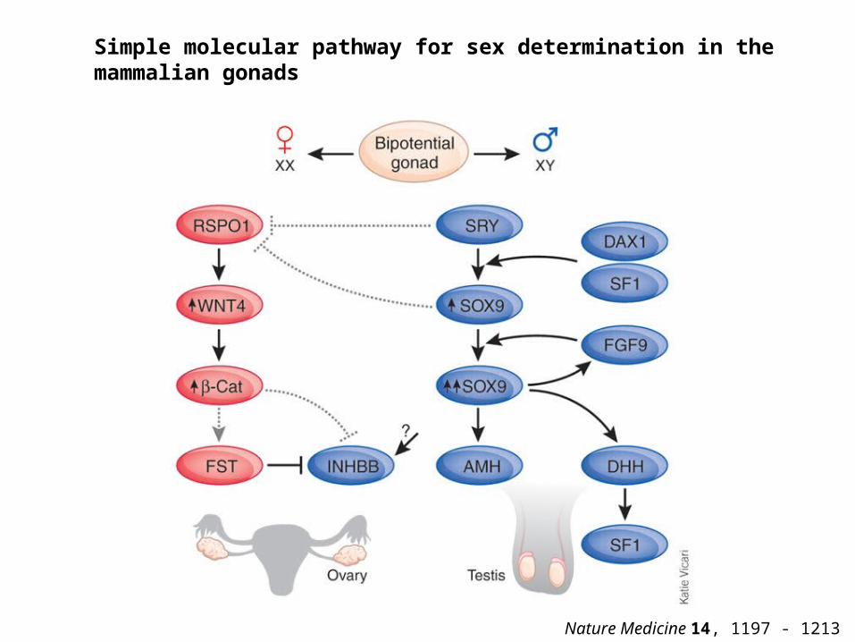
Simple molecular pathway for sex determination in the mammalian gonads
Nature Medicine 14, 1197 - 1213 (2008)
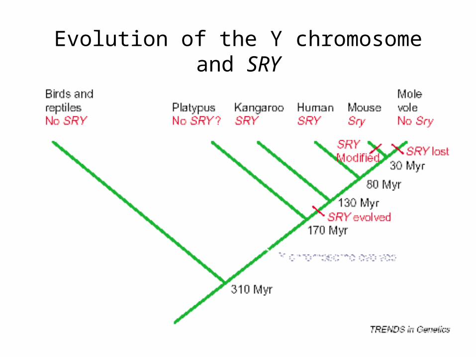
Evolution of the Y chromosome and SRY
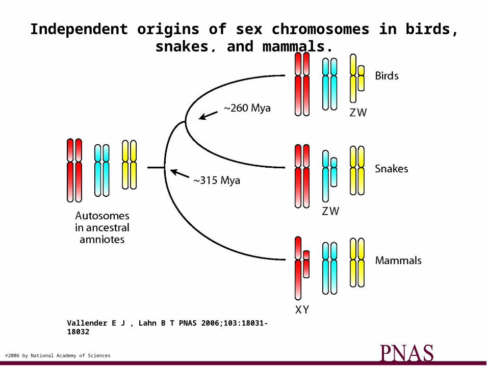
Independent origins of sex chromosomes in birds, snakes, and mammals.
Vallender E J , Lahn B T PNAS 2006;103:18031-18032
©2006 by National Academy of Sciences
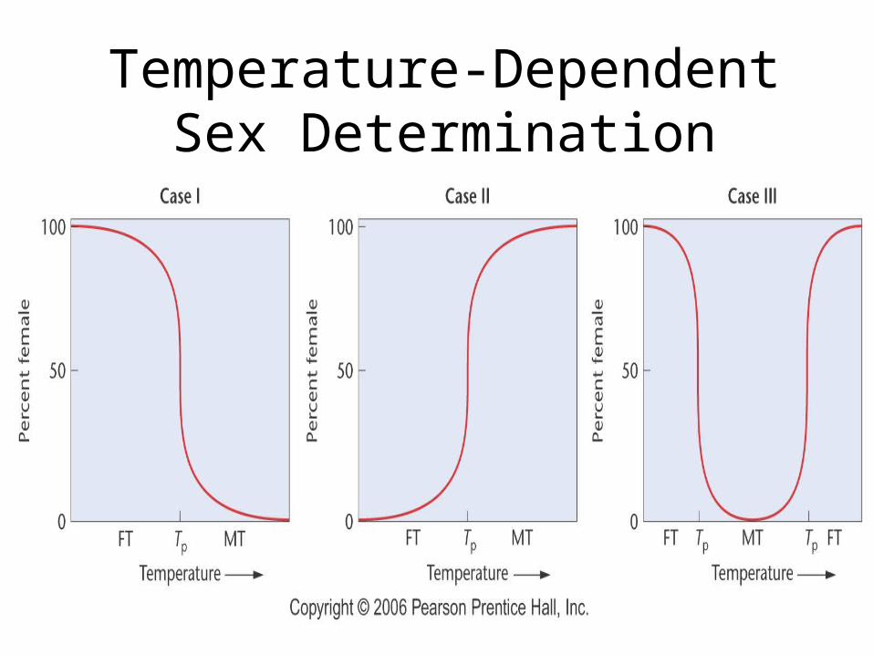
Temperature-Dependent Sex Determination
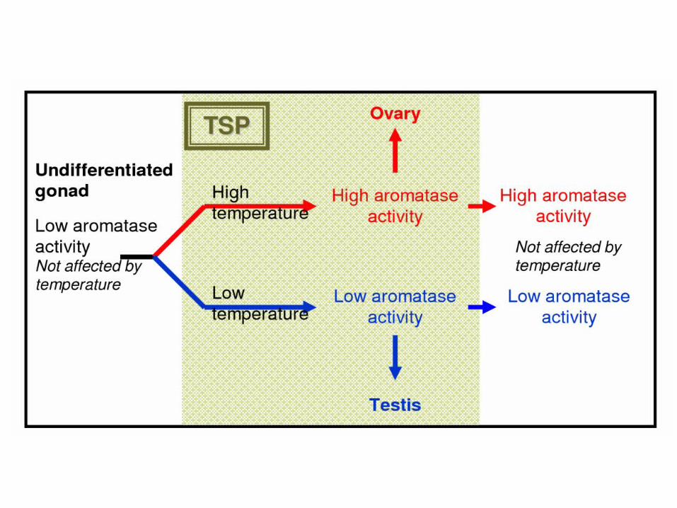
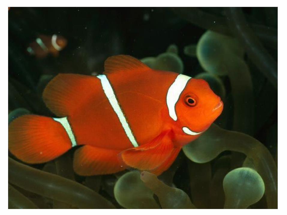
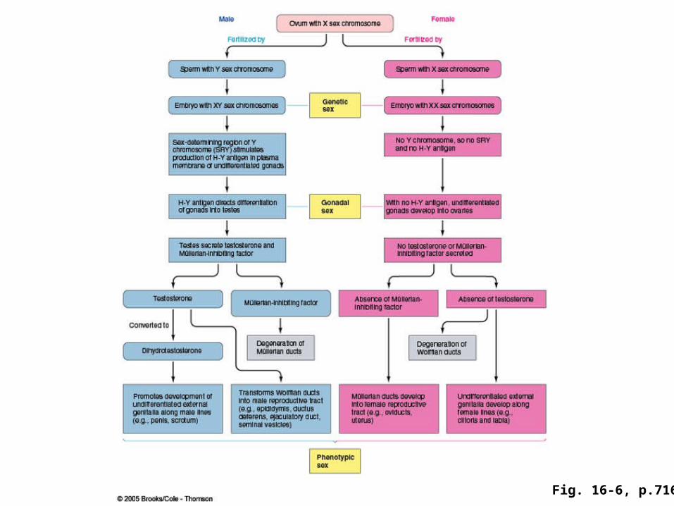
Fig. 16-6, p.716
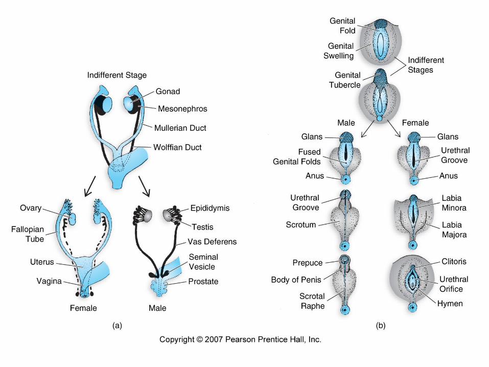
• Sexual differentiation– begins with the establishment of chromosomal sex at
fertilization, followed by the development of gonadal sex and culminating in the formation of sexual phenotypes
– Differentiation of Gonads
• differentiation of testis requires TDF
– Differentiation of accessory sex organs and external genitalia
• mullerian-inhibiting hormone
• testosterone
Sexual determination, differentiation and development
Sexual determination, differentiation and development
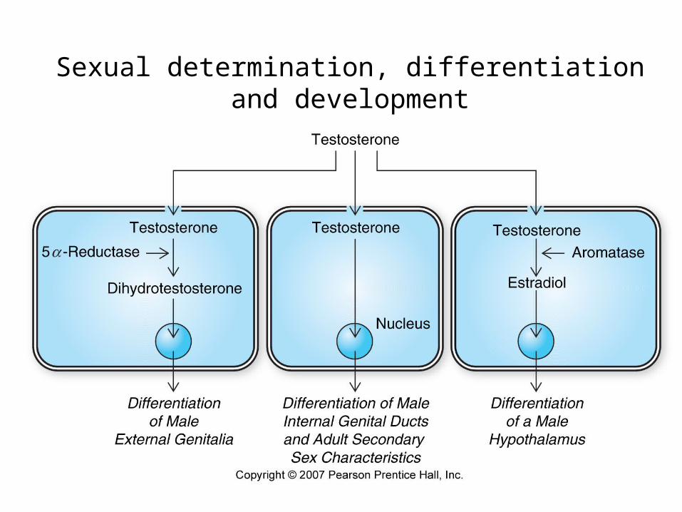
Sexual determination, differentiation and development
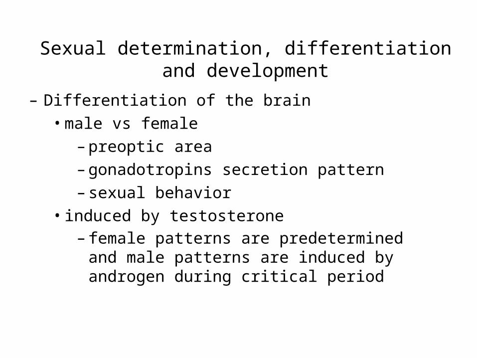
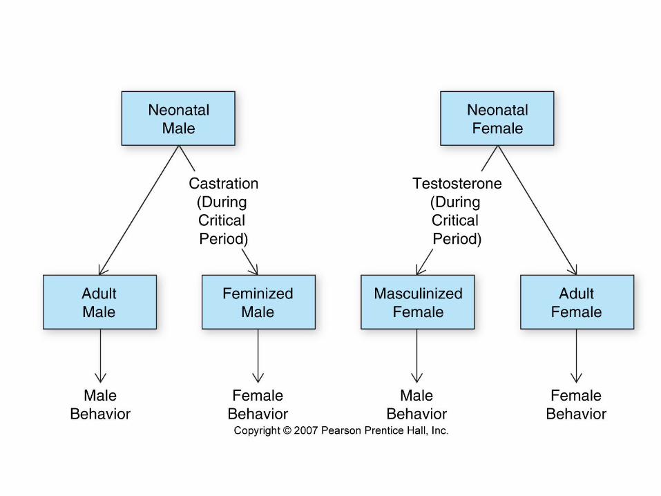
– Differentiation of the brain• male vs female
– preoptic area– gonadotropins secretion pattern– sexual behavior
• induced by testosterone – female patterns are predetermined and male
patterns are induced by androgen during critical period
Sexual determination, differentiation and development
Sexual determination, differentiation and development
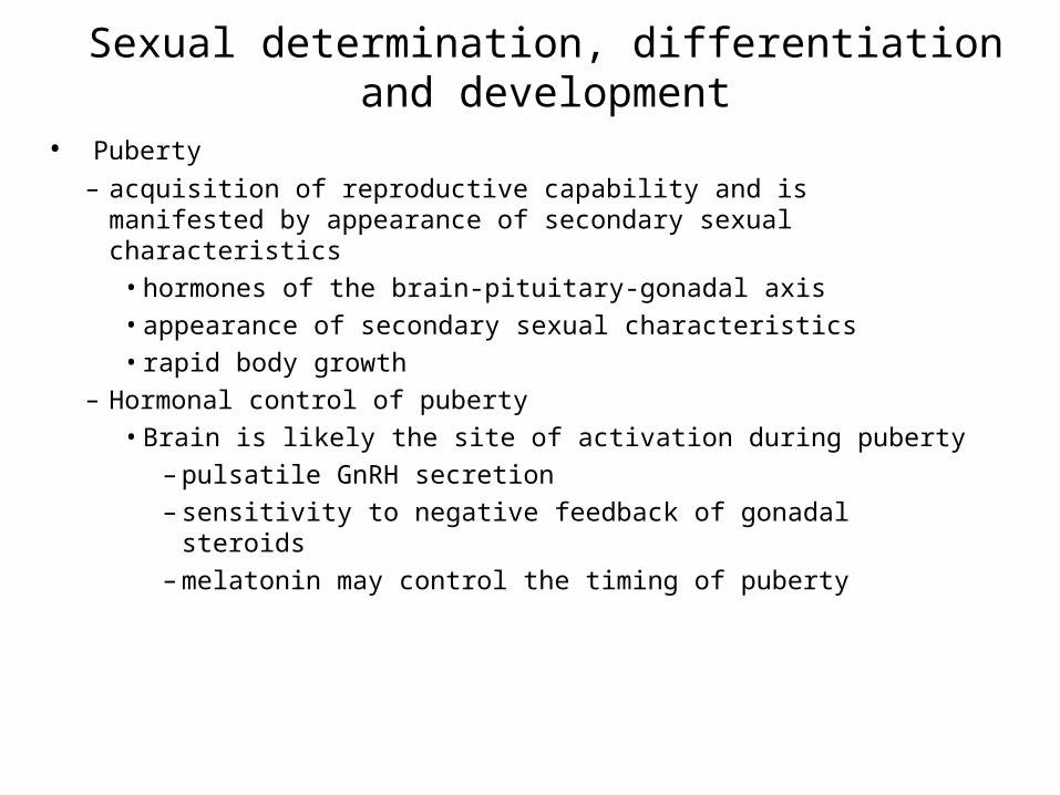
• Puberty
– acquisition of reproductive capability and is manifested by appearance of secondary sexual characteristics
• hormones of the brain-pituitary-gonadal axis• appearance of secondary sexual characteristics• rapid body growth
– Hormonal control of puberty• Brain is likely the site of activation during puberty
– pulsatile GnRH secretion– sensitivity to negative feedback of gonadal steroids – melatonin may control the timing of puberty
Sexual determination, differentiation and development
Sexual determination, differentiation and development
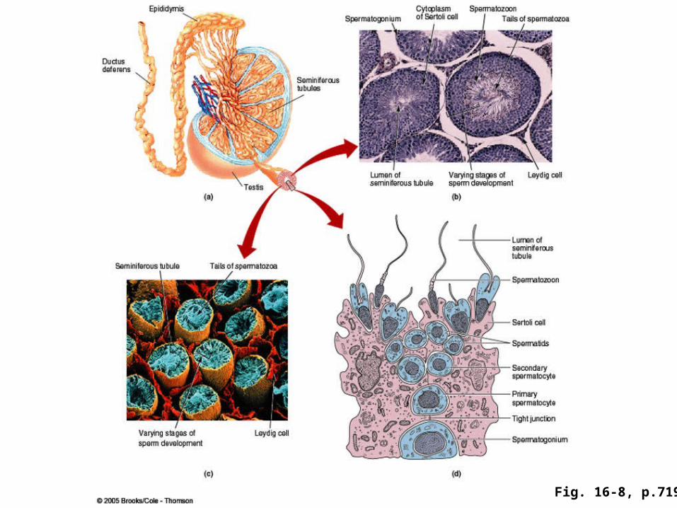
Fig. 16-8, p.719
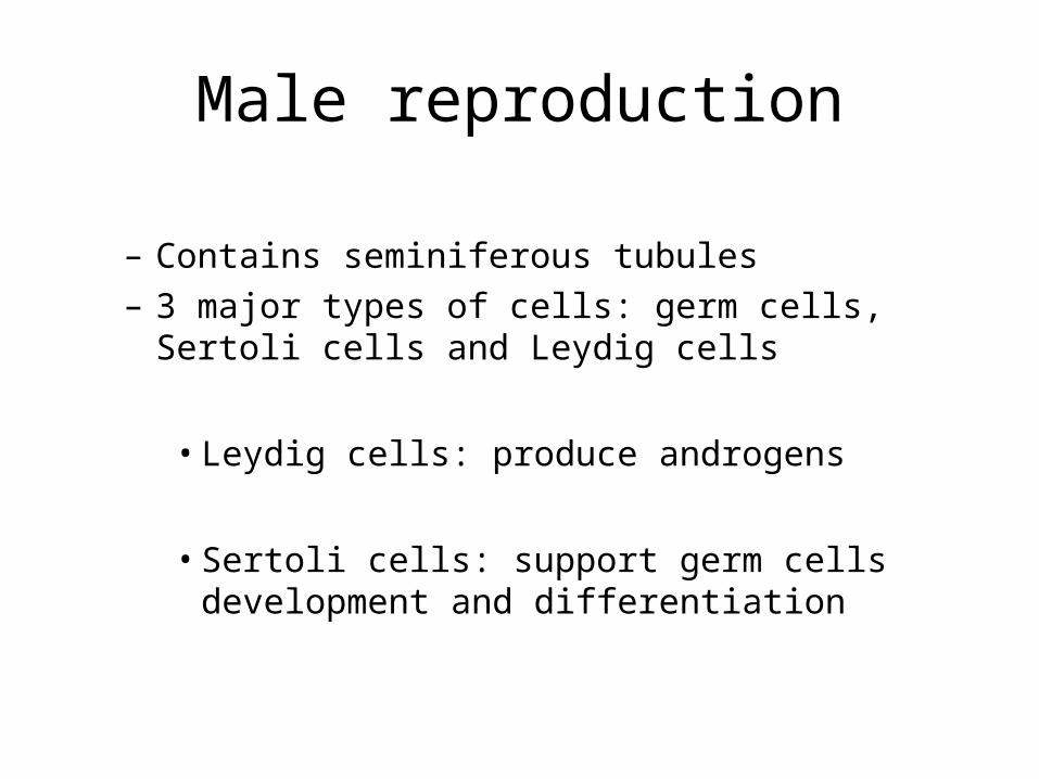
Male reproduction
– Contains seminiferous tubules – 3 major types of cells: germ cells, Sertoli cells and
Leydig cells
• Leydig cells: produce androgens
• Sertoli cells: support germ cells development and differentiation
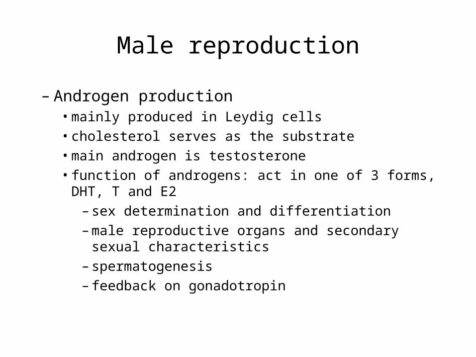
Male reproduction
– Androgen production• mainly produced in Leydig cells• cholesterol serves as the substrate• main androgen is testosterone• function of androgens: act in one of 3 forms, DHT, T and E2
– sex determination and differentiation– male reproductive organs and secondary sexual
characteristics– spermatogenesis– feedback on gonadotropin
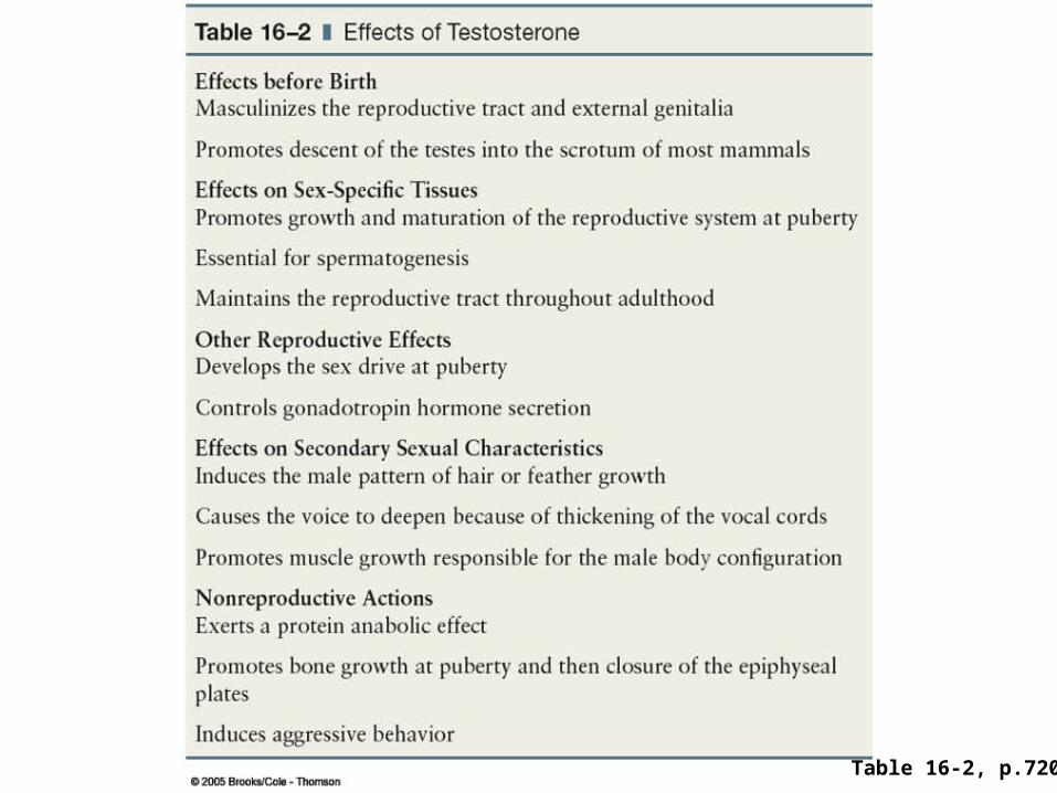
Table 16-2, p.720
Male Reproduction
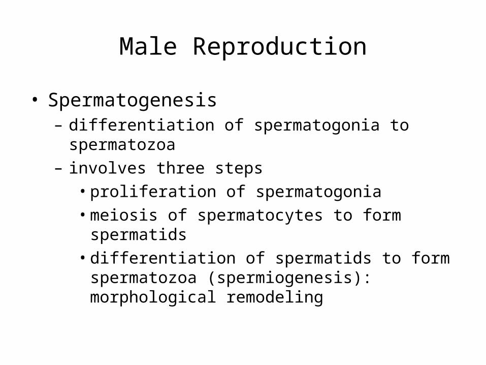
• Spermatogenesis– differentiation of spermatogonia to spermatozoa– involves three steps
• proliferation of spermatogonia• meiosis of spermatocytes to form spermatids• differentiation of spermatids to form spermatozoa
(spermiogenesis): morphological remodeling
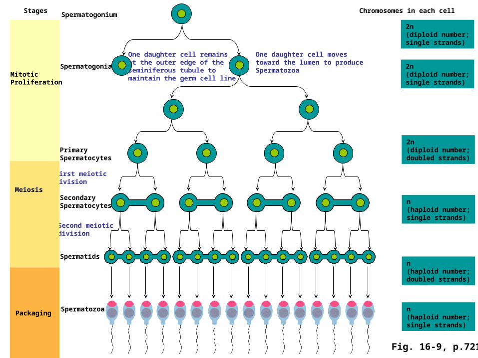
Chromosomes in each cell
2n (diploid number;single strands)
n(haploid number;doubled strands)
n(haploid number;single strands)
n(haploid number;single strands)
2n (diploid number;single strands)
One daughter cell movestoward the lumen to produceSpermatozoa
One daughter cell remainsat the outer edge of theseminiferous tubule tomaintain the germ cell line
Spermatogonia
Spermatogonium
2n (diploid number;doubled strands)
PrimarySpermatocytes
SecondarySpermatocytes
First meioticdivision
Second meioticdivision
Spermatids
Spermatozoa
MitoticProliferation
Meiosis
Packaging
Stages
Fig. 16-9, p.721
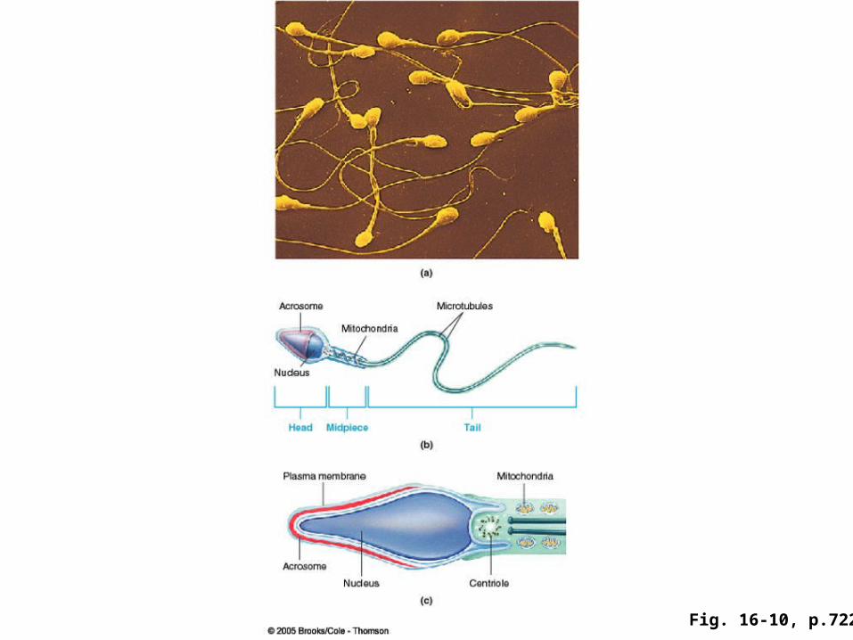
Fig. 16-10, p.722
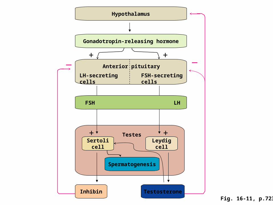
Sertolicell
Leydigcell
Spermatogenesis
Testes+ +
Hypothalamus
Gonadotropin-releasing hormone
Inhibin Testosterone
— —+ +
FSH LH
Anterior pituitary
LH-secretingcells
FSH-secretingcells
Fig. 16-11, p.723
Male reproduction• Regulation of steroidogenesis
– FSH is a major regulator, especially in the initiation of spermatogenesis
» increases the size of testis » stimulates the replication of spermatogonia» increases LH-R #, contribute to T production
– Testosterone is essential for maintenance of spermatogenesis
– Activin: replication of spermatogonia– Inhibin: inhibits differentiation of spermatogonia
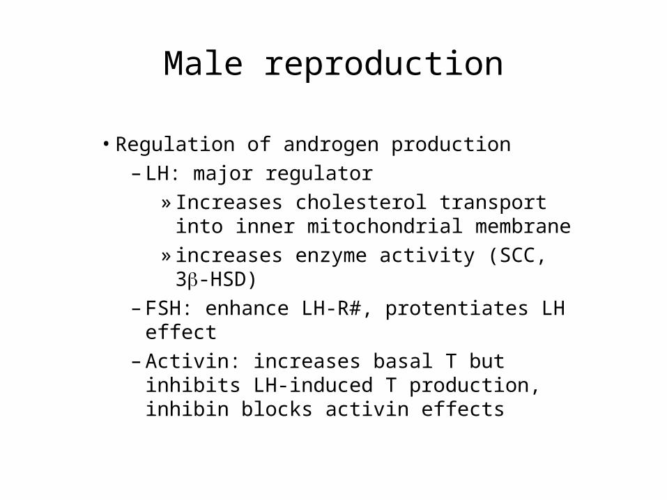
Male reproduction
• Regulation of androgen production– LH: major regulator
» Increases cholesterol transport into inner mitochondrial membrane
» increases enzyme activity (SCC, 3-HSD)
– FSH: enhance LH-R#, protentiates LH effect– Activin: increases basal T but inhibits LH-
induced T production, inhibin blocks activin effects
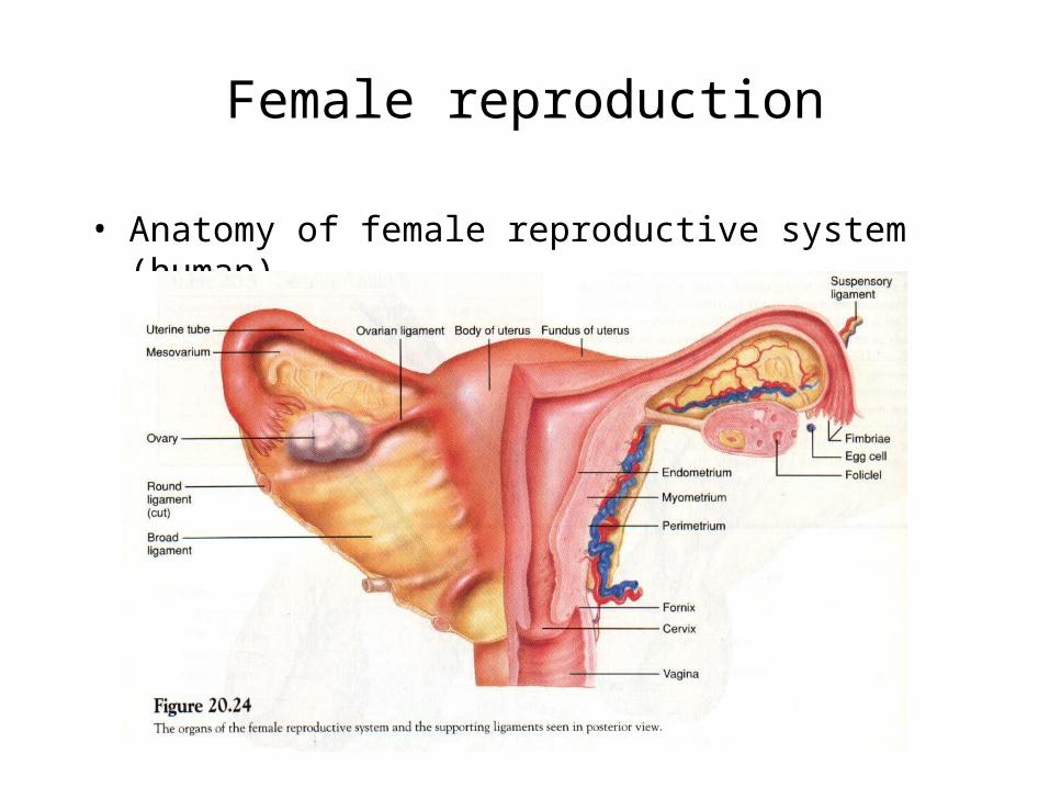
Female reproduction
• Anatomy of female reproductive system (human)
ndiploid number;
doubled strands)
2n (diploid number;single strands)
2n (diploid number;doubled strands)
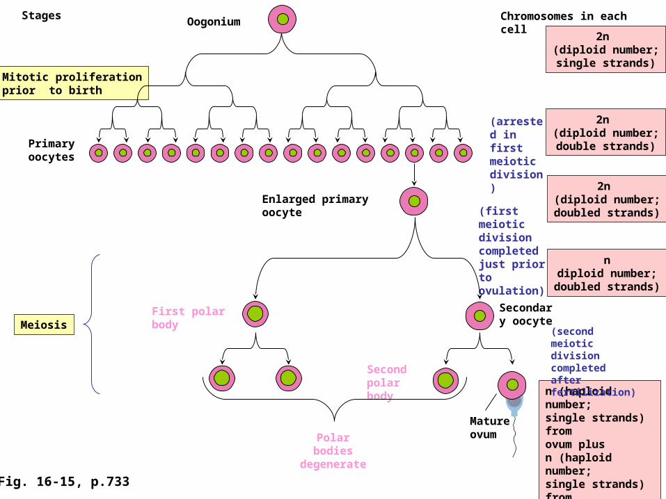
Mitotic proliferationprior to birth
Meiosis
n (haploid number;single strands) from ovum plusn (haploid number;single strands) from sperm for diploidfertilized ovum with2n chromosomes
(second meiotic division completed after fertilization)
(first meiotic division completed just prior to ovulation)
2n (diploid number;double strands)Primary
oocytes
(arrested in first meiotic division)
Enlarged primary oocyte
Chromosomes in each cellStagesOogonium
Polar bodies degenerate
Secondary oocyte
First polar body
Second polar body
Mature ovum
Fig. 16-15, p.733
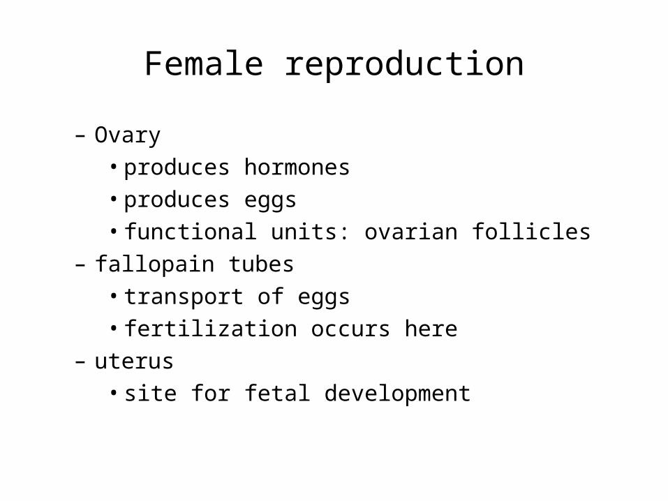
Female reproduction
– Ovary• produces hormones• produces eggs• functional units: ovarian follicles
– fallopain tubes• transport of eggs• fertilization occurs here
– uterus• site for fetal development
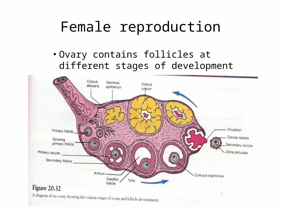
Female reproduction
• Ovary contains follicles at different stages of development
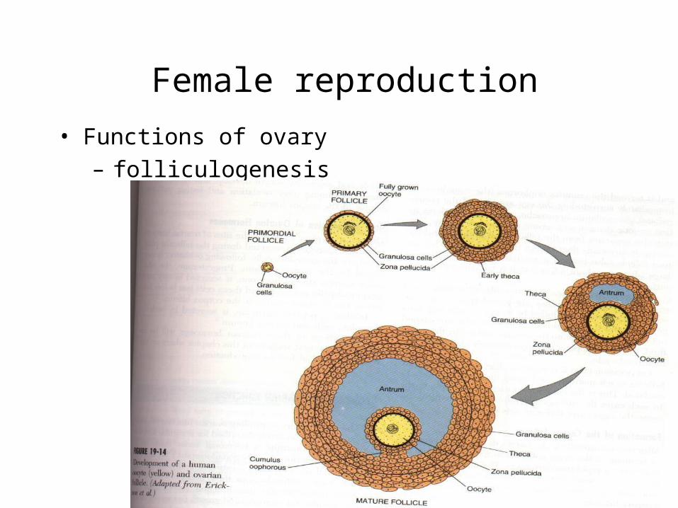
Female reproduction
• Functions of ovary– folliculogenesis
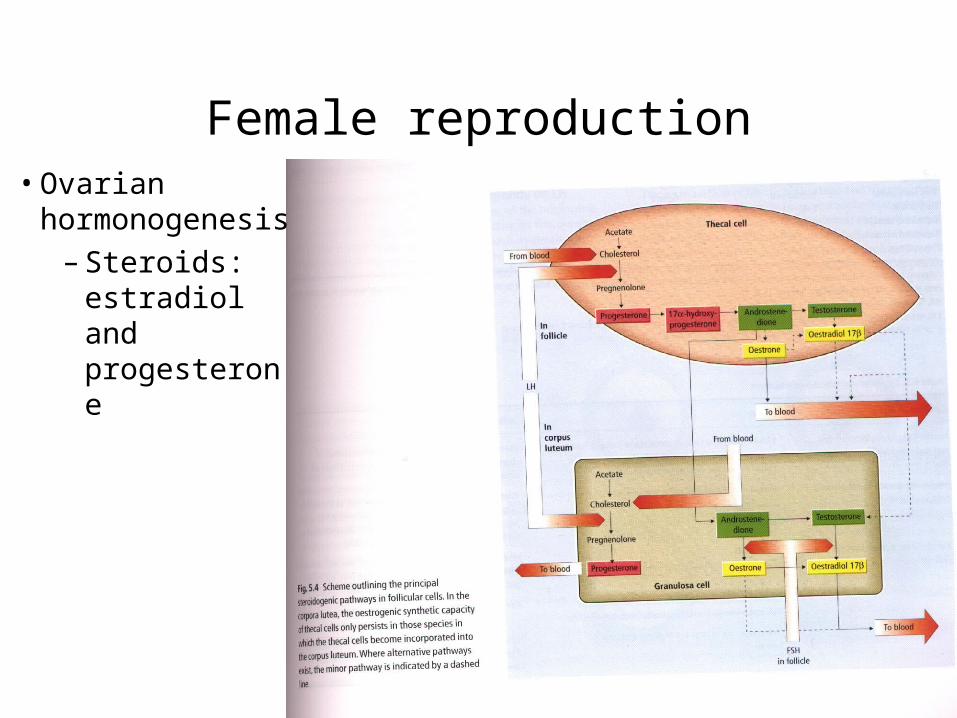
Female reproduction• Ovarian
hormonogenesis– Steroids:
estradiol and progesterone
Female reproduction
• Estradiol:
– synthesized mainly by granulosa cells
– stimulated by FSH and LH
– act on CNS to maintain libido and sexual behavior
– feedback regulation of GnRH, LH and FSH (+ve or -ve)
– function of female reproductive organs
– oocyte maturation
– parturition and lactation
– metabolic functions
» anabolic: weight gain
» bone mineral deposition
Female reproduction
• Progesterone
– synthesized mainly by corpus luteum
– stimulated by LH (primed by FSH)
– act on CNS to increase sexual receptivity
– feedback regulation of GnRH, LH and FSH (-ve)
– effects on reproductive tract
– pregnancy
– metabolic functions
» increases basal metabolic rate and thus thermogenic action
Female reproduction
• Others– ovary also produces many nonsteroidal hormones– inhibin and activin
» regulate FSH secretion and ovarian function– prostaglandins
» PGF2 induces CL regression» PGF2 and PGE2 required for ovulation
– insulin-like growth factor» stimulates granulosa cell proliferation; inhibits
apoptosis; induces steroidogenesis; induces maturation
Female reproduction
– Reproductive cycle• cyclic change of reproductive activity• seasonal reproductive cycle
– related to environmental changes, e.g. photoperiod, temperature, food availability, etc.
• estrous cycle (menstrual cycle in primates)– visible sign of ovulation– a behavior strategy to ensure that the
female is mated at the time of ovulation
Female reproduction
– human menstrual cycle• cycle of ovarian activity that repeat at
approximately one-month interval (menstru=monthly)
• menstruation is used to indicate the periodic shedding of endometrium, which become thickened prior to menstruation under stimulation by ovarian steroids
• shedding of endometrium is accompanied by bleeding
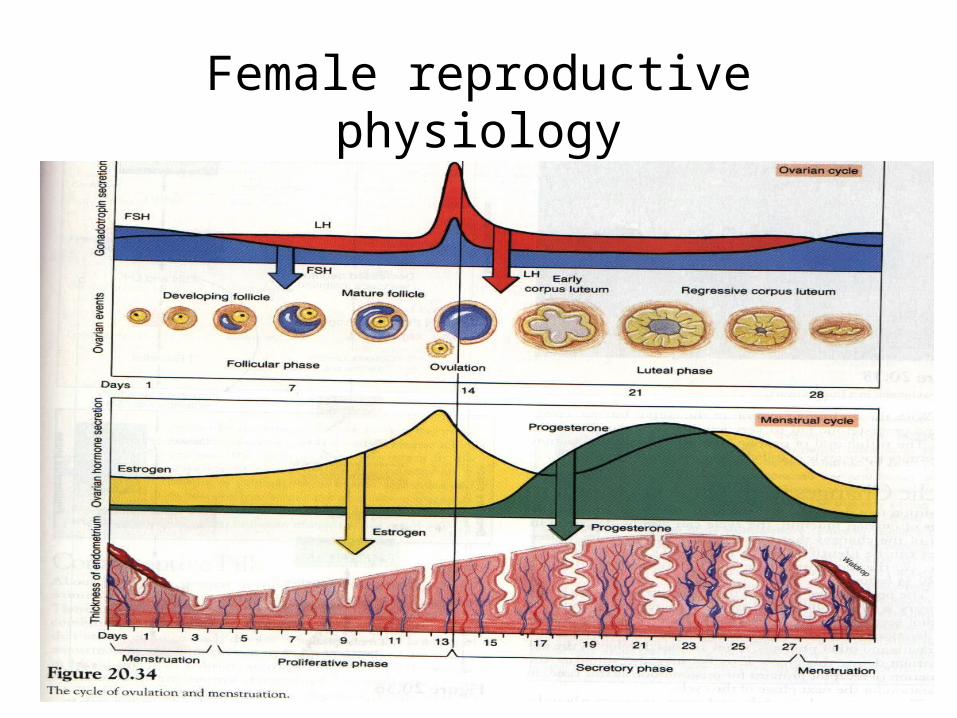
Female reproductive physiology
Female reproductive physiology
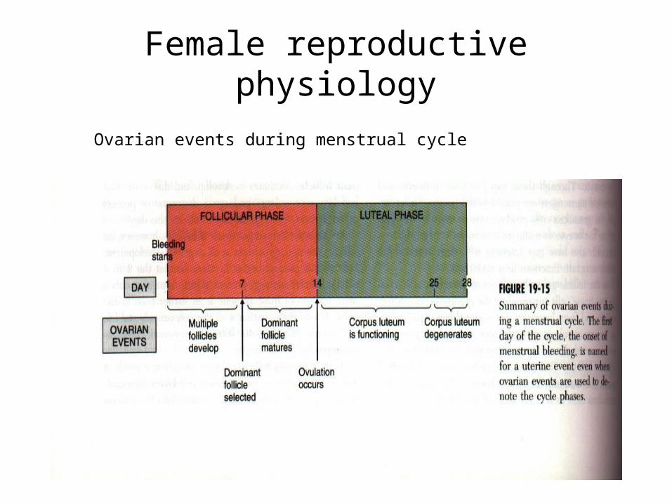
Ovarian events during menstrual cycle
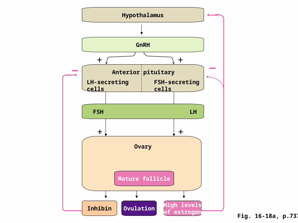
Female reproduction• Regulation of ovarian functions
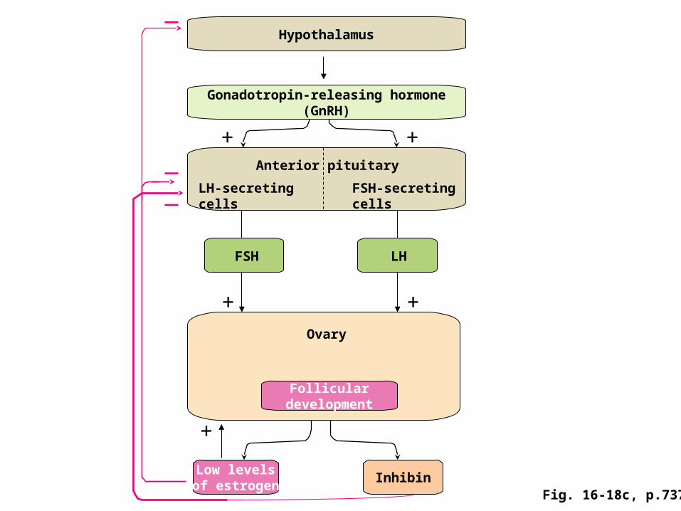
– Follicular phase:
» FSH level is elevated at the beginning of the cycles
» FSH stimulates follicular development and production of E2 and inhibin
» E2 and inhibin feedback to inhibit FSH and thus FSH level decreases
» the follicle that has the highest sensitivity to FSH will be selected and develops into a mature follicle
» growth of mature follicle is accompanied by rapid increase in E2
» E2 triggers LH surge (positive feedback)
Female reproduction
– ovulation: rupture of follicular wall and release of oocyte» triggered by LH surge» other hormones: prostaglandin: histamine
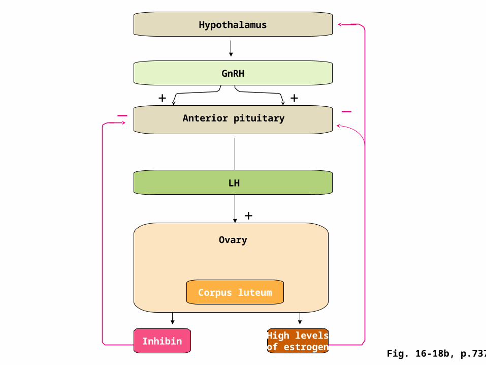
– Luteal phase» CL formed» progesterone produced by CL» together with E2 feedback to suppress FSH and LH : prevent new
follicular development» if pregnancy occurs, hCG stimulates progesterone production and
CL function maintained» if no implantation, CL regresses and progesterone level declines
(about day 22)
Female reproductive physiology Regulation of uterine events during menstrual cycle
• menstrual phase
– starts at the first day of bleeding (last 3-5 days)
– endometrium degenerates
– resulted from decrease in progesterone
• proliferative phase
– between the cessation of menstruation and ovulation (about 10 days)
– endometrium regenerates and thickens
– estradiol induces endometrium and myometrium growth, as well as progesterone receptors
Female reproduction
• secretory phase– between ovulation and the onset of next
menstruation– occurs when the ovary is at luteal phase– under the action of progesterone and estradiol,
endometrium is prepared to accept and nourish an embryo
» thick, vascular and “spongy” in appearance» accumulation of glycogen and various enzymes
– Progesterone also inhibits myometrium activity
Female reproduction
Menopause cessation of ovarian activity during postmenopause years, ovaries are
depleted of follicles and stop secreting estradiol due to failure in the ovary, not pituitary a weak estrogen (estrone) is produced by
adipose tissue from an androgen produced by the adrenal gland
withdrawal of estradiol is responsible for most symptoms of menopause
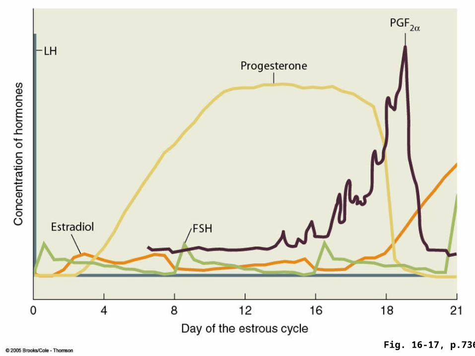
Fig. 16-17, p.736
+ +
Mature follicle
Ovary
Hypothalamus
GnRH
InhibinHigh levelsof estrogen
— —+ +
FSH LH
Anterior pituitary
LH-secretingcells
FSH-secretingcells
OvulationFig. 16-18a, p.737
+
Corpus luteum
Ovary
Hypothalamus
GnRH
InhibinHigh levelsof estrogen
— —+ +
LH
Anterior pituitary
Fig. 16-18b, p.737
+
+ +
Folliculardevelopment
Ovary
Hypothalamus
Gonadotropin-releasing hormone(GnRH)
InhibinLow levelsof estrogen
+ +
FSH
Anterior pituitary
LH-secretingcells
FSH-secretingcells
LH
—
—
—
Fig. 16-18c, p.737
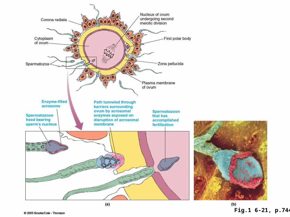
Fig.1 6-21, p.744
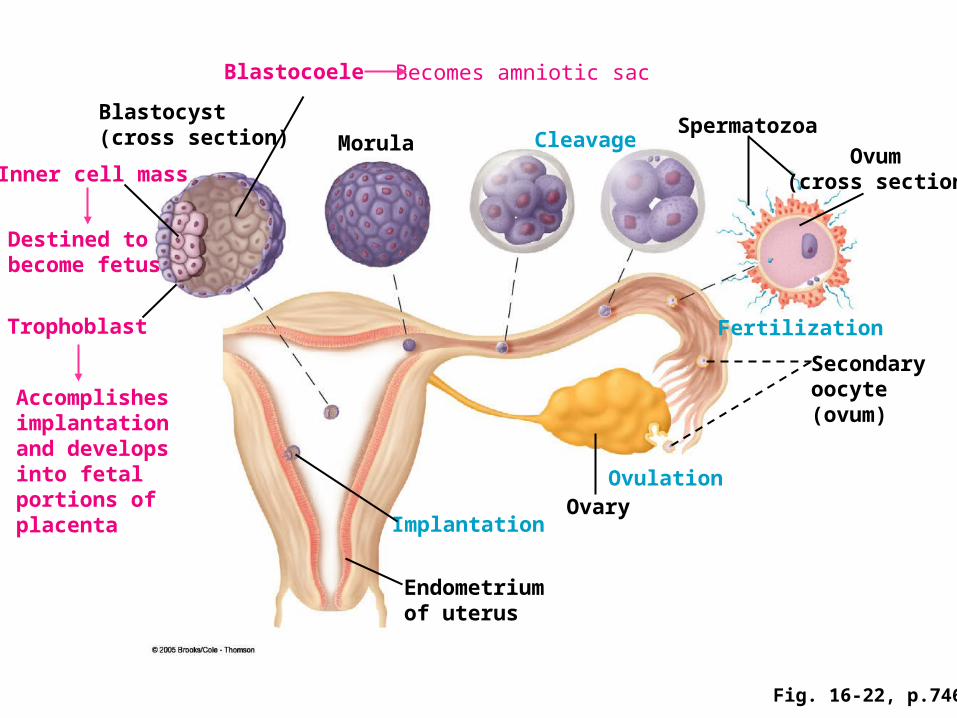
Fig. 16-22, p.746
Trophoblast
Accomplishesimplantation and developsinto fetal portions ofplacenta
Inner cell mass
Destined to become fetus
Spermatozoa
Ovum (cross section)
Blastocoele Becomes amniotic sac
Blastocyst (cross section) CleavageMorula
Fertilization
Secondaryoocyte(ovum)
OvulationOvary
Endometrium of uterus
Implantation
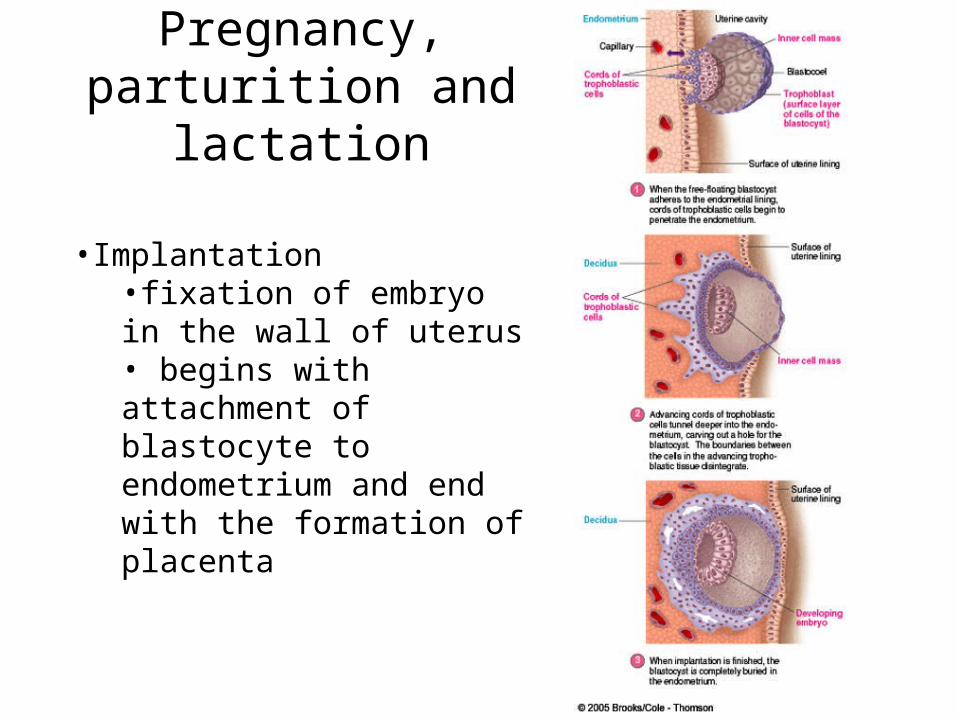
Pregnancy, parturition and lactation
•Implantation•fixation of embryo in the wall of uterus • begins with attachment of blastocyte to endometrium and end with the formation of placenta
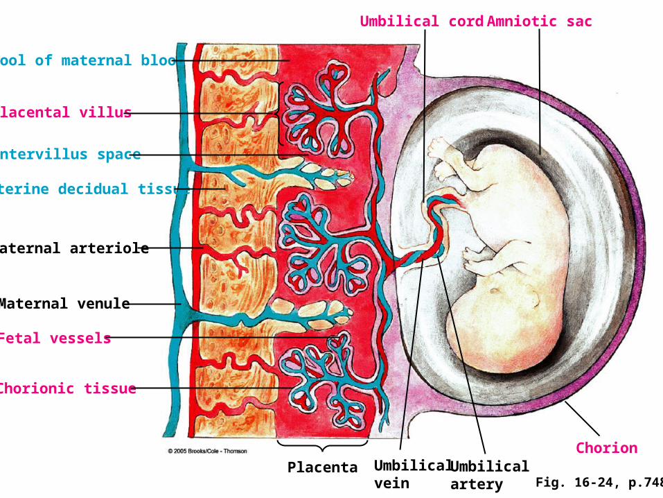
Fig. 16-24, p.748
Pool of maternal blood
Placental villus
Intervillus space
Uterine decidual tissue
Maternal arteriole
Maternal venule
Fetal vessels
Chorionic tissue
Umbilical cord Amniotic sac
PlacentaChorion
Umbilicalvein
Umbilical artery
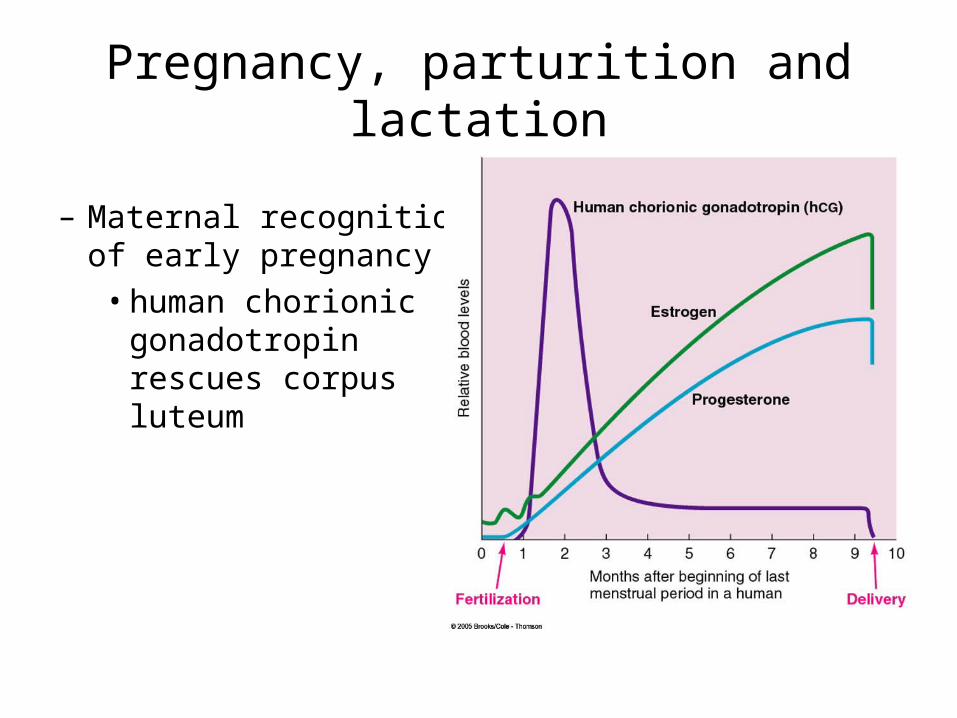
Pregnancy, parturition and lactation
– Maternal recognition of early pregnancy
• human chorionic gonadotropin rescues corpus luteum
Pregnancy, parturition and lactation
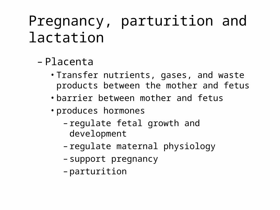
– Placenta• Transfer nutrients, gases, and waste products
between the mother and fetus• barrier between mother and fetus• produces hormones
– regulate fetal growth and development– regulate maternal physiology– support pregnancy– parturition
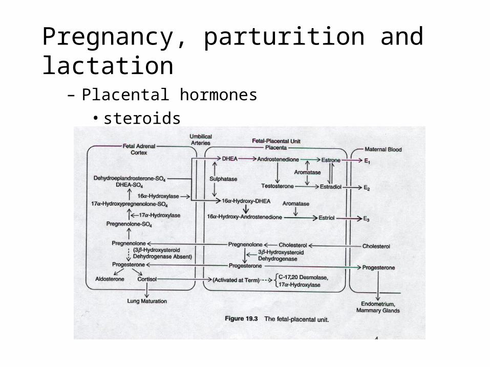
Pregnancy, parturition and lactation– Placental hormones
• steroids
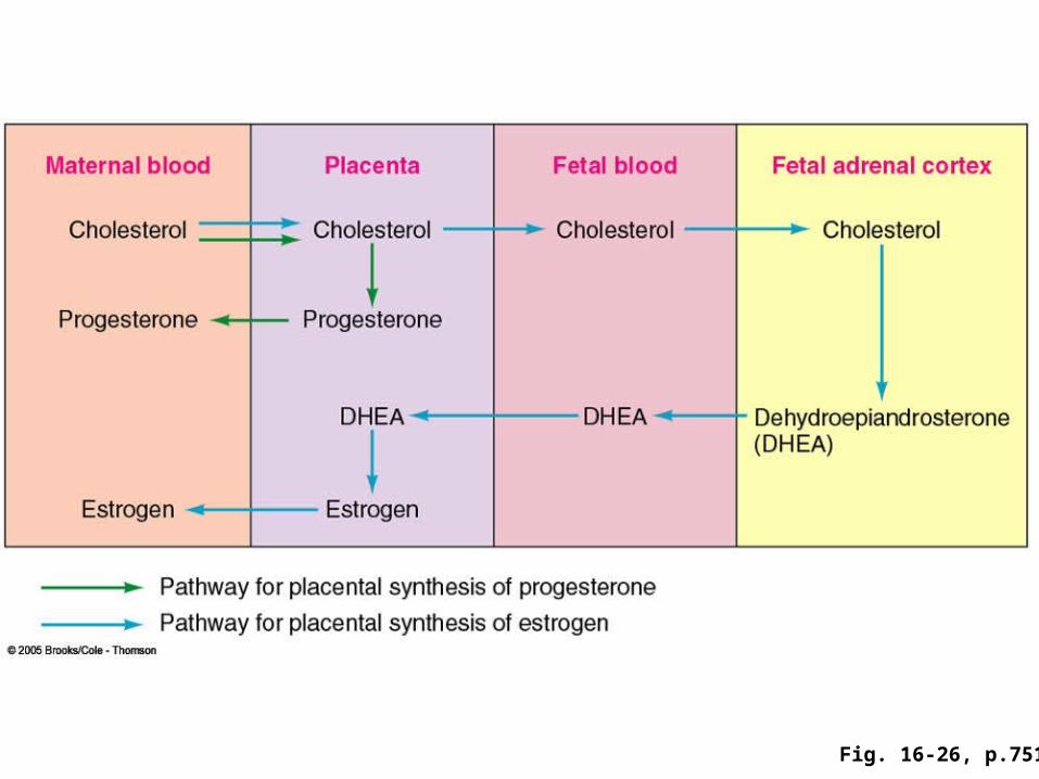
Fig. 16-26, p.751
Pregnancy, parturition and lactation
• Progesterone– produced by placenta from cholesterol– maintenance of uterine structure and function– mammary growth and development– feedback on gonadotropin– substrate for cortisol production in fetal adrenal gland
• Estrogens– produced by the placenta from precursors derived
from adrenal gland– important for parturition and lactation
Pregnancy, parturition and lactation
– Peptide hormones• hCG
– acts at same receptor as LH– stimulates progesterone production – regulate development of fetal adrenal and gonad
• hPL– maternal intermediary metabolism– fetal growth– mammary gland differentiation– steroidogenesis
Pregnancy, parturition and lactation
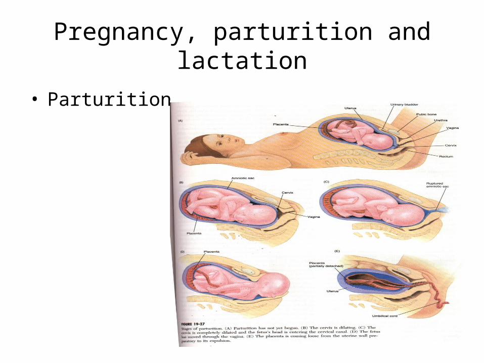
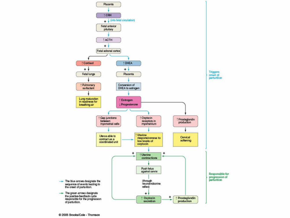
• Parturition
Pregnancy, parturition and lactation
• Parturition– delivery of baby at term– requires two physiological changes
– cervical softening to reduce the resistance to expulsion of baby
– coordinated myometrial contraction to increase intrauterine pressure
– induced by hormones
Pregnancy, parturition and lactation
• Lactation– secretion of milk by mammary glands– mammals are characterized by lactation– lactation provides a primary source of nutrition for
new-born– this process includes
• milk production• milk let-down
Pregnancy, parturition and lactation
• Regulation of mammary gland development
– stimulated by estrogen, progesterone, PRL, GH and cortisol
• Regulation of milk production
– PRL: essential for milk production
– Cortisol: synergizes with PRL to initiate lactation
– Estradiol: increases PRL and cortisol
– progesterone: inhibitory
– prostaglandins: increase PRL and cortisol
– insulin: lipogenesis
Pregnancy, parturition and lactation
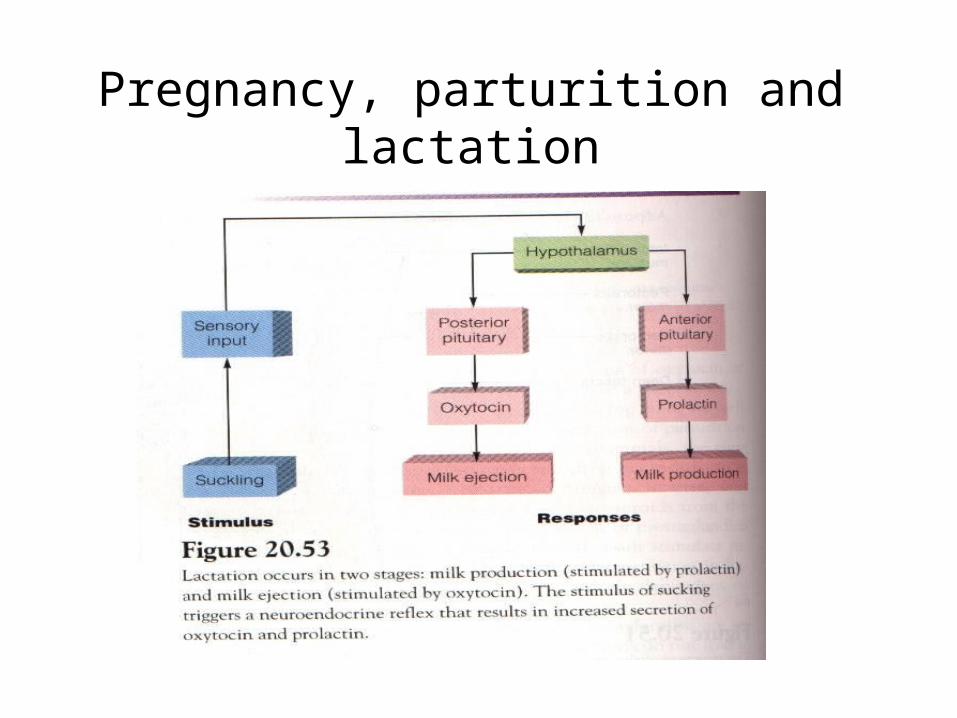
• Milk ejection– accomplished by contraction of the
myoepithelial cells surrounding the alveoli– contraction is under the control of oxytocin – oxytocin is released in response to suckling– suckling also induces prolactin release
which stimulates more milk production
Pregnancy, parturition and lactation