repeatability of corneal power and wavefront aberration measurements with a dual-scheimpflug placido...
TRANSCRIPT
ARTICLE
Repeatability of corne
al power and wavefrontaberration measurements with a dual-Scheimpflug Placido corneal topographerLi Wang, MD, PhD, Mariko Shirayama, MD, Douglas D. Koch, MD
Q 2010 A
Published
SCRS an
by Elsev
PURPOSE: To evaluate the repeatability of the Galilei dual-Scheimpflug analyzer in measuring cor-neal curvature, wavefront aberrations, pachymetry, and anterior chamber depth (ACD).
SETTING: Cullen Eye Institute, Baylor College of Medicine, Houston, Texas, USA.
METHODS: Three consecutive measurements were performed in 1 eye of each subject. The followingwere evaluated: (1) mean total corneal power at the central, paracentral, and peripheral zones (0.0 to4.0 mm, 4.0 to 7.0 mm, and 7.0 to 8.0 mm, respectively) and posterior corneal power (Kavg); (2) cor-neal higher-order wavefront aberrations (6.0 mm pupil); (3) mean pachymetry at the central, paracen-tral, and peripheral zones; and (4) ACD. Repeatability was assessed by calculating the within-subjectstandard deviation (SD), coefficient of variation (COV), and intraclass correlation coefficient (ICC).
RESULTS: The study enrolled 20 subjects. The SD was 0.09 diopter (D), 0.05 D, and 0.19 D for cen-tral, paracentral, and peripheral total corneal power, respectively, and 0.03 D for posterior Kavg. TheCOV ranged from 0.10% to 0.35%, and the ICC was 0.996 or more (P<.001). For 3rd-order comaand trefoil, the SD was 0.08 mm and 0.09 mm, respectively. For 4th-order spherical aberration, astig-matism, and tetrafoil, the SDs were lower (0.02 mm, 0.04 mm, and 0.09 mm, respectively). The SDwas 1.68 mm, 1.98 mm, and 2.82 mm for central, paracentral, and peripheral pachymetry, respec-tively, and 0.04 mm for ACD.
CONCLUSION: Dual-Scheimpflug measurements of corneal power, pachymetry, ACD, and cornealaberrations for Zernike terms in the middle of the Zernike tree showed excellent repeatability.
Financial Disclosure: No author has a financial or proprietary interest in any material or methodmentioned. Additional disclosures are found in the footnotes.
J Cataract Refract Surg 2010; 36:425–430 Q 2010 ASCRS and ESCRS
In recent years, Scheimpflug photographic deviceshave become commercially available for anterior seg-ment measurements. The Pentacam system (Oculus,Inc.) uses a single Scheimpflug camera to acquire mul-tiple photographs of the anterior segment of the eye.1–6
TheGalilei dual-Scheimpflug analyzer (ZiemerGroup)uses dual-channel Scheimpflug cameras and combinesa Placido disk with the goal of improving accuracy ofcorneal power and pachymetric measurements.7
Knowledge of repeatability of new devices is essen-tial. In a previous study,8 we assessed the repeatabilityof the simulated keratometry values measured withthe dual-Scheimpflug analyzer and compared themwith values measured using the Humphrey Atlas cor-neal topographer (Carl Zeiss), IOLMaster partial co-herence interferometry (PCI) biometer (Carl Zeiss),and a manual keratometer (Bausch & Lomb, Inc.).8
Menassa et al.9 evaluated the reproducibility and
d ESCRS
ier Inc.
comparability of central corneal thickness (CCT) andkeratometry readings using the dual-Scheimpflug de-vice, the Orbscan II scanning-slit topographer (Bausch& Lomb, Inc.), and a 50 MHz pachymeter (Sonogage).
The purpose of this studywas to evaluate the repeat-ability of the dual-Scheimpflug analyzer in measuringthe total corneal power at the central, paracentral, andperipheral zones; posterior corneal power; cornealwavefront aberrations; corneal pachymetry at the cen-tral, paracentral, and peripheral zones; and anteriorchamber depth (ACD).
SUBJECTS AND METHODS
Subjects
This study prospectively enrolled subjects who met thefollowing inclusion criteria: no previous ocular surgery ortrauma, no corneal or other ocular diseases, and not a contactlens wearer. One eye of each subject was selected; right eyes
0886-3350/10/$dsee front matter 425doi:10.1016/j.jcrs.2009.09.034
426 REPEATABILITY OF A DUAL-SCHEIMPFLUG TOPOGRAPHER
were chosen from the first 10 subjects and left eyes from thesecond 10 subjects. Institutional Review Board approval wasobtained for the study, and all subjects provided informedconsent.
Measurements
Measurements in this study were performed using theGalilei system, which has a dual-channel Scheimpflug cam-era and a Placido disk to measure anterior and posterior cor-neal surfaces. The device performs anterior cornealmeasurements by a proprietary method of merging the Plac-ido and Scheimpflug data. The posterior corneal surface ismeasured using the Scheimpflug data. Using the device’ssoftware (version 3.0), the same examiner (M.S.) performed3 sets of measurements. Subjects were asked to sit back aftereachmeasurement, and the device was realigned before eachmeasurement. Subjects were instructed to blink completelyjust before each measurement. Four categories of parameterswere evaluated.
1. Corneal power. The total corneal power displayed on thedual-Scheimpflug device is calculated by ray tracingthrough the anterior and posterior corneal surfaces usingthe Snell law. The total corneal powers at the central zone(0.0 mm to 4.0 mm), paracentral zone (4.0 to 7.0 mm), andperipheral zone (7.0 to 8.0 mm) were evaluated. For theposterior corneal surface, the mean of the posterior curva-tures at the steep meridian and flat meridian (Kavg; 1.0 to4.0mm) aswell as themagnitude of astigmatismwere cal-culated. To assess posterior corneal astigmatism, vectoranalysis was performed as proposed by Thibos et al.10
The vector components include M (spherical equivalentof refractive error), J0 (cylinder at 0-degree meridian),and J45 (cylinder at 45-degree meridian). In this study,M was equivalent to Kavg; therefore, only values for J0and J45 are shown.
2. Total corneal higher-order wavefront aberrations (3rd to 6th or-der). The dual Scheimpflug system displays the total cor-neal wavefront aberrations calculated from the frontsurface and back surface, centered on the pupil. The fol-lowing values were recorded with a 6.0 mm pupil: indi-vidual Zernike coefficient for terms in the 3rd and 4thorders; root-mean-square (RMS) for the 3rd-, 4th-, 5th-,and 6th-orders; and total higher-order RMS. In this anal-ysis, the normalized polar Zernike coefficients that
Submitted: July 9, 2009.Final revision submitted: September 29, 2009.Accepted: September 30, 2009.
From the Department of Ophthalmology, Baylor College of Medi-cine, Houston, Texas USA.
Additional financial disclosures: Dr. Wang received research sup-port and Dr. Shirayama received travel support from the ZiemerGroup, Port, Switzerland.
Supported in part by an unrestricted grant from Research to Pre-vent Blindness, New York, New York, USA.
Corresponding author: Douglas D. Koch, MD, Department of Oph-thalmology, Baylor College of Medicine, 6565 Fannin, NC205,Houston, Texas 77030, USA. E-mail: [email protected].
J CATARACT REFRACT SURG -
combine the paired terms in the same order to give a singlevalue were also calculated. For example, the 3rd-ordercoma terms of vertical coma and horizontal coma werecombined to obtain 3rd-order coma Z(3,1).
3. Corneal pachymetry. The mean corneal pachymetry atthe central zone (0.0 to 4.0 mm), paracentral zone (4.0 to7.0mm), andperipheral zone (7.0 to8.0mm)was evaluated.
4. Anterior chamber depth. The ACD is the distance betweenthe corneal endothelium and the anterior surface of thecrystalline lens. Thedirection of the distancemeasurementis perpendicular to the lens surface. In this study, the ACDvalue was the mean of all measured Scheimpflug scans.
Statistical Analysis
Statistical analysis was performed using SPSS software(version 15.0, SPSS, Inc.). Intraobserver repeatability was as-sessed using 5 parameters. The first parameter was thewithin-subject standard deviation (SD), which is also knownas the SD of repeated measurements (Sw).11 The second pa-rameter was precision; for 95% of observations, the differ-ence between a subject’s measurement and the true valuewould be expected to be less than 1.96 Sw.11 The third pa-rameter was repeatability; for 95% of pairs of observation,the difference between 2 measurements for the same subjectis expected to be less than 2.77 Sw.11 The fourth parameterwas the coefficient of variation (COV), which is defined asthe ratio of the SD of the repeated measurements to themean. A lower COV value indicates higher repeatability.The advantage of COV values is that they can be comparedbetween data sets with different units or widely differentmeans. The disadvantage is that when the mean value isnear zero, the COV is sensitive to small changes in themean, limiting its usefulness. For wavefront data, becausethe mean values for individual Zernike coefficients wereclose to zero for most terms, the COV values were deter-mined for normalized polar Zernike coefficients and RMSvalues. The fifth parameter was the intraclass correlation co-efficient (ICC), which is a measure of correlation or consis-tency for data sets of repeated measurements. The ICCvalues range from 0 to 1, with 1 indicating perfect agreement.
RESULTS
Twenty eyes (10 right, 10 left) of 20 subjects (6 men, 14women) were studied. The mean age of the subjectswas 36 years G 12.5 (SD) (range 23 to 62 years).
Corneal Power
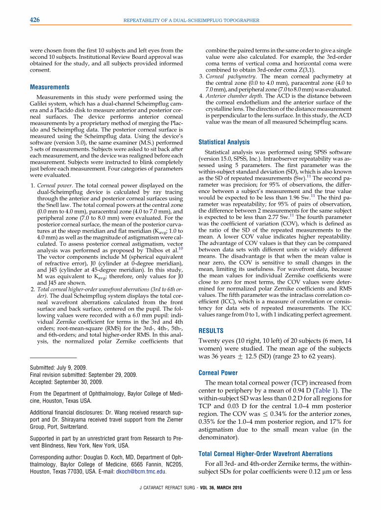
The mean total corneal power (TCP) increased fromcenter to periphery by a mean of 0.94 D (Table 1). Thewithin-subject SDwas less than 0.2 D for all regions forTCP and 0.03 D for the central 1.0–4 mm posteriorregion. The COV was % 0.34% for the anterior zones,0.35% for the 1.0–4 mm posterior region, and 17% forastigmatism due to the small mean value (in thedenominator).
Total Corneal Higher-Order Wavefront Aberrations
For all 3rd- and 4th-order Zernike terms, the within-subject SDs for polar coefficients were 0.12 mm or less
VOL 36, MARCH 2010
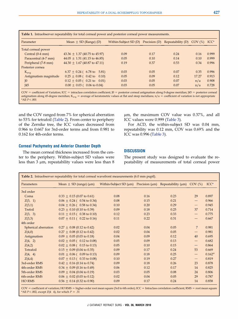
Table 1. Intraobserver repeatability for total corneal power and posterior corneal power measurements.
Parameter Mean G SD (Range) (D) Within-Subject SD (D) Precision (D) Repeatability (D) COV (%) ICC*
Total corneal powerCentral (0-4 mm) 43.36 G 1.37 (40.75 to 45.97) 0.09 0.17 0.24 0.16 0.999Paracentral (4-7 mm) 44.05 G 1.51 (41.15 to 46.85) 0.05 0.10 0.14 0.10 0.999Peripheral (7-8 mm) 44.30 G 1.67 (40.87 to 47.11) 0.19 0.37 0.53 0.34 0.996
Posterior corneaKavg �6.37 G 0.24 (�6.78 to �5.81) 0.03 0.05 0.07 0.35 0.996Astigmatism magnitude �0.25 G 0.08 (�0.42 to �0.10) 0.05 0.09 0.12 17.27 0.913J0 �0.12 G 0.05 (�0.21 to �0.01) 0.03 0.05 0.07 n/a 0.908J45 �0.00 G 0.03 (�0.06 to 0.04) 0.03 0.05 0.07 n/a 0.728
COV Z coefficient of Variation; ICC Z intraclass correlation coefficient; J0 Z posterior corneal astigmatism along 0-degree meridian; J45 Z posterior cornealastigmatism along 45-degree meridian; Kavg Z average of keratometric values at flat and steep meridians; n/a Z coefficient of variation is not appropriate*All P!.001
427REPEATABILITY OF A DUAL-SCHEIMPFLUG TOPOGRAPHER
and the COV ranged from 7% for spherical aberrationto 53% for tetrafoil (Table 2). From center to peripheryof the Zernike tree, the ICC values decreased from0.966 to 0.667 for 3rd-order terms and from 0.981 to0.162 for 4th-order terms.
Corneal Pachymetry and Anterior Chamber Depth
The mean corneal thickness increased from the cen-ter to the periphery. Within-subject SD values wereless than 3 mm, repeatability values were less than 8
Table 2. Intraobserver repeatability for total corneal wavefront measure
Parameters Mean G SD (range) (mm) Within-Subject S
3rd orderComa 0.33 G 0.15 (0.07 to 0.61) 0.08Z(3,�1) �0.06 G 0.24 (�0.54 to 0.34) 0.08Z(3,1) 0.04 G 0.26 (�0.58 to 0.34) 0.10Trefoil 0.23 G 0.10 (0.10 to 0.39) 0.09Z(3,�3) �0.11 G 0.15 (�0.38 to 0.10) 0.12Z(3,3) �0.07 G 0.11 (�0.22 to 0.16) 0.11
4th orderSpherical aberration 0.27 G 0.08 (0.12 to 0.42) 0.02Z(4,0) 0.27 G 0.08 (0.12 to 0.42) 0.02Astigmatism 0.09 G 0.05 (0.03 to 0.18) 0.04Z(4,�2) �0.02 G 0.05 (�0.12 to 0.08) 0.05Z(4,2) �0.02 G 0.08 (�0.15 to 0.13) 0.05Tetrafoil 0.15 G 0.09 (0.04 to 0.35) 0.09Z(4,�4) 0.01 G 0.06 (�0.09 to 0.13) 0.09Z(4,4) �0.07 G 0.13 (�0.33 to 0.08) 0.10
3rd-order RMS 0.42 G 0.16 (0.14 to 0.74) 0.094th-order RMS 0.34 G 0.09 (0.16 to 0.49) 0.065th-order RMS 0.09 G 0.04 (0.04 to 0.19) 0.036th-order RMS 0.06 G 0.02 (0.03 to 0.12) 0.02HO RMS 0.56 G 0.14 (0.32 to 0.90) 0.09
COV Z coefficient of variation; HORMS Z higher-order root mean square (3rd to 6*All P!.002, except Z(4�4), for which P Z .31
J CATARACT REFRACT SURG -
mm, the maximum COV value was 0.37%, and allICC values were 0.999 (Table 3).
For ACD, the within-subject SD was 0.04 mm,repeatability was 0.12 mm, COV was 0.69% and theICC was 0.996 (Table 3).
DISCUSSION
The present study was designed to evaluate the re-peatability of measurements of total corneal power
ments (6.0 mm pupil).
D (mm) Precision (mm) Repeatability (mm) COV (%) ICC*
0.16 0.23 29 0.8970.15 0.21 d 0.9660.20 0.29 d 0.9450.18 0.25 37 0.7140.23 0.33 d 0.7750.22 0.31 d 0.667
0.04 0.05 7 0.9810.04 0.05 d 0.9810.09 0.12 40 0.6950.09 0.13 d 0.6820.10 0.15 d 0.8640.17 0.24 53 0.6690.18 0.25 d 0.1624
0.19 0.27 d 0.8190.18 0.26 23 0.8780.12 0.17 14 0.8250.05 0.08 24 0.8060.04 0.05 29 0.7870.17 0.24 14 0.858
th order); ICC Z Intraclass correlation coefficient; RMS Z root mean square
VOL 36, MARCH 2010
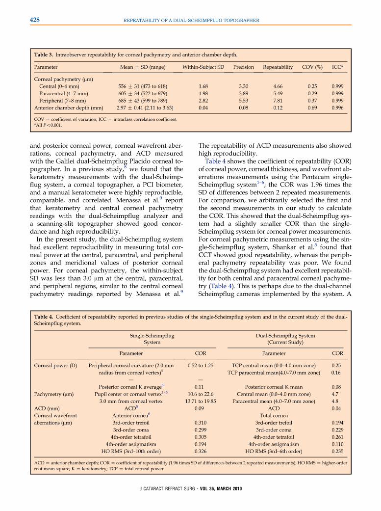
Table 3. Intraobserver repeatability for corneal pachymetry and anterior chamber depth.
Parameter Mean G SD (range) Within-Subject SD Precision Repeatability COV (%) ICC*
Corneal pachymetry (mm)Central (0–4 mm) 556 G 31 (473 to 618) 1.68 3.30 4.66 0.25 0.999Paracentral (4–7 mm) 605 G 34 (522 to 679) 1.98 3.89 5.49 0.29 0.999Peripheral (7–8 mm) 685 G 43 (599 to 789) 2.82 5.53 7.81 0.37 0.999
Anterior chamber depth (mm) 2.97 G 0.41 (2.11 to 3.63) 0.04 0.08 0.12 0.69 0.996
COV Z coefficient of variation; ICC Z intraclass correlation coefficient*All P!0.001.
428 REPEATABILITY OF A DUAL-SCHEIMPFLUG TOPOGRAPHER
and posterior corneal power, corneal wavefront aber-rations, corneal pachymetry, and ACD measuredwith the Galilei dual-Scheimpflug Placido corneal to-pographer. In a previous study,8 we found that thekeratometry measurements with the dual-Scheimp-flug system, a corneal topographer, a PCI biometer,and a manual keratometer were highly reproducible,comparable, and correlated. Menassa et al.9 reportthat keratometry and central corneal pachymetryreadings with the dual-Scheimpflug analyzer anda scanning-slit topographer showed good concor-dance and high reproducibility.
In the present study, the dual-Scheimpflug systemhad excellent reproducibility in measuring total cor-neal power at the central, paracentral, and peripheralzones and meridional values of posterior cornealpower. For corneal pachymetry, the within-subjectSD was less than 3.0 mm at the central, paracentral,and peripheral regions, similar to the central cornealpachymetry readings reported by Menassa et al.9
Table 4. Coefficient of repeatability reported in previous studies of theScheimpflug system.
Single-ScheimpflugSystem
Parameter C
Corneal power (D) Peripheral corneal curvature (2.0 mmradius from corneal vertex)5
0.52
d
Posterior corneal K average5 0Pachymetry (mm) Pupil center or corneal vertex1–5 10.6
3.0 mm from corneal vertex 13.71ACD (mm) ACD5 0Corneal wavefrontaberrations (mm)
Anterior cornea6
3rd-order trefoil 03rd-order coma 04th-order tetrafoil 0
4th-order astigmatism 0HO RMS (3rd–10th order) 0
ACD Z anterior chamber depth; COR Z coefficient of repeatability (1.96 times SDroot mean square; K Z keratometry; TCP Z total corneal power
J CATARACT REFRACT SURG -
The repeatability of ACD measurements also showedhigh reproducibility.
Table 4 shows the coefficient of repeatability (COR)of corneal power, corneal thickness, andwavefront ab-errations measurements using the Pentacam single-Scheimpflug system1–6; the COR was 1.96 times theSD of differences between 2 repeated measurements.For comparison, we arbitrarily selected the first andthe second measurements in our study to calculatethe COR. This showed that the dual-Scheimpflug sys-tem had a slightly smaller COR than the single-Scheimpflug system for corneal power measurements.For corneal pachymetric measurements using the sin-gle-Scheimpflug system, Shankar et al.5 found thatCCT showed good repeatability, whereas the periph-eral pachymetry repeatability was poor. We foundthe dual-Scheimpflug system had excellent repeatabil-ity for both central and paracentral corneal pachyme-try (Table 4). This is perhaps due to the dual-channelScheimpflug cameras implemented by the system. A
single-Scheimpflug system and in the current study of the dual-
Dual-Scheimpflug System(Current Study)
OR Parameter COR
to 1.25 TCP central mean (0.0–4.0 mm zone) 0.25
d
TCP paracentral mean(4.0–7.0 mm zone) 0.16
.11 Posterior corneal K mean 0.08to 22.6 Central mean (0.0–4.0 mm zone) 4.7to 19.85 Paracentral mean (4.0–7.0 mm zone) 4.8.09 ACD 0.04
Total cornea.310 3rd-order trefoil 0.194.299 3rd-order coma 0.229.305 4th-order tetrafoil 0.261.194 4th-order astigmatism 0.110.326 HO RMS (3rd–6th order) 0.235
of differences between 2 repeated measurements); HO RMS Z higher-order
VOL 36, MARCH 2010
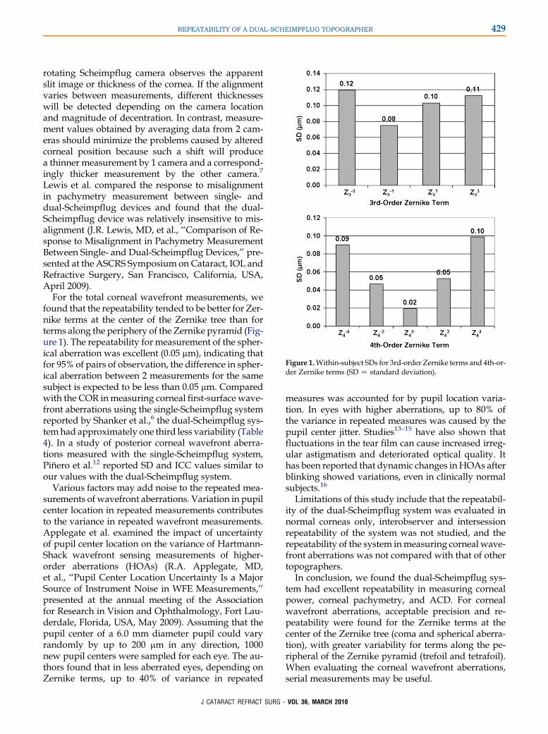
Figure 1.Within-subject SDs for 3rd-order Zernike terms and 4th-or-der Zernike terms (SD Z standard deviation).
429REPEATABILITY OF A DUAL-SCHEIMPFLUG TOPOGRAPHER
rotating Scheimpflug camera observes the apparentslit image or thickness of the cornea. If the alignmentvaries between measurements, different thicknesseswill be detected depending on the camera locationand magnitude of decentration. In contrast, measure-ment values obtained by averaging data from 2 cam-eras should minimize the problems caused by alteredcorneal position because such a shift will producea thinner measurement by 1 camera and a correspond-ingly thicker measurement by the other camera.7
Lewis et al. compared the response to misalignmentin pachymetry measurement between single- anddual-Scheimpflug devices and found that the dual-Scheimpflug device was relatively insensitive to mis-alignment (J.R. Lewis, MD, et al., ‘‘Comparison of Re-sponse to Misalignment in Pachymetry MeasurementBetween Single- and Dual-Scheimpflug Devices,’’ pre-sented at the ASCRS Symposium on Cataract, IOL andRefractive Surgery, San Francisco, California, USA,April 2009).
For the total corneal wavefront measurements, wefound that the repeatability tended to be better for Zer-nike terms at the center of the Zernike tree than forterms along the periphery of the Zernike pyramid (Fig-ure 1). The repeatability for measurement of the spher-ical aberration was excellent (0.05 mm), indicating thatfor 95% of pairs of observation, the difference in spher-ical aberration between 2 measurements for the samesubject is expected to be less than 0.05 mm. Comparedwith the COR inmeasuring corneal first-surface wave-front aberrations using the single-Scheimpflug systemreported by Shanker et al.,6 the dual-Scheimpflug sys-temhad approximately one third less variability (Table4). In a study of posterior corneal wavefront aberra-tions measured with the single-Scheimpflug system,Pinero et al.12 reported SD and ICC values similar toour values with the dual-Scheimpflug system.
Various factors may add noise to the repeated mea-surements of wavefront aberrations. Variation in pupilcenter location in repeated measurements contributesto the variance in repeated wavefront measurements.Applegate et al. examined the impact of uncertaintyof pupil center location on the variance of Hartmann-Shack wavefront sensing measurements of higher-order aberrations (HOAs) (R.A. Applegate, MD,et al., ‘‘Pupil Center Location Uncertainty Is a MajorSource of Instrument Noise in WFE Measurements,’’presented at the annual meeting of the Associationfor Research in Vision and Ophthalmology, Fort Lau-derdale, Florida, USA, May 2009). Assuming that thepupil center of a 6.0 mm diameter pupil could varyrandomly by up to 200 mm in any direction, 1000new pupil centers were sampled for each eye. The au-thors found that in less aberrated eyes, depending onZernike terms, up to 40% of variance in repeated
J CATARACT REFRACT SURG -
measures was accounted for by pupil location varia-tion. In eyes with higher aberrations, up to 80% ofthe variance in repeated measures was caused by thepupil center jitter. Studies13–15 have also shown thatfluctuations in the tear film can cause increased irreg-ular astigmatism and deteriorated optical quality. Ithas been reported that dynamic changes inHOAs afterblinking showed variations, even in clinically normalsubjects.16
Limitations of this study include that the repeatabil-ity of the dual-Scheimpflug system was evaluated innormal corneas only, interobserver and intersessionrepeatability of the system was not studied, and therepeatability of the system inmeasuring corneal wave-front aberrations was not compared with that of othertopographers.
In conclusion, we found the dual-Scheimpflug sys-tem had excellent repeatability in measuring cornealpower, corneal pachymetry, and ACD. For cornealwavefront aberrations, acceptable precision and re-peatability were found for the Zernike terms at thecenter of the Zernike tree (coma and spherical aberra-tion), with greater variability for terms along the pe-ripheral of the Zernike pyramid (trefoil and tetrafoil).When evaluating the corneal wavefront aberrations,serial measurements may be useful.
VOL 36, MARCH 2010
430 REPEATABILITY OF A DUAL-SCHEIMPFLUG TOPOGRAPHER
REFERENCES1. Barkana Y, Gerber Y, Elbaz U, Schwartz S, Ken-Dror G, Avni I,
Zadok D. Central corneal thickness measurement with the Pen-
tacam Scheimpflug system, optical low-coherence reflectometry
pachymeter, and ultrasound pachymetry. J Cataract Refract
Surg 2005; 31:1729–1735
2. Lackner B, Schmidinger G, Pieh S, Funovics MA, Skorpik C. Re-
peatability and reproducibility of central corneal thickness mea-
surement with Pentacam, Orbscan, and ultrasound. Optom Vis
Sci 2005; 82:892–899. Available at: http://www.oculus.de/chi/
downloads/dyn/sonstige/sonstige/lackner_pachymetry.pdf. Ac-
cessed December 5, 2009
3. O’Donnell C, Maldonado-Codina C. Agreement and repeatability
of central thickness measurement in normal corneas using ultra-
sound pachymetry and the OCULUS. Pentacam. Cornea 2005;
24:920–924
4. Amano S, Honda N, Amano Y, Yamagami S, Miyai T,
Samejima T, Ogata M, Miyata K. Comparison of central corneal
thickness measurements by rotating Scheimpflug camera, ultra-
sonic pachymetry, and scanning-slit corneal topography. Oph-
thalmology 2006; 113:937–941
5. Shankar H, Taranath D, Santhirathelagan CT, Pesudovs K. An-
terior segment biometry with the Pentacam: comprehensive as-
sessment of repeatability of automated measurements. J
Cataract Refract Surg 2008; 34:103–113
6. Shankar H, Taranath D, Santhirathelagan CT, Pesudovs K. Re-
peatability of corneal first-surface wavefront aberrations mea-
sured with Pentacam corneal topography. J Cataract Refract
Surg 2008; 34:727–734
7. Roberts C, Zuger BJ. GALILEI dual Scheimpflug analyzer. In:
Netto MV, Ambrosio R Jr, Schor P, Chalita MR, Chamon W,
eds, Wavefront, Topografia e Tomografia da Cornea e Segmen-
to Anterior; Atualizacao Propedeutica em Cirurgia Refrativa. Rio
de Janeiro, Brazil, Cultura Medica, 2006; 177–182
8. Shirayama M, Wang L, Weikert MP, Koch DD. Comparison of
corneal powers obtained from 4 different devices. Am J Ophthal-
mol 2009; 148:528–535
9. Menassa N, Kaufmann C, Goggin M, Job OM, Bachmann LM,
Thiel MA. Comparison and reproducibility of corneal thickness
J CATARACT REFRACT SURG -
and curvature readings obtained by the Galilei and the Orbscan
II analysis systems. J Cataract RefractSurg2008; 34:1742–1747
10. Thibos LN, Wheeler W, Horner D. Power vectors: an application
of Fourier analysis to the description and statistical analysis of
refractive error. Optom Vis Sci 1997; 74:367–375. Available at:
http://research.opt.indiana.edu/Library/AnalyzingAstigmatism/
AnalyzingAstigmatism.html. Accessed December 4, 2009
11. Bland JM, Altman DG. Statistical notes: measurement error.
BMJ 1996; 313:744
12. Pinero DP, Saenz GonzalezC, Alio JL. Intraobserverand interob-
server repeatabilityof curvatureandaberrometric measurements
of the posterior corneal surface in normal eyes using Scheimpflug
photography. J Cataract Refract Surg 2009; 35:113–120
13. Liu Z, Pflugfelder SC. Corneal surface regularity and the effect of
artificial tears in aqueous tear deficiency. Ophthalmology 1999;
106:939–943
14. Goto T, Zheng X, Klyce SD, Kataoka H, Uno T, Karon M,
Tatematsu Y, Bessyo T, Tsubota K, Ohashil Y. A new method
for tear film stability analysis using videokeratography. Am J
Ophthalmol 2003; 135:607–612
15. Montes-Mico R, Alio JL, Munoz G, Charman WN. Temporal
changes in optical quality of air-tear film interface at anterior cor-
nea after blink. Invest Ophthalmol Vis Sci 2004; 45:1752–1757.
Available at: http://www.iovs.org/cgi/reprint/45/6/1752. Ac-
cessed December 3, 2009
16. Koh S, Maeda N, Hirohara Y, Mihashi T, Ninomiya S, Bessho K,
Watanabe H, Fujikado T, Tano Y. Serial measurements of high-
er-order aberrations after blinking in normal subjects. Invest Oph-
thalmol Vis Sci 2006; 47:3318–3324. Available at: http://www.
iovs.org/cgi/reprint/47/8/3318. Accessed December 3, 2009
VO
L 36, MARCH 2010First Author:Li Wang, MD, PhD
Department of Ophthalmology, BaylorCollege of Medicine, Houston, TexasUSA