reorganization and in vivo dynamics of microtubules during
TRANSCRIPT

Reorganization and in Vivo Dynamics of Microtubulesduring Arabidopsis Root Hair Development[w]
Nathalie Van Bruaene1*, Greg Joss, and Patrick Van Oostveldt
Laboratory for Biochemistry and Molecular Cytology, Ghent University, 9000 Gent, Belgium (N.V.B., P.V.O.);and Department of Biological Sciences, Macquarie University, Sydney, New South Wales 2109, Australia (G.J.)
Root hairs emerge from epidermal root cells (trichoblasts) and differentiate by highly localized tip growth. Microtubules (MTs)are essential for establishing and maintaining the growth polarity of root hairs. The current knowledge about the configurationof the MTcytoskeleton during root hair development is largely based on experiments on fixed material, and reorganization andin vivo dynamics of MTs during root hair development is at present unclear. This in vivo study provides new insights into themechanisms of MT (re)organization during root hair development in Arabidopsis (Arabidopsis thaliana). Expression of a bindingsite of the MT-associated protein-4 tagged with green fluorescent protein enabled imaging of MT nucleation, growth, andshortening and revealed distinct MT configurations. Depending on the dynamics of the different MT populations during roothair development, either repeated two-dimensional (x, y, t) or repeated three-dimensional (x, y, z, t) scanning was performed.Furthermore, a new image evaluation tool was developed to reveal important data on MT instability. The data show how MTsreorient after apparent contact with other MTs and support a model for MT alignment based on repeated reorientation ofdynamic MT growth.
A root hair is a long, lateral, tubular extension of anepidermal root cell formed by a process called tipgrowth. Diffuse longitudinal growth of the epidermalcell undergoes transition to highly localized andpolarized growth at one specific site. After the forma-tion of an initial bulge, the root hair grows by polar-ized exocytosis and deposition of cell wall materialconfined to the tip (Schnepf, 1986). In an early phase,3-d-old Arabidopsis (Arabidopsis thaliana) root hairsgrow out at a rate of 0.4 mm/min, followed by a lategrowth phase at 1 to 2.5 mm/min (Dolan et al.,1994). Tip growth is characterized by the presence ofa vesicle-rich region, a reverse-fountain type of cyto-plasmic streaming, and a gradient of cytoplasmic Ca21
toward the hair tip (Pierson et al., 1996; Galway et al.,1997; Miller et al., 1997, 1999; Wymer et al., 1997; deRuijter et al., 1998; Yang, 1998). During growth arrestcytoplasmic streaming switches from reverse-fountainstreaming to circular streaming, and this reorganiza-tion is coupled to the disappearance of the vesicle-richregion (Miller et al., 1997).It has been accepted that root hair outgrowth is
associated with microtubule (MT) and actin filamentreorientations (Emons and Derksen, 1986; Baluskaet al., 2000). Bao et al. (2001) reported that reduceda-tubulin gene expression causes formation of multi-ple root hairs by single epidermal cells and ectopicroot hair formation in tissues that are normally roothair free. This was the first evidence, to our knowl-
edge, for a function of the MTcytoskeleton during roothair initiation, in particular suggesting a role for MTdisassembly (caused by low a-tubulin expression) inthe initiation of root hair formation. In maize (Zeamays) and lettuce (Lactuca sativa) the initiation of roothairs is associated with disorganization of the cortical-MT arrays in the trichoblast (Baluska et al., 2000;Takahashi et al., 2003a).
Filamentous actin plays a fundamental role in thetip growth process (for review, see Geitmann andEmons, 2000; Hepler et al., 2001). The function of MTsin tip growth is less clear and under debate. Bothlongitudinal and helical arrays of MTs have beendescribed in root hairs (Lloyd, 1983; for review, seeGeitmann and Emons, 2000) and do not seem to bepart of the cellular machinery required for tip growth(Bibikova et al., 1999; Ketelaar et al., 2002). However,pharmacological studies showed that MTs are re-quired to maintain growth directionality and unidi-rectional growth at the tip of root hairs (Bibikova et al.,1999; Ketelaar et al., 2003). Further evidence that MTsare involved in controlling root hair growth directioncomes from the temperature-sensitive mor1 (MTorganization 1) mutants (Whittington et al., 2001).
Recent in vivo studies have provided new insightsinto MT dynamics in plant cells. Sieberer et al. (2002)showed that in vivoMTconfigurations during root hairdevelopment obtained with the green fluorescent pro-tein (GFP)-MT-binding domain (MBD) fusion proteinwere similar to immuno-labeled fixed whole-mountsamples. Two different populations of MTs were re-ported inMedicago truncatula (Sieberer et al., 2002). Thecortical MTs (CMTs) are present in all developmentalstages, and the endoplasmic MTs (EMTs) are presentonly in growing root hairs. Until now, EMTs have beenreported only for legume root hairs and not in otherspecies (Arabidopsis; Galway et al., 1997; Sieberer et al.,
1 Present address: In Vitro Fertilization Center, Center for Radio-Immunology, Industriepark 3b, 9052 Zwijnaarde, Belgium.
* Corresponding author; e-mail [email protected]; fax 32–9–264–62–19.
[w]The online version of this article contains Web-only data.Article, publication date, and citation information can be found at
www.plantphysiol.org/cgi/doi/10.1104/pp.103.031591.
Plant Physiology, December 2004, Vol. 136, pp. 3905–3919, www.plantphysiol.org � 2004 American Society of Plant Biologists 3905 www.plantphysiol.orgon March 26, 2018 - Published by Downloaded from
Copyright © 2004 American Society of Plant Biologists. All rights reserved.

2002). Despite the valuable information about MTsobtained from in vivo and inhibitor studies, a lot ofquestions remain unanswered about MT reorganiza-tion during root hair development. MTs changetheir configuration during root hair development(Geitmann and Emons, 2000; Sieberer et al., 2002),but it is not known how the transitional processes inMT reorganization occur. It still remains enigmatichow CMTs are formed in root hairs. Are they newlyformed, or are they reorganized from existing CMTs?In addition, the reorganization of MTs during root hairoutgrowth and tip growth is not understood.
In general, reorganization can occur by polymeri-zation of new MTs, reorientation of intact MTs, orpolymerization of existing MTs in a new direction(Kropf et al., 1998). Polymerization of new MTs re-quires the presence of nucleating sites. MT nucleationor formation is initiated at the g-tubulin complexeslocated at the spindle pole body in yeast or at thecentrosome in animal cells (for review, see Schiebel,2000). In plant cells, the absence of organelles such asthe centrosome has led to a lot of debate about theorigin of MT nucleation sites (for review, see Schmit,2002). It has been shown that the nuclear surface inhigher plants has a centrosome-like activity (Mizuno,1993; Stoppin et al., 1994). In addition, spontaneousand de novo assembly of nucleation sites may occur inthe cell cortex, which was proved by semi-in vitroassays (Wasteneys et al., 1989), by studies of MTrecovery after disassembly (Cleary and Hardham,1987; Wasteneys and Williamson, 1989a), and by invivo studies (Wasteneys et al., 1993; Erhardt et al.,2002; Chan et al., 2003; Shaw et al., 2003).
Another feature of MT dynamics adds to the com-plexity of MT reorganization. MTs are capable of rapidrearrangement by subunit exchange, which allowsthem to grow and shorten. These mechanisms havebeen thoroughly investigated in animal cells (for re-view, see Desai and Mitchinson, 1997) and to a lesserextent in plant cells (Zhang et al., 1990; Hush et al.,1994; Chan et al., 2003; Dhonukshe and Gadella, 2003;Shaw et al., 2003; Vos et al., 2004). Two mechanisms ofsubunit exchange are known. Treadmilling involvesthe addition of subunits to one (plus) end of anMTandloss of subunits from the opposite (minus) end. Dy-namic instability is defined as a rapid switch betweengrowth and shortening at the plus end of an MT(Cassimeris et al., 1988; Vorobjev et al., 1999). Mostlythe minus end is less dynamic and oriented toward, orburied in, the MT organizing center-like structure.Recent experiments in BY-2 cells revealed the dynam-ics of MTs visualized with CYTOPLASMIC LINKERPROTEIN170-GFP (Dhonukshe and Gadella, 2003) orMBD-GFP (Vos et al., 2004). These studies showed thatshort periods of fast shortening are alternated withlonger periods of slower growth. A hybrid MT-tread-milling mechanism, with dynamic instability at oneend and slow depolymerization at the other end, hasbeen demonstrated in cortical arrays of Arabidopsisepidermal cells (Shaw et al., 2003).
Transgenic Arabidopsis plants expressing a GFP-MBD of MT-associated protein-4 (MAP4) fusion pro-tein (Marc et al., 1998) were used in this study toanalyze the dynamic reorganizations of MTs duringroot hair development. This marker protein hasproven to be very useful in previous cytoskeletonstudies in Arabidopsis trichomes (Mathur and Chua,2000), M. truncatula root hairs (Sieberer et al., 2002),and BY-2 cells (Dhonukshe and Gadella, 2003). Pre-vious studies on dynamic instability in plant cells werebased on two-dimensional (2-D; x, y) imaging meth-ods where data are collected only from restrictedregions in focus (Dhonukshe and Gadella, 2003;Shaw et al., 2003). As root hairs are often not lyingcompletely parallel to the focal plane, 2-D imaging (x,y) would reveal only part of the MT organization inroot hairs. Furthermore, 2-D scanning can lead toincorrect conclusions when MTs go out of focus. Tohave a complete view of MT dynamics in growingand full-grown root hairs, repeated three-dimensional(3-D) images (x, y, z, t) were taken over time (everyminute). However, at some stages in developmentdynamics seemed so fast that the MT configurationwas completely altered after 1 min. Therefore, addi-tional fast-repeated 2-D scanning (x, y, t; a frame everysecond) with increased focal depth was performed atthose stages. This study in Arabidopsis demonstratedthat at the onset of root hair outgrowth MTs werereorganized, and during outgrowth new MTs areformed from distinct initiation sites. During root hairtip growth two populations of MTs could be distin-guished. A new visualization method reveals prom-inent four-dimensional (4-D) data of MT polarity inmature root hairs. Finally, analysis of shorteningevents suggests turnover of subunits after apparentcontact with neighboring CMTs. A model is discussedin which these shortening events play a role ina mechanism for CMT alignment.
RESULTS
To allow direct in vivo observations of MTs, trans-genic plants stably expressing GFP-MBD of MAP4were used (Marc et al., 1998). Arabidopsis plantscarrying this construct were phenotypically normaland displayed normal root hair formation. As the rootand the surrounding medium were transferred asa whole to the small-growth chamber suitable formicroscopy, the root hairs continued to grow forseveral hours during imaging. The mean root hairgrowth rate during imaging was approximately 1 mm/min. This is in agreement with data from literature(Dolan et al., 1994) and suggests that root hair de-velopment was not affected by imaging.
MTs Reorganize and Apparent Initiation Sites AreFormed during Bulge Development
The cortical arrays in elongating epidermal cellsformed helical arrays in which the MTs were oriented
Van Bruaene et al.
3906 Plant Physiol. Vol. 136, 2004 www.plantphysiol.orgon March 26, 2018 - Published by Downloaded from
Copyright © 2004 American Society of Plant Biologists. All rights reserved.

transversely or at a slightly oblique angle (60–90� tothe long axis of the cell). At the onset of root hairoutgrowth, the MTs in the trichoblasts reorganized,whereas the MTs in atrichoblasts stayed in transversearrays (Fig. 1, A and B). In the apical region of thetrichoblast (near the bulge) the MTs disorganized (Fig.1, A and B). This was also observed in lettuce byTakahashi et al. (2003a). At the onset of tip growtha smooth region appeared in the tip, which is known tocontain vesicles (Galway et al., 1997). In the corticalregion and apical cytoplasm of the outgrowing roothair, several discrete spots were observed with short-radiating MTs (white arrowheads and arrows, respec-tively, in Fig. 1D). To follow the dynamics of thesestructures over time, repeated 2-D sections (x, y, t)were taken. Supplemental Movie 1 focuses on theendoplasmic region and shows that the spots and theshort MTs were very dynamic and moved simulta-neously. Occasionally, MTs stayed in focus and asa result the elongation of MTs extending from thedense spots could be followed (black arrowhead inFig. 1D; Supplemental Movie 1). Supplemental Movie2 focuses on the (sub)-cortical region and illustratesthe initiation and elongation of an MT. These obser-
vations suggest that the discrete spots are MT initia-tion sites.
Distinct Configuration Patterns of CMTs Versus EMTs
in Elongating Root Hairs
MT configuration was followed in 4-D (x, y, z, t)during root hair expansion over long time periods. The3-D time series images (x, y, z, t) were reduced tomovies by projecting the maximum fluorescence of theconfocal-optical sections for each time step to a 2-Dimage. In the cortical region an array with a highdensity of CMTs was observed (Fig. 2A; SupplementalMovies 3 and 4) similar to previous immunofluores-cence studies (Lloyd et al., 1987; Geitmann and Emons,2000). In addition to this cortical array, EMTs wereobserved in the cytoplasm of the root hair (Figs. 2, B–G, and 3). EMTs were previously demonstrated inlegumes, but not in Arabidopsis. During early expan-sion EMTs accumulated in the cytoplasm (Fig. 2, K andL) and during late-growth stage bundles were formedin the cytoplasm (Fig. 2, N–P). During early root hairgrowth and before nuclear migration, EMTs extendedbetween the subapical region of the tip and the
Figure 1. MT organization during bulge formation visualized with GFP-MBD of MAP4 and imaged with a confocal laserscanning microscope by 3-D sampling (x, y, z; A–C) and repeated 2-D sampling (x, y, t; D). A, Maximal projection of 25 opticalsections at 1-mm (z) intervals of MTs in a bulge. The small asterisk indicates an atrichoblast; the large asterisk indicatesa trichoblast. B, Single optical section at the endoplasmic region in the middle of the outgrowing root hair. C, Transmissionimage; double arrow indicates the vesicle-rich region of the outgrowing root hair. D, Time sequence showing the dynamics ofapparent initiation sites. Five representative single-optical sections are shown from a series of 40 images taken at 1-s intervals(Supplemental Movie 1). The numbers indicate the time in seconds. The pointed arrows indicate the endoplasmic-apparentinitiation sites, the arrowheads indicate the cortical-apparent initiation sites, and the gray-blocked arrow shows the growth ofa single MT. Scale bar 5 10 mm.
Microtubule Dynamics in Root Hairs
Plant Physiol. Vol. 136, 2004 3907 www.plantphysiol.orgon March 26, 2018 - Published by Downloaded from
Copyright © 2004 American Society of Plant Biologists. All rights reserved.

nucleus and occasionally between the base of the roothair and the nucleus (Fig. 2, C–J). Dense spots werelocalized at the surface of the nucleus (still in the mainbody of the cell), and MTs extended between thesesites and the endoplasmic bundles (Fig. 2, I and J;Supplemental Movie 4). In the corresponding timeseries movie, spots seem to move along existing MTs(data not shown).
During further expansion of the root hair, the nucleusmigrated into the tube and positioned at a more-or-lessconstant distance from the tip (Ketelaar et al., 2002).Figure 3A shows the situation after the nucleus hasmigrated into the root hair. Thick bundles of EMTsextended between the tip and the nucleus and formedan array around the nucleus. The cross section throughthe nucleus in Figure 3B clearly shows that the fluores-cence was present at the edge of the nucleus and wasmost intense at the ends toward the tip and the base ofthe root hair. The EMT bundles branched in the tip andseemed to extend toward the CMTs in the subapicalregion. In highly vacuolated regions of the root hairs,
the EMTs were localized close to the cortex as the en-doplasm was reduced to a thin layer between thevacuole and the plasma membrane. In this study, theEMT bundles at the subcortex were still clearly distin-guishable from the CMT array but sometimes difficultto represent in a single projection. SupplementalMovie5 shows thedynamics in the core-central sections and ata more upper level (including part of the CMTs). TheEMTs are very dynamic and continuously change theirorientation and configuration. Possibly these move-ments are related to the reorganizations of the cyto-plasmic strands. Close to the bundles, individualfluorescent spots could be distinguished with shortMTs radiating from these spots. The spots are clearlyvisible in the subcortex (lower part of Fig. 4A) and inthe endoplasm (Fig. 4B; Supplemental Movie 6). MTsextending from the nucleus toward the cortex areindicated by arrows in Figure 4C. The elongation andreorientation of single MTs were imaged by repeated2-D images (x, y, t) with increased focal depth takenevery second (Supplemental Movie 6).
Figure 2. MTorganization in a growing root hair visualized with a confocal laser scanning microscope in 3-D (x, y, z) and 4-D (x,y, z, t). The root hair is in an early stage of growth and the nucleus is not yet migrated into the tube. A–J, A series of 10 opticalsections (x, y) with a separating distance of 2.5 mm. A, Optical section at the cortical region; E, optical section at the endoplasmicregion. Arrowheads in I and J indicate the spots localized at the nuclear membrane, and arrows in C to J indicate cross sections ofEMTs extending between the nucleus (n) and the tip or between the base and the tip of the root hair. Due to orientation of the celland the EMTs, each optical section shows partial EMTs or cross sections of the EMTs. K to P, Six representative images fromrepeated 3-D scanning (x, y, z, t) over a period of 30 min. The numbers indicate the time in min. The images are represented asmaximal projections of the four median-optical sections (x, y, z) with a separating distance of 1 mm in the endoplasmic region.The root hair was growing at a velocity of 1.4 mm/min. In O apparent initiation sites at the base of the root hair are indicated byarrows. Scale bar 5 10 mm.
Van Bruaene et al.
3908 Plant Physiol. Vol. 136, 2004 www.plantphysiol.orgon March 26, 2018 - Published by Downloaded from
Copyright © 2004 American Society of Plant Biologists. All rights reserved.

The Organization Pattern of MTs in Elongating Root
Hairs Is Different from the Pattern in Full-GrownRoot Hairs
Since cytological structures rearrange duringgrowth arrest, it was interesting to investigate whether
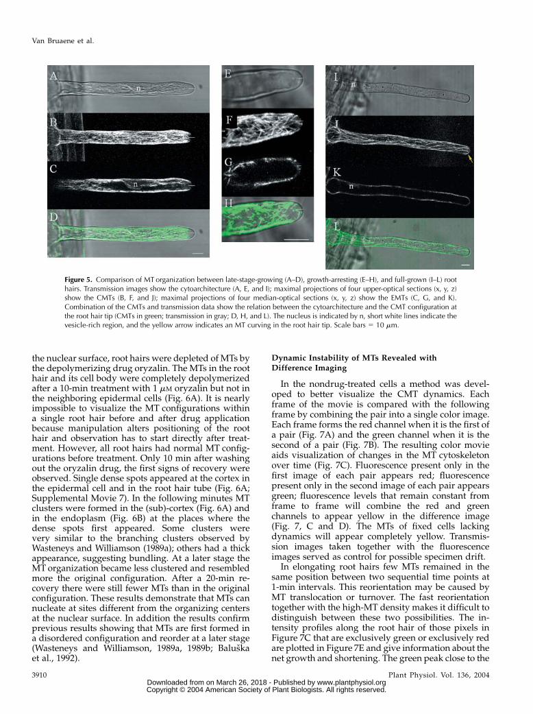
these reorganizations coincide with MT rearrange-ments. One of the features visible during growth arrestwas that the vesicle-rich region narrowed and finallycompletely disappeared in full-grown root hairs (com-pare Fig. 5, A, E, and I). The cytoplasmic streamingchanged from reverse-fountain streaming to circularstreaming, as described earlier by Miller et al. (1997)and Sieberer and Emons (2000). At that time thenucleus left its position close to the tip and kept onmoving back and forth in the mature root hair(Chytilova et al., 2000; Ketelaar et al., 2002; VanBruaene et al., 2003).
There are three major configuration differencesbetween growing and full-grown root hairs. First,and most obviously, there are fewer CMTs in full-grown root hairs compared to elongating root hairs(Fig. 5, B versus J). The growing root hairs had a high-density array of CMTs. In full-grown root hairs, CMTshad a longitudinal or helical organization (for review,see Geitmann and Emons, 2000); only very few MTswere transversely oriented. Second, the MTs in elon-gating root hairs didn’t reach the very tip, whereassome MTs reached the very tip in growth-arrestingroot hairs and could even curve in the tip of matureroot hairs (Fig. 5, B, F, and J, respectively). Third, theEMTs disappeared during growth arrest and therewere no MTs extending between the nucleus and thetip in full-grown root hairs (compare Fig. 5, C, G, andK). Occasionally, short MTs were observed close to thenucleus in full-grown root hairs.
MTs Reassemble after Complete Depolymerizationwith Oryzalin
To determine whether MTs can reassemble inde-pendently from the interior organizing sites such as
Figure 3. Configuration of EMTs around the nucleus (n) in late-stage-growing root hairs visualized with a confocal laser scanning micro-scope. A, Maximal projection of four median-optical sections ata separating distance of 1 mm; arrow indicates EMT bundle extendingtoward the tip. B, Single optical section through the nucleus. EMTfluorescence is present at the edge of the nucleus and is most intense atthe ends toward the tip and the base of the root hair (arrow). Both roothair tips are oriented to the left. Scale bar 5 10 mm.
Figure 4. Configuration of EMTs inlate-stage-growing root hairs. Imagesare shown using an inverted grayscaleto enhance the details. A, A represen-tative image is shown out of a series ofsingle optical sections taken every sec(x, y, t) with increased pinhole of 3 airydisc units. The complete sequences of30 s are available as SupplementalMaterial (Supplemental Movie 6). Theroot hair is tilted and the image grazesonly the cortex at the upper half of theimage. The lower half shows the sub-cortical region; spots are indicated byarrows. B, Single optical section at thecenter of the root hair showing EMTbundles and spots (arrows). C, Opticalsection through the nucleus, singleEMTs extend from the nucleus towardthe cortical region (arrows). Root tipis oriented downwards. Scale bar 5
10 mm.
Microtubule Dynamics in Root Hairs
Plant Physiol. Vol. 136, 2004 3909 www.plantphysiol.orgon March 26, 2018 - Published by Downloaded from
Copyright © 2004 American Society of Plant Biologists. All rights reserved.
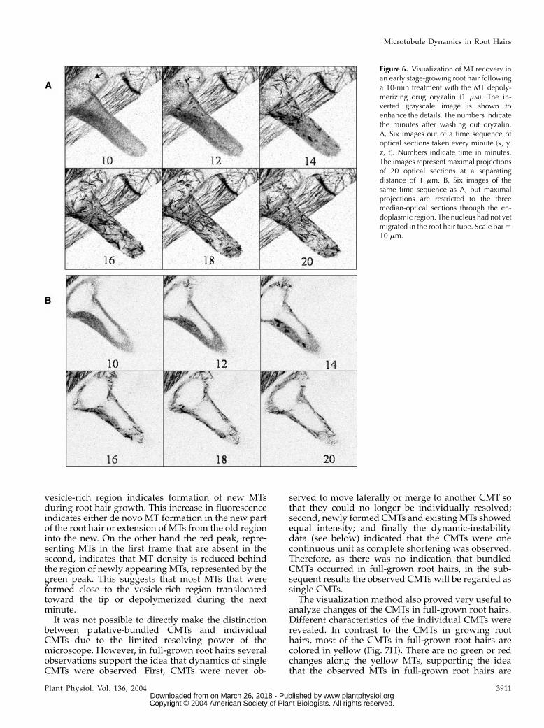
the nuclear surface, root hairs were depleted of MTs bythe depolymerizing drug oryzalin. The MTs in the roothair and its cell body were completely depolymerizedafter a 10-min treatment with 1 mM oryzalin but not inthe neighboring epidermal cells (Fig. 6A). It is nearlyimpossible to visualize the MT configurations withina single root hair before and after drug applicationbecause manipulation alters positioning of the roothair and observation has to start directly after treat-ment. However, all root hairs had normal MT config-urations before treatment. Only 10 min after washingout the oryzalin drug, the first signs of recovery wereobserved. Single dense spots appeared at the cortex inthe epidermal cell and in the root hair tube (Fig. 6A;Supplemental Movie 7). In the following minutes MTclusters were formed in the (sub)-cortex (Fig. 6A) andin the endoplasm (Fig. 6B) at the places where thedense spots first appeared. Some clusters werevery similar to the branching clusters observed byWasteneys and Williamson (1989a); others had a thickappearance, suggesting bundling. At a later stage theMT organization became less clustered and resembledmore the original configuration. After a 20-min re-covery there were still fewer MTs than in the originalconfiguration. These results demonstrate that MTs cannucleate at sites different from the organizing centersat the nuclear surface. In addition the results confirmprevious results showing that MTs are first formed ina disordered configuration and reorder at a later stage(Wasteneys and Williamson, 1989a, 1989b; Baluskaet al., 1992).
Dynamic Instability of MTs Revealed withDifference Imaging
In the nondrug-treated cells a method was devel-oped to better visualize the CMT dynamics. Eachframe of the movie is compared with the followingframe by combining the pair into a single color image.Each frame forms the red channel when it is the first ofa pair (Fig. 7A) and the green channel when it is thesecond of a pair (Fig. 7B). The resulting color movieaids visualization of changes in the MT cytoskeletonover time (Fig. 7C). Fluorescence present only in thefirst image of each pair appears red; fluorescencepresent only in the second image of each pair appearsgreen; fluorescence levels that remain constant fromframe to frame will combine the red and greenchannels to appear yellow in the difference image(Fig. 7, C and D). The MTs of fixed cells lackingdynamics will appear completely yellow. Transmis-sion images taken together with the fluorescenceimages served as control for possible specimen drift.
In elongating root hairs few MTs remained in thesame position between two sequential time points at1-min intervals. This reorientation may be caused byMT translocation or turnover. The fast reorientationtogether with the high-MT density makes it difficult todistinguish between these two possibilities. The in-tensity profiles along the root hair of those pixels inFigure 7C that are exclusively green or exclusively redare plotted in Figure 7E and give information about thenet growth and shortening. The green peak close to the
Figure 5. Comparison of MT organization between late-stage-growing (A–D), growth-arresting (E–H), and full-grown (I–L) roothairs. Transmission images show the cytoarchitecture (A, E, and I); maximal projections of four upper-optical sections (x, y, z)show the CMTs (B, F, and J); maximal projections of four median-optical sections (x, y, z) show the EMTs (C, G, and K).Combination of the CMTs and transmission data show the relation between the cytoarchitecture and the CMT configuration atthe root hair tip (CMTs in green; transmission in gray; D, H, and L). The nucleus is indicated by n, short white lines indicate thevesicle-rich region, and the yellow arrow indicates an MT curving in the root hair tip. Scale bars 5 10 mm.
Van Bruaene et al.
3910 Plant Physiol. Vol. 136, 2004 www.plantphysiol.orgon March 26, 2018 - Published by Downloaded from
Copyright © 2004 American Society of Plant Biologists. All rights reserved.
vesicle-rich region indicates formation of new MTsduring root hair growth. This increase in fluorescenceindicates either de novo MT formation in the new partof the root hair or extension of MTs from the old regioninto the new. On the other hand the red peak, repre-senting MTs in the first frame that are absent in thesecond, indicates that MT density is reduced behindthe region of newly appearing MTs, represented by thegreen peak. This suggests that most MTs that wereformed close to the vesicle-rich region translocatedtoward the tip or depolymerized during the nextminute.It was not possible to directly make the distinction
between putative-bundled CMTs and individualCMTs due to the limited resolving power of themicroscope. However, in full-grown root hairs severalobservations support the idea that dynamics of singleCMTs were observed. First, CMTs were never ob-
served to move laterally or merge to another CMT sothat they could no longer be individually resolved;second, newly formed CMTs and existingMTs showedequal intensity; and finally the dynamic-instabilitydata (see below) indicated that the CMTs were onecontinuous unit as complete shortening was observed.Therefore, as there was no indication that bundledCMTs occurred in full-grown root hairs, in the sub-sequent results the observed CMTs will be regarded assingle CMTs.
The visualization method also proved very useful toanalyze changes of the CMTs in full-grown root hairs.Different characteristics of the individual CMTs wererevealed. In contrast to the CMTs in growing roothairs, most of the CMTs in full-grown root hairs arecolored in yellow (Fig. 7H). There are no green or redchanges along the yellow MTs, supporting the ideathat the observed MTs in full-grown root hairs are
Figure 6. Visualization of MT recovery inan early stage-growing root hair followinga 10-min treatment with the MT depoly-merizing drug oryzalin (1 mM). The in-verted grayscale image is shown toenhance the details. The numbers indicatethe minutes after washing out oryzalin.A, Six images out of a time sequence ofoptical sections taken every minute (x, y,z, t). Numbers indicate time in minutes.The images represent maximal projectionsof 20 optical sections at a separatingdistance of 1 mm. B, Six images of thesame time sequence as A, but maximalprojections are restricted to the threemedian-optical sections through the en-doplasmic region. The nucleus had not yetmigrated in the root hair tube. Scale bar 510 mm.
Microtubule Dynamics in Root Hairs
Plant Physiol. Vol. 136, 2004 3911 www.plantphysiol.orgon March 26, 2018 - Published by Downloaded from
Copyright © 2004 American Society of Plant Biologists. All rights reserved.

single MTs (Supplemental Movie 9). This is in contrastwith the green and red portions appearing along theyellow MTs in the transverse arrays of nonroot hairepidermal cells (data not shown), which suggestsdynamics of multiple MTs lying along each other. Infull-grown root hairs no MT displacement was ob-served in the z-direction (Fig. 8A; MTremains yellow);green and red portions were observed only at the MTends. In addition, the majority of CMTs showed nolateral movements over time (Fig. 8, B and C). Thesetwo observations suggest that the CMTs were fixed tothe cortex, corresponding to the observations of Shawet al. (2003). However, some newly formed MTsshowed clear lateral movement as indicated in Figure8D. Vertical cross sections demonstrate that the MTwas subcortical. Newly formed MTs were probablynot yet fixed to the cortical region.
Another important feature that could be directlydetermined from the difference images is the amountof growth (green) and shortening (red) at the MT ends(Fig. 7, F–I). The dynamics of both MT ends of five
CMTs are depicted as MT life history plots in Figure 7J.The plots demonstrate that the growth rate is lowerthan the shortening rate, corresponding with the dataof Dhonukshe and Gadella (2003) and Vos et al. (2004).It should be noted that the plots may represent anunderestimation of the real situation, especially at thetransition points where growth switches to shorteningand vice versa. In contrast to these dynamic (plus)ends, the opposite minus ends were characterizedwith slow (72%) or no (28%; total n 5 146) shortening(Fig. 7J). Only limited examples of breakage in CMTswere recorded (less than 2%).
Detailed analysis of the behavior of 174 CMTs in 10different full-grown root hairs revealed that the ma-jority of CMT plus ends were oriented toward the roothair tip (82% 6 9%). Only 11% 6 6% of the plus endswere oriented toward the base and 7% 6 5% weretransversely oriented. The difference image Figure 7Hshowing shortening in red and growth in greenrevealed that fast shortening of MTs occurred overthe whole length of the full-grown root hair. Shorten-
Figure 7. CMT dynamics in late-stage-growing (A–E) and full-grown (F–J) root hairs. Configuration of CMTs in growing and full-grown root hair represented as maximal projection of 20 optical sections (separating distance 1 mm) at 0 min (A and F) and at1 min (B and G). Difference images of two time points (C and H), the images at 0 min (A and F) from the red channel, and imagesat 1 min (B and G) from the green channel. CMTs that did not change in position or length appear yellow (see also D and I).Intensity profile of red and green (not yellow) pixels along the root hair axis (length5 470 pixels; E). Root hair tip is not shown in Fto I but oriented downwards. Life history plots show individual dynamics of CMT plus and minus ends. The yellow trianglesindicate the time points where an apparent contact was detected. The deviation in orientation of the growth direction in respectto the previous growth direction for the different MTs: MT1, 8�; MT2, 7� and 22�; MT3, 5� and 23�; MT4, 27� and 11�; andMT5, 2�. Negative values indicate counter clockwise deviations (J). Scale bars 5 10 mm.
Van Bruaene et al.
3912 Plant Physiol. Vol. 136, 2004 www.plantphysiol.orgon March 26, 2018 - Published by Downloaded from
Copyright © 2004 American Society of Plant Biologists. All rights reserved.

Figure 8. CMT position and dynamics in full-grown root hairs. A, Vertical positioning of an MTover time; transects (x, z) throughz-depth of an MTat two different time points (left section 5 0 min, middle section 5 20 min). The difference image (right section)in lower section shows that the MT did not move in the z-direction. Arrow indicates the z-direction. Height and width of imagebox is 6 and 14 mm, respectively. B, Maximal projection of (x, y, z) stack at one time point. Scale bar 5 8 mm. C, Kymograph ofa transect (yellow line in B) through the time sequence of maximal projections. Straight lines indicate no lateral movement of theMTs during the 30-min experiment. Vertical bar 5 10 min, horizontal bar 5 5 mm. Yellow line indicates position of maximalprojection in B. D, Sequence of 14 images from repeated 3-D optical sections over time (x, y, z, t). Numbers indicate the time inminutes. The inverted-grayscale images are maximal projections of 20 optical sections with a separating distance of 1 mm takenat the base of the root hair. Newly formed MT shows lateral movement in the time sequence (MT colored in red). The green box in
Microtubule Dynamics in Root Hairs
Plant Physiol. Vol. 136, 2004 3913 www.plantphysiol.orgon March 26, 2018 - Published by Downloaded from
Copyright © 2004 American Society of Plant Biologists. All rights reserved.

ing events at MT plus ends could result in completedepolymerization of CMTs (MT4 in Fig. 7J). MTs thatdid not show depolymerization at the minus endbefore complete depolymerization of the plus endrecovered in the next minute from the original initia-tion site. On the other hand, MTs that showed slowdepolymerization at the minus end did not recoverafter complete depolymerization (MT4 in Fig. 7J). Thissuggests that the MT initiation site is inactivated afterminus-end depolymerization.
Dynamic Instability of CMT Plus Ends after Apparent
Contact with Other CMTs
Inspection of the movies (e.g. Fig. 8, F and G;Supplemental Movie 9) revealed another strikingfeature. When a CMT plus end apparently contactedanother CMT, the CMT partly or completely depoly-merized. Repeated 2-D (x, y, t) imaging of restrictedwell-focused regions showed that shortening afterapparent contact occurs after a pause of 30 to 60 swith no length change. To better quantify the signif-icance of this observation the number of CMT short-ening events and apparent contacts of a CMT plus endwith another CMT were counted in seven cells(Table I). The data clearly show that most shorteningevents occurred after the leading plus end encoun-
tered another CMT (80% 6 9%). Only 20% 6 10% ofthe shortenings occurred without apparent contact.Apparent contacts of plus ends with other CMTs couldalso result in crossings without shortening, but thisoccurred at a much lower frequency (27.5% 6 5%). Inaddition approximately 50% of this type of apparentcontact occurred with transversely oriented CMTs(Table I), which were markedly shorter in lengththan the other more longitudinally oriented MTs. Thedata in Table I represent only the dynamic eventsduring imaging. Only those crossing events that oc-curred during imaging were counted, as also theprevious apparent contacts could not be taken intoaccount. As the time gap between the images is 1 min,the growth or shortening within this period cannot bevisualized. Due to this limited-time resolution, thedynamics of the MTs may be underestimated; e.g. it ispossible that the growth and subsequent rapid short-ening taking place between two time points is seen asa net shortening. In this respect, the number of eventswithin the class of shortenings without apparentcontact might be overestimated. However, as there isa pause of 30 to 60 s with no change in length afterencountering another MT, the chance to observe anMT in an apparent contact state is high, and the chancethat growth, apparent contact with pause, and sub-sequent rapid shortening occurs completely within thetimeframe of 1 min (observed as a shortening withoutan apparent contact) can be expected to be low.
Interestingly, those CMTs that recovered after short-ening (n 5 64) all grew back in a slightly differentdirection. In the extra sections of Figure 8, F and G twoexamples are depicted where an MT in this wayshowed three subsequent different orientations duringone movie of 30 min. The apparent contacts followedby shortening are indicated by yellow triangles inFigure 7J, and the deviation in growth directionranged from 2� to 11� (see figure legend Fig. 7J). Theseobservations suggest that the reorientation of a CMTafter apparent contact is part of a mechanism for MTalignment. To test this hypothesis, the frequencies ofshortening events were observed in root hairs with
Figure 8. (Continued.)the extra section reveals the subcortical position of the MT in question and represents a (x, z) cross section taken through theoptical sections at time point 15 min (position indicated by the green line in 15-min image). The red arrow shows the position ofthe single MT colored in red. The black arrow is the axis of increasing z. Scale bar 5 5 mm. E, Formation of a new CMT. Sequenceof 10 images from repeated 3-D optical sections over time (x, y, z, t) taken every minute. The images are maximal projections ofthe four upper-optical sections (with a separating distance of 1 mm) taken at the level of the cortex of the root hair. The ellipseindicates a single CMT. Initial growth is followed by complete shortening. Subsequent growth from the same site results in growthin a slightly different direction. This process is repeated several times over 20 min. Scale bar 5 5 mm. F, Dynamic instability ofCMTs at the base of the root hair. The images are the result of maximal projections of four upper-optical sections at a separatingdistance of 1 mm repeated over time (every 2 min). Numbers indicate number of section. Dynamics is illustrated by the green(growth)/ red (shortening) difference image. For detailed information concerning this technique, see results section. An individualCMT is indicated by an arrow. The green, red, and white color of the arrow indicates growth, shortening, and pause, respectively.Extra section shows reorientation of CMT after a shortening event and subsequent growth; light-blue line corresponds with timepoint 3, dark blue with time point 6, and magenta with time point 14. Scale bar 5 10 mm. G, Dynamic instability at the root hairtip represented in the way as explained in F. Note that section G7 shows two arrows indicating shortening and subsequent growthin a different direction of the same CMT. Light-blue line in extra section shows CMT position at time point 5, dark blue at timepoint 9, and magenta at time point 14. All root hairs tips are oriented downwards.
Table I. Association of the CMT-shortening events with apparentcontacts of a CMT plus end with another CMT
Apparent contacts of a CMT plus end with another CMT may result inshortening or crossing. Shortening in seven cells associated or notassociated with apparent contacts. Numbers correspond with numberof events; the relative percentages are mentioned in the text.
Apparent
ContactsShortenings Crossings
100 100 –38 – 38 (17/38 with
transverse CMTs)– 25 –
Totals 138 125 38
Van Bruaene et al.
3914 Plant Physiol. Vol. 136, 2004 www.plantphysiol.orgon March 26, 2018 - Published by Downloaded from
Copyright © 2004 American Society of Plant Biologists. All rights reserved.

different densities of CMTs. In a root hair with fewerCMTs (Table II, root hairs 1 and 2), significantly fewershortening events occurred over time than in root hairswith more CMTs (root hair 3). One would expecta shorter CMT length if more shortening events occur.Indeed, the CMT lengths were significantly shorter inroot hair 3 compared to root hairs 1 and 2 (Table II).Finally, the CMTs in root hairs 1 and 2 are moreuniformly aligned than in root hair 3 (Table II). Pre-sumably, root hair 3 was more recently arrested ingrowth, showing more CMTs but less well alignedCMTs. Together with the fact that CMTs grew ina slightly different direction after shortening (Fig. 8,F and G) and the fact that some CMTs completelydepolymerized after apparent contact, this processmay finally result in fewer, longer, and better alignedCMTs (root hairs 1 and 2). Data modeling for thedifferent parameters will be performed in the future toinvestigate if this correlation is real.
DISCUSSION
MT Reorganization Precedes Change ofGrowth Direction
At the onset of root hair outgrowth MTs disorganizein Arabidopsis in trichoblasts, in contrast to the trans-verse arrays in the atrichoblasts (Fig. 1A). Similarresults have been reported for maize and lettuce(Baluska et al., 2000; Takahashi et al., 2003a), exceptfor M. truncatula, where the CMT arrays are stilltransversely organized at the onset of polar outgrowth(Sieberer et al., 2002). In lettuce it was demonstratedthat MT disorganization is necessary for bulge forma-tion and can be induced by acidification or treatmentwith MT-depolymerizing drugs (Takahashi et al.,2003a). Recent observations demonstrated that hor-mones such as ethylene and auxin might be involvedin CMT randomization (Takahashi et al., 2003b). Inaddition, reduced a-tubulin gene expression in Arabi-dopsis causes formation of multiple and ectopic roothairs (Bao et al., 2001). How MT reorganization isexactly coupled to root hair formation needs to befurther analyzed.
Formation of New MTs during Root Hair Development
Reorganization of MTs during root hair develop-ment is in part the result of polymerization of newMTs. MT initiation sites were observed as discretespots with radiating MTs at the onset of root hairoutgrowth and near the EMTs during root hair growth.MTs also initiate at the (sub)-cortex in full-grown roothairs, but the initiation sites are not distinct from theMTs. Due to the dense CMT array in growing roothairs it is impossible to see if there are discrete spots.MTs in the newly formed tip are continuously addedduring growth. If fluorescent spots indicate newinitiation sites of MT formation, there is no evidencethat new MTs are formed near the vesicle-rich region.Therefore, MTs in the region near the vesicle-richregion have to be extensions of MTs from the oldregion. At present there are different views about theorigin of the CMTs. One model suggests that all MTsare nucleated at the nuclear membrane and thentransported (overview in Lambert and Lloyd, 1994),whereas others claim that MTs originate at dispersednucleation sites (Wasteneys and Williamson, 1989a;Wasteneys et al., 1993; Erhardt et al., 2002). Data ingrowing root hairs are consistent with dispersion ofMT initiation complexes from the nuclear surfacealong existing EMTs (Wasteneys, 2002). The recoveryexperiment of reassembly of MTs after complete de-polymerization in root hairs shows that MT-nucleatingfactors can be located at sites other than the nuclearsurface during root hair outgrowth. The patterns of therecovered MTs after complete depolymerization byoryzalin (Fig. 6) closely resemble the branching clus-ters described earlier (Wasteneys and Williamson,1989a, 1989b). The formation of these structures wasexplained by a model proposing that MT-initiatingfactors move along existing MT tracks and nucleatethe assembly of new MTs at different angles frompreexisting MTs (fractal tree model; Wasteneys, 2002).At steady state this mechanism might be present aswell but too fast to visualize. It should be kept in mindthat the nucleation after disassembly is not a naturalprocess, as reassembly takes place under a biochemi-cally abnormal condition such as high tubulin concen-trations. Use of in vivo markers of MT nucleation, such
Table II. Configuration of CMTs related to the density of CMTs and number of shortening events in three different mature root hairs
CMT density is the mean number of CMTs measured on different cross sections (indicated by n) through the root hair regularly spaced along root hairaxis; no. shortening events is the mean number of shortening MTs (labeled red) in the root hair tube measured every 2 min in a movie over a period of30 min (measuring no. of shortening events as count of shortening MTs only in alternate frames ensures that they are not double counted as the eventsdon’t persist long enough); length is the mean length of all CMTs in mm; angle is the angle measured between the axis of CMT (plus/minus end) and theroot hair axis in degrees. Length and angle were measured in one typical frame. For every parameter the SEM was calculated, the number of repeatsbeing indicated between brackets.
Root Hair CMT Density No. Shortening Events Length Angle
mm �1 3.29 6 1.81 (n 5 20) 7.43 6 2.73 (n 5 15) 29.22 6 2.94 (n 5 15) 9 6 1.81 (n 5 15)2 3.90 6 1.97 (n 5 31) 7.71 6 2.78 (n 5 15) 16.65 6 0.60 (n 5 23) 14 6 1.46 (n 5 23)3 5.38 6 2.32 (n 5 40) 23.0 6 4.80 (n 5 15) 6.77 6 0.22 (n 5 40) 25 6 3.00 (n 5 40)
Microtubule Dynamics in Root Hairs
Plant Physiol. Vol. 136, 2004 3915 www.plantphysiol.orgon March 26, 2018 - Published by Downloaded from
Copyright © 2004 American Society of Plant Biologists. All rights reserved.

as AtSpc98p-GFP (a proposed MT organizing compo-nent; see Erhardt et al., 2002), might reveal moreexactly where the MTs nucleate during root hair de-velopment.
EMTs Bundles Extend from Nucleus to Root Hair Tipin Growing Root Hairs
This in vivo study demonstrated EMTs for the firsttime, to our knowledge, in Arabidopsis root hairs.EMTs could not be demonstrated by immunofluores-cence previously (Ketelaar et al., 2002). It is possiblethat the fixation procedure made the EMTs disappear,since EMTs disappear quickly during growth arrest(this study; Sieberer et al., 2002). Alternatively, it mightbe that the antitubulin antibodies do not recognize orreach the EMTstructures. In highly vacuolated regionsof the root hair the EMTs have a subcortical localiza-tion, and this might be the reason why the EMTs wereoverlooked in a previous ultrastructural study inArabidopsis (Galway et al., 1997). Staining with anti-tubulin antibodies in Vicia sativa root hairs revealedendoplasmic, axial bundles (Lloyd et al., 1987), verysimilar to the EMTs observed in this study in Arabi-dopsis. In M. truncatula root hairs the EMTs forma dense array through the subapical-dense regionbetween the tip and the nucleus, but no bundleswere observed (Sieberer et al., 2002). This differencein MT organization can be explained by the differencein configuration of the subapical region. The tip nu-cleus distance is smaller in M. truncatula (30–40 mm)than inArabidopsis (40–70mm; compareKetelaar et al.,2002, and Sieberer et al., 2002). In Arabidopsis, vacu-olar compartments or precursors can accumulate inthe region between the nucleus and the tip. This mayreduce the subapical region, resulting inmore bundledEMTs. The different EMT configuration in M. trunca-tula could be related to a specialized function inlegumes to interact with rhizobia.
Before nuclear migration perinuclear EMTs in Ara-bidopsis growing root hairs extend from the nucleustoward the tip. During nuclear migration the perinu-clear EMTs form a very tight array surrounding thenucleus and extend as bundles in the subapical regiontoward the tip. In M. truncatula root hairs EMTs arethought to configure the subapical region and to benecessary to keep the nucleus close to the root hair tip(Sieberer et al., 2002). EMTs are thought to play a rolein the growth machinery of root hairs and to stronglyinfluence the growth pattern (Bibikova et al., 1999;Weerasinghe et al., 2003). EMTs may be involved in thetransport of new nucleation complexes from the nu-cleus to other places in the root hair. Different studiesalso give evidence that MTs may act as organizingelements to position the actin filaments (Tominagaet al., 1997; Justus et al., 2004). The dynamics of theEMTs may be related to the organization of thecytoplasmic strands, as the data suggest that themovements of the EMTs and cytoplasmic strands arecorrelated. In other cells, perinuclearMTs are observed
only briefly as cells reenter interphase, probably be-cause of their transient nature (Hasezawa et al., 2000).In contrast, perinuclear MTs are prominent in fra2mutants (Burk et al., 2001), probably because thesevering of the perinuclear MTs is delayed in thesemutants. The dense spots observed at the nuclearenvelope in the fra2 mutant look similar to thoseobserved in growing root hairs.
Dynamic Instability of CMT Plus Ends after ApparentContact with Other CMTs
CMT dynamics in full-grown root hairs was ana-lyzed in detail using difference images. CMTs showedno lateral or z-direction movement, except if newlyformed. Possibly, CMTs became anchored to the cortexby specific MAPs during maturation (for review, seeLloyd and Hussey, 2001; Hussey et al., 2002; Mayerand Jurgens, 2002; Wasteneys and Galway, 2003). Themajority of the MTs showed dynamic instability at theplus end and slow depolymerization at the minus end.This process is called hybrid treadmilling and hasrecently been observed in plant cells (Shaw et al.,2003). There are no indications that CMTs are physi-cally moving in the direction of the polymer, as there isno correlation between plus- and minus-end dynam-ics. However, experiments based on fluorescence pho-tobleaching could not rule out this possibility, asrecovery was too fast with the MDB-GFP marker(data not shown). This later result demonstrates thatMDB-GFP is a reliable marker for MT dynamics, asthis MAP decorates MTs very rapidly. The observationof slow depolymerization from the minus end sug-gests release of the MT from the initiation complex.This release can occur by dissolution of the MTinitiation complex or through cleavage near the minusend by a katanin-like protein. Molecular candidatesfor severing activity have been recently discovered inArabidopsis (Bichet et al., 2001; Burk et al., 2001; Webbet al., 2002) and the in vitro severing activity of AtKSShas been demonstrated (Stoppin-Mellet et al., 2002).
Quantitative results revealed that the plus ends ofCMTs seemed to shorten due to other CMTs in theirpath (Table I). Shortening events mainly occurred(80%) when the plus end of one CMT encounteredanother CMT (Table I). Since two populations of CMTswere observed, those that appear to be anchored to theplasma membrane and those located at a slightlydifferent level in the subcortex, this may explain theapparently different behavior following an apparentcontact. If the CMTs are both anchored to the plasmamembrane, then a contact is likely to be real. Other-wise the two CMTs are likely to just cross the line ofsight at different levels. Transverse CMTs are likely tobe positioned at a slightly different level in the cortexand cross with other CMTs rather than collide. Trans-verse MTs are shorter in length and might not yet beanchored to the cortex, which can explain their differ-ent positioning. The feature of shortening after contactis probably more obvious in plant cells than in animal
Van Bruaene et al.
3916 Plant Physiol. Vol. 136, 2004 www.plantphysiol.orgon March 26, 2018 - Published by Downloaded from
Copyright © 2004 American Society of Plant Biologists. All rights reserved.

cells, as dense arrays of CMTs are confined to a thincortical layer in plant cells.One possible explanation for shortening after ap-
parent contact is based on force in encountering anobstacle. There is evidence for this in vitro (Jansonet al., 2003). Another possibility is that, after apparentcontact, molecular interactions result in the activationof destabilizing factors and/or inactivation of stabiliz-ing factors. Candidates for these interactions area shortening-inducing kinesin of the kin-1 family(XKCM1 in Xenopus, Walczak et al., 1996; KMCM1in mammalian cells, Kline-Smith and Walczak, 2002)and the stabilizing factor XMAP215. The N terminalof XMAP215 works antagonistically with XKCM1by stabilizing MTs (Tournebize et al., 2000) and ishomologous to the N-terminal domain of MOR1 inArabidopsis (Whittington et al., 2001). Mor1 mutantshave disorganized CMTs at the restrictive temperature(Whittington et al., 2001). According to Hussey et al.(2002) the N-terminal repeat of MOR1 could binddirectly to a destabilizing kinesin or compete with itfor the MT-binding site. Future experiments mayprovide new insights into whether these molecularplayers are involved in the regulation ofMT dynamics.
A New Mechanism for CMT Alignment in Full-GrownRoot Hairs
It is clear that reorganization is a distinct processfrom initiation of the MTs, as MTs first appear asdisorganized structures and order later (this study,Fig. 6; Wasteneys and Williamson, 1989b). Reorgani-zation of existing MTs could occur by either of twomechanisms, translocation or turnover. Previous stud-ies have known that factors such as light, hormones,electrical fields, calcium, pH, mechanical pressure,gravity, and wounding can elicit MT reorientations intransverse-cortical arrays (Cyr, 1994; Hush and Over-all, 1996; Granger and Cyr, 2001; Overall et al., 2001;Takahashi et al., 2003a, 2003b). However, it is notknown if the configuration is altered by depolymeriz-ing or translocation. As CMTs in full-grown root hairsseem to be fixed at the cortex, it is likely that turnoveris involved in reorientation of the CMTs. Indeed, it wasobserved that shortening after apparent contact en-abled CMTs to grow in a slightly different direction.From these results a touch-and-reorientation model
is suggested in which CMTs first grow in arbitrarydirections and reorient after contact with other MTs,finally resulting in the alignment of CMTs. When theMTs are less dense, the CMTs are less likely to shortenand more likely to grow undisturbed (Table II). In full-grown root hairs, this mechanism is observable asCMT density is low and dynamics relatively slow. It ispossible that this type of reorientation also occurs ingrowing root hairs, but may be difficult to observe asthe distance between the CMTs is small, which canresult in reorientation at a much faster rate. Duringroot hair growth CMTs have to follow the tip and arepresumably mainly ordered in a longitudinal direction
toward the root hair tip. Possibly there is a linkbetween ion gradients at the apical root hair tip andthe MTs. Bibikova et al. (1999) suggested that theaction of MTs may be mediated through interactionswith the cellular machinery that maintains the Ca21
gradients at the tip. MTs may act in concert with actin,Rop GTPases, cell wall molecules, and plasma mem-brane receptors to modulate polarity in root hairs ina manner similar to what occurs in pollen tubes (forreview, see Hepler et al., 2001). Starting from thismainly longitudinal orientation in growing root hairs,this touch-and-reorientation model will favor theCMTs in full-grown root hairs to align parallel toeach other in the longitudinal direction and cover thewhole length of the full-grown root hair. In additionthe touch-and-reorientation model resulting in CMTalignment might be important to maintain root hairgrowth direction. Root hairs treated with taxol (MT-stabilizing drug) and oryzalin (MT-depolymerizingdrug) displayed a waving growth pattern (Bibikovaet al., 1999). The effects of these drugs are most likelyattributable to alterations in MT dynamics. For exam-ple, taxol, widely known to induce MT stabilizationand bundling (plant cells; Collings et al., 1998), spe-cifically suppresses MT dynamics rather than stimu-lating directly MT polymerization or stabilization(Yvon et al., 1999). It will be challenging to analyzeMT dynamics in these wavy root hairs to reveal if thereis a relationship between MT dynamics and root hairgrowth direction.
MATERIALS AND METHODS
Plant Material and Growth Conditions
For labeling of the MTs a construct of the GFP-MBD-chimeric gene, kindly
supplied by Richard Cyr (Biology Department, Pennsylvania State University)
described by Marc et al. (1998) was used. The construct was cut out from
a pUC18 vector, cloned in pBIN19 vector, introduced in Agrobacterium
tumefaciens .C58 pMP90, and Arabidopsis (Arabidopsis thaliana) plants were
transformed via in vitro root transformation (Valvekens et al., 1988) by
Karimi Mansour (Flemish Interuniversity Institute for Biotechnology, Ghent,
Belgium). Seeds were sterilized for 15 min in 5% sodium hypochlorite
and stratified in growth medium at 4�C in the dark for 1 to 2 d. Growth
medium contained Murashige and Skoog salts with 0.5% Phytagel (Sigma-
Aldrich, St. Louis). Seedlings were grown at an angle of 60� during 6 d at
a temperature of 21�C and 65% humidity under a photoperiod of 16 h light/
8 h dark (50 mmol m22 s21). Seedlings that were growing in the medium
were selected for observation. The seedlings and the surrounding gel were
cut out and transferred to one-chamber coverglasses (Labtek II from Nunc
Plasticware, Naperville, IL or Microwell Dishes from Mattek, Ashland, MA).
Root hairs continued to grow for several hours in these small growth
chambers.
Live Cell Microscopy
A series of 3-D images (x, y, z) were recorded with a confocal laser-
scanning microscope (Radiance 2000, Bio-Rad, Hertfordshire, UK), mounted
on an Eclipse 300 Nikon microscope (Tokyo). A 403 S Fluor oil (NA 1.30) or
603 Plan Apo (NA 1.2) water immersion lens was used. Enhanced GFP was
excited with a 488-nm line of an Argon Ion laser and detected with an HQ
528/50 nm emission filter. Simultaneous transmission images were taken to
visualize the cytoplasmic architecture. A typical 3-D stack (x, y, z) consisted of
25 optical sections of whole root hairs or parts of root hairs with a separating
Microtubule Dynamics in Root Hairs
Plant Physiol. Vol. 136, 2004 3917 www.plantphysiol.orgon March 26, 2018 - Published by Downloaded from
Copyright © 2004 American Society of Plant Biologists. All rights reserved.

distance of 1 mm between the successive sections. Time lapse images were
recorded automatically. Either 3-D (x, y, z) optical sections were imaged every
minute resulting in 4-D datasets (x, y, z, t) or 2-D single optical sections (x, y)
were imaged every second (x, y, t). The 3-D (x, y, z) optical sections were
imaged with a pinhole of 1.9 airy disc units, whereas the single optical sections
(x, y) were imaged with a larger pinhole of 3 airy disc units (except
Supplemental Movie 2).
Drug Treatment
The MT-depolymerizing drug oryzalin (Sigma-Aldrich) was dissolved in
dimethyl sulfoxide (Merck, Darmstadt, Germany) as a 10-mM stock solution
and diluted in a phosphate buffered solution to a final concentration of 1 mM.
The final dimethyl sulfoxide concentration did not exceed 0.01%, and this
concentration had no effect on the MTs. The roots were treated by supple-
menting the drug solution to the Phytagel for 10 min. The drug was washed
away by applying and removing buffered saline solution to the phytagel three
times.
Computerized Image Analysis
All image analysis was performed with the public domain NIH ImageJ
program (developed at the United States National Institutes of Health and
available on the Internet at http://rsb.info.nih.gov/ij) together with a suite of
purpose-written plugin programs and macros. Plugins were written for the
reduction of the 3-D time lapse datasets to movies and for tracking of changes
between images. They are available on request from the authors. The growth
and shortening patterns of CMTs could easily be measured on the difference
images (see above ‘‘Results’’ section; shortening 5 red, growth 5 green) in
combination with the reslice tool on the movie stack. By drawing a line along
the maximal length of the MT, the reslice tool gave an output of the lengths of
the MTs, with the shortening and growth indicated in red and green,
respectively. The density of CMTs was measured by drawing lines at well-
defined distances perpendicular to the root hair axis. The plot profile of the
intensities together with visual inspection made it possible to count the
number of CMTs. The number of shortening events was measured by
counting the significant red lines in the difference images. Root hair lengths
were measured on transmission images. All movies were converted to the avi
format using ImageJ. The t test (0.05) was used to measure significant
differences in frequency of shortening for the different root hairs.
Upon request, all novel materials described in this publication will be
made available in a timely manner for noncommercial research purposes,
subject to the requisite permission from any third-party owners of all or parts
of the material. Obtaining any permission will be the responsibility of the
requestor.
ACKNOWLEDGMENTS
We thank Richard Cyr (Biology Department, Pennsylvania State Univer-
sity) for the MAP4 construct and Karimi Mansour (Flemish Interuniversity
Institute for Biotechnology, Ghent University) for establishing the stable
transformed Arabidopsis lines. We are grateful to Danny Geelen (VIB, Ghent
University) for helpful comments on the manuscript; Bjorn Sieberer for
discussions; and Alejandro Calderon-Urrea (California State University,
Fresno), Nico Thooft, and Els Van Damme (Department of Molecular Bio-
technology, Ghent University) for critical reading of the text.
Received August 12, 2003; returned for revision June 30, 2004; accepted July
21, 2004.
LITERATURE CITED
Baluska F, Parker JS, Barlow PW (1992) Specific patterns of cortical and
endoplasmic microtubules associated with cell growth and tissue
differentiation in roots of maize (Zea mays L.). J Cell Sci 103: 191–200
Baluska F, Salaj J, Mathur J, BraunM, Jasper F, Samaj J, Chua NH, Barlow
PW, Volkmann D (2000) Root hair formation: F-actin-dependent tip
growth is initiated by local assembly of profilin-supported F-actin
meshworks accumulated within expansin-enriched bulges. Dev Biol
227: 618–632
Bao YQ, Kost B, Chua NH (2001) Reduced expression of a-tubulin genes in
Arabidopsis thaliana specifically affects root growth and morphology,
root hair development and root gravitropism. Plant J 28: 145–157
Bibikova TN, Blancaflor EB, Gilroy S (1999) Microtubules regulate tip
growth and orientation in root hairs of Arabidopsis thaliana. Plant J 17:
657–665
Bichet A, Desnos T, Turner S, Grandjean O, Hofte H (2001) BOTERO1 is
required for normal orientation of cortical microtubules and anisotropic
cell expansion in Arabidopsis. Plant J 25: 137–148
Burk DH, Liu B, Zhong RQ, Morrison WH, Ye Z-H (2001) A katanin-like
protein regulates normal cell wall biosynthesis and cell elongation.
Plant Cell 13: 807–827
Cassimeris L, Pryer NK, Salmon ED (1988) Real-time observations of
microtubule dynamic instability in living cells. J Cell Biol 107: 2223–2231
Chan J, Calder GM, Doonan JH, Lloyd CW (2003) EB1 reveals mobile
microtubule nucleation sites in Arabidopsis. Nat Cell Biol 5: 967–971
Chytilova E, Macas J, Sliwinska E, Rafelski SM, Lambert GM, Galbraith
DW (2000) Nuclear dynamics in Arabidopsis thaliana. Mol Biol Cell 11:
2733–2741
Cleary AL, Hardham AR (1987) Depolymerization of microtubule arrays in
root tip cells by oryzalin and their recovery with modified nucleation
patterns. Can J Bot 66: 2353–2366
Collings DA, Asada T, Allen NS, Shibaoka H (1998) Plasma membrane-
associated actin in bright yellow 2 tobacco cells: evidence for interaction
with microtubules. Plant Physiol 118: 917–928
Cyr RJ (1994) Microtubules in plant morphogenesis: role of the cortical
array. Annu Rev Cell Biol 10: 153–180
de Ruijter NCA, Rook MB, Bisseling T, Emons AMC (1998) Lipochito-
oligosaccharides re-initiate root hair tip growth in Vicia sativa with high
calcium and spectrin-like antigen at the tip. Plant J 13: 341–350
Desai A, Mitchinson TJ (1997) Microtubule polymerization dynamics.
Annu Rev Cell Dev Biol 13: 83–117
Dhonukshe P, Gadella TWJ Jr (2003) Alteration of microtubule dynamic
instability during preprophase band formation revealed by yellow
fluorescent protein-CLIP170 microtubule plus-end labeling. Plant Cell
15: 597–611
Dolan L, Duckett CM, Grierson C, Linstead P, Schneider K, Lawson E,
Dean C, Poethig S, Roberts K (1994) Clonal relationships and cell
patterning in the root epidermis of Arabidopsis. Development 120:
2465–2474
Emons AMC, Derksen J (1986) Microfibrils, microtubules and micro-
filaments of the trichoblast of Equisetum hyemale. Acta Bot Neerl 35:
311–320
Erhardt M, Stoppin-Mellet V, Campagne S, Canaday J, Mutterer J,
Fabian T, Sauter M, Muller T, Peter C, Lambert A-M, et al (2002) The
plant Spc98p homologue colocalizes with g-tubulin at microtubule
nucleation sites and is required for microtubule nucleation. J Cell Sci
115: 2423–2431
Galway ME, Heckman JW Jr, Schiefelbein JW (1997) Growth and
ultrastructure of Arabidopsis root hairs: the rhd3 mutation alters vacuole
enlargement and tip-growth. Planta 201: 209–218
Geitmann A, Emons AMC (2000) The cytoskeleton in plant and fungal cell
tip growth. J Microsc (Oxf) 198: 218–245
Granger CL, Cyr RJ (2001) Spatiotemporal relationships between growth
and microtubule orientation as revealed in living root cells of Arabidopsis
thaliana transformed with green-fluorescent-protein gene construct
GFP-MBD. Protoplasma 216: 201–214
Hasezawa S, Ueda K, Kumugai F (2000) Time-sequence observations of
microtubule dynamics throughout mitosis in living cell suspensions of
stable transgenic Arabidopsis: direct evidence for the origin of cortical
microtubules at the M/G(1) interface. Plant Cell Physiol 41: 244–250
Hepler PK, Vidali L, Chueng AY (2001) Polarized cell growth in higher
plants. Annu Rev Cell Dev Biol 17: 159–187
Hush JM, Overall L (1996) Cortical microtubule reorientation in higher
plants: dynamics and regulation. J Microsc (Oxf) 181: 129–139
Hush JM, Wadsworth P, Callaham DA, Hepler PK (1994) Quantification of
microtubule dynamics in living plant cells using fluorescence redistri-
bution after photobleaching. J Cell Sci 107: 775–784
Hussey PJ, Hawkins TJ, Igarashi H, Kaloriti D, Smertenko A (2002) The
plant cytoskeleton: recent advances in the study of the plant microtu-
bule-associated proteins MAP-65, MAP-190 and the Xenopus MAP215-
like protein, MOR1. Plant Mol Biol 50: 915–924
Van Bruaene et al.
3918 Plant Physiol. Vol. 136, 2004 www.plantphysiol.orgon March 26, 2018 - Published by Downloaded from
Copyright © 2004 American Society of Plant Biologists. All rights reserved.

Janson ME, de Dood ME, Dogterom M (2003) Dynamic instability of
microtubules is regulated by force. J Cell Biol 161: 1029–1034
Justus CD, Anderhag P, Goins JL, Lazzaro MD (2004) Microtubules and
micofilaments coordinate to direct a fountain streaming pattern in
elongating conifer pollen tube tips. Planta 219: 103–109
Ketelaar T, de Ruijter NCA, Emons AMC (2003) Unstable F-actin specifies
the area and microtubule direction of cell expansion in Arabidopsis root
hairs. Plant Cell 15: 285–292
Ketelaar T, Faivre-Moskalenko C, Esseling JJ, de Ruijter NCA, Grierson
CS, Dogterom M, Emons AMC (2002) Positioning of nuclei in Arabi-
dopsis root hairs: an actin regulated process of tip growth. Plant Cell 14:
2941–2955
Kline-Smith SL, Walczak CE (2002) The microtubule-destabilizing kinesin
XKCM1 regulates microtubule dynamic instability in cells. Mol Biol Cell
13: 2718–2731
Kropf DL, Bisgrove SR, Hable WE (1998) Cytoskeletal control of polar
growth in plant cells. Curr Biol 10: 117–122
Lambert AM, Lloyd CW (1994) The higher plant microtubule cycle. In JS
Hyams, CW Lloyd, eds, Microtubules. John Wiley & Sons, New York, pp
325–341
Lloyd C (1983) Helical microtubular arrays in onion root hairs. Nature 305:
311–313
Lloyd C, Hussey P (2001) Microtubule-associated proteins in plants: why
we need a MAP. Nat Rev Mol Cell Biol 2: 40–47
Lloyd C, Pearce KJ, Rawlins DJ, Ridge RW, Shaw PJ (1987) Endoplasmic
microtubules connect the advancing nucleus to the tip of legume root
hairs, but F-actin is involved in basipetal migration. Cell Motil Cyto-
skeleton 8: 27–36
Marc J, Granger CL, Brincat J, Fisher DD, Kao TH, McCubbin AG, Cyr RJ
(1998) A GFP-MAP4 reporter gene for visualizing cortical microtubule
rearrangements in living epidermal cells. Plant Cell 10: 1927–1939
Mathur J, Chua NH (2000) Microtubule stabilization leads to growth
reorientation in Arabidopsis trichomes. Plant Cell 12: 465–477
Mayer U, Jurgens G (2002) Microtubule cytoskeleton: a track record. Curr
Opin Plant Biol 5: 494–501
Miller DD, de Ruijter NCA, Bisseling T, Emons AMC (1999) The role of
actin in root hair morphogenesis: studies with lipochito-oligosaccharide
as a growth stimulator and cytochalasin as an actin perturbing drug.
Plant J 17: 141–154
Miller DD, de Ruijter NCA, Emons AMC (1997) From signal to form:
aspects of the cytoskeleton-plasma membrane-cell wall continuum in
root hair tips. J Exp Bot 48: 1881–1896
Mizuno K (1993) Microtubule-nucleation sites on nuclei of higher plant
cells. Protoplasma 173: 77–85
Overall RL, Dibbayawan TP, Blackman LM (2001) Intercellular align-
ments of Plant cytoskeleton. J Plant Growth Regul 2: 162–169
Pierson ES, Miller DD, Callaham DA, vanAken D, Hackett G, Hepler PK
(1996) Tip-localized calcium entry fluctuates during pollen tube growth.
Dev Biol 174: 160–173
Schiebel E (2000) g-tubulin complexes: binding to the centrosome,
regulation and microtubule nucleation. Curr Opin Cell Biol 12:
113–118
Schmit A-C (2002) Acentrosomal microtubule nucleation in higher plants.
Int Rev Cytol 220: 257–289
Schnepf E (1986) Cellular polarity. Annu Rev Plant Physiol 37: 23–47
Shaw SL, Kamyar R, Erhardt DW (2003) Sustained microtubule treadmil-
ling in Arabidopsis cortical arrays. Science 300: 1715–1718
Sieberer B, Emons AMC (2000) Cytoarchitecture and pattern of cytoplas-
mic streaming in developing root hairs of Medicago truncatula and
during deformation by Nod factors. Protoplasma 214: 118–127
Sieberer B, Timmers ACJ, Lhuissier FGP, Emons AMC (2002) Endoplas-
mic microtubules configure the sub-apical cytoplasm and are required
for fast growth of Medicago trunculata root hairs. Plant Physiol 130:
977–988
Stoppin V, Vantard M, Schmit AC, Lambert AM (1994) Isolated plant
nuclei nucleate microtubule assembly: the nuclear surface in higher
plants has centrosome-like activity. Plant Cell 6: 1099–1106
Stoppin-Mellet V, Gaillard J, Vantard M (2002) Functional evidence for in
vitro microtubule severing by the plant katanin homolog. Biochem J 365:
337–342
Takahashi H, Hirota K, Kawahara A, Hayakawa E, Inoue Y (2003a)
Randomization of cortical microtubules in root epidermal cells induces
root hair initiation in Lettuce (Lactuca sativa L.) seedlings. Plant Cell
Physiol 44: 350–359
Takahashi H, Kawahara A, Inoue Y (2003b) Ethylene promotes the in-
duction by auxin of the cortical microtubule randomization required for
low-pH-induced root hair initiation in Lettuce (Lactuca sativa L.) seed-
lings. Plant Cell Physiol 44: 932–940
Tominaga M, Morita K, Sonobe S, Yokata E, Shimmen T (1997) Micro-
tubules regulate the organization of actin filaments at the cortical region
in root hair cells of Hydrocharis. Protoplasma 199: 83–92
Tournebize R, PopovA, Kinoshita K, AshfordAJ, Rybina S, Pozniakovsky A,
Mayer TU, Walczak CE, Karsenti E, Hyman AA (2000) Control of
microtubule dynamics by the antagonistic activities of XMAP215 and
XKCM1 in Xenopus egg extracts. Nat Cell Biol 2: 13–19
Valvekens D, Van Montagu M, Van Lijsebettens M (1988) Agrobacterium
tumefaciens-mediated transformation of Arabidopsis thaliana root ex-
plants by using kanamycin selection. Proc Natl Acad Sci USA 85:
5536–5540
Van Bruaene N, Joss G, Thas O, Van Oostveldt P (2003) Four-dimensional
imaging and computer-assisted track analysis of nuclear migration in
root hairs of Arabidopsis thaliana. J Microsc (Oxf) 211: 167–178
Vorobjev IA, Rodionov VI, Maly IV, Borisy GG (1999) Contribution of
plus and minus end pathways to microtubule turnover. J Cell Sci 112:
2277–2289
Vos JW, Dogterom M, Emons AMC (2004) Microtubules become more
dynamic but not shorter during preprophase band formation: a possible
‘‘search-and-capture’’ mechanism for microtubule translocation. Cell
Motil Cytoskeleton 57: 246–258
Walczak CE, Mitchinson TJ, Desai A (1996) XKCM1: a Xenopus kinesin-
related protein that regulates microtubule dynamics during mitotic
spindle assembly. Cell 84: 37–47
Wasteneys GO (2002) Microtubule organization in the green kingdom:
chaos or self-order? J Cell Sci 115: 1345–1354
Wasteneys GO, Galway ME (2003) Remodelling the cytoskeleton for
growth and form: an overview with some new views. Annu Rev Plant
Biol 54: 691–722
Wasteneys GO, Gunning BES, Hepler PK (1993) Microinjection of fluo-
rescent brain tubulin reveals dynamic properties of cortical micro-
tubules in living plant cells. Cell Motil Cytoskeleton 24: 205–213
Wasteneys GO, Jablonsky PP, Williamson RE (1989) Assembly of purified
brain tubulin at cortical and endoplasmic sites in perfused internodal
cells of the alga Nitella tasmanica. Cell Biol Int Rep 13: 513–528
Wasteneys GO, Williamson RE (1989a) Reassembly of microtubules in
Nitella tasmanica: assembly of cortical microtubules in branching clus-
ters and its relevance to steady-state microtubule assembly. J Cell Sci 93:
705–714
Wasteneys GO, Williamson RE (1989b) Reassembly of microtubules in
Nitella tasmanica: quantitaive analysis of assembly and orientation. Eur J
Cell Biol 50: 76–83
Webb M, Jouannic S, Foreman J, Linstead P, Dolan L (2002) Cell specifi-
cation in the Arabidopsis root epidermis requires the activity of
ECTOPIC ROOT HAIR 3: a katanin-p60 protein. Development 129:
123–131
Weerasinghe RR, Collings DA, Johannes E, Allen NS (2003) The distri-
butional changes and role of microtubules in Nod factor-challenged
Medicago sativa root hairs. Planta 218: 276–287
Whittington AT, Vugrek O, Wei KJ, Hasenbein NG, Sugimoto K,
Rashbrooke MC, Wasteneys GO (2001) MOR1 is essential for organiz-
ing cortical microtubules in plants. Nature 411: 610–613
Wymer CL, Bibikova TN, Gilroy S (1997) Cytoplasmic free calcium
distributions during the development of root hairs of Arabidopsis
thaliana. Plant J 12: 427–439
Yang ZB (1998) Signaling tip growth in plants. Curr Opin Plant Biol 1:
525–530
Yvon A-MC, Wadsworth P, Jordan MA (1999) Taxol suppresses dynamics
of individual microtubules in living human tumor cells. Mol Biol Cell
10: 947–959
Zhang DH, Wadsworth P, Hepler PK (1990) Microtubule dynamics in
living dividing plant cells: confocal imaging of microinjected fluores-
cent brain tubulin. Proc Natl Acad Sci USA 87: 8820–8824
Microtubule Dynamics in Root Hairs
Plant Physiol. Vol. 136, 2004 3919 www.plantphysiol.orgon March 26, 2018 - Published by Downloaded from
Copyright © 2004 American Society of Plant Biologists. All rights reserved.