renal failure and renal replacement therapy
TRANSCRIPT
RENAL FAILURE AND RENAL REPLACEMENT THERAPY
ByNalukenge Caroline and
Nsubuga Meddie
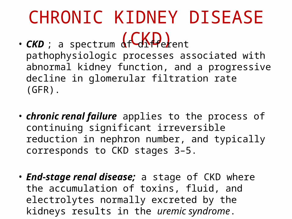
CHRONIC KIDNEY DISEASE (CKD)• CKD ; a spectrum of different pathophysiologic
processes associated with abnormal kidney function, and a progressive decline in glomerular filtration rate (GFR).
• chronic renal failure applies to the process of continuing significant irreversible reduction in nephron number, and typically corresponds to CKD stages 3–5.
• End-stage renal disease; a stage of CKD where the accumulation of toxins, fluid, and electrolytes normally excreted by the kidneys results in the uremic syndrome.
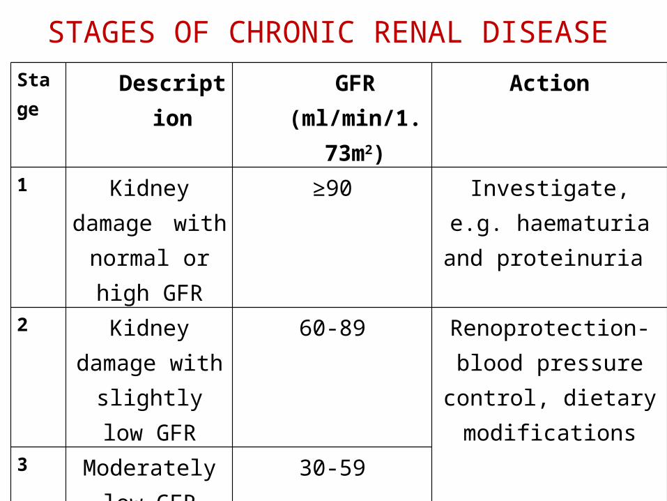
STAGES OF CHRONIC RENAL DISEASEStage Description GFR
(ml/min/1.73m2)
Action
1 Kidney damage with normal or
high GFR
≥90 Investigate, e.g. haematuria and
proteinuria 2 Kidney damage
with slightly low GFR
60-89 Renoprotection-blood pressure control, dietary
modifications3 Moderately low
GFR30-59
4 Severe low GFR 15-29 Prepare for renal replacement therapy (if
appropriate)5 Kidney failure < 15 or dialysis
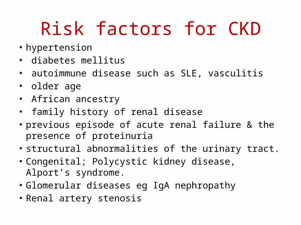
Risk factors for CKD• hypertension• diabetes mellitus• autoimmune disease such as SLE, vasculitis• older age• African ancestry• family history of renal disease• previous episode of acute renal failure & the presence of
proteinuria• structural abnormalities of the urinary tract.• Congenital; Polycystic kidney disease, Alport’s syndrome.• Glomerular diseases eg IgA nephropathy• Renal artery stenosis
Pathophysiology of CKD2 mechanisms; initiating mechanisms specific to the underlying etiology
(e.g., immune complexes and mediators of inflammation or toxin exposure)
a set of progressive mechanisms, involving hyperfiltration and hypertrophy of the remaining viable nephrons, eventually sclerosis & dropout of the remaining nephrons.
• The responses to reduction in nephron number are
mediated by vasoactive hormones, cytokines, and growth factors.
Clinical PresentationNB: Uraemia refers to clinical symptoms & signs of
RF due to loss of the excretory, metabolic and endocrine functions of the kidney.
• asymptomatic until GFR falls below 30 ml/minute
• may present as a raised blood urea and creatinine found during routine examination, often accompanied by HTN, proteinuria or anaemia.
Clinical Presentation cont’d• Nocturia• tiredness or breathlessness• Weakness• Kussmaul's breathing• anorexia and nausea• hiccoughs• pruritus• vomiting• muscular twitching• fits, drowsiness and coma ensue.
Clinical Presentation cont’d
PHYSICAL EXAMINATION;• Wasted• Yellow complexion• Pallor• Features of cardiac
tamponade• Brown line
pigmentation of nails• Excoriations
• Ecchymoses• Increased RR & depth• P. neuropathy• Absent reflexes• Reduced sensation• Paraesthesias• Restless legs
Diagnosis and management
• Proper history(causes/risk factors, symptoms, complications)
• Physical examination• INVESTIGATIONS- CBC - Urinalysis; protein- RFTs - LFTs (albumin)- Calcium and phosphate levels- PTH- Hepatitis(B & C) and HIV serology- Others tailored to underlying cause
ManagementIf diagnosis is not known• Immunoglobulins and protein electrophoresis • Urinary Bence Jones protein • Complement • ANA: and dsDNA if ANA is positive • ENA: if a connective tissue disorder is suspected • Rheumatoid factor • ANCA: in all possible inflammatory renal disease • Anti-GBM: in all possible inflammatory renal
disease
Management
• Imaging- Renal U/S ( kidneys, symmetry, size, renal masses,
evidence of obstruction)- Renal artery imaging: if renovascular disease is
suspected… doppler U/S- Voiding cystogram; reflux nephropathy- ??? Radiographic contrast imaging not recommended- Chest X-ray; heart size, pulmonary oedema- ECG; if > 40 years or there are risk factors for cardiac
disease
Mngt cont’d• Role of renal biopsy in CKD-mainly indicated in early stage CKD to establish
etiology in absence of clinical dx.Contraindications• bilaterally small kidneys• uncontrolled hypertension• active urinary tract infection• bleeding diathesis• morbid obesity. NB; bleeding time should be measured, and, if
increased, desmopressin should be administered immediately prior to the procedure.
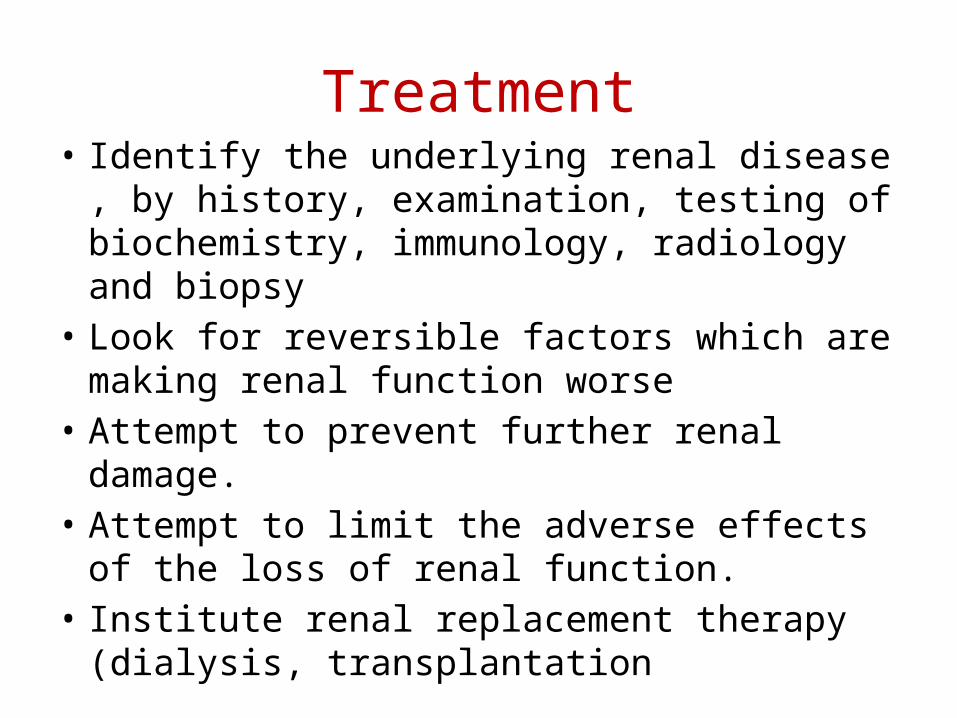
Treatment• Identify the underlying renal disease , by
history, examination, testing of biochemistry, immunology, radiology and biopsy
• Look for reversible factors which are making renal function worse
• Attempt to prevent further renal damage. • Attempt to limit the adverse effects of the loss
of renal function. • Institute renal replacement therapy (dialysis,
transplantation
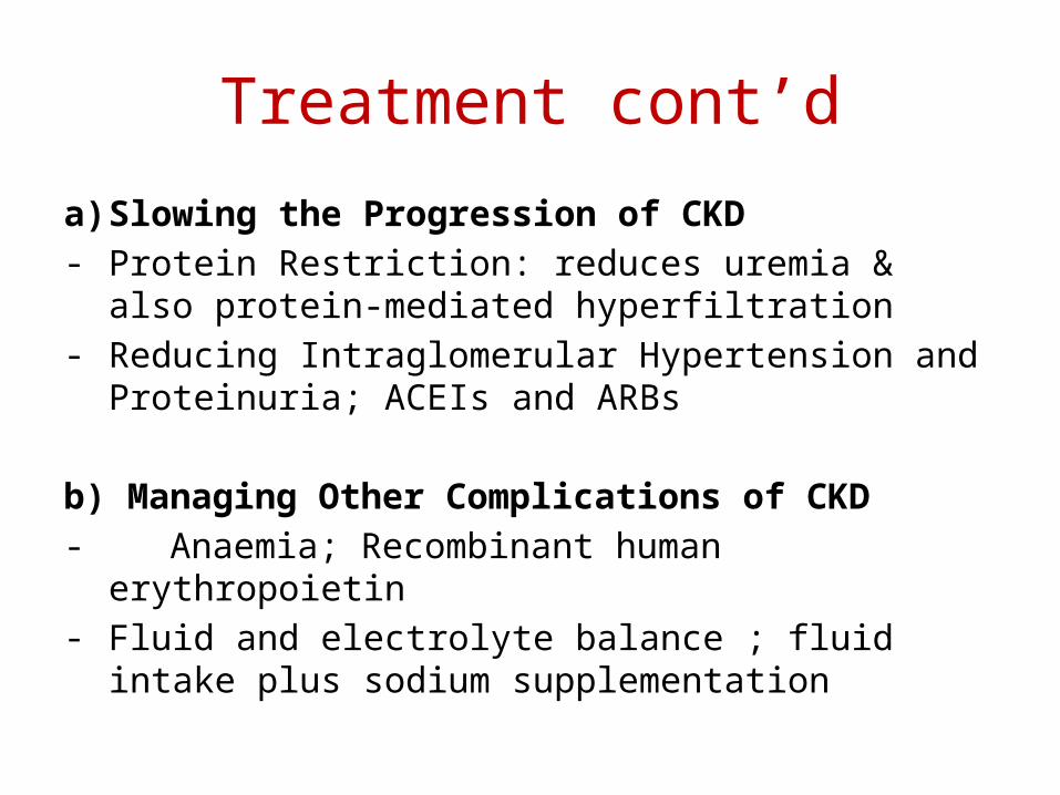
Treatment cont’d
a) Slowing the Progression of CKD- Protein Restriction: reduces uremia & also protein-
mediated hyperfiltration- Reducing Intraglomerular Hypertension and
Proteinuria; ACEIs and ARBs
b) Managing Other Complications of CKD- Anaemia; Recombinant human erythropoietin- Fluid and electrolyte balance ; fluid intake plus
sodium supplementation
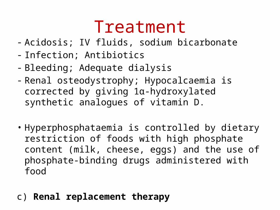
Treatment- Acidosis; IV fluids, sodium bicarbonate- Infection; Antibiotics- Bleeding; Adequate dialysis- Renal osteodystrophy; Hypocalcaemia is corrected by
giving 1α-hydroxylated synthetic analogues of vitamin D.
• Hyperphosphataemia is controlled by dietary restriction of foods with high phosphate content (milk, cheese, eggs) and the use of phosphate-binding drugs administered with food
c) Renal replacement therapy
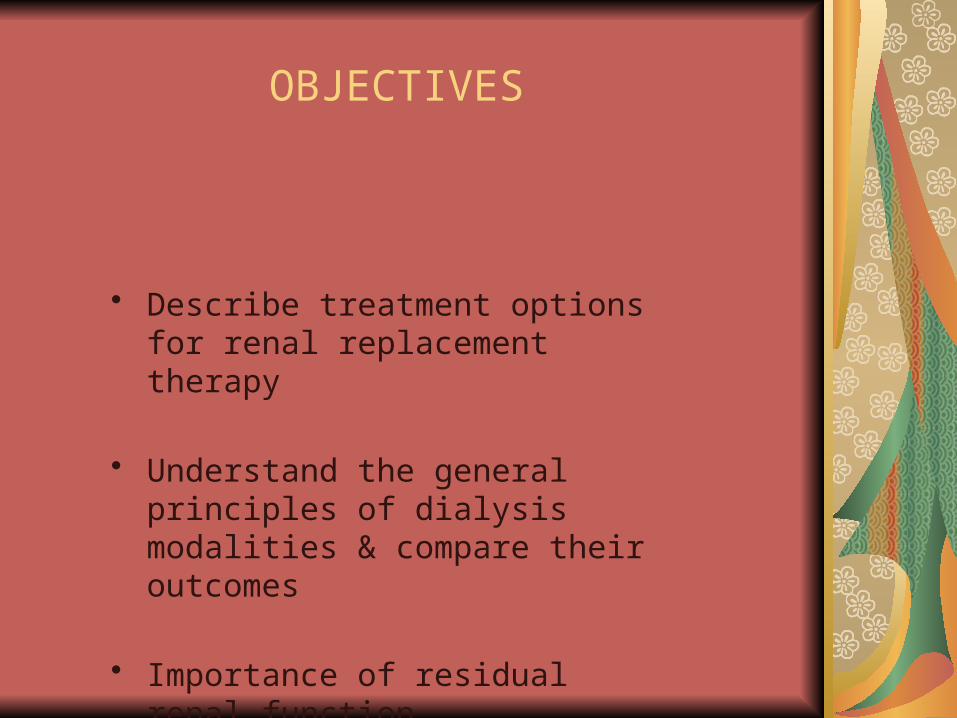
OBJECTIVES
Describe treatment options for renal replacement therapy
• Describe treatment options for renal replacement therapy
• Understand the general principles of dialysis modalities & compare their outcomes
• Importance of residual renal function
of dialysis modalities & compare th
PURPOSES OF DIALYSIS
1. Removes excess fluids and waste products.2. Restores chemical and electrolyte balance
HEMODIALYSIS‑ one of several renal replacement therapies used for the treatment of renal failure. HD involves the extracorporeal (outside of the body) passage of the client’s blood through a semi permeable membrane that serves as an artificial kidney.
Indications for Renal Replacement Therapy
• Hyperkalemia• Metabolic acidosis• Fluid overload (recurrent CHF
admissions)• Uremic pericarditis (rub)• Other non specific uremic symptoms:
anorexia and nausea, impaired nutritional status, increased sleepiness, and decreased energy level, attentiveness, and cognitive tasking, …
GENERAL GUIDELINE REQUIREMENTS FOR APPROPRIATE CLIENT SELECTION
1. Presence of fatal, irreversible renal failure when other therapies are unacceptable or ineffective.
2. Absence of illnesses that would prevent or seriously complicate HD.
3. Expectation of rehabilitation.
4. The client’s acceptance of the regimen.
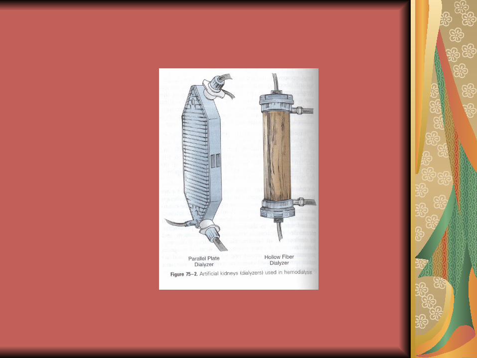
Components of HemodialysisDialyzer or artificial kidney
Dialyzer has 4 components: Blood compartment, Dialysate compartment, Semipermeable membrane, enclosed structure to support the membrane.
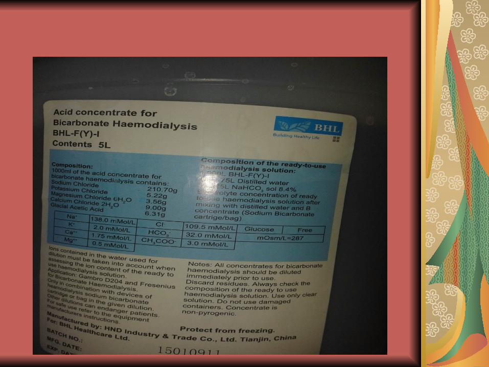
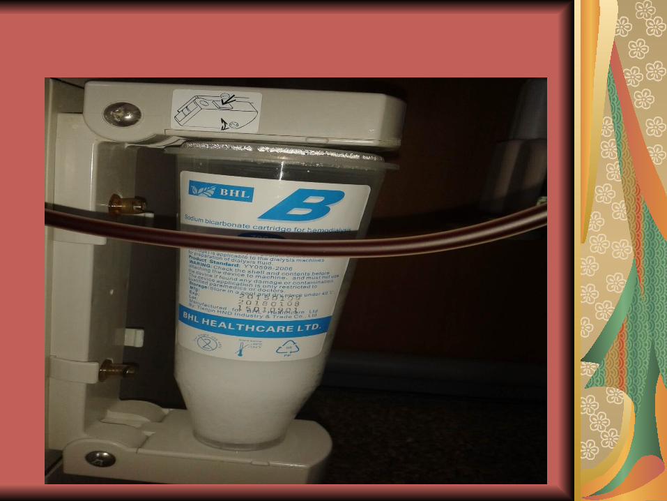
Dialysate – made up of clear H2O & chemicals. Compositions may be altered according to patient’s needs for treatment of electrolyte imbalance. Warmed to 37.8 C = to 100 F to increase efficiency of diffusion. Prevent decrease in pt’s blood temperature.
Vascular access routes – AV fistula, AV Graft, Dual Lumen Cathater, AV Shunt.
Hemodialysis machine
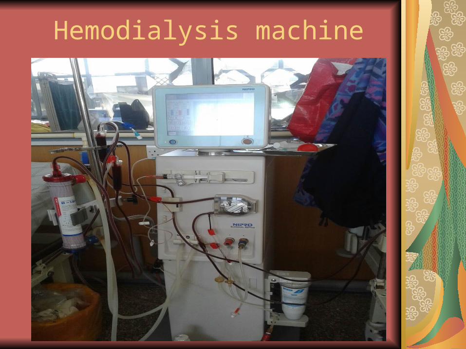
Hemodialysis machine
PROCEDURE
The principles of HD are based on the passive transfer of toxins, which is accomplished by diffusion.
When HD is initiated, blood and dialysate flow in opposite directions from their respective sides of an enclosed semi permeable membrane.
The dialysate is a balanced mix of electrolytes and water that closely resembles human plasma. On the other side of the membrane is the client’s blood, which contains metabolic waste products, excess water, and excess electrolytes.
Cont…
During HD, the waste products move from the blood into the dialysate because of the difference in their concentrations (diffusion). Excess water is also removed from the blood into the dialysate (osmosis). Electrolytes can move in either direction, as needed, and take some fluid with them. Potassium and sodium typically move out of the plasma. This process continues as the blood and the dialysate are circulated past the membrane for a preset length of time.Duration and frequency of HD treatment depends on the amount of metabolic waste to be cleared, and the amount of fluid to be removed.
COMPLICATIONS OF HEMODIALYSIS
Dialysis disequilibrium syndrome- the cause is
unknown but maybe due to rapid decrease in
blood urea nitrogen levels during HD. These change can cause cerebral edema- leads to increase intracranial pressure. Infection- transmitted by blood transfusion are
another serious complication associated with long term HD.
Hepatitis Infection- in clients with chronic renal failure.

Best Practice for Caring for the client Undergoing Hemodialysis
Weigh the client before and after dialysis.
Know the client’s dry weight.
Discuss with physician whether any of the
client’s medications should be withheld until after
dialysis.
Be aware of events that occurred during the
dialysis treatment.
Cont…Measure blood pressure, pulse
rate, respirations, and temp.
Assess for symptoms of orthostatic hypotension.
Assess the vascular access site.
Observe for bleeding
Assess the client’s level of
consciousness and assess for
headache, nausea, and vomiting.
COMPLICATIONS OF AV FISTULAE OR SYNTHETIC AV GRAFT
Stenosis‑the most frequent cause of
permanent peripheral hemodialysis access
failure is vascular stenosis.
Thrombosis‑ this complication is more
common in synthetic AV grafts than native AV
fistulae.
Failure of maturation‑ a native AV fistula
requires 1 to 4 months to mature; if blood flow
is diminished by stenosis or multiple outflow
veins, maturation will be impaired.
Cont..Infection‑ a leading cause of
complications and death in dialysis
patient. Typical S/S of an infected dialysis
access include local erythema, induration,
tenderness, and purulent drainage from
incision sites.
Ischemic steal syndrome‑ diverting blood
flow from the distal extremity through the
hemodialysis access may cause pain and
ischemia in some patients, esp.diabetic
and elderly patient.
Pseudoaneurysm‑ also called false
aneurysm or pulsating hematoma
TYPES OF VASCULAR ACCESS FOR HEMODIALYSIS
Permanent AV fistular –forearm for 2-4 months or more
Av graft- forearm for 1-2 weeks Dualumen HD – subclavian vein –
immediately post operatively and after x-ray confirmation of placement
Temporary HD catheter (dual or triple)- subclavian, internal jugular or femoral vein- immediately after insertion and x-ray confirmation
AV shunt (relatively uncommon)-forearm- immediately after insertion

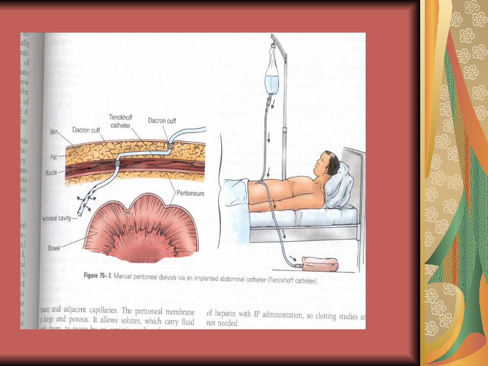
PERITONIAL DIALYSIS
Peritoneal dialysis (PD) takes place within the
peritoneal cavity. PD is slower than hemodialysis,
However , and more time is needed for the same
effect to be obtained.
TYPES OF PERITONEAL DIALYSIS
CAPD- Continuous Ambulatory Peritoneal Dialysis
MBCAPD‑ Multiple‑Bag CAPD
APD‑ Automated Peritoneal Dialysis
IPD‑ Intermittent Peritoneal Dialysis
CCPD‑ Continuous Cycle peritoneal Dialysis
PROCEDURE AND PROCESS The surgical insertion of a siliconized rubber
(Sillastic) catheter into the abdominal cavity is
required to allow the infusion of dialyzing fluid
(dialysate) is infused according to the physician
order, 1 to 2L of dialysate is infused by gravity (fill)
into the peritoneal space over a 10 to 20 minutes
period, according to the client’s tolerance. The fluid dwells in the cavity for a specified time
ordered by the physician. The fluid then flows out of the body (drain)
by gravity into a drainage bag.
Con’t of process and procedure
The peritoneal outflow contains the dialysate in
addition to the excess water, electrolytes
and nitrogenous waste products that have
accumulated in the body.
The Three Phases of the process:
1. Infusion or fill.
2. Dwell
3. Outflow or drain.
Cont.. PD occurs through diffusion and osmosis
across the Semipermeable peritoneal
membrane and adjacent capillaries.
The peritoneal membrane is large and porous.
it allows solutes, which carry fluid with
them to move by an osmotic gradient from
an area of higher concentration in the body
(blood) to an area of lower concentration in
the dialyzing fluid.
Complications of CAPD
PERITONITIS‑the major complication of PD. The
most common cause of peritonitis is contamination
of the connection site during an exchange. The
infection of peritoneum is manifested by cloudy
dialysate outflow (effluent), fever, rebound
abdominal tenderness, abdominal pain, general
malaise, nausea, and vomiting.
Cont..
Cloudy or opaque effluent is the earliest sign of
peritonitis. The best treatment of peritonitis is
prevention.
The nurse must maintain meticulous sterile
technique when caring for the PD catheter
and when hooking up or clamping off
dialysate bags.
Con’t
Pain- pain during inflow of dialysate is common
during the first few exchanges because of
peritoneal irritation; however, it disappear after a
week or two. Cold dialysate aggravates
discomfort. Thus the dialysate bags should be
warmed before instillation by use of a heating pad
to wrap the bag or use of warming chamber.
Con’t Microwave oven are not recommended for
the warming of dialysate because of their
unpredictable warming patterns and
temperatures.
Exit Site and Tunnel infections- the normal
exit site from a PD catheter should be clean,
dry, and with out pain or evidence of
inflammation.
Con’t
Insufficient flow of the Dialysate- Constipation is
the primary cause of inflow or outflow problems.
To prevent constipation, the physician orders a
bowel preparation before placing the PD catheter.
The nurse ensures that the drainage bag is lower
than the client abdomen. The nurse inspects the
connection tubing and PD system for kinking or
twisting and rechecks to make sure that clamps
are open.
Con’t
Dialysate Leakage- when dialysis is initiated,
small volumes of dialysate are used. It may
take clients 1 to 2 weeks to tolerate a full 2-L
exchange without leakage around the catheter
site.
Other Complication- The nurse notes any
change in the color of the outflow.
NURSING CARE DURING PERITONEAL DIALYSIS
Evaluate baseline vital signs
The client is weigh, always on the same scale, before
the beginning of the procedure or at least every 24
hours while receiving the treatment.
Baseline laboratory value determination, such as
electrolyte and glucose levels,
Con’t
During PD, the nurse continually monitors the
client. For the first
exchanges, record the values every 15
minutes. Ongoing assessment for
respiratory distress, pain or discomfort.
Abdominal dressing around the catheter exit
site is checked frequently for wetness.
Monitor for dwell time.
NURSING CARE CON’T
For hourly exchanges, dwell time usually ranges from 20 to 40
Minutes. Blood glucose assessment is necessary, due to
Glucose absorption occur in some patient.
The outflow is recorded accurately after each exchange.
Con’t
Visual inspection of the outflow bag and daily
weights may be sufficient to note the
adequacy of the return. If drainage return is
brown, a bowel perforation must be
suspected. If drainage return is the same
color as urine and has the same glucose
concentration, a possible bladder perforation
should be investigated. If drainage is cloudy
or opaque, an infection is suspected.