renal disease
DESCRIPTION
Renal Disease. Ricki Otten MT(ASCP)SC [email protected]. Review the Objectives. Those objectives marked with ‘*’ will not be tested over during the Student Lab Rotation. Classification of Renal Disease. Usually by specific structural component affected by disease Glomerular Disease - PowerPoint PPT PresentationTRANSCRIPT


2
Review the Objectives
• Those objectives marked with ‘*’ will not be tested over during the Student Lab Rotation

3
Classification of Renal Disease
Usually by specific structural component
affected by disease
1. Glomerular Disease
2. Tubular Disease
3. Interstitial Tissue Disease
4. Vascular Disease

4
Glomerular Disease• Most often due to damage to glomerular
basement membrane– Immunologic disease– Metabolic disease– Hereditary disease
• Basement membrane damage leads to– Morphologic changes – Altered glomerular function– Increased permeability allowing leakage of
cells and protein into urine

5
Glomerular Disease• Classification
– Primary: specifically affects the kidney• Acute glomerulonephritis• Chronic glomerulonephritis• Nephrotic syndrome
– Secondary: another disease process affects the health of the glomerulus
• Systemic disease (diabetes mellitus, SLE) • Hereditary disorder

6
Glomerular Injury
• Clinical features dependent upon– Number of glomeruli involved– Mechanism of injury– Rapidity of disease onset

7
Glomerular Injury• Clinical findings:
– Urinalysis: proteinuria, hematuria
– Oliguria
– Physical findings: edema, hypertension
– Blood evaluation: hypoproteinemia, azotemia
(increased urea, creatinine)

8
Glomerular Disease
• Acute glomerulonephritis
• Chronic glomerulonephritis
• Nephrotic syndrome
• Diabetes mellitus (nephropathy)

9
Acute Glomerulonephritis• Acute post-streptococcal glomerulonephritis
– Relatively common, often in children, also adults– Occurs 1-2 weeks post streptococcal infection– Antibody mediated: blood cultures negative
• Clinical findings:– Sudden onset, fever, malaise, nausea– Oliguria– Edema (lower extremities (ankles), eyes)– Mild hypertension

10
Acute Glomerulonephritis• Urinalysis
– Physical Color? Clear?– Chemical– Microscopic

11
Acute Glomerulonephritis• Urinalysis
– Physical yellow, hazy– Chemical ?– Microscopic

12
Acute Glomerulonephritis• Urinalysis
– Physical yellow, hazy
– Chemical + Blood
Proteinuria (mild)
(<1.0 gram/24 hour)
– Microscopic: ?

13
Acute Glomerulonephritis• Urinalysis
– Microscopic:
RBC (some dysmorphic)
WBC
RTE
Casts: RBC hemoglobin granular

14
Acute Glomerulonephritis• Other testing:
– Blood• ASO titer• Decreased complement (Antigen-Antibody mediated)• Increased BUN, increased creatinine• Decreased albumin
– Urine• Decreased CrCl = Decreased GFR• Proteinuria (mild: <1.0 grams/24 hr)

15
Acute Glomerulonephritis• Majority (>95%) of children recover
• Approx 60% of adults recover
• Only 1-2 % post-strep acute glomerulonephritis develop chronic glomerulonephritis

16
Chronic Glomerulonephritis• Numerous glomerular diseases develop
chronic glomerulonephritis
• Onset is slow and insiduous taking many years to develop clinical signs and symptoms
• If not treated, may result in death (uremia)
• Clinical findings: same as acute, but worse

17
Chronic Glomerulonephritis
• Urinalysis– Physical Color? Clear?– Chemical– Microscopic

18
Chronic Glomerulonephritis
• Urinalysis– Physical yellow, hazy– Chemical ?– Microscopic

19
Chronic Glomerulonephritis
• Urinalysis– Physical yellow, hazy
– Chemical+ BloodProteinuria (mild-moderate)
(>2.5 and < 3.5 grams/24 hr)Specific gravity: low and fixed
(isosthenuric)
– Microscopic: ?

20
Chronic Glomerulonephritis• Urinalysis
– Microscopic
RBC
WBC
RTE
Casts (RBC, hemoglobin, granular, waxy)

21
Chronic Glomerulonephritis
• Other testing: – Blood:
• Increased BUN, increased creatinine• Decreased albumin, decreased TSP
– Urine: • Decreased CrCl = decreased GFR• Proteinuria (moderate: >2.5 grams/24 hr)

22
Nephrotic Syndrome
• Selective filtering capability of glomerulus is lost
• Many conditions may lead to NS
• Clinical findings: ‘pitting edema’, azotemia, hypertension, oliguria

23
Nephrotic Syndrome
• Urinalysis– Physical Color? Clear?– Chemical– Microscopic

24
Nephrotic Syndrome
• Urinalysis– Physical yellow, hazy (cloudy ?)– Chemical ?– Microscopic
25
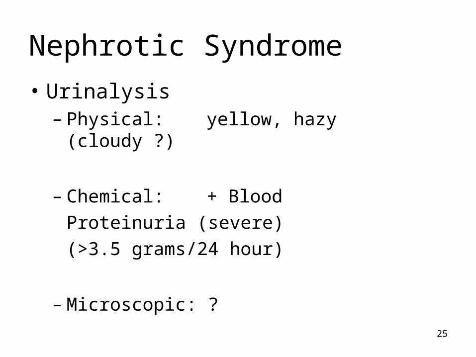
Nephrotic Syndrome
• Urinalysis– Physical: yellow, hazy (cloudy ?)
– Chemical: + Blood
Proteinuria (severe)
(>3.5 grams/24 hour)
– Microscopic: ?

26
Nephrotic Syndrome
• Urinalysis– Microscopic
RBC
WBC
RTE
Oval Fat Bodies (OFB)
Free fat droplets
Casts (granular, fatty, waxy, RTE)

27
Nephrotic Syndrome
• Other testing:– Blood:
• hypoproteinemia (decr albumin, decr TSP)• Increased lipids• Increased sodium
– Urine: • Decreased CrCl = decreased GFR• Proteinuria (severe: > 3.5 grams/24 hr)

28
Diabetes Mellitus (Nephropathy)
• Disorder of carbohydrate metabolism
• Renal disease is a major cause of death in the diabetic patient
• Diabetes is leading cause of– Blindness– End-stage renal disease– Limb amputations

29
Diabetes Mellitus (Nephropathy)
• Clinical findings:– Polyuria– Polydipsia– Nocturia

30
Diabetes Mellitus (Nephropathy)
• Urinalysis– Physical Color? Clear?– Chemical– Microscopic

31
Diabetes Mellitus (Nephropathy)
• Urinalysis– Physical Yellow, hazy– Chemical ?– Microscopic

32
Diabetes Mellitus (Nephropathy)
• Urinalysis– Physical Yellow, hazy– Chemical
+ Glucose
Proteinuria (mild-moderate)
– Microscopic ?

33
Diabetes Mellitus (Nephropathy)
• Urinalysis– Microscopic
RBC
Casts
Yeast, possibly
Depends on extent of renal involvement (disease)

34
Diabetes Mellitus (Nephropathy)
• Other testing:– Blood
• Increased glucose• Increased ketones (diabetes mellitus, type 1)
– Urine• Proteinuria: leads to chronic renal failure and death

35
Tubular Disease
• Altered tubular function
• Necrosis of tubular epithelium

36
Altered Tubular Function
• Caused by– Reabsorption-secretion capability lost– Concentrating-diluting capability lost
• Results in– Build up of waste products in bloodstream– Loss of essential substances into urine

37
Altered Tubular Function
• Renal glycosuria– Glucose in urine, renal threshold not exceeded
• Cystinuria
• Cystinosis
• Renal tubular acidosis– Tubules unable to secrete adequate H+ despite
systemic acidosis
Inherited disorders
Cystine crystals in urine

38
Urinalysis Findings
• Renal glycosuria: + glucose
• Cystinuria, cystinosis: cystine crystals
• Renal tubular acidosis: pH not as acid as is needed to compensate for systemic acidosis

39
Necrosis of Tubular Epithelium
• Destruction of tubular epithelial cells– Toxin– Ischemic event
• Most common cause of renal failure

40
Necrosis of Tubular Epithelium
• Clinical presentation: 3 phases– Onset– Renal failure
• Azotemia• Hyperkalemia• Metabolic acidosis• Oliguria
– Recovery

41
Acute Tubular Necrosis• Toxic ATN
– Drugs: AminoglycosidesAnestheticsRadiographic dyesChemotherapyAnti-rejection drugs
– Toxins: MercuryLeadCadmiumEthylene glycolPesticidesMushrooms

42
Acute Tubular Necrosis
• Ischemic ATN: decreased perfusion of kidneys as a result of hypotensive events
– Sepsis: bacterial infection of bloodstream– Shock– Trauma

43
Acute Tubular Necrosis• Urinalysis
– Physical: Yellow, hazy
– Chemical:
Proteinuria (mild), +blood, low specific gravity
– Microscopic:
RBC, WBC, RTE
Casts: RTE, granular, waxy, broad

44
Interstitial Tissue Disease
• Lower urinary tract infection– Cystitis (bladder)– Urethritis (urethra)
• Acute pyelonephritis (upper UTI)
• Yeast infection
• Any bacterial or fungal agent can cause a UTI

45
Lower UTI
• ~85% of lower UTI caused by
gram-negative rods (fecal E.coli)
• Urinalysis– Physical Color? Clear?– Chemical– Microscopic

46
Lower UTI
• Urinalysis– Physical yellow, hazy (cloudy, turbid)– Chemical ?– Microscopic

47
Lower UTI
• Urinalysis– Physical yellow, hazy (cloudy, turbid)– Chemical + protein (<0.5 grams/24 hr)
+ leukocyte esterase
+ nitrite
+ blood
– Microscopic ?

48
Lower UTI
– Microscopic
WBC
Bacteria
RBC
Transitional epithelial cells (cystitis)
Absence of casts: why?

49
Acute Pyelonephritis
• Most common upper UTI
• Two mechanisms causing infection– Bacterial moving from lower to upper urinary
tract– Septicemia localizing in the kidneys
• Incomplete voiding due to obstruction or dysfunction or anatomic abnormality
• Catheterization, pregnancy, diabetes

50
Acute Pyelonephritis
• Urinalysis– Physical Color? Clear?– Chemical– Microscopic

51
Acute Pyelonephritis
• Urinalysis– Physical Yellow, hazy (cloudy, turbid)– Chemical ?– Microscopic

52
Acute Pyelonephritis
• Urinalysis– Physical Yellow, hazy (cloudy, turbid)– Chemical + protein (<1.0 gram/24 hr)
+ leukocyte esterase (WBC)+ nitrite+ bloodspecific gravity: normal to
low
– Microscopic ?
53
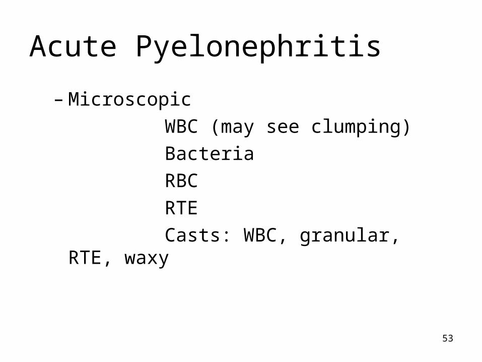
Acute Pyelonephritis
– Microscopic
WBC (may see clumping)
Bacteria
RBC
RTE
Casts: WBC, granular, RTE, waxy

54
Yeast Infection
• Urinary tract of both men and women are susceptible to yeast infection
• Most often vaginal yeast infection contaminates urine
• Often caused by Candida species
(candida albicans)

55
Yeast Infection
• Candida species (candida albicans)– Normal flora of GI tract and vagina– Normal bacterial flora keep yeast proliferation
under control– Catheters provide mode of inoculation

56
Yeast Infection
• Urinalysis– Physical Color? Clear?– Chemical– Microscopic

57
Yeast Infection
• Urinalysis– Physical Yellow, hazy (cloudy)– Chemical ?– Microscopic

58
Yeast Infection• Urinalysis
– Physical Yellow, hazy (cloudy)– Chemical + WBC ?
+ blood ?
– Microscopic
Yeast
Mycelial elements
RBC? WBC?
Casts? Why or why not?

59
Vascular Disease
• Any disorder that affects the blood flow to the kidneys can cause renal disease
– Cardiac disease (25% of cardiac output)– Atherosclerosis– Hypertension– Diabetes– Eclampsia– Etc

60