renal cell carcinoma: review of etiology, …renal cell carcinoma: review of etiology,...
TRANSCRIPT
Biomed Pap Med Fac Univ Palacky Olomouc Czech Repub. 2016 Jun; 160(2):183-194.
183
Renal cell carcinoma: Review of etiology, pathophysiology and risk factorsNadezda Petejovaa,b, Arnost Martineka,b
Background and Aims. The global incidence of renal cell cancer is increasing annually and the causes are multifacto-rial. Early diagnosis and successful urological procedures with partial or total nephrectomy can be life-saving. However, only up to 10% of RCC patients present with characteristic clinical symptoms. Over 60% are detected incidentally in routine ultrasound examination. The question of screening and preventive measures greatly depends on the cause of the tumor development. For the latter reason, this review focuses on etiology, pathophysiology and risk factors for renal neoplasm. Methods. A literature search using the databases Medscape, Pubmed, UpToDate and EBSCO from 1945 to 2015.Results and Conclusions. Genetic predisposition/hereditary disorders, obesity, smoking, various nephrotoxic indus-trial chemicals, drugs and natural/manmade radioactivity all contribute and enviromental risks are a serious concern in terms of prevention and the need to screen populations at risk. Apropos treatment, current oncological research is directed to blocking cancer cell division and inhibiting angiogenesis based on a knowledge of molecular pathways.
Key words: hereditary syndromes, nephrectomy, radioactivity, renal cell carcinoma, renal carcinogenesis, uranium toxicity
Received: May 4, 2015; Accepted with revision: September 18, 2015; Available online: November 3, 2015http://dx.doi.org/10.5507/bp.2015.050
aDepartment of Internal Medicine, University Hospital Ostrava, Czech RepublicbDepartment of Clinical Studies, Faculty of Medicine, University of Ostrava, Czech RepublicCorresponding author: Nadezda Petejova, e-mail: [email protected]
INTRODUCTION
Renal cancer (RC) accounts for around 3% of all adult malignancies and is the twelfth most common can-cer in the world, with 338,000 new cases diagnosed in 2012 and around 100,000 deaths annually1,2. Cancers of the kidney are more common in men than in women, and over the last few decades, the incidence has been increasing in many parts of the world. About 59% of RC cases occur in more developed countries. The global incidence rates are highest in Europe, North America and Australia and lowest in Africa, India and China. The Czech Republic has the highest rate of RC in the world, followed by Lithuania and Slovakia. The incidence in the Czech Republic to 2012 was reported as 24.1 (men) and 10.5 (women) per 100,000 people per year1.This accounts in the Czech Republic for around 2,000 partial or radi-cal nephrectomies yearly. Renal cell carcinoma (RCC) accounts for 80-85% of kidney cancers. It is the most common kidney variety and the third most commonly diagnosed urogenital malignancy3.
The most frequent histological type of RCC is clear cell renal cell carcinoma (ccRCC), with a prevalence of 75% of all primary kidney cancers. Papillary and chro-mophobe RCC are two less common subtypes (ref.4,5). Renal pelvic cancer accounts for the remaining 10%. Nephroblastoma (Wilms tumor), the primary renal car-cinoma in children comprises about 1.1% of all kidney cancers6. Up to 10% of RCC patients present with charac-teristic clinical symptoms consisting of hematuria, lateral dorsal or flank pain and palpable abdominal mass. Over
60% of RCC are detected incidentally in routine ultra-sound examination7. Despite the advances in diagnosis, especially improved imaging techniques, about 20–30% of all patients are diagnosed with metastatic disease. Patients with metastatic RCC have a median survival of around 13 months. The 5-year survival rate is under 10% (ref.8). More than 20% of patients undergoing nephrectomy will develop metastases during follow-up9. For those with met-astatic disease, the prognosis is extremely poor despite advances in multimodal treatment. Therapeutic options for RCC are limited due to resistance to chemotherapy and radiotherapy and to the low efficiency and toxicity of immunotherapy10,11.
CLASSIFICATION AND PATHOLOGY OF PRIMARY RENAL NEOPLASMS
The four most common malignant epithelial neo-plasms in adults are clear cell, papillary, chromophobe RCC and collecting-duct carcinoma. The rare benign pri-mary renal tumor with unique microscopic features is an oncocytoma.
Clear cell renal cell carcinomaThe genetic features most closely associated with
ccRCC are mutation, hypermethylation, loss or biallelic inactivation of the tumor suppressor - von Hipple–Lindau gene(VHL) (ref.12,13).The loss of the wild-type allele of VHL is found in hemangioblastomas, pancreatic neuro-endocrine tumors, kidney cysts, and ccRCC in patients
Biomed Pap Med Fac Univ Palacky Olomouc Czech Repub. 2016 Jun; 160(2):183-194.
184
with VHL. Inactivation of VHL results in upregulation of hypoxia inducible factors (HIF)-1α and 2α which drive an-giogenesis and proliferation and has profound effects on energy metabolism14-16. According to recent data, inactiva-tion of VHL alone is not sufficient to cause ccRCC. Other genes are likely to be important in its development includ-ing: polybromol (PBRM1), BRCA1 associated protein-1, SET domain containing 2 (SETD2) and lysine K-specific demethylase 6A (KDM6A) (ref.17-20). Histopathologically clear cell RCC appears as golden yellow but the color var-ies with tumor grade. Under light microscopy, the tissue can demonstrate a variety of growth patterns including solid, acinar and cystic papillary, pseudopapillary, tubu-lar and sarcomatoid. The cytoplasm is typically clear or granular-eosinophilic21. The clear cell RCCs are highly vascularised tumors due to upregulation of vascular endo-thelial growth factor A (VEGFA or VEGF) and platelet-derived growth factor B (PDGFB) which both promote angiogenesis22,23.
Papillary renal cell carcinomaThis type of kidney tumor comprises approximately
10% of all RCCs (ref.24,25). Two familial syndromes are associated with increased risk of papillary-type RCC: hereditary papillary RCC is an autosomal dominant syn-drome characterized by multifocal, bilateral, type 1 - RCC caused by mutation of the MET gene on 7q31. The papil-lary type 2 - RCC is the pathological type most commonly associated with Hereditary Leiomyomatosis (HLRCC) and tends to have an early age of onset. Mutation of the fumarate hydratase (FH) gene which encodes the enzyme that converts fumarate to malate in the Krebs cycle, is mutated in HLRCC. It should be noted that mutations in FH also occur in fumarate hydratase deficiency (FHD). Homozygous or compound heterozygous FH germline mutations cause autosomal recessive FHD, a metabolic disease characterised by neurological impairment and encephalopathy26,27. Multifocal disease is a pathological feature of papillary RCC and under light microscopy, necrosis is often seen. The cancer cells associated with HLRCC have typically large nuclei with inclusion-like orangiophilic or eosinophilic nucleoli and hemosiderin pigment in the cytoplasm16,21,28.
Chromophobe renal cell carcinomaChromophobe renal cell carcinoma is a distinct sub-
type of renal cell carcinoma that accounts for 5% of all renal neoplasms. This subtype is further subdivided into two variants, classic and eosinophilic (oncocytic).
The hereditary disease associated with chromophobe RCC (chRCC) is an autosomal dominant Birt-Hogg-Dubé (BHD) syndrome which is caused by germline mutations in the folliculin gene – FLCN maps to chromosome 17 and was subsequently identified at 17p11.2 (ref.29).This gene acts as a tumor suppressor and interacts with mTOR and AMP activated protein-kinase signalling pathways30. Patients with the BHD syndrome tend to develop fibrofol-liculomas, lung cysts, spontaneous pneumothorax, renal cysts, cancers and skin manifestations as multiple fibrofol-liculomas, trichodiscomas and acrochordons31. Few other
mutations in tumor suppressor genes have been identi-fied in chromophobe RCC. One example is dismutations in PTEN located in 10q23 and TP53 located at 17p13 (ref.13).
Collecting-duct carcinomaThis rare type of renal neoplasm comprises less than
1% of primary renal tumors and is also known as Bellini duct carcinoma, medullary renal carcinoma, distal renal tubular carcinoma and distal nephron carcinoma21,32. The tumor arises from the collecting duct in the renal me-dulla, is highly aggressive and most patients present with metastatic disease. Typically, the metastasis is to regional lymph nodes in approximately 80% of cases, to the lung or adrenal gland and to the liver32,33. Associated chromo-somal abnormalities are losses of various chromosomal regions on chromosomes: -1p, -8p, -9p, and gains on chro-mosome at-13q (ref.34).
Renal oncocytomaThe incidence of oncocytomas ranges from 3% to 7%
of all primary renal neoplasms. Oncocytoma has a wide age distribution, with a peak incidence in the seventh de-cade of life. Men are affected twice as commonly as fe-males35. Renal oncocytoma is rare. Benign renal epithelial tumors are composed of large cells with mitochondria-rich cytoplasm thought to arise from intercalated cells of the collecting duct. The cytological features of renal oncocyto-ma show overlap with other renal entities36. Pathologically, classic renal oncocytomas have been described as cir-cumscribed solid tumors with a central stellate scar, with focal cystification reported in 20% to 37% of cases37,38. Pathological differentiation between an oncocytoma and an RCC with oncocytic features is difficult. The most re-cently published study described the usefulness of immu-nohistochemical markers: DOG1 (discovered on GIST 1), cyclin D1, CK7, CD117 and vimentin in the differential diagnosis of renal epithelial tumors. The results showed that of these markers, DOG1 is a very sensitive and very specific marker for distinguishing chRCC from ccRCC; Cyclin D1 is useful in discriminating between chRCC and renal oncocytoma; CK7 and CD117 are useful markers for distinguishing chRCC from renal oncocytoma and ccRCC; and vimentin is helpful for distinguishing clear cell RCC from chromophobe RCC and oncocytoma39.
ETIOLOGY
Demographics, cigarette smoking, use of phenacetin-containing analgetics, obesity, lack of physical activity, exposure to industrial or environmental agents and comor-bidities such as hypertension, hyperglycemia and hypertri-glyceridemia belonging to the metabolic syndrome (MS) have been associated with RCC (ref.6). From observa-tional studies, almost half of all kidney tumors are linked to obesity with a BMI>30 kg/m2 and renal cancer risk is between 20-35% higher for every 5 kg/m2 of higher BMI. This association suggests that obesity is the chief factor in a diverse array of fatal conditions from malignancies to
Biomed Pap Med Fac Univ Palacky Olomouc Czech Repub. 2016 Jun; 160(2):183-194.
185
cardiovascular and renal diseases40,41.The mechanisms that underlie the role of the MS in RCC carcinogenesis are complex, involving insulin resistance (IR), inflammation, angiogenesis, cell-stroma interaction, and other factors42. The insulin-like growth factor (IGF) family and changes in its components that result from IR play a crucial role in tumor development and progression. Through activation of mitogen-activated protein kinase and phosphatidylino-sitol-3 kinase signaling pathways, IGF-1 may play a role in the promotion of mitosis, cell migration and apopto-sis suppression. In addition, IGF-1 can stimulate tumor angiogenesis by increasing VEGF levels43. In the case of the MS, elevated levels of reactive oxygen species (ROS) together with pro-inflammatory cytokines and mediators of inflammation, such as NF-κB are known to affect cell apoptosis, proliferation and invasion42.
Recent studies have also suggested that circulating vita-min D-binding protein (VDBP) concentration plays a role in the etiology of several cancers, directly or by modifying the association between circulating vitamin D and risk of disease44,45.VDBP can directly impact carcinogenesis via membership of the extracellular actin scavenger system and by playing a role in macrophage activation, apoptosis and angiogenesis45,46. In a prospective study, Mondul et al. investigated 262 RCC male patients to determine whether circulating VDBP concentration was associated with risk of renal cell carcinoma and whether this modified the as-sociation with 25(OH)vitaminD. Men with higher serum concentrations of VDBP had lower risk of RCC, whereas the 25(OH)vitaminD:VDBPratio, a proxy for free circu-lating 25(OH)vitaminD, showed a possible positive risk association. Thus, the VDBP association may reflect a biological mechanism unrelated to vitamin D status47.
Anticancer properties are especially attributed to vi-tamin D3 which binds to the VDR receptor. Vitamin D3 and its analogues inhibit cell proliferation and promote apoptosis in cancer cells in culture. The major site for the synthesis of vitamin D3 is the kidney. Biological and epidemiological data suggest that vitamin D3 levels have an impact on the development of renal cancer. However, gene polymorphism of the vitamin D receptor may play the most important role in its development48.
The association between Fok1 polymorphism (ff gen-otype) of VDR and higher renal cancer risk has been confirmed in two unrelated studies: in Central- Eastern European and North Indian populations. In a recent re-search study of a North Indian population, an increased number of Fok1 polymorphism alleles have been linked to a high risk of renal cell cancer, also taking into consid-eration other risk factors such as hypertension, smoking and high body mass index48-50.
Several studies have suggested that gene-environment interactions in connection to RCC are linked to genes that underlie enzymes involved in metabolism. Polymorphism in genes encoding carcinogen metabolizing enzymes with altered expression and function may increase or decrease carcinogen activation or deactivation. The most widely studied has been cytochrome P450 especially in relation to CYP1A1. This plays a key role in the metabolism of drugs and environmental chemicals e.g. polycyclic aro-
matic hydrocarbons that may contribute to carcinogenesis and in estrogen metabolism51,52.The results of a metaanaly-sis by Chinese authors showed that CYP1A1 MspI poly-morphism was significantly associated with increased risk of RCC using three genetic comparison models (allele model: OR=1.49,95%CI 1.03-2.16;homozygous model: OR=1.64, 95% CI 1.13-2.40; dominant model: OR=1.72, 95%CI 1.07-2.76). Obvious heterogeneity was observed in an allele model and a dominant model53.
Other studies have highlighted the impact and risk for tumor development associated with nutrition, especially with intake of ochratoxins and citrinin. These mycotoxins or their metabolites could significantly contribute to this disease of the kidney in the Czech population. The accu-mulation of ochratoxins, citrinin (CIT) and of their me-tabolites in the human organism could initiate cancer and negatively affect either the rest of the kidney or to increas-ing the risk of recurrence or accelerate ongoing cancer. Ochratoxins and citrinin are toxicological and agricul-turally important mycotoxins. Ochratoxins and citrinin are produced by several fungi of the genera Penicillium, which contaminate foodstuffs, food and feed54. Citrinin, often found in the same food as ochratoxin A (OTA), is a powerful nephrotoxin. After exposure, ochratoxins show nephrotoxic, hepatotoxic, immunotoxic, neurotoxic, em-bryotoxic, teratogenic, genotoxic and carcinogenic effects in laboratory and farm animals55.
Ochratoxin A is an important nephrotoxin with carci-nogenic effects (IARC, WHO, 1993) (ref.56), a possible human carcinogen, which is now even considered as both an initiator and promoter of carcinogenesis. However, the mechanism of OTA carcinogenicity is currently the sub-ject of scientific debate and there are opposing views57-60. According to some authors, OTA is a direct genotoxic carcinogen which forms covalent adducts at carbon level 8 (C8) guanine C8-dG-OTA (ref.61). Some recent data show that OTA reduces superoxide dismutase activity in porcine kidney proximal tubular cells and might decrease cellular glutathione with inhibited detoxification62.
In animal studies, CIT has similar toxic properties to OTA especially nephrotoxic ones. Citrinin modifies the distribution and excretion of OTA and increases its toxic effects. Co-administration of OTA 25 mg/kg and CIT 200 mg/kg increased the incidence of renal tumors in male mice56. This co-occurence of CIT and OTA in foods have raised concerns over possible risks for human health too61. Recent CIT research is oriented toward ne-phrotoxicity; both additive and synergistic effects have been described in combination with OTA (ref.61,63-65). With regard to the nephrotoxicity of CIT, the situation may be complicated by the fact that CIT interacts with other natu-rally occurring mycotoxins e.g. OTA. This aside CIT and OTA are also associated with alterations in renal function and/or with development of renal pathologies. It has been demonstrated that co-exposure to CIT and OTA simulta-neously modifies DNA adduct formation with increasing formation of the C-C8dG-OTA adduct61.
More information from randomized controlled studies are necessary for accurate understanding of the direct neph-rotoxicity of OTA, CIT and other mycotoxins on renal cells.
Biomed Pap Med Fac Univ Palacky Olomouc Czech Repub. 2016 Jun; 160(2):183-194.
186
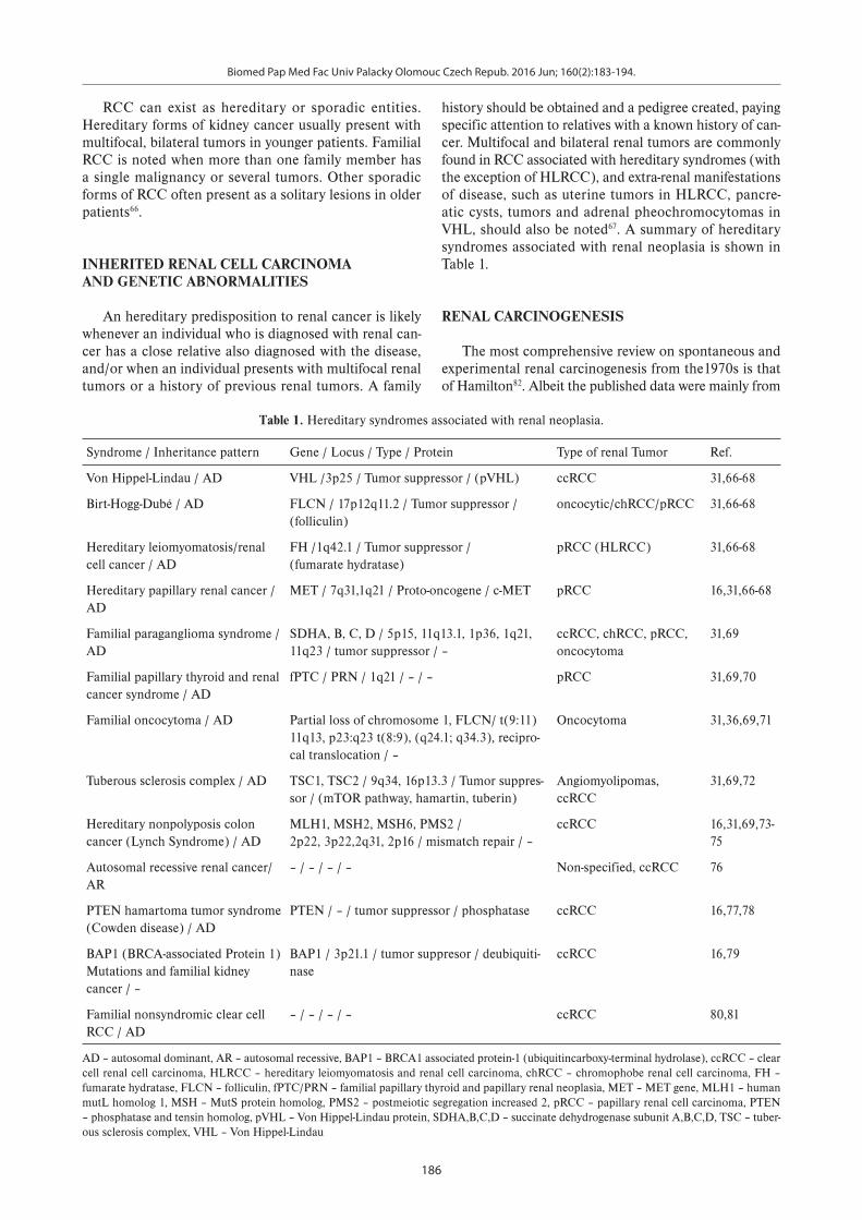
RCC can exist as hereditary or sporadic entities. Hereditary forms of kidney cancer usually present with multifocal, bilateral tumors in younger patients. Familial RCC is noted when more than one family member has a single malignancy or several tumors. Other sporadic forms of RCC often present as a solitary lesions in older patients66.
INHERITED RENAL CELL CARCINOMA AND GENETIC ABNORMALITIES
An hereditary predisposition to renal cancer is likely whenever an individual who is diagnosed with renal can-cer has a close relative also diagnosed with the disease, and/or when an individual presents with multifocal renal tumors or a history of previous renal tumors. A family
history should be obtained and a pedigree created, paying specific attention to relatives with a known history of can-cer. Multifocal and bilateral renal tumors are commonly found in RCC associated with hereditary syndromes (with the exception of HLRCC), and extra-renal manifestations of disease, such as uterine tumors in HLRCC, pancre-atic cysts, tumors and adrenal pheochromocytomas in VHL, should also be noted67. A summary of hereditary syndromes associated with renal neoplasia is shown in Table 1.
RENAL CARCINOGENESIS
The most comprehensive review on spontaneous and experimental renal carcinogenesis from the1970s is that of Hamilton82. Albeit the published data were mainly from
Table 1. Hereditary syndromes associated with renal neoplasia.
Syndrome / Inheritance pattern Gene / Locus / Type / Protein Type of renal Tumor Ref.
Von Hippel-Lindau / AD VHL /3p25 / Tumor suppressor / (pVHL) ccRCC 31,66-68
Birt-Hogg-Dubé / AD FLCN / 17p12q11.2 / Tumor suppressor /(folliculin)
oncocytic/chRCC/pRCC 31,66-68
Hereditary leiomyomatosis/renal cell cancer / AD
FH /1q42.1 / Tumor suppressor /(fumarate hydratase)
pRCC (HLRCC) 31,66-68
Hereditary papillary renal cancer /AD
MET / 7q31,1q21 / Proto-oncogene / c-MET pRCC 16,31,66-68
Familial paraganglioma syndrome / AD
SDHA, B, C, D / 5p15, 11q13.1, 1p36, 1q21, 11q23 / tumor suppressor / –
ccRCC, chRCC, pRCC, oncocytoma
31,69
Familial papillary thyroid and renal cancer syndrome / AD
fPTC / PRN / 1q21 / – / – pRCC 31,69,70
Familial oncocytoma / AD Partial loss of chromosome 1, FLCN/ t(9:11) 11q13, p23:q23 t(8:9), (q24.1; q34.3), recipro-cal translocation / –
Oncocytoma 31,36,69,71
Tuberous sclerosis complex / AD TSC1, TSC2 / 9q34, 16p13.3 / Tumor suppres-sor / (mTOR pathway, hamartin, tuberin)
Angiomyolipomas, ccRCC
31,69,72
Hereditary nonpolyposis colon cancer (Lynch Syndrome) / AD
MLH1, MSH2, MSH6, PMS2 / 2p22, 3p22,2q31, 2p16 / mismatch repair / –
ccRCC 16,31,69,73-75
Autosomal recessive renal cancer/AR
– / – / – / – Non-specified, ccRCC 76
PTEN hamartoma tumor syndrome (Cowden disease) / AD
PTEN / – / tumor suppressor / phosphatase ccRCC 16,77,78
BAP1 (BRCA-associated Protein 1) Mutations and familial kidney cancer / –
BAP1 / 3p21.1 / tumor suppresor / deubiquiti-nase
ccRCC 16,79
Familial nonsyndromic clear cell RCC / AD
– / – / – / – ccRCC 80,81
AD – autosomal dominant, AR – autosomal recessive, BAP1 – BRCA1 associated protein-1 (ubiquitincarboxy-terminal hydrolase), ccRCC – clear cell renal cell carcinoma, HLRCC – hereditary leiomyomatosis and renal cell carcinoma, chRCC – chromophobe renal cell carcinoma, FH – fumarate hydratase, FLCN – folliculin, fPTC/PRN – familial papillary thyroid and papillary renal neoplasia, MET – MET gene, MLH1 – human mutL homolog 1, MSH – MutS protein homolog, PMS2 – postmeiotic segregation increased 2, pRCC – papillary renal cell carcinoma, PTEN – phosphatase and tensin homolog, pVHL – Von Hippel-Lindau protein, SDHA,B,C,D – succinate dehydrogenase subunit A,B,C,D, TSC – tuber-ous sclerosis complex, VHL – Von Hippel-Lindau
Biomed Pap Med Fac Univ Palacky Olomouc Czech Repub. 2016 Jun; 160(2):183-194.
187
animal studies, they may reflect the same in the human organism. Experimental renal tumors were induced by hormones, viruses, chemicals and radiation. Induction of renal tumors in hamsters can be achieved by subcu-taneous implantation or injection of estrogens: namely stilbestrol or diethylstilbestrol83. However, Algard et al. de-scribed cells from an estrogen-dependent renal tumor that could be grown in a hormone-free media and in which the addition of crystalline hormone did not enhance growth or survival84. The exact genesis of the estrogen-induced renal tumors was unclear though it was assumed it was via the pituitary or through the direct action of estrogen. Surprisingly, progesterone inhibited tumor induction82,85. An exact explanation for this phenomenon has not been sufficiently confirmed.
The polyoma virus, SV 40 and adenovirus 7, aflatoxin,a number of chemicals (eg.N-nitrosodimethylnitrosamine, nitrosomethylurea, nitrosoethylurea, phosphate and acetate) and also γ- radiation can lead to renal tumori-genesis in animals82.
Radiation exposureInformative data on carcinogenesis in humans has
been acquired especially after exposure to radiation. In 1994, the first comprehensive report of cancer incidence from the Life Span Study (LSS) cohort of atomic bomb survivors in Japan (Hiroshima and Nagasaki) was pub-lished. This included data from 1958 to 1987(ref.86,87) and through 1998 (ref.88). With the follow-up period for these analyses ending in December 1998, analyses were based on more than 40 years of cancer incidence data for members of the LSS. The LSS is one of the few radia-tion effect studies that is composed of a basically healthy general population, including males and females exposed to a wide range of radiation doses at all ages. The chance of surviving depends on factors such as age at the time of exposure and ability to survive the effects (heat, blast and radiation) of the bomb. In the full LSS cohort of 120,321 individuals, about 48% overall (52% of females and 43% of males) were still alive at the end of 1998. Among the 105,427 members of the LSS cohort included in this study, 17,448 first primary solid cancers were diagnosed from 1958-1998. Stomach cancer was the most common cancer in the cohort, accounting for more than 25% of all cases. Other commonly occurring cancers included lung (10%), liver (9%), colon (9%), rectum (5%), female breast (6%), and cervix (5%). Baseline renal cell cancer rates in the LSS tend to increase with age for men but did not in-crease very much for women. At age 70, the baseline rate for women was about one third that for men. The base-line rate models suggested that age-specific renal cancer baseline rates in the LSS have increased by about 20% per decade increase in year of birth and if radiation increased the RCC incidence, the magnitude decreased over time88. However, in 2000, 13-years after the Chernobyl accident in Ukraine a higher incidence of RCC was found with an increase from 4.7 to 7.5 per 100,000 of the total popula-tion. The study was based on knowledge about 137Cesium accounting for 90% of incorporated radioactivity and this is eliminated via the kidney. The Ukrainian population
has been exposed to long-term, low dose ionizing radia-tion since 1986. A study was carried out to evaluate the histopathological features and immunohistochemical status of proliferating cell nuclear antigen (PCNA) and K-ras in RCC of 236 Ukrainian patients in comparison to an analog of 112 patients in Spain. RCCs from Ukraine presented more frequently with sarcomatoid changes with significant differences and less frequent peritumor inflammatory response. The dramatic increase in aggres-sivity and proliferative activity presenting by PCNA level (83% in comparison to 69%) and K-ras expression (56% in comparison to 38%) of RCCs in Ukrainian groups showed good correlation with the duration of radiation exposure and confirmed the effect of chronic, regular and sustained low dose ionizing radiation on renal carcinogenesis in the Ukrainian population89.
Low dose radiationProlonged low dose radiation is one major cause of hu-
man renal carcinogenesis? There is a difference between atomic bomb attack and survivors with different types of cancers and low–dose radiation over a longer period and its effects on the kidney. Maybe the answer to this ques-tion is based on the effects of low-dose radiation on living organisms and information on uranium nephrotoxicity.
Low dose radiation exposure is defined as doses up to ∼100 mSv and usually has direct or indirect effects on DNA. Direct effects are based on adsorption of radia-tion energy with structural alteration of DNA. Induction of DNA damage by low-dose radiation has been quanti-fied by foci formation, and over a range of few mGy up to 1000 mGy. A variety of changes in DNA have been identified: base damage, apyridimic/apurinic sites, single-strand breaks, double-strand breaks and cross-linkages90. DNA damage can lead to genomic instability that is a characteristic of most cancer cells with increased tenden-cy to genome damage during cell division. Maintenance of genomic stability is essential for cellular integrity to prevent errors in DNA replication, endogenous geno-toxic stress and exogenous carcinogen insult (ultraviolet light, ionizing radiation and DNA damaging chemicals). Genomic instability includes small structural variation such as increased frequencies of base pair mutations, mi-crosatellite instability and significant structural variation such as chromosome number or structural changes91.The defence barrier against cancer are the well-known tumor suppressor genes. Three major pathways of regulatory control that are targeted by mutations in human neopla-sia are: 1) Rb/p16 tumor suppressor pathway with two proto-oncogenes Cyclin D and Cdk4; 2) Apc/β-Catenin pathway with Apc tumor-suppressor gene and β-Catenin as proto-oncogene; 3) p53/Mdm2 pathway with p53 tu-mor suppressor gene and Mdm2 proto-oncogene which binds and inhibits the transcriptional activation domain of p53(ref.92). In the other hand, one of the most important factors in gene expression and transcription is DNA meth-ylation, a biochemical process where a methyl group is added to the cytosine or adenine DNA nucleotides. DNA methylation contributes to the adaptive response to ioniz-ing radiation and was investigated on B-lymphoblast cells
Biomed Pap Med Fac Univ Palacky Olomouc Czech Repub. 2016 Jun; 160(2):183-194.
188
in a study by Ye et al.93 As a putative novel diagnostic bio-marker in RCC genome-wide DNA methylation, profiling alterations in renal cell carcinoma of diverse histologies and benign adjacent kidney tissues were investigated in 96 patients. These authors found widespread methylation differences between malignant tumors and benign adja-cent tissues, particularly in immune- , G-protein coupled receptor-, and metabolism-related genes94.
UraniumThe nephrotoxic effects of uranium are known from
the uranium industry around the world from human95,96 and animal studies97. Biologically soluble uranium com-pounds such as uranyl nitrate [UO2 (NO3)2] and ammoni-um diuranate (NH3-UO3-H2O) are filtered rapidly through the renal glomeruli and are toxic to the renal proximal tubules. The lesions in the proximal convoluted tubules result in the appearance of glucose, low-molecular-weight proteins and amino acids in the urine. These substances, under normal conditions are reabsorbed from the tubular fluid. Acute damage to the proximal tubular cells and re-duced proximal tubular reabsorption are consistent with uranium nephrotoxicity98,99. Thun et al. showed signifi-cant excretion of beta-2-microglobulin and amino acids in 39 uranium industry workers99. However, proteinuria in the case of uranium nephrotoxicity was low and had no value in predicting kidney damage. A more sensitive approach for urinalysis in uranium detection is the fluo-rophotometric method100. Chronic exposure to soluble uranium and the harmful effects of low-dose radiation can be predicted in countries with a uranium industry. The Czech Republic’s mine at Rozna is the only operating uranium mine in the European Union and the only one currently operating in Europe. It is not known if there is an association between increase in RCC cases and the uranium mining and its chemical industry in the Czech Republic.
DIAGNOSIS OF RENAL CELL CARCINOMA
The detection and diagnosis of RCC have evolved in recent years. At present, the majority of RCCs are found incidentally from abdominal ultrasound or com-puter tomography examinations undertaken for various reasons. Significantly less frequent are visible signs and symptoms of RCC. The most common are: microscopic or macroscopic hematuria, lateral dorsal or flank pain and palpable abdominal mass. Important information for physicians is that RCC can become very large without any symptoms, due to the retroperitoneal position of the kidney101. Paraneoplastic manifestations of RCC, includ-ing hypercalcemia, production of adrenocorticotrophic hormone, polycytemia, hepatic dysfunction, amyloido-sis, fever and weight loss are present in up to 20% of pa-tients. Hypercalcemia is caused by release of parathyroid hormone-related peptide (PTHrP), interleukins IL-6, IL-1 and tumor necrosis factor α(TNFα) from cancer tissue101.The mechanism by which PTHrP causes hypercalcemia involves many of the normal hormonal pathways of cal-
cium homeostasis. PTHrP binds to the PTH receptor in both bone and renal tissue. This binding leads to in-creased bone resorption and decreased renal clearance of calcium as well as increased phosphorus excretion102.
Wells syndrome, a less common paraneoplastic mani-festation of metastatic RCC was reported by Rajpara et al.who described a 58-year old man suffering from dif-fuse granulomatous dermatitis with eosinophilia103, first reported by Wells in 1971 as eosinophilic cellulitis104.Nonmetastatic nephrogenic hepatic dysfunction syn-drome (Stauffer’s syndrome) is a unique and rare para-neoplastic manifestation of renal cell carcinoma that usually manifests as anicteric cholestasis. This syndrome, originally described in 1961 by M. H. Stauffer, is charac-terized by elevated alkaline phosphatase, erythrocyte sedi-mentation rate, α-2-globulin, and γ-glutamyltransferase, thrombocytosis, prolongation of prothrombin time, and hepatosplenomegaly, the absence of hepatic metasta-sis and jaundice due to the possible role of IL-6 over-expression by the primary tumor105,106. Polycytemia (or erythrocytosis) which has been noted in patients with RCC is believed to be caused by ectopic production of erythropoetin by cancer cells101. Nonspecific symptoms such as fever, weight loss, and fatigue common to many malignancies, are thought to be mediated by cytokines especially TNFα and IL-6 (ref.107). Many other endocrine abnormalities are associated with RCC, such as elevated human chorionic gonadotropin and adrenocorticotropic hormone, manifesting themselves as clinical syndromes such as Cushing’s syndrome and hyper/hypoglycaemia. Other conditions associated with RCC include amyloi-dosis due to pathological production and deposition of AA protein with typical clinical presentation related to the specific organ systems affected including the cardio-vascular, renal and gastrointestinal systems. A number of other syndromes such as light chain nephropathy, vas-culitis, coagulopathies, neuromyopathies have been also described in patients with RCC (ref.107). Most pulmonary and neurological symptoms result from lung or intracra-nial metastases and have a poor prognosis.
Imaging testsRadiological investigations of RCC should include
CT imaging, before and after intravenous contrast to confirm the diagnosis. These will provide information on the function and morphology of the contralateral kidney and assess tumor extension, including extrarenal spread and venous involvement. Abdominal ultrasound and mag-netic resonance imaging are alternatives to CT. Chest CT is the most accurate for tumor staging; a routine chest X-ray should be done as a minimum108. Case dependant is evaluation of bone and brain metastases with diagnos-tic performance of bone scintigraphy and brain CT. Our clinical approach greatly depends on a patient’s clinical status and tumor staging. Renal masses may be classified as solid or cystic by imaging criteria. For evaluating solid renal masses, the presence of enhancement is the most important criteria for differentiating malignant from be-nign lesions.
Biomed Pap Med Fac Univ Palacky Olomouc Czech Repub. 2016 Jun; 160(2):183-194.
189
However, the best measure in differentiation malignant and benign lesions is a tumor biopsy, if technically or clinically (e.g. patient’s status, solitary kidney, coagulopa-thies etc.) possible.
TNM CLASSIFICATION AND STAGING OF RENAL CELL CARCINOMA
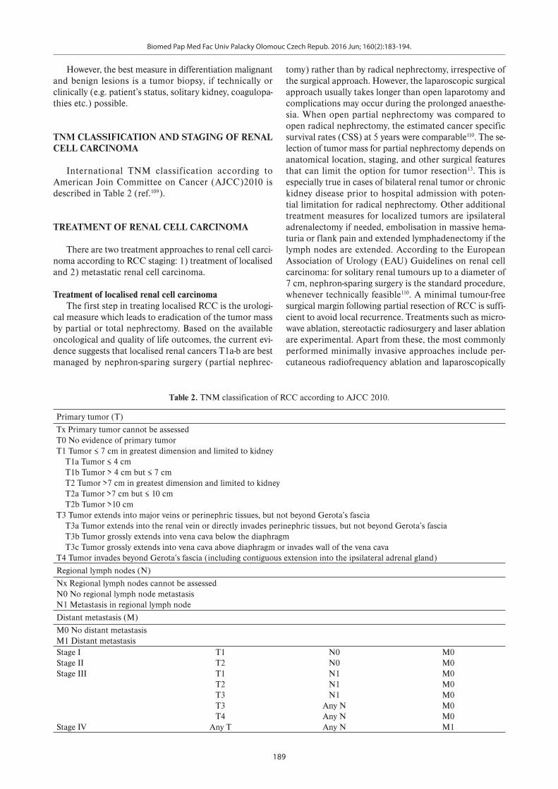
International TNM classification according to American Join Committee on Cancer (AJCC)2010 is described in Table 2 (ref.109).
TREATMENT OF RENAL CELL CARCINOMA
There are two treatment approaches to renal cell carci-noma according to RCC staging: 1) treatment of localised and 2) metastatic renal cell carcinoma.
Treatment of localised renal cell carcinomaThe first step in treating localised RCC is the urologi-
cal measure which leads to eradication of the tumor mass by partial or total nephrectomy. Based on the available oncological and quality of life outcomes, the current evi-dence suggests that localised renal cancers T1a-b are best managed by nephron-sparing surgery (partial nephrec-
Table 2. TNM classification of RCC according to AJCC 2010.
Primary tumor (T)
Tx Primary tumor cannot be assessedT0 No evidence of primary tumorT1 Tumor ≤ 7 cm in greatest dimension and limited to kidney T1a Tumor ≤ 4 cm T1b Tumor > 4 cm but ≤ 7 cm T2 Tumor >7 cm in greatest dimension and limited to kidney T2a Tumor >7 cm but ≤ 10 cm T2b Tumor >10 cmT3 Tumor extends into major veins or perinephric tissues, but not beyond Gerota’s fascia T3a Tumor extends into the renal vein or directly invades perinephric tissues, but not beyond Gerota’s fascia T3b Tumor grossly extends into vena cava below the diaphragm T3c Tumor grossly extends into vena cava above diaphragm or invades wall of the vena cavaT4 Tumor invades beyond Gerota’s fascia (including contiguous extension into the ipsilateral adrenal gland)
Regional lymph nodes (N)
Nx Regional lymph nodes cannot be assessedN0 No regional lymph node metastasisN1 Metastasis in regional lymph node
Distant metastasis (M)
M0 No distant metastasisM1 Distant metastasisStage IStage IIStage III
Stage IV
T1T2T1T2T3T3T4
Any T
N0N0N1N1N1
Any NAny NAny N
M0M0M0M0M0M0M0M1
tomy) rather than by radical nephrectomy, irrespective of the surgical approach. However, the laparoscopic surgical approach usually takes longer than open laparotomy and complications may occur during the prolonged anaesthe-sia. When open partial nephrectomy was compared to open radical nephrectomy, the estimated cancer specific survival rates (CSS) at 5 years were comparable110. The se-lection of tumor mass for partial nephrectomy depends on anatomical location, staging, and other surgical features that can limit the option for tumor resection13. This is especially true in cases of bilateral renal tumor or chronic kidney disease prior to hospital admission with poten-tial limitation for radical nephrectomy. Other additional treatment measures for localized tumors are ipsilateral adrenalectomy if needed, embolisation in massive hema-turia or flank pain and extended lymphadenectomy if the lymph nodes are extended. According to the European Association of Urology (EAU) Guidelines on renal cell carcinoma: for solitary renal tumours up to a diameter of 7 cm, nephron-sparing surgery is the standard procedure, whenever technically feasible110. A minimal tumour-free surgical margin following partial resection of RCC is suffi-cient to avoid local recurrence. Treatments such as micro-wave ablation, stereotactic radiosurgery and laser ablation are experimental. Apart from these, the most commonly performed minimally invasive approaches include per-cutaneous radiofrequency ablation and laparoscopically
Biomed Pap Med Fac Univ Palacky Olomouc Czech Repub. 2016 Jun; 160(2):183-194.
190
assisted cryoablation. Indications for thermal ablations are usually small renal masses in elderly more comorbid patients unable to undergo surgical intervention and pa-tients with bilateral tumors or solitary kidney111. Adjuvant therapy after nephrectomy has not been proven to prolong survival or to have any significant patient benefit110,111.
Treatment of metastatic renal cell carcinomaThe treatment of metastatic disease is very difficult
and greatly depends on histological subtype of renal tu-mour mass, the patient’s clinical status and has changed over the last 10-15 years. Special emphasis is given to cytoreductive nephrectomy (CN) prior to the start of adjuvant therapy112. The evidence for performing CN before cytokine therapy came from two randomized tri-als Southwest oncology group 8949 (SWOG 8949) and European Organisation for Research and Treatment of Cancer (EORTC 30947) where a survival benefit for CN followed by the immunotherapy with INF-α compared with INF-α alone was shown. The SWOG 8949 study reported the median survival of 120 eligible patients as-signed to surgery followed by interferon as 11.1 months, and among the 121 eligible patients assigned to interferon alone it was 8.1months (P = 0.05). The difference in me-dian survival between the two groups was independent of performance status, metastatic site or presence or absence
of measurable metastatic lesion113. The EORTC 30947 study involved 40 (53%) of 75 patients receiving at least 16 weeks of INF-α treatment, which was the median du-ration of treatment. Time to progression (5 vs 3 months, hazard ratio 0.60, 95% CI 0.36-0.97) and median duration of survival were significantly better in patients than in controls (17 vs 7 months, 0.54, 0.31-0.94). Five patients responded completely to combined treatment and one to INF-α alone114.
These two studies were also been the motivation to perform CN in the targeted therapy era. Hovewer, cytore-ductive nephrectomy is in principle not curative approach in case of metastatic disease and patients must have been able to undergo this surgical intervention. The rationale for CN performance is in better survival, removal of ma-lignant renal mass, immunosupressant cytokines and other hormones or molecules that underlie the paraneo-plastic syndrome112, 115. The palliative benefits of CN in case of tumor related pain and massive hematuria are also important for patient outcome.
Systemic metastatic RCC treatment has developed significantly: vascular endothelial growth factor as well as tyrosine kinase inhibitors and drugs that inhibit the mammalian target of rapamycin (mTOR) signaling path-way have become the mainstay for the management of advanced RCC. Possible treatment modalities in current
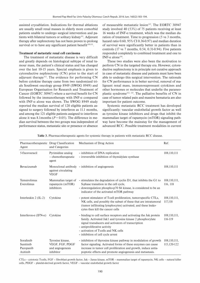
Table 3. Pharmacotherapeutic agents for systemic therapy in patients with metastatic RCC disease.
PharmacotherapeuticAgent
Drug Classification and Categories
Mechanism of Drug Action Ref.
5-fluorouracil Pyrimidine analog – chemotherapeutic agent
– inhibition of DNA replication– irreversible inhibition of thymidylate synthase
108,110,111
Bevacizumab Monoclonal antibody against circulating VEGF
– inhibition of angiogenesis 108,110,111
TemsirolimusEverolimus
Mammalian target of rapamycin (mTOR) inhibitors
– stimulates the degradation of cyclin D1, that inhibits the G1 to S-phase transition in the cell cycle,
– downregulation phospho-p70 S6 kinase, is considered to be an indicator of the activated mTOR pathway
108,110,111,116, 118
Interleukin 2 (IL-2) Cytokine – potent stimulator of T-cell proliferation, tumor-specific CTLs, NK cells, and possibly the subset of these that are intratumoral (tumor infiltrating lymphocytes) activated, and these leuko-cytes then kill the cancer cells
108,110,111,117,118
Interferon-α (IFN-α) Cytokine – binding to cell surface receptors and activating the Jak protein family. Activated Jak1 and tyrosine kinase 2 phosphorylate signal transducers and activators of transcription
– antiproliferative activity– activation of T-cells and NK cells– inhibition of cell cycle arrest
108,110,111,116-119
SorafenibSunitinibPazopanibAxitinib
Tyrosine kinase, VEGF, FGF, PDGF and angiogenesis inhibitor
– inhibition of thyrosine kinase pathway in modulation of growth factor signaling. Activated forms of these enzymes can cause increase in tumor cell proliferation and growth, induce antia-poptotic effects and promote angiogenesis and metastasis.
108,110,111,115,120-122
CTLs – cytotoxic T-cells, FGF – fibroblast growth factor, Jak – Janus kinase, mTOR – mammalian target of rapamycin, NK cells – natural killer cells, PDGF – platelet-derived growth factor, VEGF – vascular endothelial growth factor
Biomed Pap Med Fac Univ Palacky Olomouc Czech Repub. 2016 Jun; 160(2):183-194.
191
oncological and urological practice are described below in Table 3.
Unfortunately, systemic chemotherapy is not effec-tive and monotherapy with INF-α and IL-2 is not recom-mended (except in lung metastases) as a 1stline therapy in patients with histopathologically verified metastatic clear cell RCC. According to the described pathophysiology of ccRCC in this group currently preferred and recom-mended in 1stline therapy are: 1) sunitinib (50 mg daily orally for a period of 4 weeks followed by 2 weeks of rest); 2) pazopanib (800 mg orally daily); 3) and the combina-tion of INF-α (9 MU three times per week subcutane-ously) + bevacizumab (10 mg/kg biweekly intravenously). In the 2nd line therapy is recommended axitinib, sorafenib and everolimus after prior tyrosine kinase inhibitors and sorafenib, axitinib and pazopanib after prior administra-tion of cytokines110,111. In the 3rd line are preferred everoli-mus after VEGF targeted therapy and sorafenib after mTOR treatment111. However, the combination therapy has no benefit over single – agent use.
In patients with nonclear cell metastatic RCC recom-mended in 1st line therapy are: 1) sunitinib; 2) everolimus (10 mg daily orally) and 3) temsirolimus (25 mg weekly intravenously). In the 2nd line therapy any targeted agent can be used111.
Patient surveillance after nephrectomy and RCC treat-ment is an important measure and allows physicians to make an early diagnosis of complications and/or assess-ment of local tumor progression and any metastasis. The basic measures should include abdominal ultrasound, CT or MRI imaging and thoracic X-ray/CT once yearly in the subsequent 5 years.
CONCLUSIONS
Renal cell cancer is usually a serious disease with poor prognosis, especially when there are metastasis. RCC tu-mors also are difficult to diagnose due to non-specific symptoms in the early stages of the disease. Current on-cological research is directed to blocking cancer cell divi-sion or inhibiting angiogenesis based on knowledge of molecular pathways. Natural and manmade radioactivity, chemical nephrotoxicity and environmental risks are also a serious concern in terms of prevention and the need to screen populations at risk.
Author contributions: Authors contributed equally to pre-paring the manuscript.Conflict of interest statement: The authors declare there are no conflicts of interest regarding the publication of this article.
REFERENCES
1. Ferlay J, Soerjomataram I, Ervik M, Dikshit R, Eser S, Mathers C, Rebelo M, Parkin DM, Forman D, Bray, F. GLOBOCAN 2012 v1.0, Cancer Incidence and Mortality Worldwide: IARC Cancer Base No. 11Lyon, France: International Agency for Research on Cancer
[Internet]. 2013 [cited 2015 March 3]; Available from: http://globo-can.iarc.fr and www.wcrf.org
2. Remon J, Lianes P, Martínez S. Brain metastases from renal cell car-cinoma. Should we change the current standard? Cancer Treat Rev 2012;38(4):249-57.
3. Landis SH, Murray T, Bolden S, Wingo PA. Cancer statistics, 1999. CA Cancer J Clin 1999;49(1):8-31.
4. Aydin H, Chen L, Cheng L, Vaziri S, He H, Ganapathi R, Delahunt B, Magi-Galluzzi C, Zhou M. Clear cell tubulopapillary renal cell carci-noma: a study of 36 distinctive low-grade epithelial tumors of the kidney. Am J Surg Pathol 2010;34(11):1608-21.
5. Gobbo S, Eble JN, Maclennan GT, Grignon DJ, Shah RB, Zhang S, Martignoni G, Brunelli M, Cheng L. Renal cell carcinomas with papil-lary architecture and clear cell components: the utility of immuno-histochemical and cytogenetical analyses in differential diagnosis. Am J Surg Pathol 2008;32(12):1780-6.
6. Chow WH, Dong LM, Devesa SS. Epidemiology and risk factors for kidney cancer. Nat Rev Urol 2010;7(5):245-57.
7. Dmitriev AA, Rudenko EE, Kudryavtseva AV, Krasnov GS, Gordiyuk VV, Melnikova NV,Stakhovsky EO, Kononenko OA, Pavlova LS, Kondratieva TT, Alekseev BY, Braga EA, Senchenko VN, Kashuba VI. Epigenetic alterations of chromosome 3 revealed by NotI-microarrays in clear cell renal cell carcinoma. Biomed Res Int 2014;2014:735292.
8. Cairns P. Renal cell carcinoma. Cancer Biomark 2010;9(1-6):461-73. 9. Cohen HT, McGovern FJ. Renal-cell carcinoma. N Engl J Med
2005;353(23):2477-90. 10. Belldegrun AS, Klatte T, Shuch B, LaRochelle JC, Miller DC, Said JW,
Riggs SB, Zomorodian N, Kabbinavar FF, Dekernion JB, Pantuck AJ. Cancer-specific survival outcomes among patients treated during the cytokine era of kidney cancer (1989-2005): a benchmark for emerging targeted cancer therapies. Cancer 2008;113(9):2457-63.
11. McDermott DF, Regan MM, Clark JI, Flaherty LE, Weiss GR, Logan TF,Kirkwood JM, Gordon MS, Sosman JA, Ernstoff MS, Tretter CP, Urba WJ, Smith JW, Margolin KA, Mier JW, Gollob JA, Dutcher JP, Atkins MB. Randomized phase III trial of high-dose interleukin-2 versus subcutaneous interleukin-2 and interferon in patients with meta-static renal cell carcinoma. J Clin Oncol 2005;23(1):133-41. Erratum in: J Clin Oncol 2005; 23(12):2877.
12. Brauch H, Weirich G, Brieger J, Glavac D, Rödl H, Eichinger M, Feurer M, Weidt E, Puranakanitstha C, Neuhaus C, Pomer S, Brenner W, Schirmacher P, Störkel S, Rotter M, Masera A, Gugeler N, Decker HJ.VHL alterations in human clear cell renal cell carcinoma: associa-tion with advanced tumor stage and a novel hot spot mutation. Cancer Res 2000;60(7):1942-8.
13. Jonasch E, Gao J, Rathmell WK. Renal cell carcinoma. BMJ 2014;349:g4797.
14. Vortmeyer AO, Lubensky IA, Fogt F, Linehan WM, Khettry U, Zhuang Z.Allelic deletion and mutation of the von Hippel-Lindau (VHL) tu-mor suppressor gene in pancreatic microcystic adenomas. Am J Pathol 1997;151(4):951-6.
15. Tse JY, Wong JH, Lo KW, Poon WS, Huang DP, Ng HK. Molecular ge-netic analysis of the von Hippel-Lindau disease tumor suppressor gene in familial and sporadic cerebellar hemangioblastomas. Am J Clin Pathol 1997;107(4):459-66.
16. Haas NB, Nathanson KL. Hereditary kidney cancer syndromes. Adv Chronic Kidney Dis 2014;21(1):81-90.
17. Gossage L, Murtaza M, Slatter AF, Lichtenstein CP, Warren A, Haynes B,Marass F, Roberts I, Shanahan SJ, Claas A, Dunham A, May AP, Rosenfeld N, Forshew T, Eisen T. Clinical and pathological impact of VHL, PBRM1, BAP1, SETD2, KDM6A, and JARID1c in clear cell renal cell carcinoma. Genes Chromosomes Cancer 2014;53(1):38-51.
18. Varela I, Tarpey P, Raine K, Huang D, Ong CK, Stephens P, Davies H, Jones D, Lin ML, Teague J, Bignell G, Butler A, Cho J, Dalgliesh GL, Galappaththige D, Greenman C, Hardy C, Jia M, Latimer C, Lau KW, Marshall J, McLaren S, Menzies A, Mudie L, Stebbings L, Largaespada DA, Wessels LF, Richard S, Kahnoski RJ, Anema J, Tuveson DA, Perez-Mancera PA, Mustonen V, Fischer A, Adams DJ, Rust A, Chan-on W, Subimerb C, Dykema K, Furge K, Campbell PJ, Teh BT, Stratton MR, Futreal PA. Exome sequencing identifies frequent mutation of the SWI/SNF complex gene PBRM1 in renal carcinoma. Nature 2011;469(7331):539-42. Erratum in: Nature 2012;484(7392):130.
19. Peña-Llopis S, Vega-Rubín-de-Celis S, Liao A, Leng N, Pavía-Jiménez A, Wang S, Yamasaki T, Zhrebker L, Sivanand S, Spence P, Kinch L,
Biomed Pap Med Fac Univ Palacky Olomouc Czech Repub. 2016 Jun; 160(2):183-194.
192
Hambuch T, Jain S, Lotan Y, Margulis V, Sagalowsky AI, Summerour PB, Kabbani W, Wong SW, Grishin N, Laurent M, Xie XJ, Haudenschild CD, Ross MT, Bentley DR, Kapur P, Brugarolas J. BAP1 loss defines a new class of renal cell carcinoma. Nat Genet 2012; 44(7):751-9. Erratum in: Nat Genet 2012; 44(9):1072.
20. Dalgliesh GL, Furge K, Greenman C, Chen L, Bignell G, Butler A, Davies H, Edkins S, Hardy C, Latimer C, Teague J, Andrews J, Barthorpe S, Beare D, Buck G, Campbell PJ, Forbes S, Jia M, Jones D, Knott H, Kok CY, Lau KW, Leroy C, Lin ML, McBride DJ, Maddison M, Maguire S, McLay K, Menzies A, Mironenko T, Mulderrig L, Mudie L, O'Meara S, Pleasance E, Rajasingham A, Shepherd R, Smith R, Stebbings L, Stephens P, Tang G, Tarpey PS, Turrell K, Dykema KJ, Khoo SK, Petillo D, Wondergem B, Anema J, Kahnoski RJ, Teh BT, Stratton MR, Futreal PA.Systematic sequencing of renal carcinoma reveals inactivation of histone modifying genes. Nature 2010;463(7279):360-3.
21. George CM, Stadler WM, Vogelzang NJ. Primary neoplasms of the kidney and renal pelvis. In: Schrier RW, editor. Diseases of the kidney and urinary tract, Philadelphia: Lippincott Williams &Wilkins; 2001. p. 831-49.
22. Breen EC. VEGF in biological control. J Cell Biochem 2007;102(6):1358-67. 23. Rydzanicz M, Wrzesiński T, Bluyssen HA, Wesoły J. Genomics and
epigenomics of clear cell renal cell carcinoma: recent developments and potential applications. Cancer Lett 2013;341(2):111-26.
24. Teloken PE, Houston Thompson R, Tickoo SK, Cronin A, Savage C, Reuter VE. Russo P. Prognostic impact of histological subtype in pa-tients with surgically treated localized renal cell carcinoma. J Urol 2009;182(5): 2132-6.
25. Fernandes DS, Lopes JM. Pathology, therapy and prognosis of papil-lary renal carcinoma. Future Oncol 2015;11(1):121-32.
26. Wei MH, Toure O, Glenn GM, Pithukpakorn M, Neckers L, Stolle C,Choyke P, Grubb R, Middelton L, Turner ML, Walther MM, Merino MJ, Zbar B, Linehan WM, Toro JR. Novel mutations in FH and ex-pansion of the spectrum of phenotypes expressed in families with hereditary leiomyomatosis and renal cell cancer. J Med Genet 2006; 43(1):18-27.
27. Gellera C, Uziel G, Rimoldi M, Zeviani M, Laverda A, Carrara F, DiDonato S. Fumarase deficiency is an autosomal recessive encepha-lopathy affecting both the mitochondrial and the cytosolic enzymes. Neurology 1990;40:495-9.
28. Merino MJ, Torres-Cabala C, Pinto P, Linehan WM. The morpho-logic spectrum of kidney tumors in hereditary leiomyomatosis and renal cell carcinoma (HLRCC) syndrome. Am J Surg Pathol 2007;31(10):1578-85.
29. Schmidt LS, Warren MB, Nickerson ML, Weirich G, Matrosova V, Toro JR,Turner ML, Duray P, Merino M, Hewitt S, Pavlovich CP, Glenn G, Greenberg CR, Linehan WM, Zbar B.Birt-Hogg-Dubé syndrome, a genodermatosis associated with spontaneous pneumothorax and kidney neoplasia, maps to chromosome 17p11.2. Am J Hum Genet 2001;69(4):876-82.
30. Johannesma PC, van Moorselaar RJ, Horenblas S, van der Kolk LE, Thunnissen E, van Waesberghe JH,Menko FH, Postmus PE. Bilateral renal tumour as indicator for birt-hogg-dubé syndrome. Case Rep Med 2014;2014:618675.
31. Morrison PJ, Donnelly DE, Atkinson AB, Maxwell AP. Advances in the genetics of familial renal cancer.Oncologist 2010;15(6):532-8.
32. Gupta R, Billis A, Shah RB, Moch H, Osunkoya AO, Jochum W, Hes O, Bacchi CE, de Castro MG, Hansel DE, Zhou M, Vankalakunti M, Salles PG, Cabrera RA, Gown AM, Amin MB. Carcinoma of the collecting ducts of Bellini and renal medullary carcinoma: clinicopathologic analysis of 52 cases of rare aggressive subtypes of renal cell car-cinoma with a focus on their interrelationship. Am J Surg Pathol 2012;36(9):1265-78.
33. Davis CJ Jr, Mostofi FK, Sesterhenn IA. Renal medullary carcinoma. The seventh sickle cell nephropathy. Am J Surg Pathol 1995;19(1):1-11.
34. Becker F, Junker K, Parr M, Hartmann A, Füssel S, Toma M,Grobholz R, Pflugmann T, Wullich B, Strauss A, Behnes CL, Otto W, Stöckle M, Jung V. Collecting duct carcinomas represent a unique tumor entity based on genetic alterations. PLoS One 2013;8(10):e78137.
35. Reuter VE, Gaudin PB. Adult renal tumors. In: Sternberg SS, editor. Diagnostic surgical pathology. Philadelphia: JB Lippincott; 1999. p. 1785-818.
36. Biswas B, Wahal SP, Gulati A. Renal oncocytoma: A diagnostic di-lemma on cytology. J Cytol 2014;31(1):59-60.
37. Trpkov K, Yilmaz A, Uzer D, Dishongh KM, Quick CM, Bismar TA, Gokden N. Renal oncocytoma revisited: a clinicopathological study of 109 cases with emphasis on problematic diagnostic features. Histopathology 2010;57:893-906.
38. Perez-Ordonez B, Hamed G, Campbell S, Erlandson RA, Russo P, Gaudin PB, Reuter VE. Renal oncocytoma: a clinicopathologic study of 70 cases. Am J Surg Pathol 1997;21:871-83.
39. Zhao W, Tian B, Wu C, Peng Y, Wang H, Gu WL, Gao FH. DOG1, cy-clin D1, CK7, CD117 and vimentin are useful immunohistochemical markers in distinguishing chromophobe renal cell carcinoma from clear cell renal cell carcinoma and renal oncocytoma. Pathol Res Pract 2015;211(4):303-7.
40. Renehan AG, Tyson M, Egger M, Heller RF, Zwahlen M. Body-mass in-dex and incidence of cancer: a systematic review and meta-analysis of prospective observational studies. Lancet 2008;371(9612):569-78.
41. Li L, Kalantar-Zadeh K. Obesity that makes kidney cancer more likely but helps fight it more strongly. J Natl Cancer Inst 2013;105(24):1848-9.
42. Zhang GM, Zhu Y, Ye DW. Metabolic syndrome and renal cell carci-noma. World J Surg Oncol 2014;12:236.
43. Hoeben A, Landuyt B, Highley MS, Wildiers H, Van Oosterom AT, De Bruijn EA. Vascular endothelial growth factor and angiogenesis. Pharmacol Rev 2004;56(4):549-80.
44. Ying HQ, Sun HL, He BS, Pan YQ, Wang F, Deng QW, Chen J, Liu X, Wang SK. Circulating vitamin D binding protein, total, free and bio-available 25-hydroxyvitamin D and risk of colorectal cancer. Sci Rep 2015;5:7956.
45. Anic GM, Weinstein SJ, Mondul AM, Männistö S, Albanes D. Serum vitamin D, vitamin D binding protein, and lung cancer survival. Lung Cancer 2014;86(3):297-303.
46. Speeckaert M, Huang G, Delanghe JR, Taes YE. Biological and clini-cal aspects of the vitamin D binding protein (Gc-globulin) and its polymorphism. Clin Chim Acta 2006;372(1-2):33-42.
47. Mondul AM, Weinstein SJ, Moy KA, Männistö S, Albanes D. Vitamin D-binding protein, circulating vitamin D and risk of renal cell carci-noma. Int J Cancer 2014; 134(11):2699-706.
48. Khan MI, Bielecka ZF, Najm MZ, Bartnik E, Czarnecki JS, Czarnecka AM, Szczylik C. Vitamin D receptor gene polymorphisms in breast and renal cancer: current state and future approaches (review). Int J Oncol 2014;44(2):349-63.
49. Karami S, Brennan P, Hung RJ, Boffetta P, Toro J, Wilson RT, Zaridze D, Navratilova M, Chatterjee N, Mates D, Janout V, Kollarova H, Bencko V, Szeszenia-Dabrowska N, Holcatova I, Moukeria A, Welch R, Chanock S, Rothman N, Chow WH, Moore LE.Vitamin D receptor polymorphisms and renal cancer risk in Central and Eastern Europe.J Toxicol Environ Health A 2008;71(6):367-72.
50. Arjumand W, Ahmad ST, Seth A, Saini AK, Sultana S. Vitamin D re-ceptor FokI and BsmI gene polymorphism and its association with grade and stage of renal cell carcinoma in North Indian population. Tumour Biol 2012;33(1):23-31.
51. Agundez JA. Cytochrome P450 gene polymorphism and cancer. Curr Drug Metab 2004;5(3):211-24.
52. Khlifi R, Messaoud O, Rebai A, Hamza-Chaffai A. Polymorphisms in the human cytochrome P450 and arylamine N-acetyltransferase: susceptibility to head and neck cancers. Biomed Res Int 2013;2013:582768.
53. Meng FD, Ma P, Sui CG, Tian X, Jiang YH. Association between cy-tochrome P450 1A1 (CYP1A1) gene polymorphisms and the risk of renal cell carcinoma: a meta-analysis. Sci Rep 2015;5:8108.
54. Ostry V, Malir F, Ruprich J. Producers and important dietary sources of ochratoxin A and citrinin.Toxins (Basel) 2013;5(9):1574-86.
55. Malir F, Ostry V, Pfohl-Leszkowicz A, Novotna E. Ochratoxin A: devel-opmental and reproductive toxicity-an overview. Birth Defects Res B Dev Reprod Toxicol 2013;98(6):493-502.
56. International Agency for research on cancer[Internet]. 1993 [cited 2015 June 22]; Available from: http://monographs.iarc.fr/.
57. Malir F, Ostry V, Pfohl-Leszkowicz A, Roubal T. Ochratoxin A expo-sure biomarkers in the Czech Republic and comparison with foreign countries. Biomarkers 2012;17(7):577-89.
58. Pfohl-Leszkowicz A, Manderville RA. An update on direct genotoxic-ity as a molecular mechanism of ochratoxin a carcinogenicity. Chem Res Toxicol 2012;25(2):252-62.
59. Hibi D, Suzuki Y, Ishii Y, Jin M, Watanabe M, Sugita-Konishi Y, Yanai T, Nohmi T, Nishikawa A, Umemura T. Site-specific in vivo mutagenicity
Biomed Pap Med Fac Univ Palacky Olomouc Czech Repub. 2016 Jun; 160(2):183-194.
193
in the kidney of gpt delta rats given a carcinogenic dose of ochra-toxin A.Toxicol Sci 2011;122(2):406-14.
60. Akman SA, Adams M, Case D, Park G, Manderville RA. Mutagenicity of ochratoxin A and its hydroquinone metabolite in the SupF gene of the mutation reporter plasmid Ps189. Toxins (Basel) 2012;4(4):267-80.
61. Pfohl-Leszkowicz A, Molinié A, Tozlovanu M, Manderville RA. Combined Toxic Effects of Ochratoxin A and Citrinin in Vitro and in Vivo, In: Food Contaminats: Mycotoxins & Food Allergens; Siantar DP, Trucksess MW, Scott PM, Herman EM Eds; American Chemical Society Symposium Series, 1001, American Society of Microbiology: Washington D.C., USA, 2008; pp.56-80.
62. Boesch-Saadatmandi C, Loboda A, Jozkowicz A, Huebbe P, Blank R, Wolffram S, Dulak J, Rimbach G. Effect of ochratoxin A on redox-regulated transcription factors, antioxidant enzymes and glutathione-S-transferase in cultured kidney tubulus cells. Food Chem Toxicol 2008;46(8):2665-71.
63. Heussner AH, Dietrich DR, O'Brien E. In vitro investigation of indi-vidual and combined cytotoxic effects of ochratoxin A and other selected mycotoxins on renal cells. Toxicol In Vitro 2006;20(3):332-41.
64. Grenier, B; Oswald, IP. Mycotoxin co-contamination of food and feed: meta-analysis of publications describing toxicological interactions. World Mycotoxin J 2011;4(3):285-313.
65. Manderville, R.; Pfohl-Leszkowicz, A. Bioactivation and DNA Adduction as a Rationale for Ochratoxin A Carcinogenesis. World Mycotoxin J 2008;1:357-67.
66. Barrisford GW, Singer EA, Rosner IL, Linehan WM, Bratslavsky G. Familial renal cancer: molecular genetics and surgical management. Int J Surg Oncol 2011; 2011: 658767.
67. Pavlovich CP, Schmidt LS. Searching for the hereditary causes of renal-cell carcinoma. Nat Rev Cancer 2004;4(5):381-93.
68. Genetics of kidney cancer (Renal Cell Cancer): National Cancer Institute. [Internet]. [cited 2015 March 22]; Available from: http:// www. cancer.gov/.
69. FaCD online, Familial Cancer Database.[Internet]. [cited 2015 March 25]; Available from: http://www.familialcancerdatabase.nl/
70. Malchoff CD, Sarfarazi M, Tendler B, Forouhar F, Whalen G, Joshi V, Arnold A, Malchoff DM. Papillary thyroid carcinoma associated with papillary renal neoplasia: genetic linkage analysis of a distinct heritable tumor syndrome. J Clin Endocrinol Metab 2000;85(5):1758-64.
71. De la Rosette JMCH, Sternberg CN, Van Poppel HPA. Renal Cell Cancer Diagnosis and therapy. London: Springer Verlag; 2008: p. 166.
72. De Waele L, Lagae L, Mekahli D. Tuberous sclerosis complex: the past and the future. Pediatr Nephrol 2014; DOI 10.1007_s00467-014-3027-9.
73. Ferrer Márquez M, Moreno Serrano A, Maturana Ibáñez V, Reina Duarte A. Lynch syndrome-associated kidney cancer. Rev Esp Enferm Dig 2011;103(6):339-40.
74. Alvaro E, Alegre C, Perea J. Association between Lynch syndrome and renal carcinoma. Rev Esp Enferm Dig 2011;103(9):500-1.
75. Zhang T, Boswell EL, McCall SJ, Hsu DS. Mismatch repair gone awry: Management of Lynch syndrome. Crit Rev Oncol Hematol 2015;93(3):170-9.
76. Hemminki K, Li X. Familial renal cell cancer appears to have a reces-sive component. J Med Genet 2004;41(5):e58.
77. Moch H, Montironi R, Lopez-Beltran A, Cheng L, Mischo A. Oncotargets in different renal cancer subtypes. Curr Drug Targets 2015;16(2):125-35.
78. Hobert JA, Eng C. PTEN hamartoma tumor syndrome: an overview. Genet Med 2009;11(10):687-94.
79. Farley MN, Schmidt LS, Mester JL, Peña-Llopis S, Pavia-Jimenez A, Christie A, Vocke CD, Ricketts CJ, Peterson J, Middelton L, Kinch L, Grishin N, Merino MJ, Metwalli AR, Xing C, Xie XJ, Dahia PL, Eng C, Linehan WM, Brugarolas J. A novel germline mutation in BAP1 predisposes to familial clear-cell renal cell carcinoma. Mol Cancer Res 2013;11(9):1061-71.
80. Tollefson MK, Boorjian SA, Lohse CM, Blute ML, Leibovich BC. The impact of family history on pathological and clinical out-comes in non-syndromic clear cell renal cell carcinoma. BJU Int 2010;106(11):1638-42.
81. Woodward ER. Familial non-syndromic clear cell renal cell carcinoma. Curr Mol Med 2004;4(8):843-8.
82. Hamilton JM. Renal carcinogenesis. Adv Cancer Res 1975;22:1-56.
83. Kirkman H, Bacon RL. Estrogen-induced tumors of the kidney. II. Effect of dose, administration, type of estrogen, and age on the induction of renal tumors in intact male golden hamsters. J Natl Cancer Inst 1952;13(3):757-71.
84. Algard FT. Action of sex hormones on dependent tumors in cell and organ-culture systems. Natl Cancer Inst Monogr 1963;11:215-26.
85. Ward DN, Putch JD, Adams-Mayne M. Estrogen-induced kidney tumors in the golden hamster. 3. Luteinizing hormone levels in the pituitary gland during tumorigenesis. Cancer Res 1965;25(10):1781-3.
86. Mabuchi K, Soda M, Ron E, Tokunaga M, Ochikubo S, Sugimoto S, Ikeda T, Terasaki M, Preston DL, Thompson DE. Cancer inci-dence in atomic bomb survivors. Part I: Use of the tumor regis-tries in Hiroshima and Nagasaki for incidence studies. Radiat Res 1994;137(2 Suppl):S1-16.
87. Thompson DE, Mabuchi K, Ron E, Soda M, Tokunaga M, Ochikubo S, Sugimoto S, Ikeda T, Terasaki M, Izumi S, Preston DL. Cancer incidence in atomic bomb survivors. Part II: Solid tumors, 1958-1987.Radiat Res 1994;137(2 Suppl):S17-67. Erratum in: Radiat Res 1994;139(1):129.
88. Preston DL, Ron E, Tokuoka S, Funamoto S, Nishi N, Soda M, Mabuchi K, Kodama K. Solid cancer incidence in atomic bomb survivors: 1958-1998.Radiat Res 2007;168(1):1-64.
89. Romanenko A, Morell-Quadreny L, Nepomnyaschy V, Vozianov A, Llombart-Bosch A. Pathology and proliferative activity of renal-cell carcinomas (RCCS) and renal oncocytomas in patients with different radiation exposure after the Chernobyl accident in Ukraine. Int J Cancer2000;87(6):880-3.
90. Suzuki K, Yamashita S. Low-dose radiation exposure and carcino-genesis. Jpn J Clin Oncol 2012;42(7):563-8.
91. Yao Y, Dai W. Genomic Instability and Cancer. J Carcinog Mutagen. 2014;5. pii: 1000165.
92. Kern SE. Progressive Genetic Abnormalities in Human Neoplasia. In: Mendelsohn J, Howley PM, Israel MA, Liotta LA, editors. The Molecular Basis of Cancer, Philadelphia: W.B. Saunders Company; 2001. p.41-63.
93. Ye S, Yuan D, Xie Y, Pan Y, Shao C. Role of DNA methylation in the adaptive responses induced in a human B lymphoblast cell line by long-term low-dose exposures to γ-rays and cadmium. Mutat Res Genet Toxicol Environ Mutagen 2014;773:34-8.
94. Lasseigne BN, Burwell TC, Patil MA, Absher DM, Brooks JD, Myers RM. DNA methylation profiling reveals novel diagnostic biomarkers in renal cell carcinoma. BMC Med 2014;12(1):235.
95. Anderson JL, Daniels RD, Fleming DA, Tseng CY. Exposure assess-ment for a cohort of workers at a former uranium processing facility. J Expo Sci Environ Epidemiol 2012;22(4):324-30.
96. Medley DW, Kathren RL, Miller AG. Diurnal urinary volume and ura-nium output in uranium workers and unexposed controls. Health Phys 1994;67(2):122-30.
97. Haley DP, Bulger RE, Dobyan DC.The long-term effects of uranyl nitrate on the structure and function of the rat kidney. Virchows Arch B Cell Pathol Incl Mol Pathol 1982;41(1-2):181-92.
98. Committee on the Biological Effects of Ionizing Radiation BEIR IV. Health risk of radon and other internally deposited alpha-emitters. Washington D.C: National Academy Press; 1988. pp.276-303.
99. Thun MJ, Baker DB, Steenland K, Smith AB, Halperin W, Berl T. Renal toxicity in uranium mill workers. Scand J Work Environ Health 1985;11(2):83-90.
100. Milligan MF. Fluorophotometric determinations of uranium in urine and air. Proceedings of the Bioassay and Analytical Meeting. USAES Report NLCO-595.1955.
101. Montie JE, Levin SH. Detection and Diagnosis of Renal Cell Carcinoma In: Montie JE, Pontes JE, Bukowski RM, Editors. Clinical Management of Renal Cell Cancer. Chicago, London, Boca Raton, Littleton, Mass: Year Book Medical Publishers INC;1987. pp.6-90.
102. Fukumoto S, Matsumoto T, Yamoto H, Kawashima H, Ueyama Y, Tamaoki N, Ogata E. Suppression of serum 1,25-dihydroxyvitamin D in humoral hypercalcemia of malignancy is caused by elaboration of a factor that inhibits renal 1,25-dihydroxyvitamin D3 production. Endocrinology1989;124(5):2057-62.
103. Rajpara A, Liolios A, Fraga G, Blackmon J. Recurrent paraneoplas-tic wells syndrome in a patient with metastatic renal cell cancer. Dermatol Online J 2014;20(6):pii:13030/qt35w8r1g3.
Biomed Pap Med Fac Univ Palacky Olomouc Czech Repub. 2016 Jun; 160(2):183-194.
194
104. Wells GC. Recurrent granulomatous dermatitis with eosinophilia. Trans St Johns Hosp Dermatol Soc 1971;57(1):46-56.
105. Morla D, Alazemi S, Lichtstein D. Stauffer's Syndrome Variant with Cholestatic Jaundice A Case Report. J Gen Intern Med 2006;21(7):C11-C13.
106. Stauffer MH. Nephrogenic hepatomegaly. Gastroenterology 1961;40:694.
107. Palapattu GS, Kristo B, Rajfer J. Paraneoplastic Syndromes in Urologic Malignancy: The Many Faces of Renal Cell Carcinoma. Reviews in urology 2002;4:163-70.
108. Ljungberg B, Cowan N, Hanbury D.C, Hora M, Kuczyk M.A, Merseburger A.S, Mulders P.F.A, Patard J-J, Sinescu I.C. Guidelines on Renal Cell Carcinoma. Eur Urol 2007;51(6):1502-10.
109. Edge SB, Byrd DR, Compton CC, Fritz AG, Greene FL, Trotti A. AJCC Cancer Staging Manual. 7thed. New York, NY: Springer-Verlag; 2010 , p.43.
110. Ljungberg B, Bensalah K, Bex A, Canfield S, Dabestani S, Hofmann F Hora M, Kuczyk MA, Lam T, Marconi L, Merseburger AS, Mulders PFA, Powles T, Staehler M, Volpe A. Guidelines on renal cell carci-noma. European Association of Urology [Internet]. 2013 [cited 2015 January 10]; Available from:http://www.uroweb.org/
111. Ljungberg B, Bensalah K, Canfield S, Dabestani S, Hofmann F, Hora M, Kuczyk MA, Lam T, Marconi L, Merseburger AS, Mulders P, Powles T, Staehler M, Volpe A, Bex A. EAU Guidelines on Renal Cell Carcinoma: 2014 Update. Eur Urol 2015;67(5):913-24.
112. Pantuck AJ, Belldegrun AS, Figlin RA. Cytoreductive nephrectomy for metastatic renal cell carcinoma: is it still imperative in the era of targeted therapy? Clin Cancer Res 2007;13(2 Pt 2):693-6.
113. Flanigan RC, Salmon SE, Blumenstein BA, Bearman SI, Roy V, McGrath PC, Caton JR Jr, Munshi N, Crawford ED. Nephrectomy fol-lowed by interferon alfa-2b compared with interferon alfa-2b alone for metastatic renal-cell cancer. N Engl J Med 2001; 345(23):1655-9.
114. Mickisch GH, Garin A, van Poppel H, de Prijck L, Sylvester R;
European Organisation for Research and Treatment of Cancer (EORTC) Genitourinary Group. Radical nephrectomy plus inter-feron-alfa-based immunotherapy compared with interferon alfa alone in metastatic renal-cell carcinoma: a randomised trial. Lancet 2001;358(9286):966-70.
115. Stroup SP, Raheem OA, Palazzi KL, Liss MA, Mehrazin R, Kopp RP, Patel N, Cohen SA, Park SK, Patterson AL, Kane CJ, Millard F, Derweesh IH. Does timing of cytoreductive nephrectomy im-pact patient survival with metastatic renal cell carcinoma in the tyrosine kinase inhibitor era? A multi-institutional study. Urology 2013;81(4):805-11.
116. Han X, Shang D, Han T, Xu X, Tian Y. Interferon-α enhances the susceptibility of renal cell carcinoma to rapamycin by suppressing mTOR activity. Exp Ther Med 2014;8(1):267-73.
117. Payne R, Glenn L, Hoen H, Richards B, Smith JW 2nd, Lufkin R, Crocenzi TS, Urba WJ, Curti BD. Durable responses and revers-ible toxicity of high-dose interleukin-2 treatment of melanoma and renal cancer in a Community Hospital Biotherapy Program. J Immunother Cancer 2014;2:13.
118. Fishman M, Seigne J. Immunotherapy of metastatic renal cell can-cer. Cancer Control 2002;9(4):293-304.
119. Asmana Ningrum R. Human interferon alpha-2b: a therapeutic pro-tein for cancer treatment. Scientifica (Cairo) 2014;2014:970315.
120. Arora A, Scholar EM. Role of tyrosine kinase inhibitors in cancer therapy. J Pharmacol Exp Ther 2005;315(3):971-9.
121. Czarnecka AM, Szczylik C, Rini B. The use of sunitinib in renal cell carcinoma: where are we now? Expert Rev Anticancer Ther 2014;14(9):983-99.
122. Livne-Segev D, Gottfried M, Maimon N, Peer A, Neumann A, Hayat H, Kovel S, Sella A, Mermershtain W, Rouvinov K, Boursi B, Weitzen R, Berger R, Keizman D. Experience with sunitinib treatment for metastatic renal cell carcinoma in a large cohort of Israeli patients: outcome and associated factors. Isr Med Assoc J 2014;16(6):347-51.