relationship between cytotoxicity, drug accumulation, dna damage and repair of human ovarian cancer...
TRANSCRIPT
Cancer Chemother Pharmacol (1989) 25:77-83 ancer hemotherapy and harmacology
�9 Springer-Verlag 1989
Relationship between cytotoxicity, drug accumulation, DNA damage and repair of human ovarian cancer cells treated with doxorubicin: modulation by the tiapamil analog ROll-2933"
M. Abdellah Alaoui Jamal i , Ming-biao Yin**, Alessandra Mazzoni***, Issam Bankusli, and Youcef M. Rustum
Grace Cancer Drug Center, Roswell Park Memorial Institute, 666 Elm Street, Buffalo, NY 14263, USA
Summary. The effect of N-(3,4-dimethoxyphenyl)N-meth- yl-2-(naphthyl)-m-dithiane-2-propylamine hydrochloride (RO11-2933), an analog of the calcium channel blocker tiapamil, on doxorubicin (DOX)-induced cytotoxicity and DNA damage in human ovarian cancer cells sensitive and resistant to DOX was investigated. A2780-DX2, A2780-DX3, and A2780-DX6 cell sublines were charac- terized by 7-, 26-, and 48-fold resistance after 2 h DOX ex- posure and 30-, 50-, and 500-fold resistance after 72 h DOX exposure, respectively. Increased drug efflux re- sulting in a lower intracellular drug accumulation, de- creased DOX-induced DNA single-strand breaks (DNA SSBs), and rapid DNA repair correlated with the degree of resistance. In addition, DNA SSBs were rapidly repaired within 8 h in A2780-DX3 cells, whereas no significant re- pair of DNA SSBs was observed in sensitive cells. In com- parison with verapamil, RO11-2933 was found to reverse DOX resistance at lower and nontoxic concentrations (2 g M as compared with 10 ktM verapamil). This reversion was complete in cells with a low degree of resistance (A2780-DX1 and A2780-DX2) but partial in highly resis- tant cells (A2780-DX3 and A2780-DX6), and continuous exposure to RO11-2933 was essential for optimal reversal of drug resistance. Interestingly, RO11-2933 was found to inhibit the repair of DNA SSBs induced by DOX but not those induced by X-ray. These results suggest that the potentiation of DNA SSBs and the specific inhibition of DNA repair by ROll-2933 in multidrug-resistant cells could be of particular value in overcoming MDR in the clinic.
Introduction
Although doxorubicin (DOX) is a key chemotherapeutic agent widely used in the clinical management of ovarian cancer [7, 23, 24, 40], its efficacy is often hampered by the
* This work was supported in part by CA 18420 and CA 21071 ** Visiting Scientist from Chinese Academy of Medical Sciences, Beijing, China *** Present address: Servizio di Farmacologia, Istituto Nazionale per la Ricerca sul Cancro, viale Benedetto XV 10, 16132 Genova, Italy Offprint requests to: Youcef M. Rustum Abbrevations: ROll-2933, N-(3,4-dimethoxyphenethyl)-N-meth- yl-2-(2-naphthyl)- m-dithiane-2-propylamine hydrochloride; DOX, doxorubicin-HCl; SSBs, single-strand breaks; MDR, multidrug resistance
development of tumor cells resistant to this agent [28, 39] and cross-resistant to a variety of chemotherapeutic agents with different structures and mechanisms of action [4, 39]. Studies of multidrug resistance (MDR) in a variety of sys- tems indicate that the mechanism of action of DOX is not restricted to its interaction with the DNA helix [1, 25[ but may also involve other cellular targets. DOX has been demonstrated to maintain its cytotoxic activity without en- tering the nucleus [33, 35]. On the other hand, other mech- anisms have been reported, such as: (1) the alteration of membrane integrity [12, 21] and (2) the induction of free radicals that lead to lipid peroxidation of intracellular membranes as well as direct macromolecular damage [2, 5, 22]. These multiple targets increase the likelihood that resistance to DOX can be a multifactorial event. Thus, although reduced drug uptake by resistant cells is an im- portant determinant of resistance to DOX, its initial dem- onstration was not well correlated with the development of resistance.
In an effort to understand this phenomenon, we fo- cused on the identification of determinants involved in re- sistance to DOX in human ovarian cancer cells. These studies included drug accumulation and retention and DNA damage and repair. The effects of RO11-2933, a cal- cium channel blocker, on these parameters were also in- vestigated.
Materials and methods
Drugs and chemicals. Doxorubicin HC1 was purchased from Farmitalia Carlo Erba (Milan, Italy). ROll-2933 (Fig. 1) was kindly supplied by Hoffmann-LaRoche AG (Basel, Switzerland). [14C]-Thymidine (sp. act., 56 mCi/ mmol) and [3H]-thymidine (sp. act., 91 Ci/mmol) were purchased from Moravek Biochemicals, Inc., and Amer- sham. Proteinase K and tetrapropylammonium hydroxide (TPOH) were obtained from Bethesda Research Laborato-
r y and Fisher Scientific Company, respectively.
Selection of DOX-resistant sublines. Human ovarian cancer cell line A2780 was established by Rogan et al. from cells from an untreated patient [30]. Selection of the resistant A2780-DX1, -DX2, -DX3, and -DX6 sublines was accom- plished as follows. A2780-DX1 cells were obtained by the exposure of sensitive cells to 0.01 ~tM DOX (IC20) for 14days. A2780-DX2 and -DX3 were selected from A2780-DX1 by increasing the concentration of DOX in
78
CH 3 S .S I
~ N v ~ O C H s .HC I
~ O C H s
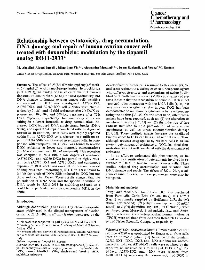
Fig. 1. Structure of N-(3,4-dimethoxyphenethyl)- N-methyl- 2-(naphthyl)- m-dithiane-2-propylamine hydrochloride (RO 11-2933)
CELL LINES RELATIVE FOLD OF RESISTANCE 2h exposure 72h exposure
A2780 | t
t4 doy$ ~ IO'2/j.M DOX
A2780-DXI 4 5
t4 doy$ ~ 5 x 10⁻²~M DOX m
A 2 7 8 0 - DX 2 7 30
14 doys ~ IO-IFM DOX /
A 2 7 8 0 - D X 3 26 53 /
2t doys ~ 5xto'l/u.M DOX
A 2 7 8 0 - D X 4 :5t 77 /
2t days l tFM OOX
A 2 7 8 0 - D X 5 35 24 I /
55 day', / 5/~M DOX +
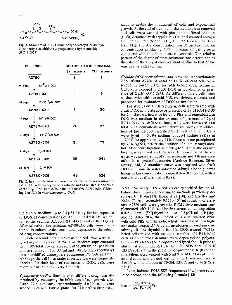
A 2 7 8 0 - D X 6 48 526 Fig. 2. In vitro selection of ovarian cancer cell sublines resistant to DOX. The relative degree of resistance was calculated as the ratio of the ICs0 of resistant cells to that of sensitive A2780 ceils, follow- ing 2 or 72 h in vitro exposure to DOX
the culture medium up to 0.1 ~M. Using further exposure to DOX at concentrations of 0.5, 1.0, and 5.0 gM, we ob- tained the sublines A2780-DX4, -DX5, and -DX6 (Fig. 2). After selection, the resistant A2780-DX cells were main- tained in culture under continuous exposure to the select- ed drug concentrations.
Both parental and DOX-resistant cell lines were cul- tured in monolayers in RPMI 1640 medium supplemented with 10% fetal bovine serum, 2 mM glutamine, penicillin and streptomycin (100 IU/ml and 100 p~g/ml, respectively) in a humidified atmosphere containing 5% CO2 at 37 ~ C. Although the cell lines under investigation were frequently checked for their level of resistance to DOX, cells were taken out of the bank every 3 months.
Cytotoxicity studies. Sensitivity to different drugs was de- termined by measuring the inhibition of cell growth after 2and 72h exposure. Approximately 5x 10⁴ cells were seeded in 24-well Falcon plates for 24 h before drug treat-
ment to enable the attachment of cells and exponential growth. At the end of treatment, the medium was removed and cells were washed with phosphate-buffered solution (PBS), detached with trypsin-EDTA, and counted using a Coulter Counter (Model ZBI, Coulter Electronics, Hia- leah, Fla). The ICs0 concentration was defined as the drug concentration producing 50% inhibition of cell growth compared with that in nontreated controls. The relative pattern of the degree of cross-resistance was determined as the ratio of the ICs0 of each resistant subline to that of the sensitive parental cell line.
Cellular DOX accumulation and retention. Approximately 2.5 • 105/ml A2780 parental or DOX-resistant cells were seeded in 6-well plates for 24 h before drug treatment. Cells were exposed to 2 ~M DOX in the absence or pres- ence of 2 p~M RO11-2933. At different times, cells were washed twice with ice-cold PBS, trypsinized, counted, and processed for evaluation of DOX accumulation.
For studies on DOX retention, cells were treated with 2 p~M DOX in the absence or presence of 2 p~M RO 11-2933 for 2 h, then washed with ice-cold PBS and reincubated in DOX-free medium in the absence of presence of 2 p.M ROll-2933. At different times, cells were harvested and total DOX equivalents were determined using a modifica- tion of the method described by Finkel et al. [10]. Cells were lysed in 0.05% sodium dodecyl sulfate (SDS) at - 20 ~ C for approximately 24 h. Proteins were precipitated by 3.3% AgNO3 before the addition of 4.0 mI n-butyl alco- hol. After centrifugation at 3,500 g for 10 min, the organic layer was removed and the total fluorescence of the ex- tracts was measured at 588 nm emission and 480 nm exci- tation in a spectrofiuorometer (Aminco Bowman, Silver Spring, Md). A standard curve was prepared with fresh DOX dilutions in water-saturated n-butyl alcohol; it was linear in the concentration range 0.02-10.0 ~tg/ml, with a correlation coefficient of > 0.998.
DNA SSB assay. DNA SSBs were quantified by the al- kaline elution assay according to methods previously de- scribed by Kohn [17], Kohn et al. [18], and Bradley and Kohn [6]. Approximately 0.125 x 10⁶/ml sensitive or resis- tant A2780 cells were grown in RPMI 1640 medium sup- plemented with 10% fetal bovine serum containing either 0.0251xCi/ml [14C]-thymidine or 0.5~xCi/ml [3H]-thy- midine. After 20 h, the labeled cells were washed twice with cold PBS and the radioactivity was chased into high- molecular-weight DNA by an incubation in medium con- taining 10 -5 M thymidine for 4 h. DOX-treated [t4C]-la- beled cells mixed with an equal number of [3H]-labeled cells as an internal standard were deposited on polycar- bonate (PC) filters (Nucleopore) and lysed for 1 h prior to elution at room temperature with 2% SDS and 0.025 M EDTA (pH 9.7) in the presence of proteinase K (0.25 mg/ ml). Filters were washed with 5 ml 0.02 MEDTA (pH 10.3) and elution was carried out in a dark environment at 2 ml/h with a solution of TPOH/EDTA and 0.1% SDS at pH 12.1.
Drug-induced DNA SSB frequencies (PBD) were calcu- lated according to the following formula [18]:
log (rl/ro) PBD = log (Ro/ro) PBR,
o "E 0 ~)
r
o (.9
t.)
t 0 0
~ ! . - - 72h exposure i " ~ " - - - - - 2 h exposure
~.~ �9 A2780-DX2 "/'5 \~ ~ , " A2780- DX3
\ l ",.~, x .z78o-ox6
50 '(~X
25 N \ "\',
I I I
I0 20 30 4 0 50
RO11-2933 Concentration EpM]
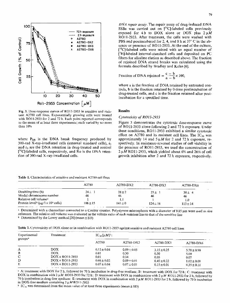
Fig. 3. Dose-response curves of RO11-2933 in sensitive and resis- tant A2780 cell lines. Exponentially growing cells were treated with RO11-2933 for 2 and 72 h. Each point reported corresponds to the mean of at least three experiments; each varied by no more than 10%
where P~R is the D N A break frequency produced by 300-rad X-ray-irradiated cells (external standard cells), rl and r0 are the D N A retention in drug-treated and control []4C]-labeled cells, respectively, and Ro is the D N A reten- tion of 300-rad X-ray-irradiated cells.
79
DNA repair assay. The repair assay of drug-induced D N A SSBs was carried out on [14C]-labeled cells previously exposed for 4 h to D O X alone or D O X plus 2~tM ROll -2933. After treatment, the cells were washed with PBS and postincubated for 2, 4, and 8 h at 37 ~ C in the ab- sence or presence of RO11-2933. At the end of the culture, [14C]-labeled cells were mixed with an equal number of [3H]-labeled internal-standard cells and deposited on PC filters for alkaline elution as described above. The fraction of rejoined D N A strand breaks was calculated using the formula described by Bradley and Kohn [6].
c - b Fraction of D N A rejoined = a - b x 100,
where a is the fraction of D N A retained by untreated con- trols, b is the fraction retained by 0-time postincubation of drug-treated cells, and c is the fraction retained after post- incubation for a specified time.
Results Cytotoxicity ofR011-2933
Figure 3 demonstrates the cytotoxic dose-response curve of RO11-2933 alone following 2 and 72 h exposure. Under these conditions, RO11-2933 exhibited a similar cytotoxic effect on A2780 and its resistant cell lines. The ICs0 was approximately 14 and 5 IxM for 2 and 72 h exposure, re- spectively. In resistance-reversal studies of cell viability in the presence of R O l 1-2933, we used" the concentrat ion of 2 ~tM RO11-2933, which yielded about 6% and 26% Of cell growth inhibit ion after 2 and 72 h exposure, respectively.
Table 1. Characteristics of sensitive and resistant A2780 cell lines
A2780 A2780-DX2 A2780-DX3 A2780-DX6
Doubling time (h) 24+ 1 28+2 25+ 1 30+ 4 Modal chromosome number 46 46 46 46 Relative cell volume" 1 1.1 1 1.0 Protein level b (p.g/1 x 106 cells) 106 _+ 15 141 +0 124_+ 16 113 + 14
a Determined with a channelizer connected to a Coulter counter. Polystyrene microspheres with a diameter of 9.85 gm were used as size reference. The relative cell volume was evaluated as the volume ratio of each resistant line to that of the sensitive line b Determined by the Lowry method [20] (mean_+ SD)
Table 2. Cytotoxicity of DOX alone or in combination with RO 11-2933 against sensitive and resistant A2780 cell lines
Experimental Treatment IC 50 [ktM] b: groups a
A2780 A2780-DX2 A2780-DX3 A2780-DX6
A DOX 0.12 + 0.04 0.89 + 0.03 3.15 + 0.25 5.78 _ 0.90 B DOX 0.01 0.30 0.50 5.00 C DOX + RO 11-2933 0.01 0.34 0.05 0.07 D DOX + RO 11-2933 0.08 _ 0.02 0.09 _+ 0.01 0.45 +_ 0.22 1.02 ___ 0.09 E DOX + RO 11-2933 0.07 + 0.04 0.07 + 0.01 0.15 ___ 0.01 0.35 ___ 0.11
" A: treatment with DOX for 2 h, followed by 70 h incubation in drug-free medium; B: treatment with DOX for 72 h; C: treatment with DOX in combination with 2 txM RO 11-2933 for 72 h; D: treatment with DOX in combination with 2 IxM RO 11-2933 for 2 h, followed by 70 h incubation in drug-free medium; E: treatment with DOX in combination with 2 ~tM RO11-2933 for 2 h, followed by 70 h incubation in DOX-free medium containing 2 IxM RO 11-2933 b IC 50 was determined from the mean value of at least three experiments (mean + SD)
80
A 2 7 8 0 A 2 7 8 0 - DX2 A2780-DX5
t~176 V , , m
=.,
't~ 2 0 0
100
I I i I
I 2 1 2
--~,~. L~,
I I i I I I L I
A 2 7 8 0 - D X 6
A
i
t 2
B
r L i f
1 2 5 4 1 2 3 4 I 2 5 4 1 2 :5 4
Time ( h )
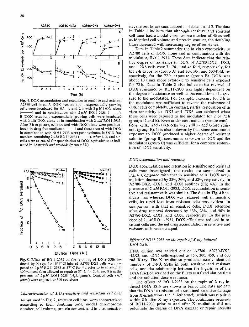
Fig. 4. DOX accumulation and retention in sensitive and resistant A2780 cell lines. A DOX accumulation: exponentially growing cells were incubated for 0.5, 1, and 2 h with 2 txM DOX alone ( ) and in combination with 21xM ROll-2933 ( - - - - - ) . B DOX retention: exponentially growing cells were incubated with 2 ~tM DOX alone or in combination with 2 ~tM RO11-2933. After 2 h exposure, ceils treated with DOX alone were postincu- bated in drug-free medium ( ) and those treated with DOX in combination with RO11-2933 were postincubated in DOX-free medium containing 2 IxM RO11-2933 ( - - - - - ) . After 1, 2, and 4 h, cells were extracted for quantitation of DOX equivalence as indi- cated in Materials and methods (mean ___ SD)
ity; the results are summarized in Tables 1 and 2. The data in Table 1 indicate that although sensitive and resistant cell lines had a modal chromosome number of 46 as well as identical cell volume and protein content, the doubling times increased with increasing degree of resistance.
Data in Table 2 summarize the in vitro cytotoxicity to A2780 cells of DOX alone and in combination with the modulator, RO11-2933. These data indicate that the rela- tive degree of resistance to DOX of A2780-DX2, -DX3, and -DX6 cells were 7-, 26-, and 48-fold, respectively, for the 2-h exposure (group A) and 30-, 50-, and 500-fold, re- spectively, for the 72-h exposure (group B). DOX was about 10 times more cytotoxic to sensitive cells exposed for 72 h. Data in Table 2 also indicate that reversal of DOX resistance by ROll -2933 was highly dependent on the degree of resistance as well as the conditions of expo- sure to the modulator. For example, exposure for 2 h to the modulator was sufficient to reverse the resistance of -DX2 cells completely. In contrast, partial restoration of in vitro sensitivity to -DX3 and -DX6 was achieved when these cells were exposed to the modulator for 2 or 72 h (groups D and E). Even under continuous-exposure condi- tions, -DX3 and -DX6 cells were still 2- and 6-fold resis- tant (group E). It is also noteworthy that since continuous exposure to DOX produced a higher degree of resistant colonies (group B), continuous exposure to DOX and the modulator (group C) was sufficient for a complete restora- tion o f - D X 2 sensitivity.
1.0
.8 "1o
' - . 6
03 fie
<t .4 Z E3 I
r ,r
Control
8
g .2
o o T ime
14-
I I I I I 0 3 6 9 12 15
= _ Control
0 5 6 9 12 15
Elution Time (h)
Fig. 5. Effect of ROll-2933 on the rejoining of DNA SSBs in- duced by X-ray: 1 • 106 [MC]-labeled A2780-DX3 cells were ex- posed to 2 ~xM RO11-2933 at 37 ~ C for 4 h prior to irradiation at 300 tad and then allowed to repair at 37 ~ C for 2, 4, and 8 h in the presence of 2~tM ROll-2933 (right panel). Control cells (left panel) were exposed to 300 rad alone
Characterization of DOX-sensitive and -resistant cell lines
As outlined in Fig. 2, resistant cell lines were characterized according to their doubling time, modal chromosome number, cell volume, protein content, and in vitro sensitiv-
DOX accumulation and retention
DOX accumulation and retention in sensitive and resistant cells were investigated; the results are summarized in Fig. 4. Compared with that in sensitive ceils, DOX accu- mulation decreased by 23%, 30%, and 52%, respectively, in A2780-DX2, -DX3, and -DX6 sublines (Fig. 4A). In the presence of 2 lxM RO 11-2933, DOX accumulation in sensi- tive and resistant cells was similar. The data in Fig. 4 B in- dicate that whereas DOX was retained well in sensitive cells, its rapid loss from resistant cells was evident. In comparison with that in sensitive cells, DOX retention after drug removal decreased by 13%, 25%, and 56% in A2780-DX2, -DX3, and -DX6, respectively. In the pres- ence of 2 IxM ROll-2933, DOX efflux was reduced in re- sistant cells and the net drug accumulation in sensitive and resistant cells became equal.
Effect of RO11-2933 on the repair of X-ray induced DNA SSBs
D N A elution was carried out on A2780, A2780-DX2, -DX3, and -DX6 cells exposed to 150, 300, 450, and 600 rad X-ray. The X-irradiation produced nearly identical numbers of D N A SSBs in both sensitive and resistant cells, and the relationship between the logarithm of the D N A fraction retained on the filters at a fixed elution time and the radiation dose was linear.
The effects of ROll -2933 on the rapir of X-ray-in- duced D N A SSBs are shown in Fig. 5. The data indicate that the D N A in resistant cells sustained extensive damage from X-irradiation (Fig. 5, left panel), which was rapaired within 8 h after X-ray exposure. The continuing presence of ROll-2933 prior to and after X-irradiation did not potentiate the degree of D N A damage or repair. Results
81
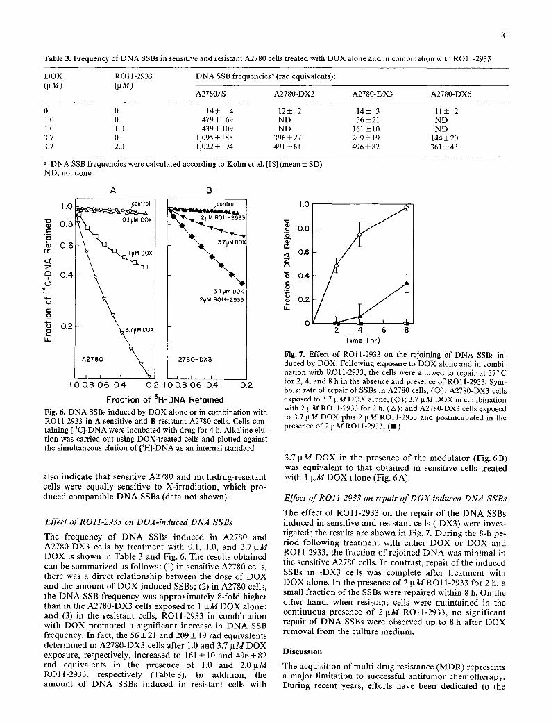
Table 3. Frequency of DNA SSBs in sensitive and resistant A2780 cells treated with DOX alone and in combination with RO11-2933
DOX RO11-2933 (p~M) (]lM)
DNA SSB frequencies ~ (rad equivalents):
A2780/S A2780-DX2 A2780-DX3 A2780-DX6
0 0 14+ 4 12+ 2 14+ 3 11+ 2 1.0 0 479+ 69 ND 56+21 ND 1.0 1.0 439+ 109 ND 161 _ 10 ND 3.7 0 1,095 + 185 396 _ 27 209 _+ 19 144 + 20 3.7 2.0 1,022+ 94 491 ___61 496+82 361 _+43
a DNA SSB frequencies were calculated according to Kohn et al. [181 (mean+ SD)
rr"
Z C3
I ( .9
"3 t - O ~6
ta_
ND, not done
A B
| . 0 /control
~o 0.8
0.6 X
0.4
0.2 ~.THM DO>
\ A2780 " ~ 2780 - DX3
I I I
1.0 0.8 0 6 0.4 0 2 t.0 0.8 0.6 0.4 0.2
Froction of 3H-DNA Retoined
Fig. 6. DNA SSBs induced by DOX alone or in combination with RO11-2933 in A sensitive and B resistant A2780 cells. Cells con- taining []4C]-DNA were incubated with drug for 4 h. Alkaline elu- tion was carried out using DOX-treated cells and plotted against the simultaneous elution of [3H]-DNA as an internal standard
371JM DOX 2vM R0~-2955
also indicate that sensitive A2780 and mult idrug-resis tant cells were equally sensitive to X-ir radia t ion, which pro- duced comparable D N A SSBs (data not shown).
Effect ofR011-2933 on DOX-induced DNA SSBs
The frequency of D N A SSBs induced in A2780 and A2780-DX3 cells by t reatment with 0.1, 1.0, and 3.7 IxM DOX is shown in Table 3 and Fig. 6. The results obta ined can be summarized as follows: (1) in sensitive A2780 cells, there was a direct re la t ionship between the dose of DOX and the amount of DOX- induced SSBs; (2) in A2780 cells, the D N A SSB frequency was approximate ly 8-fold higher than in the A2780-DX3 cells exposed to 1 ixM DOX alone; and (3) in the resistant ceils, ROl l -2933 in combinat ion with DOX promoted a significant increase in D N A SSB frequency. In fact, the 56___ 21 and 209___ 19 t ad equivalents de termined in A2780-DX3 cells after 1.0 and 3.7 ~tM DOX exposure, respectively, increased to 161 ___ 10 and 496___ 82 rad equivalents in the presence of 1.0 and 2.011M RO 11-2933, respectively (Table 3). In addi t ion, the amount of D N A SSBs induced in resistant cells with
I.O
c 0.8
r r < 0.6 Z s
"6 0.4 c .9 o 0.2 LL
0 vm 2 4 6 8
Time (hr)
Fig. 7. Effect of RO11-2933 on the rejoining of DNA SSBs in- duced by DOX. Following exposure to DOX alone and in combi- nation with RO11-2933, the cells were allowed to repair at 37 ~ C for 2, 4, and 8 h in the absence and presence of RO11-2933. Sym- bols: rate of repair of SSBs in A2780 cells, (O); A2780-DX3 cells exposed to 3.7 txM DOX alone, (O); 3,7 p~M DOX in combination with 2 I.tMRO11-2933 for 2 h, (A); and A2780-DX3 cells exposed to 3.7 gM DOX plus 2 llM RO11-2933 and postincubated in the presence of 2 I.tM RO11-2933, (m)
3.7 g M DOX in the presence of the modula to r (Fig. 6B) was equivalent to that obta ined in sensitive cells t reated with 1 l lM DOX alone (Fig. 6A).
Effect ofR011-2933 on repair of DOX-indueed DNA SSBs
The effect of RO11-2933 on the repai r of the D N A SSBs induced in sensitive and resistant cells (-DX3) were inves- t igated; the results are shown in Fig. 7. During the 8-h pe- r iod following t reatment with either DOX or DOX and RO11-2933, the fraction o f re joined D N A was minimal in the sensitive A2780 cells. In contrast, repair of the induced SSBs in -DX3 cells was complete after t reatment with DOX alone. In the presence of 2 IxMRO11-2933 for 2 h, a small fraction of the SSBs were repai red within 8 h. On the other hand, when resistant cells were main ta ined in the cont inuous presence of 2 g M ROl l -2933 , no significant repair of D N A SSBs were observed up to 8 h after DOX removal from the culture medium.
Discussion
The acquisit ion of mult i -drug resistance (MDR) represents a major l imitat ion to successful ant i tumor chemotherapy. During recent years, efforts have been dedicated to the
82
identification of mechanisms leading to acquisition of MDR as well as the definition of new treatment strategies to circumvent resistance. In the present investigation, DOX resistance was induced in human ovarian cancer cells by stepwise increases in drug concentration. This procedure enabled the selection of cells with a low level of resistance, e.g., A2780-DX2 (7-fold resistance), and higher levels of resistance, e.g., A2780-DX3 and -DX6 (26- and 48-fold, respectively). The procedure outlined in Fig. 2 in- dicates that the acquisition of resistance was initially rapid but became progressively slower with increasing drug con- centration, which suggests that at least one modification occurs in the process of acquiring a highly resistant pheno- type. Moreover, the selective procedure induced an atypi- cal MDR. In fact, the A2780-DX sublines were also cross- resistant to cisplatin, navelbin, and colchicine, to a degree that increased as the cell population became more resistant to DOX (data not shown). This evidence suggests that common mechanism(s) of resistance may be shared by these drugs, probably as the result of the expression of a modified single gene (or genes) under coordinated regu- lation.
Previous investigations have demonstrated that an ac- tive outward drug transport, resulting in lower intracellu- lar drug accumulation, is partly responsible for DOX re- sistance [9, 19, 30-32, 37]. In the A2780-DX sublines, im- paired accumulation and retention were observed. As shown in Fig. 4, the degree of drug accumulation and re- tention decreased as the degree of resistance increased. These data are in agreement with those reported in other cell lines. Overexpression of p-glycoprotein, PG170, was detected in A2780-DX3 and -DX6 (data not shown). In these cell lines, lower intracellular DOX accumulation and retention correlated with the overexpression of P-170. However, differences in the amount of DOX accumulation and retention could not account for the differences in re- sistance level, since the drug level in A2780-DX3 cells was 33% lower than that in the sensitive A2780 line (Fig. 3), al- though the former were about 50-fold resistant (Fig. 2). Moreover, in A2780-DX6 cells, the drug level was only 52% lower than that in A2780 cells (Fig. 3), whereas the former were 526-fold resistant after long-term exposure (72 h, Fig. 2).
Since the major biological activity of DOX is thought to reside in its ability to interact with DNA [14], studies on damage and repair were also carried out. Our results showed an induction of DNA SSBs in sensitive and resis- tant cells treated with 1.0 or 3.7 ~tM DOX. However, the extent of DNA SSBs decreased with increasing drug resis- tance (Table 3). The results are in agreement with previ- ously reported findings in other cell lines [29, 34, 41]. In addition, DNA SSB repair was in resistant cells almost complete within 8 h, whereas no significant DNA repair was observed in sensitive cells. These data suggest that the mechanism(s) of resistance to DOX are multifactorial events associated with reduced drug accumulation and re- tention as well as a modification of the drug's interaction with DNA that results in decreased SSB induction and in- creased repair capacities.
We also investigated the effects of RO11-2933 on DOX resistance. Previous studies have shown that verapamil, a calcium channel blocker, can successfully potentiate DOX cytotoxicity in resistant cells [8, 13, 15, 24, 30, 36]. The clinical use of verapamil in combination with DOX is lira-
ited by the relatively high verapamil dose required to achieve the plasma drug concentration necessary for in vi- tro reversal of DOX resistance without unacceptable host toxicity. In addition, Rabkin et al. [26] have shown that pretreatment with verapamil may potentiate the myocardi- al toxicity caused by conventional DOX treatment [26, 27]. For these reasons, new calcium-channel-blocker analogs with greater selectivity were identified by Kessel and Wilberding [16].
Among the agents evaluated, RO11-2933 was found to be more potent than verapamil (1 txMvs 10 ~M) [3, 27, 38] but less toxic in vivo (data not shown). Results of these studies revealed that restoration of drug sensitivity was complete in cells with a low resistance level (A2780-DX1 and -DX2) and correlated with greater drug accumulation and retention as well as increased DNA damage. In con- trast, we found that ROll-2933 could only partially reverse DOX resistance in highly resistant cells (A2780-DX3 and -DX6), even when they were continu- ously exposed to the optimal concentration of the modu- lator.
As shown by our cytotoxicity experiments (Table 2), a residual 2- and 5-fold resistance was observed in A2780-DX3 and -DX6, respectively, when 2p~M ROll-2933 (the maximally tolerated concentration) was added to the incubation medium. Furthermore, RO11-2933 effectively retarded the repair of DOX-in- duced DNA SSBs in the resistant cells, and this effect was strictly related to the treatment schedule. In fact, A2780-DX3 cells treated with 3.7 lxM DOX alone showed complete DNA repair that decreased to 33% when the drug was combined with ROll-2933 and to approximately 0 when 2.0 ~tM RO11-2933 was maintained in the posttreat- ment medium.
The evidence that RO11-2933 completely reverses cell sensitivity to DOX in ceils with a lower degree of resis- tance (A2780-DX1 and -DX2) has some clinical value, since only low-level resistance may be expected to occur in clinical situations. Furthermore, inhibition of DNA repair by the modulator in resistant cells may offer an additional selective advantage for the clinical use of this agent over other calcium channel blockers.
Acknowledgements. We wish to thank C. Frank for her technical help and C. Melancon and G. Wagner for their secretarial assis- tance.
References
1. Bachur NR (1982) Mechanism of action of doxorubicin. In: Hansen HH (ed) Anthracyclines and cancer chemotherapy. Proceedings of a symposium, Ronneby Brunn, Sweden, Oc- tober 6-7, p 2
2. Bachur NR, Gordon SL, Gee MV (1977) Anthracycline anti- biotic augmentation of microsomal electron transport and free radical formation. Mol Pharmacol 13:901
3. Bankusli I, Yin M-B, Rustum YM (1987) The use of "Ca +2 channel blocker" N-(3,4-dimethoxyphenethyl)-N-methyl- 2-(naphthyl)- m-dithione-2-propylamine hydrochloride (DMDP) to overcome multidrug resistance of P-388 cell line. 15th International Congress on Chemotherapy, Istanbul, Tur- key, contribution D-30
4. Behrens BC, Louie KG, Hamilton TC, Curt G, Kinsella T, Young RC, Ozols RF (1984) Resistance and cross resistance of human ovarian cancer cell lines to Adriamycin, melphalan and irradiation. Proc Am Assoc Cancer Res 25 : 336
83
5. Berlin V, Hazeltine WA (1981) Reduction of Adriamycin to a semiquinone-free radical by NADPH cytochrome P-450 re- ductase produces DNA cleavage in a reaction mediated by molecular oxygen. J Biol Chem 256:4747
6. Bradley MO, Kohn KW (1979) X-ray induced DNA double strand break production and repair in mammalian cells as measured by neutral filter elution. Nucleic Acids Res 7:793
7. Crook ST, Reich SD (eds) (1980) Anthracyclines: current status and new developments. Academic, New York
8. Dahllof B, Martinsson T, Mannervik B, Jensson H, Levan G (1987) Chracterization of multidrug resistance in SEWA mouse tumor cells: increased glutathione transferase activ- ity and reversal of resistance with verapamil. Anticancer Res 7:65
9. Dano K (1973) Active outward transport of daunomycin in resistant Ehrlich ascites tumor cells. Biochim Biophys Acta 323 : 466
10. Finkel JM, Knapp KT, Mulligan LT (1969) Fluorometric de- termination of the serum levels and urinary excretion of dau- nomycin (NCS-82151) in mice and rats. Cancer Chemother Rep 53:159
11. Frankfurt OS, Chin JL, Englander LS, Greco WR, Pontes JE, Rustum YM (1985) Relationship between DNA ploidy, glan- dular differentiation, and tumor spread in human prostate cancer. Cancer Res 45:1418
12. Garman D, Center MS (1982) Alterations in cell surface mem- branes in Chinese hamster lung cells resistant to Adriamycin. Biochem Biophys Res Commun 105:157
13. Harker WG, Bauer D, Etiz BB, Newman RA, Sikic BI (1986) Verapamil-mediated sensitization of doxorubicin-selected pleiotropic resistance in human sarcoma cells: selectivity for drug which produce DNA scission. Cancer Res 46:2369
14. Harris JF, Karran P, Lindahi T (1983) O6-Methylguanine - DNA methyltransferase of human lymphoid cells: structural and kinetic properties and absence in repair-deficient cells. Cancer Res 43 : 3247
15. Hindenburg AA, Baker MA, Gleyzer E, Stewart VJ, Case N, Taub RN (1987) Effect of verapamil and other agents on the distribution of anthracyclines and on reversal of drug resis- tance. Cancer Res 47:1421
16. Kessel D, Wilberding C (1985) Promotion of daunorubicin uptake and toxicity by the calcium antagonist tiapamil and its analogs. Cancer Treat Rep 69:673
17. Kohn KW (1979) DNA as a target in cancer chemotherapy: measurement of macromolecular DNA damage produced in mammalian cells by anticancer agents and carcinogens. Meth- ods Cancer Res 16:291
18. Kohn KW, Ewig RAG, Erickson LC, Zwelling LA (1981) Measurement of strand breaks and cross-links by alkaline elu- tion. In: Friedberg EC, Hanawalt PC (eds) DNA repair: a laboratory manual of research techniques. Marcel Dekker, New York, p 379
19. Louie KG, Hamilton TC, Winker MA, Behrens BC, Tsu- ruo T, Klecker RW Jr, McKoy WM, Grotzinger KR, Myers CE, Young RC, Ozols RT (1986) Adriamycin accumulation and metabolism in Adriamycin-sensitive and -resistant human ovarian cancer cell lines. Biochem Pharmaco135:467
20. Lowry OH, Rosebrough NJ, Farr AL, Randall RJ (1951) Pro- tein measurement with the folin phenol reagent. J Biot Chem 193 : 265
21. Murphree SA, Tritton TR, Smith PL, Sartorelli AC (1981) Adriamycin-induced changes in the surface membrane of sar- coma 180 ascites cells. Biochim Biophys Acta 649:317
22. Myers CE, McGuire WP, Liss RH, Ifrian I, Grotyinger K, Young RC (1977) Adriamycin: the role of lipid peroxidation in cardiac toxicity and tumor response. Science 197:165
23. Ozols RF, Young RC, Speyer JL, Sugarbaker PH, Greene R, Jenkins J, Myers CE (1982) Phase I and pharmacologic stud- ies of Adriamycin administered intraperitoneally to patients with ovarian cancer. Cancer Res 42:4265
24. Ozols RF, Cunnion RE, Klecker RW, Hamilton TC Jr, Ostchega Y, Parillo JE, Young RC (1987) Verapamil and Adriamycin in the treatment of drug-resistant ovarian cancer patients. J Clin Oncol 5:641
25. Pigram WJ, Fuller W, Hamilton LD (1972) Stereochemistry of intercalation: interaction of daunomycin with DNA. Nature 235:17
26. Rabkin SW, Otten M, Polimeni PI (1983) Increased mortality with cardiotoxic doses of Adriamycin after verapamil pre- treatment despite prevention of myocardial calcium accumu- lation. Can J Physiol Pharmacol 61 : 1050
27. Radel S, Bankusli I, Mayhew E, Rustum YM (1988) The ef- fect of verapamil and a tiapamil analogue, "DMDP", on Adriamycin-induced cytotoxicity in P388 Adriamycin-resis- tant and -sensitive leukemia in vitro and in vivo. Anticancer Res 7: 1105
28. Ramu A, Fuks Z, Gatt S, Gluabiger D (1984) Reversal of ac- quired resistance to doxorubicin in P388 murine leukemia cells by perhexiline maleate. Cancer Res 44:144
29. Richon VM, Schulte N, Eastman A (1987) Multiple mecha- nisms of resistance to cis-diamminedichloroplatinum(II) in murine leukemia L1210 cells. Cancer Res 47:2056
30. Rogan AM, Hamilton TC, Young RC, Klecker RW, Ozols RF (1984) Reversal of Adriamycin resistance by verapamil in human ovarian cancer. Science 224:994
31. Skovsgaard T (1978) Mechanism of resistance to daunorubi- cin by sensitive and anthracycline-resistant sublines of P388 leukemia. Biochem Pharmacol 27: 2123
32. Slater LM, Murray SL, Wetzel MW (1982) Verapamil restora- tion of daunorubicin responsiveness in daunorubicin-resistant Ehrlich ascites carcinoma. J Clin Invest 70:1131
33. Tokes ZA, Rogers KE, Rembaum A (1982) Synthesis of Adriamycin-coupled polyglutaraldehyde microspheres and evaluation of their cytostatic activity. Proc Natl Acad Sci USA 79: 2026
34. Tong WP, Kirk MC, Ludlum DB (1981) Molecular pharma- cology of the haloethyl nitrosoureas: formation of 6-hydroxy- ethylguanine in DNA treated with BCNU (N,N'-bis[2-chloro- ethyl]-N-nitrosourea). Biochem Biophys Res Commun 100: 351
35. Tritton T, Yee G (1982) The anticancer agent Adriamycin can be actively cytotoxic without entering cells. Science 217:248
36. Tsuruo T, Iida H, Tsukagoshi S, Sakurai Y (1982) Increased accumulation of vincristine and Adriamycin in drug-resistant P388 tumor cells following incubation with calcium antago- nists and calmodulin inhibitors. Cancer Res 42:4730
37. Tsuruo T, Lida H, Jojiri M, Tsukagoshi S, Sakurai Y (1983) Circumvention of vincristine and Adriamycin resistance in vi- tro and in vivo by calcium influx blockers. Cancer Res 43: 2905
38. Yin M-B, Bankusli I, Rustum YM (1988) Modulation of Adriamycin resistance by a calcium channel blocker , N-(3,4-demethoxyphenethyl)- N-methyl-2-(naphthyl)- m-di- thiane-2-propylamine hydrochloride (DMDP). Am Assoc Cancer Res 2002:504
39. Young RC (1984) Ovarian cancer treatment: progress or pa- ralysis. Semin Oncol 11 : 327
40. Young RC, Ozols RF, Myers CE (1981) The anthracycline antineoptastic drugs. N Engl J Med 305:139
41. Zijlstra JG, Vries EGE de, Mulder NH (1987) Multifactorial drug resistance in an Adriamycin-resistant human small cell lung carcinoma cell line. Cancer Res 47:1780
Received 7 December 1988/Accepted 20 April 1989