relationship between central corneal thickness and progression of visual field loss in patients with...
TRANSCRIPT

Relationship between central corneal thickness and progressionof visual field loss in patients with open-angle glaucomaKathy Y. Cao, MD, FRCSC*, Mustafa Kapasi, MD†, Janet A. Betchkal, MD‡,Catherine M. Birt, MA, MD, FRCSC*
ABSTRACT ● RÉSUMÉ
Objective: To determine whether a relationship exists between central corneal thickness (CCT) and visual field (VF) progression intreated patients with open-angle glaucoma and asymmetric corneal thickness.
Design: Retrospective chart review.Participants: We studied 100 charts of patients with open-angle glaucoma and also bilateral CCT and VF data.Methods: Charts from 2 glaucoma subspecialty practices were reviewed. The CCT and the rate of progression and event analysis of
visual field data were assessed in all subjects. Subanalysis was performed for subjects whose CCT asymmetry was � 16 �m.Results: The mean CCT was 544 � 40 �m OD and 541 � 40 �m OS. The mean CCT difference between fellow eyes was 15 � 11 �m
(range, 1 to 52 �m). There was no significant intrasubject difference in the mean deviation (MD) and the pattern standard deviation(PSD) (p �0.917 and p � 0.704, respectively; paired t test). The more advanced VF MDs and PSDs were found in the thin eyes of47 and 50 subjects, respectively (p � 0.459 and p � 0.317, respectively; �2). Of the 65 subjects whose visual field indexes wereavailable, 34 had the more rapid visual field index rates of progression in the thin eye (p � 0.400; �2). Of the 27 subjects for whomevent analysis was available, 15 had the worse progression category in the thin eye (p � 0.453, �2). Subgroup analysis of 48 subjectswith � 16 �m CCT asymmetry did not find any significant difference in analyses of field progression between fellow eyes.
Conclusions: No relationship was found between CCT and VF loss in treated patients with primary open-angle glaucoma or normal-tension glaucoma with asymmetrical CCT. Specifically, the thin eye did not have the more advanced VF loss or more rapid VFprogression.
Objet : Établir s’il y a une relation entre de l’épaisseur du centre de la cornée (ÉCC) et la progression du champ visuel (CV) chez lespatients traités pour un glaucome à angle ouvert avec une épaisseur asymétrique de la cornée.
Nature : Étude rétrospective des dossiers.Participants : 100 dossiers de patients atteints de glaucome à angle ouvert, ayant des données d’ÉCC et de CV.Méthodes : Examen des dossiers de deux pratiques surspécialisées en glaucome. L’ÉCC et le taux de progression et l’analyse de
l’événement des données du CV ont été évalués chez tous les patients. La suranalyse a été effectuée chez les sujets dont l’asymétriede l’ÉCC était �16�m.
Résultats : La moyenne d’ÉCC était de 544�40�m OD et 541�40�m OS (??). La moyenne d’écart de l’ÉCC entre les deux yeux étaitde 15�11�m (écarts, 1-52�m). Il n’y avait pas d’écart significatif des MD et PSD dans un même sujet (p � 0,917 et p � 0,704respectivement, test-t apparié). Les MD et PSD du champs visuel le plus faible ont été trouvés dans l’œil mince de 47 et 50 sujetsrespectivement (p � 0,459 and p � 0,317 respectivement, �2). Parmi les 65 sujets ayant VFI disponible, 34 avaient eu un taux deprogression de CVI (?) plus rapide dans l’œil mince (p � 0.400, �2). Parmi les 27 sujets qui avaient une analyse d’événementdisponible, 15 avaient la pire catégorie de progression dans l’œil mince (p � 0,453, �2). L’analyse du sous-groupe de 48 sujets ayantune asymétrie de l’ÉCC de �16�m n’a pas indiqué d’écart significatif dans les analyses de progression du champ entre les deuxyeux.
Conclusions : L’on n’a pas trouvé de relation entre la perte d’ÉCC et de CV chez les patients traités avec GPAO ou GPN pour ÉCC.Spécifiquement, l’œil mince n’avait pas de perte plus prononcée de CV ni de progression plus rapide de CV.
Although elevated intraocular pressure (IOP) is themain risk factor for the development and progression ofglaucomatous optic neuropathy, other risk factors havebeen identified. One of these factors, central corneal thick-ness (CCT), became a routine evaluation in glaucoma carefollowing the publication of the Ocular HypertensionTreatment Study, which showed that conversion from oc-ular hypertension to open-angle glaucoma was more com-mon in hypertensive patients with thinner CCT.1 CCTvariation is also an important factor in the accuracy ofmeasurement of IOP by Goldmann applanation tonome-try.2 The Goldmann applanation tonometry technique
From the *University of Toronto, Toronto, Ont.; the †University ofOttawa, Ottawa, Ont.; and the ‡St. Vincent Medical Center, Jacksonville,Fla.
Originally received Aug. 20, 2011. Final revision Oct. 28, 2011. AcceptedNov. 2, 2011Correspondence to Catherine Birt, M1 302a, 2075 Bayview Ave., Toronto
ON M4N 3M5; email: [email protected]was developed on the basis of an estimated ideal cornealthickness of 500 �m, and corneas that are thicker or thin-ner than this can lead to overestimation or underestima-tion of the actual IOP. However, previous research alsosuggests that a thin CCT may be an independent risk fac-tor for glaucomatous optic neuropathy, possibly reflectingabnormalities in the posterior sclera and lamina cribrosa.The effect of glaucomatous neuropathy on visual functionis assessed by the degree of damage shown on automatedvisual field testing.3,4 Recent studies have shown that eyeswith thinner CCTs tend to have more advanced visual fieldloss.5,6
Can J Ophthalmol 2012;47:155–1580008-4182/11/$-see front matter © 2012 Canadian Ophthalmological Society.Published by Elsevier Inc. All rights reserved.doi:10.1016/j.jcjo.2012.01.008
CAN J OPHTHALMOL—VOL. 47, NO. 2, APRIL 2012 155
CCT and VF progression in OAG—Cao et al.
This study was undertaken to examine whether a rela-tionship between CCT and progression of visual field (VF)changes exists in patients treated for open-angle glaucomawho have asymmetrical CCT. We hypothesized that eyeswith thinner CCT may have either a more severe VF defectat presentation or may show more rapid field progressiondespite treatment compared to eyes with thicker CCT. Ifso, it would provide further evidence to suggest that CCTis a risk for glaucomatous field loss independent of IOP,and perhaps eyes with thinner CCT require more aggres-sive treatment.
METHOD
After approval of the project by the Research EthicsBoard of Sunnybrook Health Sciences Centre, a retrospec-tive review of 100 charts was performed at the offices of 2glaucoma subspecialists (J.B., C.M.B.). The inclusion cri-teria required a diagnosis of primary open-angle glaucoma(POAG) or normal tension glaucoma (NTG) based oneither or both characteristic visual field defects (nasal step,arcuate scotoma, paracentral defect) or optic nerve changes(enlarged cup to disc ratio, localized notch, disc hemor-rhage) in at least 1 eye. A reliable VF examination takenwithin 6 months of the pachymetry reading had to beavailable for both eyes. To exclude the possibility that dif-ferences between eyes was due to IOP control, we requiredan IOP difference of � 4 mm Hg between fellow eyes atthe time of pachymetry. Exclusion criteria were non-POAGglaucoma diagnosis, monocular patients, and patients withsystemic diseases that might result in VF changes, or cornealpathology or surgery that might influence either pachymetryor IOP measurements. Data gathered included demographicinformation (age, sex, and race); glaucoma diagnosis; cup-to-disc ratio; IOP level; number of medications used (recordedon the date the CCT was assessed); CCT for both eyes; andVF information (mean deviation [MD]; pattern standard de-viation [PSD]; the VF index trend and its significance level;and the presence of progression in the glaucoma progression
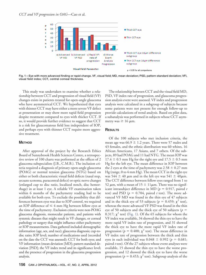
Fig. 1—Eye with more advanced finding or rapid change. VF, visvisual field index; CCT, central corneal thickness.
analysis.
156 CAN J OPHTHALMOL—VOL. 47, NO. 2, APRIL 2012
The relationship between CCT and the visual field MD,PSD, VF index rate of progression, and glaucoma progres-sion analysis event were assessed. VF index and progressionanalysis were calculated in a subgroup of subjects becausesome patients were not present for enough follow-up toprovide calculations of trend analysis. Based on pilot data,a subanalysis was performed in subjects whose CCT asym-metry was � 16 �m.
RESULTS
Of the 100 subjects who met inclusion criteria, themean age was 66.9 � 1.2 years. There were 57 males and43 females, and the ethnic distribution was 60 whites, 16African Americans, 17 Asians, and 7 others. Of the sub-jects, 89 had POAG and 11 had NTG. The mean IOP was17.4 � 0.5 mm Hg for the right eye and 17.5 � 0.5 mmHg for the left eye. The mean difference in IOP betweenthe 2 eyes at the time of pachymetry was 2.58 � 0.27 mmHg (range, 0 to 4 mm Hg). The mean CCT in the right eyewas 544 � 40 �m and in the left eye was 541 � 40�m.The CCT difference between fellow eyes ranged from 1 to52 �m, with a mean of 15 � 11�m. There was no signif-icant intrasubject difference in MD (p � 0.917, paired ttest) and PSD (p � 0.704, paired t test). The more ad-vanced VF MD was found in the thin eye of 47 subjectsand in the thick eye of 53 subjects (p � 0.459; �2 test),whereas the more advanced VF PSD was found in the thineye of 50 subjects and the thick eye of 50 subjects (p �0.317; �2 test) (Fig. 1). Of the 65 subjects for whom theVF index was available, 34 showed the thin eye to have themore rapid VF index rate of progression, and 31 showedthe thick eye to have the more rapid VF index rate ofprogression (p � 0.400; �2 test). The mean difference inVF index rate of progression between the thick and thineyes in each individual was 0.048 � 0.992 (p � 0.681;paired t test). Of the 27 subjects whose event analyses wereavailable, 15 showed the thin eye to have the worse pro-gression, and 12 showed the thick eye to have the worse
field; MD, mean deviation; PSD, pattern standard deviation; VFI,
ualprogression (p � 0.453; �2 test). Subgroup analysis of the

CCT and VF progression in OAG—Cao et al.
48 subjects whose CCT asymmetry was �16 �m also didnot find any significant difference in the MD and PSDbetween fellow eyes. The more advanced VF MD wasfound in the thin eye of 21 subjects in this subgroup and inthe thick eye of 27 subjects (p � 0.386; �2 test), and themore advanced VF PSD was found in the thin eye of 22subjects and in the thick eye of 26 subjects (p � 0.564; �2
test).Subgroup analysis of the subjects whose CCT asymme-
try was � 16 �m also did not find any significant differ-ence in the VF index rate of progression and event analysisbetween fellow eyes. In this subgroup, of the 37 subjectswhose VF indices were available, 16 showed the thin eye tohave the more rapid VF index rate of progression, and 11showed the thick eye to have the more rapid VF index rateof progression (p � 0.336; �2 test). The mean difference inthe VF index rate of progression between the thick andthin eyes for each individual was 0.273 � 0.880 (p �0.119; paired t test). There were 12 subjects in this sub-group for whom event analysis was available: 9 had thin eyein the worse progression category; 3 had thin eye (p �0.083; �2 test). No relationship was found between CCTand VF loss in treated patients with POAG or NTG whohad asymmetrical CCT.
DISCUSSION
In our study sample, the thin eye did not have the moreadvanced VF loss or more rapid VF progression, as wouldbe expected from prior reports that CCT is an independentrisk factor for glaucomatous optic neuropathy. This sug-gests that other factors may have stronger influences on VFloss in this study population, and further research involv-ing larger sample sizes is required for better definition ofthe relationship between CCT and VF loss.
Whether thin corneas independently increase the riskfor the development or the progression of glaucoma is dif-ficult to determine. There are many ways the relationshipbetween CCT and glaucoma can be examined, even usingonly visual fields as a marker for disease. Some studies havelooked at the rate of conversion from high risk nonglau-coma status to frank glaucoma, usually using ocular hyper-tension as the risk factor; others have looked at the stage ofdisease at presentation, and still others have reported onthe rate of progression during disease management. Acrossmany different studies, results have varied, some showingCCT as a risk factor and others not. When looking atconversion to POAG, the Ocular Hypertension Treat-ment Study provided prospective evidence for the impor-tance of CCT measurement, where thinner CCT readingswere a risk factor for conversion from ocular hypertensionto open angle glaucoma.1 However, it is not possible to besure whether the increased risk was simply related to theconfounding of IOP measurements by corneal thickness;patients with thick corneas may actually have had lower
IOP than was recorded. In our study, all patients had glau-coma. Hence the progression of the disease rather thanconversion to the disease was followed in relation to CCT.
When examining disease stage at presentation, Jonas et al.,7
Sullivan-Mee et al.,8 and Fernandez-Bahamonde et al.9 foundthat patients with thinner CCTs had more advanced stages ofglaucoma and worsened visual field. Mokbel et al.10 did astudy in which POAG patients were divided into a thin-CCT group (� 540 �m) and a thick-CCT group (� 540�m). They found that thin-CCT group had significantlyworse VF than did the thick-CCT group. Likewise, Lin etal.6 examined the relationship between the stage of glau-coma based on the VF defect and found better VF index inpatients with thicker CCTs. However, they were not ableto find an association between CCT and VF defects in eyeswith POAG. Sullivan-Mee et al.8 and Fernandez-Ba-hamonde et al.9 were not able to show any trend analysisbecause their studies covered a short time period.
When reporting on the rate of progression, both Kim etal.11 and Medeiros et al.12 reported that progressing pa-tients had thinner CCTs than did stable patients. This isthe inverse of our study. It looked at CCT in patientsknown to be progressing, whereas we looked at patients’CCTs and then examined whether they progressed or not.It is therefore possible that patients who progress are morelikely to have thin corneas, but patients with thin corneasdo not necessarily progress. However, in a recent study, DeMoraes et al.13 found that a thinner cornea was a risk factorfor progression of glaucoma in their treated patients. Incontrast, the Early Manifest Glaucoma Trial14 and workby Chauhan et al.15 did not find evidence of CCT as a riskfactor for field progression, and the Jonas study found that,despite a relationship between CCT and disease stage, inhis sample, progression was independent of CCT.7
This study intended to examine rates of progression intreated patients with POAG; if thin CCT were confirmedas a risk factor for poorer outcomes, treatment goals mightbe different for these patients. We found no significantdifference in progression analyses between fellow eyes,However, there was a trend toward the eye with the thinnercornea having a more rapid rate of progression of VF indexand worse event analysis progression, especially in thosewhose CCT asymmetry was � 16 �m. Because of the smallsample size in this subgroup, further study involving a largersample is needed. Another limitation is the retrospective na-ture of the study. There was no standardized protocol for thetreatments given to the different patients. Treatment targetswere individualized to each patient based on the usual pre-senting levels of IOP and nerve and VF damage, but the num-bers and types of intervention provided over the follow-upperiod were determined only by the opinion of the treatingphysician and may have varied widely in patients. This mayhave resulted in different apparent rates of progression inde-pendent of CCT. A prospective trial could examine both dis-ease stage at presentation and rates of progression in treatedpatients, but that would be a long-term project. The VF is a
functional marker of the progression of glaucoma; however,CAN J OPHTHALMOL—VOL. 47, NO. 2, APRIL 2012 157

CCT and VF progression in OAG—Cao et al.
optic-nerve–related markers are also used to follow the dis-ease. Further studies could examine the relationship betweenCCT and the other structural indicators of glaucoma.
Disclosure: The authors have no proprietary or commercial interestin any material disclosed in this article.
REFERENCES
1. Gordon MO, Beiser JA, Brandt JD, et al. The Ocular HypertensionTreatment Study: Baseline factors that predict the onset of primaryopen-angle glaucoma. Arch Ophthalmol. 2002;120:714-20.
2. Goldmann H, Schmidt T. Applanation tonometry. Ophthalmologica.1957;134:221-42.
3. Vesti E, Johnson C, Chauhan B. Comparison of different methods fordetecting glaucomatous visual field progression. Invest Ophthalmol VisSci. 2003;44:3873-9.
4. Jampel H, Singh K, Lin S, et al. Assessment of visual function in glau-coma. Ophthalmology. 2011;118:986-1002.
5. Rogers DL, Cantor RN, Catoira Y, et al. Central corneal thickness andvisual field loss in fellow eyes of patients with open-angle glaucoma. Am JOphthalmol. 2007;143:159-61.
6. Lin W, Aoyama Y, Kawase K, Yamamoto T. Relationship between cen-tral corneal thickness and visual field defect in open-angle glaucoma. Jpn
J Ophthalmol. 2009;53:477-81.158 CAN J OPHTHALMOL—VOL. 47, NO. 2, APRIL 2012
7. Jonas JB, Stroux A, Velten I, et al. Central corneal thickness correlated withglaucoma damage and rate of progression. Invest Ophthalmol Vis Sci. 2005;46:1269-74.
8. Sullivan-Mee M, Gentry JM, Qualls C. Relationship between asymmet-ric central corneal thickness and glaucomatous visual field loss within thesame patient. Optom Vis Sci, 2006;83:516-9.
9. Fernandez-Bahamonde J, Roman-Rodriguez C, Fernandez-Riuz M.Central corneal thickness as a predictor of visual field loss in primaryopen-angle glaucoma for a Hispanic population. Sem Ophthalmol. 2011;26:28-32.
10. Mokbel T, Ghanem A. Correlation of central corneal thickness andoptic nerve head topography in patients with primary open-angle glau-coma. Oman J Ophthalmol. 2010;3:75-80.
11. Kim JW, Chen PP. Central corneal pachymetry and visual field progres-sion in patients with open-angle glaucoma. Ophthalmology. 2004;111:2126-32.
12. Medeiros FA, Sample PA, Zangwill LM, et al. Corneal thickness as arisk factor for visual field loss in patients with preperimetric glauco-matous optic neuropathy. Am J Ophthalmol. 2003;136:805-13.
13. De Moraes C, Juthani V, Liebmann JM, et al. Risk factors for visual fieldprogression in treated glaucoma. Arch Ophthalmol. 2011;129:562-8.
14. Leske MC, Heijl A, Hussein M, et al. The Early Manifest GlaucomaTrial Group: Factors for glaucoma progression and the effect of treat-ment: The Early Manifest Glaucoma Trial. Arch Ophthalmol. 2003;121:48-56.
15. Chauhan BC, Hutchison DM, LeBlanc RP, et al. Central corneal thick-ness and progression of the visual field and optic disc in glaucoma. Br J
Ophthalmol. 2005;89:1008-12.