regulation of substrate adhesion dynamics during cell motility
TRANSCRIPT

The International Journal of Biochemistry & Cell Biology 34 (2002) 746–761
Review
Regulation of substrate adhesion dynamics during cell motility
Irina Kaverina, Olga Krylyshkina, J. Victor Small∗Department of Cell Biology, Institute of Molecular Biology, Austrian Academy of Sciences, Billrothstrasse 11, Salzburg 5020, Austria
Received 26 September 2001; received in revised form 20 November 2001; accepted 5 December 2001
Abstract
The movement of a metazoan cell entails the regulated creation and turnover of adhesions with the surface on which itmoves. Adhesion sites form as a result of signaling between the extracellular matrix on the outside and the actin cytoskeletonon the inside, and they are associated with specific assembles of actin filaments. Two broad categories of adhesion sites can bedistinguished: (1) “focal complexes” associated with lamellipodia and filopodia that support protrusion and traction at the cellfront; and (2) “focal adhesions” at the termini of stress fibre bundles that serve in longer term anchorage. Focal complexes aresignaled via Rac1 or Cdc42 and can either turnover on a minute scale or differentiate, via intervention of the RhoA pathway, intolonger-lived focal adhesions. All classes of adhesion sites depend on the stress in the actin cytoskeleton for their formation andmaintenance. Different cell types use different adhesion strategies to move, in terms of the relative engagement of filopodia andlamellipodia in focal complex formation and protrusion and the extent of focal adhesion formation. These differences can beattributed to variations in the relative activities of Rho family members. However, the Rho GTPases alone are unable to signalasymmetry in the actin cytoskeleton, necessary for polarisation and movement. Polarisation requires the collaboration of themicrotubule cytoskeleton. Changes in the polymerisation state of microtubules influences the activities of both Rac1 and RhoAand microtubules interact directly with adhesion foci and promote their turnover. Possible mechanisms of cross-talk betweenthe microtubule and actin cytoskeletons in determining polarity are discussed. © 2002 Published by Elsevier Science Ltd.
Keywords: Actin cytoskeleton; Microtubules; Polarisation; Signaling; Rho
Contents
1. Introduction. . . . . . . . . . . . . . . . . . . . . . . . . . . . . . . . . . . . . . . . . . . . . . . . . . . . . . . . . . . . . . . . . . . . . . . . . . . . . . . . 7472. Adhesion foci and the actin cytoskeleton. . . . . . . . . . . . . . . . . . . . . . . . . . . . . . . . . . . . . . . . . . . . . . . . . . . . . . 7473. Rho GTPases and adhesion complexes. . . . . . . . . . . . . . . . . . . . . . . . . . . . . . . . . . . . . . . . . . . . . . . . . . . . . . . . 7494. Alternative strategies of adhesion formation in motile cells. . . . . . . . . . . . . . . . . . . . . . . . . . . . . . . . . . . . . 7495. Tension, adhesion and retraction. . . . . . . . . . . . . . . . . . . . . . . . . . . . . . . . . . . . . . . . . . . . . . . . . . . . . . . . . . . . . 752
∗ Corresponding author. Tel.:+43-662-63961-11;fax: +43-662-63961-40.
E-mail addresses: [email protected] (I. Kaverina),[email protected] (J.V. Small).
1357-2725/02/$ – see front matter © 2002 Published by Elsevier Science Ltd.PII: S1357-2725(01)00171-6

I. Kaverina et al. / The International Journal of Biochemistry & Cell Biology 34 (2002) 746–761 747
6. Cross-talk of microtubules with adhesion foci. . . . . . . . . . . . . . . . . . . . . . . . . . . . . . . . . . . . . . . . . . . . . . . . . . 7527. Rho GTPases and microtubule engagement. . . . . . . . . . . . . . . . . . . . . . . . . . . . . . . . . . . . . . . . . . . . . . . . . . . . . 753
7.1. RhoA. . . . . . . . . . . . . . . . . . . . . . . . . . . . . . . . . . . . . . . . . . . . . . . . . . . . . . . . . . . . . . . . . . . . . . . . . . . . . . . . . . 7537.2. Rac1. . . . . . . . . . . . . . . . . . . . . . . . . . . . . . . . . . . . . . . . . . . . . . . . . . . . . . . . . . . . . . . . . . . . . . . . . . . . . . . . . . . 7557.3. Cdc42. . . . . . . . . . . . . . . . . . . . . . . . . . . . . . . . . . . . . . . . . . . . . . . . . . . . . . . . . . . . . . . . . . . . . . . . . . . . . . . . . . 756
8. Other potential regulators linked to microtubules. . . . . . . . . . . . . . . . . . . . . . . . . . . . . . . . . . . . . . . . . . . . . . . 7568.1. Src. . . . . . . . . . . . . . . . . . . . . . . . . . . . . . . . . . . . . . . . . . . . . . . . . . . . . . . . . . . . . . . . . . . . . . . . . . . . . . . . . . . . . 7568.2. Ras. . . . . . . . . . . . . . . . . . . . . . . . . . . . . . . . . . . . . . . . . . . . . . . . . . . . . . . . . . . . . . . . . . . . . . . . . . . . . . . . . . . . 757
9. Delivery of components to the cell front. . . . . . . . . . . . . . . . . . . . . . . . . . . . . . . . . . . . . . . . . . . . . . . . . . . . . . . 75710. Concluding remarks. . . . . . . . . . . . . . . . . . . . . . . . . . . . . . . . . . . . . . . . . . . . . . . . . . . . . . . . . . . . . . . . . . . . . . . . . . 758Acknowledgements. . . . . . . . . . . . . . . . . . . . . . . . . . . . . . . . . . . . . . . . . . . . . . . . . . . . . . . . . . . . . . . . . . . . . . . . . . . . . . . 758References. . . . . . . . . . . . . . . . . . . . . . . . . . . . . . . . . . . . . . . . . . . . . . . . . . . . . . . . . . . . . . . . . . . . . . . . . . . . . . . . . . . . . . 758
1. Introduction
The adhesion of a cell to a substrate is a necessaryrequirement for it to spread and crawl. Studies usingthe technique of interference reflection microscopy(IRM) [1] were the first to show that cells do notattach uniformly to a surface but at specialised foci,the largest of which have been termed focal contactsor focal adhesions [2,3]. From the interference pat-terns in the IRM images it was estimated that the cellto substrate separation at focal adhesions lies in therange of 10–15 nm. The same studies [2] revealed thegeneral immobility of focal adhesions relative to thesubstrate, consistent with an adhesive function. Andthe adhesive nature of these foci was confirmed in ex-periments whereby cells were mechanically shearedfrom the surface on which they were grown: aftersuch treatments, focal adhesion sites were left behind,isolated and still attached to the substrate [4,5].
It is now well established that adhesion foci arecomplex molecular assemblies that link the extra-cellular matrix, via transmembrane matrix receptors(integrins) to the actin cytoskeleton [6,7]. And theidentification of component proteins of focal adhe-sions, starting with vinculin [8] and now numberingover 50 [7] has resulted in alternative tools to visualiseadhesion sites in living and fixed cells. In particular,the possibility to tag adhesion site proteins with flu-orescent probes, including green fluorescent protein(GFP) has allowed the detection in living cells ofadhesion complexes below the resolution offered bythe IRM method [9–11]. It has also become apparentthat focal adhesions are only one of a few classes of
adhesion complexes observed in spreading and mi-grating cells. In discussing what is known about thegenesis and turnover of adhesion sites during cellmovement we will highlight alternative strategiesof adhesion site dynamics adopted by selected celltypes to move. We will then survey the current ideasabout the role microtubules play in determining cellpolarity, through their influence on adhesion site dy-namics. And finally comment will be made on thepurported pathways signaling adhesion site formationand turnover.
2. Adhesion foci and the actin cytoskeleton
In discussing the types of adhesion complexeswe first note that they are all exclusively coupled tothe actin cytoskeleton; and second, that the differenttypes of adhesion complex can be conveniently clas-sified according to the assemblies of actin filamentswith which they are associated. This implicit inter-relationship between adhesion site genesis and actincytoskeleton assembly necessitates a brief descrip-tion of the actin filament subcompartments generatedin spreading and moving cells. These subcompart-ments (see also reviews [12,13]) are schematicallyrepresented in Fig. 1A and conveniently illustratedin the image in Fig. 1B of a fibroblast labelled withfluorescent phalloidin.
The first compartment is the lamellipodium and itsramifications at the advancing cell front, which in-clude membrane ruffles (Fig. 1C). The lamellipodiumis made up of a laminar meshwork of actin filaments

748 I. Kaverina et al. / The International Journal of Biochemistry & Cell Biology 34 (2002) 746–761
[14], up to about 5�m in width and around 0.2�mor less thick [15], [unpublished findings]. It is of-ten punctuated by radially oriented bundles of actinfilaments, ranging from 0.1 to 0.25�m in diameter
[16], termed microspikes or filopodia. The filamentsof these bundles merge into the meshwork of thelamellipodium, from which they clearly arise [16]and they can extend as finger-like projections beyondthe lamellipodium tip. Lamellipodia and filopodia arecomposed of filaments polarised with their fast grow-ing ends directed to the cell front, consistent with aprotrusive function. As protrusive “organelles” theyare both engaged in cell motility. Adhesion sites inlamellipodia are commonly of a punctate or oblongnature and may be elongated beneath microspikes orfilopodia that are adherent [9,17]. We will refer tothese collectively as “focal complexes” [17–19].Actinfilaments behind the lamellipodium are organised ei-ther into bundled arrays, or into more loose networks.At least five types of bundled arrays can be distin-guished (see also [12,13]), three of which are evidentin Fig. 1: (1) linear bundles, or “stress fibres” thattraverse the cytoplasm; (2) concave bundles at thecell edge, either alone or at the base of lamellipodia;(3) convex, circumferential bundles at the cell edge(characteristic of epithelial cells [20]); (4) polygonalnetworks; and (5) dorsal arcs. In contrast to lamellipo-dia and filopodia, these bundles feature anti-parallelarrays of actin that contain myosin II and are there-fore contractile. Dorsal arcs [12] and polygonal arrays[21] are not directly associated with the substrate andsince they are inconsistent features of motile cells,will not be discussed further. Stress fibres and con-cave bundles are anchored to the substrate at theirends to well defined, mainly elongated adhesion sites,corresponding to the focal adhesions visible by IRM.
�
Fig. 1. Subcompartments in the actin cytoskeleton and substrateadhesion complexes. (A) Schematic representation of subcompart-ments in the actin cytoskeleton (green) and adhesion complexes(red). Lam: lamellipodium; Fil: filopodium (microspike); Rf: ruf-fle; SF: stress fibres; Arc: dorsal arc; CB: concave bundle; LM:loose meshwork; FA: focal adhesions; FFX: filopodia-based focalcomplexes; LFX: lamellipodia-based focal complexes. (B) Fluo-rescence image of a mouse Swiss 3T3 fibroblast that was fixedand then immuno-labelled for vinculin (red) and counterstainedfor F-actin with phalloidin (green). FA: focal adhesion; FX: fo-cal complexes. Image was kindly provided by K. Rottner. and(C) Ruffle. Panels show sequential video frames (15 s apart) ofa GFP-actin expressing B16 melanoma cell. Only the peripheralregion is included to show the lamellipodium and its backfolding,to produce a ruffle (Rf). Bars: 5�m.
I. Kaverina et al. / The International Journal of Biochemistry & Cell Biology 34 (2002) 746–761 749
3. Rho GTPases and adhesion complexes
Experiments involving the manipulation of starvedcell models have established the Rho family of smallGTPases as central players in the regulatory pathwayssignaling the assembly of the actin cytoskeleton andadhesion formation (reviewed in [22,23]). Of thosestudies dealing with adhesion, one or other Rho GT-Pase was injected into starved cells and the adhesionpatterns analysed either after fixation [17,18] or di-rectly in living cells [19]. In the present context wemay note that in this experimental set-up, the cellswere depolarised and non-motile, but as we shall latersee, the findings are relevant also to motile cells.
To summarise: RhoA signals the formation of focaladhesions associated with actin stress fibre bundlesand Rac1 and Cdc42, the formation of focal com-plexes in association with, respectively, lamellipodiaand filopodia. At the same time it has become clearthat a balance between Rho GTPase activities, influ-enced by mutual antagonism [19,24–26,27], is criticalin determining the final patterns of adhesion andcytoskeleton organization. Different flavours of RhoGTPase activities can have profound effects on therelative proportions of lamellipodia and filopodia atthe cell front and on the extent of actin filament bundleassembly [17]. Bundle assembly is further modulatedby the relative activities of two downstream targetsof RhoA, Rho kinase and mDia, one signaling thick,compact bundles and the other parallel arrays of finebundles [28]. The influence of the balance of RhoGTPase activities on adhesion formation has been il-lustrated in living 3T3 fibroblasts in which adhesionswere marked by the injection of rhodamine-taggedvinculin [19]. In these studies it was shown that focalcomplexes formed in lamellipodia by the injectionof constitutively active Rac1 could be converted intofocal adhesions by the subsequent injection of consti-tutively active RhoA. The same result identified focalcomplexes as potential precursors of focal adhesions.It could also be shown that the injection of dominantnegative Rac1 into a normal, migrating fibroblastcaused the suppression of membrane ruffling and fo-cal complexes and an accompanying increase in thesize of focal adhesions. And inhibition of Rho kinasein immobile 3T3 fibroblasts, expressing only focaladhesions, induced lamellipodia protrusion associatedwith transitory focal complexes [19]. Observations of
macrophages injected with Rho GTPases [17] havegenerally confirmed those on fibroblasts. However, itwas additionally found that membrane ruffling depen-dent on Rac1 was suppressed by high concentrationsof constitutively active Cdc42, again illustrating themutual antagonism between Rho family members.
4. Alternative strategies of adhesionformation in motile cells
So what types of adhesion dynamics are shown bymoving cells? The early, pioneering investigations onthe dynamics of the molecular components of substrateadhesions in living cells were restricted to the moreprominent focal adhesions [29]. In more recent years,the development of more sensitive cameras, as wellas of new fluorescent analogues of adhesion compo-nents, has opened the way for renewed analysis of theorigin and turnover of adhesion sites [11,19,30–38].For convenience, we shall illustrate the variations inadhesion dynamics during cell motility by comparingthree different cell types (Figs. 2–4).
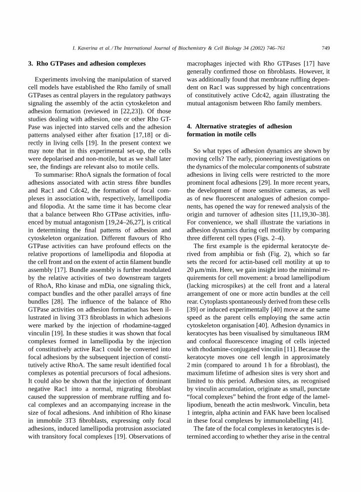
The first example is the epidermal keratocyte de-rived from amphibia or fish (Fig. 2), which so farsets the record for actin-based cell motility at up to20�m/min. Here, we gain insight into the minimal re-quirements for cell movement: a broad lamellipodium(lacking microspikes) at the cell front and a lateralarrangement of one or more actin bundles at the cellrear. Cytoplasts spontaneously derived from these cells[39] or induced experimentally [40] move at the samespeed as the parent cells employing the same actincytoskeleton organisation [40]. Adhesion dynamics inkeratocytes has been visualised by simultaneous IRMand confocal fluorescence imaging of cells injectedwith rhodamine-conjugated vinculin [11]. Because thekeratocyte moves one cell length in approximately2 min (compared to around 1 h for a fibroblast), themaximum lifetime of adhesion sites is very short andlimited to this period. Adhesion sites, as recognisedby vinculin accumulation, originate as small, punctate“focal complexes” behind the front edge of the lamel-lipodium, beneath the actin meshwork. Vinculin, beta1 integrin, alpha actinin and FAK have been localisedin these focal complexes by immunolabelling [41].
The fate of the focal complexes in keratocytes is de-termined according to whether they arise in the central
750 I. Kaverina et al. / The International Journal of Biochemistry & Cell Biology 34 (2002) 746–761
Fig. 2. Adhesion complexes in epidermal keratocytes. (A)Schematic representation of adhesion complexes (red) in relationto the actin cytoskeleton (green). The general organisation of actinin this and Figs. 3–8 is based on electron microscope observations.(B) Confocal microscope image of living, trout keratocyte thatwas injected with rhodamine-vinculin to label substrate adhesionsites. Image was kindly provided by K. Anderson.
region, in front of the cell body, or in the flanks. In thecentral region, the focal complexes remain stationaryrelative to the substrate and either disassemble beneaththe cell body as it moves over them, or are removedfrom the substrate as the rear edge of the cell rollsupwards [11,42]. In the regions of the lamellipodiumflanking the cell body, focal complexes do not dis-solve, but fuse together in the trailing edge to formlarger adhesions, resembling focal adhesions. Theselateral adhesions are not stationary, are short-lived andare drawn into the flanks of the cell body in a slid-ing motion, driven by contractility in the laterally or-ganised bundles of actin filaments [42,43]. As well asproviding the most direct example of recycling of ad-hesion sites, the example of the keratocyte promptsthe question of whether the trailing, lateral adhesionsserve a useful role in motility. Indeed they do, for with-out them, the lateral tension required for the tractionof the cell body [42–44] could not be developed.
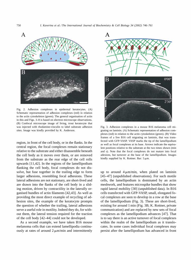
As a second example, we have taken B16 mousemelanoma cells that can extend lamellipodia continu-ously at rates of around 2�m/min and intermittently
Fig. 3. Adhesion complexes in a mouse B16 melanoma cell mi-grating on laminin. (A) Schematic representation of adhesion com-plexes (red) in relation to the actin cytoskeleton (green). (B) Videoframes of a live B16 cell migrating on laminin, that was trans-fected with GFP-VASP. VASP marks the tip of the lamellipodiumas well as focal complexes at its base. Arrows indicate the equiva-lent positions relative to the substrate at the two times shown (minand s). Note that the focal complexes do not mature into focaladesions, but turnover at the base of the lamellipodium. Imageskindly supplied by K. Rottner. Bar: 5�m.
up to around 4�m/min, when plated on laminin[45–47] (unpublished observations). For such motilecells, the lamellipodium is dominated by an actinmeshwork, and features microspike bundles that showrapid lateral mobility [30] (unpublished data). In B16cells transfected with GFP-VASP, small, elongated fo-cal complexes are seen to develop in a row at the baseof the lamellipodium (Fig. 3). These are short-lived,existing for around 1 min (Fig. 3B; K. Rottner, privatecommunication) and are replaced by new sets of focalcomplexes as the lamellipodium advances [47]. Thatis to say there is an active turnover of focal complexeswithin the realm of the lamellipodium as it translo-cates. In some cases individual focal complexes maypersist after the lamellipodium has advanced in front

I. Kaverina et al. / The International Journal of Biochemistry & Cell Biology 34 (2002) 746–761 751
of them and grow into longer-lived focal adhesions,together with associated stress fibre bundles of actin[47]. However, the region behind rapidly advancinglamellipodia of B16 cells is dominated by a loosenetwork of actin filaments [45,47] that is continuouswith a fraction of the filaments that make up thelamellipodium (unpublished observations). This typeof loose network is likely an important component ofmigrating cells and can provide structural continuityin the absence of macroscopic bundles.
As a third example we take a goldfish fibroblastcell line (Fig. 4). In these cells the migrating frontis dominated by filopodia that can extend 10–20�mfrom the cell edge. Lamellipodia segments link ad-jacent filopodia, and both filopodia and lamellipodiacan undergo active upfolding and ruffling activitybetween phases of protrusion. The notable featureof these cells is the creation of resolvable adhesionfoci mainly in association with the base of filopodia(Fig. 4B). These “focal complexes” are either tran-sitory, with lifetimes in the range of 5–15 min, orpersist and differentiate into focal adhesions. In livingcells, discrete adhesion sites are not recognised inthe lamellipodia segments, which show two distinctactin filament organisations in the electron micro-scope worth mentioning (unpublished observations;Fig. 4A). In one case, the lamellipodium shows thecharacteristic meshwork (reviewed in [48]) with adecrease in the filament density from front to rear,with some filaments trailing off into the loose mesh-work behind. In the second case, the lamellipodiumis narrower and is bordered at its base by a concavebundle of actin filaments that shows continuity withthe flanking filopodia. These two morphologies maybe correlated respectively with phases of protrusionand retraction, whereby “hammocking of filaments”[49] between filopodia and their associated adhesionscontribute to the support of the cell edge followinglamellipodia retraction. In the context of Rho GTPaseregulation the injection of constitutively active Rac1into fish fibroblasts expressing GFP-VASP causes atransformation of the cell front from one dominatedby filopodia to one dominated by lamellipodia and fo-cal complexes (Kaverina, unpublished observations),resembling that seen in B16 melanoma cells Thesefindings emphasises again that adhesion strategiesare the outcome of a subtle balance in Rho GTPaseactivities.
Fig. 4. Adhesion complexes in a migrating goldfish fibroblast. (A)Schematic representation of adhesion complexes (red) in relation tothe actin cytoskeleton (green). (B) Paired video frames, in fluore-scence (left) and phase contrast (right), of a living goldfish fibrob-last expressing GFP-zyxin to mark adhesion sites. Arrows indicateequivalent positions in each video pair. Note the creation of focalcomplexes in association with the base of filopodia. Focal com-plexes can develop into focal adhesions behind the lamellipodium(central arrow at 7 min). Time (min and s). Bar: 10�m.
Final mention should be made of the unique ad-hesion foci found in cells of the monocyte lineage,referred to as podosomes [50–55]. First observed insome virally transformed cells [56,57], podosomes ex-hibit a ring structure composed of an actin core and

752 I. Kaverina et al. / The International Journal of Biochemistry & Cell Biology 34 (2002) 746–761
adhesion proteins at the periphery. In differentiatedosteoclasts, podosomes form in a prominent band atthe cell periphery which appears to function as a sealaround the bone resorbing zone [50]. Podosomes donot move, but they are dynamic, dissolving and thenreforming in new locations, with lifetimes of 2–12 min([50]; F. Bard, private communication). Data on thedynamics of podosomes in moving cells does not yetexist. But it appears that podosomes lie behind a nar-row lamellipodium ([54,56]; F. Bard, private commu-nication) which presumably functions in protrusion.
5. Tension, adhesion and retraction
Chrzanowska-Wodnicka and Burridge [58] haveshown that the formation of focal adhesions is de-pendent on the development of tension in the actinfilament cytoskeleton, through actin–myosin interac-tions (reviewed in [58]). Likewise, focal complexesrely on actomyosin tension for their formation and in-tegrity [19] and, like focal adhesions exert traction onthe substrate [36]. The dependence of focal adhesiondevelopment on tension has been elegantly illustratedby the mechanical manipulation of cells with mi-croneedles [37]: these experiments have shown thatexternally applied forces can induce adhesion sitegrowth in the same way as intracellular contractility.An interesting difference in this case was the require-ment for mDia activity downstream of RhoA, but notRho kinase [37]. Using the same approach, we havebeen able to show that the restraint of the cell bodyin migrating B16 melanoma cells induces the forma-tion of actin filament bundles from the loose networkbehind the lamellipodium, and the transformationof focal complexes into focal adhesions (Kaverina,unpublished observations).
In a migrating cell, tension in the actin cytoskele-ton is necessary for adhesion at the front, illustratedby the retraction induced by myosin inhibitors [32,59]as well as for the retraction of the trailing cell bodyin the last phase of motility [60,61]. Using flexiblesubstrates, Beningo et al. [36] and Balaban et al. [62]have recently quantitated the forces exerted at adhe-sion sites in living cells. Whereas, Balaban et al. [62]found that larger adhesions exerted more force perunit area, Beningo et al. [36] found the opposite. Thisdifference may partly be explained by the analysis
of stationary cells in one study [62] and motile cellsin the other [36]. In motile fish fibroblasts, the ante-rior focal complexes and early focal adhesions exertmore stress than mature focal adhesions. This findingis in line with decreased substrate deformation aroundthe trailing tail of fibroblasts [63] and the observedsliding of trailing contacts during cell edge retraction[30,32,34,64]. Taken together, these studies underlinetension as a central factor in the development and dis-sociation of adhesion complexes, an aspect that wewill return to below.
6. Cross-talk of microtubules with adhesion foci
In fibroblasts, the depolymerisation of microtubulesleads to the depolarisation of cell shape [65], an in-crease in the contractility of the cytoskeleton [66]and an amplification in the size of focal adhesions[58]. This response is paralleled by the activation ofRhoA [67,68]. Conversely, the repolymerisation ofmicrotubules following the disassembly is associatedwith the activation of Rac1 [69]. A direct correlationtherefore exists between microtubule polymerisationdynamics and the activity of the Rho GTPases pro-teins that direct actin cytoskeleton organisation andsubstrate adhesion dynamics.
A link between microtubules and adhesion sites wasindependently illustrated in another context. Thus,observations of living cells in which microtubules andadhesion components were labelled with fluorescentprobes revealed that microtubules specifically targetadhesion foci as they polymerise towards the cell pe-riphery [31] (Fig. 5). This interaction is close range,since focal adhesions are able to capture microtubulesand stabilise them temporarily against depolymerisa-tion by nocodazole [31]. And the closeness of thisassociation has more recently been corroborated bytotal internal reflection fluorescence microscopy ofcells expressing GFP-tubulin and GFP-zyxin ([70];unpublished observations), whereby microtubulesover adhesions were seen to dip into the excitingevanescent wave formed within 150 nm of the sub-strate [70]. Interestingly, the assembly of podosomesin macrophages is microtubule-dependent [54] andmicrotubules are required for stabilisation of the beltof podosomes at the periphery of osteoclasts (F. Bard,private communication).

I. Kaverina et al. / The International Journal of Biochemistry & Cell Biology 34 (2002) 746–761 753
Fig. 5. Targeting of focal adhesions by microtubules. Figure showssuperimposed video frames of a fish fibroblast that was expressingGFP-tubulin (green channel) to label microtubules and injectedwith rhodamine-tagged vinculin to label adhesion sites (red chan-nel). The time separation between the two channels was less than2 s. Arrows indicate typical targeting events. Bar: 5�m.
Such an intimate cross-talk between microtubulesand adhesion foci must serve a function, and otherdata indicate that this function is to modulate adhesionsite dynamics [32,33]. Accordingly, experiments onliving cells have demonstrated that the targeting of fo-cal adhesions or focal complexes by microtubules ei-ther retards the growth of adhesions or promotes theirdisassembly [32]. Similar dynamics of disassemblyassociated with microtubule targeting could be mim-icked by the local application of inhibitors of myosincontractility to a cell edge [32]. And taking this ob-servation one step further, it was demonstrated that
depolarised cells lacking microtubules could be po-larised and induced to move by the asymmetricalapplication of the same myosin inhibitor [33]. It wastherefore concluded that microtubules exert their in-fluence on cell polarisation by modulating adhesionsite turnover through the point delivery of signalsthat antagonise myosin contractility at adhesion foci.Bershadsky et al. [68] have also attributed a role of mi-crotubules in the general or local suppression of con-tractility. They showed that microtubule disruption inserum starved cells induces focal adhesions and stressfibre formation, and that this effect was prevented bythe inhibition of cell contractility modulated via RhoA.
7. Rho GTPases and microtubule engagement
As already indicated, RhoA, Rac1 and Cdc42 actin three signal-transduction pathways regulating theassembly of actin stress fibre bundles, lamellipodiaand filopodia respectively. Rho GTPases signal todiverse effectors to initiate a downstream response.Each of these GTPases act as a molecular switch, cy-cling between an active GTP-bound, and an inactiveGDP-bound, state. Guanosine nucleotide exchangefactors (GEFs) facilitate the exchange of GDP for GTP,and GTPase-activating proteins (GAPs) increase therate of GTP hydrolysis of Rho GTPases [71]. Here, wediscuss the possible pathways linking regulators andeffectors of Rho GTPases to microtubules (Table 1).
7.1. RhoA
As we have seen, highly dynamic focal complexesformed in association with lamellipodia are depen-dent on Rac1, while those formed beneath filopodiaare initiated via Cdc42. Both types of complexes canmature into focal adhesions in a process that requiresRhoA. At which regulatory stage microtubules exerttheir influence on contractility is not yet known. How-ever, existing data suggest that Rac1-dependent focalcomplexes can turnover and disassemble indepen-dently of microtubules ([46]; Kaverina unpublished).In the same context, we are reminded that keratocytespolarise quite happily without microtubules [39] asso do some primary fibroblasts [72]. Common tothese examples is the absence of established stressfibre bundles and focal adhesions. We are prompted

754 I. Kaverina et al. / The International Journal of Biochemistry & Cell Biology 34 (2002) 746–761
Table 1Regulatory proteins implicated with microtubules
Protein Relation to actin cytoskeleton Relation to microtubules Reference
Rac1 Promotes lamellipodia and associated focalcomplex formation
Activated during microtubule re-polymerisation [17–19,69]
RhoA Promotes stress fibre formation and focaladhesion maturation
Activated upon microtubule depolymerisation [17–19,73]
Cdc42 Promotes filopodia and associated focalcomplex formation. Important for polarisedmotility
[17–19,92,94]
P190RhoGEF Rho-specific GEF Binds microtubules in vivo and in vitro [74]Lfc GEF, activates Rho in vitro. Overexpression
leads to stress fibre and ruffle formationBinds microtubules in vivo and in vitro [75]
GEF H1 Activates Rac and Rho in vitro Binds microtubules [76]mDia Rho effector Promotes formation of parallel
arrays of fine actin bundlesOver-expression promotes the formation of“Glu” microtubules
[28,77]
Pak Rac and Cdc42 effectors; can modulateactomyosin contractility downstream from Rho
[83,84]
ASEF Rac specific GEF Binds APC and could be delivered bymicrotubules
[85]
RhoG Rac and Cdc42-activating GTPase Localisation and activity depends onmicrotubule integrity. Binds kinectin and couldbe transported by kinesin
[86,88]
Trio GEF for Rho and RhoG/Rac/Cdc42 Localisation and activity depends onmicrotubule integrity
[87]
CIP4 and WASP Cdc42 effectors; involved in actin dynamics Bind to microtubules [93]mPar6/PKCzeta
complexCdc42 effector Involved in MTOC polarization [92]
Src Involved in RhoA activation cycle; importantfor mDia bundle formation
Binds Tau and MAP2 colocalises withmicrotubules when inactive
[96,98–102]
Ras Involved in regulation downstream of RhoA Ki-Ras localisation depends on microtubuleintegrity
[104,107]
MAPKs: MLK-2,JNK, ERK
Involved in Ras-Rho interplay MLK-2 and JNK localise to microtubules [104,105]
to conclude that below a certain threshold of stressat adhesion sites, cell asymmetry can be induced andmaintained by the actomyosin system [40] and thatabove this level microtubules are required to promotethe adhesion disassembly to control cell shape. Thisstress threshold may be determined by the engage-ment of RhoA to promote the maturation of adhesionsinitiated via Rac1 and Cdc42. According to theseconsiderations microtubules most likely exert theirinfluence selectively on the RhoA pathway.
RhoA-GTP pull down assays confirm the find-ings already cited (Section 6) that RhoA is stronglyactivated upon microtubule depolymerisation [73].But, how is RhoA activity influenced by micro-tubules? RhoA itself does not bind microtubulesnor to microtubule-binding proteins and it’s intra-cellular localisation is not microtubule-dependent.
Some of RhoA upstream regulators, however, showmicrotubule-binding activities (Fig. 6). An interestingcandidate, P190RhoGEF binds microtubules in vivoas well as in vitro. This exchange factor is specific forRhoA and elevates RhoA activity when overexpressed.Interestingly, in cells in which microtubules were dis-assembled, overexpression of p190RhoGEF failed toamplify RhoA activation, indicating that p190RhoGEFis involved in the microtubule-dependent upregula-tion of RhoA [74]. Similar functions could be at-tributed to other related Dbl-like microtubule-bindingGEFs, such as Lfc, which activates RhoA in vitroand, when overexpressed, stress fibre and ruffle for-mation in vivo, consistent with activation of bothRac1 and RhoA [75]. Also, GEF H1 binds mi-crotubules and activates both Rac1 and RhoA invitro [76].

I. Kaverina et al. / The International Journal of Biochemistry & Cell Biology 34 (2002) 746–761 755
Fig. 6. Potential pathways linking microtubules to the regulation ofRhoA. MT: microtubule; FX: focal complex; FA: focal adhesion.See text for details.
In another scheme, stabilised “Glu“ microtubuleshave been attributed a role in polarisation, involv-ing the RhoA effector, mDia [77]. Over-expression ofmDia was reported to promote the formation of “Glu”microtubules oriented towards the wounded edge in afibroblast monolayer, but the preferred polarisation ofthis subset of microtubules was not compelling. Fur-thermore, Glu microtubules are capped, non dynamic[77] and far removed from the advancing front as com-pared to dynamic microtubules [31]. It is therefore dif-ficult to accept the idea of microtubule stabilisation asa “key event” in polarisation [77].
Further, since contact growth upon tension applica-tion requires mDia [37], and mDia is likely to be im-portant for microtubule interaction with the cell cor-tex [78] Geiger and Bershadsky [79] have suggested adual role for microtubules in adhesion regulation. Oneinvolves signaling disassembly and the other foreseesthe delivery of specific components (such as membersof the mDia pathway) that are necessary for the de-velopment of focal contacts and stress fibres. In thiscontext, a balance between the two could determineadhesion site turnover.
7.2. Rac1
Waterman-Storer et al. [69] observed that Rac1 isactivated during microtubule re-polymerisation afterdrug-induced disassembly. This result prompted thesuggestion that polymerising microtubules mediatethe activation of Rac1 at the cell front, to induceprotrusion, whereas depolymerising microtubules atthe cell rear mediate the activation of RhoA, leading
Fig. 7. Potential pathways linking microtubules to the regulationof Rac1. Rac1, in turn, promotes focal adhesion turnover viaits antagonism of RhoA. MT TP: microtubule tip proteins; kin:kinesin; KC: kinectin. See text for details.
to contractility and retraction [80,81]. Lamellipodiaadvance can however occur without microtubules[33,39,46,82] and the spatial arrangement of micro-tubules in moving cells does not locally correlate withlamellipodium protrusion events. There is also no ev-idence that microtubule depolymerisation is enhancedat the cell rear as compared to the cell front. If Rac1is involved in the regulation of polarisation via micro-tubules, it most likely acts at the level of focal adhe-sions. In this case, the antagonism between the Rac1and RhoA pathways may play a role. Hence, Rac1via Pak could reduce actomyosin contractility down-stream from RhoA ([33], reviewed in [34]), (Fig. 7).
A good candidate for microtubule-dependent Rac1stimulation is ASEF [35]. It binds APC and activatesRac1 in an APC-dependent manner. Since APC canbe transported on the growing tips of a subset of mi-crotubules in a complex with other microtubule tipproteins, ASEF could be delivered by microtubules tocertain adhesion sites.
Another candidate pathway potentially involved inthe microtubule-dependent regulation of Rac1 is theRac1 and Cdc42-activating GTPase RhoG and it’sexchange factor Trio. Trio is a multifunctional GEFthat contains 2 exchange domains, one RhoA specific,another specific for RhoG and less active on Rac1.Microtubule disruption prevents localisation of RhoGto the plasma membrane and inhibits its activity

756 I. Kaverina et al. / The International Journal of Biochemistry & Cell Biology 34 (2002) 746–761
[36]. The N-terminal part of the Trio molecule whichspecifically activates RhoG also localises to the mem-brane and activates Rac1 and Cdc42 and all theseactivities are blocked by microtubule disruption [37].RhoG was reported to bind kinectin, a link suggestedas essential for the transport of this GTPase to the cellperiphery by kinesin [86,88]. Inhibition of kinesintransport by antibody injection, as well as inhibitionof kinectin–RhoG interaction, is sufficient to blockRhoG-induced ruffling [88]. RhoG might then be de-livered in a complex with Trio, since Trio localisationalso depends on microtubules (Fig. 7).
7.3. Cdc42
In line with the role of Cdc42 in the polarisation ofyeast [89] some evidence is now emerging for an in-volvement of this Rho family member in the polarisa-tion of vertebrate cells [90]. In particular, Nobes andHall [91] showed that inhibition of Cdc42 in a woundhealing assay inhibited polarisation of cells into thewound. However, the involvement of microtubules onthe activity of Cdc42 was not analysed. Since an asym-metric distribution of Cdc42 was also found necessaryto transduce it’s effect on polarity [92] it is not unlikelythat microtubules function to provide this asymmetry.
The influence of Cdc42 on overall adhesion dynam-ics in motile cells is not yet clear, but one can spec-ulate that this GTPase establishes contact asymmetryin collaboration with microtubules. In fish fibroblasts,focal complexes associated with filopodia are targetedby microtubules and intense targeting results in fo-cal complex disassembly (Kaverina, unpublished re-sults). This finding indicates that microtubules couldintervene in the Cdc42 pathway leading to focal adhe-sion assembly. A potential regulator of Cdc42 linkedto microtubules could again be RhoG, which may betransported along microtubules in a kinesin-dependentmanner through its ability to bind kinectin [86,88].
Alternatively, regulatory molecules downstreamof Cdc42 may collaborate with microtubules. Somespecific Cdc42 effector molecules, involved in actincytoskeleton regulation, such as CIP4 and WASP,have been shown to bind to microtubules in a regu-lated way [93]. Other Cdc42 effectors are involvedin polarized localization of the microtubule organiz-ing centre (MTOC) in cells moving into an artificialwound [92,94], and in astrocytes the mPar6/PKCzeta
complex was identified as such an effector [92]. Therelocalization of the MTOC is interestingly depen-dent on dynein. However, we have recently found thatdynein activity is not required for polarised motilityof fibroblasts (Krylyshkina, unpublished). Taken to-gether, these data suggest separate signalling routesfrom Cdc42: to the polarisation of the MTOC viamPar6/PKCzeta; and to the polarized distributionof actin structures responsible for motility (includ-ing adhesions), probably via CIP4 and WASP. Also,mDia-dependent formation of polarized stable mi-crotubule arrays is regulated independently of Cdc42[94]. Clearly, there is still a lot to do to establish howCdc42 influences cell polarity in coordination withother GTPases.
Some alternative ideas of how microtubules maydetermine the polarity via interfacing with Rho GT-Pases have been recently reviewed by Wittmann andWaterman-Storer [81].
8. Other potential regulators linked tomicrotubules
8.1. Src
Different lines of evidence suggest that Src ki-nase activity is involved in regulating focal adhesionturnover. The v-Src temperature-sensitive mutant (aswell as c-Src in its active conformation [95]) translo-cates to focal adhesions at the permissive temper-ature [96] and the kinase activity of v-Src leads toeventual focal adhesion disassembly upon phospho-rylation and degradation of FAK. FAK-containingfocal adhesions also grow faster in Src−/− cells incomparison with wild type cells [97], resulting inthe suppression of cell motility. The link betweenthe Src- and RhoA-dependent pathways regulatingfocal adhesions is, however, unclear. On the onehand, Src kinases can downregulate RhoA activityvia phosporylation of P190RhoGAP (a major Src andFyn substrate) [98]. On the other hand, RhoA activa-tion in starved cells results in the translocation of Srcto focal adhesions, a translocation that is blocked bymyosin inhibition [99]. Interestingly, the localisationof kinase-dead v-Src to focal adhesions blocks theirturnover and causes them to enlarge, unless Src kinaseis activated [90]. Additionally, the RhoA downstream

I. Kaverina et al. / The International Journal of Biochemistry & Cell Biology 34 (2002) 746–761 757
effector mDia was found to interact with Src, andinhibition of Src blocked mDia-dependent stress fi-bre formation [100]. There is no direct evidence formicrotubules-binding Src, but Src kinases have beenshown to bind the microtubule-binding proteins Tauand MAP2 in neurons [101,102]. And the localisationof inactive c-Src as well as v-Src mutant at restric-tive temperature has been shown to correspond withzones of high microtubule density [99]. If there is anyinteraction it may be speculated that microtubules actas transitory docking sites for inactive c-Src.
8.2. Ras
Nobes and Hall [91] have reported that inhibi-tion of Ras by antibody injection induces large focaladhesions and blocks cell motility, presumably bydisrupting focal adhesion dynamics. A temporal as-sociation of active Ras with focal adhesions has beenobserved and several possibilities exist for the inter-play between Ras and Rho GTPases on the molecularlevel, which could be essential for actin cytoskeletonregulation (reviewed in [103]). For example, it hasbeen recently shown that the lack of stress fibres inRas-transformed cells is a result of a functional un-coupling of RhoA from Rho kinase, dependent onthe ERK-MAP kinase pathway downstream of Ras[104]. Interestingly, activity of the MAP kinase path-way members has long been known to be influencedby microtubule-specific agents. Of special interest isthe finding that such kinases as MLK2 and JNK arelocalised in punctate structures along microtubulesin fibroblasts [105]. MLK2 activates ERK and there-fore can be involved in down-regulation of the RhoAdownstream effect on stress fibre and focal adhesionformation. In a two hybrid, screen MLK2 associatedwith KIF3, a kinesin superfamily motor, suggestingthat it might be one of the regulators delivered bykinesins to adhesion sites [105].
It is not excluded that the intracellular localisationof Ras itself could be defined by microtubules. Ki-Ras4B appears to be the isoform of Ras most importantfor cell motility [106] and it has been shown that thefunctionally essential membrane targeting of Ki-Rasdepends on microtubule integrity and dynamics [107].This Ras isoform associates with microtubules uponprenylation and when microtubules are stabilised anddisorganised by taxol it fails to be transported to the
Fig. 8. Potential pathways linking Ras to focal adhesion turnover.Scissors indicate uncoupling of RhoA from Rho kinase by Erk.See text for details.
plasma membrane. Thus, microtubules could poten-tially influence focal adhesion dynamics by modulat-ing delivery of active Ki-Ras to adhesion sites (Fig. 8).
9. Delivery of components to the cell front
Alternative ideas of how microtubules may influ-ence cell polarity have been discussed by Nabi [108].These hinge in the main on the delivery of membranevia vesicle traffic along microtubules. We cannot rig-orously exclude the possibility that structural compo-nents of adhesions are delivered to or removed fromadhesion sites by microtubules. In this connection,paxillin was found to bind alpha- and gamma-tubulin,as well as to co-localize with microtubule organisingcentres in lymphocytes [109]. We consider this un-likely, however, since adhesion site formation per seis not microtubule-dependent.
It has also been suggested that integrins and othermembrane components of adhesion plaques are de-livered to the sites of adhesion site assembly viamicrotubule-driven membrane traffic [110]. In sup-port of this idea, a block in vesicle transport wasfound to inhibit cell spreading [111] and deliveryof integrins to the cell membrane. The question ofhow membrane is replenished at the cell front is aninteresting one that remains to be clarified and couldinvolve members of the Rab family of small GTPases[112,113]. We only note that cells can spread in theabsence of microtubules and for this do not dependon microtubule-linked vesicle trafficking.

758 I. Kaverina et al. / The International Journal of Biochemistry & Cell Biology 34 (2002) 746–761
10. Concluding remarks
Much has still to be learned about adhesion sitedynamics during motility. Not least is the problem ofadhesion site composition, which is far from complete[7], as well as the temporal association of the compo-nent molecules with adhesion sites. Here we are onlyjust beginning to scratch the surface [79,35,114,115].Other questions include the localisation of regulatorsand regulatory complexes, which can only properlybe defined in living cells, requiring probes and in-strumentation that are just now becoming available.Added to this is the question of how the movement ofcells in vitro relates to migration in vivo. Steps in thisdirection are promising [116,117] and set the stagefor further progress.
Acknowledgements
We thank Drs. Klemens Rottner and Kurt Andersonfor providing figures and Drs. Benny Geiger, ClareWaterman-Storer, Torsten Wittmann and FredericBard for permission to cite unpublished work. Studiesforming the background of this review were supportedby grants from the Austrian Science Foundation.
References
[1] A.S.G. Curtis, The mechanism of adhesion of cells to glass,J. Cell Biol. 20 (1964) 199–215.
[2] C.S. Izzard, L.R. Lochner, Formation of cell-to-substratecontacts during fibroblast motility: an interference–reflexionstudy, J Cell Sci. 42 (1980) 81–116.
[3] K. Burridge, K. Fath, T. Kelly, G. Nuckolls, C. Turner,Focal adhesions: transmembrane junctions between theextracellular matrix and the cytoskeleton, Annu. Rev. CellBiol. 4 (1988) 487–525.
[4] Z. Avnur, J.V. Small, B. Geiger B, Actin-independentassociation of vinculin with the cytoplasmic aspect of theplasma membrane in cell-contact areas, J. Cell Biol. 96(1983) 1622–1630.
[5] A.A. Neyfakh, T.M. Svitkina, Isolation of focal contactmembrane using saponin, Exp. Cell Res. 149 (1983) 582–586.
[6] S.M. Schoenwaelder, K. Burridge, Bidirectional signalingbetween the cytoskeleton and integrins, Curr. Opin. CellBiol. 11 (1999) 274–286.
[7] E. Zamir, B. Geiger, Molecular complexity and dynamics ofcell-matrix adhesions, J. Cell Sci. 114 (2001) 3583–3590.
[8] B. Geiger, A 130 K protein from chicken gizzard: itslocalization at the termini of microfilament bundles incultured chicken cells, Cell 18 (1979) 193–205.
[9] G. Rinnerthaler, B. Geiger, J.V. Small, Contact formationduring fibroblast locomotion: involvement of membraneruffles and microtubules, J. Cell Biol. 106 (1988) 747–760.
[10] B. Yuruker, V. Niggli, Alpha-actinin and vinculin in humanneutrophils: reorganization during adhesion and relation tothe actin network, J. Cell Sci. 101 (1992) 403–414.
[11] K.I. Anderson, R. Cross, Contact dynamics during keratocytemotility, Curr. Biol. 10 (2000) 253–260.
[12] J.P. Heath, B.F. Holifield, On the mechanisms of corticalactin flow and its role in cytoskeletal organisation offibroblasts, Symp. Soc. Exp. Biol. 47 (1993) 35–56.
[13] J.V. Small, K. Rottner, I. Kaverina, K.I. Anderson,Assembling an actin cytoskeleton for cell attachment andmovement, Biochim. Biophys. Acta 1404 (1998) 271–481.
[14] J.V. Small, J.E. Celis, Direct visualization of the 10-nm(100-A)-filament network in whole and enucleated culturedcells, J. Cell Sci. 31 (1978) 393–409.
[15] M. Abercrombie, J.E. Heaysman, S.M. Pegrum, Thelocomotion of fibroblasts in culture. Part IV. Electronmicroscopy of the leading lamella, Exp. Cell Res. 67 (1971)359–367.
[16] J.V. Small, Organization of actin in the leading edge ofcultured cells: influence of osmium tetroxide and dehydrationon the ultrastructure of actin meshworks, J. Cell Biol. 91(1981) 695–705.
[17] W.E. Allen, G.E. Jones, J.W. Pollard, A.J. Ridley, Rho, Racand Cdc42 regulate actin organization and cell adhesion inmacrophages, J. Cell Sci. 110 (1997) 707–720.
[18] C.D. Nobes, A. Hall, Rho, Rac, and Cdc42 GTPases regulatethe assembly of multimolecular focal complexes associatedwith actin stressfibers, lamellipodia and filopodia, Cell 81(1995) 53–62.
[19] K. Rottner, A. Hall, J.V. Small, Interplay between Rac andRho in the control of substrate contact dynamics, Curr. Biol.9 (1999) 640–648.
[20] N.A. Gloushankova, N.A. Alieva, M.F. Krendel, E.M.Bonder, H.H. Feder, J.M. Vasiliev, I.M. Gelfand, Cell–cellcontact changes the dynamics of lamellar activity innontransformed epitheliocytes but not in theirras-trans-formed descendants, Proc. Natl. Acad. Sci. U. S. A. 94(1997) 879–883.
[21] P.C. Rathke, M. Osborn, K. Weber, Immunological andultrastructural characterization of microfilament bundles:polygonal nets and stress fibers in an established cell line,J. Cell Biol. 19 (1979) 40–48.
[22] A. Hall, Rho GTPases and the actin cytoskeleton, Science279 (1998) 509–514.
[23] A.J. Ridley, Rho proteins, PI 3-kinases and monocyte/macrophage motility, FEBS Lett. 498 (2001) 168–171.
[24] R. Kozma, S. Sarner, S. Ahmed, L. Lim, Rho familyGTPases and neuronal growth cone remodeling: relationshipbetween increased complexity induced by Cdc42Hs, Rac1and acetylcholine and collapse induced by RhoA andlysophosphatide acid, Mol. Cell Biol. 17 (1997) 1201–1211.

I. Kaverina et al. / The International Journal of Biochemistry & Cell Biology 34 (2002) 746–761 759
[25] M. Hirose, T. Ishizaki, N. Watanabe, M. Uehata,O. Kranenburg, W.H. Moolenaar, F. Matsumura, M.Maekawa, H. Bito, S. Narumiya, Molecular dissection ofthe Rho-associated protein kinase (p160ROCK)-regulatedneurite remodeling in neuroblastoma N1E-115 cells, J. CellBiol. 41 (1998) 1625–1636.
[26] J.P. Moorman, D. Luu, J. Wickham, D.A. Bobak, C.S. Hahn,A balance of signaling by Rho family small GTPases RhoA,Rac1 and Cdc42 coordinates cytoskeletal morphology butno cell survival, Oncogene 18 (1999) 149–158.
[27] E.E. Sander, J.P. ten Klooster, S. van Delft, R.A. van derKammen, J.G. Collard, Rac downregulates Rho activity:reciprocal balance between both GTPases determines cellularmorphology and migratory behavior, J. Cell Biol. 147 (1999)1009–1022.
[28] N. Watanabe, T. Kato, A. Fujita, T. Ishizaki, S. Narumiya,Cooperation between mDia1 and ROCK in Rho-inducedactin reorganization, Nat. Cell Biol. 1 (1999) 136–143.
[29] J.B. Meigs, Y.L. Wang, Reorganization of alpha-actinin andvinculin induced by a phorbol ester in living cells, J. CellBiol. 102 (1986) 1430–1438.
[30] K. Rottner, J.V. Small, GFP in motion. Trends Cell Biol.(2001).
[31] I. Kaverina, K. Rottner, J.V. Small, Targeting, capture, andstabilization of microtubules at early focal adhesions, J. CellBiol. 142 (1998) 181–190.
[32] I. Kaverina, O. Krylyshkina, J.V. Small, Microtubuletargeting of substrate contacts promotes their relaxation anddissociation, J. Cell Biol. 146 (1999) 1033–1044.
[33] I. Kaverina, O. Krylyshkina, M. Gimona, K. Beningo, Y.L.Wang, J.V. Small, Enforced polarisation and locomotion offibroblasts lacking microtubules, Curr. Biol. 10 (2000) 739–742.
[34] L.B. Smilenov, A. Mikhailov, R.J. Pelham, E.E.Marcantonio, G.G. Gundersen, Focal adhesion motilityrevealed in stationary fibroblasts, Science 286 (1999) 1172–1174.
[35] C.M. Laukaitis, D.J. Webb, K. Donais, A.F. Horwitz,Differential dynamics of alpha 5 integrin, paxillin, and alphaactinin during formation and disassembly of adhesions inmigrating cells, J. Cell Biol. 153 (2001) 1427–1440.
[36] K.A. Beningo, M. Dembo, I. Kaverina, J.V. Small,Y.L. Wang, Nascent focal adhesions are responsible forthe generation of strong propulsive forces in migratingfibroblasts, J. Cell Biol. 153 (2001) 881–888.
[37] D. Riveline, E. Zamir, N.Q. Balaban, U.S. Schwarz, T.Ishizaki, S. Narumiya, Z. Kam, B. Geiger, A.D. Bershadsky,Focal contacts as mechanosensors: externally applied localmechanical force induces growth of focal contacts by anmDia1-dependent and ROCK-independent mechanism, J.Cell Biol. 153 (2001) 1175–1186.
[38] M. Edlund, M.A. Lotano, C.A. Otey, Dynamics ofalpha-actinin in focal adhesions and stress fibers visualizedwith alpha-actinin-green fluorescent protein, Cell Motil.Cytoskeleton 48 (2001) 190–200.
[39] U. Euteneuer, M. Schliwa, Persistent, directional motilityof cells and cytoplasmic fragments in the absence ofmicrotubules, Nature 310 (1984) 58–61.
[40] A.B. Verkhovsky, T.M. Svitkina, G.G. Borisy, Self-polarization and directional motility of cytoplasm, Curr. Biol.9 (1999) 11–20.
[41] J. Lee, K. Jacobson, The composition and dynamics ofcell-substratum adhesions in locomoting fish keratocytes, J.Cell Sci. 110 (1997) 2833–2844.
[42] K.I. Anderson, Y.L. Wang, J.V. Small, Coordination ofprotrusion and translocation of the keratocyte involvesrolling of the cell body, J. Cell Biol. 134 (1996) 1209–1218.
[43] J. Lee, M. Leonard, T. Oliver, A. Ishihara, K. Jacobson,Traction forces generated by locomoting keratocytes, J. CellBiol. 127 (1994) 1957–1964.
[44] T.M. Svitkina, A.B. Verkhovsky, K.M. McQuade, G.G.Borisy, Analysis of the actin-myosin II system in fishepidermal keratocytes: mechanism of cell body translocation,J. Cell Biol. 139 (1997) 397–415.
[45] C. Ballestrem, B. Wehrle-Haller, B.A. Imhof, Actindynamics in living mammalian cells, J. Cell Sci. 111 (1998)1649–1658.
[46] C. Ballestrem, B. Wehrle-Haller, B. Hinz, B.A. Imhof,Actin-dependent lamellipodia formation and microtubule-dependent tail retraction control-directed cell migration, Mol.Biol. Cell 11 (2000) 2999–3012.
[47] K. Rottner, B. Behrendt, J.V. Small, J. Wehland, VASPdynamics during lamellipodia protrusion, Nat. Cell Biol. 1(1999) 321–322.
[48] J.V. Small, The actin cytoskeleton, Electron. Microsc. Rev.1 (1988) 155–174.
[49] A.S. Hoglund, R. Karlsson, E. Arro, B.A. Fredriksson,U. Lindberg, Visualization of the peripheral weave ofmicrofilaments in glia cells, J. Muscle Res. Cell Motil. 1(1980) 127–146.
[50] J. Kanehisa, T. Yamanaka, S. Doi, K. Turksen, J.N. Heersche,J.E. Aubin, H. Takeuchi, A band of F-actin containingpodosomes is involved in bone resorption by osteoclasts,Bone 11 (1990) 287–293.
[51] A. Tourkin, M. Bonner, E. Mantrova, E.C. LeRoy, S.Hoffman, Dot-like focal contacts in adherent eosinophils,their redistribution into peripheral belts, and correlatedeffects on cell migration and protected zone formation, J.Cell Sci. 109 (1996) 2169–2177.
[52] T. Suzuki, S. Shoji, K. Yamamoto, S. Nada, M. Okada,T. Yamamoto, Z. Honda, Essential roles of lyn infibronectin-mediated filamentous actin assembly and cellmotility in mast cells, J. Immun. 161 (1998) 3694–3701.
[53] S. Ory, Y. Munari-Silem, P. Fort, P. Jurdic, Rho and Rac exertantagonistic functions on spreading of macrophage-derivedmultinucleated cells and are not required for actin fiberformation, J. Cell Sci. 113 (2000) 1177–1188.
[54] S. Linder, K. Hüfner, U. Wintergerst, M. Aepfelbacher,Microtubule-dependent formation of podosomal adhesionstructures in primary human macrophages, J. Cell Sci. 113(2000) 4165–4176.
[55] S. Burns, A.J. Thrasher, M.P. Blundell, L. Machesky, G.E.Jones, Configuration of human dendritic cell cytoskeleton byRho GTPases, the WAS protein, and differentiation, Blood98 (2001) 1142–1149.
760 I. Kaverina et al. / The International Journal of Biochemistry & Cell Biology 34 (2002) 746–761
[56] K. Burridge, L. Connell, Talin: a cytoskeletal componentconcentrated in adhesion plaques and other sites ofactin-membrane interaction, Cell Motil. 3 (1983) 405–417.
[57] G. Tarone, D. Cirillo, F.G. Giancotti, P.M. Comoglio,P.C. Marchisio, Rous sarcoma virus-transformed fibroblastsadhere primarily at discrete protrusions of the ventralmembrane called podosomes, Exp. Cell Res. 159 (1985)141–157.
[58] M. Chrzanowska-Wodnicka, K. Burridge, Rho-stimulatedcontractility drives the formation of stress fibers and focaladhesions, J. Cell Biol. 133 (1996) 1403–1415.
[59] L.P. Cramer, T.J. Mitchison, Myosin is involved inpostmitotic cell spreading, J. Cell Biol. 131 (1995) 179–189.
[60] W.T. Chen, Induction of spreading during fibroblastmovement, J. Cell Biol. 81 (1979) 684–691.
[61] G.A. Dunn, The locomotory machinery of fibroblasts, Eur.J. Cancer 16 (1980) 6–8.
[62] N.Q. Balaban, U.S. Schwarz, D. Riveline, P. Goichberg,G. Tzur, I. Sabanay, D. Mahalu, S. Safran, A. Bershadsky,L. Addadi, B. Geiger, Force and focal adhesion assembly:a close relationship studied using elastic micropatternedsubstrates, Nat. Cell Biol. 3 (2001) 466–472.
[63] R.J. Pelham, Y.L. Wang, High resolution detection ofmechanical forces exerted by locomoting fibroblasts on thesubstrate, Mol. Biol. Cell 10 (1999) 935–945.
[64] E. Zamir, M. Katz, Y. Posen, N. Erez, K.M. Yamada,B.Z. Katz, S. Lin, D.C. Lin, A. Bershadsky, Z. Kam, B.Geiger, Dynamics and segregation of cell-matrix adhesionsin cultured fibroblasts, Nat. Cell Biol. 2 (2000) 191–196.
[65] J.M. Vasiliev, I.M. Gelfand, Effects of colcemid onmorphogenetic processes and locomotion of fibroblasts, in:R. Goldman, T. Pollard, J. Rosenbaum (Eds.), Cell Motility,Cold, Spring Harbor Laboratory, Cold Spring Harbor, NY,1976, pp. 279–304.
[66] B.A. Danowski, Fibroblast contractility and actinorganization are stimulated by microtubule inhibitors, J. CellSci. 93 (1989) 255–266.
[67] T. Enomoto, Microtubule disruption induces the formationof actin stress fibers and focal adhesions in cultured cells:possible involvement of the rho signal cascade, Cell Struct.Funct. 21 (1996) 317–326.
[68] A. Bershadsky, A. Chausovsky, E. Becker, A. Lyubimova,B.B. Geiger, Involvement of microtubules in the control ofadhesion-dependent signal transduction, Curr. Biol. 6 (1996)1279–1289.
[69] C.M. Waterman-Storer, R.A. Worthylake, B.P. Liu, K.Burridge, E.D. Salmon, Microtubule growth activates Rac1to promote lamellipodial protrusion in fibroblasts, Nat. CellBiol. 1 (1999) 45–50.
[70] D. Toomre, D.J. Manstein, Lighting up the cell surface withevanescent wave Microscopy, Trends Cell Biol. 11 (2001)298–303.
[71] A.L. Bishop, A. Hall, Rho GTPases and their effectorproteins, Biochem. J. 348 (2000) 241–255.
[72] C.A. Middleton, A.F. Brown, R.M. Brown, I.D. Karavanova,D.J. Roberts, J.M. Vasiliev, The polarization of fibroblastsin early primary cultures is independent of microtubuleintegrity, J. Cell Sci. 94 (1989) 25–32.
[73] X.D. Ren, W.B. Kiosses, M.A. Schwartz, Regulation of thesmall GTP-binding protein Rho by cell adhesion and thecytoskeleton, EMBO J. 18 (1999) 578–585.
[74] F.P.G. van Horck, M.R. Ahmadian, L.C. Haeusler,W.H. Moolenaar, O. Kranenburg, Characterization ofp190RhoGEF: a RhoA-specific guanine nucleotide exchangefactor that interacts with microtubules, J. Biol. Chem. 276(2001) 4948–4956.
[75] J.A. Glaven, I. Whitehead, S. Bagrodia, R. Kay, R.A.Cerione, The Dbl-related protein, Lfc, localizes tomicrotubules and mediates the activation of Rac signalingpathways in cells, J. Biol. Chem. 274 (1999) 2279–2285.
[76] Y. Ren, R. Li, Y. Zheng, H. Busch, Cloning andCharacterization of GEF-H1: a microtubule-associatedguanine nucleotide exchange factor for Rac and RhoGTPases, J. Biol. Chem. 273 (1998) 34954–34960.
[77] A.F. Palazzo, T.A. Cook, A.S. Alberts, G.G. Gundersen,mDia mediates Rho-regulated formation and orientation ofstable microtubules, Nat. Cell Biol. 3 (2001) 723–729.
[78] T. Ishizaki, Y. Morishima, M. Okamoto, T. Furuyashiki, T.Kato, S. Narumiya, Coordination of microtubules and theactin cytoskeleton by the Rho effector mDia1, Nat. CellBiol. 3 (2001) 8–14.
[79] B. Geiger, A. Bershadsky, Assembly and mechanosensoryfunction of focal contacts, Curr. Opin. Cell Biol. 13 (2001)584–592.
[80] C.M. Waterman-Storer, E. Salmon, Positive feedbackinteractions between microtubule and actin dynamics duringcell motility, Curr. Opin. Cell Biol. 11 (1999) 61–67.
[81] T. Wittmann, C.M. Waterman-Storer, Cell motility: can RhoGTPases and microtubules point the way? J. Cell Sci. 114(2001) 3795–3803.
[82] A.D. Bershadsky, E.A. Vaisberg, J.M. Vasiliev, Pseudopodialactivity at the active edge of migrating fibroblast isdecreased after drug-induced microtubule depolymerization,Cell Motil. Cytoskeleton 19 (1991) 152–158.
[83] L.C. Sanders, F. Matsumura, G.M. Bokoch, P. de Lanerolle,Inhibition of myosin light chain kinase by p21-activatedkinase, Science 283 (1999) 2083–2085.
[84] R.H. Daniels, G.M. Bokoch, p21-Activated protein kinase:a crucial component of morphological signaling, TrendsBiochem. Sci. 24 (1999) 350–355.
[85] Y. Kawasaki, T. Senda, T. Ishidate, R. Koyama, T. Morishita,Y. Iwayama, O. Higuchi, T. Akiyama, ASEF: a link betweenthe tumor suppressor APC and G-protein signalling, Science289 (2000) 1194–1197.
[86] C. Gauthier-Rouviere, E. Vignal, M. Meriane, P. Roux, P.Montcourier, P. Fort, RhoG GTPase controls a pathway thatindependently activates Rac1 and Cdc42Hs, Mol. Biol. Cell9 (1998) 1379–1394.
[87] A. Blangy, E. Vignal, S. Schmidt, A. Debant, C.Gauthier-Rouvière, P. Fort, TrioGEF1 controls Rac- andCdc42-dependent cell structures through the direct activationof RhoG, J. Cell Sci. 113 (2000) 729–739.
[88] E. Vignal, A. Blangy, M. Martin, C. Gauthier-Rouviere,P. Fort, Kinectin is a key effector of RhoG microtubule-dependent cellular activity, Mol. Cell Biol. 21 (2001) 8022–8034.

I. Kaverina et al. / The International Journal of Biochemistry & Cell Biology 34 (2002) 746–761 761
[89] D. Pruyne, A. Bretscher, Polar, Polarization of cell growthin yeast. Part I. Establishment and maintenance of polaritystates, J. Cell Sci. 113 (2000) 365–375.
[90] R. Kroschewski, A. Hall, I. Mellman, Cdc42 controlssecretory and endocytic transport to the basolateral plasmamembrane of MDCK cells, Nat. Cell Biol. 1 (1999) 8–13.
[91] C.D. Nobes, A. Hall, Rho GTPases control polarity,protrusion, and adhesion during cell movement, J. Cell Biol.144 (1999) 1235–1244.
[92] S. Etienne-Manneville, A. Hall, Integrin-mediated activationof Cdc42 controls cell polarity in migrating astrocytesthrough PKCzeta, Cell 106 (2001) 489–498.
[93] L. Tian, D.L. Nelson, D.M. Stewart, Cdc42-interactingprotein 4 mediates binding of the Wiskott–Aldrich syndromeprotein to microtubules, J. Biol. Chem. 275 (2000) 7854–7861.
[94] A. Palazzo, H.L. Joseph, Y.-J. Chen, D.L. Dujardin,A.S. Alberts, K.K. Pfister, R.B. Vallee, G.G. Gundersen,Cdc42, dynein, and dynactinregulate MTOC reorientationindependent of Rho-regulated microtubule stabilization,Curr. Biol. 11 (2001) 1536–1541.
[95] K.B. Kaplan, K.B. Bibbins, J.R. Swedlow, M. Arnaud, D.O.Morgan, H.E. Varmus, Association of the amino-terminalhalf of c-Src with focal adhesions alters their properties andis regulated by phosphorylation of tyrosine 527, EMBO J.17 (1994) 4745–4756.
[96] V.J. Fincham, M.C. Frame, The catalytic activity of Src isdispensable for translocation to focal adhesions but controlsthe turnover of these structures during cell motility, EMBOJ. 17 (1998) 81–92.
[97] T. Volberg, L. Romer, E. Zamir, B. Geiger, pp60 (c-Src)and related tyrosine kinases: a role in the assembly andreorganization of matrix adhesions, J. Cell Sci. 114 (2001)2279–2289.
[98] M.R. Brouns, S.F. Matheson, J. Settleman, p190 RhoGAPis the principal Src substrate in brain and regulates axonoutgrowth, guidance and fasciculation, Nat. Cell Biol. 3(2001) 361–367.
[99] V.J. Fincham, V.G. Brunton, M.C. Frame, The SH3 domaindirects acto-myosin-dependent targeting of v-Src to focaladhesions via phosphatidylinositol 3-kinase, Mol. Cell. Biol.20 (2000) 6518–6536.
[100] T. Tominaga, E. Sahai, P. Chardin, F. McCormick, S.A.Courtneidge, A.S. Alberts, Diaphanous-related forminsbridge Rho GTPase and Src tyrosine kinase signaling, Mol.Cell. 5 (2000) 13–25.
[101] G. Lee, S.T. Newman, D.L. Gard, Tau interacts withsrc-family non-receptor tyrosine kinases, J. Cell Sci. 111(1998) 3167–3177.
[102] R.W.L. Lim, S. Halpain, Regulated association ofmicrotubule-associated protein 2 (MAP2) with Src and Grb2:evidence for MAP2 as a scaffolding protein, J. Biol. Chem.275 (2000) 20578–20587.
[103] D. Bar-Sagi, A. Hall, Ras and Rho GTPases: a familyreunion, Cell 103 (2000) 227–238.
[104] E. Sahai, M.F. Olson, C.J. Marshall, Cross-talk betweenRas and Rho signalling pathways in transformation favoursproliferation and increased motility, EMBO J. 20 (2001)755–766.
[105] K. Nagata, A. Puls, C. Futter, P. Aspenstrom, E. Schaefer,T. Nakata, N. Hirokawa, A. Hall, The MAP kinase MLK2co-localizes with activated JNK along microtubules andassociates with kinesin superfamily motor KIF3, EMBO J.17 (1998) 149–158.
[106] J.K. Voice, R.L. Klemke, J.H. Le A, A. Jackson, Four humanras homologs differ in their abilities to activate Raf-1, inducetransformation, and stimulate cell motility, J. Biol. Chem.274 (1999) 17164–17170.
[107] J.A. Thissen, J.M. Gross, K. Subramanian, T. Meyer, P.J.Casey, Prenylation-dependent association of Ki-Ras withmicrotubules. Evidence for a role in subcellular trafficking,J. Biol. Chem. 272 (1997) 30362–30370.
[108] I. Nabi, The polarization of the motile cell, J. Cell Sci. 112(1999) 1803–1811.
[109] L. Herreros, J.L. Rodriguez-Fernandez, M.C. Brown, J.L.Alonso-Lebreroi, C. Caban, F. Sanchez-Madridi, N. Longo,C.E. Turner, P. Sanchez-Mateos, Paxillin localizes to thelymphocyte microtubule organizing center and associateswith the microtubule cytoskeleton, J. Biol. Chem. 275 (2000)26436–26440.
[110] M.S. Bretscher, Moving membrane up to the front ofmigrating cells, Cell 85 (1996) 465–467.
[111] M. Roberts, S. Barry, A. Woods, P. van der Sluijs, J. Norman,PDGF-regulated Rab4-dependent recycling of alphavbeta3integrin from early endosomes is necessary for cell adhesionand spreading, Curr. Biol. 11 (18) (2001) 1392–1402.
[112] M. Spaargaren, J.L. Bos, Rab5 induces Rac-independentlamellipodia formation and cell migration, Mol. Biol. Cell10 (1999) 3239–3250.
[113] I. de Curtis, Cell migration: GAPs between membrane trafficand the cytoskeleton, EMBO Rep. 2 (2001) 1–5.
[114] E. Zamir, B.Z. Katz, S. Aota, K.M. Yamada, B. Geiger, Z.Kam, Molecular diversity of cell-matrix adhesions, J. CellSci. 112 (1999) 1655–1669.
[115] K. Rottner, M. Krause, M. Gimona, J.V. Small, J. Wehland,Zyxin is not co-localised with VASP at lamellipodia tipsand exhibits different dynamics to vinculin and paxillin infocal adhesions, Mol. Biol. Cell 12 (2001) 3013–3103.
[116] A. Jacinto, W. Wood, T. Balayo, M. Turmaine, A.Martinez-Arias, P. Martin, Dynamic actin-based epithelialadhesion and cell matching during Drosophila dorsal closure,Curr. Biol. 10 (2000) 1420–1426.
[117] C.A. Mandato, W.M. Bement, Contraction and polymeri-zation cooperate to assemble and close actomyosin ringsaround Xenopus oocyte wounds, J. Cell Biol. 154 (2001)785–797.