regulation of microrna biogenesis and turnover by animals ... · regulation of microrna biogenesis...
TRANSCRIPT
REVIEW
Regulation of microRNA biogenesis and turnover by animalsand their viruses
Valentina Libri • Pascal Miesen • Ronald P. van Rij •
Amy H. Buck
Received: 22 November 2012 / Revised: 27 December 2012 / Accepted: 27 December 2012 / Published online: 26 January 2013
� The Author(s) 2013. This article is published with open access at Springerlink.com
Abstract MicroRNAs (miRNAs) are a ubiquitous
component of gene regulatory networks that modulate the
precise amounts of proteins expressed in a cell. Despite
their small size, miRNA genes contain various recognition
elements that enable specificity in when, where and to what
extent they are expressed. The importance of precise con-
trol of miRNA expression is underscored by functional
studies in model organisms and by the association between
miRNA mis-expression and disease. In the last decade,
identification of the pathways by which miRNAs are pro-
duced, matured and turned-over has revealed many aspects
of their biogenesis that are subject to regulation. Studies in
viral systems have revealed a range of mechanisms by
which viruses target these pathways through viral proteins
or non-coding RNAs in order to regulate cellular gene
expression. In parallel, a field of study has evolved around
the activation and suppression of antiviral RNA interfer-
ence (RNAi) by viruses. Virus encoded suppressors of
RNAi can impact miRNA biogenesis in cases where
miRNA and small interfering RNA pathways converge.
Here we review the literature on the mechanisms by which
miRNA biogenesis and turnover are regulated in animals
and the diverse strategies that viruses use to subvert or
inhibit these processes.
Keywords MicroRNA � MicroRNA biogenesis �MicroRNA turnover � RNA degradation � Herpesviruses �Host–pathogen � Viral suppressor of RNA interference
Introduction
Small RNA classification
The specific recognition of nucleic acid sequences by
RNA–protein complexes (RNPs) is central to transcrip-
tional and post-transcriptional gene regulation. Small
RNAs are incorporated into many RNPs in order to
mediate the specific recognition of target nucleic acids
through Watson–Crick base-pairing. Different classes of
small RNAs continue to be discovered, including some that
are specific to plants or animal lineages, reviewed in [1, 2].
There are three major classes in animals: microRNAs
(miRNAs), short interfering RNAs (siRNAs), and piwi-
interacting RNAs (piRNAs). These classes differ in their
origin and biogenesis, the proteins with which they inter-
act, the mechanism of action of the RNP in which they are
contained, and the nature of their targets. MiRNAs are
derived from single-stranded (ss) RNAs that fold back on
themselves into stem-loop structures. Endogenous siRNAs
originate from double-stranded (ds) RNA precursors that
result from convergent bi-directional transcription, inverted
repeat regions in structured RNA, or base-pairing between
protein-coding genes and pseudogene-derived antisense
V. Libri and P. Miesen contributed equally to this work.
V. Libri � A. H. Buck (&)
Centre for Immunity, Infection and Evolution,
University of Edinburgh, King’s Buildings, West Mains Road,
Edinburgh, EH9 3JT, UK
e-mail: [email protected]
V. Libri
Centre for Human Immunology, Immunology Department,
Pasteur Institute, 25 rue du Docteur Roux, 75015 Paris, France
P. Miesen � R. P. van Rij (&)
Department of Medical Microbiology, Nijmegen Centre
for Molecular Life Sciences, Radboud University Nijmegen
Medical Centre, 6500 HB, Nijmegen, The Netherlands
e-mail: [email protected]
Cell. Mol. Life Sci. (2013) 70:3525–3544
DOI 10.1007/s00018-012-1257-1 Cellular and Molecular Life Sciences
123
transcripts. The detailed mechanism(s) of piRNA biogenesis
remains somewhat elusive, but the primary piRNAs origi-
nate from single-stranded precursor RNAs and are only
found in animals, and specifically in the germline [3]. Each
class of small RNAs binds to a member of the Argonaute
(Ago) family of proteins: siRNAs and miRNAs associate
with the Ago clade, whereas piRNAs associate with the Piwi
clade, reviewed in [4]. The Ago protein bound to the small
RNA comprises the RNA-induced silencing complex
(RISC). There is increasing diversity in the mechanisms by
which RISCs function and in the genes they target [5]. The
RISCs containing miRNAs are found throughout the
eukaryal domain and primarily target messenger RNAs
(mRNAs), causing the inhibition of translation and/or
de-adenylation and degradation of the mRNAs, reviewed in
[6]. Recognition of the mRNA target does not require perfect
complementarity with the miRNA and is generally dictated
by the ‘‘seed region’’ within the 50 terminal region of the
miRNA (nucleotides 2–8), reviewed in [7]. Based on this
low sequence requirement for recognition, each miRNA is
predicted to target several hundred genes. The majority of
human protein-coding genes have miRNA binding sites that
are maintained under selective pressure [8].
miRNAs in hosts and viruses
Based on the large number of genes targeted by miRNAs,
together with the ability of miRNAs to operate synergis-
tically with one another, these small RNAs are involved in
regulating numerous aspects of cellular biology including
proliferation, tumorigenesis, metabolism, differentiation,
development, apoptosis, and innate and adaptive immune
responses, reviewed in [9–14]. Viruses have evolved to
exploit and manipulate these same cellular pathways.
Therefore, it is not surprising that they use the miRNA
pathway to do this, either by encoding their own miRNAs,
or encoding molecules that activate or inhibit cellular
miRNA expression. Seven different virus families have
been reported to encode miRNAs or miRNA-like
molecules: herpesviruses, polyomaviruses, adenoviruses,
baculoviruses, an ascovirus, and recently a retrovirus and a
flavivirus [15–18]. Analysis of a wide range of RNA
viruses failed to identify viral miRNAs [17], apart from the
identification of miRNAs in bovine leukemia virus (BLV),
a retrovirus that replicates in the nucleus [18] and the
identification of a miRNA-like species in West Nile virus,
a cytoplasmic RNA virus that encodes a stem-loop struc-
ture in its 30UTR [16]. In the latter study the small RNA
was detected in infected mosquito cells, but not infected
mammalian cells, raising the question of how biogenesis
factors differ in the two animals. There have been several
reports, some controversial, suggesting that additional ret-
roviruses may encode miRNAs [19–21], but it remains
unclear if this strategy would be advantageous to cyto-
plasmic RNA viruses [17]. However, both DNA and RNA
viruses can modulate the expression of host miRNAs to
enhance replication or facilitate the progression through
their life cycles, reviewed in [22].
Given the intricate role of miRNAs in regulating cell
biology, it is not surprising that miRNA expression is
subject to various levels of regulation, which viruses can
also exploit. miRNA biogenesis encompasses a series of
sequential processing steps to convert the primary miRNA
(pri-miRNA) transcript into the biologically active, mature
miRNA (Fig. 1), reviewed in [1, 5]. Following transcrip-
tion, the pri-miRNA is cleaved by the RNase III-like
enzyme Drosha in the nucleus [23] to generate a *60–70 nt
precursor miRNA (pre-miRNA). The pre-miRNA is then
exported into the cytoplasm [24] and processed into a
*22 nt duplex by the RNase III-like enzyme Dicer
[25–29]. One strand of this duplex is then loaded into RISC
which is comprised of at least one Ago protein [30, 31] and
GW182, a glycine–tryptophan repeat containing protein
required for gene silencing (also known as trinucleotide
repeat containing 6, TNRC6) [32]. Each stage in the
miRNA biogenesis pathway is subject to regulation. Here
we summarise the current literature on the regulation of
miRNA biogenesis and turnover and detail the mechanisms
by which viruses exploit or manipulate these processes. We
focus primarily on animal miRNAs, but highlight some
common and distinct properties of plant miRNAs, which
evolved separately [33].
Regulation of miRNA transcription
The first regulatory layer governing miRNA abundance
occurs at the stage of transcription of the pri-miRNA. The
stem-loop structures from which miRNAs are derived are
disseminated throughout the genome, either within intronic
sequences of protein-coding genes, within intronic or exo-
nic regions of noncoding RNAs, or set between independent
transcription units (intergenic). The majority of intronic
miRNAs are transcribed from the same promoter as the
host gene. However, approximately one-third of intronic
miRNAs are transcribed from independent promoters,
enabling separate control of their transcription [34–36].
Most pri-miRNAs are transcribed by RNA polymerase II
(Pol II) [37], however, a subset of miRNAs, including viral
miRNAs, are transcribed by Pol III [35, 38–40]. Like mRNAs,
Pol II-derived pri-miRNAs are poly-adenylated at their 30
end and bear 7-methyl-guanosine caps at their 50 end [37].
The promoters of pri-miRNAs also contain CpG islands,
TATA box sequences, initiation elements and certain his-
tone modifications, indicating potential for regulation by
transcription factors (TFs), enhancers, silencing elements
3526 V. Libri et al.
123

and chromatin modifications [9, 35]. Therefore, many of
the properties dictating the transcriptional regulation of
miRNAs are the same as those regulating protein-coding
genes. Following transcription, the stem-loop sequence of
the pri-miRNA is recognized by a series of enzymes that
orchestrate a tightly controlled maturation process.
-
SMAD
p53
Er p72 AAAA
p68
BRCA1
SF2/AF
TDP-43
hnRNPA1
KSRP
miR-709
Lin-28B
VA RNA
TDP-43
KSRP
Lin-28A
MCPIP1
rncs-1
Dicer
TRBP
passenger strand degradation
AAAAAAA
m7G
pre-miRNA
pri-miRNA
Exportin 5
DGCR8
Drosha
p72 m 7 G
AAAAAAA
p68
m 7 G AAAAAAA
m7G
transcription
Drosha cleavage
export
Dicer cleavage
silencing
GW182
Ago 1-4
translational repression
mRNA degradation
miRISC
B
C
D
NF90 NF45
ADAR
Tudor-SN degradation
bmnpv-miR-1
TUT4 degradation
uridylation
PACT
PACT Ago 1-4
TRBP
Dicer
ADAR
BCDIN3D
miR-BART6-5p
Ran GTP
Ago 1-4
Dicer
Ran GTP
Pol II
miRNA
let-7
Ago 1-4
VA RNA
A
MBNL1
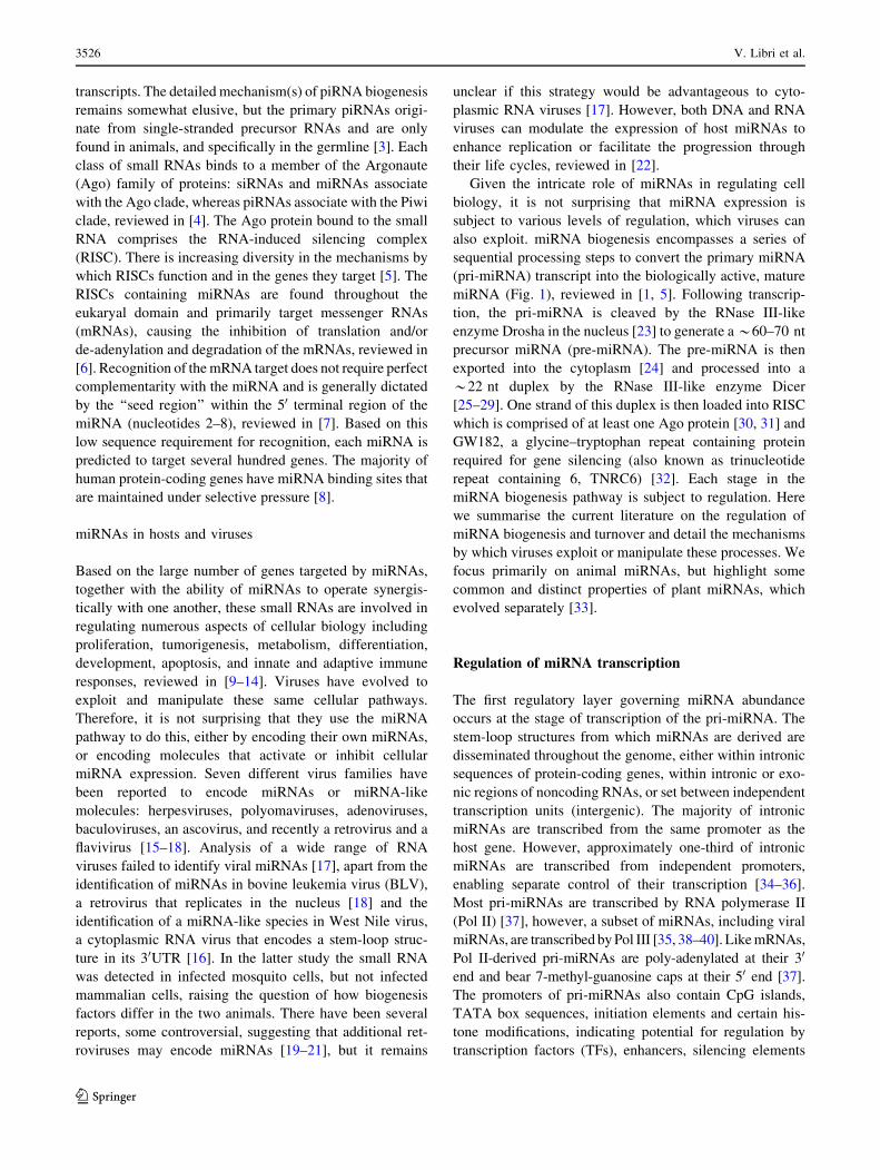
Fig. 1 Schematic overview of microRNA biogenesis and regulation
in animals. a The canonical biogenesis pathway. Pri-miRNAs are
transcribed in the nucleus by polymerase II with a cap (m7G,
7-methylguanosine-cap) and poly A tail. The pri-miRNA can harbour
a single pre-miRNA or a cluster of pre-miRNAs; the mature miRNA
sequence is depicted in red. Cleavage of the pri-miRNA occurs in the
nucleus by the Microprocessor complex, composed minimally of
Drosha and DGCR8, which interact with helicases p68 and p72. The
pre-miRNA is then exported through the nuclear pore complex into the
cytoplasm where the stem is cleaved by Dicer, supported by TRBP or
PACT. The miRNA/miRNA* duplex is loaded into the Ago protein
within RISC, where one part of the strand is preferentially retained;
this complex contains an Ago protein and GW182, which is required
for gene silencing. b Regulation of pri-miRNA cleavage. Proteins can
either positively (green) or negatively (red) influence cleavage of pri-
miRNAs by Drosha, based on direct interactions with the pri-miRNA
or interactions with auxiliary proteins p68/p72 (indicated by arrows).
Factors depicted in both green and red can behave as positive or
negative regulators depending on the identity of the miRNA and the
presence of other factors. Mature miRNAs can also regulate pri-
miRNA processing through interactions downstream of the stem-loop:
let-7 promotes processing of pri-let-7 whereas miR-709 inhibits
processing of pri-miR 15/16. c Regulation of pre-miRNA export. Two
viral non-coding RNAs inhibit miRNA translocation to the cytoplasm:
VA1 competes with endogenous pre-miRNAs for binding to Exportin-
5 whereas the viral miRNA, Bmnp-miR-1, regulates export indirectly
(dotted line) by targeting RanGTP. d Regulation of pre-miRNA
cleavage by Dicer. Proteins that regulate Dicer processing include:
(1) Lin28 (Lin28A), which recruits TUT4 that oligo-uridylates pre-
miRNAs leading to degradation, (2) MCPIP1 which cleaves the loop,
(3) TDP-43 and KSRP, which bind to the loops of both pri-miRNAs
and pre-miRNAs and (4) BCDIN3D, which can add methyl groups to
the 50 end of pre-miRNA and inhibit recognition by Dicer. The RNA
factors that are known to inhibit Dicer processing include an *800
non-coding RNA termed rnsc-1, VA RNAs from Vaccinia virus
(black) and a viral miRNA that regulates Dicer indirectly (dotted line)
miRNA biogenesis and turnover 3527
123

Pri-miRNA cleavage by the Microprocessor
In the canonical pathway, the pri-miRNA is cleaved in the
nucleus by the RNase III enzyme Drosha into a *60–70 nt
pre-miRNA. Cleavage by Drosha requires the co-factor
DGCR8 (DiGeorge critical region 8), also known as Pasha
[41]. Together these two proteins comprise the minimum
components of the Microprocessor complex (Fig. 1b).
DGCR8 functions at least in part by binding to the junction
between single-stranded and double-stranded regions of the
pri-miRNA and directing Drosha to cleave approximately
11 bp downstream of this junction [42], generating prod-
ucts with 2 nt 30 overhangs. It is thought that cleavage of
the pri-miRNA by Drosha occurs co-transcriptionally along
with splicing [43, 44], supported by the fact that Drosha
co-localizes to sites of active transcription [45]. Processing
of a pri-miRNA into a pre-miRNA can be regulated by a
variety of protein co-factors that are either recruited to the
Microprocessor through protein–protein interactions or
through direct interactions with the pri-miRNAs.
Regulation of pri-miRNA processing by proteins
that interact with the Microprocessor
Many proteins have been identified that interact with
Drosha, including the DEAD-box helicase proteins p68 (also
known as DDX5) and p72 (DDX17) [41]. These helicases
facilitate processing of nearly one-third of pri-miRNAs,
according to studies with p68/p72 knock-out mice [46]. In
some cases they do this by mediating interactions of TFs
with the Microprocessor. A well-characterized example is
the stimulation of maturation of specific pri-miRNAs by
SMAD proteins, which are TFs induced upon stimulation
with tumour growth factor b (TGF-b). The SMAD proteins
associate with p68 to enhance processing through binding a
consensus sequence in pri-miRNAs that strongly resembles
the DNA SMAD-binding element (Fig. 2) [47–49]. Other
TFs that regulate processing include the tumour suppressor
p53, which promotes pri-miRNA processing via interaction
with p68 [50] and ERa (estrogen receptor a), which inhibits
the processing of specific pri-miRNAs via interactions with
p68/p72 [51]. Another tumour suppressor, BRCA1 (breast
cancer susceptibility gene 1), also associates with Drosha,
p68, SMAD3 and p53 to accelerate processing of specific
pri-miRNAs associated with cancer [52]. In contrast to the
SMAD-regulated miRNAs, no consensus sequence has
been identified within the miRNAs regulated by these TFs
and the mechanisms underlying specificity in their regula-
tory functions are unknown. In addition to p68/p72, NF90
and NF45 (nuclear factor 90 and 45) also associate with the
Microprocessor [41] and can inhibit processing of several
miRNAs, including let-7 family members [53]. Other pro-
teins that associate with Drosha and positively regulate
processing include the multifunctional protein SNIP1
(SMAD nuclear interacting protein) [54] and ARS2 (arse-
nite-resistance protein 2) [55, 56]. However the precise
mechanisms by which these multi-functional proteins
influence biogenesis are unclear.
Regulation of pri-miRNA processing by recognition
of the stem-loop sequence or structure
Comparative analysis of pri-miRNA sequences suggests
that 14 % of human pri-miRNAs have conserved nucleo-
tides in their terminal loops, which may relate to
interactions with regulatory proteins [57]. One of the first
proteins identified to operate in this way was hnRNP-A1
(heterogeneous nuclear ribonucleoprotein A1), which binds
to the terminal loop and stem of pri-miR-18a and facilitates
processing by alteration of the stem structure [57, 58]
(Figs. 1, 2). Interestingly, this protein can also interact with
pri-let-7a, but in this case it negatively regulates processing
[59]. The inhibitory effect appears to result from compe-
tition between hnRNP-A1 and KSRP (KH-type splicing
regulatory protein), which both bind to the loop of pri-let-
7a. KSRP positively regulates a subset of miRNAs and
recognition has been proposed to derive from 2 or 3
sequential guanidines in the loop sequences [60] (Figs. 1b,
2). Interestingly, KSRP activity is modulated through its
phosphorylation state in response to different stimuli and
provides a link between PI3K/AKT signalling and miRNA
processing [61, 62] (Figs. 1b, 2). Other RNA-binding
proteins that interact with pri-miRNAs and promote their
biogenesis include TDP-43 (TAR DNA-binding protein-
43) [63] and the serine/arginine-rich SR protein SF2/ASF.
The SF2/ASF protein binds to a motif in the stem of pri-
miR-7 and has been proposed to alter the structure as
observed for hnRNP-A1 [64]. Interestingly, miR-7 targets
the 30UTR of SF2/ASF, providing a negative feedback loop
that may be important for controlling the steady-state
expression level of this miRNA [64].
A key protein involved in regulating multiple aspects of
miRNA biogenesis is Lin28 (abnormal cell lineage factor
28), which was originally discovered as a heterochronic
gene regulating developmental timing in worms [65].
Lin28 can inhibit both pri-let-7 processing [66–68] and
pre-let-7 processing [69–74] and recognition is mediated
by the primary sequence and structure of the terminal loop
(Fig. 2) [75]. Two Lin28 paralogs are present in mammals,
Lin28A and Lin28B. Lin28A is predominantly cytoplasmic
whereas Lin28B contains nuclear localisation signals and
accumulates in the nucleolus. It has been proposed that
Lin28B blocks let-7 processing by sequestering pri-let-7
miRNAs in the nucleoli away from the Microprocessor
[68], suggesting a new mechanism by which other RNA-
binding proteins might inhibit pri-miRNA biogenesis.
3528 V. Libri et al.
123
Regulation of pri-miRNAs by other miRNAs
A recent study by Zisoulis and colleagues [76] demonstrates
the pri-let-7 processing is also regulated by mature let-7,
providing the first example of a direct auto-regulatory loop
for let-7 biogenesis. In C. elegans, the ALG-1 (Argonaute-
like protein-1) binds to a specific site at the 30 end of the pri-
let-7 and thereby promotes processing of the pri-miRNA.
The interaction between ALG-1 and pri-let-7 is mediated by
mature let-7 through a conserved site in the pri-miRNA
transcript (Figs. 1b, 2). Immunoprecipitation of Ago
proteins in human cells also suggests an interaction with
pri-let-7, though it is not clear if this is mediated by a
miRNA [76]. Interaction between a mature miRNA and a
pri-miRNA can also have inhibitory effects on processing
(Figs. 1b, 2). For example, miR-709 binds to a stretch of
19 nt in the sequence of pri-miR-15a/16-1, preventing pri-
miRNA processing, leading to reduced levels of mature
miR-15a/16-1 [77]. The factors underlying nuclear locali-
sation of miR-709 remain unknown but this appears to be
associated with apoptotic stimuli, and may be a dynamic
mechanism for altering miR-15a/16 levels in response to
external signals. Transfection of a miR-709 mimic into cells
results in nuclear localisation of the synthetic RNA, indi-
cating that the localisation signal is contained within the
mature miRNA sequence. Nuclear localisation of miRNAs
was first reported in a study showing that a hexanucleotide
element within the mature miRNA sequence of miR-29b
directs its nuclear transport [78]. However, this element is
not present in miR-709 and the mechanism of nuclear
transport is unknown. It appears that miR-709 and its
binding site in pri-miR-15a/16 have co-evolved recently, as
they are both only present in the mouse [77]. Further
analyses are required to understand the breadth of regula-
tion of pri-miRNAs by mature miRNAs and whether this
relates to the nuclear localisation of Ago proteins that has
been reported previously [79].
The Drosha–DGCR8 regulatory loop and additional
substrates of the Microprocessor
Regulatory feedback loops are thought to be a key feature
of how miRNAs function in biological systems; for
example, miRNAs that are induced by Toll-like receptor
signalling target genes in this pathway, thereby dampening
the inflammatory response [80]. The miRNA biogenesis
machinery is also subject to regulation by feedback loops,
as observed for the Drosha–DGCR8 complex [81–83].
DGCR8 stabilizes the Drosha protein in the Microproces-
sor complex and the Microprocessor complex in turn
cleaves hairpin structures embedded in the 50UTR of
DGCR8 mRNA, leading to degradation of the DGCR8
transcript. This auto-regulatory loop is postulated to be
critical to maintain the appropriate balance between the
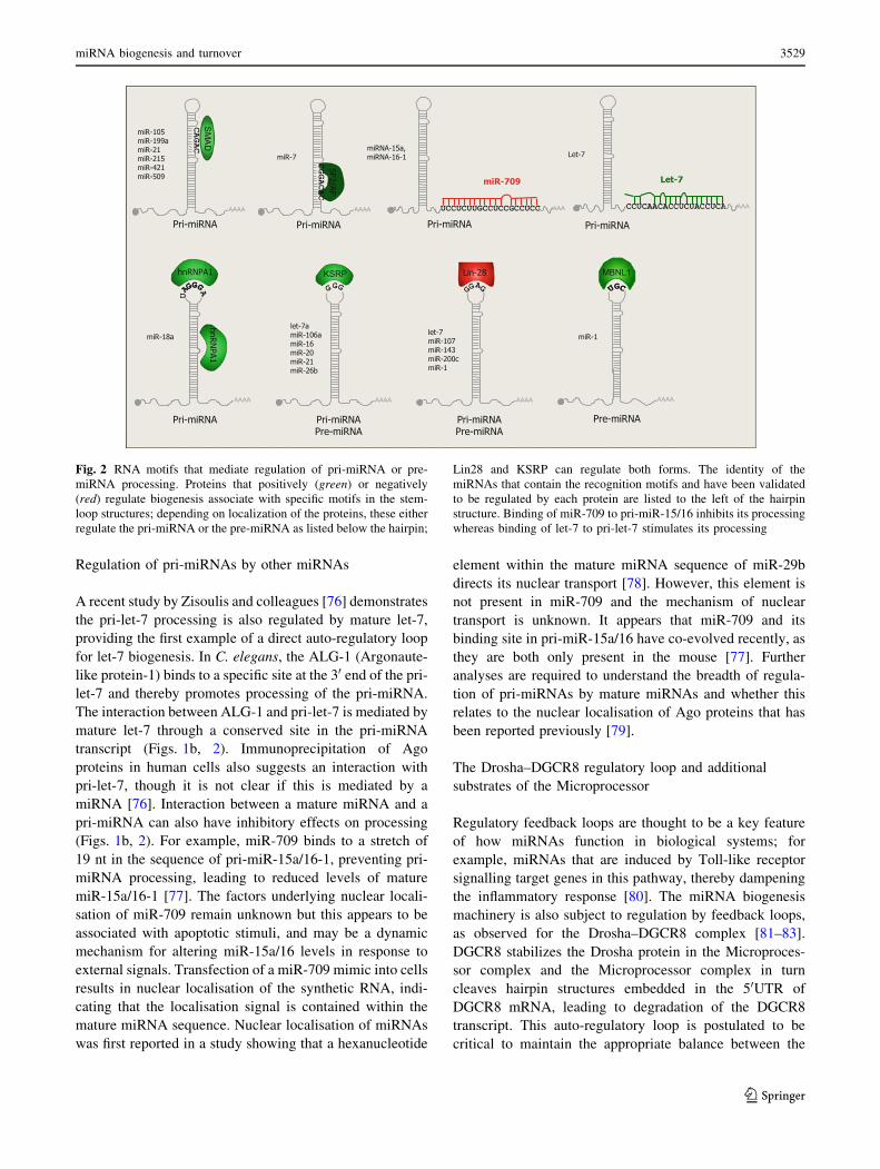
Fig. 2 RNA motifs that mediate regulation of pri-miRNA or pre-
miRNA processing. Proteins that positively (green) or negatively
(red) regulate biogenesis associate with specific motifs in the stem-
loop structures; depending on localization of the proteins, these either
regulate the pri-miRNA or the pre-miRNA as listed below the hairpin;
Lin28 and KSRP can regulate both forms. The identity of the
miRNAs that contain the recognition motifs and have been validated
to be regulated by each protein are listed to the left of the hairpin
structure. Binding of miR-709 to pri-miR-15/16 inhibits its processing
whereas binding of let-7 to pri-let-7 stimulates its processing
miRNA biogenesis and turnover 3529
123
levels of the Drosha–DGCR8 complex and its substrates:
when the Drosha–DGCR8 complex expression level is too
low there is suboptimal miRNA processing; when the
Drosha–DGCR8 complex expression level is too high,
cleavage of non-miRNA substrates such as mRNAs may
occur. Barad et al. [84] propose that efficient miRNA
processing and minimal off-target cleavage is obtained
only for a narrow range of Microprocessor concentration
values. These studies also suggest that, apart from miRNA
processing, the Microprocessor might play roles in mRNA
stability control [83]. Consistent with this, HITS-CLIP
analysis identified hundreds of mRNAs bound to DGCR8,
including DGCR8 mRNA [85]. This study further dem-
onstrated that cleavage within exonic cassettes can
influence ratios of alternative spliced isoforms, suggesting
complex roles of the Microprocessor in various modes of
gene regulation. A viral mRNA was also shown to be
regulated by Drosha in Kaposi’s sarcoma-associated her-
pesvirus (KSHV) infection: the KapB (kaposin B) mRNA
includes two pre-miRNAs in its 30UTR and excision of
these by Drosha alters the stability of the mRNA, thereby
reducing KapB protein expression [86]. This mode of
regulating viral gene expression during lytic or latent
infection could represent an alternative function of viral
miRNAs, where their processing serves a purpose, rather
than (or in addition to) their activities in gene silencing.
Regulation of pre-miRNA export
Once produced, the pre-miRNA is translocated to the
cytoplasm through the nuclear pore complex by Exportin-
5, which requires the co-factor RanGTP (Fig. 1) [24, 87,
88]. Structural analyses suggest that the length of the
double-stranded stem and presence of 30 overhangs are
important for Exportin-5 recognition [1, 89]. Interestingly,
Exportin-5 interacts with the RNA-binding protein NF90,
also known as ILF-3 (interleukin enhancer-binding factor
3) [90], which is found in the Microprocessor complex
[41]. It is possible that there is coordination between pri-
miRNA cleavage and export but this has not been exam-
ined. Exportin-5 also shuttles tRNAs and other abundant
RNAs to the cytoplasm and several studies suggest that
export of pre-miRNAs can be regulated by these RNAs
through competition. For example, Adenovirus produces a
*160 nt hairpin RNA (VA1 in Fig. 1c) that binds to
Exportin-5 and inhibits nuclear export of pre-miRNAs [91].
Over-expression of short hairpin RNAs (shRNAs) in ani-
mals can also be toxic due to saturation of Exportin-5 and
subsequent inhibition of pre-miRNA export [92]. Interest-
ingly, Exportin-5 was also reported to interact with Dicer
mRNA and high levels of pre-miRNAs or other Exportin-5
substrates can lead to accumulation of Dicer mRNA in the
nucleus, providing another feedback loop for regulating the
miRNA biogenesis factors [93]. The insect virus Bombyx
mori nucleopolyhedrosis virus (BmNPV) negatively regu-
lates nucleocytoplasmic transport of miRNAs by encoding
a viral miRNA that targets RanGTP [94], although the
functional relevance of this is not yet known.
Dicer processing of pre-miRNAs
Once in the cytoplasm, the pre-miRNA hairpin associates
with the RNase III-like enzyme Dicer that, in association
with dsRNA binding domain (dsRBD) proteins, cleaves it
into a double stranded miRNA duplex comprised of the
mature miRNA and the miRNA* (or passenger strand)
[25, 28, 95]. In flies, the dsRBD required for Dicer activity is
Loquacious [96–98], whereas the proteins in mammals are
TRBP (TAR RNA Binding Protein) and PACT (protein
activator of PKR) [99–101]. In general, the thermodynamic
asymmetry of the miRNA duplex determines which strand is
incorporated in RISC: the miRNA strand whose 50 end is less
stably base-paired is more frequently retained [102, 103].
Regulation of pre-miRNA processing: proteins
and RNA motifs involved
Dicer-mediated processing of pre-miRNAs is subject to
regulation by co-factors that interact with Dicer and RNA-
binding proteins that recognize RNA elements within
the pre-miRNAs. The Dicer protein alone can catalyse
the cleavage of pre-miRNA, however, the specificity of
cleavage is enhanced by TRBP and PACT [104]. Binding
of TRBP and PACT also stabilizes Dicer and knockdown
of TRBP and PACT reduces mature miRNA levels [99,
101]. TRBP also provides a link between MAPK (mitogen-
activated protein kinase) signalling and miRNA processing
since it is phosphorylated by Erk (extracellular signal
regulated protein) [105]. The phosphorylated form of
TRBP is more stable and leads to increased levels of many
growth-promoting miRNAs in HEK293 cells and also
causes a decrease in let-7 members. The mechanism for
differential effects of phosphorylated TRBP on individual
miRNAs is not yet clear [105].
The best-studied regulator of pre-miRNA processing by
Dicer is Lin28 (Fig. 1d). Lin28A, the cytoplasmic isoform,
binds a tetra-nucleotide sequence motif (GGAG) in the
terminal loop of let-7 precursors and recruits TUT4 (ter-
minal uridylyltransferase-4, also known as ZCCHC11),
which adds an oligo U-tail to pre-let-7. This U tail
blocks Dicer processing and mediates decay of pre-let-7,
presumably through recruitment of 30 to 50 exonucleases
[73, 106]. Lin-28A-dependent uridylation has also been
observed for several other pre-miRNAs that contain the
3530 V. Libri et al.
123

GGAG motif in their terminal loops, including miR-107,
miR-143 and miR-200c [106, 107]. Kim and colleagues
have recently shown that TUT4, as well as TUT2 and
TUT7, can also add a single uridine to the 30 end of
a specific set of pre-miRNAs (termed ‘‘group 2’’ pre-
miRNAs), which is independent of Lin28A. Up to 30 % of
pre-let-7 family members have an untemplated uridine at
the 30 end in cells not expressing Lin28A [107, 108]. The
pre-miRNAs that are modified lack a classical 2 nt 30 end
overhang, such that monouridylation results in the 2 nt
overhang and thereby improves processing by Dicer [108].
Like Lin28, KSRP and TDP-43 are also involved in both
pri- and pre-miRNA processing but they serve to promote,
rather than inhibit, processing (Fig. 1b, d) [60, 63]. These
findings suggest that the terminal loop is an important
platform for both ‘‘activators’’ (for example, hnRNP A1,
KSRP and TDP-43) and ‘‘repressors’’ (for example, Lin28)
to modulate miRNA levels and thereby gene regulation,
reviewed in [109]. There also appears to be some interplay
between the activators and repressors. For example, the
RNA binding protein MBNL1 (muscleblind-like splicing
regulatory protein 1) binds to pre-miR-1 through recogni-
tion of a UGC motif that overlaps with a binding site for
Lin28 (Fig. 2), such that MBNL1 binding blocks Lin28-
mediated oligouridylation and subsequent degradation of
pre-miRNA-1 [110]. Similar competition is seen with the
mammalian immune regulator MCPIP1 (monocyte che-
moattractant protein induced protein-1) and Lin-28:
MCPIP1 is a ribonuclease that inhibits miRNA biogenesis
by competing with Dicer for the cleavage of the terminal
loop of pre-miRNAs. Addition of Lin28 abolishes
MCPIP1-mediated cleavage in vitro, presumably through
competition for binding to the terminal loop [111]. Other
negative regulators of processing might also stabilize pre-
miRNAs against degradation, but it is not clear if this is
one of their functions in vivo. Recently Kouzarides’s group
showed that Dicer processing can also be regulated by
methylation of the 50 end of the pre-miRNA by the human
RNA-methyltransferase, BCDIN3D [112]. BCDIN3D adds
two methyl groups to the 50 phosphate of pre-miR-145
in vitro and in vivo; since Dicer specifically recognizes the
50 monophosphate [113], this modification inhibits pro-
cessing (Fig. 1d). A noncoding RNA in C. elegans was also
shown to inhibit pre-miRNA processing: the *800 nt
noncoding RNA, rncs-1 (RNA noncoding, starvation
up-regulated), competes with endogenous dsRNAs for bind-
ing to Dicer or accessory dsRBD proteins [114] (Fig. 1d).
The VA RNAs in Adenovirus have also been shown to
operate as competitive inhibitors for Dicer processing of pre-
miRNA [91, 115], in addition to their inhibitory effects on
Exportin 5.
Other viruses also inhibit this step in miRNA biogenesis.
For example, Vaccinia Virus (VACV) infection leads to a
drastic reduction in Dicer protein expression and a con-
comitant defect in pre-miRNA processing. The mechanism
by which the virus abrogates Dicer expression remains
unclear [116]. The human herpesvirus Epstein–Barr virus
(EBV) influences Dicer processing through a more subtle
mechanism: the viral-encoded miRNA miR-BART6-5p
targets human Dicer mRNA [117]; it is expected that this
could form a feedback loop to regulate the level of viral
miRNAs. The host-encoded let-7 also regulates Dicer
levels through target sites in the coding sequence, sug-
gesting that feedback loops for controlling miRNA
biogenesis may be inherent to miRNA homeostasis [118],
which viruses can exploit.
Regulation of miRNA expression by Argonaute proteins
MiRNAs function in partnership with Ago proteins, and
a number of studies suggest that expression levels of
miRNAs are tied to the expression levels of Agos. For example,
ectopically expressed Ago proteins (Ago1–4) enhance
expression of miRNAs under conditions where the
miRNAs saturate the endogenous machinery [119], and
endogenous miRNAs are reduced in mouse embryonic
fibroblasts from Ago2-knockout mice [120]. Ago proteins
are also subject to various levels of transcriptional and
post-transcriptional regulation that might therefore influ-
ence miRNA expression. For example, the expression level
of the Ago2 protein is specifically up-regulated in breast
cancer cells lacking ERa, which is dependent on the
EGFR/MAPK signalling pathway and leads to enhanced
miRNA activity [121]. Ago2 can also be phosphorylated
within the RNA binding pocket, which inhibits small RNA
binding and is expected to thereby influence miRNA sta-
bility [122]. In addition to its role in miRNA stabilization,
Ago2 has also been shown to catalyse an alternative pre-
miRNA processing event [120]. Cleavage occurs within the
30 arm of a pre-miRNA such that only the small RNA
generated from the 50 arm can be functional. The relevance
of this alternative processing pathway remains elusive, but
it may play a role in passenger strand dissociation for
hairpins with a high degree of complementarity, where this
might otherwise be inefficient [120].
Non-canonical pathways of biogenesis: breaking
the rules
In addition to the canonical biogenesis pathway, some
miRNAs are processed by Drosha-independent and Dicer-
independent pathways (Fig. 3) [123]. Studies of
viral-encoded miRNAs in particular illuminate a range of non-
canonical possibilities. For example, murine c-herpesvirus
miRNA biogenesis and turnover 3531
123
68 (MHV68) expresses its miRNAs in the same Pol III
primary transcripts as the viral-encoded tRNAs [39, 40].
The pre-miRNAs are generated following cleavage by
RNase Z and are subsequently processed by Dicer, thus
bypassing the Microprocessor complex [124]. The retro-
virus BLV also encodes Pol III-dependent pre-miRNA-like
species that bypass Drosha cleavage and are subsequently
processed by Dicer. Importantly, this mechanism provides
a route for viral miRNA biogenesis that does not result in
cleavage of the retroviral genomic RNA [18]. A miRNA-
like species was also recently reported in West Nile virus (a
cytoplasmic RNA virus) [16] and several reports have
shown that artificial miRNAs engineered into RNA viruses
are processed to a detectable level [125–127]. However,
the mechanism(s) for biogenesis of these viral RNAs are
not reported. Another alternative processing pathway has
been described for miRNAs encoded by Herpesvirus
Saimiri (HVS). These miRNAs are derived from the same
Pol II transcripts that encode another class of viral non-
coding RNA, HSURs (H. saimiri U-rich RNAs), which
resemble small nuclear RNAs (snRNAs). The pre-miRNAs
are located directly downstream of the 30 end processing
signals of HSURs and processing of the viral miRNAs does
not require the Microprocessor [128]. Rather, the 50 ends of
the viral pre-miRNAs are produced by the Integrator, a
nuclear complex of 12 proteins that associates with Pol II
and is required for HSUR biogenesis. As in the canonical
miRNA biogenesis pathway, HVS pre-miRNAs require
Exportin-5 for transit to the cytoplasm, where they are
processed by Dicer. An Integrator-dependent mechanism
has not been reported for biogenesis of endogenous
miRNAs. However, a range of reports suggest other
mechanisms by which RNAs can be processed into miRNA-
like species without a requirement for Drosha. For example,
some miRNAs are derived from ‘‘mirtrons’’, which are
generated by splicing and debranching of short hairpin
introns (Fig. 3) [129, 130]. The 50 and 30 ends are defined by
donor and acceptor splice sites, but in some cases include
additional unstructured tails [131, 132]. The biogenesis of
30-tailed mirtrons in Drosophila was recently reported to
utilize the RNA exosome, the major 30–50 exoribonuclease
in eukaryotes [133]. Indeed, there is increasing overlap in
the factors involved in miRNAs biogenesis and other RNA
processing pathways. The list of RNAs that feed into the
miRNA biogenesis pathway is also increasing: snoRNAs
(small nucleolar RNAs), tRNAs and endogenous shRNAs
can be processed by Dicer into small RNA fragments that
then mediate gene silencing [131, 134–136].
Dicer is generally considered essential for the biogenesis
of miRNAs, but at least one highly conserved miRNA,
miR-451, is produced by a Dicer-independent mechanism
in human, mouse and zebrafish [137–139]. The mature
miRNA maps to the stem as well as loop sequence of the
pre-miRNA and directly binds to Ago proteins (Fig. 3b).
Ago1 and Ago3 can actively load pre-mir-451 but only
Ago2 can process the miRNA since this requires the
endonuclease activity [140]. To date, no other Dicer-
independent miRNAs have been identified and the specific
features that dictate routing to Dicer versus Ago are under
investigation [140]. A recent report showed that pre-
miRNAs could be designed to be processed by Ago2 as
well as Integrator, eliminating the need for either Drosha or
Dicer and opening up the possibility that such pathways
could exist naturally [141].
Regulation of miRNA biogenesis by single nucleotide
polymorphisms and RNA editing
Natural sequence variations in pri-miRNAs, pre-miRNAs
or mature miRNAs can influence their processing, stability
and target selection. These sequence variations originate
from changes in the DNA-coding sequence or from post-
transcriptional modifications to the RNA [142–147]. In
humans, differences in processing by Drosha were
observed for alleles of miR-125a, miR-126, miR-146a,
miR-502, miR-510, miR-890, and miR-892b [143–145,
147], while alteration of processing by Dicer was
postulated for SNPs (single nucleotide polymorphisms) in
miR-196a [146]. A natural variant of miR-934 was found
to contain a mutation in the first nucleotide of the pre-
miRNA, which affects strand selection for incorporation
into RISC [145].
MiRNAs can also be post-transcriptionally modified by
the ADAR family members (adenosine deaminase acting
on RNA proteins) which convert adenosines to inosine,
reviewed in [148]. The hairpin structures of pre-miRNAs
are favourable substrates for ADARs [149], which recog-
nize dsRNA. Blow et al. [150] sequenced 99 miRNAs from
10 human tissues and identified 6 % of pri-miRNA tran-
scripts with A to I conversions in at least one of the
analysed tissues. Another survey reported that 16 % of pri-
miRNAs are edited in the brain, where there is generally a
higher frequency of RNA editing [151]. Editing can affect
pri-miRNA and pre-miRNA processing and can also alter
the target repertoire of the miRNA when editing occurs in
the mature sequence [152–155]. For example, editing of
pri-miR-142 substantially reduces processing by Drosha
and leads to cleavage by Tudor-SN (Tudor staphylococcal
nuclease), a component of RISC with ribonuclease activity
specific for inosine-containing dsRNAs [154, 155]. In
contrast, editing of pri-miR-151 by ADAR1 does not affect
pri-miRNA processing but interferes with pre-miRNA
cleavage by Dicer, as seen by accumulation of edited pre-
miR-151 (Fig. 1d) [153]. The A to I conversion within the
mature miRNA can retarget the miRNA to a new set of
3532 V. Libri et al.
123

mRNAs since inosine base pairs with cytosine rather than
uridine. For example, editing of sites within the miR-376
seed alters its target repertoire both in vitro and in vivo
[152]. Interestingly, Heale et al. reported that ADAR
enzymes can also influence miRNA processing indepen-
dently of their catalytic activity, suggesting that in some
cases binding of the ADAR proteins alone might be suf-
ficient to interfere with miRNA processing [156].
Some viral miRNAs have also been found to be edited,
for example KSHV miR-K12-10 [40], Marek’s disease
virus miR-M7 [157] and EBV miR-BART6 [117]. To date
the functional relevance of this editing has only been
suggested for the latter. In HEK-293 cells, editing of EBV
miR-BART6-3p decreases the efficiency with which the
miRNA encoded on the opposite strand, miR-BART6-5p,
is loaded into RISC. Strikingly, miR-BART6-5p targets
human Dicer via 4 binding sites in its 30UTR. Therefore,
editing of miR-BART6-3p relieves Dicer from post-
transcriptional gene silencing. Dicer levels affect the
expression levels of multiple genes that regulate the
infectious and lytic states of EBV and it is postulated that
editing of miR-BART6-3p could be an indirect way to
modulate miRNA biogenesis and thereby the viral life
cycle [117].
Regulation of miRNA stability
Once a mature miRNA is incorporated into RISC it is
generally considered to be extraordinary stable [158, 159].
pre-miRNA
tRNA
MHV68 miRNAs
export
Dicer cleavage
Drosha cleavage
pre-miRNA
Drosha-independent Dicer-independent A B
Ago 2
slicing
m7G GW182
Ago
miRISC
AAAAAAA
Cellular miRNAs
exon exon mirtron
splicing
mature mRNA
debranching
trimming
Cellular miRNAs
HSUR
int11
Integrator
HVS miRNAs
Pol III Pol II Pol II Pol II
m 7 G AAAAAAA
Fig. 3 Alternative miRNA biogenesis pathways in animals and
viruses. a Drosha-independent biogenesis. Pre-miRNAs are
co-transcribed with tRNAs in Pol III transcripts in MHV68 and
bypass processing by Drosha. Pre-miRNA like miRNAs in HVS are
derived from the same Pol II transcripts as HSURs and require the
Integrator for generation of their 50 ends. Cellular miRNAs termed
mirtrons also do not require Drosha: they are Pol II transcripts that are
excised by splicing and linearized by lariat debranching; tailed
mirtrons require further 50 or 30 trimming by nucleases and then they
are directly processed by Dicer. b Dicer-independent biogenesis. The
highly conserved miRNA, miR-451 is produced in a dicer-indepen-
dent mechanism involving cleavage by Ago. The mature miRNA
(red) derives from the stem as well as loop sequence of the pre-
miRNA
miRNA biogenesis and turnover 3533
123
Indeed, upon inactivation of miRNA transcription or pro-
cessing the majority of mature miRNAs in human and
rodent cell lines have half-lives in the range of many hours
to days [160, 161]. However, recent reports from various
model systems have demonstrated differences in the sta-
bilities of individual miRNAs, suggesting that regulated
degradation of specific miRNAs is a physiologically rele-
vant way to modulate their expression, reviewed further in
[162]. In particular, active miRNA decay seems to play a
prominent role in neurons. In mouse retinal cells the sen-
sory neuron-specific miR-183/96/182 cluster and miR-204
and miR-211 are differentially expressed in response to
light. The mature miRNAs are rapidly down regulated
upon dark-adaptation due to active degradation by a yet
unidentified enzyme [163]. Several other brain-enriched
miRNAs have short half lives both in primary human
neuronal cell culture and post mortem brain tissue [164].
The fast turnover is recapitulated in primary neurons out-
side the retina as well as in neurons derived from mouse
embryonic stem cells. Strikingly, blocking of action
potentials by inhibition of sodium channels prevented the
degradation of selected miRNAs, indicating that activation
of neurons is required for the regulated decay of some
neuronal miRNAs [163]. In line with this observation, a
small RNA deep sequencing approach identified several
brain-enriched miRNAs that also were rapidly down reg-
ulated upon transient exposure to the neurotransmitter
serotonin in the marine snail Aplysia [165]. Active miRNA
decay represents an elegant way to re-activate neuronal
transcripts, which might be important for a rapid response
to various external stimuli [166–169]. Regulated miRNA
turnover also occurs during viral infection (described
below), although to date the mechanisms of miRNA turn-
over in neurons or during infection in mammals remain
unknown. However, studies from other model systems
have identified molecular determinants of regulated
miRNA decay and here we will summarize the current
knowledge on these determinants and their modes of
action.
Modifications to the 30 end of miRNAs
Chemical modifications of mature miRNAs play a crucial
role in regulating their stabilities. The first appreciation for
miRNA stability factors came from studies in plants, where
the methyltransferase HEN1 (Hua enhancer 1) methylates
the 20 hydroxyl group of the 30 terminal nucleotide of a
miRNA [170–172]. Methylation of plant miRNAs protects
their 30 ends from terminal uridylation by the nucleotidyl
transferase HESO1 (HEN1 suppressor 1), which triggers
their degradation [173–175]. Uridylation at the 30 ends of
RNAs is also associated with reduced stability of piRNAs,
siRNA and mRNAs [176–178]. Similarly, a nucleotidyl
transferase in the unicellular alga Chlamydomonas rein-
hardtii, MUT68, uridylates small RNAs leading to their
degradation by the peripheral exosomal subunit RRP6
(ribosomal binding protein 6) [179].
Animal miRNAs generally lack a protective 20-O methyl
group at their 30 terminus and display template-independent
nucleotide addition, mostly adenylation or uridylation
that may regulate miRNA stability [180–182]. Several
enzymes, including MTPAP, PAPD4/GLD2, PAPD5,
ZCCHC6, TUT4/ZCCHC11, and PAPD2/TUT1 display
terminal nucleotidyl transferase activity and knockdown
experiments indicate that these proteins are responsible for
miRNA 30 end variation to various extents [183, 184].
However, functional implications have thus far been
described for only a few of these enzymes. For example,
TUT4, the nucleotidyl transferase implicated in the deg-
radation of histone mRNA and several pre-miRNAs
[73, 110, 178], regulates cytokine levels by uridylation of
mature miR-26 family members [185]. In the human A549
cell line, miR-26b targets the IL6 (interleukin 6) transcript
but terminal uridylation of this miRNA interferes with its
function. Knockdown of TUT4 results in reduced miR-26a
uridylation along with decreased expression of a reporter
containing the IL6 30UTR. Conversely, overexpression of
TUT4 leads to enhanced levels of the same reporter,
indicating that uridylated miR-26a is less effective in tar-
geting IL6. Notably, knockdown of TUT4 does not
increase miR-26 expression levels, indicating that uridy-
lation of the miRNA affects its activity without affecting its
expression [185].
Adenylation at the 30 ends of miRNAs is associated with
both enhanced and decreased miRNA stability [186–189].
For example, the most highly expressed miRNA in the
liver, miR-122, is monoadenylated by the cytoplasmic
poly(A) polymerase GLD2 (germline development defec-
tive-2). In GLD2 knockout mice, miR-122 is selectively
destabilized whereas the levels of 10 other miRNAs remain
unchanged. The stability of the miR-122 precursors is not
affected by GLD2 knockout, suggesting a role for adeny-
lation in modulating stability of the mature form [186].
Recently it was demonstrated that VACV induces pol-
yadenylation of endogenous miRNAs during infection. The
viral poly(A) polymerase is responsible for the non-tem-
plated adenylation that results in a *30-fold reduction of
endogenous miRNA levels in infected mouse embryonic
fibroblasts; other small RNAs such as tRNAs and snRNAs
remain largely unaffected by VACV infection. It was
suggested that viral poly(A) polymerase operates only on
Ago-bound small RNAs, but the mechanism is unknown.
Whereas polyadenylation of miRNAs is mediated by a
viral gene product, the actual degrading activity is postu-
lated to stem from a yet undefined cellular protein [189]. It
is not clear if and how the modification of miRNAs by
3534 V. Libri et al.
123
VACV is linked to the reduction in Dicer expression that
was described previously [116]; it may be that this virus
uses two different mechanisms to shut-off cellular miRNA
expression. Poxviruses infect a wide range of vertebrate
and invertebrate hosts. Infection of Drosophila cells with
VACV leads to global reduction in miRNA expression
whereas the levels of endogenous siRNAs are unaffected.
Like plant miRNAs, insect siRNAs are methylated, which
protects them from polyadenylation by the virus. Indeed 30
methylation of a transfected miRNA prevents it from being
polyadenylated and degraded during infection [189]. The
advent of deep sequencing technology has enabled a much
greater appreciation for the extent of heterogeneity and
modifications at the 30 ends of miRNAs [182, 183, 190]. In
the coming years it will be important to further characterise
the enzymes that write and read these modifications and to
understand their impact on miRNA stability and function.
Sequence motifs regulating miRNA stability
Several reports have demonstrated altered kinetics in the
turnover of individual miRNAs under conditions where the
expression levels of most miRNAs are unchanged [160,
161]. This suggests that cis acting elements in the mature
miRNA sequence provide specificity to the miRNA deg-
radation process. In a survey to characterise the role of
miRNA turnover during the cell cycle, Rissland and col-
leagues [191] found that miR-503 and other members of
the extended miR-16 family are constitutively unstable in
NIH-3T3 cells. The high turnover rate allows dynamic
transcriptional regulation of these miRNAs during the cell
cycle. For example, miR-503 is rapidly down regulated
upon cell cycle re-entry but accumulates during cell cycle
arrest by serum starvation. Sequence elements within the
seed and 30 end of the miRNA appear to be required for the
degradation. Similarly, miR-382 is selectively unstable in
HEK293 cells and an element in the 30 end of the miRNA
is required for its enhanced turnover in vitro [160]. Optimal
paradigms to study cis acting elements with a role in
miRNA decay are miRNAs that are co-transcribed and
highly similar on a sequence level, yet differ in their decay
rates. The miR-29 family provides such an example:
miR-29b is unstable in cycling cells and only accumulates
during mitosis whereas miR-29a is stable throughout the
cell cycle [78]. The miR-29a and miR-29b share the same
seed sequence but are distinguished by a C to U substitu-
tion at position 10 and miR-29b contains a hexanucleotide
motif (AGUGUU) at its 30 end that is responsible for its
nuclear localisation. However, the motif does not account
for the accelerated miRNA decay. Instead, uridines at
position 9–11 in miR-29b seem to enhance destabilisation
and many, but not all, miRNAs that contain a uridine
stretch at this position are reported to display faster
turnover rates [192]. Therefore, additional factors must
dictate the differential stability of miRNAs. Altogether
these studies show that miRNAs, though limited in coding
space, contain sequence elements outside the classical seed
that may critically influence miRNA abundance and func-
tion. To date, no viral miRNAs have been reported to
contain such motifs, but this could provide another strategy
for viruses to diversify miRNA function and regulation
during their life cycles. Identification of the trans-acting
factors that recognise these motifs is important for further
investigations in this area.
Trans-acting factors regulating miRNA stability
The first report of enzymes that are capable of degrading
single-stranded small RNAs came from a candidate gene
approach in plants. In Arabidopsis, SDN1 (small RNA
degrading nuclease 1) possesses 30–50 exonuclease activity
on small RNAs including miRNAs. In a cell free assay
system, SDN1 specifically degrades ssRNA but not
dsRNA. The 20 O-methylation present on the 30 terminal
nucleotide of plant miRNAs is protective against SDN1
activity [193]. The enzyme belongs to a family of exori-
bonucleases with partially overlapping functions in vivo
that are responsible for miRNA turnover in plants. Inter-
estingly, members of this protein family are conserved in
all eukaryotes and it seems likely that animal homologues
of SDNs have similar functions but these have not yet been
reported [194]. The XRN family of enzymes play various
roles in miRNA stability in different organisms: in
Arabidopsis, XRN2 and XRN3 are involved in degrading
the loop sequence of pre-miRNAs [195], in mammalian
cells, XRN2 degrades the pri-miRNA following processing
by Drosha [43, 196]. In C. elegans, XRN2 degrades mature
miRNAs once released from the RISC complex and may
also influence the rate at which they are released [197].
Interestingly, the presence of target RNA counteracts the
decay of miRNAs by XRN2 both in vitro and in vivo
[197, 198]. Whether this is due to direct competition
between the target and XRN2 for miRNA binding or
through another molecular mechanism is not yet known.
The exoribonuclease XRN1 and the exosome core subunit
Rrp42 (ribosomal RNA-processing protein-42) are pro-
posed to be involved in turnover of miR-382 in HEK293
cells, as knock-down of these factors selectively increases
miR-382 expression levels [160].
In a human melanoma cell line, ectopic expression of
hPNPaseold-35 (human polynucleotide phosphorylase pro-
tein) leads to the selective down regulation of several
miRNAs (miR-221, miR-222 and miR-106b). Immuno-
precipitation studies show that this 30–50 exoribonuclease
directly associates with these miRNAs and causes their
degradation in vitro. However, it remains unclear whether
miRNA biogenesis and turnover 3535
123
hPNPaseold-35 is also able to actively dislodge them from
the RISC complex. Interestingly, hPNPaseold-35 is an
interferon-stimulated gene and mediates IFN-b-induced
down regulation of miR-221. One of the direct targets of
miR-221 is the cell-cycle suppressor p27kip1. Conse-
quently, both miR-221 overexpression and knockdown of
hPNPaseold-35 protect human melanoma cells from INF-b-
induced growth arrest, indicating a pivotal role of con-
trolled miRNA decay in tuning cell proliferation [199]. The
30–50 exoribonuclease Eri1 was recently implicated in
regulating miRNA stability in mouse lymphocytes, based
on the global increase in miRNA levels observed in NK
and T cells from Eri1 knockout mice [200]. The regulation
of miRNA levels by Eri1 appears to be required for
NK-cell development and antiviral immunity, but its
mechanism of action remains to be established.
Besides promoting miRNA degradation, RNA binding
proteins can also enhance the stability of mature miRNAs.
For example, Quaking, a member of the STAR (signal
transduction and activation of RNA) family of RNA
binding proteins, is up regulated in response to p53 sig-
nalling and stabilises mature miR-20a [201]. The
identification of proteins that stabilise and de-stabilise
mature miRNAs supports the idea that regulation of miR-
NA decay is important in controlling the miRNA repertoire
of the cell. Yet, there are still major gaps in understanding
how specificity in degradation or stabilization is mediated.
Target mediated miRNA turnover
In contrast to target-mediated stabilization of miRNA in
C. elegans, binding of miRNAs to RNAs can promote
miRNA degradation in Drosophila and mammals. In flies,
most miRNAs are incorporated in Ago1-containing RISC
complexes whereas siRNAs, usually derived from dsRNA
from viruses and transposons, are loaded into Ago2 [202]
and are 30 methylated by the Drosophila homolog of HEN-
1 [203]. Intriguingly, binding of Ago1 associated miRNAs
to target sites with extensive complementarity results in
destabilization of the miRNAs [204]. Deep sequencing the
small RNAs revealed that a large proportion of these
miRNAs are either shortened or have non-templated
nucleotide additions at their 30 ends (mostly adenines and
uridines). This mechanism of trimming and tailing, medi-
ated by as yet unknown enzymes, seems to precede miRNA
decay (Fig. 4). In contrast, miRNAs that associate with
Ago2 and thus are methylated appear to be protected from
degradation. In human cells, miRNAs are also subject to
this target-directed destabilisation, as evidenced by trim-
ming and tailing in Hela cells in vitro [204]. Baccarini and
colleagues [205] examined in more detail the fate of a
miRNA molecule after target recognition and demonstrated
that miRNAs generally out-live their targets, whether the
target is perfectly complementary or contains a central
bulge. However, target recognition promotes post-tran-
scriptional modification of miRNAs (mostly 30 uridylation)
which is postulated to induce their degradation, thereby
limiting miRNA recycling. It is not yet known what fea-
tures in the target RNA direct the posttranscriptional
modification of a miRNA but this may involve extensive
pairing as proposed in flies [204].
Two distinct mammalian herpesviruses, a gamma her-
pesvirus infecting new world primates and a beta
herpesvirus infecting mice, exploit the mechanism of tar-
get-directed miRNA degradation (Fig. 4). Several HSURs
are expressed in Herpesvirus saimiri (HVS)-transformed
T-cells and one of these, HSUR1, contains an interaction
site for the endogenous miRNA, miR-27 [206]. Cazalla
et al. [206] showed that binding of HSUR1 to miR-27
accelerates its rate of turnover and replacing the miR-27
interaction site with a binding site for miR-20 re-targets
HSUR1 to the other miRNA. Similarly, miR-27 is also
rapidly down regulated in murine cytomegalovirus
(MCMV) infection of several mouse cell lines as well as
primary macrophages. Yet, the expression levels of miR-27
precursors remain stable, indicating that the mature form is
subject to enhanced degradation, presumably by a viral
inhibitor [207]. Indeed, the MCMV m169 gene contains a
binding site for miR-27 in its 30 UTR and miR-27 levels are
rescued if the m169 gene is knocked down or deleted from
the virus [208, 209]. During lytic MCMV infection, m169 is
among the most highly transcribed genes [209] and it rep-
resents the most frequent non-miRNA segment sequenced
in Ago2 immunoprecipitations [208]. Down regulation of
miR-27 is linked to its 30 end tailing and trimming, indi-
cating that a similar mechanism as suggested in flies and
human cells might underlie the degradation process [204,
209]. As reported for HSUR1, replacing the miR-27 binding
site with an interaction site for an unrelated miRNA is able
to redirect m169 to target that specific miRNA [206, 208,
209]. The degradation of miR-27 by two distinct herpe-
sviruses might suggest that this miRNA plays an important
role in the viral life cycles. Indeed, miR-27 represses
MCMV replication when over-expressed in cell culture
experiments [207] and MCMV mutants incapable of down
regulating miR-27 display attenuated viral growth in mice
[209]. So far, however, it remains unclear which cellular
miR-27 target(s) are responsible for modulating MCMV
replication and whether it plays the same role in both
MCMV and HVS infections. In summary, the pairing pat-
terns of miRNAs with their targets as well as the relative
amounts of each seem to be crucial factors that determine
the extent of target-mediated miRNA decay [205]. A range
of reports suggest that endogenous mRNAs, noncoding
RNAs and pseudogenes also play a role in regulating
miRNA activity and/or stability, reviewed in [210].
3536 V. Libri et al.
123
Viral suppressors of RNA interference may modulate
miRNA expression
In insects and plants, RNA silencing pathways mediate a
potent antiviral response. For efficient replication, viruses
that infect these hosts therefore rely on virus-encoded
suppressors of RNAi (VSRs) [211]. Also in mammalian
viruses, proteins with RNAi suppressive activity have been
identified, although the importance of this suppressive
activity in vivo remains to be established [212, 213]. In the
following section we will discuss how the expression of
these VSRs affects miRNA biosynthesis in insects and
plants and we will further speculate about their possible
influence on miRNA expression in mammals.
The RNA interference machinery in insects recognizes
viral dsRNA in the cytoplasm and processes it into
vsiRNAs (viral siRNAs) [211]. These vsiRNAs associate
with Ago2-containing RISC complexes, which then act as
antiviral effectors by cleaving viral RNA in the cytoplasm
[214]. Whereas the production of siRNA and miRNA
molecules in mammals largely rely on the same biogenesis
factors, the miRNA and antiviral RNAi pathways in insects
are governed by a distinct set of processing and effector
complexes. Specifically, pre-miRNAs are processed by
Dicer-1 to be loaded into Ago1-containing RISC com-
plexes. In contrast, cytoplasmic long dsRNA is sensed and
cleaved by Dicer-2 and the resulting 21 nt siRNAs are
predominantly loaded into Ago2-containing RISC [202,
215, 216]. Insect VSRs interfere with the RNAi machinery
at different stages of the pathway. Drosophila C virus 1A
for example binds long dsRNA, thereby preventing its
efficient processing into siRNA [214]. Flock house virus
B2 binds both long dsRNA and siRNAs [217–220]. Cricket
Paralysis virus 1A and Noravirus VP1 directly interact with
the small RNA-loaded Ago2 effector complex and prevent
its target RNA cleavage activity [221, 222] (and unpub-
lished observations).
Although the siRNA and miRNA biogenesis machiner-
ies are distinct in insects and plants, many VSRs have
dsRNA binding properties, and it might be expected that
they could affect miRNA processing too. However, this
does not seem to be the case in flies. VSR expression in
transgenic Drosophila does not alter levels of mature
miRNAs, nor does it affect the activities of miRNA
reporters. Furthermore, in contrast to Ago1 loss-of-function
mutants, transgenic animals expressing VSRs do not dis-
play developmental defects, suggesting that VSRs do not
affect global miRNA biogenesis and function [214, 219,
Viral transcripts Transgenes
(artificial microRNA targets)
Pol II Pol II
Pol II
?
Extensive base pairing
Tailing and Trimming
miRNAdegradation
?
Target RNA
miRNA
? ?
Endogenous mRNAs
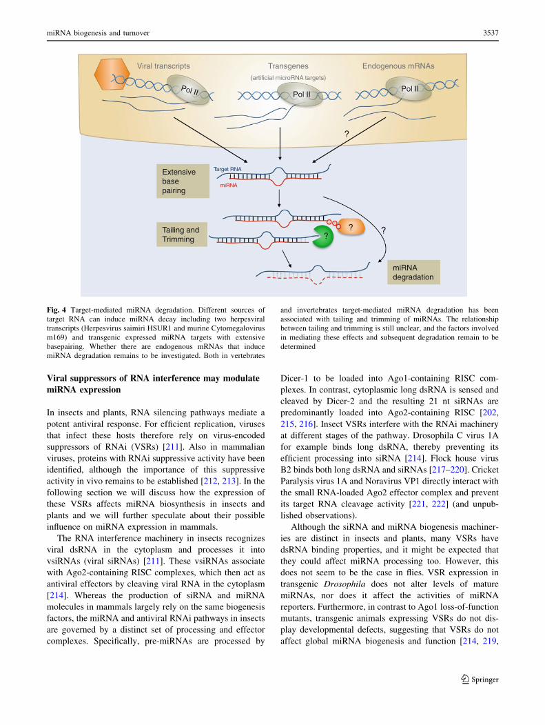
Fig. 4 Target-mediated miRNA degradation. Different sources of
target RNA can induce miRNA decay including two herpesviral
transcripts (Herpesvirus saimiri HSUR1 and murine Cytomegalovirus
m169) and transgenic expressed miRNA targets with extensive
basepairing. Whether there are endogenous mRNAs that induce
miRNA degradation remains to be investigated. Both in vertebrates
and invertebrates target-mediated miRNA degradation has been
associated with tailing and trimming of miRNAs. The relationship
between tailing and trimming is still unclear, and the factors involved
in mediating these effects and subsequent degradation remain to be
determined
miRNA biogenesis and turnover 3537
123
221–223]. In contrast, transgenic expression of VSRs in
plants leads to pleiotropic, developmental defects due to
alterations in miRNA-mediated gene regulation [224–226].
This is likely based on the convergence of the plant siRNA
and miRNA biogenesis pathways, which share several
processing factors. For instance, both miRNAs and anti-
viral siRNAs can be loaded into Ago1 effector complexes
in plants [227–229]. Yet, for many plant VSRs, it remains
elusive how they manipulate the miRNA machinery
in vivo. A number of VSRs have dsRNA binding activity
in vitro, which has been hypothesised to explain their
interference with miRNA biogenesis [230–235]. For
instance, Tombusvirus P19 directly binds siRNA duplexes
preventing their efficient loading into effector RISC com-
plexes in vitro [224, 225, 230, 236–238]. In transgenic
Arabidopsis, P19 also prevents miRNA loading into Ago1-
containing RISC. However, this seems to be a rather
exceptional property as three other VSRs tested, Turnip
crinckle virus P38, Peanut Clump virus P15, and Turnip
mosaic virus Hc-Pro, blocked siRNA loading into Ago1
but did not disturb its association with miRNAs [238].
A number of plant VSRs may act on the miRNA
machinery in other ways than by small RNA sequestration.
Turnip crinckle virus (TCV) P38 and Sweet potato mild
mottle virus (SPMMV) P1 directly interact with the siR-
NA/miRNA effector Ago1 by mimicking the glycine/
tryptophan (GW)/WG repeats normally found in host
proteins that associate with Ago proteins [239, 240].
Indeed, host miRNA levels were reduced in TCV infec-
tions [240] and P1 expression suppresses silencing of a
miRNA sensor [239]. However, in a study using transgenic
Arabidopsis, P38 did not suppress accumulation of miR-
NAs in Ago1-containing RISC complexes [238], which
might reflect the differences between the two model sys-
tems (TCV infection versus P38 transgenic plants). Beet
western yellow virus P0 has been suggested to target Ago1
for degradation by acting as a F-box protein [241–244].
F-box proteins are components of E3 ubiquitin ligase
complexes, which target proteins for ubiquitination and
subsequent proteasomal degradation [245]. Interestingly,
the VSR activity of P0 is insensitive to proteasome inhi-
bition, indicating that P0 induces Ago1 degradation via a
non-canonical pathway [241]. Besides suppression of
dsRNA-induced RNAi, transgenic expression of P0 in
Arabidopsis causes developmental defects reminiscent of
miRNA pathway-defective plants. Indeed, six out of twelve
analysed miRNA target genes have elevated expression
levels suggesting that P0 also affects the miRNA pathway
[242]. The indications that P38, P1 and P0 inhibit both
(v)siRNA and miRNA biogenesis may reflect the conver-
gence of these two pathways on Ago1 [227–229].
In mammalian cells, virus infection triggers a potent
protein-based immune response and it remains unclear to
what extent RNAi-based mechanisms contribute to antivi-
ral immunity. Yet, three lines of evidence support the idea
that vsiRNAs could contribute to antiviral immune defence
in mammals. First, in a broad small RNA deep-sequencing
survey of six different RNA virus infecting multiple hosts,
virus-derived small RNAs were discovered in 4 positive
(?) strand RNA viruses and 1 negative (-) strand RNA
virus [246]. However, the origin, Dicer-dependence, and
functional importance of these small RNAs remains to be
established. Second, siRNAs engineered to target viruses
restrict virus growth in several mammalian model systems
[247, 248]. This suggests that the RNAi pathway could
have intrinsic antiviral activity, provided that vsiRNAs are
naturally generated at sufficient levels. Third, several
viruses were suggested to encode proteins that suppress
RNAi in mammalian cells, including Influenza virus NS1,
Vaccinia virus E3L, Nodamura virus B2, La Crosse virus
NSs, HIV Tat and Ebola virus VP30, VP35 and VP40 [216,
249–253]. Many of these VSRs, including NS1, E3, VP30
and VP35, have dsRNA binding activity. Influenza NS1
protein has been demonstrated to function as VSR only in
heterologous plant and Drosophila cell systems [216, 254,
255]. In mammalian cells this protein fails to suppress
RNAi induced by exogenous shRNA or siRNAs [256]. The
VSR activity of Nodamura virus B2 has also been attrib-
uted to its RNA binding properties. The B2 binds both
siRNAs and shRNAs and interferes with Dicer processing
in mammalian cells in vitro [249]. Since pre-miRNAs are
structurally similar to shRNAs, it is expected that this VSR
could bind pre-miRNAs and thereby hinder their process-
ing. Indeed, human cells stably expressing NoV B2 display
elevated levels of pre-let-7d, suggesting that efficient Dicer
processing of this pre-miRNA is inhibited [249]. However,
this effect was not observed for two other endogenous
miRNAs and the mechanism has not been examined further
[249]. Nonetheless, these results demonstrate that viral
RNA binding proteins have the potential to interfere with
miRNA biogenesis through RNA–protein interactions.
In contrast to RNA binding, VSRs may also function
through direct interaction with protein components of the
mammalian RNAi machinery. Ebola virus VP30 and VP35
can directly interact with Dicer or with Dicer-associated
factors TRBP and PACT, and thereby inhibit the produc-
tion of functional siRNAs [252, 253]. Unlike the small
RNA biogenesis machinery in insects, mammalian cells
only express one Dicer that is responsible for both the
production of siRNAs and miRNAs [5]. Inhibition of Dicer
processing by VP30 and VP35 is, therefore, expected to
interfere with pre-miRNA processing but this requires
further experimental validation. Similarly, the HIV Tat
protein has been suggested to interfere with Dicer pro-
cessing of shRNAs in vitro [251]. Tat associates with Dicer
in an RNA-dependent manner but the molecular identity of
3538 V. Libri et al.
123
the required RNA is still unknown [257]. Furthermore, it
remains elusive if the Tat-Dicer interaction is necessary for
the VSR activity of Tat. A retrovirus, Primate foamy virus
(PFV) type 1 encodes the Tas protein, which has been
suggested to be a non-specific suppressor of miRNA-
mediated silencing with an as yet unknown mode of action
[258]. Interestingly, PFV is efficiently targeted by the host
miR-32 and inhibiting this cellular miRNAs with locked
nucleic acid miRNA antagonists enhances PFV replication.
Blocking the miRNA-virus interaction may thus represent
a major function of Tas VSR activity. However, the anti-
viral activity of miR-32 remains an item of debate [259], as
does the functional importance of retroviral VSRs. For
example, Qian et al. [260] suggest that HIV Tat protein
suppresses RNAi by inhibiting a step downstream of
siRNA processing. In another study, overexpression of both
HIV tat and PFV Tas failed to suppress shRNA-induced
RNAi in human cells [261].
To conclude, a number of mammalian VSRs have the
potential to actively manipulate host miRNA biogenesis
either through interactions with RNA or protein compo-
nents of the small RNA processing machinery. Yet, for
most candidate VSRs, firm support for a global change of
miRNA levels or activity in the context of an authentic
infection is lacking. Making use of high throughput
sequencing and screening approaches it will be possible to
assess to what extent VSRs contribute to changes in
miRNA expression or activity in infected mammalian cells.
Conclusions
Since their initial discovery nearly 20 years ago miRNAs
have been shown to play fundamental roles in virtually all
cell-biological processes. Therefore it is not surprising that
their expression is tightly regulated in a spatio-temporal
fashion. There are many mechanisms by which miRNAs can
be produced and subsequently regulated in mammalian cells.
Studies of viral systems have revealed diversity in the origin
of miRNAs, the factors required for their synthesis, and the
factors that can control their turnover. In some cases, viruses
influence global expression levels of miRNAs, in line with
their mode of action in targeting RNAi pathways in plants
and insects. However, as reviewed here, miRNAs play
diverse functional roles in a cell and there are numerous
mechanisms for regulating specific subsets of miRNAs, or
individual miRNAs, rather than the global machinery. It
appears that some viruses such as HVS and MCMV have
tapped into these modes of regulation, most likely in order to
precisely control specific pathways in the host cell. With the
advancement of RNA–protein mapping techniques and
sequencing technologies, it is likely that many more viral-
host interactions targeting miRNA regulation will emerge.
Acknowledgments We thank G. Michlewski and D. Santhakumar
for comments on the manuscript. This work was financially supported
by a VIDI fellowship (Project Number 864.08.003) from the
Netherlands Organization for Scientific Research to RvR, by a PhD
fellowship from Radboud University Nijmegen Medical Centre to
PM. Research in AHB’s lab is supported by the BBSRC (BB/
J001279) and a Wellcome Trust RCDF (WT097394A1A).
Open Access This article is distributed under the terms of the
Creative Commons Attribution License which permits any use, dis-
tribution, and reproduction in any medium, provided the original
author(s) and the source are credited.
References
1. Kim VN, Han J, Siomi MC (2009) Biogenesis of small RNAs in
animals. Nat Rev Mol Cell Biol 10(2):126–139
2. Ghildiyal M, Zamore PD (2009) Small silencing RNAs: an
expanding universe. Nat Rev Genet 10(2):94–108
3. Malone CD, Hannon GJ (2009) Small RNAs as guardians of the
genome. Cell 136(4):656–668
4. Cenik ES, Zamore PD (2011) Argonaute proteins. Curr Biol
21(12):R446–R449
5. Carthew RW, Sontheimer EJ (2009) Origins and mechanisms of
miRNAs and siRNAs. Cell 136(4):642–655
6. Fabian MR, Sonenberg N (2012) The mechanics of miRNA-
mediated gene silencing: a look under the hood of miRISC. Nat
Struct Mol Biol 19(6):586–593
7. Bartel DP (2009) MicroRNAs: target recognition and regulatory
functions. Cell 136(2):215–233
8. Friedman RC et al (2009) Most mammalian mRNAs are con-
served targets of microRNAs. Genome Res 19(1):92–105
9. Lee YS, Dutta A (2009) MicroRNAs in cancer. Annu Rev
Pathol 4:199–227
10. Mendell JT, Olson EN (2012) MicroRNAs in stress signaling
and human disease. Cell 148(6):1172–1187
11. Xiao C, Rajewsky K (2009) MicroRNA control in the immune
system: basic principles. Cell 136(1):26–36
12. Chang TC, Mendell JT (2007) microRNAs in vertebrate phys-
iology and human disease. Annu Rev Genomics Hum Genet
8:215–239
13. Schickel R et al (2008) MicroRNAs: key players in the immune
system, differentiation, tumorigenesis and cell death. Oncogene
27(45):5959–5974
14. Ambros V (2004) The functions of animal microRNAs. Nature
431(7006):350–355
15. Grundhoff A, Sullivan CS (2011) Virus-encoded microRNAs.
Virology 411(2):325–343
16. Hussain M et al (2012) West Nile virus encodes a microRNA-
like small RNA in the 30 untranslated region which up-regulates
GATA4 mRNA and facilitates virus replication in mosquito
cells. Nucleic Acids Res 40(5):2210–2223
17. Cullen BR (2011) Viruses and microRNAs: RISCy interactions
with serious consequences. Genes Dev 25(18):1881–1894
18. Kincaid RP, Burke JM, Sullivan CS (2012) RNA virus micro-
RNA that mimics a B-cell oncomiR. Proc Natl Acad Sci USA
109(8):3077–3082
19. Ouellet DL et al (2008) Identification of functional microRNAs
released through asymmetrical processing of HIV-1 TAR ele-
ment. Nucleic Acids Res 36(7):2353–2365
20. Omoto S, Fujii YR (2005) Regulation of human immunodefi-
ciency virus 1 transcription by nef microRNA. J Gen Virol
86(Pt 3):751–755
miRNA biogenesis and turnover 3539
123
21. Omoto S et al (2004) HIV-1 nef suppression by virally encoded
microRNA. Retrovirology 1:44
22. Skalsky RL, Cullen BR (2010) Viruses, microRNAs, and host
interactions. Annu Rev Microbiol 64:123–141
23. Lee Y et al (2003) The nuclear RNase III Drosha initiates
microRNA processing. Nature 425(6956):415–419
24. Yi R et al (2003) Exportin-5 mediates the nuclear export of pre-
microRNAs and short hairpin RNAs. Genes Dev 17(24):3011–
3016
25. Bernstein E et al (2001) Role for a bidentate ribonuclease in the
initiation step of RNA interference. Nature 409(6818):363–366
26. Knight SW, Bass BL (2001) A role for the RNase III enzyme
DCR-1 in RNA interference and germ line development in
Caenorhabditis elegans. Science 293(5538):2269–2271
27. Grishok A et al (2001) Genes and mechanisms related to RNA
interference regulate expression of the small temporal RNAs
that control C. elegans developmental timing. Cell 106(1):23–34
28. Hutvagner G et al (2001) A cellular function for the RNA-
interference enzyme Dicer in the maturation of the let-7 small
temporal RNA. Science 293(5531):834–838
29. Ketting RF et al (2001) Dicer functions in RNA interference and
in synthesis of small RNA involved in developmental timing in
C. elegans. Genes Dev 15(20):2654–2659
30. Mourelatos Z et al (2002) miRNPs: a novel class of ribonu-
cleoproteins containing numerous microRNAs. Genes Dev
16(6):720–728
31. Hammond SM et al (2001) Argonaute2, a link between genetic
and biochemical analyses of RNAi. Science 293(5532):1146–
1150
32. Chen CY et al (2009) Ago-TNRC6 triggers microRNA-medi-
ated decay by promoting two deadenylation steps. Nat Struct
Mol Biol 16(11):1160–1166
33. Millar AA, Waterhouse PM (2005) Plant and animal microR-
NAs: similarities and differences. Funct Integr Genomics 5(3):
129–135
34. Monteys AM et al (2010) Structure and activity of putative
intronic miRNA promoters. RNA 16(3):495–505
35. Ozsolak F et al (2008) Chromatin structure analyses identify
miRNA promoters. Genes Dev 22(22):3172–3183
36. Wang X et al (2009) High-resolution human core-promoter
prediction with CoreBoost_HM. Genome Res 19(2):266–275
37. Lee Y et al (2004) MicroRNA genes are transcribed by RNA
polymerase II. EMBO J 23(20):4051–4060
38. Borchert GM, Lanier W, Davidson BL (2006) RNA polymerase
III transcribes human microRNAs. Nat Struct Mol Biol
13(12):1097–1101
39. Diebel KW, Smith AL, van Dyk LF (2010) Mature and func-
tional viral miRNAs transcribed from novel RNA polymerase III
promoters. RNA 16(1):170–185
40. Pfeffer S et al (2005) Identification of microRNAs of the her-
pesvirus family. Nat Methods 2(4):269–276
41. Gregory RI et al (2004) The Microprocessor complex mediates
the genesis of microRNAs. Nature 432(7014):235–240
42. Han J et al (2006) Molecular basis for the recognition of primary
microRNAs by the Drosha–DGCR8 complex. Cell 125(5):887–901
43. Morlando M et al (2008) Primary microRNA transcripts are
processed co-transcriptionally. Nat Struct Mol Biol 15(9):
902–909
44. Kim YK, Kim VN (2007) Processing of intronic microRNAs.
EMBO J 26(3):775–783
45. Pawlicki JM, Steitz JA (2008) Primary microRNA transcript
retention at sites of transcription leads to enhanced microRNA
production. J Cell Biol 182(1):61–76
46. Fukuda T et al (2007) DEAD-box RNA helicase subunits of the
Drosha complex are required for processing of rRNA and a
subset of microRNAs. Nat Cell Biol 9(5):604–611
47. Davis BN et al (2008) SMAD proteins control DROSHA-
mediated microRNA maturation. Nature 454(7200):56–61
48. Davis BN et al (2010) Smad proteins bind a conserved RNA
sequence to promote microRNA maturation by Drosha. Mol
Cell 39(3):373–384
49. Blahna MT, Hata A (2012) Smad-mediated regulation of micr-
oRNA biosynthesis. FEBS Lett 586(14):1906–1912
50. Suzuki HI et al (2009) Modulation of microRNA processing by
p53. Nature 460(7254):529–533
51. Yamagata K et al (2009) Maturation of microRNA is hormon-
ally regulated by a nuclear receptor. Mol Cell 36(2):340–347
52. Kawai S, Amano A (2012) BRCA1 regulates microRNA bio-
genesis via the DROSHA microprocessor complex. J Cell Biol
197(2):201–208
53. Sakamoto S et al (2009) The NF90–NF45 complex functions as
a negative regulator in the microRNA processing pathway. Mol
Cell Biol 29(13):3754–3769
54. Yu B et al (2008) The FHA domain proteins DAWDLE in
Arabidopsis and SNIP1 in humans act in small RNA biogenesis.
Proc Natl Acad Sci USA 105(29):10073–10078
55. Gruber JJ et al (2009) Ars2 links the nuclear cap-binding
complex to RNA interference and cell proliferation. Cell
138(2):328–339
56. Sabin LR et al (2009) Ars2 regulates both miRNA- and siRNA-
dependent silencing and suppresses RNA virus infection in
Drosophila. Cell 138(2):340–351
57. Michlewski G et al (2008) Posttranscriptional regulation of
miRNAs harboring conserved terminal loops. Mol Cell 32(3):
383–393
58. Guil S, Caceres JF (2007) The multifunctional RNA-binding
protein hnRNP A1 is required for processing of miR-18a. Nat
Struct Mol Biol 14(7):591–596
59. Michlewski G, Caceres JF (2010) Antagonistic role of hnRNP
A1 and KSRP in the regulation of let-7a biogenesis. Nat Struct
Mol Biol 17(8):1011–1018
60. Trabucchi M et al (2009) The RNA-binding protein KSRP
promotes the biogenesis of a subset of microRNAs. Nature
459(7249):1010–1014
61. Zhang X et al (2011) The ATM kinase induces microRNA
biogenesis in the DNA damage response. Mol Cell 41(4):
371–383
62. Briata P et al (2012) PI3K/AKT signaling determines a dynamic
switch between distinct KSRP functions favoring skeletal
myogenesis. Cell Death Differ 19(3):478–487
63. Kawahara Y, Mieda-Sato A (2012) TDP-43 promotes microR-
NA biogenesis as a component of the Drosha and Dicer
complexes. Proc Natl Acad Sci USA 109(9):3347–3352
64. Wu H et al (2010) A splicing-independent function of SF2/ASF
in microRNA processing. Mol Cell 38(1):67–77
65. Moss EG, Lee RC, Ambros V (1997) The cold shock domain
protein LIN-28 controls developmental timing in C. elegans and
is regulated by the lin-4 RNA. Cell 88(5):637–646
66. Newman MA, Thomson JM, Hammond SM (2008) Lin-28
interaction with the Let-7 precursor loop mediates regulated
microRNA processing. RNA 14(8):1539–1549
67. Viswanathan SR, Daley GQ, Gregory RI (2008) Selective
blockade of microRNA processing by Lin28. Science
320(5872):97–100
68. Piskounova E et al (2011) Lin28A and Lin28B inhibit let-7
microRNA biogenesis by distinct mechanisms. Cell 147(5):
1066–1079
69. Heo I et al (2008) Lin28 mediates the terminal uridylation of let-
7 precursor MicroRNA. Mol Cell 32(2):276–284
70. Lehrbach NJ et al (2009) LIN-28 and the poly(U) polymerase
PUP-2 regulate let-7 microRNA processing in Caenorhabditis
elegans. Nat Struct Mol Biol 16(10):1016–1020
3540 V. Libri et al.
123
71. Rybak A et al (2009) The let-7 target gene mouse lin-41 is a
stem cell specific E3 ubiquitin ligase for the miRNA pathway
protein Ago2. Nat Cell Biol 11(12):1411–1420
72. Rybak A et al (2008) A feedback loop comprising lin-28 and let-
7 controls pre-let-7 maturation during neural stem-cell com-
mitment. Nat Cell Biol 10(8):987–993
73. Hagan JP, Piskounova E, Gregory RI (2009) Lin28 recruits the
TUTase Zcchc11 to inhibit let-7 maturation in mouse embryonic
stem cells. Nat Struct Mol Biol 16(10):1021–1025
74. Piskounova E et al (2008) Determinants of microRNA pro-
cessing inhibition by the developmentally regulated RNA-
binding protein Lin28. J Biol Chem 283(31):21310–21314
75. Nam Y et al (2011) Molecular basis for interaction of let-7
microRNAs with Lin28. Cell 147(5):1080–1091
76. Zisoulis DG et al (2012) Autoregulation of microRNA biogen-
esis by let-7 and Argonaute. Nature 486(7404):541–544
77. Tang R et al (2012) Mouse miRNA-709 directly regulates
miRNA-15a/16-1 biogenesis at the posttranscriptional level in
the nucleus: evidence for a microRNA hierarchy system. Cell
Res 22(3):504–515
78. Hwang HW, Wentzel EA, Mendell JT (2007) A hexanucleotide
element directs microRNA nuclear import. Science 315(5808):
97–100
79. Robb GB et al (2005) Specific and potent RNAi in the nucleus
of human cells. Nat Struct Mol Biol 12(2):133–137
80. O’Neill LA, Sheedy FJ, McCoy CE (2011) MicroRNAs: the
fine-tuners of Toll-like receptor signalling. Nat Rev Immunol
11(3):163–175
81. Triboulet R et al (2009) Post-transcriptional control of DGCR8
expression by the Microprocessor. RNA 15(6):1005–1011
82. Han J et al (2009) Posttranscriptional crossregulation between
Drosha and DGCR8. Cell 136(1):75–84
83. Kadener S et al (2009) Genome-wide identification of targets of
the drosha–pasha/DGCR8 complex. RNA 15(4):537–545
84. Barad O et al (2012) Efficiency and specificity in microRNA
biogenesis. Nat Struct Mol Biol 19(6):650–652
85. Macias S et al (2011) DGCR8 HITS-CLIP reveals novel func-
tions for the Microprocessor. Nat Struct Mol Biol 19(8):760–766
86. Lin YT, Sullivan CS (2011) Expanding the role of Drosha to the
regulation of viral gene expression. Proc Natl Acad Sci USA
108(27):11229–11234
87. Bohnsack MT, Czaplinski K, Gorlich D (2004) Exportin 5 is a
RanGTP-dependent dsRNA-binding protein that mediates
nuclear export of pre-miRNAs. RNA 10(2):185–191
88. Lund E et al (2004) Nuclear export of microRNA precursors.
Science 303(5654):95–98
89. Okada C et al (2009) A high-resolution structure of the pre-micr-
oRNA nuclear export machinery. Science 326(5957):1275–1279
90. Brownawell AM, Macara IG (2002) Exportin-5, a novel kary-
opherin, mediates nuclear export of double-stranded RNA
binding proteins. J Cell Biol 156(1):53–64
91. Lu S, Cullen BR (2004) Adenovirus VA1 noncoding RNA can
inhibit small interfering RNA and MicroRNA biogenesis.
J Virol 78(23):12868–12876
92. Grimm D et al (2006) Fatality in mice due to oversaturation of
cellular microRNA/short hairpin RNA pathways. Nature
441(7092):537–541
93. Bennasser Y et al (2011) Competition for XPO5 binding
between Dicer mRNA, pre-miRNA and viral RNA regulates
human Dicer levels. Nat Struct Mol Biol 18(3):323–327
94. Singh CP, Singh J, Nagaraju J (2012) A baculovirus-encoded
MicroRNA (miRNA) suppresses its host miRNA biogenesis by
regulating the exportin-5 cofactor Ran. J Virol 86(15):7867–7879
95. Gregory RI et al (2005) Human RISC couples microRNA bio-
genesis and posttranscriptional gene silencing. Cell 123(4):
631–640
96. Forstemann K et al (2005) Normal microRNA maturation and
germ-line stem cell maintenance requires Loquacious, a double-
stranded RNA-binding domain protein. PLoS Biol 3(7):e236
97. Jiang F et al (2005) Dicer-1 and R3D1-L catalyze microRNA
maturation in Drosophila. Genes Dev 19(14):1674–1679
98. Saito K et al (2005) Processing of pre-microRNAs by the Dicer-
1-Loquacious complex in Drosophila cells. PLoS Biol 3(7):e235
99. Chendrimada TP et al (2005) TRBP recruits the Dicer complex
to Ago2 for microRNA processing and gene silencing. Nature
436(7051):740–744
100. Haase AD et al (2005) TRBP, a regulator of cellular PKR and
HIV-1 virus expression, interacts with Dicer and functions in
RNA silencing. EMBO Rep 6(10):961–967
101. Lee Y et al (2006) The role of PACT in the RNA silencing
pathway. EMBO J 25(3):522–532
102. Khvorova A, Reynolds A, Jayasena SD (2003) Functional
siRNAs and miRNAs exhibit strand bias. Cell 115(2):209–216
103. Schwarz DS et al (2003) Asymmetry in the assembly of the
RNAi enzyme complex. Cell 115(2):199–208
104. Koscianska E, Starega-Roslan J, Krzyzosiak WJ (2011) The role
of Dicer protein partners in the processing of microRNA pre-
cursors. PLoS ONE 6(12):e28548
105. Paroo Z et al (2009) Phosphorylation of the human microRNA-
generating complex mediates MAPK/Erk signaling. Cell 139(1):
112–122
106. Heo I et al (2009) TUT4 in concert with Lin28 suppresses
microRNA biogenesis through pre-microRNA uridylation. Cell
138(4):696–708
107. Newman MA, Mani V, Hammond SM (2011) Deep sequencing
of microRNA precursors reveals extensive 30 end modification.
RNA 17(10):1795–1803
108. Heo I et al (2012) Mono-uridylation of pre-microRNA as a key
step in the biogenesis of group ii let-7 microRNAs. Cell
151(3):521–532
109. Choudhury NR, Michlewski G (2012) Terminal loop-mediated
control of microRNA biogenesis. Biochem Soc Trans 40(4):
789–793
110. Rau F et al (2011) Misregulation of miR-1 processing is asso-
ciated with heart defects in myotonic dystrophy. Nat Struct Mol
Biol 18(7):840–845
111. Suzuki HI et al (2011) MCPIP1 ribonuclease antagonizes dicer
and terminates microRNA biogenesis through precursor micro-
RNA degradation. Mol Cell 44(3):424–436
112. Xhemalce B, Robson SC, Kouzarides T (2012) Human RNA
methyltransferase BCDIN3D regulates microRNA processing.
Cell 151(2):278–288
113. Park JE et al (2011) Dicer recognizes the 50 end of RNA for
efficient and accurate processing. Nature 475(7355):201–205
114. Hellwig S, Bass BL (2008) A starvation-induced noncoding
RNA modulates expression of Dicer-regulated genes. Proc Natl
Acad Sci USA 105(35):12897–12902
115. Andersson MG et al (2005) Suppression of RNA interference by
adenovirus virus-associated RNA. J Virol 79(15):9556–9565
116. Grinberg M et al (2012) Vaccinia virus infection suppresses the
cell microRNA machinery. Arch Virol 157(9):1719–1727
117. Iizasa H et al (2010) Editing of Epstein–Barr virus-encoded
BART6 microRNAs controls their dicer targeting and conse-
quently affects viral latency. J Biol Chem 285(43):33358–
33370
118. Forman JJ, Legesse-Miller A, Coller HA (2008) A search for
conserved sequences in coding regions reveals that the let-7
microRNA targets Dicer within its coding sequence. Proc Natl
Acad Sci USA 105(39):14879–14884
119. O’Carroll D et al (2007) A Slicer-independent role for Arg-
onaute 2 in hematopoiesis and the microRNA pathway. Genes
Dev 21(16):1999–2004
miRNA biogenesis and turnover 3541
123
120. Diederichs S, Haber DA (2007) Dual role for argonautes in
microRNA processing and posttranscriptional regulation of
microRNA expression. Cell 131(6):1097–1108
121. Adams BD, Claffey KP, White BA (2009) Argonaute-2
expression is regulated by epidermal growth factor receptor and
mitogen-activated protein kinase signaling and correlates with a
transformed phenotype in breast cancer cells. Endocrinology
150(1):14–23
122. Rudel S et al (2012) Phosphorylation of human Argonaute
proteins affects small RNA binding. Nucleic Acids Res 39(6):
2330–2343
123. Yang JS, Lai EC (2011) Alternative miRNA biogenesis path-
ways and the interpretation of core miRNA pathway mutants.
Mol Cell 43(6):892–903
124. Bogerd HP et al (2010) A mammalian herpesvirus uses nonca-
nonical expression and processing mechanisms to generate viral
MicroRNAs. Mol Cell 37(1):135–142
125. Shapiro JS et al (2010) Noncanonical cytoplasmic processing of
viral microRNAs. RNA 16(11):2068–2074
126. Rouha H, Thurner C, Mandl CW (2010) Functional microRNA
generated from a cytoplasmic RNA virus. Nucleic Acids Res
38(22):8328–8337
127. Varble A et al (2010) Engineered RNA viral synthesis of
microRNAs. Proc Natl Acad Sci USA 107(25):11519–11524
128. Cazalla D, Xie M, Steitz JA (2011) A primate herpesvirus uses
the integrator complex to generate viral microRNAs. Mol Cell
43(6):982–992
129. Okamura K et al (2007) The mirtron pathway generates micro-
RNA-class regulatory RNAs in Drosophila. Cell 130(1):89–100
130. Ruby JG, Jan CH, Bartel DP (2007) Intronic microRNA pre-
cursors that bypass Drosha processing. Nature 448(7149):83–86
131. Babiarz JE et al (2008) Mouse ES cells express endogenous
shRNAs, siRNAs, and other Microprocessor-independent.
Dicer-dependent small RNAs. Genes Dev 22(20):2773–2785
132. Glazov EA et al (2008) A microRNA catalog of the developing
chicken embryo identified by a deep sequencing approach.
Genome Res 18(6):957–964
133. Flynt AS et al (2010) MicroRNA biogenesis via splicing and exo-
some-mediated trimming in Drosophila. Mol Cell 38(6):900–907
134. Ender C et al (2008) A human snoRNA with microRNA-like
functions. Mol Cell 32(4):519–528
135. Saraiya AA, Wang CC (2008) snoRNA, a novel precursor of
microRNA in Giardia lamblia. PLoS Pathog 4(11):e1000224
136. Cole C et al (2009) Filtering of deep sequencing data reveals the
existence of abundant Dicer-dependent small RNAs derived
from tRNAs. RNA 15(12):2147–2160
137. Cheloufi S et al (2010) A dicer-independent miRNA biogenesis
pathway that requires Ago catalysis. Nature 465(7298):584–589
138. Cifuentes D et al (2010) A novel miRNA processing pathway
independent of Dicer requires Argonaute2 catalytic activity.
Science 328(5986):1694–1698
139. Yang JS et al (2010) Conserved vertebrate mir-451 provides a
platform for Dicer-independent, Ago2-mediated microRNA
biogenesis. Proc Natl Acad Sci USA 107(34):15163–15168
140. Yang JS, Maurin T, Lai EC (2012) Functional parameters of
Dicer-independent microRNA biogenesis. RNA 18(5):945–957
141. Maurin T et al (2012) RNase III-independent microRNA bio-
genesis in mammalian cells. RNA 18(12):2166–2173
142. Iwai N, Naraba H (2005) Polymorphisms in human pre-miR-
NAs. Biochem Biophys Res Commun 331(4):1439–1444
143. Duan R, Pak C, Jin P (2007) Single nucleotide polymorphism
associated with mature miR-125a alters the processing of pri-
miRNA. Hum Mol Genet 16(9):1124–1131
144. Jazdzewski K et al (2008) Common SNP in pre-miR-146a
decreases mature miR expression and predisposes to papillary
thyroid carcinoma. Proc Natl Acad Sci USA 105(20):7269–7274
145. Sun G et al (2009) SNPs in human miRNA genes affect bio-
genesis and function. RNA 15(9):1640–1651
146. Hu Z et al (2008) Genetic variants of miRNA sequences and
non-small cell lung cancer survival. J Clin Invest 118(7):
2600–2608
147. Harnprasopwat R et al (2010) Alteration of processing induced
by a single nucleotide polymorphism in pri-miR-126. Biochem
Biophys Res Commun 399(2):117–122
148. Nishikura K (2010) Functions and regulation of RNA editing by
ADAR deaminases. Annu Rev Biochem 79:321–349
149. Luciano DJ et al (2004) RNA editing of a miRNA precursor.
RNA 10(8):1174–1177
150. Blow MJ et al (2006) RNA editing of human microRNAs.
Genome Biol 7(4):R27
151. Kawahara Y et al (2008) Frequency and fate of microRNA
editing in human brain. Nucleic Acids Res 36(16):5270–5280
152. Kawahara Y et al (2007) Redirection of silencing targets by
adenosine-to-inosine editing of miRNAs. Science 315(5815):
1137–1140
153. Kawahara Y et al (2007) RNA editing of the microRNA-151
precursor blocks cleavage by the Dicer–TRBP complex. EMBO
Rep 8(8):763–769
154. Yang W et al (2006) Modulation of microRNA processing and
expression through RNA editing by ADAR deaminases. Nat
Struct Mol Biol 13(1):13–21
155. Scadden AD (2005) The RISC subunit Tudor-SN binds to
hyper-edited double-stranded RNA and promotes its cleavage.
Nat Struct Mol Biol 12(6):489–496
156. Heale BS et al (2009) Editing independent effects of ADARs on
the miRNA/siRNA pathways. EMBO J 28(20):3145–3156
157. Yao Y et al (2008) MicroRNA profile of Marek’s disease virus-
transformed T-cell line MSB-1: predominance of virus-encoded
microRNAs. J Virol 82(8):4007–4015
158. Bhattacharyya SN et al (2006) Relief of microRNA-mediated
translational repression in human cells subjected to stress. Cell
125(6):1111–1124
159. van Rooij E et al (2007) Control of stress-dependent cardiac
growth and gene expression by a microRNA. Science
316(5824):575–579
160. Bail S et al (2010) Differential regulation of microRNA stabil-
ity. RNA 16(5):1032–1039
161. Gantier MP et al (2011) Analysis of microRNA turnover in
mammalian cells following Dicer1 ablation. Nucleic Acids Res
39(13):5692–5703
162. Ruegger S, Grosshans H (2012) MicroRNA turnover: when,
how, and why. Trends Biochem Sci 37(10):436–446
163. Krol J et al (2010) Characterizing light-regulated retinal mi-
croRNAs reveals rapid turnover as a common property of
neuronal microRNAs. Cell 141(4):618–631
164. Sethi P, Lukiw WJ (2009) Micro-RNA abundance and stability
in human brain: specific alterations in Alzheimer’s disease
temporal lobe neocortex. Neurosci Lett 459(2):100–104
165. Rajasethupathy P et al (2009) Characterization of small RNAs in
Aplysia reveals a role for miR-124 in constraining synaptic
plasticity through CREB. Neuron 63(6):803–817
166. Ashraf SI et al (2006) Synaptic protein synthesis associated with
memory is regulated by the RISC pathway in Drosophila. Cell
124(1):191–205
167. Banerjee S, Neveu P, Kosik KS (2009) A coordinated local
translational control point at the synapse involving relief from
silencing and MOV10 degradation. Neuron 64(6):871–884
168. Schratt GM et al (2006) A brain-specific microRNA regulates
dendritic spine development. Nature 439(7074):283–289
169. Siegel G et al (2009) A functional screen implicates microRNA-
138-dependent regulation of the depalmitoylation enzyme APT1
in dendritic spine morphogenesis. Nat Cell Biol 11(6):705–716
3542 V. Libri et al.
123
170. Yu B et al (2005) Methylation as a crucial step in plant micr-
oRNA biogenesis. Science 307(5711):932–935
171. Yang Z et al (2006) HEN1 recognizes 21–24 nt small RNA
duplexes and deposits a methyl group onto the 20 OH of the 30
terminal nucleotide. Nucleic Acids Res 34(2):667–675
172. Huang Y et al (2009) Structural insights into mechanisms of the
small RNA methyltransferase HEN1. Nature 461(7265):823–827
173. Li J et al (2005) Methylation protects miRNAs and siRNAs from
a 30-end uridylation activity in Arabidopsis. Curr Biol
15(16):1501–1507
174. Ren G, Chen X, Yu B (2012) Uridylation of miRNAs by hen1
suppressor1 in Arabidopsis. Curr Biol 22(8):695–700
175. Zhao Y et al (2012) The Arabidopsis nucleotidyl transferase
HESO1 uridylates unmethylated small RNAs to trigger their
degradation. Curr Biol 22(8):689–694
176. Kamminga LM et al (2010) Hen1 is required for oocyte
development and piRNA stability in zebrafish. EMBO J 29(21):
3688–3700
177. van Wolfswinkel JC et al (2009) CDE-1 affects chromosome
segregation through uridylation of CSR-1-bound siRNAs. Cell
139(1):135–148
178. Schmidt MJ, West S, Norbury CJ (2011) The human cytoplas-
mic RNA terminal U-transferase ZCCHC11 targets histone
mRNAs for degradation. RNA 17(1):39–44
179. Ibrahim F et al (2010) Uridylation of mature miRNAs and
siRNAs by the MUT68 nucleotidyltransferase promotes their
degradation in Chlamydomonas. Proc Natl Acad Sci USA
107(8):3906–3911
180. Landgraf P et al (2007) A mammalian microRNA expression atlas
based on small RNA library sequencing. Cell 129(7):1401–1414
181. Chiang HR et al (2010) Mammalian microRNAs: experimental
evaluation of novel and previously annotated genes. Genes Dev
24(10):992–1009
182. Westholm JO et al (2012) Common and distinct patterns of
terminal modifications to mirtrons and canonical microRNAs.
RNA 18(2):177–192
183. Burroughs AM et al (2010) A comprehensive survey of 30 ani-
mal miRNA modification events and a possible role for 30
adenylation in modulating miRNA targeting effectiveness.
Genome Res 20(10):1398–1410
184. Wyman SK et al (2011) Post-transcriptional generation of miRNA
variants by multiple nucleotidyl transferases contributes to miR-
NA transcriptome complexity. Genome Res 21(9):1450–1461
185. Jones MR et al (2009) Zcchc11-dependent uridylation of micr-
oRNA directs cytokine expression. Nat Cell Biol 11(9):
1157–1163
186. Katoh T et al (2009) Selective stabilization of mammalian mi-
croRNAs by 30 adenylation mediated by the cytoplasmic
poly(A) polymerase GLD-2. Genes Dev 23(4):433–438
187. Lu S, Sun YH, Chiang VL (2009) Adenylation of plant miR-
NAs. Nucleic Acids Res 37(6):1878–1885
188. Burns DM et al (2011) CPEB and two poly(A) polymerases
control miR-122 stability and p53 mRNA translation. Nature
473(7345):105–108
189. Backes S et al (2012) Degradation of host microRNAs by
poxvirus poly(A) polymerase reveals terminal RNA methylation
as a protective antiviral mechanism. Cell Host Microbe 12(2):
200–210
190. Cerutti H, Ibrahim F (2011) Turnover of mature miRNAs and
siRNAs in plants and algae. Adv Exp Med Biol 700:124–139
191. Rissland OS, Hong SJ, Bartel DP (2011) MicroRNA destabili-
zation enables dynamic regulation of the miR-16 family in
response to cell-cycle changes. Mol Cell 43(6):993–1004
192. Zhang Z et al (2011) Uracils at nucleotide position 9–11 are
required for the rapid turnover of miR-29 family. Nucleic Acids
Res 39(10):4387–4395
193. Ramachandran V, Chen X (2008) Degradation of microRNAs
by a family of exoribonucleases in Arabidopsis. Science
321(5895):1490–1492
194. Kai ZS, Pasquinelli AE (2010) MicroRNA assassins: factors that
regulate the disappearance of miRNAs. Nat Struct Mol Biol
17(1):5–10
195. Gy I et al (2007) Arabidopsis FIERY1, XRN2, and XRN3 are endo-
genous RNA silencing suppressors. Plant Cell 19(11):3451–3461
196. Ballarino M et al (2009) Coupled RNA processing and tran-
scription of intergenic primary microRNAs. Mol Cell Biol
29(20):5632–5638
197. Chatterjee S, Grosshans H (2009) Active turnover modulates
mature microRNA activity in Caenorhabditis elegans. Nature
461(7263):546–549
198. Chatterjee S et al (2011) Target-mediated protection of endog-
enous microRNAs in C. elegans. Dev Cell 20(3):388–396
199. Das SK et al (2010) Human polynucleotide phosphorylase
selectively and preferentially degrades microRNA-221 in human
melanoma cells. Proc Natl Acad Sci USA 107(26):11948–11953
200. Thomas MF et al (2012) Eri1 regulates microRNA homeostasis
and mouse lymphocyte development and antiviral function.
Blood 120(1):130–142
201. Chen AJ et al (2012) STAR RNA-binding protein Quaking
suppresses cancer via stabilization of specific miRNA. Genes
Dev 26(13):1459–1472
202. Tomari Y, Du T, Zamore PD (2007) Sorting of Drosophila small
silencing RNAs. Cell 130(2):299–308
203. Horwich MD et al (2007) The Drosophila RNA methyltrans-
ferase, DmHen1, modifies germline piRNAs and single-stranded
siRNAs in RISC. Curr Biol 17(14):1265–1272
204. Ameres SL et al (2010) Target RNA-directed trimming and
tailing of small silencing RNAs. Science 328(5985):1534–1539
205. Baccarini A et al (2011) Kinetic analysis reveals the fate of a
microRNA following target regulation in mammalian cells. Curr
Biol 21(5):369–376
206. Cazalla D, Yario T, Steitz JA (2010) Down-regulation of a host
microRNA by a Herpesvirus saimiri noncoding RNA. Science
328(5985):1563–1566
207. Buck AH et al (2010) Post-transcriptional regulation of miR-27
in murine cytomegalovirus infection. RNA 16(2):307–315
208. Libri V et al (2012) Murine cytomegalovirus encodes a miR-27
inhibitor disguised as a target. Proc Natl Acad Sci USA 109(1):
279–284
209. Marcinowski L et al (2012) Degradation of cellular mir-27 by a
novel, highly abundant viral transcript is important for efficient
virus replication in vivo. PLoS Pathog 8(2):e1002510
210. Pasquinelli AE (2012) MicroRNAs and their targets: recogni-
tion, regulation and an emerging reciprocal relationship. Nat
Rev Genet 13(4):271–282
211. Ding SW, Voinnet O (2007) Antiviral immunity directed by
small RNAs. Cell 130(3):413–426
212. Haasnoot J, Westerhout EM, Berkhout B (2007) RNA interfer-
ence against viruses: strike and counterstrike. Nat Biotechnol
25(12):1435–1443
213. Umbach JL, Cullen BR (2009) The role of RNAi and microR-
NAs in animal virus replication and antiviral immunity. Genes
Dev 23(10):1151–1164
214. van Rij RP et al (2006) The RNA silencing endonuclease Arg-
onaute 2 mediates specific antiviral immunity in Drosophila
melanogaster. Genes Dev 20(21):2985–2995
215. Okamura K et al (2004) Distinct roles for Argonaute proteins in
small RNA-directed RNA cleavage pathways. Genes Dev
18(14):1655–1666
216. Li WX et al (2004) Interferon antagonist proteins of influenza
and vaccinia viruses are suppressors of RNA silencing. Proc
Natl Acad Sci USA 101(5):1350–1355
miRNA biogenesis and turnover 3543
123
217. Li H, Li WX, Ding SW (2002) Induction and suppression of RNA
silencing by an animal virus. Science 296(5571):1319–1321
218. Lu R et al (2005) Animal virus replication and RNAi-mediated
antiviral silencing in Caenorhabditis elegans. Nature 436(7053):
1040–1043
219. Chao JA et al (2005) Dual modes of RNA-silencing suppression
by Flock House virus protein B2. Nat Struct Mol Biol 12(11):
952–957
220. Aliyari R et al (2008) Mechanism of induction and suppression
of antiviral immunity directed by virus-derived small RNAs in
Drosophila. Cell Host Microbe 4(4):387–397
221. Nayak A et al (2010) Cricket paralysis virus antagonizes Arg-
onaute 2 to modulate antiviral defense in Drosophila. Nat Struct
Mol Biol 17(5):547–554
222. van Mierlo JT et al (2012) Convergent evolution of argonaute-2
slicer antagonism in two distinct insect RNA viruses. PLoS
Pathog 8(8):e1002872
223. Berry B et al (2009) Viral suppressors of RNA silencing hinder
exogenous and endogenous small RNA pathways in Drosophila.
PLoS ONE 4(6):e5866
224. Chapman EJ et al (2004) Viral RNA silencing suppressors
inhibit the microRNA pathway at an intermediate step. Genes
Dev 18(10):1179–1186
225. Dunoyer P et al (2004) Probing the microRNA and small
interfering RNA pathways with virus-encoded suppressors of
RNA silencing. Plant Cell 16(5):1235–1250
226. Kasschau KD et al (2003) P1/HC-Pro, a viral suppressor of RNA
silencing, interferes with Arabidopsis development and miRNA
unction. Dev Cell 4(2):205–217
227. Voinnet O (2009) Origin, biogenesis, and activity of plant mi-
croRNAs. Cell 136(4):669–687
228. Morel JB et al (2002) Fertile hypomorphic ARGONAUTE
(ago1) mutants impaired in post-transcriptional gene silencing
and virus resistance. Plant Cell 14(3):629–639
229. Zhang X et al (2006) Cucumber mosaic virus-encoded 2b sup-
pressor inhibits Arabidopsis Argonaute1 cleavage activity to
counter plant defense. Genes Dev 20(23):3255–3268
230. Lakatos L et al (2004) Molecular mechanism of RNA silencing
suppression mediated by p19 protein of tombusviruses. EMBO J
23(4):876–884
231. Merai Z et al (2006) Double-stranded RNA binding may be a
general plant RNA viral strategy to suppress RNA silencing.
J Virol 80(12):5747–5756
232. Merai Z et al (2005) Aureusvirus P14 is an efficient RNA
silencing suppressor that binds double-stranded RNAs without
size specificity. J Virol 79(11):7217–7226
233. Lakatos L et al (2006) Small RNA binding is a common strategy
to suppress RNA silencing by several viral suppressors. EMBO J
25(12):2768–2780
234. Csorba T et al (2007) The p122 subunit of tobacco mosaic virus
replicase is a potent silencing suppressor and compromises both
small interfering RNA- and microRNA-mediated pathways.
J Virol 81(21):11768–11780
235. Hemmes H et al (2007) The NS3 protein of Rice hoja blanca
tenuivirus suppresses RNA silencing in plant and insect hosts by
efficiently binding both siRNAs and miRNAs. RNA 13(7):
1079–1089
236. Vargason JM et al (2003) Size selective recognition of siRNA
by an RNA silencing suppressor. Cell 115(7):799–811
237. Ye K, Malinina L, Patel DJ (2003) Recognition of small inter-
fering RNA by a viral suppressor of RNA silencing. Nature
426(6968):874–878
238. Schott G et al (2012) Differential effects of viral silencing
suppressors on siRNA and miRNA loading support the existence
of two distinct cellular pools of ARGONAUTE1. EMBO J
31(11):2553–2565
239. Giner A et al (2010) Viral protein inhibits RISC activity by
argonaute binding through conserved WG/GW motifs. PLoS
Pathog 6(7):e1000996
240. Azevedo J et al (2010) Argonaute quenching and global changes
in Dicer homeostasis caused by a pathogen-encoded GW repeat
protein. Genes Dev 24(9):904–915
241. Baumberger N et al (2007) The Polerovirus silencing suppressor
P0 targets ARGONAUTE proteins for degradation. Curr Biol
17(18):1609–1614
242. Bortolamiol D et al (2007) The Polerovirus F box protein P0
targets ARGONAUTE1 to suppress RNA silencing. Curr Biol
17(18):1615–1621
243. Csorba T et al (2010) Polerovirus protein P0 prevents the
assembly of small RNA-containing RISC complexes and leads
to degradation of ARGONAUTE1. Plant J 62(3):463–472
244. Pazhouhandeh M et al (2006) F-box-like domain in the pole-
rovirus protein P0 is required for silencing suppressor function.
Proc Natl Acad Sci USA 103(6):1994–1999
245. Ho MS et al (2008) The utility F-box for protein destruction.
Cell Mol Life Sci 65(13):1977–2000
246. Parameswaran P et al (2010) Six RNA viruses and forty-one
hosts: viral small RNAs and modulation of small RNA reper-
toires in vertebrate and invertebrate systems. PLoS Pathog
6(2):e1000764
247. van Rij RP, Andino R (2006) The silent treatment: RNAi as a
defense against virus infection in mammals. Trends Biotechnol
24(4):186–193
248. Shah PS, Schaffer DV (2011) Antiviral RNAi: translating sci-
ence towards therapeutic success. Pharm Res 28(12):2966–2982
249. Sullivan CS, Ganem D (2005) A virus-encoded inhibitor that
blocks RNA interference in mammalian cells. J Virol 79(12):
7371–7379
250. Soldan SS et al (2005) La Crosse virus nonstructural protein NSs
counteracts the effects of short interfering RNA. J Virol 79(1):
234–244
251. Bennasser Y et al (2005) Evidence that HIV-1 encodes an siRNA
and a suppressor of RNA silencing. Immunity 22(5):607–619
252. Fabozzi G et al (2011) Ebolavirus proteins suppress the effects
of small interfering RNA by direct interaction with the mam-
malian RNA interference pathway. J Virol 85(6):2512–2523
253. Haasnoot J et al (2007) The Ebola virus VP35 protein is a
suppressor of RNA silencing. PLoS Pathog 3(6):e86
254. Bucher E et al (2004) The influenza A virus NS1 protein binds
small interfering RNAs and suppresses RNA silencing in plants.
J Gen Virol 85(Pt 4):983–991
255. Delgadillo MO et al (2004) Human influenza virus NS1 protein
enhances viral pathogenicity and acts as an RNA silencing
suppressor in plants. J Gen Virol 85(Pt 4):993–999
256. Kok KH, Jin DY (2006) Influenza A virus NS1 protein does not
suppress RNA interference in mammalian cells. J Gen Virol
87(Pt 9):2639–2644
257. Bennasser Y, Jeang KT (2006) HIV-1 Tat interaction with
Dicer: requirement for RNA. Retrovirology 3:95
258. Lecellier CH et al (2005) A cellular microRNA mediates anti-
viral defense in human cells. Science 308(5721):557–560
259. Mahajan VS, Drake A, Chen J (2009) Virus-specific host
miRNAs: antiviral defenses or promoters of persistent infection?
Trends Immunol 30(1):1–7
260. Qian S et al (2009) HIV-1 Tat RNA silencing suppressor activity
is conserved across kingdoms and counteracts translational
repression of HIV-1. Proc Natl Acad Sci USA 106(2):605–610
261. Lin J, Cullen BR (2007) Analysis of the interaction of primate
retroviruses with the human RNA interference machinery.
J Virol 81(22):12218–12226
3544 V. Libri et al.
123