regulation of amino acid, nucleotide, and phosphate ...yeastbook gene expression & metabolism...
TRANSCRIPT

YEASTBOOK
GENE EXPRESSION & METABOLISM
Regulation of Amino Acid, Nucleotide, andPhosphate Metabolism in Saccharomyces cerevisiaePer O. Ljungdahl*,1 and Bertrand Daignan-Fornier†,1*Wenner-Gren Institute, Stockholm University, S-10691 Stockholm, Sweden, and †Université de Bordeaux, Institut de Biochimie et GénétiqueCellulaires, Centre National de la Recherche Scientifique Unité Mixte de Recherche 5095, F-33077 Bordeaux Cedex, France
ABSTRACT Ever since the beginning of biochemical analysis, yeast has been a pioneering model for studying the regulation ofeukaryotic metabolism. During the last three decades, the combination of powerful yeast genetics and genome-wide approaches hasled to a more integrated view of metabolic regulation. Multiple layers of regulation, from suprapathway control to individual generesponses, have been discovered. Constitutive and dedicated systems that are critical in sensing of the intra- and extracellularenvironment have been identified, and there is a growing awareness of their involvement in the highly regulated intracellularcompartmentalization of proteins and metabolites. This review focuses on recent developments in the field of amino acid, nucleotide,and phosphate metabolism and provides illustrative examples of how yeast cells combine a variety of mechanisms to achievecoordinated regulation of multiple metabolic pathways. Importantly, common schemes have emerged, which reveal mechanismsconserved among various pathways, such as those involved in metabolite sensing and transcriptional regulation by noncoding RNAs orby metabolic intermediates. Thanks to the remarkable sophistication offered by the yeast experimental system, a picture of the intimateconnections between the metabolomic and the transcriptome is becoming clear.
TABLE OF CONTENTS
Abstract 885
Introduction 886
Amino Acids 887Nitrogen source utilization: the flow of nitrogen to amino acids, purines, and pyrimidines 887
Nitrogen source: quality of amino acids 888
Biosynthesis of amino acids 889
Nitrogen-regulated gene expression 889
Target of rapamycin (TOR) signaling and NCR are functionally distinct 890
General amino acid control 892
Nitrogen utilization and amino acid biosynthetic pathways are coordinately regulated 892
Integration of general and specific modes of regulation 894Arginine metabolism: 894
Continued
Copyright © 2012 by the Genetics Society of Americadoi: 10.1534/genetics.111.133306Manuscript received March 20, 2011; accepted for publication August 1, 20111Corresponding authors: Wenner-Gren Institute, Stockholm University, Svante Arrhenius väg 20B, Stockholm SE-106 91, Sweden. E-mail: [email protected]; IBGC CNRS UMR5095,1, rue Camille Saint Saëns, F-33077 Bordeaux Cedex, France. E-mail: [email protected]
Genetics, Vol. 190, 885–929 March 2012 885
CONTENTS, continued
Lysine metabolism: 897Methionine metabolism: 898Serine biosynthesis: 899
SPS-sensor signaling: extracellular amino acid-induced nitrogen source uptake 900
Membrane transporter systems and compartmentalization 905
Nucleotides 906Regulation of pyrimidine metabolism 906
Regulation of the purine de novo synthesis pathway 908
Regulation of GTP synthesis 909
Nucleotide balance 910
Regulation in response to growth phase 910
Phosphate 911Identification of phosphate-responsive genes 911
Phosphorylation of Pho4 and subcellular localization in response to phosphate availability 912
Role of an intermediate metabolite (IP7) in the regulation of Pho81 913
Phosphate uptake and sensing 915
Purine phosphate connection: more signal molecules 915
Polyphosphates as a means to save and buffer intracellular phosphate 916
Regulation by noncoding RNAs 916
Future Directions 916
IN addition to being the building blocks of proteins, aminoacids have a central role in general metabolism. A major
achievement of yeast research has been the determination ofthe complete metabolic pathways for amino acid utilization ascarbon and nitrogen sources, amino acid biosynthesis, and theconversion of amino acids to other metabolites including nu-cleotides. Key reviews on these processes, of almost biblicalstature, by Cooper (1982a) and Jones and Fink (1982) arenotable since they summarized and integrated results fromboth biochemical and genetic analyses and thereby provideda solid framework to incorporate findings that have beenhighlighted in subsequent major reviews (Hinnebusch 1992;Johnston and Carlson 1992; Magasanik 1992). Extensive, al-beit not fully complete, information regarding the metabolicnetworks involving amino acids and nucleotides is available inwell-established databases with excellent user interfaces, e.g.,the Saccharomyces Genome Database (SGD) (Hong et al.2008) and the Kyoto Encyclopedia of Genes and Genomes(KEGG) (Aoki-Kinoshita and Kanehisa 2007).
In cells, catabolic nitrogen source utilization and anabolicamino acid and nucleotide biosynthetic pathways functionin parallel. These competing processes must be coordinatedto enable cells to manifest a proper response to nutrientavailability. A requisite for coordination of metabolism is theability to monitor concentrations of nutrients in the extra-cellular environment and within cells (for review see Zamanet al. 2008). Plasma membrane-localized sensors that re-spond to the availability of diverse sets of nutrients, includ-ing many nitrogen sources, have recently been identified.
These environmental sensors operate together with net-works of intracellular sensing systems that are spread andfunction in the cytosol, vacuole/endosome, mitochondria,peroxisome, and nucleus. Furthermore, catabolic and ana-bolic pathways generate multiple metabolic intermediatesthat significantly contribute to the complexity of the chem-ical composition of cells. These metabolic intermediates arenot necessarily inert, and there are examples of intermedi-ates providing information (signals) regarding the metabolicstatus of cells and exerting regulatory effects. Yeast cells canclearly integrate multiple nutrient-based signals derivedfrom spatially separated sensing systems.
Here we focus on regulatory mechanisms and highlightnewly attained information regarding aspects of both catabolicand anabolic processes affecting amino acid and nucleotidemetabolism. In addition, because nucleotide synthesis is phos-phate consuming, regulation of phosphate uptake and utili-zation is included. Specific examples have been chosen toillustrate how multiple layers of metabolic control are co-ordinated. Briefly, yeast cells possess suprapathway mecha-nisms that, in response to metabolic changes, can reprogramlarge-scale patterns of gene expression. Suprapathway controlis exerted at both the transcriptional and the translationallevels. In contrast to these general modes of control, cells canalso respond very precisely by regulating the activity ofspecialized transcription factors that bind a particular metab-olite and in response activate or repress the expression ofspecific sets of genes. These mechanisms are complementedby post-translational modes of regulation, which provide cells
886 P. O. Ljungdahl and B. Daignan-Fornier

with the means to rapidly adjust the catalytic properties ofenzymes, modulating the degradation rates of enzymes andpermeases and regulating the flow of metabolites in and outof intracellular organelles.
Amino Acids
Nitrogen source utilization: the flow of nitrogen to aminoacids, purines, and pyrimidines
Yeast cells react to the nitrogen content of the growthenvironment by controlling nitrogen source uptake and byregulating catabolic and anabolic processes. As reviewed byCooper (1982a) and schematically depicted in Figure 1,yeast can use a variety of nitrogenous compounds as solesources of nitrogen for growth. Although some strain vari-ability exists, all L-amino acids, with the exception of lysine,histidine, and cysteine, can support growth as the sole ni-trogen source (Table 1). However, each amino acid supportsa distinct rate of growth; in media with glucose as the maincarbon source, generation times vary from �2 h (e.g., aspar-agine, glutamine, and arginine) to .4 h (e.g., methionineand tryptophan). The ability to use amino acids and othernitrogenous compounds requires their internalization, andaccordingly, yeast cells possess multiple permeases to facil-
itate their transport across the plasma membrane (Table 4).Notably, the presence of external amino acids induces theexpression of several broad-specificity permeases; hence,amino acids induce their own uptake. This transcriptionalresponse is mediated by the plasma membrane localizedSsy1-Ptr3-Ssy5 (SPS) sensor (reviewed in Ljungdahl 2009).Once internalized, nitrogenous compounds can be used di-rectly in biosynthetic processes, be deaminated to generateammonium, or serve as substrates of transaminases thattransfer amino groups to a-ketoglutarate to form glutamate(reviewed in Cooper 1982a; Magasanik 1992; Magasanikand Kaiser 2002). In cells grown on glucose, ammoniumcan be assimilated by two anabolic reactions, i.e., the syn-thesis of glutamate from ammonium and a-ketoglutaratecatalyzed by the NADPH-dependent glutamate dehydroge-nase (GDH1) (reaction 1) (Figure 1), and the synthesis ofglutamine from ammonium and glutamate by glutaminesynthetase (GLN1) (reaction 2). In cells grown on ethanolas a carbon source, a Gdh1 isozyme encoded by GDH3 isexpressed and contributes to the assimilation of ammonium(Avendano et al. 1997; DeLuna et al. 2001). When gluta-mine is the sole nitrogen source, the NADH-dependent glu-tamate synthase (GLT1) is required to catalyze the synthesisof glutamate (reaction 3). The catabolic release of ammoniafrom glutamate (reaction 4) is catalyzed by the NAD+-linked
Figure 1 Schematic diagram of themain pathways of nitrogen metabolism.The entry routes of several nitrogensources into the central core reactionsare shown. The class A preferred andclass B nonpreferred nitrogen sourcesare in green and red text, respectively.The nitrogen of preferred nitrogen sour-ces is incorporated into glutamate, andthe resulting carbon skeletons areshunted into pyruvate and a-ketogluta-rate. Nitrogen from branched-chainamino acids, aromatic amino acids, andmethionine (within box) is transferred toa-ketoglutarate by transaminases form-ing glutamate; the resulting deaminatedcarbon skeletons are converted to non-catabolizable and growth-inhibitoryfusel oils (Hazelwood et al. 2008). Ni-trogenous compounds are synthesizedwith nitrogen derived from glutamateor glutamine as indicated (blue arrows).Central anabolic reactions 1 and 2 arecatalyzed by NADPH-dependent gluta-mate dehydrogenase (GDH1) and gluta-mine synthetase (GLN1). Centralcatabolic reactions 3 and 4 are catalyzedby NADH-dependent glutamate syn-thase (GLT1) and NAD+-linked gluta-mate dehydrogenase (GDH2). Fordetailed descriptions of the pathways,the reader is referred to the SGD (http://pathway.yeastgenome.org/) or KEGG(http://www.genome.jp/kegg/pathway.html) databases.
Nitrogen and Phosphate Metabolism 887

glutamate dehydrogenase (GDH2). This latter reaction isalso required to provide ammonium for the synthesis ofglutamine when glutamate is the sole nitrogen source.The central importance of glutamate and glutamine inbiosynthesis of nitrogenous compounds is illustrated in Fig-ure 1 (blue arrows); �85% of the total cellular nitrogen isincorporated via the amino nitrogen of glutamate, and theremaining 15% is derived from the amide nitrogen of glu-tamine (Cooper 1982a).
Nitrogen source: quality of amino acids
The various nitrogen sources used by yeast are oftenqualitatively referred to as being preferred (good) or non-preferred (poor). This less-than-precise classification hasbeen empirically based on two criteria. The first criterion ishow well the individual compounds support growth whenpresent as sole source of nitrogen. The second criterionreflects the finding that preferred nitrogen sources generallyrepress processes required for the utilization of nonpreferrednitrogen sources (reviewed in Cooper 1982a; Magasanik1992; Magasanik and Kaiser 2002). Nitrogen regulation oftranscription is a general suprapathway response that is com-monly referred to as nitrogen catabolite repression (NCR).
NCR primarily functions to ensure that cells selectively usepreferred nitrogen sources when they are available, and inthe absence of a preferred nitrogen source, the general de-repression of NCR-regulated genes enables cells to indis-criminately scavenge alternative, nonpreferred nitrogensources. The classification of nitrogen sources is not abso-lute, and their repressive effects can vary significantly be-tween different yeast strain backgrounds. For example,ammonium and, to a lesser extent glutamate are repressingnitrogen sources for S1278b-derived strains, whereas, formany S288c-derived strains, they are not, even though thesenitrogen sources promote high rates of growth (Magasanikand Kaiser 2002). Genetic analyses have shown that thephenotypic differences between these genetic backgroundsare multifactorial and not fully understood (Magasanik andKaiser 2002; Georis et al. 2009a).
Godard et al. (2007) carefully analyzed the patterns ofgene expression in prototrophic wild-type cells (S1278b)growing in media containing glucose as the carbon sourceand different sources of sole nitrogen, including 16 individ-ual amino acids. Importantly, the patterns of gene expres-sion were monitored in cells from logarithmically expandingcultures fully adapted to growth with each individual
Table 1 Compilation of literature values: generation times and glutamate and glutamine pool sizes in cells grown on various solenitrogen sources
Generation time (hours.minutes)strain background
mmol 100 mg21
dry weightd
Nitrogen source S1278ba S1278bb S288cb S288cc Y48d Glu Gln
Preferred class Aa: high-moderate active NCR/high-moderate active UPR/inactive GAACNH4
+ 2.00 2.28 2.24 1.52 2.08 7.4 2.8Asn 2.00 2.53 2.14 1.49 2.42 5.2 3.4Gln 2.05 2.24 2.12 2.14 2.16 22.7 43.1Ser 2.15 2.40 2.33 2.23 5.53 7.0 1.5Asp 2.10 2.51 2.55 2.19 2.57 5.1 2.9Ala 2.30 2.43 3.00 3.28 4.33 2.9 0.6Arg 2.25 3.22 2.49 2.06 2.11 8.2 1.5Glu 2.15 2.16 2.29 2.29 2.38 37.9 15.6
Intermediatea: slight active NCR/moderate active UPR/inactive GAACOrn 4.30 3.26 3.13 6.56 3.42 12.2 1.8Pro 3.15 4.28 4.28 4.57 4.33 27.4 3.1Val 3.00 3.32 3.24 4.05 8.20 4.5 1.0Phe 3.20 2.33 3.44 3.39 2.51 5.3 0.9
Intermediatea: inactive NCR/inactive-slight active UPR/inactive GAACUrea 3.35 2.38 2.44 — — — —
Cit 4.30 3.06 3.28 — 3.42 2.6 22.1
Non-preferred class Ba: inactive NCR/slight active UPR/active GAACLeu 3.25 4.47 4.31 6.18 3.14 10.1 1.5Ile 3.55 4.48 3.42 6.18 9.06 5.4 0.5Met 4.05 — 5.29 5.47 5.33 10.7 6.9Tyr 4.10 2.44 5.26 5.20 4.21 9.1 1.1Thr 4.20 2.34 5.12 5.47 10.00 2.3 0.6Trp 4.45 4.01 4.20 6.18 8.20 6.4 1.0a Godard et al. (2007).b Cooper (1982a).c Niederberger et al. (1981).d Watson (1976).
888 P. O. Ljungdahl and B. Daignan-Fornier

nitrogen source. This analysis revealed several significantfindings. First, the yeast cultures grew at variable rates char-acteristic for the nitrogen source (Table 1); however, in thecomparisons with gene expression patterns, no significantvariations in the levels of general stress response genes weredetected. Consequently, cells clearly adapt to the quality ofthe nitrogen source to achieve a balanced state of growth.Second, the pattern of gene expression in urea-grown cellscould be used as the reference for comparisons; urea sup-ports intermediate growth and, notably, the major transcrip-tional regulatory systems, i.e., NCR, general amino acidcontrol (GAAC), and the unfolded protein response (UPR),as well as the SPS-sensing system, are not active. Third, theability of cells to sense the presence of extracellular aminoacids via the SPS-sensing pathway and to prioritize theiruptake is relatively independent of nitrogen source. Fourth,several of the nitrogen sources could unambiguously beclassified as follows: class A, preferred nitrogen sources—nitrogen-sensitive gene expression is repressed (NCR is ac-tive), the UPR is moderately active, and GAAC is inactive;conversely, class B, nonpreferred nitrogen sources—nitrogen-sensitive gene expression is derepressed (NCR inactive), UPRis less active, and GAAC is highly active (Table 1). Finally, aspointed out by Godard et al. (2007), the utilization of thepreferred amino acids as nitrogen sources yields carbon skel-etons that are readily integrated in metabolism (Figure 1). Sixof the seven preferred amino acids are substrates of trans-aminases or deaminases that yield pyruvate (alanine andserine), tricarboxylic acid (TCA) cycle intermediates oxaloac-etate (asparagine and aspartate) or a-ketoglutarate (gluta-mate and glutamine). The nonpreferred class B amino acidsare subject to transamination resulting in carbon skeletonsthat are converted via the Ehrlich pathway to noncataboliz-able and growth-inhibitory fusel oils (Hazelwood et al. 2008).
Biosynthesis of amino acids
As schematically depicted in Figure 2, yeast cells providedwith an appropriate source of carbon and ammonium cansynthesize all L-amino acids used in protein synthesis (Jonesand Fink 1982). Ammonia is incorporated during the forma-tion of glutamate from a-ketoglutarate (reaction 1) byNADPH-dependent glutamate dehydrogenase (GDH1), andglutamine from glutamate (reaction 2) by glutamine synthe-tase (GLN1) (reviewed in Magasanik 2003). The families ofamino acids derived from a common molecule are readilyidentifiable and include the glutamate family (glutamate,glutamine, arginine, proline, and lysine); the aromatic fam-ily (phenylalanine, tyrosine, and tryptophan); the serinefamily (serine, glycine, cysteine, and methionine); the aspar-tate family (aspartate, asparagine, threonine, and the sulfur-containing amino acids cysteine and methionine); and thepyruvate family (alanine and the branched amino acids va-line, leucine, and isoleucine). The histidine and nucleotidebiosynthetic pathways are connected. The importance ofglutamate and glutamine, and consequently the central corereactions in nitrogen metabolism, becomes apparent by
highlighting their involvement in transamination reactionsrequired in the synthesis of each amino acid (Cooper 1982a;Magasanik 1992; Magasanik and Kaiser 2002).
Nitrogen-regulated gene expression
NCR was first recognized as a physiological response in theearly 1960s, and the literature regarding NCR is extensive;however, the primary mechanism underlying how cells sensethe overall nitrogen status remains unknown (Cooper 2002;Magasanik and Kaiser 2002). This represents a major hole inunderstanding and a challenge for the future. The aim of thefollowing discussion of NCR is to provide the basis for un-derstanding the rapidly evolving concepts of how nitrogensource utilization pathways are regulated. The gene namesdefined as standard in the SGD will be used.
Although the nitrogen-sensing mechanism(s) operatingupstream of NCR remain elusive, a rather comprehensiveunderstanding of the downstream events of NCR can beoutlined as follows. NCR-sensitive genes are controlled bya core set of regulatory components, including Ure2 and thefour GATA transcription factors Gln3, Gat1, Dal80, and Gzf3.Gln3 and Gat1 function as activators of gene expression thatare efficiently targeted to the nucleus under conditions thatderepress the expression of NCR-sensitive genes. In contrast,Dal80 and Gzf3 act as repressors that constitutively localizeto the nucleus. All four transcription factors possess zinc-finger DNA-binding motifs that bind core GATAAG consen-sus sequences present in the promoters of NCR-sensitivegenes. The ability of the GATA factors to compete for cis-acting GATAAG sequence elements is influenced by nitrogensource availability and is even modulated by events withinthe nucleus (Georis et al. 2009b, 2011).
The expression of NCR-sensitive genes is constitutivelydepressed by mutations that inactivate Ure2 (Drillien andLacroute 1972), indicating that Ure2 participates in repres-sing gene expression in cells grown in the presence of pre-ferred nitrogen sources (Wiame et al. 1985; Courchesne andMagasanik 1988; Coschigano and Magasanik 1991). The de-repression of NCR genes in the absence of Ure2 is largelydependent on Gln3; cells lacking GLN3 are unable to dere-press NCR-sensitive gene expression (Mitchell and Magasanik1984; Minehart and Magasanik 1991). Cells carrying muta-tions that inactivate URE2 are able to utilize nonpreferrednitrogen sources even in the presence of preferred ones,a finding that has been exploited to optimize industrialfermentations (Salmon and Barre 1998). The inactivationof Ure2 results in constitutive nuclear localization of Gln3.Microscopic analysis and subcellular fractionation studiessuggest that a significant portion of Gln3 is membrane asso-ciated in cells grown in the presence of a preferred nitrogensource, which may have important consequences for the reg-ulation of the Ure2–Gln3 interaction (Cox et al. 2002; Puriaet al. 2008). Gat1 also targets the nucleus in cells grown innonpreferred nitrogen sources (Kulkarni et al. 2006). How-ever, in contrast to Gln3, Gat1 is not specifically excludedfrom the nucleus, and the loss of Ure2 does not greatly affect
Nitrogen and Phosphate Metabolism 889

Gat1 localization. Consequently, Gat1 localization appearslargely independent of Ure2; other factors thus must beimportant in determining Gat1 function (Georis et al. 2008,2009a,b). This notion is consistent with the finding that Gzf3interacts directly with Gat1 in the nucleus, an interaction thatregulates Gat1 promoter binding (Georis et al. 2009b).
With the notable exception of GLN3, the genes for theother three GATA factors (GAT1, GZF3, and DAL80) areexpressed under the control of promoters containing multi-ple GATAAG sequences, and their expression is sensitive toNCR (Cunningham and Cooper 1991; Coffman et al. 1996;Rowen et al. 1997; Soussi-Boudekou et al. 1997). Thesefactors participate in regulating each other’s expression(cross-regulation), exhibiting either positive or negativeregulation dependent on their corresponding roles. In cer-tain instances, the factors regulate their own expression(Figure 3) (Coffman et al. 1997; Rowen et al. 1997;Soussi-Boudekou et al. 1997; Georis et al. 2009b).
In growing cells, URE2 and GLN3 expression are nottightly regulated in response to nitrogen (Coschigano andMagasanik 1991; Georis et al. 2009b). Consequently, the
Ure2–Gln3 interaction provides cells with a stably expressedregulatory complex, or switch, that can be rapidly controlledto directly activate gene expression. The Ure2–Gln3 switchappears to function as the master controller, which, togetherwith the overlapping regulatory activities of the GATA fac-tors, enables cells to adjust GATA factor levels in a mannerappropriate for prevailing nitrogen source availability(Zaman et al. 2008). Activation of the switch in cells grownin the presence of nonrepressing (nonpreferred) nitrogensources results in the suprapathway induction of �90 genes(Table 2). Although several models have been proposed forthe regulation of the Ure2–Gln3 switch, the current litera-ture does not support a consensus view, and clearly, deci-phering the mechanism(s) controlling the Ure2–Gln3 switchremains the Holy Grail of the NCR field.
Target of rapamycin (TOR) signaling and NCRare functionally distinct
It has also been proposed that TOR signaling directly re-gulates NCR by controlling the Ure2-mediated cytoplasmicretention of Gln3 (Beck and Hall 1999). Consistent with
Figure 2 General scheme for the biosynthesis of amino acids from glucose and ammonia. Ammonia is incorporated during the formation of glutamatefrom a-ketoglutarate (reaction 1) by NADPH-dependent glutamate dehydrogenase (GDH1) and of glutamine from glutamate (reaction 2) by glutaminesynthetase (GLN1). The transamination reactions transferring nitrogen from glutamate (yellow) or glutamine (green) are shown. For detailed descriptionsof the pathways, the reader is referred to the SGD (http://pathway.yeastgenome.org/) or KEGG (http://www.genome.jp/kegg/pathway.html) databases.
890 P. O. Ljungdahl and B. Daignan-Fornier

this notion, cells treated with rapamycin, a specific inhibitorof the TORC1 complex (Loewith et al. 2002), exhibit dere-pressed expression of NCR-sensitive genes. Rapamycintreatment reduces levels of Gln3 phosphorylation, whichcorrelates with its nuclear targeting. In apparent supportof this model, the TORC1-regulated phosphatase Tap42-Sit4, negatively controlled by TORC1, has been shown toinfluence the extent of Gln3 phosphorylation (Beck and Hall1999; Cardenas et al. 1999; Hardwick et al. 1999; Bertramet al. 2000; Carvalho et al. 2001).
Although very important insights regarding NCR havebeen gained by examining rapamycin inhibition of TORC1signaling, and without doubt TORC1 activity can influenceNCR, this major signaling hub appears to operate indepen-dently, perhaps in parallel of the nitrogen sensor that “nat-urally” regulates NCR. Consistent with this notion, there isaccumulating evidence that rapamycin exerts its effects ina manner that does not faithfully mimic nitrogen starvation(Cox et al. 2004; Crespo et al. 2004; Kulkarni et al. 2006;Georis et al. 2008, 2009a; Puria and Cardenas 2008; Puriaet al. 2008; Tate et al. 2009, 2010). For example, in directopposition to rapamycin treatment, a functional myc-taggedGln3 construct becomes hyperphosphorylated during nitro-gen and carbon starvation (Cox et al. 2004; Kulkarni et al.2006), and the phosphorylation status of Gln3 does notaffect its ability to bind Ure2 (Bertram et al. 2000). Also,Gln3 phosphorylation levels do not correlate with the pres-ence of preferred or nonpreferred nitrogen sources, the in-tracellular localization of Gln3, or the capacity to supportNCR-sensitive transcription (Cox et al. 2004; Tate et al.2005; Kulkarni et al. 2006). Consequently, the mechanismscontrolling Gln3 localization remain to be clarified.
Since the inactivation of TORC1 induces signals thatimpinge on the NCR-mediated transcriptional control path-way, it is imperative to distinguish between direct andindirect effects. There are several examples where this hasbeen problematic. For example, in ammonium-grown cells,the mutational inactivation of NPR1 results in Gln3-depen-dent derepression of NCR-sensitive genes (Crespo et al.2004). The kinase activity of Npr1 is required for properpost-transcriptional control of several ammonium-sensitivepermeases (Boeckstaens et al. 2007). On the basis of experi-ments indicating that Npr1 is rapidly dephosphorylatedupon rapamycin treatment (Schmidt et al. 1998), the dere-pression of nitrogen-regulated genes was interpreted to sup-port the placement of TORC1 in a pathway negativelycontrolling NCR (Crespo et al. 2004). However, further anal-ysis has shown that the derepression of NCR-regulatedgenes is linked to the known requirement of Npr1 in facili-tating efficient ammonium uptake, i.e., the nitrogen sourceused in the initial studies. Indeed, NCR is active in npr1mutants when ammonium uptake is restored using bufferedmedia (see Wiame et al. 1985) or in heterologous expressionof a non-Npr1-regulated ammonium transporter from thefungus Hebeloma cylindrosporum (Feller et al. 2006). Con-sistently, the presence of preferred amino acids glutamine,
serine, or asparagine also represses NCR-sensitive genes incells lacking Npr1 function (Tate et al. 2006). Also, Crespoet al. (2004) reported that inactivating mutations affectingthe function of the E3-ubiquitin ligase Rsp5, or its associatedproteins Bul1/Bul2, restores repression of NCR-regulatedgenes in cells lacking NPR1. In accordance with the currentunderstanding of these components, and their role in gov-erning the stability of plasma membrane permeases (for re-view see Lauwers et al. 2010), loss of Rsp5-mediatedubiquitylation prevents the degradation of nitrogen-sensi-tive permeases. Consequently, suppression of the npr1mu-tant phenotype is accounted for by the increased capacity ofthe npr1 rsp5 double mutants to take up ammonium (Felleret al. 2006). These data demonstrate that Npr1 and TORC1have indirect roles in regulating NCR, presumably by con-trolling the functional expression of ammonium permeases.
Figure 3 Model of GATA factor and NCR-controlled gene expression.The promoters of GAT1, GZF3, and DAL80 contain multiple GATAAGsequences, and their expression is sensitive to NCR. These factors regulateeach other’s expression (cross-regulation) and in certain instances exhibitpartial autogenous regulation. GAT1 and DAL80 expression is primarilydependent on Gln3 and Dal80; the expression of these factors is thehighest in cells grown under nonrepressive conditions. Inactivation ofGZF3 results in the derepressed expression of several NCR-sensitive genesincluding GAT1, indicating that, in contrast to Dal80, Gzf3 is expressed atfunctionally significant levels and active in the presence of repressingnitrogen sources. Consistent with this latter finding, GZF3 expression isinduced by Gat1 under conditions when Gln3 is apparently inactive(Rowen et al. 1997). Gzf3 maintains low levels of GAT1 expression bycompeting with Gat1 at GATAAG-binding sites; in essence, these twofactors participate in an autoregulatory loop. Green lines and arrows in-dicate positive regulation; red lines and bars indicate negative regulation;and dashed lines reflect relatively weaker regulation. The model is mod-ified from Coffman et al. (1997) and Georis et al. (2009a).
Nitrogen and Phosphate Metabolism 891

General amino acid control
As noted by Jones and Fink (1982), many enzymes inmultiple amino acid biosynthetic pathways are inducedin response to starvation for any amino acid. This supra-,cross-pathway regulation is termed general amino acidcontrol, or GAAC (reviewed in Hinnebusch and Natarajan2002; Hinnebusch 2005). Amino acid starvation can berapidly induced by the addition of antimetabolites [e.g.,3-amino-1,2,4-triazole, a competitive inhibitor of imida-zoleglycerol-phosphate dehydratase (HIS3) that catalyzesthe sixth step of histidine biosynthesis, and metsulfuronmethyl, an inhibitor of acetohydroxyacid synthase (Ilv2)that catalyzes the first step of branched-chain amino acidbiosynthesis] or by the removal of an amino acid requiredfor growth of auxotrophic strains. GAAC is required forsurvival of cells grown on media prepared with aminoacid compositions that elicit starvation through feedbackinhibition of enzymes in shared pathways. For example,when cells are grown in the presence of both tyrosine andphenylalanine, mutants lacking GAAC cannot grow onmedia lacking tryptophan (Niederberger et al. 1981). Ineither of the starvation conditions just described, cellsactivate the expression of a large set of genes (.500)(Figure 4C), including representatives in every amino acidbiosynthetic pathway, with the exception of cysteine (Ta-ble 3) (Natarajan et al. 2001; Hinnebusch and Natarajan2002).
The transcriptional activator Gcn4, which binds to pro-moters of genes possessing the consensus UASGCRE sequencemotif GAGTCA, mediates GAAC. GCN4 expression is inducedin starved cells at the translational level by a reinitiationmechanism involving four short upstream open readingframes (uORFs) (Mueller and Hinnebusch 1986). The anal-ysis of how the GAAC pathway controls GCN4 expressionhas defined the central mechanisms governing the initiation
of protein synthesis in eukaryotes and has provided the basisfor understanding translational control of gene expression(Hinnebusch 2005; Altmann and Linder 2010). The mech-anistic details of GAAC regulation have advanced to a veryprecise level of understanding (Sonenberg and Hinnebusch2009), and a detailed discussion is out of the scope of thisreview. However, in subsequent sections, the role of GAAC asintegrated into the overall metabolic adjustments in growingand nonstarved cells will be discussed, as will its role in thetranscriptional regulation of genes associated with aminoacid biosynthesis.
An outline of the GAAC pathway and the global conse-quence of the induced expression of Gcn4 in metabolic reg-ulation is presented in Figure 4. Briefly, upon amino acidstarvation, multiple tRNAs become deacylated (Zaborskeet al. 2009, 2010). Gcn2 has an auto-inhibited kinase do-main that is allosterically activated in starved cells throughbinding of uncharged tRNAs to an adjacent histidyl-tRNAsynthetase-like domain (Wek et al. 1989; Dong et al.2000). The activated Gcn2 kinase phosphorylates the a-sub-unit of eIF2, leading to reduced levels of ternary complex(TC; eIF2-GTP-Met-tRNAi
Met). The diminished levels of TCdecrease the efficiency of scanning ribosomes to reinitiatetranslation, increasing the proportion of ribosomes thatreinitiate translation at GCN4. In addition to translationalcontrol, GCN4 transcription is induced under conditions thatderepress NCR (Godard et al. 2007), and starvation leads todecreased degradation of Gcn4 by the proteasome (Kornitzeret al. 1994).
Nitrogen utilization and amino acid biosynthetic pathwaysare coordinately regulated
The comparisons of the transcriptional response in expo-nentially growing cells adapted to growth in the presence ofa single nitrogen source make the global analyses carried
Table 2 NCR-controlled genes
Category Experimentally verifieda Predictedb
Amino acid – nitrogen metabolism ASP3c, BAT2, CAR1, DAL1, DAL2,DAL3, DAL7, DCG1, DUR1,-2, GDH2,GDH3, GLN1, PUT1, PUT2, UGA1
ARG4, CHA1, GDH1, GLT1, NIT1, SDL1
Plasma membrane nitrogen uptake AGP1, CAN1, DAL4, DAL5, DUR3, GAP1,MEP1, MEP2, MEP3, PUT4, UGA4
DIP5, OPT1, OPT2. , PTR2
Transcription factors DAL80, GAT1, GZF3 GCN4, MIG2, UGA3Vacuole function ATG14, CPS1, LAP4, PEP4, PRB1 AVT1, AVT4, AVT7, MOH1, VBA1,
YHR138cMitochondrial carrier proteins GGC1Regulatory proteins NPR2, PMP1, RTS3, YGK3Nucleotide salvage pathways AAH1, GUD1, NRK1, URK1Carbon metabolism ALD4, HXK1Other ECM38, VID30, YHI9, YGR125w ECM37, LEE1, RNY1, RPS0B, RSM10,
SLX9, UGX2, YDL237w, YDR090c,YGL196w,YJR011c, YLR149c, YLR257w, YOR052c
a Forty-one known NCR-regulated genes as compiled by (Godard et al. (2007).b Forty-four genes identified by (Godard et al. (2007).c Four copies are present in genome reference strain S288c.
892 P. O. Ljungdahl and B. Daignan-Fornier

out by Godard et al. (2007) unique. Specifically, the absenceof gross experimental impositions, such as a shift in nitrogensource, mutational inactivation of genes, or use of inhibitors,provides novel insight as to how multiple general modes ofregulation, i.e. UPR and GAAC, are integrated with NCR tocoordinate the pathways regulating nitrogen source utiliza-tion. The UPR is required to modulate processes promotingefficient protein folding in the endoplasmic reticulum(ER); in response to increased levels of folding intermedi-ates or the presence of misfolded proteins in the ER, theUPR is activated to restore folding homeostasis (reviewedin Bernales et al. 2006). Accordingly, UPR activation in cellsusing preferred nitrogen sources presumably reflects an in-creased demand for ER lumenal chaperones to support thehigher rates of protein synthesis in rapidly growing cells.Finally, this study demonstrated that SPS-sensor-controlledgene expression is induced in the presence of high concen-trations of most amino acids. This latter finding indicatesthat cells maintain the expression of broad-specificity aminoacid permeases to ensure an enhanced amino acid uptakecapacity.
The derepression of GAAC gene expression in cellsconditioned for growth using nonpreferred nitrogen sourcesis significant and clearly supports the notion that GAAC isnot merely a starvation response, but is integral to theproper regulation of the amino acid biosynthetic capacity of
growing cells. The pronounced activation of GAAC in cellsgrown in amino acids classified as class B nonpreferrednitrogen sources (Table 1) (Godard et al. 2007) suggestedthat Gcn2 is activated. This was tested by examining thegrowth of strains lacking GCN2, and consistently, the gcn2Dstrains exhibited reduced growth in the presence of nonpre-ferred nitrogen sources. These results clearly indicate thatGcn2 is important to achieving a balanced logarithmic modeof growth on these nitrogen sources. Consistently, it hasbeen demonstrated that shifting cells from ammonium tononpreferred nitrogen sources leads to alterations in thepools sizes of charged tRNA, leading to Gcn2-dependentphosphorylation of eIF2a (Staschke et al. 2010). Finally,GCN4 mRNA abundance also correlated with derepressionof NCR-sensitive genes (Godard et al. 2007). Thus, cellsusing nonpreferred nitrogen sources modulate Gcn4 levelsat both the transcriptional and the translational levels ina manner that synergistically increases NCR- and GAAC-regulated gene expression. In the absence of a preferrednitrogen source, the combined derepression of NCR- andGAAC-regulated gene expression reflects the ability of cellsto reprogram patterns of gene expression to optimize thecatabolic utilization of nonpreferred amino acids and to si-multaneously modulate protein synthesis in a manner thatprioritizes processes leading to increased amino acid uptakeand biosynthesis.
Figure 4 Schematic depiction of the GAACpathway and the global affect of Gcn4-depen-dent transcription. (A) GAAC is activated whenthe levels of any amino acid become limiting,leading to alterations in the pools of chargedtRNAs (Zaborske et al. 2009, 2010). UnchargedtRNAs bind and activate the Gcn2 kinase,which phosphorylates Ser-51 of the a-subunitof the translation initiation factor eIF2 (Weket al. 1989; Dong et al. 2000; Qiu et al.2001). The phosphorylated eIF2a exhibits anenhanced affinity for the GTP-GDP exchangefactor eIF2Β (GEF), competitively inhibiting therate of nucleotide exchange, resulting in a re-duction in the rate of TC eIF2-GTP-Met-tRNAiformation (gray dashed arrows). (B) The geneencoding the transcription factor Gcn4, the ef-fector of GAAC, is transcribed as an mRNA withfour small open reading frames in the 59-un-translated region (uORF; boxes 1–4) (Muellerand Hinnebusch 1986). As a scanning 40S ribo-some with a TC (light green) encounters thefirst initiator codon of uORF1, the GTP boundto the TC is hydrolyzed to GDP, releasing the
eIF2-GDP, and the 60S ribosome is recruited and translation initiates (80S ribosome, dark green). Translation terminates at the uORF1 stop codon, andthe 60S ribosome dislocates; the 40S ribosome continues to scan but is unable (red) to initiate translation until it reacquires a TC. Under non-inducingconditions with a high level of TC, the 40S ribosome regains competence (light green) to initiate translation at uORF4. The translation of uORF4interferes with initiation at GCN4. Under GAAC-inducing conditions, due to a low level of ternary complex, the 40S ribosome regains competence afterpassing uORF4 and initiates translation at GCN4. (C) Gcn4 binds to promoters of genes possessing the consensus UASGCRE sequence motif GAGTCA.Activation of GAAC leads to major reprograming of transcription (.500 genes are induced and .1000 are repressed) (Natarajan et al. 2001). Thenumber of induced genes (parentheses) in the categories of proteins relevant to amino acid and nucleotide metabolism is indicated. As indicated in A,a variety of stimuli have been shown to result in increased levels of Gcn4 (Hinnebusch and Natarajan 2002). Some of these responses impinge directly onGcn2 (Cherkasova et al. 2010; Zaborske et al. 2009, 2010) and some function independently, apparently in parallel. Notably, Gcn4 stability is increasedunder amino acid starvation (Kornitzer et al. 1994; Shemer et al. 2002; Bomeke et al. 2006; Aviram et al. 2008; Streckfuss-Bomeke et al. 2009).
Nitrogen and Phosphate Metabolism 893

Integration of general and specific modes of regulation
The metabolic pathways in yeast are tuned to nutrientavailability. Amino acid utilization and biosynthetic path-ways rely on multiple modes of regulation to coordinatemetabolic events. Here aspects of the arginine, lysine,methionine, and serine metabolic pathways will be dis-cussed as paradigm examples to illustrate important con-cepts and the diversity of regulatory mechanisms. In thesepathways, both general (NCR and GAAC) and specificmodes of transcriptional control operate and functiontogether to achieve nuanced responses. Integration ofsignals to generate the correct transcriptional response oftenoccurs at the level of the promoters of regulated genes andrequires the ordered combinatory assembly of multisubunitcomplexes. Arginine metabolism provides good examples ofend product (arginine) inhibition at the enzyme (allostericcontrol), transcriptional (direct binding to the ArgR/Mcm1transcription factor), and translational (CPA1 expression)levels. Lysine biosynthesis illustrates how end-product inhibi-tion coupled with the direct transcriptional regulation by thepathway intermediate a-aminoadipate semialdehyde(a-AAS) permits the fine-tuning of biosynthetic pathways.Recent advances regarding the regulation of methioninebiosynthesis indicates that polyubiquitylation is not neces-sarily linked to metabolite-induced degradation of the tran-scriptional activator Met4. Finally, the expression of SER3,encoding an enzyme in the serine biosynthetic pathway,provides an example of the recently uncovered role ofsmall noncoding RNAs in governing gene expression.
Arginine metabolism: The arginine utilization (Cooper1982b) and biosynthetic pathways (Jones and Fink 1982)are outlined and described in Figure 5. If available, arginineis transported into cells by the arginine permease Can1(Hoffmann 1985), after which it is transported into the vac-uole by the Vba2 transporter (Ohsumi and Anraku 1981;Sato et al. 1984; Shimazu et al. 2005). Greater than 90%of free arginine within cells, i.e., arginine not incorporated in
protein, is compartmentalized within the vacuole (Messen-guy et al. 1980; Kitamoto et al. 1988; Ohsumi et al. 1988).The mechanisms enabling cells to properly coordinate andregulate levels of arginine in cytoplasmic pools are not wellunderstood. However, it is well established that noncom-partmentalized arginine exerts a positive effect on the ex-pression of genes encoding enzymes required for arginineutilization and a repressive effect on genes required for ar-ginine biosynthesis (Dubois et al. 1978).
The positive and negative effects on arginine-dependentgene expression are mediated by the multimeric ArgR/Mcm1 complex that binds upstream regulatory motifs in thepromoters of arginine-sensitive promoters. The ArgR/Mcm1complex minimally consists of Arg80, Arg81, Arg82, andMcm1. Arg80 and Mcm1 are MADS box proteins that areknown to coregulate gene expression by facilitating the co-operative binding of diverse sequence-specific factors to cog-nate promoters (Messenguy and Dubois 2003). Mcm1 andArg80 form heterodimers that bind arginine-regulated pro-moters more efficiently than Mcm1 or Arg80 homodimers(Amar et al. 2000). The N-terminal domain of Arg81 con-tains a region related to bacterial arginine repressors, andmutations affecting the conserved residues alter the arginineconcentration required for DNA-binding activity of ArgR/Mcm1. Consequently, Arg81 is a good candidate for beingthe arginine sensor that regulates the activity of the complex(Amar et al. 2000). Consistent with a sensor/regulator func-tion, Arg81 facilitates the arginine-dependent recruitment ofthe Arg80/Mcm1 dimers to promoters. Arg82 is thought tostabilize Arg80/Mcm1 dimers. Arg82 exhibits inositol poly-phosphate kinase activity; however, this activity is dispens-able for proper ArgR-dependent repression of ARG genes(Dubois et al. 2000). In contrast, the kinase activity ofArg82 is required for proper Gat1-mediated derepressionof NCR-sensitive genes and for phosphate-mediated repres-sion of PHO genes (El Alami et al. 2003).
The promoters of four arginine-sensitive promoters areschematically depicted in Figure 6; CAR1 (PCAR1) and CAR2
Table 3 General Amino Acid Control (GAAC) - Gcn4-controlled genes linked to amino acid biosynthesis
Pathway Genes
Histidine HIS1, HIS2, HIS3, HIS4, HIS5, HIS7Glutamate/Glutamine GLT1, GDH2Proline PRO2Arginine ARG1, ARG2, ARG3, ARG4, ARG5, 6, ARG7, ARG8, ARG80, CPA1, CPA2Lysine LYS1, LYS2, LYS4, LYS5, LYS9, LYS12, LYS14, LYS20, LYS21Aromatic (Phe, Trp, Tyr) ARO1, ARO2, ARO3, ARO4, ARO8, ARO9, ARO10, TRP2, TRP3, TRP4, TRP5Serine/Glycine SER1, SER3, SER33, ICL1, AGX1, GCV1, GCV2, GCV3, LPD1, SHM2Aspartate AAT2Asparagine ASN1, ASN2Threonine HOM2, HOM3, THR1, THR4Methionine SUL1, SUL2, MET1, MET2, MET3, MET4, MET10, MET13, MET14,
MET16, MET17, MET22, MET28Branched chain (Leu, Ile, Val) ILV1, ILV2, ILV3, ILV6, LEU1, LEU4, BAT1, BAT2, LEU3Alanine ALT1TCA cycle ACO2, IDP1, CIT3, RTG3NCR GLN3, GAT1
Data are from (Natarajan et al. (2001). Genes in boldface type encode transcription factors.
894 P. O. Ljungdahl and B. Daignan-Fornier

(PCAR2) are positively controlled, whereas ARG1 (PARG1) andARG3 (PARG3) are negatively controlled by arginine. PCAR1contains up to 14 different cis-acting promoter elements, ofwhich at least 11 are functionally active and contribute toregulate CAR1 expression (Smart et al. 1996; Dubois andMessenguy 1997). Only three of these promoter elementswill be discussed, i.e., the repressing URS1 motif, the UASirequired for arginine induction, and the UASNTR required forNCR-controlled transcription. Expression of CAR1 is largelydependent on overcoming the strongly repressing effect ofURS1; mutations that modify the URS1 lead to constitutiveexpression in the absence of arginine induction. Under non-inducing conditions, Ume6 binding to URS1 recruits thecomponents of the histone deacetylase complex Sin3–Rpd3–Sap30, which results in a repressed state (Messenguyet al. 2000). Upon nitrogen starvation, the repression atURS1 is released and Ume6 interacts with the ArgR/Mcm1complex, presumably enhancing the binding of this complexto the three UASi motifs in a manner that facilitates inducedexpression. To achieve full derepression of CAR1, Gln3 andGat1 binding to GATA sequences in the UASNTR elements isrequired (Smart et al. 1996). Finally, CAR1 expression is non-specifically induced by the addition of micromolar amountsof the amino acids (Dubois and Wiame 1976; Godard et al.2007). Although the precise mechanism responsible for thiseffect has not been established, mutations inactivating theSPS-sensing pathway prevent this nonspecific induction(Klasson et al. 1999). The regulatory mechanisms control-ling CAR2 expression appear similar to CAR1 (Figure 6);however, PCAR2 is not under NCR control (Deschamps et al.1979), but rather is responsive to allophanate, a degradationproduct of urea. Consistently, PCAR2 lacks an UASNTR, butinstead has an upstream inducing sequence motif (UISALL)required for allophanate induction (Park et al. 1999). Theallophanate-regulated factor Dal82 and its coactivator Dal81bind the UISALL.
Regulation of the functional expression of the argininebiosynthetic pathway genes is complex. The transcription ofARG1, ARG3, ARG4, ARG5, 6, and ARG8 is repressed by argi-nine via the ArgR/Mcm1 complex (Messenguy and Dubois2003; Godard et al. 2007), and in addition, nine of the genes(i.e., ARG1–ARG8, CPA1, and CPA2) are targets of GAAC
Figure 5 Arginine metabolic network. Arginine is primarily transportedinto cells by the arginine permease Can1 (Table 4), and once internalized,the bulk of arginine is transported into the vacuole by the Vba2 trans-porter (Table 6). Cytoplasmic arginine exerts positive (green) and negative(red) effects on gene expression encoding enzymes required for arginineutilization and catabolism, respectively. Both positive and negative regula-tion relies on the ArgM/Mcm1 complex, which in an arginine-dependentmanner participates in activating the expression of the genes in green andrepressing the genes in red. (Arginine utilization; bottom) Arginine is de-graded to form glutamate. Arginine is initially degraded in the cytoplasmto form proline; this requires the concerted action of arginase (CAR1) andornithine aminotransferase (CAR2) to form glutamate g-semialdehyde,which spontaneously converts to D1-pyrroline-5-carboxylate (P5C). P5Cis converted to proline by the PRO3 gene product. Cytoplasmic prolineis transported into the mitochondria where it is converted back to P5Cby proline oxidase (PUT1). Finally, the mitochondrial P5C is convertedto glutamate by the PUT2 gene product. Whereas CAR1 and CAR2 arepositively regulated by the presence of arginine (discussed below), theexpression of PUT1 and PUT2 is induced by proline (Marczak and Brandriss1989; Siddiqui and Brandriss 1989). Proline binds directly to the transcrip-tion factor Put3, a member of the well-studied Zn(II)2Cys6 binuclear clusterfamily of transcriptional regulators (Des Etages et al. 2001). The activationof Put3 requires no additional components and can be induced by certainproline analogs with an unmodified pyrrolidine ring (Sellick and Reece2003). Detailed structural analysis indicates that proline directly controlsthe regulatory properties of transcriptional activator, providing a cleardemonstration of how metabolite recognition and transcriptional controlcan be directly coupled (Sellick and Reece 2005). (Arginine biosynthesis;top) The first five steps of biosynthesis take place in the mitochondria(ARG2, ARG5,-6, ARG8, ARG7) and result in the synthesis of ornithine.ARG5,-6 encode the enzymes that catalyze the second and third steps
and are translated into a pre-protein that is imported into mitochondria,where it is cleaved, resulting in separate proteins, i.e., N-acetylglutamatekinase (Arg6) and N-acetylglutamyl-phosphate reductase (Arg5) (Boonchirdet al. 1991). The first two enzymes in the pathway, N-acetylglutamatesynthase (Arg2) and N-acetylglutamate kinase (Arg6), bind each other,forming a complex that is necessary for their stability and for feedbackinhibition by arginine (Abadjieva et al. 2000, 2001; Pauwels et al. 2003).The ornithine synthesized in mitochondria is transported to the cytoplasmvia the mitochondrial carrier protein Ort1 (Table 5), and the remainingsteps are carried out in the cytoplasm. Carbomoyl phosphate reacts withornithine to form arginine in three steps (ARG3, ARG1, ARG4). Carbo-moyl phosphate is synthesized from CO2, ATP, and the amide nitrogen ofglutamine in a reaction catalyzed by the arginine-specific carbomoylphosphate synthetase, a heterodimeric enzyme composed of a small reg-ulatory subunit (CPA1) and a catalytic subunit (CPA2).
Nitrogen and Phosphate Metabolism 895

(Natarajan et al. 2001). Chromatin-immunoprecipitation ex-periments, used to probe ArgR/Mcm1 repressor binding tothe ARG1 promoter PARG1 (Figure 6), have revealed thatGcn4 binding to PARG1 strongly enhances the subsequent argi-nine-dependent assembly of ArgR/Mcm1 repressor complexes(Yoon et al. 2004). Arg80/Mcm1 heterodimers lacking Arg81and Arg82 are efficiently recruited to PARG1 in a Gcn4-dependentand an Arg81-independent manner either in the presence orthe absence of exogenous arginine. The presence of argininestimulates the recruitment of Arg81 and Arg82. These find-ings suggest that Gcn4 facilitates the binding of an Arg80/Mcm1 heterodimer to UASr motifs and that, under condi-tions of arginine excess, arginine binding promotes the sub-sequent assembly of a functional holo-ArgR/Mcm1 repressorcomplex. Conversely, during arginine starvation Mcm1exerts a positive role in ARG1 transcription. Mcm1 bindingto PARG1 enhances Gcn4 binding and recruitment of the pos-
itively acting SWI/SNF ATP-dependent chromatin-remodel-ing complex (Yoon and Hinnebusch 2009; Hong and Yoon2011). In summary, Gcn4 and Mcm1 function cooperatively,and arginine availability controls the repressor or activatorfunctions of the ArgR/Mcm1 complex at PARG1.
The role of Gcn4 binding at PARG1 under GAAC-inducingconditions in the absence of arginine has been intensivelystudied (Govind et al. 2005; Kim et al. 2005). In responseto amino acid starvation, the binding of Gcn4 to the PARG1UASGCRE is facilitated by its interactions with the Cyc8–Tup1complex (Kim et al. 2005). Gcn4 binding initiates the nearlysimultaneous recruitment of SAGA histone acetylase (HAT),SWI/SNF, and Mediator components [Mediator facilitatesinteractions between specific coactivation complexes andRNA polymerase II (Pol II)] (Govind et al. 2005). Thesecoactivators, together with RSC (ATP-dependent chroma-tin-remodeling complex), coordinate the rapid recruitmentof TATA-binding protein (TBP)–TFIID and Pol II to the pro-moter, stimulating preinitiation complex assembly and elon-gation through ARG1 (see Roeder 2005 for a review ontranscriptional coactivators). The finding that amino acidstarvation-induced Gcn4 binding results in the rapid recruit-ment of coactivators suggests cooperative or synergisticinteractions between these factors (Govind et al. 2005).Consistent with this notion, the SAGA HAT subunit Gcn5is required for wild-type kinetics of SWI/SNF recruitment,and RSC function is needed for optimal SAGA recruitment.Also, Mediator is strongly required for activator recruitmentof both SAGA and SWI/SNF.
Deletion of CYC8 confers sensitivity to metsulfuronmethyl, an inhibitor of isoleucine/valine biosynthesis, sug-gesting that Cyc8-Tup1 is broadly required in facilitatingGcn4-dependent activation of GAAC-controlled genes. Thepositive role of Cyc8 in GAAC is consistent with its require-ment in activating Rtg3-dependent CIT2 transcription in re-sponse to mitochondrial dysfunction (Conlan et al. 1999). Inthe absence of Cyc8, the diminished binding of Gcn4 toPARG1 is severe enough to reduce the recruitment of SAGA,Mediator, TBP, and RNA Pol II (Kim et al. 2005). The over-expression of GCN4 does not suppress these defects, raisingthe possibility that Gcn4 may enhance its own binding to theUASGCRE by recruiting Cyc8-Tup1. Together, these findingsclearly demonstrate the important role of Cyc8 in the in-duction of amino acid biosynthetic gene expression.
Two sequence motifs involved in arginine-mediatedrepression of ARG3 lie immediately downstream of the TATAbox in PARG3 (de Rijcke et al. 1992). To mediate ArgR/Mcm1-dependent repression, these motifs must be locatedclose to the transcription initiation start site; however, theyremain functional even when the TATA box is moved toa downstream location. The placement of the arginine re-sponse element in PARG3 is consistent with ArgR/Mcm1binding interfering with the assembly (or functioning) ofthe transcriptional preinitiation complex. Interestingly, thedisplacement of the UASr to a far upstream position 59 of themost proximal UASGCRE abolishes its repressive effect. In this
Figure 6 Schematic diagram of the arginine-sensitive promoters PCAR1,PCAR2, PARG1, and PARG3. PCAR1 and PCAR2 are induced, whereas PARG1 andPARG3 are repressed by arginine in an ArgR/Mcm1-dependent manner.The promoter elements, i.e., the sites for specific DNA-binding proteins,are color coded as follows: red, URS1 (Ume6 binding); blue, UASi andUASr (ArgR/MCM1 binding); black, GC rich; light yellow, Rap1; purple,Abf1; green, UASNTR (PCAR1; GATA factor Gln3 and Gat1 binding), UISALL(PCAR2; Dal82/Dal81 binding), or UASGCRE (PAGR1 and PARG3; Gcn4 bind-ing). Coordinates are relative to the translation start sites.
896 P. O. Ljungdahl and B. Daignan-Fornier

new context, the ArgR/Mcm1-binding motif serves to en-hance ARG3 expression in the presence of arginine, but onlyin the absence of Gcn4. These findings are consistent withthe more recent studies analyzing the arginine-sensitive pro-moters discussed above.
Carbomoyl phosphate is required during the synthesis ofarginine (Figure 5). This intermediate is derived from CO2,ATP, and the amide nitrogen of glutamine in a reactioncatalyzed by the arginine-specific carbomoyl phosphate syn-thetase, a heterodimeric enzyme composed of a small regu-latory subunit (CPA1) and catalytic subunit (CPA2). Theexpression of CPA1 is regulated at the level of translationin manner that is fundamentally distinct from the mecha-
nism controlling the translation of GCN4 (Werner et al.1987; Delbecq et al. 1994; Gaba et al. 2001). The CPA1mRNA has a 250-nt leader that encodes a uORF composedof 25 codons, termed the arginine attenuator peptide. In theabsence of arginine, ribosomes are able to reach the down-stream start codon of the Cpa1-coding sequence by scanningpast the uORF. However, in the presence of high levelsof cytoplasmic arginine, ribosomes synthesizing the uORFpolypeptide stall at its termination codon in an arginineattenuator peptide sequence-dependent manner. As a conse-quence of stalled ribosomes, CPA1 mRNA is degraded by theinduction of nonsense-mediated mRNA decay (Gaba et al.2005). Thus, the translational regulation of CPA1 occursby impairing ribosome scanning and not by affecting reini-tiation, as is the case of the translational control of GCN4expression (Hood et al. 2009).
Lysine metabolism: Lysine is synthesized from a-ketoglutaratevia the fungal-specific a-aminoadipate (AAA) pathway(Figure 7) (Xu et al. 2006). This pathway is composed ofeight enzymatic steps involving nine gene products; with theexception of the gene encoding the a-aminoadipate amino-transferase, catalyzing the fourth step of the pathway,all LYS genes have been defined. The first step of the path-way (homocitrate synthase) is catalyzed by either Lys20 orLys21. The homocitrate synthase activity of both Lys20 andLys21 is feedback-inhibited by lysine (Andi et al. 2005).These proteins are 90% identical (Chen et al. 1997), andalthough Lys20 activity accounts for �70% of the fluxthrough the pathway, both Lys20 and Lys21 can individuallysupport wild-type growth in the absence of the other duringfermentative growth on glucose (Feller et al. 1999; Quezadaet al. 2008). However, during respiratory growth, e.g., usingethanol as carbon source, inactivating mutations in LYS21but not LYS20 impair growth. Under these conditions, thelevels of Lys20 are reduced post-transcriptionally, as LYS20mRNA is unaffected by carbon source. Together, these find-ings suggest that, during respiratory growth, cells controlthe activity of Lys20 to avoid diverting a-ketoglutarate intolysine biosynthesis (Quezada et al. 2008). Interestingly, incells grown in the absence of exogenously added lysine,under conditions where LYS gene expression is derepressed,Lys20 and Lys21 specifically localize to the nucleus (Chenet al. 1997); the functional significance of their nuclear lo-calization remains unknown. The next two steps of the path-way from homocitrate to a-ketoadipate (LYS4 and LYS12),and the final three steps from a-aminoadipate to lysine(LYS2/LYS5, LYS9, and LYS1) are thought to take place inthe mitochondria and cytosol, respectively (Xu et al. 2006).Several mitochondrial carrier family members (Odc1, Odc2,and Ctp1) are implicated in the transport of AAA intermedi-ates across the mitochondrial inner membrane (Table 5)(Breitling et al. 2002; Palmieri et al. 2006).
A fundamental feature of this pathway is that expressionof the LYS genes is controlled in response to the levels ofa-AAS. This pathway intermediate binds and activates the
Figure 7 Lysine biosynthetic pathway. LYS gene expression is controlledin response to the levels of a-AAS. This pathway intermediate binds andactivates the pathway-specific transcription factor Lys14. As a conse-quence of a pathway intermediate controlling Lys14, conditions that in-crease or decrease the flux through the pathway, positively or negatively,affect LYS gene expression, respectively. The pathway is stimulated by theprecursor a-ketoglutarate and consistently activated in cells lackingMKS1. Conversely, due to feedback inhibition of the first step of thepathway (catalyzed by either Lys20 or Lys21), excess lysine reduces theproduction of a-AAS and causes apparent repression of the LYS genes.LYS14 is subject to GAAC regulation, which suggests that derepression ofall eight LYS genes under amino acid starvation conditions is mediatedthrough Gcn4-induced LYS14 expression (Natarajan et al. 2001).
Nitrogen and Phosphate Metabolism 897

pathway-specific transcription factor Lys14 (Becker et al.1998; El Alami et al. 2002). Lys14 is a member of the Zn(II)2Cys6 binuclear cluster family of transcriptional regula-tors, which are constitutively nuclear and found associatedwith promoter sequences of the genes they regulate(reviewed in Campbell et al. 2008). As a consequence ofa pathway intermediate controlling the capacity of Lys14to activate gene expression, conditions that increase or de-crease the flux through the pathway, positively or negatively,affect LYS gene expression. The pathway is stimulated by theprecursor a-ketoglutarate, which accounts for the observedactivation of the pathway in cells lacking MKS1 (LYS80)(Feller et al. 1997). Inactivation of MKS1 leads to increaseda-ketoglutarate levels due to activation of the retrograderesponse, which induces Rgt1/Rtg3-dependent transcriptionof genes encoding the TCA cycle enzymes citrase synthase(CIT1), aconitase (ACO1), and isocitrate dehydrogenase(IDH1,-2) (Liu et al. 2003, 2005). Consequently, the in-creased flux in the pathway results in elevated productionof a-AAS, turning on Lys14-dependent expression of all LYSgenes (Dilova et al. 2002). Conversely, due to feedback in-hibition of the first step of the pathway (catalyzed by eitherLys20 or Lys21) (Andi et al. 2005), excess lysine reduces theproduction of a-AAS and causes apparent repression ofthe LYS genes. Similar regulatory schemes integratingboth the final product concentration and the flux in thepathway (sensed via the concentration of strategic metabolicintermediates) are found in the leucine, purine, and pyrim-idine synthesis pathways (Figure 8) (Flynn and Reece1999). This dual mechanism permits fine-tuning of biosyn-thetic pathways.
Finally, LYS gene expression is coordinately induced incells lacking functional peroxisomes, suggesting that a-AASis normally sequestered within this organelle (Breitling et al.2002). This latter finding raises the possibility that one ormore steps of basal lysine biosynthesis may occur within per-oxisomes, which would restrict a-AAS from entering the nu-
cleus and preventing improper induction of Lys14-dependentgene expression.
Methionine metabolism: The synthesis of the sulfur-con-taining amino acids methionine and cysteine has beenextensively studied (reviewed in Thomas and Surdin-Kerjan1997). The synthetic pathways of these amino acids alsoprovide cells with glutathione and S-adenosylmethionine(SAM) (Figure 9). Glutathione functions as a major redoxbuffer maintaining the reducing environment of the cyto-plasm and is required for cell survival under cadmium andarsenic stress (Dormer et al. 2000; Baudouin-Cornu andLabarre 2006). SAM serves as a methyl donor and is an im-portant precursor for the synthesis of polyamines, vitamins,phospholipids, and modified nucleotides.
The expression of the majority of genes encodingenzymes of the sulfur metabolic network requires thetranscriptional activator Met4 (Thomas and Surdin-Kerjan1997; Lee et al. 2010). Although the C-terminal region ofMet4 contains a dimerization/DNA-binding domain of thebasic-leucine zipper family, Met4 lacks DNA-binding activity.Hence the ability of Met4 to activate transcription dependson interactions with DNA-binding factors that act as dedi-cated adaptors for recruiting Met4 to promoters. Met4 inter-acts directly with either of two highly similar zinc-fingerproteins, Met31 and Met32, or with the basic-helix-loop-helix protein Cbf1. In a recent transcriptome analysis, 45core Met4-dependent promoters were identified, and eachcontained a Met31/Met32-binding site that consisted of aCTGTGGC motif; in 24 of these promoters, a Cbf1 motif withan invariant sequence of CACGTGA is present (Lee et al.2010). Thus, the association of Cbf1 and Met31/32 withtheir respective DNA elements in MET promoters appearsto provide platforms for recruiting and interacting withMet4. An additional cofactor, Met28, which also lacksDNA-binding activity, is thought to stabilize DNA-boundMet4 complexes (Kuras et al. 1997; Blaiseau and Thomas
Figure 8 Transcriptional regulation of biosynthetic path-ways by metabolic intermediates. The expression of genesencoding catalytic components in the lysine (green), leu-cine (red), pyrimidine (blue), and purine (black) is con-trolled by pathway-specific transcription factors thatinduce transcription upon binding a metabolic intermedi-ate of the pathway. In these pathways, feedback inhibitionby the end product of the first and committing step of thepathway provides the means to decrease the productionof the inducer and cause the apparent repression of thepathway. This dual-sensing mechanism permits fine-tun-ing of biosynthetic pathways by integrating both the finalend-product concentration, whether synthesized or trans-ported into cells via salvage mechanisms, and the flux inthe pathway (as sensed via the concentration of strategicmetabolic intermediates).
898 P. O. Ljungdahl and B. Daignan-Fornier
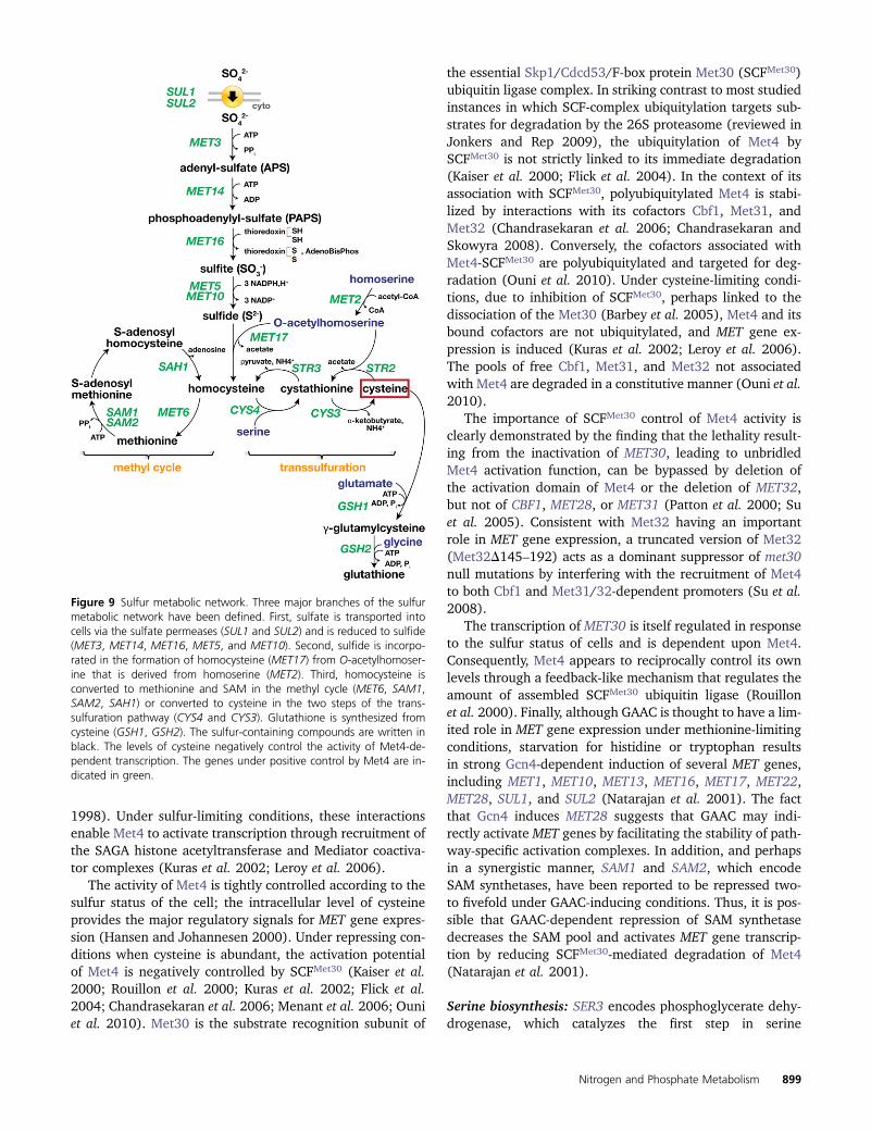
1998). Under sulfur-limiting conditions, these interactionsenable Met4 to activate transcription through recruitment ofthe SAGA histone acetyltransferase and Mediator coactiva-tor complexes (Kuras et al. 2002; Leroy et al. 2006).
The activity of Met4 is tightly controlled according to thesulfur status of the cell; the intracellular level of cysteineprovides the major regulatory signals for MET gene expres-sion (Hansen and Johannesen 2000). Under repressing con-ditions when cysteine is abundant, the activation potentialof Met4 is negatively controlled by SCFMet30 (Kaiser et al.2000; Rouillon et al. 2000; Kuras et al. 2002; Flick et al.2004; Chandrasekaran et al. 2006; Menant et al. 2006; Ouniet al. 2010). Met30 is the substrate recognition subunit of
the essential Skp1/Cdcd53/F-box protein Met30 (SCFMet30)ubiquitin ligase complex. In striking contrast to most studiedinstances in which SCF-complex ubiquitylation targets sub-strates for degradation by the 26S proteasome (reviewed inJonkers and Rep 2009), the ubiquitylation of Met4 bySCFMet30 is not strictly linked to its immediate degradation(Kaiser et al. 2000; Flick et al. 2004). In the context of itsassociation with SCFMet30, polyubiquitylated Met4 is stabi-lized by interactions with its cofactors Cbf1, Met31, andMet32 (Chandrasekaran et al. 2006; Chandrasekaran andSkowyra 2008). Conversely, the cofactors associated withMet4-SCFMet30 are polyubiquitylated and targeted for deg-radation (Ouni et al. 2010). Under cysteine-limiting condi-tions, due to inhibition of SCFMet30, perhaps linked to thedissociation of the Met30 (Barbey et al. 2005), Met4 and itsbound cofactors are not ubiquitylated, and MET gene ex-pression is induced (Kuras et al. 2002; Leroy et al. 2006).The pools of free Cbf1, Met31, and Met32 not associatedwith Met4 are degraded in a constitutive manner (Ouni et al.2010).
The importance of SCFMet30 control of Met4 activity isclearly demonstrated by the finding that the lethality result-ing from the inactivation of MET30, leading to unbridledMet4 activation function, can be bypassed by deletion ofthe activation domain of Met4 or the deletion of MET32,but not of CBF1, MET28, or MET31 (Patton et al. 2000; Suet al. 2005). Consistent with Met32 having an importantrole in MET gene expression, a truncated version of Met32(Met32D145–192) acts as a dominant suppressor of met30null mutations by interfering with the recruitment of Met4to both Cbf1 and Met31/32-dependent promoters (Su et al.2008).
The transcription of MET30 is itself regulated in responseto the sulfur status of cells and is dependent upon Met4.Consequently, Met4 appears to reciprocally control its ownlevels through a feedback-like mechanism that regulates theamount of assembled SCFMet30 ubiquitin ligase (Rouillonet al. 2000). Finally, although GAAC is thought to have a lim-ited role in MET gene expression under methionine-limitingconditions, starvation for histidine or tryptophan resultsin strong Gcn4-dependent induction of several MET genes,including MET1, MET10, MET13, MET16, MET17, MET22,MET28, SUL1, and SUL2 (Natarajan et al. 2001). The factthat Gcn4 induces MET28 suggests that GAAC may indi-rectly activate MET genes by facilitating the stability of path-way-specific activation complexes. In addition, and perhapsin a synergistic manner, SAM1 and SAM2, which encodeSAM synthetases, have been reported to be repressed two-to fivefold under GAAC-inducing conditions. Thus, it is pos-sible that GAAC-dependent repression of SAM synthetasedecreases the SAM pool and activates MET gene transcrip-tion by reducing SCFMet30-mediated degradation of Met4(Natarajan et al. 2001).
Serine biosynthesis: SER3 encodes phosphoglycerate dehy-drogenase, which catalyzes the first step in serine
Figure 9 Sulfur metabolic network. Three major branches of the sulfurmetabolic network have been defined. First, sulfate is transported intocells via the sulfate permeases (SUL1 and SUL2) and is reduced to sulfide(MET3, MET14, MET16, MET5, and MET10). Second, sulfide is incorpo-rated in the formation of homocysteine (MET17) from O-acetylhomoser-ine that is derived from homoserine (MET2). Third, homocysteine isconverted to methionine and SAM in the methyl cycle (MET6, SAM1,SAM2, SAH1) or converted to cysteine in the two steps of the trans-sulfuration pathway (CYS4 and CYS3). Glutathione is synthesized fromcysteine (GSH1, GSH2). The sulfur-containing compounds are written inblack. The levels of cysteine negatively control the activity of Met4-de-pendent transcription. The genes under positive control by Met4 are in-dicated in green.
Nitrogen and Phosphate Metabolism 899

biosynthesis from 3-phosphoglycerate (Figure 2). SER3 ex-pression is negatively regulated by serine availability bya newly discovered mechanism that involves the expressionof SRG1, a small noncoding RNA (Martens et al. 2004). Highserine levels induce transcription of SRG1, and its expressionis associated with repositioning of nucleosomes in a regionthat overlaps the SER3 promoter, which consequentlyrepresses SER3 (Figure 10) (Hainer et al. 2011). Expressionof SRG1 is activated by the well-characterized transcriptionfactor Cha4 (Martens et al. 2005), a member of the Zn(II)2Cys6 binuclear cluster family of transcriptional regu-lators (Holmberg and Schjerling 1996). In a serine-depen-dent manner, Cha4 recruits the SAGA and Swi/Snfcoactivator complexes to the SRG1 promoter, events alsorequired for SER3 repression. Importantly, Cha4 binds toUASCHA elements in the promoter of genes required for ser-ine/threonine catabolism and, in response to serine or thre-onine induction, activates their expression, e.g., CHA1encoding the catabolic serine/threonine deaminase (Holm-berg and Schjerling 1996). Taken together, these findingsdemonstrate that serine repression of SER3 transcriptionoccurs by activating SRG1 intergenic transcription. Thus,yeast uses the same transcription factor to simulta-neously activate and repress opposing pathways to reg-ulate serine biosynthesis and catabolism.
SPS-sensor signaling: extracellular amino acid-inducednitrogen source uptake
During the past 10 years, it has become clear that yeast cellspossess and use plasma membrane-localized sensing systemsto obtain information regarding concentrations of nutrients inthe extracellular environment, including the availability ofamino acids, ammonium, and glucose (reviewed in Forsbergand Ljungdahl 2001b; Zaman et al. 2008; Rubio-Texeira et al.2010). Several of these newly discovered nutrient sensorshave components that are members of protein families ofwell-characterized nutrient transporters. Interestingly, theability of these transporter homologs to transduce nutrient(ligand)-induced signals across the plasma membrane ap-pears to be independent of nutrient uptake, and thus thesesensor components apparently function analogously to tradi-tional ligand-activated receptors.
Growing yeast cells respond to the presence of micro-molar amounts of extracellular amino acids by inducingthe expression of genes required for their uptake. Thisnutrient-induced response is mediated by the SPS-sensingpathway (reviewed in Ljungdahl 2009). This pathwayderives its name from the three core components of theplasma membrane-localized SPS sensor, i.e., Ssy1, Ptr3,and Ssy5 (Forsberg and Ljungdahl 2001a). The SPS sensorregulates gene expression by controlling the activity of twotranscription factors, Stp1 and Stp2 (Figure 11A) (Andréassonand Ljungdahl 2002). These factors are synthesized as latentcytoplasmic proteins with N-terminal regulatory domainsthat function as nuclear exclusion determinants (Andréassonand Ljungdahl 2004). Upon induction by extracellular
amino acids, the SPS sensor catalyzes an endoproteolyticprocessing event that cleaves the regulatory N-terminaldomains. The shorter forms of Stp1 and Stp2 efficientlytarget to the nucleus where they bind promoters of a lim-ited set of genes, including a subset of broad-specificityamino acid permeases (cluster 1, Table 4) and the peptidetransporter Ptr2 (Didion et al. 1996, 1998; de Boer et al. 1998,2000; Iraqui et al. 1999; Klasson et al. 1999; Wielemans et al.2010; Tumusiime et al. 2011).
Ssy1 is a unique member of the amino acid permeasefamily of proteins (Table 4). Ssy1 does not catalyze measur-able amino acid uptake (Didion et al. 1998; Iraqui et al.1999; Klasson et al. 1999), but instead functions as a recep-tor of extracellular amino acids (Figure 11B) (Wu et al.2006). In addition to a core membrane transporter-likedomain composed of 12 hydrophobic membrane-spanningsegments, Ssy1 has an extended cytoplasmically orientedN-terminal domain that is not present in other amino acidpermeases. Consistent with being a receptor, Ssy1 exhibits
Figure 10 Model for the repression of SER3 by SRG1 intergenic tran-scription. In the absence of serine, the Cha4 activator is bound to theSRG1 promoter but is unable to initiate transcription. The SER3 promoteris depleted of nucleosomes allowing proteins, either an as-yet-unknownsequence-specific activator or general transcription factors, to bind andactivate SER3 transcription. In response to serine, Cha4 recruits SAGA andSwi/Snf to reposition the nucleosomes at the 59 end of SRG1 toward theSER3 promoter, permitting initiation of SRG1 transcription. These reposi-tioned nucleosomes are then disassembled ahead of the transcribing RNAPol II and reassembled after passage of RNA Pol II by the Spt6 and Spt16histone chaperones. The nucleosomes being maintained by SRG1 tran-scription occlude the SER3 promoter, preventing the binding of transcrip-tion factors and SER3 transcription. This figure and legend, orginallypublished in Pruneski and Martens (2011), are reproduced in accordancewith Landes Bioscience policy, the publishers of Cell Cycle, with permis-sion of the authors.
900 P. O. Ljungdahl and B. Daignan-Fornier

marked substrate (ligand) preferences; nonpolar aminoacids (leucine, isoleucine, methionine, phenylalanine, andtryptophan) and polar uncharged amino acids (tyrosine,threonine) are strong inducers, whereas valine, cysteine,alanine, serine, and even citrulline induce intermediatelevels, and arginine, lysine, and proline are poor inducers(Iraqui et al. 1999; Gaber et al. 2003). Ssy1 monitors theratio of external vs. internal amino acids across the plasmamembrane by undergoing transporter-like conformationalchanges between an outward-facing (signaling) and an in-
ward-facing (nonsignaling) conformation (Wu et al. 2006;Poulsen et al. 2008). Thus, in contrast to functional trans-porters, but in accordance with a receptor function, aminoacid binding to a single substrate-binding site appears toimpose a reaction barrier that inhibits the conversion from anoutward- to an inward-facing conformation. Consequently,Ssy1 signaling is sensitive to both external and internal lev-els of amino acids, and the SPS sensor induces gene expres-sion only when the levels of external amino acids are higherthan the levels of free amino acids in cytoplasmic pools. This
Figure 11 Schematic diagram of the SPS-sensing pathwayof extracellular amino acids. (A) In cells grown in the ab-sence of inducing amino acids (left), the SPS sensor ofextracellular amino acids is present in the plasma mem-brane (PM) in its preactivation conformation (Forsberg andLjungdahl 2001a), and the transcription of SPS-sensor-regulated genes, i.e., amino acid permeases (AAP), occursat basal levels, and cells exhibit low rates of amino aciduptake. The transcription factors Stp1 and Stp2 (DNA-binding motifs, green boxes) are synthesized as inactiveprecursors that localize to the cytosol due to the presenceof their N-terminal regulatory domain (anchor) that pre-vents them from efficiently entering the nucleus. Low lev-els of full-length Stp1 and Stp2 that escape cytoplasmicretention (dashed arrow, left panel) are prevented fromderepressing AAP gene expression due to activity of theAsi complex (Asi1–Asi2–Asi3) (Boban et al. 2006; Zargariet al. 2007). In the presence of extracellular amino acids(right panel), the SPS (Ssy1-Ptr3-Ssy5) sensor activates theintrinsic proteolytic activity of the Ssy5 protease, resultingin the endoproteolytic processing of Stp1 and Stp2 (scis-sors). The shorter activated forms of Stp1 and Stp2 lackingregulatory domains are targeted to the nucleus where,together with Dal81, they bind SPS-sensor-regulated pro-moters (UASaa) and induce transcription (Abdel-Sater et al.2004b; Boban and Ljungdahl 2007). The increased tran-scription of AAP genes results in increased rates of aminouptake. AAPs are cotranslationally inserted into the ERmembrane, which is contiguous with the outer nuclearmembrane. Movement of AAPs to the PM (representedby the dashed arrow, right panel) requires the ER mem-brane-localized chaperone Shr3 (Ljungdahl et al. 1992; Kotaand Ljungdahl 2005; Kota et al. 2007). (B) Transporter-based model for Ssy1 amino acid receptor function (Wuet al. 2006). Similar to canonical transporters, Ssy1 can at-tain four conformational states. However, in contrast totransporters, interconversion between the outward-facingligand bound state and the inward-facing ligand boundstate (reaction 3) is prevented by a ligand-induced reactionbarrier. The outward-facing conformations of the Ssy1 sen-sor are thought to be signaling (green), and the inward-facing conformations are nonsignaling (red). (C) Multistep
regulation of the Ssy5 endoprotease. Ssy5 is expressed as an inactive zymogen (left) composed of a prodomain that assists the folding of the Cat domain.Ssy5 is auto-processed when the Cat domain attains an active conformation. The noncovalently attached prodomain remains bound to the Cat domain,forming an inactive but catalytically competent “primed” protease complex. Primed Ssy5 is incorporated as a subcomplex of the SPS sensor via protein–protein interactions involving Ptr3, where it binds, but does not cleave, its substrates Stp1 and Stp2. In the absence of extracellular amino acids, i.e., undernon-inducing conditions (left), the basal level of phosphorylation of a phosphodegron in the prodomain is likely to be determined by counteracting activitiesof casein kinase I (Yck1 and Yck2) and the phosphatase PP2A with its regulatory subunit Rts1 (Eckert-Boulet et al. 2006). In the presence of extracellularamino acids (right), the primary amino acid sensor Ssy1 is stabilized in a conformation that triggers intracellular signaling. This conformation increases thelevel of phosphorylation of the prodomain phosphodegron (Omnus et al. 2011), presumably by increasing the accessibility of Yck1 or Yck2. An increasedlevel of phosphorylation within the degron provides the requisite surface recognized by the SCFGrr1 complex and subsequent polyubiquitylation of lysineresidues of the degron. Concomitant with being ubiquitylated, the prodomain is directly targeted for degradation by the 26S proteasome, unfettering theStp1 and Stp2 processing activity of the Cat domain.
Nitrogen and Phosphate Metabolism 901
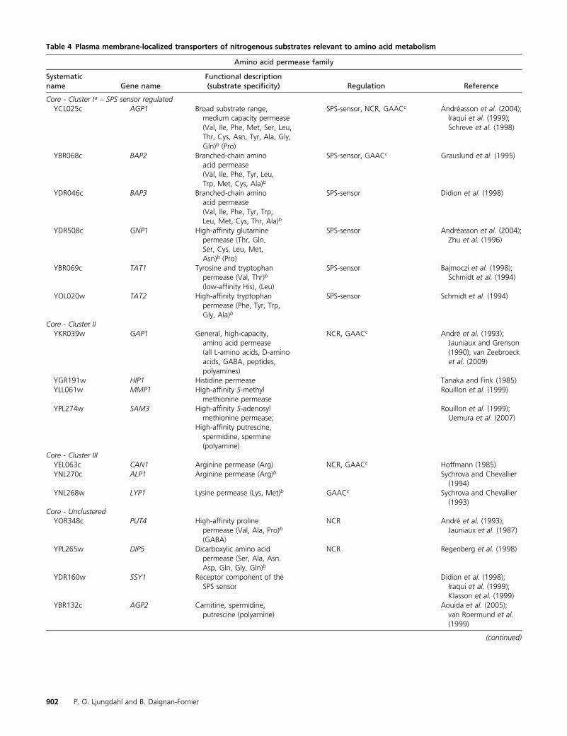
Table 4 Plasma membrane-localized transporters of nitrogenous substrates relevant to amino acid metabolism
Amino acid permease family
Systematicname Gene name
Functional description(substrate specificity) Regulation Reference
Core - Cluster Ia – SPS sensor regulatedYCL025c AGP1 Broad substrate range,
medium capacity permease(Val, Ile, Phe, Met, Ser, Leu,Thr, Cys, Asn, Tyr, Ala, Gly,Gln)b (Pro)
SPS-sensor, NCR, GAACc Andréasson et al. (2004);Iraqui et al. (1999);Schreve et al. (1998)
YBR068c BAP2 Branched-chain aminoacid permease(Val, Ile, Phe, Tyr, Leu,Trp, Met, Cys, Ala)b
SPS-sensor, GAACc Grauslund et al. (1995)
YDR046c BAP3 Branched-chain aminoacid permease(Val, Ile, Phe, Tyr, Trp,Leu, Met, Cys, Thr, Ala)b
SPS-sensor Didion et al. (1998)
YDR508c GNP1 High-affinity glutaminepermease (Thr, Gln,Ser, Cys, Leu, Met,Asn)b (Pro)
SPS-sensor Andréasson et al. (2004);Zhu et al. (1996)
YBR069c TAT1 Tyrosine and tryptophanpermease (Val, Thr)b
(low-affinity His), (Leu)
SPS-sensor Bajmoczi et al. (1998);Schmidt et al. (1994)
YOL020w TAT2 High-affinity tryptophanpermease (Phe, Tyr, Trp,Gly, Ala)b
SPS-sensor Schmidt et al. (1994)
Core - Cluster IIYKR039w GAP1 General, high-capacity,
amino acid permease(all L-amino acids, D-aminoacids, GABA, peptides,polyamines)
NCR, GAACc André et al. (1993);Jauniaux and Grenson(1990); van Zeebroecket al. (2009)
YGR191w HIP1 Histidine permease Tanaka and Fink (1985)YLL061w MMP1 High-affinity S-methyl
methionine permeaseRouillon et al. (1999)
YPL274w SAM3 High-affinity S-adenosylmethionine permease;
High-affinity putrescine,spermidine, spermine(polyamine)
Rouillon et al. (1999);Uemura et al. (2007)
Core - Cluster IIIYEL063c CAN1 Arginine permease (Arg) NCR, GAACc Hoffmann (1985)YNL270c ALP1 Arginine permease (Arg)b Sychrova and Chevallier
(1994)YNL268w LYP1 Lysine permease (Lys, Met)b GAACc Sychrova and Chevallier
(1993)Core - Unclustered
YOR348c PUT4 High-affinity prolinepermease (Val, Ala, Pro)b
(GABA)
NCR André et al. (1993);Jauniaux et al. (1987)
YPL265w DIP5 Dicarboxylic amino acidpermease (Ser, Ala, Asn.Asp, Gln, Gly, Gln)b
NCR Regenberg et al. (1998)
YDR160w SSY1 Receptor component of theSPS sensor
Didion et al. (1998);Iraqui et al. (1999);Klasson et al. (1999)
YBR132c AGP2 Carnitine, spermidine,putrescine (polyamine)
Aouida et al. (2005);van Roermund et al.(1999)
(continued)
902 P. O. Ljungdahl and B. Daignan-Fornier

Table 4, continued
Amino acid permease family
Systematicname Gene name
Functional description(substrate specificity) Regulation Reference
YFL055w AGP3 Broad-substrate specificityamino acid permease(Asp, Glu, Ser)b (Met)
Menant et al. (2006);Regenberg et al. (1999)
Non-core - MUP ClusterYGR055w MUP1 High-affinity methionine
permease (Cys)Isnard et al. (1996);Kosugi et al. (2001)
YHL036w MUP3 Low-affinity methioninepermease; inhibited bybroad-substratespectrum (Met)
GAACc Isnard et al. (1996)
Non-core - UGA ClusterYDL210w UGA4 GABA permease
(d-aminolevulinic acid,putrescine) vacuolelocalization
NCR André et al. (1993);Uemura et al. (2004)
YKL174c TPO5 Polyamine secretion, Golgilocalization
Tachihara et al. (2005)
YGL077c HNM1 Choline permease(ethanolamine)
INO regulon Nikawa et al. (1986)
YNR056c BIO5 Biotin permease(7-keto 8-aminopelargonicacid)
Phalip et al. (1999)
Ammonium permease family
YGR121C MEP1 Medium-affinity,high-capacityammonium permease
NCR Marini et al. (1994)
YNL142W MEP2 High-affinity,low-capacity ammoniumpermease, ammoniumsensor
NCR Lorenz and Heitman (1998);Marini et al. (1997)
YPR138C MEP3 Low-affinity, high-capacityammonium permease
NCR Marini et al. (1997)
Other transporters/permeases
DAL5 ClusterYJR152w DAL5 Allantonate permease
(ureidosuccinate/dipeptide)NCR Cai et al. (2007);
Rai et al. (1987)YLL055w YCT1 Cysteine transporter Kaur and Bachhawat (2007)YGR260w TNA1 Nicotinic acid transporter Llorente and Dujon (2000)YGR065c VHT1 Biotin transporter GAACc Stolz et al. (1999)YCR028c FEN2 Panthothenate transporter Stolz and Sauer (1999)YAL067c SEO1 Not determined – sulfur
compound?Isnard et al. (1996)
FUR4 ClusterYBR021w FUR4 Uracil permease Jund et al. (1988)YIR028w DAL4 Allantoin permease (uracil) NCR Yoo et al. (1992)YBL042c FUI1 Uridine permease Wagner et al. (1998)YLR237w THI7 Thiamine permease Thi3 Enjo et al. (1997)YOR192c THI72 Thiamine permease Enjo et al. (1997)YOR071c NRT1 Nicotinamide riboside
(high-affinity),thiamine (low-affinity)
Belenky et al. (2008)
FCY2 ClusterYER056c FCY2 Nucleobase permease
(cytidine, cytosine,purine, adenine, guanine,hypoxanthine)
Weber et al. (1990)
(continued)
Nitrogen and Phosphate Metabolism 903

model elegantly accounts for the accumulated experimentaldata and also provides a framework for understanding thetransceptor concept as applied to active nutrient carriersthat combine nutrient transporter and receptor functions,including Gap1 and Pho84 (Thevelein and Voordeckers2009; van Zeebroeck et al. 2009; Popova et al. 2010;Rubio-Texeira et al. 2010).
The extended N-terminal domain of Ssy1 functions asa scaffold for binding of Ptr3 and Ssy5 (Bernard and André2001; Forsberg and Ljungdahl 2001b; Liu et al. 2008). Ssy5exhibits homology to chymotrypsin-like serine proteasesand is expressed as a zymogen (Abdel-Sater et al. 2004a;Andréasson et al. 2006; Poulsen et al. 2006). After trans-lation, and concomitantly with folding to achieve catalyticcompetence, Ssy5 cleaves itself into an N-terminal prodo-main and a C-terminal catalytic (Cat) domain. The prodo-main remains noncovalently attached with the Cat domain,forming a primed, but inactive, protease subcomplex withinthe SPS sensor that associates with Stp1 and Stp2 (Andréassonet al. 2006). The prodomain functions as the inhibitory subunitof the SPS sensor, and its amino acid-induced degradation viathe 26S proteasome correlates with the signal-propagating,endoproteolytic processing of Stp1 and Stp2 (Pfirrmann et al.2010; Omnus et al. 2011).
Consistent with 26S proteasome involvement, aminoacid-induced Stp1 and Stp2 processing requires the SCFGrr1
ubiquitin ligase complex (Abdel-Sater et al. 2004a; Andréassonand Ljungdahl 2004; Spielewoy et al. 2004; Liu et al. 2008).SCF complexes achieve specificity through association withexchangeable F-box proteins (Jonkers and Rep 2009). Grr1is one of the best-characterized F-box proteins and is re-quired for Ssy5 prodomain downregulation and Cat domainactivation (Andréasson et al. 2006). Also, casein kinaseI-dependent phosphorylation (either Yck1 or Yck2) is re-quired for SPS-sensor signaling (Abdel-Sater et al. 2004a;Spielewoy et al. 2004; Liu et al. 2008). It has been shownthat Yck1/2-dependent Ssy5 prodomain phosphorylation pre-cedes SCFGrr1-dependent ubiquitylation (Abdel-Sater et al.2011). In other signaling pathways, Yck1/2-catalyzed phos-phorylation has been shown to lead to Grr1-dependent poly-ubiquitylation and subsequent degradation of the modifiedsubstrates by the 26S proteasome (Moriya and Johnston2004; Spielewoy et al. 2004). Hence, the SPS sensor coordi-nates the activity of general signaling components to achievea highly specific output (Omnus et al. 2011) (Figure 11C).
The constitutive nuclear factor Dal81 has an importantand synergistic role in amplifying the induced expression ofSPS-sensor genes (Boban and Ljungdahl 2007). Dal81 isalso required for several other well-characterized nitrogensource utilization pathways, including urea, allantoin, andGABA. Dal81 functions together with an inducer-specifictranscription factor, i.e., Uga3 (Jacobs et al. 1981; Turoscy
Table 4, continued
Other transporters/permeases
Systematicname Gene name
Functional description(substrate specificity) Regulation Reference
YGL186c TPN1 Pyridoxine transporter(vitamin B6)
GAACc Stolz and Vielreicher 2003)
UnclusteredYHL016c DUR3 High-affinity polyamine
(urea)NCR El Berry et al. (1993);
Uemura et al. (2007)YKR093w PTR2 Peptide transporter
(di-/tripeptides)SPS-sensor, Cup9, NCR Hauser et al. (2001);
Island et al. (1991)YJL212c OPT1 Oligopeptide transporter
(glutathione, phytochelatin)NCR Hauser et al. (2001)
YPR194c OPT2 Homologous to OPT1(role as transporteruncertain;inactivation leads tofragmented vacuoles)
NCR Aouida et al. (2009)
Transporters involved with excretion of nitrogenous compoundsYDR384c ACO3 Ammonium transport
outwardSPS-sensor, GAAC Guaragnella and Butow
(2003);Palkova et al. (2002)
YNL065w AQR1 H+-antiporter, localized tomultiple intracellularmembranes/vesicles(secretion of homoserine,Thr, Ala, Asp, Glu)
Velasco et al. (2004)
YLL028w TPO1 Polyamine transporter Tomitori et al. (1999);Uemura et al. (2005)
a Clustered based onon the basis of sequence homology according to Nelissen et al. (1997).b Substrate specificity as reported in Regenberg et al. (1999).c GAAC-regulated expression according to Natarajan et al. (2001).
904 P. O. Ljungdahl and B. Daignan-Fornier

and Cooper 1982; Coornaert et al. 1991), Dal82 (Visserset al. 1990), or Stp1 and Stp2 (Iraqui et al. 1999; Abdel-Sater et al. 2004b; Boban and Ljungdahl 2007) to activatetarget genes. The fact that these pathways share a commoncoactivator enables cells to integrate the signals derivedfrom these different nitrogen sources due to competitivebinding of the specific factors to Dal81 (Abdel-Sater et al.2004b). A distinct hierarchy exists, and the induced target-ing of activated forms of Stp1 and Stp2 overrides allantoate-and GABA-induced signals, enabling cells to take advantageof the availability of amino acids as preferred nitrogen sour-ces. Integration appears to be achieved by the preferentialrecruitment of Dal81 to SPS-sensor-regulated promoters.Accordingly, in an SPS-sensor-dependent manner, the pres-ence of extracellular amino acids impair Dal82-dependentexpression of DAL5, encoding allantoate/ureidosuccinatepermease (Cai et al. 2007), and Uga3-dependent gene ex-pression of UGA4, the GABA permease (Cardillo et al. 2010).
Overlapping transcriptional regulatory networks contrib-ute to the expression of several SPS-sensor-controlled genes.The promoter region of AGP1 encoding a broad-specificityamino acid permease is under NCR control; however, its ex-pression is strictly dependent on inducing signals mediated bythe SPS sensor (Iraqui et al. 1999; Abdel-Sater et al. 2004b;Wielemans et al. 2010). Stp1 binding to UASaa motifs in thepromoters of SPS-sensor-regulated genes is insensitive to thenitrogen status of the cell and to factors controlling NCR, i.e.,Gln3, Ure2, and Gzf3 (Godard et al. 2007). In cells grownwith the preferred nitrogen source ammonium, the additionof amino acids leads to the rapid induction of AGP1 expres-sion. In cells lacking a functional SPS-sensing pathway, AGP1expression is not induced, and the PAGP1 promoter is unre-sponsive to mutations that inactivate Ure2. Conversely, aminoacid-induced expression of AGP1 is greatly augmented in cellsgrown in the presence of nonpreferred nitrogen sources(Abdel-Sater et al. 2004b; Godard et al. 2007). Thus, in con-trast to other permease genes responsive to NCR control (e.g.,GAP1, DAL5, and MEP2), Gln3 is not able to activate AGP1expression in the absence of synergistic signals initiated bythe presence of extracellular amino acids that are transducedvia the SPS-sensing pathway.
The analysis of dipeptide uptake in yeast also providesa clear demonstration of how SPS-sensor-induced signalsare integrated with other nutritionally regulated activities.Amino acid-induced and SPS-sensor-mediated activation ofdipeptide transporter PTR2 expression is a requisite for di-peptide uptake (Island et al. 1987; Barnes et al. 1998;Hauser et al. 2001); however, full induction of peptideuptake requires an additional activation step (Byrd et al.1998; Turner et al. 2000; Hwang and Varshavsky 2008;Xia et al. 2008). Peptides entering the cell that containN-terminal amino acids recognized according to the N-endrule as destabilizing (Dohmen et al. 1994; Varshavsky2008), allosterically activate Ubr1, an E3 ubiquitin ligase,which in turn accelerates degradation of the transcriptionalrepressor Cup9. A decreased level of Cup9 gives rise to fully
derepressed PTR2 expression and dipeptide transport. Al-though Ubr1 and Cup9 are absolutely required for fullinduction of PTR2 transcription, they do not affect PAGP1-lacZ (Bernard and André 2001) or BAP2 (Alagramam et al.1995) expression.
Membrane transporter systems and compartmentalization
The compartmentalized eukaryotic cell architecture is acomplicating factor in evaluating the flow of metabolitesassociated with amino acid and nucleotide biosynthesis. Tofully understand metabolic regulation, the dynamic distri-bution of compartmentalized metabolites will eventuallyhave to be taken into account. Although this was alreadypointed out by Cooper (1982b) and Jones and Fink (1982),it remains a substantial challenge. Great progress has beenmade in understanding how metabolites are transported in-to cells and into or out of organelles. Here full-genome se-quence data have contributed greatly to the identification offamilies of transport proteins (Saier 2000; Brohee et al.2010). Subsequent cell biological analyses have establishedthe intracellular localization of these transporters, and inmany cases purification and reconstitution experimentshave provided precise mechanistic understanding of theirfunction.
Membrane transport systems of nitrogenous compoundsrelevant to amino acid metabolism are schematicallydepicted in Figure 12. Transporters and permeases thatfunction at the plasma membrane and primarily facilitatemetabolite uptake into cells are listed in Table 4. Transportacross the plasma membrane is facilitated via H+-symportenergized by the plasma membrane H+-ATPase Pma1(Serrano et al. 1986; Horák 1997). A fundamental themethat has emerged from the functional analysis of plasmamembrane transport is that, in many instances, individualsubstrates are transported by several different systems,which, although displaying different kinetic specificities,function redundantly (Regenberg et al. 1999). The expressionof redundant systems with differing catalytic properties allowsyeast cells to extract necessary nutrients from a great varietyof environments.
Transport across the inner mitochondrial membrane iscatalyzed by the mitochondrial carrier protein (MCP)family; a selected subset of MCPs that are intimately linkedto amino acid metabolism is listed in Table 5 (Palmieri et al.2006). The MCPs exhibit a variety of transport mechanisms,including uniport, symport, and antiport, and the transportcan occur in an electroneutral, proton-mediated, or electro-phoretic manner. In the case of bidirectional antiport exchange,the direction of transport is determined by the relative con-centrations of substrates in the cytoplasmic and mitochondrialsubstrate.
The vacuole is an organelle with a well-established role inamino acid homeostasis (Matile and Wiemken 1967; Cooper1982a; Kitamoto et al. 1988; Klionsky et al. 1990; Jacquemin-Faure et al. 1994; Sekito et al. 2008). The vacuolar transportsystems are listed in Table 6 (Sekito et al. 2008). Metabolite
Nitrogen and Phosphate Metabolism 905
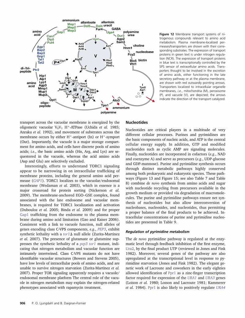
transport across the vacuolar membrane is energized by theoligomeric vacuolar V0V1 H+-ATPase (Uchida et al. 1985;Anraku et al. 1992), and movement of substrates across themembrane occurs by either H+-antiport (In) or H+-symport(Out). Importantly, the vacuole is a major storage compart-ment for amino acids, and cells have discrete pools of aminoacids; i.e., the basic amino acids (His, Arg, and Lys) are se-questered in the vacuole, whereas the acid amino acids(Asp and Glu) are selectively excluded.
Interestingly, efforts to understand TORC1 signalingappear to be narrowing in on intracellular trafficking ofmembrane proteins, including the general amino acid per-mease (GAP1). TORC1 localizes to the vacuolar/endosomalmembrane (Wedaman et al. 2003), which in essence is amajor crossroad for protein sorting (Nickerson et al.2009). The membrane-anchored EGO–GSE complex, foundassociated with the late endosome and vacuolar mem-branes, is required for TORC1 localization and activation(Dubouloz et al. 2005; Binda et al. 2009) and for properGap1 trafficking from the endosome to the plasma mem-brane during amino acid limitation (Gao and Kaiser 2006).Consistent with a link to vacuole function, null alleles ofgenes encoding class C-VPS components, e.g., PEP3, exhibitsynthetic lethality with a tor1D null allele (Zurita-Martinezet al. 2007). The presence of glutamate or glutamine sup-presses the synthetic lethality of a pep3 tor1 mutant, indi-cating that nitrogen metabolism and vacuolar function areintimately intertwined. Class C-VPS mutants do not haveidentifiable vacuolar structures (Bowers and Stevens 2005),have low levels of intracellular pools of amino acids, and areunable to survive nitrogen starvation (Zurita-Martinez et al.2007). Proper TOR signaling apparently requires a vacuole/endosomal membrane platform The central role of the vacu-ole in nitrogen metabolism may explain the nitrogen-relatedphenotypes associated with rapamycin treatment.
Nucleotides
Nucleotides are critical players in a multitude of verydifferent cellular processes. Purines and pyrimidines arethe basic components of nucleic acids, and ATP is the centralcellular energy supply. In addition, GTP and modifiednucleotides such as cyclic AMP are signaling molecules.Finally, nucleotides are incorporated in cofactors (e.g., NADand coenzyme A) and serve as precursors (e.g., UDP-glucoseand GDP-mannose). Purine and pyrimidine synthesis occursthrough distinct metabolic pathways highly conservedamong both prokaryotic and eukaryotic species. These path-ways (Figure 13 and Figure 15; see also Table 7 and Table8) combine de novo synthesis from amino acids and sugarwith nucleotide recycling from precursors available in thegrowth medium or provided via degradation of macromole-cules. The purine and pyrimidine pathways ensure net syn-thesis of nucleotides but also allow interconversion ofnucleobases, nucleosides, and nucleotides, thus permittinga proper balance of the final products to be achieved. In-tracellular concentrations of purine and pyrimidine nucleo-tides are presented in Table 9.
Regulation of pyrimidine metabolism
The de novo pyrimidine pathway is regulated at the enzy-matic level through feedback inhibition of the first enzyme,Ura2, by the final product UTP (reviewed in Jones and Fink1982). Moreover, several genes of the pathway are alsoupregulated at the transcriptional level in response to py-rimidine starvation (Jones and Fink 1982). The elegant ge-netic work of Lacroute and coworkers in the early eightiesallowed identification of Ppr1 as a zinc-finger transcriptionfactor required for expression of the URA1 and URA3 genes(Loison et al. 1980; Losson and Lacroute 1981; Kammereret al. 1984). Ppr1 is also likely to positively regulate URA4
Figure 12 Membrane transport systems of ni-trogenous compounds relevant to amino acidmetabolism. Plasma membrane-localized per-meases/transporters are shown with their corre-sponding substrates. The expression of transportproteins in green text is under nitrogen regula-tion (NCR). The expression of transport proteinsin blue text is transcriptionally controlled by theSPS sensor of extracellular amino acids. Trans-porters thought to be involved in the excretionof amino acids, either functioning in the latesecretory pathway or at the plasma membrane,are shown with red outwardly pointing arrows.Transporters localized to intracellular organellemembranes, i.e., mitochondria (M), peroxisome(P), and vacuole (V), are depicted; the arrowsindicate the direction of the transport catalyzed.
906 P. O. Ljungdahl and B. Daignan-Fornier

and URA10 on the basis of data from in vitro binding topromoter sequences (Roy et al. 1990; Roy 1992). However,the role of Ppr1 in transcription has not been evaluatedgenome-wide, which could reveal interesting cross-pathwayregulation.
The early observation that yeast mutants accumulatingorotic acid (OA) and dihydro-orotate (DHO) show increasedDHO dehydrogenase (Ura1) and orotidine 59-monophosphatedecarboxylase (Ura3) activity (Lacroute 1968) suggestedthat these metabolic intermediates could play a direct rolein the pyrimidine pathway regulation. In this model, a spe-cific pathway intermediate functions as a cofactor for thetranscriptional activator, and the abundance of the metabo-lite is downregulated by feedback inhibition (Figure 8). Thisassumption was tested directly using purified Ppr1 in vitro(Flynn and Reece 1999), where addition of either DHO orOA to the reaction stimulated Ppr1-dependent transcription(Flynn and Reece 1999). This reconstitution experimentestablished that Ppr1 directly senses the levels of pyrimidinebiosynthesis intermediates and modulates pyrimidine en-zyme synthesis in response to the flux in the pathway. In-terestingly, it was also found that efficient binding of Ppr1 toDNA required a yet-unidentified small molecule (Flynn andReece 1999). Identification of this molecule in the futurecould uncover new aspects of pyrimidine biosynthesisregulation.
Transcription of URA2, encoding the first committed stepof UTP synthesis, is also upregulated in response to pyrim-idine depletion (Exinger and Lacroute 1992; Kwapisz et al.2008). However, expression of URA2 is not Ppr1 dependent(Losson and Lacroute 1981; Kwapisz et al. 2008), but isregulated by a novel mechanism recently described. Thismechanism involves alternative transcription start sites thatlead to the synthesis of small “cryptic unstable transcripts”(CUTs) (Thiebaut et al. 2008) but also to a downstreamT-rich region (Kwapisz et al. 2008). “Nonproductive” tran-scription from the upstream sites downregulates expressionof the URA2 mRNA while uracil deprivation activates selec-tion of the functional URA2 mRNA start site by a mechanismthat does not involve lower expression of the CUTs (Thiebautet al. 2008) (Figure 14). Interestingly, mechanisms involvingnoncoding RNAs appear to regulate other nucleotide genessuch as URA8, IMD2, and ADE12 (Davis and Ares 2006;Steinmetz et al. 2006; Kuehner and Brow 2008; Kwapiszet al. 2008; Thiebaut et al. 2008). Strikingly, these fourgenes encode the first committed steps of UTP, CTP, GTP,and ATP synthesis (see Figures 13 and 15). Although notfully understood at the molecular level, these regulatoryprocesses apparently involve direct sensing of nucleotideconcentration by the transcription machinery and thus effi-ciently connect individual nucleotide synthesis to its actualavailability.
Table 5 Mitochondrial carrier family: transporters directly coupled to amino acid metabolism
Systematic name Gene name Description (substrate specificity) Transport in/out organelle Reference
YPR021c AGC1a Aspartate-glutamatetransport (Asp, Glu)
aspartate / out, in ) glutamate Cavero et al. (2003)
YPL134c ODC1 Lysine and glutamate biosynthesis/lysinecatabolism/nitrogen assimilation(a-ketoglutarate, a-ketoadipate)
a-ketoadipate / out,in ) a-ketoglutarate
Palmieri et al. (2001)
YOR222w ODC2a Lysine and glutamate biosynthesis/lysinecatabolism/nitrogen assimilation(a-ketoglutarate, a-ketoadipate)
a-ketoadipate / out,in ) a-ketoglutarate
Palmieri et al. (2001)
YOR130c ORT1a
(ARG11)Arginine biosynthesis (ornithine) ornithine / out, in ) H+ Crabeel et al. (1996);
Palmieri et al. (1997);Soetens et al. (1998)
YBR291c CTP1 Lysine biosynthesis - Lys14 regulated;suggested role in peroxisome membrane(citrate, malate)
citrate / out, in ) malate Breitling et al. (2002);Kaplan et al. (1995)
YKL120w OAC1a Leucine biosynthesis(oxalacetate, a-isopropyl malate)
a-isopropylmalate /out, in ) oxalacetate
Marobbio et al. (2008)
YBL089w YMH2 Arginine biosynthesis (citrate,a-ketoglutarate)
citrate / out, in / a-ketoglutarate Castegna et al. (2010)
YHR002w LEU5 Leucine biosynthesis - CoA transport(cofactor A)
in Prohl et al. (2001)
YPR058w YMC1a Lysine and glutamate biosynthesis(a-ketoglutarate)
n.d. Trotter et al. (2005)
YBR104w YMC2a Lysine and glutamate biosynthesis(a-ketoglutarate)
n.d. Trotter et al. (2005)
YNL003c SAM5(PET8)
Methionine biosynthesis(S-adenosylmethionine (SAM),S-adenosylhomocysteine (SAHC))
In ) SAM, SAHC / out Marobbio et al. (2003)
ThPP = Thiamine pyrophosphateTMP = Thiamine monophosphate
a GAAC-regulated expression (Natarajan et al. 2001)
Nitrogen and Phosphate Metabolism 907

Regulation of the purine de novo synthesis pathway
The transcription factors responsible for activation of purinede novo pathway genes were identified while studying thehistidine pathway regulation. The pioneering work by Finkand coworkers on the transcriptional regulation of the HIS4gene revealed the important role of the Myb-related Bas1and homeodomain Bas2 transcription factors (Arndt et al.1987; Tice-Baldwin et al. 1989). Knockout of these geneswas found to result in adenine bradytrophy, suggesting thatthey could also regulate purine biosynthesis genes. This hy-pothesis was correct, and Bas1 and Bas2 were found to berequired for expression of several ADE genes in vivo and tobind to their promoter in vitro (Daignan-Fornier and Fink1992). On the basis of proteome, transcriptome, and gene-by-gene analysis, it was found that Bas1 and Bas2 activatethe expression of all 10 AMP-biosynthesis genes exceptADE16 (Daignan-Fornier and Fink 1992; Denis et al. 1998)and bind to a specific promoter region of these genes(Daignan-Fornier and Fink 1992; Rolfes et al. 1997; Pinsonet al. 1998). Furthermore, Bas1/Bas2 also mediate adenine-repressible transcriptional activation of genes of other path-ways metabolically connected to the purine pathway, suchas histidine (HIS1, HIS4, and HIS7), glutamine (GLN1), orone-carbon-unit (SHM2, MTD1) metabolism genes (Figure15) (Arndt et al. 1987; Springer et al. 1996; Denis andDaignan-Fornier 1998). Bas1 binding to the promoter regionof most of these genes has been confirmed in vivo by ChIP-CHIP analysis (Mieczkowski et al. 2006). Importantly, Bas2is also known as Pho2, a major regulator of phosphateutilization in yeast (see Purine phosphate connection: moresignal molecules) (Arndt et al. 1987). In general, both Bas1and Pho2 are required for transcriptional activation al-
though one-carbon-unit metabolism genes are much moredependent on Bas1 than on Bas2/Pho2 (Denis and Daignan-Fornier 1998; Subramanian et al. 2005).
All the genes activated by Bas1 and Bas2/Pho2 alsorespond to extracellular adenine, their expression beinglow when adenine is abundant in the growth medium(Tice-Baldwin et al. 1989; Daignan-Fornier and Fink 1992;Springer et al. 1996; Denis et al. 1998; Denis and Daignan-Fornier 1998). To get an insight into the molecular mecha-nisms linking adenine availability to transcriptional activa-tion, Rolfes et al. (1997) used chimeras between LexA andeither Bas1 or Pho2. They found that lexA-Pho2 couldactivate transcription independently of Bas1 and in an ade-nine-independent way, while lexA-Bas1 activation was bothPho2 dependent and adenine responsive (Zhang et al.1997). They proposed that adenine limitation favors forma-tion of a complex between Bas1 and Pho2, thus unmaskingthe Bas1 activation domain (Zhang et al. 1997). Consis-tently, a Bas1–Pho2 fusion chimera activated expressionof the ADE genes in an adenine-independent way (Pinsonet al. 2000). Further analyses using Bas1 and Pho2 fused tothe transcription activation domain of VP16 suggested thatBas1 binds to the ADE gene promoters and recruits Pho2(Pinson et al. 2000). This was further demonstrated bychromatin immunoprecipitation (ChIP) analysis (Som et al.2005).
How does adenine limitation stimulate Bas1–Pho2 inter-action? A genetic analysis of mutants unresponsive to ade-nine has shown that adenine needs to be taken up andmetabolized to ADP to exert its regulatory role (Guetsovaet al. 1997; Rébora et al. 2001). This is consistent with theobservation that extracellular hypoxanthine downregulatesexpression of an ADE1-lacZ fusion while guanine has little
Table 6 Vacuole-localized amino acid transport proteins
Systematic nameGenename Substrate specificity
Transport in/outof organelle Reference
AVT subfamilyYJR001w AVT1a Ile, Leu, Asn, Gln, Tyr In Russnak et al. (2001)YEL064c AVT2 Not known, may localize to ER — Russnak et al. (2001)YKL146w AVT3b Ile, Leu, Asn, Gln, Tyr Out Russnak et al. (2001); Yang et al. (2006)YNL101w AVT4a,b Ile, Leu, Asn, Gln, Tyr Out Russnak et al. (2001); Yang et al. (2006)YBL089w AVT5 Not known — Russnak et al. (2001)YER119c AVT6b Asp, Glu Out Chahomchuen et al. (2009);
Russnak et al. (2001)YIL088c AVT7a Not known — Russnak et al. (2001)
VBA subfamilyYMR088c VBA1a His, Lys In Shimazu et al. (2005)YBR293w VBA2 Arg, His, Lys, Tyr In Shimazu et al. (2005)YCL069w VBA3 His, Lys In Shimazu et al. (2005)YDR119w VBA4 Not known — Shimazu et al. (2005)YKR105c VBA5 Not known — Shimazu et al. (2005)
Other transport proteinsYCL038c ATG22 Ile, Leu, Tyr Out Sychrova and Chevallier (1994);
Yang et al. (2006)YEL063c UGA4 GABA, putrescine In Uemura et al. (2004)YNL268w ERS1 Cys Out Gao et al. (2005)
a NCR-sensitive expression.b Required for viability upon nitrogen starvation.
908 P. O. Ljungdahl and B. Daignan-Fornier

effect (Guetsova et al. 1997) because hypoxanthine can beconverted to adenylic nucleotides while guanine cannot(Figure 15). Strikingly, most of the deregulated mutantsaffected the ADE13 gene encoding adenylosuccinate lyase,suggesting a key role for this step of the pathway (Figure15) (Rébora et al. 2001). Further genetic analyses revealedthat AICAR (59-phosphoribosyl-5-amino-4-imidazole carbox-amide) and SAICAR (succinyl-AICAR), the metabolic in-termediates just downstream and upstream of Ade13,respectively, play a pivotal role by promoting interactionsbetween Bas1 and Pho2 (Rébora et al. 2001, 2005). Severallines of evidence supported this idea. First, a direct correla-tion was found between intracellular AICAR concentrations,measured by HPLC, and ADE gene expression (Pinson et al.2009). Second, a Bas1–Pho2 fusion makes expression of theADE genes (S)AICAR independent (Rébora et al. 2001;Pinson et al. 2009). Finally, two-hybrid studies revealed thatthe Bas1–Pho2 interaction is stimulated under conditionswhere (S)AICAR accumulates (Rébora et al. 2001; Pinsonet al. 2009), and consistently, these conditions stimulateBas1-dependent recruitment of Pho2 to the ADE5, 7 andADE17 promoters, as determined by ChIP (Pinson et al.2009). Affinity chromatography revealed that AICAR bindsPho2 but not Bas1 in vitro strongly, suggesting that Pho2could be an AICAR sensor through direct interaction withthe small molecule (Pinson et al. 2009). Importantly AICAR,but not SAICAR, also affects interaction of Pho2 with an-
other transcription factor, Pho4, and thus modulates expres-sion of phosphate utilization genes (see Purine phosphateconnection: more signal molecules and Figure 8) (Pinsonet al. 2009). Together, these results suggest that somehowAICAR binding to Pho2 potentiates this transcription factorby stimulating the interaction of Pho2 with its partners.
The link between exogenous adenine and (S)AICARsynthesis is thought to occur through enzymatic regulationof the first step of the pathway, catalyzed by Ade4, which isdownregulated by ADP and ATP in vitro (Rébora et al.2001). Indeed, the intracellular concentration of ADP andATP is clearly higher in adenine-replete conditions (Gauthieret al. 2008), while the (S)AICAR concentration decreases(Hurlimann et al. 2011). Thus, both ATP, the final productof the pathway, and a metabolic intermediate, (S)AICAR,are required for proper transcriptional regulation of adenylicnucleotides. Although the mechanisms are different, thegeneral scheme is very close to that of the pyrimidine path-way, where UTP feedback inhibits Ura2, the first enzyme ofthe pathway, thus modulating the synthesis of orotate anddihydroorotate, two metabolic intermediates that directlystimulate the transcription factor Ppr1 (see Regulation ofpyrimidine metabolism). Dual-sensing mechanisms are wellsuited for regulation of nonlinear pathways, such as thepurine de novo pathway, which can be fed on the side bythe histidine pathway and which branches to allow ATP andGTP synthesis (Figure 15).
Regulation of GTP synthesis
Expression of genes required for GTP synthesis is notcoregulated with that of genes for AMP synthesis. The IMDgenes and GUA1 do not respond to adenine as ADE genes do(Escobar-Henriques and Daignan-Fornier 2001). In fact,IMD2/3/4 genes are strongly downregulated when guanineis added to the growth medium, while GUA1 expression isunaffected (Escobar-Henriques and Daignan-Fornier 2001).This effect of guanine is abolished in the fcy2, hpt1, andguk1 guanine utilization mutants, indicating that guaninemetabolism is required for GTP to exert its regulatory role(Escobar-Henriques and Daignan-Fornier 2001). In contrast,inhibitors of IMPDH activity, such as mycophenolic acid(MPA) or 6-azauracil (6AU) that lower GTP pools (Exingerand Lacroute 1992), strongly induce IMDs genes (Shaw andReines 2000; Escobar-Henriques and Daignan-Fornier 2001;Saint-Marc et al. 2009).
An analysis of the IMD2 promoter, aimed at decipheringthe molecular mechanisms leading to transcriptional regu-lation, revealed several unusual features. First, a regulatoryelement lying .200 bp upstream of the transcription start,carries a typical TATA box motif and is required for bindingof the TATA-box-binding protein on the IMD2 promoter(Escobar-Henriques and Daignan-Fornier 2001; Escobar-Henriques et al. 2003a). Deletion of this TATA box motifresulted in constitutive expression of IMD2 mRNA at a levelthat was intermediary between fully repressed and fullyinduced levels (Shaw et al. 2001; Escobar-Henriques et al.
Figure 13 Pyrimidine synthesis and salvage pathways. DHO, dihydro-orotate; OA, orotic acid; USA, ureidosuccinic acid. Gene names are itali-cized. Regulatory molecules are shown in red.
Nitrogen and Phosphate Metabolism 909

2003b; Kuehner and Brow 2008). A repressive element wasalso identified between the TATA box and the transcriptionalstart site (Shaw et al. 2001) and was shown to have transcrip-tion terminator properties (Jenks et al. 2008).
Recently, two groups studying RNA stability and transcrip-tion termination at the genome-wide scale observed unusualfeatures at the IMD2 locus. These studies revealed multipleunstable transcripts (CUTs) produced from the IMD2 pro-moter region (Figure 14) (Davis and Ares 2006; Steinmetzet al. 2006). Importantly, abundance of the CUTs appears in-versely correlated to that of IMD2 mRNA (Davis and Ares2006; Steinmetz et al. 2006). Since the CUTs always startwith a G while IMD2 mRNA starts with an A, Brow and co-workers proposed that somehow the transcription machinerysenses GTP concentration and responds to it via differentialstart-site utilization (Steinmetz et al. 2006). This model wasglobally supported by an analysis of various mutations in theIMD2 promoter (Kuehner and Brow 2008). The precise mech-anism is not yet understood, and it could involve transcriptionelongation factors, since the corresponding mutants poorlyinduce IMD2 expression in the presence of MPA or 6AU andare hypersensitive to these drugs (Shaw and Reines 2000;Desmoucelles et al. 2002; Riles et al. 2004).
Nucleotide balance
Proper nucleotide balance appears to result from individualnucleotide sensing and adjustment. However, since CTP issynthesized from UTP, and since both GTP and ATP aremade from IMP, the synthesis of individual nucleotides doesnot occur independently. Yet, synthesis of each nucleotideappears to be finely regulated by nucleotide-dependenttranscriptional regulations involving noncoding RNAs andspecific responses to limitation in each given nucleotide.Enzymatic mechanisms are also involved in ensuring the
nucleotide balance; for example, GMP synthesis requiresATP at the Gua1-catalyzed step, while AMP synthesisrequires GTP for Ade12-dependent activity (Figure 15).What happens if the nucleotide balance is disturbed? Totackle this question, specific mutations disrupting the purinenucleotide balance that result in lower ATP or GTP concen-tration (Gauthier et al. 2008; Saint-Marc et al. 2009; Igle-sias-Gato et al. 2011) or that strongly increase the GTP pool(Breton et al. 2008) were constructed. While these muta-tions had a strong impact on yeast growth and resulted ingeneral transcriptional and translational responses, no evi-dence for a common specific cellular response to defectivenucleotide balance emerged. It thus seems that yeast cellsdo not have an integrated response to nucleotide imbalance,most probably because regulatory mechanisms ensuringproper nucleotide balance are highly robust.
Regulation in response to growth phase
As building blocks, nucleotides are required mainly duringgrowth and division. Their synthesis should therefore bemuch lower in quiescent cells, in which efficient recyclingshould fulfill most requirements. Indeed, most purinebiosynthesis genes are downregulated at the transcriptionallevel upon entry into stationary phase. Interestingly, inBacillus subtilis, diminution of intracellular GTP concen-tration is required for proper entry into stationary phase(Ratnayake-Lecamwasam et al. 2001). In yeast, expressionof IMPDH-coding genes (IMDs) is strongly affected bygrowth phase (Shaw et al. 2001; Escobar-Henriques et al.2003a). A mutagenesis of the IMD2 gene revealed thatthe sequence involved in growth phase regulation lies inthe coding region of the gene (Escobar-Henriques et al.2003a). Somehow this sequence affects recruitment of theTATA-box-binding protein to the upstream IMD2 promoter
Table 7 Pyrimidine metabolism genes
Gene ORF Activity in UTP synthesis References
URA2 YJL130C Bifunctional: carbamoylphosphate synthetase -aspartate transcarbamylase
Denis-Duphil (1989); Nagy et al. (1989);Souciet et al. (1987)
URA4 YLR420W Dihydroorotase Guyonvarch et al. (1988)URA1 YKL216W Dihydroorotate dehydrogenase Roy (1992)URA5 YML106W Major orotate phosphoribosyltransferase isozyme de Montigny et al. (1989)URA10 YMR271C Minor orotate phosphoribosyltransferase isozyme de Montigny et al. (1990)URA3 YEL021W Orotidine-59-phosphate decarboxylase Storms et al. (1979)URA6 YKL024C Uridylate kinase Liljelund and Lacroute (1986)YNK1 YKL067W Nucleoside diphosphate kinase Fukuchi et al. (1993)
Activity in CTP synthesis
URA7 YBL039C Major CTP synthase isozyme Ozier-Kalogeropoulos et al. (1991)URA8 YJR103W Minor CTP synthase isozyme Ozier-Kalogeropoulos et al. (1994)
Activity in salvage
FCY1 YPR062W Cytosine deaminase Erbs et al. (1997)FUR1 YHR128W Uracil phosphoribosyltransferase de Montigny et al. (1990)SDT1 YGL224C Pyrimidine nucleotidase Nakanishi and Sekimizu (2002)URK1 YNR012W Uridine kinase Kern (1990)URH1 YDR400W Uridine-cytidine N-ribohydrolase Kurtz et al. (2002)CDD1 YLR245C Cytidine deaminase Kurtz et al. (1999)
910 P. O. Ljungdahl and B. Daignan-Fornier

regions (Escobar-Henriques et al. 2003a), but the precisemechanisms have not yet been elucidated.
For adenine deaminase (Aah1), regulation upon entryinto stationary phase occurs both at the transcriptionaland the post-transcriptional levels. A genetic screen formutants abolishing this regulation revealed that transcrip-tional regulation involves the kinase Ssn3 and its cyclin Ssn8(Escusa et al. 2006), which together are involved in phos-phorylation of the RNA polymerase II C-terminal domain.The post-transcriptional regulation occurs through degra-dation of Aah1 by the proteasome and is mediated bya SCF complex involving the F-box protein Saf1, which isitself upregulated in stationary phase (Escusa et al. 2006,2007). However, a saf1 mutant can enter and exit stationaryphase normally, and the role of nucleotide synthesis in sta-tionary-phase establishment is still unclear.
Several purine biosynthesis enzymes (Ade4, Ade5,7,Ade17, Ade12) were found to form punctate cytoplasmicfoci in cells grown to stationary phase (Narayanaswamyet al. 2009). For Ade4-GFP, these foci could be reversedby addition of adenine or hypoxanthine to the growth me-dium (Narayanaswamy et al. 2009). This intriguing propertyis reminiscent of that of purine biosynthesis enzymes inHeLa cells that tend to cluster upon purine limitation (Anet al. 2008). The existence of such complexes in yeast re-mains to be established.
Phosphate
Identification of phosphate-responsive genes
Phosphate is an abundant molecule that is incorporated intoATP and transferred from ATP to a large number of small
Table 8 Purine metabolism genes
Gene ORF Activity in IMP synthesis References
ADE4 YMR300C Phosphoribosyl-pyrophosphate amidotransferase Mantsala and Zalkin (1984)ADE5,-7 YGL234W glycinamide Glycinamide ribotide synthetase Henikoff (1986)ADE8 YDR408C Phosphoribosyl-glycinamide transformylase White et al. (1988)ADE6 YGR061C Formylglycinamidine-ribonucleotide -synthetase Giani et al. (1991)ADE5,-7 YGL234W Amino-imidazole ribotide synthetase Henikoff (1986)ADE2 YOR128C Phosphoribosyl-amino-imidazole carboxylase Stotz and Linder (1990)ADE1 YAR015W N-succinyl-5-aminoimidazole-4-carboxamide
ribotide synthetaseCrowley and Kaback (1984)
ADE13 YLR359W Adenylosuccinate lyase Guetsova et al. (1997)ADE16 YLR028C Isoform of 5-aminoimidazole-4-carboxamide
ribonucleotide transformylase and inosinemonophosphate cyclohydrolase
Tibbetts and Appling (1997)
ADE17 YMR120C Isoform of 5-aminoimidazole-4-carboxamideribonucleotide transformylase and inosinemonophosphate cyclohydrolase
Tibbetts and Appling (1997)
Activity in ADP synthesis
ADE12 YNL220W Adenylosuccinate synthase Andreichuk Iu et al. (1995)ADE13 YLR359W Adenylosuccinate lyase Guetsova et al. (1997)ADK1 YDR226W Adenylate kinase Konrad (1988); Magdolen et al. (1987)
Activity in GDP synthesis
IMD1 YAR073W IMP dehydrogenase isoform, probable pseudogene Escobar-Henriques and Daignan-Fornier(2001); Hyle et al. (2003)
IMD2 YHR216W IMP dehydrogenase isoform Escobar-Henriques and Daignan-Fornier(2001); Hyle et al. (2003)
IMD3 YLR432W IMP dehydrogenase isoform Escobar-Henriques and Daignan-Fornier(2001); Hyle et al. (2003)
IMD4 YML056C IMP dehydrogenase isoform Escobar-Henriques and Daignan-Fornier(2001); Hyle et al. (2003)
GUA1 YMR217W GMP synthase Dujardin et al. (1994)GUK1 YDR454C Guanylate kinase Konrad (1992)
Activity in purine salvage
AAH1 YNL141W Adenine deaminase Ribard et al. (2003)AMD1 YML035C AMP deaminase Meyer et al. (1989)APT1 YML022W Adenine phosphoribosyltransferase Alfonzo et al. (1999)HPT1 YDR399W Hypoxanthine-guanine phosphoribosyltransferase Guetsova et al. (1997)XPT1 YJR133W Xanthine phosphoribosyl transferase Alfonzo et al. (1999)ADO1 YJR105W Adenosine kinase Lecoq et al. (2001a)ISN1 YOR155C Inosine 59-monophosphate (IMP)-59-nucleotidase Itoh et al. (2003)PNP1 YLR209C Inosine and guanosine phosphorylase Lecoq et al. (2001b)GUD1 YDL238C Guanine deaminase Saint-Marc and Daignan-Fornier (2004)
Nitrogen and Phosphate Metabolism 911

biomolecules including nucleotides, sugars, and lipids, butalso macromolecules such as proteins. In yeast, the totalphosphate concentration is in the hundred-millimolar range.Phosphate, like carbon, nitrogen, or sulfur, is required forprogression through the cell cycle, and cells starved forphosphate arrest in G1 (Saldanha et al. 2004). Yeast cellshave developed complex responses to adapt to phosphatescarcity, and the physiological response to inorganic phos-phate availability has been studied for a long time. Most ofthe work in the past was based on the expression of excretedphosphatases that were detected using a chromogenic sub-strate (reviewed in Johnston and Carlson 1992). More re-cent transcriptomic approaches have provided an overviewof the phosphate response in yeast.
In pioneering work, Brown and coworkers used micro-array analysis to identify phosphate-responsive genes thatare induced when cells are shifted from high to lowinorganic phosphate (Pi) medium (Ogawa et al. 2000). Sub-sequently, Piper and coworkers used chemostat cultures toanalyze phosphate-responsive genes and identified a set ofgenes specifically up- or downregulated in cells grown under
low-phosphate conditions (Boer et al. 2003). These studiesgave very consistent results, but also revealed sets of genesinduced only upon starvation or only when cells are adaptedto growth on low Pi (Table 10). Some of these differencescould be due to differences in Pi concentration in the twoexperiments. Indeed, it is known that intermediate phos-phate concentration (low Pi vs. starvation) may differen-tially affect expression of the PHO genes (Lam et al. 2008).
Phosphate-responsive genes belong to several functionalcategories: organic phosphate utilization genes (phospha-tases and transporters), polyphosphate metabolism, regula-tory factors (PHO81, SPL2), and other metabolic pathways(histidine, trehalose, glycogen, inositol phosphate) (Table10). While upregulation of organic phosphate utilizationgenes in response to phosphate limitation is not surprising,induction of polyphosphate synthesis genes or cross-path-way regulations is less intuitive and will be discussed indi-vidually in the following sections.
Phosphorylation of Pho4 and subcellular localization inresponse to phosphate availability
On the basis of thorough genetic analysis of the phosphateresponse using acid phosphatase activity as a readout, threepositive regulators (PHO2, PHO4, PHO81) and two negativeregulators (PHO80 and PHO85) have been identified. Epis-tasis studies carried out in the early 1970s by the Oshimagroup indicated that these genes function within a linearregulatory cascade (Figure 16) (Johnston and Carlson1992). The most downstream effectors of the cascade,Pho2 and Pho4, are transcription factors (Johnston andCarlson 1992) that are modulated by the proteins in theupper part of the cascade through complex molecular mech-anisms that were only recently uncovered. The elegant workin the mid-1990s by the O’Shea laboratory provided a clearbreakthrough in our understanding of how yeast cells regu-late phosphate utilization. Pho85 and Pho80 were shownto be a protein kinase and a cyclin, respectively, that co-immunoprecipitated as a complex (Kaffman et al. 1994).Furthermore, the Pho80–Pho85 complex was found to co-immunoprecipitate with Pho4, could phosphorylate Pho4in vitro (Kaffman et al. 1994), and is required for Pho4
Table 9 Intracellular nucleotide concentrations
Nucleotide Concentration (mM)
ATP 4.6 6 0.6GTP 1.3 6 0.2UTP 1.6 6 0.1CTP 0.73 6 0.03ADP 0.82 6 0.081GDP 0.39 6 0.036UDP 0.093 6 0.007AMP 0.049 6 0.012GMP 0.12 6 0.011UMP 0.394 6 0.061CMP 0.017 6 0.006IMP 0.178 6 0.019dATP 0.031 6 0.008dGTP 0.016 6 0.005dCTP 0.023 6 0.004dTTP 0.022 6 0.007
FY4 prototrophic strain was grown in SD CASA (0.5% ammonium sulfate, 0.17%yeast nitrogen base, and 2% glucose, supplemented with 0.2% casamino acids)(Benoît Pinson, personal communication).
Figure 14 Organization of the URA2 and IMD2promoter regions. The transcription start sitesare shown, and their respective distances fromthe mRNA start site are indicated. Unstabletranscripts are shown in gray.
912 P. O. Ljungdahl and B. Daignan-Fornier

hyperphosphorylation in vivo (Kaffman et al. 1994). Fivephosphorylated peptides dependent on the Pho80–Pho85complex were identified (Kaffman et al. 1994), and thecognate-phosphorylated serine residues were identified bymutagenesis and phosphopeptide mapping (O’Neill et al.1996). Importantly, phosphorylation of Pho4 by Pho80–Pho85 impedes nuclear localization; Pho4 is localized inthe nucleus under low phosphate conditions or when thephospho-acceptor serines are mutated, and conversely,Pho4 is cytosolic in the presence of high phosphate whenPho80–Pho85 is active (Figure 6) (O’Neill et al. 1996). Phos-phorylation of two serine residues is specifically required forproper export of Pho4 to the cytosol in response to highphosphate via interaction of Pho4 with the exportin Msn5(Komeili and O’Shea 1999). Phosphorylation of a third ser-ine residue inhibited interaction of Pho4 with the Pse1importin and Pho4 nuclear import (Komeili and O’Shea1999). Finally, a fourth serine residue was found to modu-late interaction of Pho4 with Pho2 (Komeili and O’Shea1999). Conversely, a mutation in a serine residue in Pho2,which can be phosphorylated in vitro by Cdc28, affects itsinteraction with Pho4 (Liu et al. 2000). However, there is noevidence that this potential modification of Pho2 could beresponsive to the phosphate switch in vivo. Since both Pho2and Pho4 are required for expression of the phosphate-responsive genes because their interaction is reduced byphosphate as revealed by two-hybrid experiments (Hirstet al. 1994) and since the two proteins bind cooperatively
in vitro to the promoters of the target genes (Barbaricet al. 1996), their ability to interact probably limits theirtranscriptional activation capacity. Importantly, a Pho4 mu-tant lacking all the Pho80–Pho85 phosphorylation sites isfully derepressed for Pho5 expression under high-phosphateconditions, indicating that phosphorylation of Pho4 is themain way to regulate PHO5 expression (Komeili and O’Shea1999). It should be stressed that the regulation of Pho4activity by phosphorylation is not an on/off mechanismand that intermediary situations leading to partial phosphor-ylation of Pho4 and specific enrichment of the transcriptionfactor at specific promoters as revealed by ChIP exist(Springer et al. 2003). A study of Pho4 binding, utilizinggenomic nucleotide arrays to analyze DNA sequences thatcoprecipitate (ChIP on CHIP), revealed that Pho4 is associ-ated with promoters under high-phosphate conditions, in-dicating that phosphorylation of Pho4 does not fully excludeit from the nucleus (Nishizawa et al. 2008).
Role of an intermediate metabolite (IP7) in the regulationof Pho81
While it became clear that subcellular localization of Pho4was critical for the phosphate response, two major questionsremained to be answered: what is the nature of the molec-ular signal generated in response to inorganic phosphateavailability? And how is it transmitted to the Pho80–Pho85 complex? On the basis of epistasis studies, Pho81,the regulator just upstream of the Pho80–Pho85 complex,is considered to be the best candidate as a phosphate sensor.
Pho81 is a positive regulator of the PHO pathway thatacts through negative regulation of Pho80–Pho85 (Uedaet al. 1975) (Figure 16). Indeed, the kinase–cyclin com-plex could be co-immunoprecipitated with Pho81 and
Figure 15 Purine and histidine pathways in yeast. Ado, adenosine;AICAR, 59-phosphoribosyl-5-amino-4-imidazole carboxamide; Ino,inosine; guo, guanosine; IMP, inosine 59-monophosphate; PRPP,5-phosphoribosyl-1-pyrophosphate; SAICAR, 59-phosphoribosyl-4-(N-succinocarboxamide)-5-amino-imidazole. Gene names are itali-cized. Regulatory molecules are shown in red.
Figure 16 Regulation of phosphate utilization. The phosphate regulatorycascade is shown. New genes recently identified as important for regulationof phosphate utilization are shown in red. Question marks designate thesteps for which no molecular mechanism has been documented yet.
Nitrogen and Phosphate Metabolism 913

immunoprecipitated Pho80–Pho85 activity was higher ina strain lacking PHO81 while it was inhibited in vitro byaddition of purified Pho81 (Schneider et al. 1994). A smallregion of Pho81 has similarity to CDK inhibitors, and thisregion is sufficient to inhibit kinase activity although witha much higher IC50 than the entire Pho81 protein (Schneideret al. 1994). Several studies aimed at identifying functionaldomains in the large Pho81 protein have allowed narrowingdown the minimal regulatory domain (Ogawa et al. 1995;Huang et al. 2001); however, the roles of the other parts ofthe protein are not clearly established. Still, a regulatory roleof the Pho81 amino-terminus is expected, as there are fourdifferent point mutations in this region, leading to its consti-tutive activation (Creasy et al. 1993; Ogawa et al. 1995).Pho81 is found mainly in the nucleus although it is alsodetected in the cytosol and at the plasma membrane (Huanget al. 2001). It is clear that nuclear localization of Pho81 is notregulated by inorganic phosphate availability (Huang et al.2001). Interestingly, PHO81 itself is a phosphate-responsivegene, thus resulting in a positive feedback loop.
The molecular nature of the signal received by Pho81 wasrecently identified using an in vitro assay based on the for-mation of the Pho81–Pho80–Pho85 complex (Lee et al.2007). These authors observed that a cellular extract fromcells grown in low phosphate was sufficient to inhibitthe kinase activity. Further fractionation revealed that theinhibitor is a small molecule that was identified as a-myo-d-inositol heptakisphosphate (IP7) by NMR and mass spec-trometry and was further confirmed using the syntheticcompound (Lee et al. 2007). Consistently, IP7 was moreabundant in cells under low-phosphate conditions, andPho4 localization was reduced or elevated, respectively, bymutations impairing IP7 synthesis (vip1) or degradation(ddp1) (Figure 6) (Lee et al. 2007). Since Pho81 constitu-tively interacts with the Pho80–Pho85 kinase complex, IP7is thought to reversibly change Pho81 conformation and af-
fect accessibility of the kinase substrate (Lee et al. 2008).Although several earlier reports, including transcriptomeanalyses, had linked insositol phosphate metabolism withthe phosphate response (Flick and Thorner 1998; El Alamiet al. 2003; Steger et al. 2003; Auesukaree et al. 2005), theresults were not entirely consistent. Mutations in genes ofthe inositol polyphosphate pathway—PLC1, ARG82, or KCS1(Figure 6)—constitutively express several genes of the PHOregulon (El Alami et al. 2003), suggesting that their regu-latory role is not limited to synthesis of IP7 under low-phosphate conditions. In another report, Plc1 and Arg82were found necessary for chromatin remodeling of thePHO5 promoter upon induction in a pho80ts mutant (Stegeret al. 2003). These studies have incontestably uncovered acomplex interplay between inositol polyphosphate metabo-lism and regulation of phosphate-responsive genes.
Thus the current model of phosphate response is asfollows: under low-phosphate conditions, IP7 becomes moreabundant and binding of IP7 to Pho81 results in inhibition ofthe protein kinase activity of Pho80–Pho85. The resultinglower phosphorylation of Pho4 favors its nuclear localizationand its interaction with Pho2 and attendant transcriptionalactivation of the phosphate-responsive genes. At this point,the molecular mechanism leading to increased IP7 concen-tration in response to phosphate starvation is not elucidated.In this perspective, the regulation of the inositol phosphatepathway genes in response to phosphate limitation appearsinteresting (Table 10). Kcs1 and Ddp1 that contribute todiminish IP7 concentration (by degradation or competitionfor the IP7 synthesis substrate) are induced only in the che-mostat experiment (Boer et al. 2003) while Vip1, whichsynthesizes IP7, is induced immediately after Pi limitation(Ogawa et al. 2000) and maintained high in the chemostatexperiment (Boer et al. 2003). It is tempting to speculatethat transcriptional regulation of these genes could contrib-ute to finely tune IP7 concentration upon Pi limitation.
Table 10 Phosphate-responsive genes
Starvationa Chemostatb Both experiments
Phosphate utilization andregulation
PHO5 PHO12 PHO8 PHO3 SPL2 PHO89 PHO84 PHO11,PHO81 PHO86
Polyphosphate metabolism PPN1 VTC2 VTC1 VTC3 VTC4Phospholipid metabolism GPD1 PLB1 PLB3 INM1 GIT1 KCS1 DDP1 TAX4 HOR2 GDE1 PHM8 VIP1 SUR1Sugar metabolism GLK1 PGM2 TSL1 TPS2 GSY1GLC3
CIT2 GRE3PYK2 NTH2
Amino acid and nucleotidemetabolism
HIS1 MET6 CHA1 PRS4 PPR1 SDT1 IMD4
Cell wall DAN1 DAN4 TIR1 TIR3 KRE2 RCR1Other KRE29 PLM2 CTF19 RTC3 MGA1 PTK2
MAF1 DDR48 EMI2 ERG28 LAS1 TMA10RTS3 AMS1 PMC1 YCR007C RCN1 CMK2SPC110 HSP42 SSA4 YPK2 ZRT1 CTT1DIA1 YNL208W MSC1 PHM7 AIM17YJR061W YOR385WYLR149C YOR289WYMR291W YMR007W YBR051W YJL107C
ALR1 TRK2 ERC1 AST1 UFO1 ZAP1APG2 AUT4 BUD23 FLO9 YLH47PMU1 SHE9 YDL109C QDR1DML1 SQS1 ZPS1 COS10 GFD2YHR210C YGR079W YER186CYLR346C YNR014W YNL046WYBL070C YOR343C YMR279C
ICY1 PHM6 YNL217WYAR069C YJL119C
a Ogawa et al. (2000)b Boer et al. (2003)
914 P. O. Ljungdahl and B. Daignan-Fornier

Phosphate uptake and sensing
A lot of recent work has been devoted to understanding howyeast cells sense phosphate. Do yeast cells sense externaland/or internal phosphate concentration? How is it con-verted into a transduction signal? As for other nutrients suchas glucose or amino acids (Forsberg and Ljungdahl 2001b),phosphate uptake and sensing appear intimately connected.
Inorganic phosphate uptake in Saccharomyces cerevisiaeinvolves several transporters able to ensure phosphate up-take over a wide range of Pi concentrations. Two high-affin-ity transporters were identified as Pho84 and Pho89 (Bun-Yaet al. 1991; Martínez and Persson 1998). A Pho84–GFP fu-sion was found at the plasma membrane (Petersson et al.1999). Both PHO84 and PHO89 were among the mosthighly inducible genes upon phosphate limitation (Ogawaet al. 2000). While Pho84 cotransports phosphate with H+,Pho89 is a phosphate/Na+ symporter and works most effi-ciently under basic conditions, which are not usual physio-logical conditions for yeast cells (Zvyagilskaya et al. 2008).Indeed, on the basis of knockout experiments, Pho84 ap-pears to be the major high-affinity phosphate transporter(Pattison-Granberg and Persson 2000).
Importantly, mutations in the PHO84 gene result in con-stitutive expression of PHO5, and epistasis studies haveplaced PHO84 upstream of all the regulatory componentsof the PHO pathway (Lenburg and O’Shea 1996). Phosphateuptake was significantly reduced in the pho84 mutant evenat high-orthophosphate concentrations, thus suggesting thatconstitutive expression of PHO5 in the pho84 mutant couldbe due to decreased internal phosphate concentration(Wykoff and O’Shea 2001); This assumption was furthersupported by direct measurement of internal phosphate con-centration by 31P NMR (Auesukaree et al. 2004; Pinson et al.2004). This regulatory defect of PHO5 expression in thepho84 mutant could be compensated by overexpressinglow-affinity phosphate transporters, and thus it is unlikelythat Pho84 acts as a critical phosphate sensor in the phos-phate regulatory pathway (Wykoff and O’Shea 2001). How-ever, in work based on mutant analysis and use of agonistssuch as glycerol-3-phosphate, Pho84 was found to be in-volved in phosphate signaling to the protein kinase A path-way (Popova et al. 2010). Importantly, a good correlationwas observed between internal phosphate concentrationand PHO5 expression in several mutants (Auesukareeet al. 2004), thus suggesting that there is an internal phos-phate-concentration-sensing mechanism. How it is con-nected to Pho81 regulation of the Pho80-85 kinase via anIP7-dependent and/or -independent mechanism remains tobe determined.
Three low-affinity phosphate transporters—Pho87, Pho90and Pho91—were characterized and found to be strictly re-quired in the absence of high-affinity transporters (Wykoffand O’Shea 2001). However, more recently, Pho91 wasshown to be a vacuolar phosphate transporter (Hurlimannet al. 2007). Mutations in the low-affinity phosphate trans-
porter genes led to upregulation of phosphate-regulatedgenes (Auesukaree et al. 2003; Pinson et al. 2004). Impor-tantly, this transcriptional response was not associated witha lower intracellular phosphate concentration, suggestingthat these transporters contribute to phosphate sensing inde-pendently of internal phosphate concentration (Pinson et al.2004). This result suggests the existence of an extracellularphosphate-sensing mechanism that could be mediated by thelow-affinity transporters. Strikingly, the low-affinity phos-phate transporters carry an hydrophilic amino-terminal exten-sion that is reminiscent of the carboxy-terminus extensionfound in the well-described glucose sensors Snf3 and Rgt2(Özcan et al. 1996). More recently, the amino-terminalregions of the Pho87 and Pho90 transporters (named SPXdomains) and the small Spl2 protein were found to negativelyregulate phosphate uptake by these transporters (Wykoffet al. 2007; Hurlimann et al. 2009). The effect of Spl2 over-expression on phosphate uptake was dependent on the pres-ence of the SPX domain, and consistently Spl2 was foundto interact with the SPX domain in split ubiquitin and co-immunoprecipitation assays (Hurlimann et al. 2009). Spl2is a phosphate-responsive gene (Ogawa et al. 2000) initiallyisolated as a suppressor of a phospholipase C mutant (Flickand Thorner 1998). Under low-phosphate conditions, in-duction of Spl2 expression leads to downregulation oflow-affinity transport, which is thought to result in lower in-tracellular phosphate and attendant activation of the PHOregulon (Wykoff et al. 2007) although this aspect has notyet been addressed experimentally. This feedback loop isrequired for the bistable properties of the system that resultin heterogeneous (either low or high) expression of thePHO84 in individual cells of a clonal population, as revealedby flow cytometry using a GFP construct driven by the PHO84promoter (Wykoff et al. 2007).
It should be stressed that SPX domains are found in 10yeast proteins (PHO81, PHO87, PHO90, PHO91, VTC2,VTC3, VTC4, GDE1, SYG1, and YDR089W), of which 8 areclosely linked to phosphate metabolism; whether all theSPX domains are regulated by Spl2 is not known. Impor-tantly, in plants, the SPX domain occurs as well in proteinsinvolved in maintaining phosphate homeostasis (Duan et al.2008), suggesting that it could be directly involved in phos-phate sensing.
Purine phosphate connection: more signal molecules
Systematic high-throughput screens for mutants constitu-tively expressing PHO5 revealed, among many mutants, twopurine metabolism mutants: ado1 and adk1 (Auesukareeet al. 2005; Huang and O’Shea 2005). ADO1 and ADK1,respectively, encode adenosine and adenylate kinase, whichsuccessively phosphorylate adenosine to AMP and then ADP(Figure 15). The corresponding mutants affect the phos-phate response through different pathways. The effect ofado1 is abolished in a vip1 but not in a spl2 background,whereas the adk1 effect is vip1 independent and spl2 de-pendent (Gauthier et al. 2008). While the role of Ado1 in
Nitrogen and Phosphate Metabolism 915

the phosphate response is unclear, Adk1 effects could be dueto lower ATP concentrations in the mutant strain. Indeed,other conditions resulting in lower ATP, such as growth inthe absence of adenine, lead to upregulation of PHO84 ex-pression (Gauthier et al. 2008). However, it should bestressed that this presumed regulatory role for ATP is basedon coincidental observations and that no molecular mecha-nism linking ATP concentration to Spl2-dependent regula-tion has been identified yet. Whether direct or not, sucha central role for ATP could reflect the fact that incorpora-tion of phosphate into biomolecules always requires ATP atsome point.
More surprisingly, an important role for the purinepathway metabolic intermediate AICAR in the regulationof PHO genes was revealed by transcriptome analysis ofmutants accumulating various amounts of AICAR (Pinsonet al. 2009). AICAR, but not SAICAR, was found to promoteinteraction between Pho2 and Pho4 in vivo in a two-hybridassay, and a Pho2-Pho4 chimera was not responsive toAICAR (Figure 16) (Pinson et al. 2009). Both Pho2 andPho4 bound an AICAR column in vitro, indicating that theeffect of AICAR on these transcription factors could be direct(Pinson et al. 2009). Because AICAR is a major regulator ofpurine biosynthesis genes, it is possible that coregulation ofthe two pathways reflects the fact that purine synthesis sig-nificantly contributes to phosphate consumption. It is note-worthy that HIS1, which encodes the first committed step inthe histidine pathway, is upregulated at the transcriptionallevel under low Pi conditions (Ogawa et al. 2000). The in-duction of HIS1 was not observed in the chemostat experi-ment (Table 10), suggesting that this response is transientand that AICAR could be used to boost the transcriptionalresponse.
Importantly, the above mentioned upregulation of PHO84in the adk1 mutant is independent of the AICAR response.Indeed, PHO84 is still upregulated in an adk1 ade8 his1mutant unable to synthesize AICAR as testified by the in-ability of this triple mutant to upregulate ADE17 expression(Gauthier et al. 2008). Thus, upregulation of the PHO genesin the AICAR-accumulating mutant ade16 ade17 or in theadk1 mutant most likely occurs through different means,thereby illustrating the complex regulatory network be-tween purine and phosphate metabolism.
Polyphosphates as a means to save and bufferintracellular phosphate
While it is still unclear whether intracellular orthophosphateis directly sensed and used as a signal, the situation is evenmore complicated due to compartmentalization and storageof inorganic polyphosphate (polyP). PolyP is composed oflinear phosphate polymers, which can represent .30 mMequivalent Pi while free cytosolic orthophosphate concentra-tions are �20 mM (Pinson et al. 2004). Most polyP is storedin the vacuole, although some is found in other cellular com-partments (e.g., nucleus and mitochondria) (Urech et al.1978; Saito et al. 2005).
Genome-wide analysis of phosphate-responsive genes hasallowed the identification of several genes involved in polyPmetabolism (Ogawa et al. 2000). Importantly, measurementof phosphate uptake in the polyP synthesis mutants revealedthat polyP accumulation acts as a phosphate sink requiredfor sustained Pi uptake (Ogawa et al. 2000). This led tothe hypothesis that polyP acts as a buffer that can be mobi-lized when extracellular phosphate is transiently limiting(Thomas and O’Shea 2005) or when intracellular consump-tion fluctuates along the cell cycle (Neef and Kladde 2003).
A systematic search for knockout mutants affecting polyPcontent revealed 255 genes connected to this process(Freimoser et al. 2006), although the reasons why most ofthese mutants affect polyP concentration are largely unknown.
Regulation by noncoding RNAs
While the idea of pervasive transcription is emerging, it isstriking that transcription of noncoding RNAs has beenreported at several yeast loci connected to phosphatemetabolism where they were suspected to play importantregulatory roles. Antisense transcripts have been detected atthe PHO5 and PHO84 loci (Camblong et al. 2007; Uhleret al. 2007). These transcripts, which are degraded by thenuclear exosome, are clearly more abundant in exosomemutants (Camblong et al. 2007; Uhler et al. 2007). In thecase of PHO5, antisense transcription appears to affect thespeed of chromatin remodeling during transcription activa-tion (Uhler et al. 2007). For PHO84, antisense transcriptionwas observed during chronological aging (Camblong et al.2007). In this case, antisense transcription was found toresult in the recruitment of the histone deacetylase Hda1and thereby downregulate the authentic PHO84 promoter(Camblong et al. 2007). Importantly, antisense at PHO84was found to act both in cis and in trans, as shown by ectopicexpression of the antisense and use of a ribozyme inserted inthe antisense sequence to cleave the antisense after its syn-thesis (Camblong et al. 2009). In a third study carried out onthe KCS1 locus, both antisense and intragenic transcriptswere detected (Nishizawa et al. 2008). Importantly, expres-sion of the noncoding transcripts was dependent on thetranscription factor Pho4 and responded to phosphate avail-ability (Nishizawa et al. 2008). However, since the abun-dance of these transcripts did not significantly affect KCS1expression (Nishizawa et al. 2008), it is not yet clearwhether these transcripts play a regulatory role such asthe one established in the case of PHO84 (Camblong et al.2007). Further roles for noncodingRNAs in regulation ofphosphate utilization are waiting to be discovered.
Future Directions
The cellular components required for amino acid, nucleo-tide, and phosphate metabolism in yeast can be subdividedin four interrelated rudimental categories: (1) the per-meases in the plasma membrane that facilitate uptake andsecretion of metabolites; (2) the metabolic sensors that
916 P. O. Ljungdahl and B. Daignan-Fornier

directly control enzymatic activity or indirectly regulatemetabolic pathways by altering patterns of gene expression;(3) the enzymes catalyzing the synthesis, catabolism, andinterconversion of metabolites; and (4) the intracellularorganelles, including the vacuole, mitochondria, and perox-isome that compartmentalize metabolic processes and alsoserve as storage compartments. Although great progress hasbeen made in understanding these rudiments, major holesin understanding remain, in particular how individualcomponents are coordinated to function in synchrony.
The compartmentalized structure of eukaryotic cells re-quires intracellular targeting mechanisms to ensure correctlocalization of transport systems. Cell biological approachescontinue to provide insight into the intracellular traffic ofnutrient permeases/transporters in the early (ER) and late(Golgi and post-Golgi) stages of the secretory pathway. Inparticular, an increased understanding of the routing mech-anisms may provide answers as to how cells sense theiroverall metabolic state. For example, it is intriguing toconsider the general amino acid permease (Gap1) as an ex-ample. Gap1 is known to function as a transceptor in aminoacid-starved cells that responds to the reintroduction ofamino acids and initiates signals activating the cAMP-proteinkinase pathway (Thevelein and Voordeckers 2009; van Zee-broeck et al. 2009; Popova et al. 2010; Rubio-Texeira et al.2010). Proper routing of Gap1 from endosomal compart-ments to the plasma membrane depends on it being function-ally active (Gao and Kaiser 2006; Risinger et al. 2006); i.e., tobe sorted correctly, Gap1 must be able to switch its confor-mation between an outward facing (lumenal) and an inwardfacing (cytoplasm) conformation (Cain and Kaiser 2011). Thecombination of these findings raises the possibility that, asGap1 progresses through the secretory pathway, it may havethe capacity to sample the amino acid levels within cells.Hence, in similarity to the amino acid receptor of the externalamino acid Ssy1 (Wu et al. 2006; Poulsen et al. 2008), trans-ceptors may be the ultimate source of internal nutrient-basedsignals affecting important signaling components, e.g.,TORC1.
Finally, there is a growing appreciation of the importanceof metabolites in regulating many biological processes. Inthe future, we expect that the ability to correlate dataobtained from metabolomic and classical gene expressionanalyses will be highly informative and critical for makingprogress toward a more complete understanding. Forexample, in the case of the cellular response to phosphatelimitation, it is clear that transcriptome data obtained frombatch (phosphate starvation) and from chemostat cultures(adapted to phosphate limitation) reveal different, albeitoverlapping, sets of genes. The differences in gene expres-sion are likely due to differences in the metabolic state ofcells; consequently, metabolomic analysis should providecrucial information to help decipher the molecular nature ofthe phosphate limitation signal(s). Also, the great expand-sion of whole-genome information on many different fungienables sophisticated comparisons to be carried out, which
will likely yield interesting insights regarding metabolicregulation and may reveal precise information of generalfeatures and specific adaptations.
Acknowledgments
We thank members of the Ljungdahl and Daignan-Fornierlaboratories for fruitful discussions and patience. J. A.Martens (University of Pittsburgh) is gratefully acknowl-edged for granting permission to reproduce the schematicdiagram depicted in Figure 10. The vastness of the subjectmatter and space limitations have precluded referencing allrelevant papers, and undoubtedly we have failed to citesome papers of equal or greater value than the ones cited;we apologize for the inadvertent omission of uncited work.Original research in our laboratories is supported by theSwedish Research Council (P.O.L.) and Conseil Régionald’Aquitaine and Agence Nationale de la Recherche grantno. BLAN06-1_136912 (B.D.-F.).
Literature Cited
Abadjieva, A., P. Hilven, K. Pauwels, and M. Crabeel, 2000 Theyeast ARG7 gene product is autoproteolyzed to two subunitpeptides, yielding active ornithine acetyltransferase. J. Biol.Chem. 275: 11361–11367.
Abadjieva, A., K. Pauwels, P. Hilven, and M. Crabeel, 2001 A newyeast metabolon involving at least the two first enzymes of ar-ginine biosynthesis: acetylglutamate synthase activity requirescomplex formation with acetylglutamate kinase. J. Biol. Chem.276: 42869–42880.
Abdel-Sater, F., M. El Bakkoury, A. Urrestarazu, S. Vissers, and B.André, 2004a Amino acid signaling in yeast: casein kinase Iand the Ssy5 endoprotease are key determinants of endoproteo-lytic activation of the membrane-bound Stp1 transcription fac-tor. Mol. Cell. Biol. 24: 9771–9785.
Abdel-Sater, F., I. Iraqui, A. Urrestarazu, and B. André, 2004b Theexternal amino acid signaling pathway promotes activation ofStp1 and Uga35/Dal81 transcription factors for induction of theAGP1 gene in Saccharomyces cerevisiae. Genetics 166: 1727–1739.
Abdel-Sater, F., C. Jean, A. Merhi, S. Vissers, and B. André,2011 Amino-acid signalling in yeast: activation of the Ssy5protease is associated with its phosphorylation-induced ubiqui-tylation. J. Biol. Chem. 286: 12006–12015.
Alagramam, K., F. Naider, and J. M. Becker, 1995 A recognitioncomponent of the ubiquitin system is required for peptide trans-port in Saccharomyces cerevisiae. Mol. Microbiol. 15: 225–234.
Alfonzo, J. D., T. R. Crother, M. L. Guetsova, B. Daignan-Fornier,and M. W. Taylor, 1999 APT1, but not APT2, codes for a func-tional adenine phosphoribosyltransferase in Saccharomycescerevisiae. J. Bacteriol. 181: 347–352.
Altmann, M., and P. Linder, 2010 Power of yeast for analysis ofeukaryotic translation initiation. J. Biol. Chem. 285: 31907–31912.
Amar, N., F. Messenguy, M. El Bakkoury, and E. Dubois,2000 ArgRII, a component of the ArgR-Mcm1 complex in-volved in the control of arginine metabolism in Saccharomycescerevisiae, is the sensor of arginine. Mol. Cell. Biol. 20: 2087–2097.
An, S., R. Kumar, E. D. Sheets, and S. J. Benkovic, 2008 Reversiblecompartmentalization of de novo purine biosynthetic complexesin living cells. Science 320: 103–106.
Nitrogen and Phosphate Metabolism 917

Andi, B., A. H. West, and P. F. Cook, 2005 Regulatory mechanismof histidine-tagged homocitrate synthase from Saccharomycescerevisiae. I. Kinetic studies. J. Biol. Chem. 280: 31624–31632.
André, B., C. Hein, M. Grenson, and J. C. Jauniaux, 1993 Cloningand expression of the UGA4 gene coding for the inducibleGABA-specific transport protein of Saccharomyces cerevisiae.Mol. Gen. Genet. 237: 17–25.
Andréasson, C., and P. O. Ljungdahl, 2002 Receptor-mediatedendoproteolytic activation of two transcription factors in yeast.Genes Dev. 16: 3158–3172.
Andréasson, C., and P. O. Ljungdahl, 2004 The N-terminal regu-latory domain of Stp1p is modular and, fused to an artificialtranscription factor, confers full Ssy1p-Ptr3p-Ssy5p sensor con-trol. Mol. Cell. Biol. 24: 7503–7513.
Andréasson, C., E. P. A. Neve, and P. O. Ljungdahl, 2004 Fourpermeases import proline and the toxic proline analogue azeti-dine-2-carboxylate into yeast. Yeast 21: 193–199.
Andréasson, C., S. Heessen, and P. O. Ljungdahl, 2006 Regulationof transcription factor latency by receptor-activated proteolysis.Genes Dev. 20: 1563–1568.
Andreichuk Iu, V., A. V. Shabes, T. A. Ryzhova, I. A. Kotova, and V.D. Domkin, 1995 Saccharomyces cerevisiae ADE12 gene, codingfor adenylosuccinate synthetase (EC 6.3.4.4). Cloning, sequenc-ing, expression, and superproduction. [Translated from Rus-sian.] Mol. Gen. Mikrobiol. Virusol. No. 1, 21–28.
Anraku, Y., R. Hirata, Y. Wada, and Y. Ohya, 1992 Moleculargenetics of the yeast vacuolar H(+)-ATPase. J. Exp. Biol. 172:67–81.
Aoki-Kinoshita, K. F., and M. Kanehisa, 2007 Gene annotationand pathway mapping in KEGG. Methods Mol. Biol. 396: 71–91.
Aouida, M., A. Leduc, R. Poulin, and D. Ramotar, 2005 AGP2encodes the major permease for high affinity polyamine importin Saccharomyces cerevisiae. J. Biol. Chem. 280: 24267–24276.
Aouida, M., A. Khodami-Pour, and D. Ramotar, 2009 Novel rolefor the Saccharomyces cerevisiae oligopeptide transporter Opt2in drug detoxification. Biochem. Cell Biol. 87: 653–661.
Arndt, K. T., C. Styles, and G. R. Fink, 1987 Multiple global reg-ulators control HIS4 transcription in yeast. Science 237: 874–880.
Auesukaree, C., T. Homma, Y. Kaneko, and S. Harashima,2003 Transcriptional regulation of phosphate-responsivegenes in low-affinity phosphate-transporter-defective mutantsin Saccharomyces cerevisiae. Biochem. Biophys. Res. Commun.306: 843–850.
Auesukaree, C., T. Homma, H. Tochio, M. Shirakawa, Y. Kanekoet al., 2004 Intracellular phosphate serves as a signal for theregulation of the PHO pathway in Saccharomyces cerevisiae.J. Biol. Chem. 279: 17289–17294.
Auesukaree, C., H. Tochio, M. Shirakawa, Y. Kaneko, and S.Harashima, 2005 Plc1p, Arg82p, and Kcs1p, enzymes in-volved in inositol pyrophosphate synthesis, are essential forphosphate regulation and polyphosphate accumulation inSaccharomyces cerevisiae. J. Biol. Chem. 280: 25127–25133.
Avendano, A., A. Deluna, H. Olivera, L. Valenzuela, and A. Gonza-lez, 1997 GDH3 encodes a glutamate dehydrogenase isozyme,a previously unrecognized route for glutamate biosynthesis inSaccharomyces cerevisiae. J. Bacteriol. 179: 5594–5597.
Aviram, S., E. Simon, T. Gildor, F. Glaser, and D. Kornitzer,2008 Autophosphorylation-induced degradation of thePho85 cyclin Pcl5 is essential for response to amino acid limita-tion. Mol. Cell. Biol. 28: 6858–6869.
Bajmoczi, M., M. Sneve, D. J. Eide, and L. R. Drewes, 1998 TAT1encodes a low-affinity histidine transporter in Saccharomycescerevisiae. Biochem. Biophys. Res. Commun. 243: 205–209.
Barbaric, S., M. Munsterkotter, J. Svaren, and W. Horz, 1996 Thehomeodomain protein Pho2 and the basic-helix-loop-helix pro-
tein Pho4 bind DNA cooperatively at the yeast PHO5 promoter.Nucleic Acids Res. 24: 4479–4486.
Barbey, R., P. Baudouin-Cornu, T. A. Lee, A. Rouillon, P. Zarzovet al., 2005 Inducible dissociation of SCF(Met30) ubiquitinligase mediates a rapid transcriptional response to cadmium.EMBO J. 24: 521–532.
Barnes, D., W. Lai, M. Breslav, F. Naider, and J. M. Becker,1998 PTR3, a novel gene mediating amino acid-inducible reg-ulation of peptide transport in Saccharomyces cerevisiae. Mol.Microbiol. 29: 297–310.
Baudouin-Cornu, P., and J. Labarre, 2006 Regulation of the cad-mium stress response through SCF-like ubiquitin ligases: com-parison between Saccharomyces cerevisiae, Schizosaccharomycespombe and mammalian cells. Biochimie 88: 1673–1685.
Beck, T., and M. N. Hall, 1999 The TOR signalling pathway con-trols nuclear localization of nutrient-regulated transcription fac-tors. Nature 402: 689–692.
Becker, B., A. Feller, M. el Alami, E. Dubois, and A. Pierard,1998 A nonameric core sequence is required upstream of theLYS genes of Saccharomyces cerevisiae for Lys14p-mediated acti-vation and apparent repression by lysine. Mol. Microbiol. 29:151–163.
Belenky, P. A., T. G. Moga, and C. Brenner, 2008 Saccharomycescerevisiae YOR071C encodes the high affinity nicotinamide ribo-side transporter Nrt1. J. Biol. Chem. 283: 8075–8079.
Bernales, S., F. R. Papa, and P. Walter, 2006 Intracellular signal-ing by the unfolded protein response. Annu. Rev. Cell Dev. Biol.22: 487–508.
Bernard, F., and B. André, 2001 Genetic analysis of the signallingpathway activated by external amino acids in Saccharomycescerevisiae. Mol. Microbiol. 41: 489–502.
Bertram, P. G., J. H. Choi, J. Carvalho, W. Ai, C. Zeng et al.,2000 Tripartite regulation of Gln3p by TOR, Ure2p, and phos-phatases. J. Biol. Chem. 275: 35727–35733.
Binda, M., M. P. Peli-Gulli, G. Bonfils, N. Panchaud, J. Urban et al.,2009 The Vam6 GEF controls TORC1 by activating the EGOcomplex. Mol. Cell 35: 563–573.
Blaiseau, P. L., and D. Thomas, 1998 Multiple transcriptional ac-tivation complexes tether the yeast activator Met4 to DNA.EMBO J. 17: 6327–6336.
Boban, M., and P. O. Ljungdahl, 2007 Dal81 enhances Stp1- andStp2-dependent transcription necessitating negative modulationby inner nuclear membrane protein Asi1 in Saccharomyces cer-evisiae. Genetics 176: 2087–2097.
Boban, M., A. Zargari, C. Andréasson, S. Heessen, J. Thyberg et al.,2006 Asi1 is an inner nuclear membrane protein that restrictspromoter access of two latent transcription factors. J. Cell Biol.173: 695–707.
Boeckstaens, M., B. André, and A. M. Marini, 2007 The yeastammonium transport protein Mep2 and its positive regula-tor, the Npr1 kinase, play an important role in normal andpseudohyphal growth on various nitrogen media throughretrieval of excreted ammonium. Mol. Microbiol. 64: 534–546.
Boer, V. M., J. H. de Winde, J. T. Pronk, and M. D. Piper, 2003 Thegenome-wide transcriptional responses of Saccharomyces cerevi-siae grown on glucose in aerobic chemostat cultures limited forcarbon, nitrogen, phosphorus, or sulfur. J. Biol. Chem. 278:3265–3274.
Bomeke, K., R. Pries, V. Korte, E. Scholz, B. Herzog et al.,2006 Yeast Gcn4p stabilization is initiated by the dissociationof the nuclear Pho85p/Pcl5p complex. Mol. Biol. Cell 17: 2952–2962.
Boonchird, C., F. Messenguy, and E. Dubois, 1991 Determinationof amino acid sequences involved in the processing of the ARG5/ARG6 precursor in Saccharomyces cerevisiae. Eur. J. Biochem.199: 325–335.
918 P. O. Ljungdahl and B. Daignan-Fornier

Bowers, K., and T. H. Stevens, 2005 Protein transport from thelate Golgi to the vacuole in the yeast Saccharomyces cerevisiae.Biochim. Biophys. Acta 1744: 438–454.
Breitling, R., O. Sharif, M. L. Hartman, and S. K. Krisans,2002 Loss of compartmentalization causes misregulation oflysine biosynthesis in peroxisome-deficient yeast cells. Eukaryot.Cell 1: 978–986.
Breton, A., B. Pinson, F. Coulpier, M. F. Giraud, A. Dautant et al.,2008 Lethal accumulation of guanylic nucleotides in Saccha-romyces cerevisiae HPT1-deregulated mutants. Genetics 178:815–824.
Brohee, S., R. Barriot, Y. Moreau, and B. André, 2010 YTPdb:a wiki database of yeast membrane transporters. Biochim. Bio-phys. Acta 1798: 1908–1912.
Bun-Ya, M., M. Nishimura, S. Harashima, and Y. Oshima,1991 The PHO84 gene of Saccharomyces cerevisiae encodesan inorganic phosphate transporter. Mol. Cell. Biol. 11: 3229–3238.
Byrd, C., G. C. Turner, and A. Varshavsky, 1998 The N-end rulepathway controls the import of peptides through degradation ofa transcriptional repressor. EMBO J. 17: 269–277.
Cai, H., M. Hauser, F. Naider, and J. M. Becker, 2007 Differentialregulation and substrate preferences in two peptide transportersof Saccharomyces cerevisiae. Eukaryot. Cell 6: 1805–1813.
Cain, N. E., and C. A. Kaiser, 2011 Transport activity-dependentintracellular sorting of the yeast general amino acid permease.Mol. Biol. Cell 22: 1919–1929.
Camblong, J., N. Iglesias, C. Fickentscher, G. Dieppois, and F. Stutz,2007 Antisense RNA stabilization induces transcriptional genesilencing via histone deacetylation in S. cerevisiae. Cell 131:706–717.
Camblong, J., N. Beyrouthy, E. Guffanti, G. Schlaepfer, L. M.Steinmetz et al., 2009 Trans-acting antisense RNAs mediatetranscriptional gene cosuppression in S. cerevisiae. GenesDev. 23: 1534–1545.
Campbell, R. N., M. K. Leverentz, L. A. Ryan, and R. J. Reece,2008 Metabolic control of transcription: paradigms and les-sons from Saccharomyces cerevisiae. Biochem. J. 414: 177–187.
Cardenas, M. E., N. S. Cutler, M. C. Lorenz, C. J. Di Como, and J.Heitman, 1999 The TOR signaling cascade regulates gene ex-pression in response to nutrients. Genes Dev. 13: 3271–3279.
Cardillo, S. B., M. Bermudez Moretti, and S. Correa Garcia,2010 Uga3 and Uga35/Dal81 transcription factors regulateUGA4 transcription in response to gamma-aminobutyric acidand leucine. Eukaryot. Cell 9: 1262–1271.
Carvalho, J., P. G. Bertram, S. R. Wente, and X. F. Zheng,2001 Phosphorylation regulates the interaction betweenGln3p and the nuclear import factor Srp1p. J. Biol. Chem.276: 25359–25365.
Castegna, A., P. Scarcia, G. Agrimi, L. Palmieri, H. Rottensteineret al., 2010 Identification and functional characterization ofa novel mitochondrial carrier for citrate and oxoglutarate inSaccharomyces cerevisiae. J. Biol. Chem. 285: 17359–17370.
Cavero, S., A. Vozza, A. del Arco, L. Palmieri, A. Villa et al.,2003 Identification and metabolic role of the mitochondrialaspartate-glutamate transporter in Saccharomyces cerevisiae.Mol. Microbiol. 50: 1257–1269.
Chahomchuen, T., K. Hondo, M. Ohsaki, T. Sekito, and Y. Kakinuma,2009 Evidence for Avt6 as a vacuolar exporter of acidic aminoacids in Saccharomyces cerevisiae cells. J. Gen. Appl. Microbiol. 55:409–417.
Chandrasekaran, S., and D. Skowyra, 2008 The emerging regula-tory potential of SCFMet30-mediated polyubiquitination and pro-teolysis of the Met4 transcriptional activator. Cell Div. 3: 11.
Chandrasekaran, S., A. E. Deffenbaugh, D. A. Ford, E. Bailly, N.Mathias et al., 2006 Destabilization of binding to cofactors
and SCFMet30 is the rate-limiting regulatory step in degradationof polyubiquitinated Met4. Mol. Cell 24: 689–699.
Chen, S., J. S. Brockenbrough, J. E. Dove, and J. P. Aris,1997 Homocitrate synthase is located in the nucleus in theyeast Saccharomyces cerevisiae. J. Biol. Chem. 272: 10839–10846.
Cherkasova, V., H. Qiu, and A. G. Hinnebusch, 2010 Snf1 pro-motes phosphorylation of the alpha subunit of eukaryotic trans-lation initiation factor 2 by activating Gcn2 and inhibitingphosphatases Glc7 and Sit4. Mol. Cell. Biol. 30: 2862–2873.
Coffman, J. A., R. Rai, T. Cunningham, V. Svetlov, and T. G. Cooper,1996 Gat1p, a GATA family protein whose production is sen-sitive to nitrogen catabolite repression, participates in transcrip-tional activation of nitrogen-catabolic genes in Saccharomycescerevisiae. Mol. Cell. Biol. 16: 847–858.
Coffman, J. A., R. Rai, D. M. Loprete, T. Cunningham, V. Svetlovet al., 1997 Cross regulation of four GATA factors that controlnitrogen catabolic gene expression in Saccharomyces cerevisiae.J. Bacteriol. 179: 3416–3429.
Conlan, R. S., N. Gounalaki, P. Hatzis, and D. Tzamarias,1999 The Tup1-Cyc8 protein complex can shift from a tran-scriptional co-repressor to a transcriptional co-activator. J. Biol.Chem. 274: 205–210.
Cooper, T., 1982a Nitrogen metabolism in Saccharomyces cerevi-siae, pp. 39–100 in The Molecular Biology of the Yeast Saccharo-myces: Metabolism and Gene Expression, edited by J. N.Strathern, E. W. Jones, and J. R. Broach. Cold Spring HarborLaboratory Press, Cold Spring Harbor, NY.
Cooper, T., 1982b Transport in Saccharomyces cerevisiae, pp. 399–461 in The Molecular Biology of the Yeast Saccharomyces: Metab-olism and Gene Expression, edited by J. N. Strathern, E. W.Jones, and J. R. Broach. Cold Spring Harbor Laboratory Press,Cold Spring Harbor, NY.
Cooper, T. G., 2002 Transmitting the signal of excess nitrogen inSaccharomyces cerevisiae from the Tor proteins to the GATAfactors: connecting the dots. FEMS Microbiol. Rev. 26: 223–238.
Coornaert, D., S. Vissers, and B. André, 1991 The pleiotropicUGA35(DURL) regulatory gene of Saccharomyces cerevisiae:cloning, sequence and identity with the DAL81 gene. Gene 97:163–171.
Coschigano, P. W., and B. Magasanik, 1991 The URE2 gene prod-uct of Saccharomyces cerevisiae plays an important role in thecellular response to the nitrogen source and has homology toglutathione s-transferases. Mol. Cell. Biol. 11: 822–832.
Courchesne, W. E., and B. Magasanik, 1988 Regulation of nitro-gen assimilation in Saccharomyces cerevisiae: roles of the URE2and GLN3 genes. J. Bacteriol. 170: 708–713.
Cox, K. H., J. J. Tate, and T. G. Cooper, 2002 Cytoplasmic com-partmentation of Gln3 during nitrogen catabolite repression andthe mechanism of its nuclear localization during carbon starva-tion in Saccharomyces cerevisiae. J. Biol. Chem. 277: 37559–37566.
Cox, K. H., A. Kulkarni, J. J. Tate, and T. G. Cooper, 2004 Gln3phosphorylation and intracellular localization in nutrient limi-tation and starvation differ from those generated by rapamycininhibition of Tor1/2 in Saccharomyces cerevisiae. J. Biol. Chem.279: 10270–10278.
Crabeel, M., O. Soetens, M. De Rijcke, R. Pratiwi, and R. Pankiewicz,1996 The ARG11 gene of Saccharomyces cerevisiae encodes a mi-tochondrial integral membrane protein required for arginine bio-synthesis. J. Biol. Chem. 271: 25011–25018.
Creasy, C. L., S. L. Madden, and L. W. Bergman, 1993 Molecularanalysis of the PHO81 gene of Saccharomyces cerevisiae. NucleicAcids Res. 21: 1975–1982.
Crespo, J. L., S. B. Helliwell, C. Wiederkehr, P. Demougin, B. Fowleret al., 2004 NPR1 kinase and RSP5–BUL1/2 ubiquitin ligase
Nitrogen and Phosphate Metabolism 919

control GLN3-dependent transcription in Saccharomyces cerevi-siae. J. Biol. Chem. 279: 37512–37517.
Crowley, J. C., and D. B. Kaback, 1984 Molecular cloning of chro-mosome I DNA from Saccharomyces cerevisiae: isolation of theADE1 gene. J. Bacteriol. 159: 413–417.
Cunningham, T. S., and T. G. Cooper, 1991 Expression of theDAL80 gene, whose product is homologous to the GATA factorsand is a negative regulator of multiple nitrogen catabolic genesin Saccharomyces cerevisiae, is sensitive to nitrogen cataboliterepression. Mol. Cell. Biol. 11: 6205–6215.
Daignan-Fornier, B., and G. R. Fink, 1992 Coregulation of purineand histidine biosynthesis by the transcriptional activators BAS1and BAS2. Proc. Natl. Acad. Sci. USA 89: 6746–6750.
Davis, C. A., and M. Ares Jr.. 2006 Accumulation of unstable pro-moter-associated transcripts upon loss of the nuclear exosomesubunit Rrp6p in Saccharomyces cerevisiae. Proc. Natl. Acad. Sci.USA 103: 3262–3267.
de Boer, M., J. P. Bebelman, P. M. Gonçalves, J. Maat, H. vanHeerikhuizen et al., 1998 Regulation of expression of theamino acid transporter gene BAP3 in Saccharomyces cerevisiae.Mol. Microbiol. 30: 603–613.
de Boer, M., P. S. Nielsen, J. P. Bebelman, H. Heerikhuizen, H. A.Andersen et al., 2000 Stp1p, Stp2p and Abf1p are involved inregulation of expression of the amino acid transporter geneBAP3 of Saccharomyces cerevisiae. Nucleic Acids Res. 28: 974–981.
Delbecq, P., M. Werner, A. Feller, R. K. Filipkowski, F. Messenguyet al., 1994 A segment of mRNA encoding the leader peptideof the CPA1 gene confers repression by arginine on a heterolo-gous yeast gene transcript. Mol. Cell. Biol. 14: 2378–2390.
DeLuna, A., A. Avendano, L. Riego, and A. Gonzalez, 2001 NADP-glutamate dehydrogenase isoenzymes of Saccharomyces cerevi-siae. Purification, kinetic properties, and physiological roles. J.Biol. Chem. 276: 43775–43783.
de Montigny, J., A. Belarbi, J. C. Hubert, and F. Lacroute,1989 Structure and expression of the URA5 gene of Saccharo-myces cerevisiae. Mol. Gen. Genet. 215: 455–462.
de Montigny, J., L. Kern, J. C. Hubert, and F. Lacroute,1990 Cloning and sequencing of URA10, a second gene encod-ing orotate phosphoribosyl transferase in Saccharomyces cerevi-siae. Curr. Genet. 17: 105–111.
Denis, V., and B. Daignan-Fornier, 1998 Synthesis of glutamine,glycine and 10-formyl tetrahydrofolate is coregulated with pu-rine biosynthesis in Saccharomyces cerevisiae. Mol. Gen. Genet.259: 246–255.
Denis, V., H. Boucherie, C. Monribot, and B. Daignan-Fornier,1998 Role of the myb-like protein bas1p in Saccharomyces cer-evisiae: a proteome analysis. Mol. Microbiol. 30: 557–566.
Denis-Duphil, M., 1989 Pyrimidine biosynthesis in Saccharomycescerevisiae: the ura2 cluster gene, its multifunctional enzymeproduct, and other structural or regulatory genes involved inde novo UMP synthesis. Biochem. Cell Biol. 67: 612–631.
de Rijcke, M., S. Seneca, B. Punyammalee, N. Glansdorff, and M.Crabeel, 1992 Characterization of the DNA target site for theyeast ARGR regulatory complex, a sequence able to mediaterepression or induction by arginine. Mol. Cell. Biol. 12: 68–81.
Deschamps, J., E. Dubois, and J. M. Wiame, 1979 L-Ornithinetransaminase synthesis in Saccharomyces cerevisiae: regulationby inducer exclusion. Mol. Gen. Genet. 174: 225–232.
Des Etages, S. A., D. Saxena, H. L. Huang, D. A. Falvey, D. Barberet al., 2001 Conformational changes play a role in regulatingthe activity of the proline utilization pathway-specific regulatorin Saccharomyces cerevisiae. Mol. Microbiol. 40: 890–899.
Desmoucelles, C., B. Pinson, C. Saint-Marc, and B. Daignan-Fornier,2002 Screening the yeast “disruptome” for mutants affectingresistance to the immunosuppressive drug, mycophenolic acid.J. Biol. Chem. 277: 27036–27044.
Didion, T., M. Grauslund, M. C. Kielland-Brandt, and H. A. Andersen,1996 Amino acids induce expression of BAP2, a branched-chainamino acid permease in Saccharomyces cerevisiae. J. Bacteriol.178: 2025–2029.
Didion, T., B. Regenberg, M. U. Jørgensen, M. C. Kielland-Brandt,and H. A. Andersen, 1998 The permease homologue Ssy1pcontrols the expression of amino acid and peptide transportergenes in Saccharomyces cerevisiae. Mol. Microbiol. 27: 643–650.
Dilova, I., C. Y. Chen, and T. Powers, 2002 Mks1 in concert withTOR signaling negatively regulates RTG target gene expressionin S. cerevisiae. Curr. Biol. 12: 389–395.
Dohmen, R. J., P. Wu, and A. Varshavsky, 1994 Heat-inducibledegron: a method for constructing temperature-sensitive mu-tants. Science 263: 1273–1276.
Dong, J., H. Qiu, M. Garcia-Barrio, J. Anderson, and A. G. Hinne-busch, 2000 Uncharged tRNA activates GCN2 by displacingthe protein kinase moiety from a bipartite tRNA-binding do-main. Mol. Cell 6: 269–279.
Dormer, U. H., J. Westwater, N. F. McLaren, N. A. Kent, J. Melloret al., 2000 Cadmium-inducible expression of the yeast GSH1gene requires a functional sulfur-amino acid regulatory net-work. J. Biol. Chem. 275: 32611–32616.
Drillien, R., and F. Lacroute, 1972 Ureidosuccinic acid uptakein yeast and some aspects of its regulation. J. Bacteriol. 109:203–208.
Duan, K., K. Yi, L. Dang, H. Huang, W. Wu et al.,2008 Characterization of a sub-family of Arabidopsis geneswith the SPX domain reveals their diverse functions in planttolerance to phosphorus starvation. Plant J. 54: 965–975.
Dubois, E., and F. Messenguy, 1997 Integration of the multiplecontrols regulating the expression of the arginase gene CAR1 ofSaccharomyces cerevisiae in response to different nitrogen sig-nals: role of Gln3p, ArgRp-Mcm1p, and Ume6p. Mol. Gen.Genet. 253: 568–580.
Dubois, E., D. Hiernaux, M. Grennon, and J. M. Wiame,1978 Specific induction of catabolism and its relation to re-pression of biosynthesis in arginine metabolism of Saccharomy-ces cerevisiae. J. Mol. Biol. 122: 383–406.
Dubois, E., V. Dewaste, C. Erneux, and F. Messenguy,2000 Inositol polyphosphate kinase activity of Arg82/ArgRIIIis not required for the regulation of the arginine metabolism inyeast. FEBS Lett. 486: 300–304.
Dubois, E. L., and J.-M. Wiame, 1976 Non specific induction ofarginase in Saccharomyces cerevisiae. Biochimie 58: 207–211.
Dubouloz, F., O. Deloche, V. Wanke, E. Cameroni, and C. De Virgilio,2005 The TOR and EGO protein complexes orchestrate micro-autophagy in yeast. Mol. Cell 19: 15–26.
Dujardin, G., M. Kermorgant, P. P. Slonimski, and H. Boucherie,1994 Cloning and sequencing of the GMP synthetase-encodinggene of Saccharomyces cerevisiae. Gene 139: 127–132.
Eckert-Boulet, N., K. Larsson, B. Wu, P. Poulsen, B. Regenberg et al.,2006 Deletion of RTS1, encoding a regulatory subunit ofprotein phosphatase 2A, results in constitutive amino acidsignaling via increased Stp1p processing. Eukaryot. Cell 5:174–179.
El Alami, M., A. Feller, A. Pierard, and E. Dubois, 2002 The properfolding of a long C-terminal segment of the yeast Lys14p regu-lator is required for activation of LYS genes in response to themetabolic effector. Mol. Microbiol. 43: 1629–1639.
El Alami, M., F. Messenguy, B. Scherens, and E. Dubois,2003 Arg82p is a bifunctional protein whose inositol poly-phosphate kinase activity is essential for nitrogen and PHO geneexpression but not for Mcm1p chaperoning in yeast. Mol. Micro-biol. 49: 457–468.
El Berry, H. M., M. L. Majumdar, T. S. Cunningham, R. A. Sumrada,and T. G. Cooper, 1993 Regulation of the urea active trans-
920 P. O. Ljungdahl and B. Daignan-Fornier

porter gene (DUR3) in Saccharomyces cerevisiae. J. Bacteriol.175: 4688–4698.
Enjo, F., K. Nosaka, M. Ogata, A. Iwashima, and H. Nishimura,1997 Isolation and characterization of a thiamin transportgene, THI10, from Saccharomyces cerevisiae. J. Biol. Chem.272: 19165–19170.
Erbs, P., F. Exinger, and R. Jund, 1997 Characterization of theSaccharomyces cerevisiae FCY1 gene encoding cytosine deami-nase and its homologue FCA1 of Candida albicans. Curr. Genet.31: 1–6.
Escobar-Henriques, M., and B. Daignan-Fornier, 2001 Tran-scriptional regulation of the yeast gmp synthesis pathway byits end products. J. Biol. Chem. 276: 1523–1530.
Escobar-Henriques, M., M. A. Collart, and B. Daignan-Fornier,2003a Transcription initiation of the yeast IMD2 gene is abol-ished in response to nutrient limitation through a sequence in itscoding region. Mol. Cell. Biol. 23: 6279–6290.
Escobar-Henriques, M., B. Daignan-Fornier, and M. A. Collart,2003b The critical cis-acting element required for IMD2 feed-back regulation by GDP is a TATA box located 202 nucleotidesupstream of the transcription start site. Mol. Cell. Biol. 23:6267–6278.
Escusa, S., J. Camblong, J. M. Galan, B. Pinson, and B. Daignan-Fornier, 2006 Proteasome- and SCF-dependent degradation ofyeast adenine deaminase upon transition from proliferation toquiescence requires a new F-box protein named Saf1p. Mol.Microbiol. 60: 1014–1025.
Escusa, S., D. Laporte, A. Massoni, H. Boucherie, A. Dautant et al.,2007 Skp1-Cullin-F-box-dependent degradation of Aah1p re-quires its interaction with the F-box protein Saf1p. J. Biol.Chem. 282: 20097–20103.
Exinger, F., and F. Lacroute, 1992 6-Azauracil inhibition of GTPbiosynthesis in Saccharomyces cerevisiae. Curr. Genet. 22: 9–11.
Feller, A., F. Ramos, A. Pierard, and E. Dubois, 1997 Lys80p ofSaccharomyces cerevisiae, previously proposed as a specific re-pressor of LYS genes, is a pleiotropic regulatory factor identicalto Mks1p. Yeast 13: 1337–1346.
Feller, A., F. Ramos, A. Pierard, and E. Dubois, 1999 In Saccharo-myces cerevisae, feedback inhibition of homocitrate synthase iso-enzymes by lysine modulates the activation of LYS geneexpression by Lys14p. Eur. J. Biochem. 261: 163–170.
Feller, A., M. Boeckstaens, A. M. Marini, and E. Dubois,2006 Transduction of the nitrogen signal activating Gln3-me-diated transcription is independent of Npr1 kinase and Rsp5-Bul1/2 ubiquitin ligase in Saccharomyces cerevisiae. J. Biol.Chem. 281: 28546–28554.
Flick, J. S., and J. Thorner, 1998 An essential function of a phos-phoinositide-specific phospholipase C is relieved by inhibition ofa cyclin-dependent protein kinase in the yeast Saccharomycescerevisiae. Genetics 148: 33–47.
Flick, K., I. Ouni, J. A. Wohlschlegel, C. Capati, W. H. McDonaldet al., 2004 Proteolysis-independent regulation of the tran-scription factor Met4 by a single Lys 48-linked ubiquitin chain.Nat. Cell Biol. 6: 634–641.
Flynn, P. J., and R. J. Reece, 1999 Activation of transcription bymetabolic intermediates of the pyrimidine biosynthetic pathway.Mol. Cell. Biol. 19: 882–888.
Forsberg, H., and P. O. Ljungdahl, 2001a Genetic and biochemicalanalysis of the yeast plasma membrane Ssy1p-Ptr3p-Ssy5psensor of extracellular amino acids. Mol. Cell. Biol. 21: 814–826.
Forsberg, H., and P. O. Ljungdahl, 2001b Sensors of extracellularnutrients in Saccharomyces cerevisiae. Curr. Genet. 40: 91–109.
Freimoser, F. M., H. C. Hurlimann, C. A. Jakob, T. P. Werner, and N.Amrhein, 2006 Systematic screening of polyphosphate (polyP) levels in yeast mutant cells reveals strong interdependencewith primary metabolism. Genome Biol. 7: R109.
Fukuchi, T., J. Nikawa, N. Kimura, and K. Watanabe, 1993 Iso-lation, overexpression and disruption of a Saccharomyces cerevi-siae YNK gene encoding nucleoside diphosphate kinase. Gene129: 141–146.
Gaba, A., Z. Wang, T. Krishnamoorthy, A. G. Hinnebusch, and M. S.Sachs, 2001 Physical evidence for distinct mechanisms oftranslational control by upstream open reading frames. EMBOJ. 20: 6453–6463.
Gaba, A., A. Jacobson, and M. S. Sachs, 2005 Ribosome occu-pancy of the yeast CPA1 upstream open reading frame termina-tion codon modulates nonsense-mediated mRNA decay. Mol.Cell 20: 449–460.
Gaber, R. F., K. Ottow, H. A. Andersen, and M. C. Kielland-Brandt,2003 Constitutive and hyperresponsive signaling by mutantforms of Saccharomyces cerevisiae amino acid sensor Ssy1. Eu-karyot. Cell 2: 922–929.
Gao, M., and C. A. Kaiser, 2006 A conserved GTPase-containingcomplex is required for intracellular sorting of the generalamino-acid permease in yeast. Nat. Cell Biol. 8: 657–667.
Gao, X. D., J. Wang, S. Keppler-Ross, and N. Dean, 2005 ERS1encodes a functional homologue of the human lysosomal cystinetransporter. FEBS J. 272: 2497–2511.
Gauthier, S., F. Coulpier, L. Jourdren, M. Merle, S. Beck et al.,2008 Co-regulation of yeast purine and phosphate pathwaysin response to adenylic nucleotide variations. Mol. Microbiol.68: 1583–1594.
Georis, I., J. J. Tate, T. G. Cooper, and E. Dubois, 2008 Tor path-way control of the nitrogen-responsive DAL5 gene bifurcates atthe level of Gln3 and Gat1 regulation in Saccharomyces cerevi-siae. J. Biol. Chem. 283: 8919–8929.
Georis, I., A. Feller, J. J. Tate, T. G. Cooper, and E. Dubois,2009a Nitrogen catabolite repression-sensitive transcriptionas a readout of Tor pathway regulation: the genetic background,reporter gene and GATA factor assayed determine the outcomes.Genetics 181: 861–874.
Georis, I., A. Feller, F. Vierendeels, and E. Dubois, 2009b Theyeast GATA factor Gat1 occupies a central position in nitrogencatabolite repression-sensitive gene activation. Mol. Cell. Biol.29: 3803–3815.
Georis, I., J. J. Tate, A. Feller, T. G. Cooper, and E. Dubois,2011 Intranuclear function for protein phosphatase 2A: Pph21and Pph22 are required for rapamycin-induced GATA factor bind-ing to the DAL5 promoter in yeast. Mol. Cell. Biol. 31: 92–104.
Giani, S., M. Manoni, and D. Breviario, 1991 Cloning and tran-scriptional analysis of the ADE6 gene of Saccharomyces cerevi-siae. Gene 107: 149–154.
Godard, P., A. Urrestarazu, S. Vissers, K. Kontos, G. Bontempi et al.,2007 Effect of 21 different nitrogen sources on global geneexpression in the yeast Saccharomyces cerevisiae. Mol. Cell. Biol.27: 3065–3086.
Govind, C. K., S. Yoon, H. Qiu, S. Govind, and A. G. Hinnebusch,2005 Simultaneous recruitment of coactivators by Gcn4p stim-ulates multiple steps of transcription in vivo. Mol. Cell. Biol. 25:5626–5638.
Grauslund, M., T. Didion, M. C. Kielland-Brandt, and H. A. Andersen,1995 BAP2, a gene encoding a permease for branched-chainamino acids in Saccharomyces cerevisiae. Biochim. Biophys. Acta1269: 275–280.
Guaragnella, N., and R. A. Butow, 2003 ATO3 encoding a putativeoutward ammonium transporter is an RTG-independent re-trograde responsive gene regulated by GCN4 and the Ssy1-Ptr3-Ssy5 amino acid sensor system. J. Biol. Chem. 278:45882–45887.
Guetsova, M. L., K. Lecoq, and B. Daignan-Fornier, 1997 The iso-lation and characterization of Saccharomyces cerevisiae mutantsthat constitutively express purine biosynthetic genes. Genetics147: 383–397.
Nitrogen and Phosphate Metabolism 921

Guyonvarch, A., M. Nguyen-Juilleret, J. C. Hubert, and F. Lacroute,1988 Structure of the Saccharomyces cerevisiae URA4 gene en-coding dihydroorotase. Mol. Gen. Genet. 212: 134–141.
Hainer, S. J., J. A. Pruneski, R. D. Mitchell, R. M. Monteverde, andJ. A. Martens, 2011 Intergenic transcription causes repressionby directing nucleosome assembly. Genes Dev. 25: 29–40.
Hansen, J., and P. F. Johannesen, 2000 Cysteine is essential fortranscriptional regulation of the sulfur assimilation genes inSaccharomyces cerevisiae. Mol. Gen. Genet. 263: 535–542.
Hardwick, J. S., F. G. Kuruvilla, J. K. Tong, A. F. Shamji, and S. L.Schreiber, 1999 Rapamycin-modulated transcription definesthe subset of nutrient-sensitive signaling pathways directly con-trolled by the Tor proteins. Proc. Natl. Acad. Sci. USA 96:14866–14870.
Hauser, M., V. Narita, A. M. Donhardt, F. Naider, and J. M. Becker,2001 Multiplicity and regulation of genes encoding peptidetransporters in Saccharomyces cerevisiae. Mol. Membr. Biol. 18:105–112.
Hazelwood, L. A., J. M. Daran, A. J. van Maris, J. T. Pronk, and J. R.Dickinson, 2008 The Ehrlich pathway for fusel alcohol produc-tion: a century of research on Saccharomyces cerevisiae metabo-lism. Appl. Environ. Microbiol. 74: 2259–2266.
Henikoff, S., 1986 The Saccharomyces cerevisiae ADE5,7 protein ishomologous to overlapping Drosophila melanogaster Gart poly-peptides. J. Mol. Biol. 190: 519–528.
Hinnebusch, A. G., 1992 General and pathway-specific regulatorymechanisms controlling the synthesis of amino acid biosyntheticenzymes in Saccharomyces cerevisae, pp. 319–414 in The Molec-ular and Cellular Biology of the Yeast Saccharomyces, edited byE. W. Jones, J. R. Pringle, and J. R. Broach. Cold Spring HarborLaboratory Press, Cold Spring Harbor, NY.
Hinnebusch, A. G., 2005 Translational regulation of GCN4 andthe general amino acid control of yeast. Annu. Rev. Microbiol.59: 407–450.
Hinnebusch, A. G., and K. Natarajan, 2002 Gcn4p, a masterregulator of gene expression, is controlled at multiple levelsby diverse signals of starvation and stress. Eukaryot. Cell 1:22–32.
Hirst, K., F. Fisher, P. C. McAndrew, and C. R. Goding, 1994 Thetranscription factor, the Cdk, its cyclin and their regulator: di-recting the transcriptional response to a nutritional signal.EMBO J. 13: 5410–5420.
Hoffmann, W., 1985 Molecular characterization of the CAN1 lo-cus in Saccharomyces cerevisiae. A transmembrane protein with-out N-terminal hydrophobic signal sequence. J. Biol. Chem. 260:11831–11837.
Holmberg, S., and P. Schjerling, 1996 Cha4p of Saccharomycescerevisiae activates transcription via serine/threonine responseelements. Genetics 144: 467–478.
Hong, E. L., R. Balakrishnan, Q. Dong, K. R. Christie, J. Park et al.,2008 Gene Ontology annotations at SGD: new data sourcesand annotation methods. Nucleic Acids Res. 36: D577–D581.
Hong, S., and S. Yoon, 2011 Mcm1p binding sites in the ARG1promoter positively regulate ARG1 transcription and S. cerevi-siae growth in the absence of arginine and Gcn4p. Amino Acids40: 623–631.
Hood, H. M., D. E. Neafsey, J. Galagan, and M. S. Sachs,2009 Evolutionary roles of upstream open reading frames inmediating gene regulation in fungi. Annu. Rev. Microbiol. 63:385–409.
Horák, J., 1997 Yeast nutrient transporters. Biochim. Biophys.Acta 1331: 41–79.
Huang, S., and E. K. O’Shea, 2005 A systematic high-throughputscreen of a yeast deletion collection for mutants defective inPHO5 regulation. Genetics 169: 1859–1871.
Huang, S., D. A. Jeffery, M. D. Anthony, and E. K. O’Shea,2001 Functional analysis of the cyclin-dependent kinase inhib-
itor Pho81 identifies a novel inhibitory domain. Mol. Cell. Biol.21: 6695–6705.
Hurlimann, H. C., M. Stadler-Waibel, T. P. Werner, and F. M. Frei-moser, 2007 Pho91 is a vacuolar phosphate transporter thatregulates phosphate and polyphosphate metabolism in Saccha-romyces cerevisiae. Mol. Biol. Cell 18: 4438–4445.
Hurlimann, H. C., B. Pinson, M. Stadler-Waibel, S. C. Zeeman, andF. M. Freimoser, 2009 The SPX domain of the yeast low-affin-ity phosphate transporter Pho90 regulates transport activity.EMBO Rep. 10: 1003–1008.
Hurlimann, H. C., B. Laloo, B. Simon-Kayser, C. Saint-Marc, F.Coulpier et al., 2011 Physiological and toxic effects of the pu-rine intermediate 5-amino-4-imidazolecarboxamide Ribonucle-otide (AICAR) in yeast. J. Biol. Chem. 286: 30994–31002.
Hwang, C. S., and A. Varshavsky, 2008 Regulation of peptideimport through phosphorylation of Ubr1, the ubiquitin ligaseof the N-end rule pathway. Proc. Natl. Acad. Sci. USA 105:19188–19193.
Hyle, J. W., R. J. Shaw, and D. Reines, 2003 Functional distinc-tions between IMP dehydrogenase genes in providing mycophe-nolate resistance and guanine prototrophy to yeast. J. Biol.Chem. 278: 28470–28478.
Iglesias-Gato, D., P. Martin-Marcos, M. A. Santos, A. G. Hinne-busch, and M. Tamame, 2011 Guanine nucleotide pool imbal-ance impairs multiple steps of protein synthesis and disruptsGCN4 translational control in Saccharomyces cerevisiae. Genetics187: 105–122.
Iraqui, I., S. Vissers, F. Bernard, J. O. de Craene, E. Boles et al.,1999 Amino acid signaling in Saccharomyces cerevisiae: a per-mease-like sensor of external amino acids and F-Box proteinGrr1p are required for transcriptional induction of the AGP1gene, which encodes a broad-specificity amino acid permease.Mol. Cell. Biol. 19: 989–1001.
Island, M. D., F. Naider, and J. M. Becker, 1987 Regulation ofdipeptide transport in Saccharomyces cerevisiae by micromolaramino acid concentrations. J. Bacteriol. 169: 2132–2136.
Island, M. D., J. R. Perry, F. Naider, and J. M. Becker,1991 Isolation and characterization of S. cerevisiae mutantsdeficient in amino acid-inducible peptide transport. Curr. Genet.20: 457–463.
Isnard, A. D., D. Thomas, and Y. Surdin-Kerjan, 1996 The studyof methionine uptake in Saccharomyces cerevisiae revealsa new family of amino acid permeases. J. Mol. Biol. 262:473–484.
Itoh, R., C. Saint-Marc, S. Chaignepain, R. Katahira, J. M. Schmitteret al., 2003 The yeast ISN1 (YOR155c) gene encodes a newtype of IMP-specific 59-nucleotidase. BMC Biochem. 4: 4.
Jacobs, E., E. Dubois, C. Hennaut, and J. M. Wiame, 1981 Positiveregulatory elements involved in urea amidolyase and urea uptakeinduction in Saccharomyces cerevisiae. Curr. Genet. 4: 13–18.
Jacquemin-Faure, I., D. Thomas, J. Laporte, C. Cibert, and Y. Sur-din-Kerjan, 1994 The vacuolar compartment is required forsulfur amino acid homeostasis in Saccharomyces cerevisiae.Mol. Gen. Genet. 244: 519–529.
Jauniaux, J. C., and M. Grenson, 1990 GAP1, the general aminoacid permease gene of Saccharomyces cerevisiae. Nucleotide se-quence, protein similarity with the other bakers yeast aminoacid permeases, and nitrogen catabolite repression. Eur. J. Bio-chem. 190: 39–44.
Jauniaux, J. C., M. Vandenbol, S. Vissers, K. Broman, and M. Grenson,1987 Nitrogen catabolite regulation of proline permease in Sac-charomyces cerevisiae. Cloning of the PUT4 gene and study of PUT4RNA levels in wild-type and mutant strains. Eur. J. Biochem. 164:601–606.
Jenks, M. H., T. W. O’Rourke, and D. Reines, 2008 Properties ofan intergenic terminator and start site switch that regulate IMD2transcription in yeast. Mol. Cell. Biol. 28: 3883–3893.
922 P. O. Ljungdahl and B. Daignan-Fornier

Johnston, M., and M. Carlson, 1992 Regulation of carbon andphosphate utilization, pp. 193–281 in The Molecular and CellularBiology of the Yeast Saccharomyces, edited by E. W. Jones, J. R.Pringle, and J. R. Broach. Cold Spring Harbor Laboratory Press,Cold Spring Harbor, NY.
Jones, E. W., and G. R. Fink, 1982 Regulation of amino acid andnucleotide biosynthesis in yeast, pp. 181–299 in The MolecularBiology of the Yeast Saccharomyces: Metabolism and Gene Expres-sion, edited by J. N. Strathern, E. W. Jones, and J. R. Broach.Cold Spring Harbor Laboratory Press, Cold Spring Harbor, NY.
Jonkers, W., and M. Rep, 2009 Lessons from fungal F-box pro-teins. Eukaryot. Cell 8: 677–695.
Jund, R., E. Weber, and M. R. Chevallier, 1988 Primary structureof the uracil transport protein of Saccharomyces cerevisiae. Eur. J.Biochem. 171: 417–424.
Kaffman, A., I. Herskowitz, R. Tjian, and E. K. O’Shea,1994 Phosphorylation of the transcription factor PHO4 by a cy-clin-CDK complex, PHO80–PHO85. Science 263: 1153–1156.
Kaiser, P., K. Flick, C. Wittenberg, and S. I. Reed, 2000 Regulationof transcription by ubiquitination without proteolysis: Cdc34/SCF(Met30)-mediated inactivation of the transcription factorMet4. Cell 102: 303–314.
Kammerer, B., A. Guyonvarch, and J. C. Hubert, 1984 Yeast reg-ulatory gene PPR1. I. Nucleotide sequence, restriction map andcodon usage. J. Mol. Biol. 180: 239–250.
Kaplan, R. S., J. A. Mayor, D. A. Gremse, and D. O. Wood,1995 High level expression and characterization of the mito-chondrial citrate transport protein from the yeast Saccharomycescerevisiae. J. Biol. Chem. 270: 4108–4114.
Kaur, J., and A. K. Bachhawat, 2007 Yct1p, a novel, high-affinity,cysteine-specific transporter from the yeast Saccharomyces cere-visiae. Genetics 176: 877–890.
Kern, L., 1990 The URK1 gene of Saccharomyces cerevisiae encod-ing uridine kinase. Nucleic Acids Res. 18: 5279.
Kim, S. J., M. J. Swanson, H. Qiu, C. K. Govind, and A. G. Hinnebusch,2005 Activator Gcn4p and Cyc8p/Tup1p are interdependent forpromoter occupancy at ARG1 in vivo. Mol. Cell. Biol. 25: 11171–11183.
Kitamoto, K., K. Yoshizawa, Y. Ohsumi, and Y. Anraku,1988 Dynamic aspects of vacuolar and cytosolic amino acidpools of Saccharomyces cerevisiae. J. Bacteriol. 170: 2683–2686.
Klasson, H., G. R. Fink, and P. O. Ljungdahl, 1999 Ssy1p andPtr3p are plasma membrane components of a yeast system thatsenses extracellular amino acids. Mol. Cell. Biol. 19: 5405–5416.
Klionsky, D. J., P. K. Herman, and S. D. Emr, 1990 The fungalvacuole: composition, function, and biogenesis. Microbiol. Rev.54: 266–292.
Komeili, A., and E. K. O’Shea, 1999 Roles of phosphorylation sitesin regulating activity of the transcription factor Pho4. Science284: 977–980.
Konrad, M., 1988 Analysis and in vivo disruption of the genecoding for adenylate kinase (ADK1) in the yeast Saccharomycescerevisiae. J. Biol. Chem. 263: 19468–19474.
Konrad, M., 1992 Cloning and expression of the essential genefor guanylate kinase from yeast. J. Biol. Chem. 267: 25652–25655.
Kornitzer, D., B. Raboy, R. G. Kulka, and G. R. Fink, 1994 Regu-lated degradation of the transcription factor Gcn4. EMBO J. 13:6021–6030.
Kosugi, A., Y. Koizumi, F. Yanagida, and S. Udaka, 2001 MUP1,high affinity methionine permease, is involved in cysteine up-take by Saccharomyces cerevisiae. Biosci. Biotechnol. Biochem.65: 728–731.
Kota, J., and P. O. Ljungdahl, 2005 Specialized membrane-local-ized chaperones prevent aggregation of polytopic proteins in theER. J. Cell Biol. 168: 79–88.
Kota, J., C. F. Gilstring, and P. O. Ljungdahl, 2007 Membranechaperone Shr3 assists in folding amino acid permeases prevent-ing precocious ERAD. J. Cell Biol. 176: 617–628.
Kuehner, J. N., and D. A. Brow, 2008 Regulation of a eukaryoticgene by GTP-dependent start site selection and transcriptionattenuation. Mol. Cell 31: 201–211.
Kulkarni, A., T. D. Buford, R. Rai, and T. G. Cooper,2006 Differing responses of Gat1 and Gln3 phosphorylationand localization to rapamycin and methionine sulfoximinetreatment in Saccharomyces cerevisiae. FEMS Yeast Res. 6:218–229.
Kuras, L., R. Barbey, and D. Thomas, 1997 Assembly of a bZIP-bHLH transcription activation complex: formation of the yeastCbf1-Met4-Met28 complex is regulated through Met28 stimula-tion of Cbf1 DNA binding. EMBO J. 16: 2441–2451.
Kuras, L., A. Rouillon, T. Lee, R. Barbey, M. Tyers et al., 2002 Dualregulation of the met4 transcription factor by ubiquitin-dependent degradation and inhibition of promoter recruitment.Mol. Cell 10: 69–80.
Kurtz, J. E., F. Exinger, P. Erbs, and R. Jund, 1999 New insightsinto the pyrimidine salvage pathway of Saccharomyces cerevi-siae: requirement of six genes for cytidine metabolism. Curr.Genet. 36: 130–136.
Kurtz, J. E., F. Exinger, P. Erbs, and R. Jund, 2002 The URH1uridine ribohydrolase of Saccharomyces cerevisiae. Curr. Genet.41: 132–141.
Kwapisz, M., M. Wery, D. Despres, Y. Ghavi-Helm, J. Soutourinaet al., 2008 Mutations of RNA polymerase II activate key genesof the nucleoside triphosphate biosynthetic pathways. EMBO J.27: 2411–2421.
Lacroute, F., 1968 Regulation of pyrimidine biosynthesis in Sac-charomyces cerevisiae. J. Bacteriol. 95: 824–832.
Lam, F. H., D. J. Steger, and E. K. O’Shea, 2008 Chromatin de-couples promoter threshold from dynamic range. Nature 453:246–250.
Lauwers, E., Z. Erpapazoglou, R. Haguenauer-Tsapis, and B. André,2010 The ubiquitin code of yeast permease trafficking. TrendsCell Biol. 20: 196–204.
Lecoq, K., I. Belloc, C. Desgranges, and B. Daignan-Fornier,2001a Role of adenosine kinase in Saccharomyces cerevisiae:identification of the ADO1 gene and study of the mutant phe-notypes. Yeast 18: 335–342.
Lecoq, K., I. Belloc, C. Desgranges, M. Konrad, and B. Daignan-Fornier, 2001b YLR209c encodes Saccharomyces cerevisiaepurine nucleoside phosphorylase. J. Bacteriol. 183: 4910–4913.
Lee, T. A., P. Jorgensen, A. L. Bognar, C. Peyraud, D. Thomas et al.,2010 Dissection of combinatorial control by the Met4 tran-scriptional complex. Mol. Biol. Cell 21: 456–469.
Lee, Y. S., S. Mulugu, J. D. York, and E. K. O’Shea,2007 Regulation of a cyclin-CDK-CDK inhibitor complex byinositol pyrophosphates. Science 316: 109–112.
Lee, Y. S., K. Huang, F. A. Quiocho, and E. K. O’Shea,2008 Molecular basis of cyclin-CDK-CKI regulation by revers-ible binding of an inositol pyrophosphate. Nat. Chem. Biol. 4:25–32.
Lenburg, M. E., and E. K. O’Shea, 1996 Signaling phosphate star-vation. Trends Biochem. Sci. 21: 383–387.
Leroy, C., L. Cormier, and L. Kuras, 2006 Independent recruitmentof mediator and SAGA by the activator Met4. Mol. Cell. Biol. 26:3149–3163.
Liljelund, P., and F. Lacroute, 1986 Genetic characterization andisolation of the Saccharomyces cerevisiae gene coding for uridinemonophosphokinase. Mol. Gen. Genet. 205: 74–81.
Liu, C., Z. Yang, J. Yang, Z. Xia, and S. Ao, 2000 Regulation ofthe yeast transcriptional factor PHO2 activity by phosphoryla-tion. J. Biol. Chem. 275: 31972–31978.
Nitrogen and Phosphate Metabolism 923

Liu, Z., T. Sekito, M. Spirek, J. Thornton, and R. A. Butow,2003 Retrograde signaling is regulated by the dynamic inter-action between Rtg2p and Mks1p. Mol. Cell 12: 401–411.
Liu, Z., M. Spirek, J. Thornton, and R. A. Butow, 2005 A noveldegron-mediated degradation of the RTG pathway regulator,Mks1p, by SCFGrr1. Mol. Biol. Cell 16: 4893–4904.
Liu, Z., J. Thornton, M. Spirek, and R. A. Butow, 2008 Activationof the SPS amino acid-sensing pathway in Saccharomyces cere-visiae correlates with the phosphorylation state of a sensor com-ponent, Ptr3. Mol. Cell. Biol. 28: 551–563.
Ljungdahl, P. O., 2009 Amino-acid-induced signalling via the SPS-sensing pathway in yeast. Biochem. Soc. Trans. 37: 242–247.
Ljungdahl, P. O., C. J. Gimeno, C. A. Styles, and G. R. Fink,1992 SHR3: a novel component of the secretory pathway spe-cifically required for the localization of amino acid permeases inyeast. Cell 71: 463–478.
Llorente, B., and B. Dujon, 2000 Transcriptional regulation of theSaccharomyces cerevisiae DAL5 gene family and identification ofthe high affinity nicotinic acid permease TNA1 (YGR260w).FEBS Lett. 475: 237–241.
Loewith, R., E. Jacinto, S. Wullschleger, A. Lorberg, J. L. Crespoet al., 2002 Two TOR complexes, only one of which is rapa-mycin sensitive, have distinct roles in cell growth control. Mol.Cell 10: 457–468.
Loison, G., R. Losson, and F. Lacroute, 1980 Constitutive mutantsfor orotidine 5 phosphate decarboxylase and dihydroorotic aciddehydrogenase in Saccharomyces cerevisiae. Curr. Genet. 2:39–44.
Lorenz, M. C., and J. Heitman, 1998 The MEP2 ammonium per-mease regulates pseudohyphal differentiation in Saccharomycescerevisiae. EMBO J. 17: 1236–1247.
Losson, R., and F. Lacroute, 1981 Cloning of a eukaryotic regula-tory gene. Mol. Gen. Genet. 184: 394–399.
Magasanik, B., 1992 Regulation of nitrogen utilization, pp. 283–317 in The Molecular and Cellular Biology of the Yeast Saccharo-myces: Gene Expression, edited by E. W. Jones, J. R. Pringle, andJ. R. Broach. Cold Spring Harbor Laboratory Press, Cold SpringHarbor, NY.
Magasanik, B., 2003 Ammonia assimilation by Saccharomyces cer-evisiae. Eukaryot. Cell 2: 827–829.
Magasanik, B., and C. A. Kaiser, 2002 Nitrogen regulation in Sac-charomyces cerevisiae. Gene 290: 1–18.
Magdolen, V., U. Oechsner, and W. Bandlow, 1987 The completenucleotide sequence of the gene coding for yeast adenylate ki-nase. Curr. Genet. 12: 405–411.
Mantsala, P., and H. Zalkin, 1984 Glutamine nucleotide sequenceof Saccharomyces cerevisiae ADE4 encoding phosphoribosylpyr-ophosphate amidotransferase. J. Biol. Chem. 259: 8478–8484.
Marczak, J. E., and M. C. Brandriss, 1989 Isolation of constitutivemutations affecting the proline utilization pathway in Saccharo-myces cerevisiae and molecular analysis of the PUT3 transcrip-tional activator. Mol. Cell. Biol. 9: 4696–4705.
Marini, A. M., S. Vissers, A. Urrestarazu, and B. André,1994 Cloning and expression of the MEP1 gene encoding anammonium transporter in Saccharomyces cerevisiae. EMBO J. 13:3456–3463.
Marini, A. M., S. Soussi-Boudekou, S. Vissers, and B. André,1997 A family of ammonium transporters in Saccharomycescerevisiae. Mol. Cell. Biol. 17: 4282–4293.
Marobbio, C. M., A. Vozza, M. Harding, F. Bisaccia, F. Palmieriet al., 2002 Identification and reconstitution of the yeast mi-tochondrial transporter for thiamine pyrophosphate. EMBO J.21: 5653–5661.
Marobbio, C. M., G. Agrimi, F. M. Lasorsa, and F. Palmieri,2003 Identification and functional reconstitution of yeast mi-tochondrial carrier for S-adenosylmethionine. EMBO J. 22:5975–5982.
Marobbio, C. M., G. Giannuzzi, E. Paradies, C. L. Pierri, and F.Palmieri, 2008 a-Isopropylmalate, a leucine biosynthesis inter-mediate in yeast, is transported by the mitochondrial oxalace-tate carrier. J. Biol. Chem. 283: 28445–28453.
Martens, J. A., L. Laprade, and F. Winston, 2004 Intergenic tran-scription is required to repress the Saccharomyces cerevisiaeSER3 gene. Nature 429: 571–574.
Martens, J. A., P. Y. Wu, and F. Winston, 2005 Regulation of anintergenic transcript controls adjacent gene transcription in Sac-charomyces cerevisiae. Genes Dev. 19: 2695–2704.
Martínez, P., and B. L. Persson, 1998 Identification, cloning andcharacterization of a derepressible Na+-coupled phosphatetransporter in Saccharomyces cerevisiae. Mol. Gen. Genet. 258:628–638.
Matile, P., and A. Wiemken, 1967 The vacuole as the lysosome ofthe yeast cell. Arch. Mikrobiol. 56: 148–155.
Menant, A., R. Barbey, and D. Thomas, 2006 Substrate-mediatedremodeling of methionine transport by multiple ubiquitin-dependent mechanisms in yeast cells. EMBO J. 25: 4436–4447.
Messenguy, F., and E. Dubois, 2003 Role of MADS box proteinsand their cofactors in combinatorial control of gene expressionand cell development. Gene 316: 1–21.
Messenguy, F., D. Colin, and J. P. ten Have, 1980 Regulation ofcompartmentation of amino acid pools in Saccharomyces cerevi-siae and its effects on metabolic control. Eur. J. Biochem. 108:439–447.
Messenguy, F., F. Vierendeels, B. Scherens, and E. Dubois,2000 In Saccharomyces cerevisiae, expression of arginine cata-bolic genes CAR1 and CAR2 in response to exogenous nitrogenavailability is mediated by the Ume6 (CargRI)-Sin3 (CargRII)-Rpd3 (CargRIII) complex. J. Bacteriol. 182: 3158–3164.
Meyer, S. L., K. L. Kvalnes-Krick, and V. L. Schramm, 1989 Char-acterization of AMD, the AMP deaminase gene in yeast. Pro-duction of amd strain, cloning, nucleotide sequence, andproperties of the protein. Biochemistry 28: 8734–8743.
Mieczkowski, P. A., M. Dominska, M. J. Buck, J. L. Gerton, J. D. Liebet al., 2006 Global analysis of the relationship between thebinding of the Bas1p transcription factor and meiosis-specificdouble-strand DNA breaks in Saccharomyces cerevisiae. Mol.Cell. Biol. 26: 1014–1027.
Minehart, P. L., and B. Magasanik, 1991 Sequence and expressionof GLN3, a positive nitrogen regulatory gene of Saccharomycescerevisiae encoding a protein with a putative zinc finger DNA-binding domain. Mol. Cell. Biol. 11: 6216–6228.
Mitchell, A. P., and B. Magasanik, 1984 Regulation of glutamine-repressible gene products by the GLN3 function in Saccharomy-ces cerevisiae. Mol. Cell. Biol. 4: 2758–2766.
Moriya, H., and M. Johnston, 2004 Glucose sensing and signalingin Saccharomyces cerevisiae through the Rgt2 glucose sensor andcasein kinase I. Proc. Natl. Acad. Sci. USA 101: 1572–1577.
Mueller, P. P., and A. G. Hinnebusch, 1986 Multiple upstreamAUG codons mediate translational control of GCN4. Cell 45:201–207.
Nagy, M., M. Le Gouar, S. Potier, J. L. Souciet, and G. Herve,1989 The primary structure of the aspartate transcarbamylaseregion of the URA2 gene product in Saccharomyces cerevisiae.Features involved in activity and nuclear localization. J. Biol.Chem. 264: 8366–8374.
Nakanishi, T., and K. Sekimizu, 2002 SDT1/SSM1, a multicopysuppressor of S-II null mutant, encodes a novel pyrimidine 59-nucleotidase. J. Biol. Chem. 277: 22103–22106.
Narayanaswamy, R., M. Levy, M. Tsechansky, G. M. Stovall, J. D.O’Connell et al., 2009 Widespread reorganization of metabolicenzymes into reversible assemblies upon nutrient starvation.Proc. Natl. Acad. Sci. USA 106: 10147–10152.
Natarajan, K., M. R. Meyer, B. M. Jackson, D. Slade, C. Robertset al., 2001 Transcriptional profiling shows that Gcn4p is
924 P. O. Ljungdahl and B. Daignan-Fornier

a master regulator of gene expression during amino acid star-vation in yeast. Mol. Cell. Biol. 21: 4347–4368.
Neef, D. W., and M. P. Kladde, 2003 Polyphosphate loss promotesSNF/SWI- and Gcn5-dependent mitotic induction of PHO5. Mol.Cell. Biol. 23: 3788–3797.
Nelissen, B., R. De Wachter, and A. Goffeau, 1997 Classification ofall putative permeases and other membrane plurispanners ofthe major facilitator superfamily encoded by the complete ge-nome of Saccharomyces cerevisiae. FEMS Microbiol. Rev. 21:113–134.
Nickerson, D. P., C. L. Brett, and A. J. Merz, 2009 Vps-C com-plexes: gatekeepers of endolysosomal traffic. Curr. Opin. CellBiol. 21: 543–551.
Niederberger, P., G. Miozzari, and R. Hütter, 1981 Biological roleof the general control of amino acid biosynthesis in Saccharo-myces cerevisiae. Mol. Cell. Biol. 1: 584–593.
Nikawa, J., Y. Tsukagoshi, and S. Yamashita, 1986 Cloning ofa gene encoding choline transport in Saccharomyces cerevisiae.J. Bacteriol. 166: 328–330.
Nishizawa, M., T. Komai, Y. Katou, K. Shirahige, T. Ito et al.,2008 Nutrient-regulated antisense and intragenic RNAs mod-ulate a signal transduction pathway in yeast. PLoS Biol. 6:2817–2830.
Ogawa, N., J. DeRisi, and P. O. Brown, 2000 New components ofa system for phosphate accumulation and polyphosphate metab-olism in Saccharomyces cerevisiae revealed by genomic expres-sion analysis. Mol. Biol. Cell 11: 4309–4321.
Ogawa, N., K. Noguchi, H. Sawai, Y. Yamashita, C. Yompakdeeet al., 1995 Functional domains of Pho81p, an inhibitor ofPho85p protein kinase, in the transduction pathway of Pi signalsin Saccharomyces cerevisiae. Mol. Cell. Biol. 15: 997–1004.
Ohsumi, Y., and Y. Anraku, 1981 Active transport of basic aminoacids driven by a proton motive force in vacuolar membranevesicles of Saccharomyces cerevisiae. J. Biol. Chem. 256: 2079–2082.
Ohsumi, Y., K. Kitamoto, and Y. Anraku, 1988 Changes inducedin the permeability barrier of the yeast plasma membrane bycupric ion. J. Bacteriol. 170: 2676–2682.
Omnus, D. J., T. Pfirrmann, C. Andréasson, and P. O. Ljungdahl,2011 A phosphodegron controls nutrient-induced proteasomalactivation of the signaling protease Ssy5. Mol. Biol. Cell 22:2754–2765.
O’Neill, E. M., A. Kaffman, E. R. Jolly, and E. K. O’Shea,1996 Regulation of PHO4 nuclear localization by thePHO80–PHO85 cyclin-CDK complex. Science 271: 209–212.
Ouni, I., K. Flick, and P. Kaiser, 2010 A transcriptional activator ispart of an SCF ubiquitin ligase to control degradation of itscofactors. Mol. Cell 40: 954–964.
Özcan, S., J. Dover, A. G. Rosenwald, S. Wölfl, and M. Johnston,1996 Two glucose transporters in Saccharomyces cerevisiae areglucose sensors that generate a signal for induction of geneexpression. Proc. Natl. Acad. Sci. USA 93: 12428–12432.
Ozier-Kalogeropoulos, O., F. Fasiolo, M. T. Adeline, J. Collin, and F.Lacroute, 1991 Cloning, sequencing and characterization ofthe Saccharomyces cerevisiae URA7 gene encoding CTP synthe-tase. Mol. Gen. Genet. 231: 7–16.
Ozier-Kalogeropoulos, O., M. T. Adeline, W. L. Yang, G. M. Carman,and F. Lacroute, 1994 Use of synthetic lethal mutants to cloneand characterize a novel CTP synthetase gene in Saccharomycescerevisiae. Mol. Gen. Genet. 242: 431–439.
Palkova, Z., F. Devaux, M. Icicova, L. Minarikova, S. Le Crom et al.,2002 Ammonia pulses and metabolic oscillations guide yeastcolony development. Mol. Biol. Cell 13: 3901–3914.
Palmieri, F., G. Agrimi, E. Blanco, A. Castegna, M. A. Di Noia et al.,2006 Identification of mitochondrial carriers in Saccharomycescerevisiae by transport assay of reconstituted recombinant pro-teins. Biochim. Biophys. Acta 1757: 1249–1262.
Palmieri, L., V. De Marco, V. Iacobazzi, F. Palmieri, M. J. Runswicket al., 1997 Identification of the yeast ARG-11 gene as a mito-chondrial ornithine carrier involved in arginine biosynthesis.FEBS Lett. 410: 447–451.
Palmieri, L., G. Agrimi, M. J. Runswick, I. M. Fearnley, F. Palmieriet al., 2001 Identification in Saccharomyces cerevisiae of twoisoforms of a novel mitochondrial transporter for 2-oxoadipateand 2-oxoglutarate. J. Biol. Chem. 276: 1916–1922.
Park, H. D., S. Scott, R. Rai, R. Dorrington, and T. G. Cooper,1999 Synergistic operation of the CAR2 (ornithine transami-nase) promoter elements in Saccharomyces cerevisiae. J. Bacter-iol. 181: 7052–7064.
Pattison-Granberg, J., and B. L. Persson, 2000 Regulation of cat-ion-coupled high-affinity phosphate uptake in the yeast Saccha-romyces cerevisiae. J. Bacteriol. 182: 5017–5019.
Patton, E. E., C. Peyraud, A. Rouillon, Y. Surdin-Kerjan, M. Tyerset al., 2000 SCF(Met30)-mediated control of the transcrip-tional activator Met4 is required for the G(1)-S transition.EMBO J. 19: 1613–1624.
Pauwels, K., A. Abadjieva, P. Hilven, A. Stankiewicz, and M. Cra-beel, 2003 The N-acetylglutamate synthase/N-acetylgluta-mate kinase metabolon of Saccharomyces cerevisiae allows co-ordinated feedback regulation of the first two steps in argininebiosynthesis. Eur. J. Biochem. 270: 1014–1024.
Petersson, J., J. Pattison, A. L. Kruckeberg, J. A. Berden, and B. L.Persson, 1999 Intracellular localization of an active green fluo-rescent protein-tagged Pho84 phosphate permease in Saccharo-myces cerevisiae. FEBS Lett. 462: 37–42.
Pfirrmann, T., S. Heessen, D. J. Omnus, C. Andreasson, and P. O.Ljungdahl, 2010 The prodomain of Ssy5 protease controls re-ceptor-activated proteolysis of transcription factor Stp1. Mol.Cell. Biol. 30: 3299–3309.
Phalip, V., I. Kuhn, Y. Lemoine, and J. M. Jeltsch, 1999 Char-acterization of the biotin biosynthesis pathway in Saccharomycescerevisiae and evidence for a cluster containing BIO5, a novelgene involved in vitamer uptake. Gene 232: 43–51.
Pinson, B., I. Sagot, F. Borne, O. S. Gabrielsen, and B. Daignan-Fornier, 1998 Mutations in the yeast Myb-like protein Bas1presulting in discrimination between promoters in vivo but notin vitro. Nucleic Acids Res. 26: 3977–3985.
Pinson, B., T. L. Kongsrud, E. Ording, L. Johansen, B. Daignan-Fornier et al., 2000 Signaling through regulated transcrip-tion factor interaction: mapping of a regulatory interactiondomain in the Myb-related Bas1p. Nucleic Acids Res. 28:4665–4673.
Pinson, B., M. Merle, J. M. Franconi, and B. Daignan-Fornier,2004 Low affinity orthophosphate carriers regulate PHO geneexpression independently of internal orthophosphate concentra-tion in Saccharomyces cerevisiae. J. Biol. Chem. 279: 35273–35280.
Pinson, B., S. Vaur, I. Sagot, F. Coulpier, S. Lemoine et al.,2009 Metabolic intermediates selectively stimulate transcrip-tion factor interaction and modulate phosphate and purinepathways. Genes Dev. 23: 1399–1407.
Popova, Y., P. Thayumanavan, E. Lonati, M. Agrochao, and J. M.Thevelein, 2010 Transport and signaling through the phos-phate-binding site of the yeast Pho84 phosphate transceptor.Proc. Natl. Acad. Sci. USA 107: 2890–2895.
Poulsen, P., L. Lo Leggio, and M. C. Kielland-Brandt,2006 Mapping of an internal protease cleavage site in theSsy5p component of the amino acid sensor of Saccharomycescerevisiae and functional characterization of the resulting pro-and protease domains by gain-of-function genetics. Eukaryot.Cell 5: 601–608.
Poulsen, P., R. F. Gaber, and M. C. Kielland-Brandt, 2008 Hyper-and hyporesponsive mutant forms of the Saccharomyces cerevi-siae Ssy1 amino acid sensor. Mol. Membr. Biol. 25: 164–176.
Nitrogen and Phosphate Metabolism 925

Prohl, C., W. Pelzer, K. Diekert, H. Kmita, T. Bedekovics et al.,2001 The yeast mitochondrial carrier Leu5p and its humanhomologue Graves’ disease protein are required for accumula-tion of coenzyme A in the matrix. Mol. Cell. Biol. 21: 1089–1097.
Pruneski, J. A., and J. A. Martens, 2011 Transcription of inter-genic DNA deposits nucleosomes on promoter to silence geneexpression. Cell Cycle 10: 1021–1022.
Puria, R., and M. E. Cardenas, 2008 Rapamycin bypasses vesicle-mediated signaling events to activate Gln3 in Saccharomycescerevisiae. Commun. Integr. Biol. 1: 23–25.
Puria, R., S. A. Zurita-Martinez, and M. E. Cardenas,2008 Nuclear translocation of Gln3 in response to nutrientsignals requires Golgi-to-endosome trafficking in Saccharomycescerevisiae. Proc. Natl. Acad. Sci. USA 105: 7194–7199.
Qiu, H., J. Dong, C. Hu, C. S. Francklyn, and A. G. Hinnebusch,2001 The tRNA-binding moiety in GCN2 contains a dimeriza-tion domain that interacts with the kinase domain and is re-quired for tRNA binding and kinase activation. EMBO J. 20:1425–1438.
Quezada, H., C. Aranda, A. DeLuna, H. Hernandez, M. L. Calcagnoet al., 2008 Specialization of the paralogue LYS21 determineslysine biosynthesis under respiratory metabolism in Saccharo-myces cerevisiae. Microbiology 154: 1656–1667.
Rai, R., F. Genbauffe, H. Z. Lea, and T. G. Cooper,1987 Transcriptional regulation of the DAL5 gene in Saccha-romyces cerevisiae. J. Bacteriol. 169: 3521–3524.
Ratnayake-Lecamwasam, M., P. Serror, K. W. Wong, and A. L.Sonenshein, 2001 Bacillus subtilis CodY represses early-stationary-phase genes by sensing GTP levels. Genes Dev. 15:1093–1103.
Rébora, K., C. Desmoucelles, F. Borne, B. Pinson, and B. Daignan-Fornier, 2001 Yeast AMP pathway genes respond to adeninethrough regulated synthesis of a metabolic intermediate. Mol.Cell. Biol. 21: 7901–7912.
Rébora, K., B. Laloo, and B. Daignan-Fornier, 2005 Revisitingpurine-histidine cross-pathway regulation in Saccharomycescerevisiae: a central role for a small molecule. Genetics 170:61–70.
Regenberg, B., S. Holmberg, L. D. Olsen, and M. C. Kielland-Brandt,1998 Dip5p mediates high-affinity and high-capacity transportof L-glutamate and L-aspartate in Saccharomyces cerevisiae. Curr.Genet. 33: 171–177.
Regenberg, B., L. During-Olsen, M. C. Kielland-Brandt, and S.Holmberg, 1999 Substrate specificity and gene expression ofthe amino-acid permeases in Saccharomyces cerevisiae. Curr.Genet. 36: 317–328.
Ribard, C., M. Rochet, B. Labedan, B. Daignan-Fornier, P. Alzariet al., 2003 Sub-families of alpha/beta barrel enzymes:a new adenine deaminase family. J. Mol. Biol. 334: 1117–1131.
Riles, L., R. J. Shaw, M. Johnston, and D. Reines, 2004 Large-scalescreening of yeast mutants for sensitivity to the IMP dehydro-genase inhibitor 6-azauracil. Yeast 21: 241–248.
Risinger, A. L., N. E. Cain, E. J. Chen, and C. A. Kaiser,2006 Activity-dependent reversible inactivation of the generalamino acid permease. Mol. Biol. Cell 17: 4411–4419.
Roeder, R. G., 2005 Transcriptional regulation and the role ofdiverse coactivators in animal cells. FEBS Lett. 579: 909–915.
Rolfes, R. J., F. Zhang, and A. G. Hinnebusch, 1997 The transcrip-tional activators BAS1, BAS2, and ABF1 bind positive regulatorysites as the critical elements for adenine regulation of ADE5,7.J. Biol. Chem. 272: 13343–13354.
Rouillon, A., Y. Surdin-Kerjan, and D. Thomas, 1999 Transport ofsulfonium compounds. Characterization of the S-adenosyl-methionine and S-methylmethionine permeases from theyeast Saccharomyces cerevisiae. J. Biol. Chem. 274: 28096–28105.
Rouillon, A., R. Barbey, E. E. Patton, M. Tyers, and D. Thomas,2000 Feedback-regulated degradation of the transcriptionalactivator Met4 is triggered by the SCF(Met30)complex. EMBOJ. 19: 282–294.
Rowen, D. W., N. Esiobu, and B. Magasanik, 1997 Role of GATAfactor Nil2p in nitrogen regulation of gene expression in Saccha-romyces cerevisiae. J. Bacteriol. 179: 3761–3766.
Roy, A., 1992 Nucleotide sequence of the URA1 gene of Saccha-romyces cerevisiae. Gene 118: 149–150.
Roy, A., F. Exinger, and R. Losson, 1990 cis- and trans-actingregulatory elements of the yeast URA3 promoter. Mol. Cell. Biol.10: 5257–5270.
Rubio-Texeira, M., G. van Zeebroeck, K. Voordeckers, and J. M.Thevelein, 2010 Saccharomyces cerevisiae plasma membranenutrient sensors and their role in PKA signaling. FEMS YeastRes. 10: 134–149.
Russnak, R., D. Konczal, and S. L. McIntire, 2001 A family of yeastproteins mediating bidirectional vacuolar amino acid transport.J. Biol. Chem. 276: 23849–23857.
Saier, M. H. Jr.. 2000 Families of transmembrane transportersselective for amino acids and their derivatives. Microbiology146(Pt. 8): 1775–1795.
Saint-Marc, C., and B. Daignan-Fornier, 2004 GUD1 (YDL238c)encodes Saccharomyces cerevisiae guanine deaminase, an en-zyme expressed during post-diauxic growth. Yeast 21: 1359–1363.
Saint-Marc, C., B. Pinson, F. Coulpier, L. Jourdren, O. Lisova et al.,2009 Phenotypic consequences of purine nucleotide imbal-ance in Saccharomyces cerevisiae. Genetics 183: 529–538,521SI–527SI.
Saito, K., R. Ohtomo, Y. Kuga-Uetake, T. Aono, and M. Saito,2005 Direct labeling of polyphosphate at the ultrastructurallevel in Saccharomyces cerevisiae by using the affinity of thepolyphosphate binding domain of Escherichia coli exopolyphos-phatase. Appl. Environ. Microbiol. 71: 5692–5701.
Saldanha, A. J., M. J. Brauer, and D. Botstein, 2004 Nutritionalhomeostasis in batch and steady-state culture of yeast. Mol.Biol. Cell 15: 4089–4104.
Salmon, J. M., and P. Barre, 1998 Improvement of nitrogen as-similation and fermentation kinetics under enological condi-tions by derepression of alternative nitrogen-assimilatorypathways in an industrial Saccharomyces cerevisiae strain. Appl.Environ. Microbiol. 64: 3831–3837.
Sato, T., Y. Ohsumi, and Y. Anraku, 1984 An arginine/histidineexchange transport system in vacuolar-membrane vesicles ofSaccharomyces cerevisiae. J. Biol. Chem. 259: 11509–11511.
Schmidt, A., M. N. Hall, and A. Köller, 1994 Two FK506 resis-tance-conferring genes in Saccharomyces cerevisiae, TAT1 andTAT2, encode amino acid permeases mediating tyrosine andtryptophan uptake. Mol. Cell. Biol. 14: 6597–6606.
Schmidt, A., T. Beck, A. Koller, J. Kunz, and M. N. Hall, 1998 TheTOR nutrient signalling pathway phosphorylates NPR1 and in-hibits turnover of the tryptophan permease. EMBO J. 17: 6924–6931.
Schneider, K. R., R. L. Smith, and E. K. O’Shea, 1994 Phosphate-regulated inactivation of the kinase PHO80–PHO85 by the CDKinhibitor PHO81. Science 266: 122–126.
Schreve, J. L., J. K. Sin, and J. M. Garrett, 1998 The Saccharomy-ces cerevisiae YCC5 (YCL025c) gene encodes an amino acid per-mease, Agp1, which transports asparagine and glutamine. J.Bacteriol. 180: 2556–2559.
Sekito, T., Y. Fujiki, Y. Ohsumi, and Y. Kakinuma, 2008 Novelfamilies of vacuolar amino acid transporters. IUBMB Life 60:519–525.
Sellick, C. A., and R. J. Reece, 2003 Modulation of transcriptionfactor function by an amino acid: activation of Put3p by proline.EMBO J. 22: 5147–5153.
926 P. O. Ljungdahl and B. Daignan-Fornier

Sellick, C. A., and R. J. Reece, 2005 Eukaryotic transcription factorsas direct nutrient sensors. Trends Biochem. Sci. 30: 405–412.
Serrano, R., M. C. Kielland-Brandt, and G. R. Fink, 1986 Yeastplasma membrane ATPase is essential for growth and has ho-mology with (Na+/K+), K+- and Ca2+-ATPases. Nature 319:689–693.
Shaw, R. J., and D. Reines, 2000 Saccharomyces cerevisiae tran-scription elongation mutants are defective in PUR5 induction inresponse to nucleotide depletion. Mol. Cell. Biol. 20: 7427–7437.
Shaw, R. J., J. L. Wilson, K. T. Smith, and D. Reines, 2001 Regu-lation of an IMP dehydrogenase gene and its overexpression indrug-sensitive transcription elongation mutants of yeast. J. Biol.Chem. 276: 32905–32916.
Shemer, R., A. Meimoun, T. Holtzman, and D. Kornitzer,2002 Regulation of the transcription factor Gcn4 by Pho85cyclin PCL5. Mol. Cell. Biol. 22: 5395–5404.
Shimazu, M., T. Sekito, K. Akiyama, Y. Ohsumi, and Y. Kakinuma,2005 A family of basic amino acid transporters of the vacuolarmembrane from Saccharomyces cerevisiae. J. Biol. Chem. 280:4851–4857.
Siddiqui, A. H., and M. C. Brandriss, 1989 The Saccharomycescerevisiae PUT3 activator protein associates with proline-specificupstream activation sequences. Mol. Cell. Biol. 9: 4706–4712.
Smart, W. C., J. A. Coffman, and T. G. Cooper, 1996 Com-binatorial regulation of the Saccharomyces cerevisiae CAR1 (ar-ginase) promoter in response to multiple environmental signals.Mol. Cell. Biol. 16: 5876–5887.
Soetens, O., M. Crabeel, B. El Moualij, C. Duyckaerts, and F. Sluse,1998 Transport of arginine and ornithine into isolated mito-chondria of Saccharomyces cerevisiae. Eur. J. Biochem. 258: 702–709.
Som, I., R. N. Mitsch, J. L. Urbanowski, and R. J. Rolfes,2005 DNA-bound Bas1 recruits Pho2 to activate ADE genesin Saccharomyces cerevisiae. Eukaryot. Cell 4: 1725–1735.
Sonenberg, N., and A. G. Hinnebusch, 2009 Regulation of trans-lation initiation in eukaryotes: mechanisms and biological tar-gets. Cell 136: 731–745.
Souciet, J. L., S. Potier, J. C. Hubert, and F. Lacroute,1987 Nucleotide sequence of the pyrimidine specific carba-moyl phosphate synthetase, a part of the yeast multifunctionalprotein encoded by the URA2 gene. Mol. Gen. Genet. 207: 314–319.
Soussi-Boudekou, S., S. Vissers, A. Urrestarazu, J. C. Jauniaux, andB. André, 1997 Gzf3p, a fourth GATA factor involved in nitro-gen-regulated transcription in Saccharomyces cerevisiae. Mol.Microbiol. 23: 1157–1168.
Spielewoy, N., K. Flick, T. I. Kalashnikova, J. R. Walker, and C.Wittenberg, 2004 Regulation and recognition of SCFGrr1 tar-gets in the glucose and amino acid signaling pathways. Mol.Cell. Biol. 24: 8994–9005.
Springer, C., M. Kunzler, T. Balmelli, and G. H. Braus,1996 Amino acid and adenine cross-pathway regulation actthrough the same 59-TGACTC-39 motif in the yeast HIS7 pro-moter. J. Biol. Chem. 271: 29637–29643.
Springer, M., D. D. Wykoff, N. Miller, and E. K. O’Shea,2003 Partially phosphorylated Pho4 activates transcription ofa subset of phosphate-responsive genes. PLoS Biol. 1: E28.
Staschke, K. A., S. Dey, J. M. Zaborske, L. R. Palam, J. N. McClinticket al., 2010 Integration of general amino acid control and tar-get of rapamycin (TOR) regulatory pathways in nitrogen assim-ilation in yeast. J. Biol. Chem. 285: 16893–16911.
Steger, D. J., E. S. Haswell, A. L. Miller, S. R. Wente, and E. K.O’Shea, 2003 Regulation of chromatin remodeling by inositolpolyphosphates. Science 299: 114–116.
Steinmetz, E. J., C. L. Warren, J. N. Kuehner, B. Panbehi, A. Z.Ansari et al., 2006 Genome-wide distribution of yeast RNA
polymerase II and its control by Sen1 helicase. Mol. Cell 24:735–746.
Stolz, J., and N. Sauer, 1999 The fenpropimorph resistance geneFEN2 from Saccharomyces cerevisiae encodes a plasma membraneH+-pantothenate symporter. J. Biol. Chem. 274: 18747–18752.
Stolz, J., and M. Vielreicher, 2003 Tpn1p, the plasma membranevitamin B6 transporter of Saccharomyces cerevisiae. J. Biol.Chem. 278: 18990–18996.
Stolz, J., U. Hoja, S. Meier, N. Sauer, and E. Schweizer,1999 Identification of the plasma membrane H+-biotin sym-porter of Saccharomyces cerevisiae by rescue of a fatty acid-auxotrophic mutant. J. Biol. Chem. 274: 18741–18746.
Storms, R. K., J. B. McNeil, P. S. Khandekar, G. An, J. Parker et al.,1979 Chimeric plasmids for cloning of deoxyribonucleicacid sequences in Saccharomyces cerevisiae. J. Bacteriol. 140:73–82.
Stotz, A., and P. Linder, 1990 The ADE2 gene from Saccharomycescerevisiae: sequence and new vectors. Gene 95: 91–98.
Streckfuss-Bomeke, K., F. Schulze, B. Herzog, E. Scholz, and G. H.Braus, 2009 Degradation of Saccharomyces cerevisiae transcrip-tion factor Gcn4 requires a C-terminal nuclear localization sig-nal in the cyclin Pcl5. Eukaryot. Cell 8: 496–510.
Su, N. Y., K. Flick, and P. Kaiser, 2005 The F-box protein Met30 isrequired for multiple steps in the budding yeast cell cycle. Mol.Cell. Biol. 25: 3875–3885.
Su, N. Y., I. Ouni, C. V. Papagiannis, and P. Kaiser, 2008 A dom-inant suppressor mutation of the met30 cell cycle defect sug-gests regulation of the Saccharomyces cerevisiae Met4-Cbf1transcription complex by Met32. J. Biol. Chem. 283: 11615–11624.
Subramanian, M., W. B. Qiao, N. Khanam, O. Wilkins, S. D. Deret al., 2005 Transcriptional regulation of the one-carbon me-tabolism regulon in Saccharomyces cerevisiae by Bas1p. Mol.Microbiol. 57: 53–69.
Sychrova, H., and M. R. Chevallier, 1993 Cloning and sequencingof the Saccharomyces cerevisiae gene LYP1 coding for a lysine-specific permease. Yeast 9: 771–782.
Sychrova, H., and M. R. Chevallier, 1994 APL1, a yeast gene en-coding a putative permease for basic amino acids. Yeast 10:653–657.
Tachihara, K., T. Uemura, K. Kashiwagi, and K. Igarashi, 2005 Ex-cretion of putrescine and spermidine by the protein encoded byYKL174c (TPO5) in Saccharomyces cerevisiae. J. Biol. Chem. 280:12637–12642.
Tanaka, J., and G. R. Fink, 1985 The histidine permease gene(HIP1) of Saccharomyces cerevisiae. Gene 38: 205–214.
Tate, J. J., R. Rai, and T. G. Cooper, 2005 Methionine sulfoximinetreatment and carbon starvation elicit Snf1-independent phos-phorylation of the transcription activator Gln3 in Saccharomycescerevisiae. J. Biol. Chem. 280: 27195–27204.
Tate, J. J., R. Rai, and T. G. Cooper, 2006 Ammonia-specific reg-ulation of Gln3 localization in Saccharomyces cerevisiae by pro-tein kinase Npr1. J. Biol. Chem. 281: 28460–28469.
Tate, J. J., I. Georis, A. Feller, E. Dubois, and T. G. Cooper,2009 Rapamycin-induced Gln3 dephosphorylation is insuffi-cient for nuclear localization: Sit4 and PP2A phosphatases areregulated and function differently. J. Biol. Chem. 284: 2522–2534.
Tate, J. J., I. Georis, E. Dubois, and T. G. Cooper, 2010 Distinctphosphatase requirements and GATA factor responses to nitro-gen catabolite repression and rapamycin treatment in Saccharo-myces cerevisiae. J. Biol. Chem. 285: 17880–17895.
Thevelein, J. M., and K. Voordeckers, 2009 Functioning and evo-lutionary significance of nutrient transceptors. Mol. Biol. Evol.26: 2407–2414.
Thiebaut, M., J. Colin, H. Neil, A. Jacquier, B. Seraphin et al.,2008 Futile cycle of transcription initiation and termination
Nitrogen and Phosphate Metabolism 927

modulates the response to nucleotide shortage in S. cerevisiae.Mol. Cell 31: 671–682.
Thomas, D., and Y. Surdin-Kerjan, 1997 Metabolism of sulfuramino acids in Saccharomyces cerevisiae. Microbiol. Mol. Biol.Rev. 61: 503–532.
Thomas, M. R., and E. K. O’Shea, 2005 An intracellular phosphatebuffer filters transient fluctuations in extracellular phosphatelevels. Proc. Natl. Acad. Sci. USA 102: 9565–9570.
Tibbetts, A. S., and D. R. Appling, 1997 Saccharomyces cerevisiaeexpresses two genes encoding isozymes of 5-aminoimidazole-4-carboxamide ribonucleotide transformylase. Arch. Biochem.Biophys. 340: 195–200.
Tice-Baldwin, K., G. R. Fink, and K. T. Arndt, 1989 BAS1 hasa Myb motif and activates HIS4 transcription only in combina-tion with BAS2. Science 246: 931–935.
Tomitori, H., K. Kashiwagi, K. Sakata, Y. Kakinuma, and K. Igarashi,1999 Identification of a gene for a polyamine transport proteinin yeast. J. Biol. Chem. 274: 3265–3267.
Trotter, P. J., A. L. Adamson, A. C. Ghrist, L. Rowe, L. R. Scott et al.,2005 Mitochondrial transporters involved in oleic acid utiliza-tion and glutamate metabolism in yeast. Arch. Biochem. Bio-phys. 442: 21–32.
Tumusiime, S., C. Zhang, M. S. Overstreet, and Z. Liu,2011 Differential regulation of transcription factors Stp1 andStp2 in the Ssy1-Ptr3-Ssy5 amino acid sensing pathway. J. Biol.Chem. 286: 4620–4631.
Turner, G. C., F. Du, and A. Varshavsky, 2000 Peptides acceleratetheir uptake by activating a ubiquitin-dependent proteolyticpathway. Nature 405: 579–583.
Turoscy, V., and T. G. Cooper, 1982 Pleiotropic control of fiveeucaryotic genes by multiple regulatory elements. J. Bacteriol.151: 1237–1246.
Uchida, E., Y. Ohsumi, and Y. Anraku, 1985 Purification andproperties of H+-translocating, Mg2+-adenosine triphosphatasefrom vacuolar membranes of Saccharomyces cerevisiae. J. Biol.Chem. 260: 1090–1095.
Ueda, Y., E. A. To, and Y. Oshima, 1975 Isolation and character-ization of recessive, constitutive mutations for repressible acidphosphatase synthesis in Saccharomyces cerevisiae. J. Bacteriol.122: 911–922.
Uemura, T., Y. Tomonari, K. Kashiwagi, and K. Igarashi,2004 Uptake of GABA and putrescine by UGA4 on the vacuo-lar membrane in Saccharomyces cerevisiae. Biochem. Biophys.Res. Commun. 315: 1082–1087.
Uemura, T., K. Tachihara, H. Tomitori, K. Kashiwagi, and K. Igar-ashi, 2005 Characteristics of the polyamine transporter TPO1and regulation of its activity and cellular localization by phos-phorylation. J. Biol. Chem. 280: 9646–9652.
Uemura, T., K. Kashiwagi, and K. Igarashi, 2007 Polyamine up-take by DUR3 and SAM3 in Saccharomyces cerevisiae. J. Biol.Chem. 282: 7733–7741.
Uhler, J. P., C. Hertel, and J. Q. Svejstrup, 2007 A role for non-coding transcription in activation of the yeast PHO5 gene. Proc.Natl. Acad. Sci. USA 104: 8011–8016.
Urech, K., M. Durr, T. Boller, A. Wiemken, and J. Schwencke,1978 Localization of polyphosphate in vacuoles of Saccharo-myces cerevisiae. Arch. Microbiol. 116: 275–278.
van Roermund, C. W., E. H. Hettema, M. van den Berg, H. F. Tabak,and R. J. Wanders, 1999 Molecular characterization of carni-tine-dependent transport of acetyl-CoA from peroxisomes to mi-tochondria in Saccharomyces cerevisiae and identification ofa plasma membrane carnitine transporter, Agp2p. EMBO J.18: 5843–5852.
van Zeebroeck, G., B. M. Bonini, M. Versele, and J. M. Thevelein,2009 Transport and signaling via the amino acid binding siteof the yeast Gap1 amino acid transceptor. Nat. Chem. Biol. 5:45–52.
Varshavsky, A., 2008 The N-end rule at atomic resolution. Nat.Struct. Mol. Biol. 15: 1238–1240.
Velasco, I., S. Tenreiro, I. L. Calderon, and B. André,2004 Saccharomyces cerevisiae Aqr1 is an internal-membranetransporter involved in excretion of amino acids. Eukaryot. Cell3: 1492–1503.
Vissers, S., B. André, F. Muyldermans, and M. Grenson,1990 Induction of the 4-aminobutyrate and urea-catabolicpathways in Saccharomyces cerevisiae. Specific and commontranscriptional regulators. Eur. J. Biochem. 187: 611–616.
Wagner, R., J. de Montigny, P. de Wergifosse, J. L. Souciet, and S.Potier, 1998 The ORF YBL042 of Saccharomyces cerevisiae enc-odes a uridine permease. FEMS Microbiol. Lett. 159: 69–75.
Watson, T. G., 1976 Amino-acid pool composition of Saccharomy-ces cerevisiae as a function of growth rate and amino acid nitro-gen source. J. Gen. Microbiol. 96: 263–268.
Weber, E., C. Rodriguez, M. R. Chevallier, and R. Jund, 1990 Thepurine-cytosine permease gene of Saccharomyces cerevisiae: pri-mary structure and deduced protein sequence of the FCY2 geneproduct. Mol. Microbiol. 4: 585–596.
Wedaman, K. P., A. Reinke, S. Anderson, J. Yates III. J. M. McCaff-ery et al., 2003 Tor kinases are in distinct membrane-associ-ated protein complexes in Saccharomyces cerevisiae. Mol. Biol.Cell 14: 1204–1220.
Wek, R. C., B. M. Jackson, and A. G. Hinnebusch,1989 Juxtaposition of domains homologous to protein kinasesand histidyl-tRNA synthetases in GCN2 protein suggests a mech-anism for coupling GCN4 expression to amino acid availability.Proc. Natl. Acad. Sci. USA 86: 4579–4583.
Werner, M., A. Feller, F. Messenguy, and A. Pierard, 1987 Theleader peptide of yeast gene CPA1 is essential for the transla-tional repression of its expression. Cell 49: 805–813.
White, J. H., J. F. DiMartino, R. W. Anderson, K. Lusnak, D. Hilbertet al., 1988 A DNA sequence conferring high postmeioticsegregation frequency to heterozygous deletions in Saccharo-myces cerevisiae is related to sequences associated with eu-caryotic recombination hotspots. Mol. Cell. Biol. 8: 1253–1258.
Wiame, J. M., M. Grenson, and H. N. Arst Jr.. 1985 Nitrogencatabolite repression in yeasts and filamentous fungi. Adv. Mi-crob. Physiol. 26: 1–88.
Wielemans, K., C. Jean, S. Vissers, and B. André, 2010 Amino acidsignaling in yeast: post-genome duplication divergence of theStp1 and Stp2 transcription factors. J. Biol. Chem. 285: 855–865.
Wu, B., K. Ottow, P. Poulsen, R. F. Gaber, E. Albers et al.,2006 Competitive intra- and extracellular nutrient sensing bythe transporter homologue Ssy1p. J. Cell Biol. 173: 327–331.
Wykoff, D. D., and E. K. O’Shea, 2001 Phosphate transport andsensing in Saccharomyces cerevisiae. Genetics 159: 1491–1499.
Wykoff, D. D., A. H. Rizvi, J. M. Raser, B. Margolin, and E. K.O’Shea, 2007 Positive feedback regulates switching of phos-phate transporters in S. cerevisiae. Mol. Cell 27: 1005–1013.
Xia, Z., G. C. Turner, C. S. Hwang, C. Byrd, and A. Varshavsky,2008 Amino acids induce peptide uptake via accelerated deg-radation of Cup9, the transcriptional repressor of the PTR2 pep-tide transporter. J. Biol. Chem. 283: 28958–28968.
Xu, H., B. Andi, J. Qian, A. H. West, and P. F. Cook, 2006 Thealpha-aminoadipate pathway for lysine biosynthesis in fungi.Cell Biochem. Biophys. 46: 43–64.
Yang, Z., J. Huang, J. Geng, U. Nair, and D. J. Klionsky,2006 Atg22 recycles amino acids to link the degradativeand recycling functions of autophagy. Mol. Biol. Cell 17:5094–5104.
Yoo, H. S., T. S. Cunningham, and T. G. Cooper, 1992 The allan-toin and uracil permease gene sequences of Saccharomyces cer-evisiae are nearly identical. Yeast 8: 997–1006.
928 P. O. Ljungdahl and B. Daignan-Fornier

Yoon, S., and A. G. Hinnebusch, 2009 Mcm1p binding sites inARG1 positively regulate Gcn4p binding and SWI/SNF recruit-ment. Biochem. Biophys. Res. Commun. 381: 123–128.
Yoon, S., C. K. Govind, H. Qiu, S. J. Kim, J. Dong et al.,2004 Recruitment of the ArgR/Mcm1p repressor is stimulatedby the activator Gcn4p: a self-checking activation mechanism.Proc. Natl. Acad. Sci. USA 101: 11713–11718.
Zaborske, J. M., J. Narasimhan, L. Jiang, S. A. Wek, K. A. Dittmaret al., 2009 Genome-wide analysis of tRNA charging and acti-vation of the eIF2 kinase Gcn2p. J. Biol. Chem. 284: 25254–25267.
Zaborske, J. M., X. Wu, R. C. Wek, and T. Pan, 2010 Selectivecontrol of amino acid metabolism by the GCN2 eIF2 kinasepathway in Saccharomyces cerevisiae. BMC Biochem. 11: 29.
Zaman, S., S. I. Lippman, X. Zhao, and J. R. Broach, 2008 HowSaccharomyces responds to nutrients. Annu. Rev. Genet. 42:27–81.
Zargari, A., M. Boban, S. Heessen, C. Andréasson, J. Thyberget al., 2007 Inner nuclear membrane proteins Asi1, Asi2,and Asi3 function in concert to maintain the latent properties
of transcription factors Stp1 and Stp2. J. Biol. Chem. 282:594–605.
Zhang, F., M. Kirouac, N. Zhu, A. G. Hinnebusch, and R. J. Rolfes,1997 Evidence that complex formation by Bas1p and Bas2p(Pho2p) unmasks the activation function of Bas1p in an ade-nine-repressible step of ADE gene transcription. Mol. Cell. Biol.17: 3272–3283.
Zhu, X., J. Garrett, J. Schreve, and T. Michaeli, 1996 GNP1, thehigh-affinity glutamine permease of S. cerevisiae. Curr. Genet.30: 107–114.
Zurita-Martinez, S. A., R. Puria, X. Pan, J. D. Boeke, and M. E.Cardenas, 2007 Efficient Tor signaling requires a functionalclass C Vps protein complex in Saccharomyces cerevisiae. Genet-ics 176: 2139–2150.
Zvyagilskaya, R. A., F. Lundh, D. Samyn, J. Pattison-Granberg, J. M.Mouillon et al., 2008 Characterization of the Pho89 phosphatetransporter by functional hyperexpression in Saccharomyces cer-evisiae. FEMS Yeast Res. 8: 685–696.
Communicating editor: A. Hinnebusch
Nitrogen and Phosphate Metabolism 929