regional anesthesia in patients with preexisting ... grand rounds w... · regional anesthesia in...
TRANSCRIPT
SPECIAL ARTICLE
Regional Anesthesia in Patients With PreexistingNeurologic Disease
Sandra L. Kopp, MD, Adam K. Jacob, MD, and James R. Hebl, MD
What’s New: Since publication of initial recommendations in 2008,there is limited new information regarding the performance of regional an-esthesia in patients with preexisting neurologic diseases. However, thestrength of evidence has increased since 2008 regarding (1) the concernthat diabetic nerves are more sensitive to local anesthetics and perhapsmore susceptible to injury and (2) the concern that performing neuraxialanesthesia and analgesia in patients with preexisting spinal canal pathologymay increase the risk of new or worsening neurologic symptoms. This in-creased evidence reinforces our initial recommendations. In addition, sincethe initial recommendations in 2008, the concept of postsurgical inflamma-tory neuropathy has been described and is potentially a contributor topostoperative neurologic dysfunction.
(Reg Anesth Pain Med 2015;40: 467–478)
P reexisting disorders of the peripheral nervous system (hereditaryneuropathies, diabetic polyneuropathy [DPN], chemotherapy-
induced neuropathies, inflammatory neuropathies), central nervoussystem (multiple sclerosis [MS], postpolio syndrome [PPS], amyo-trophic lateral sclerosis [ALS]), and spinal canal pathology presenta challenge to patients and anesthesiologists who desire to use re-gional anesthetic techniques. Because each of these clinical condi-tions involves compromise to neural structures, the concern is thatfurther insult from surgical (eg, intraoperative stretch or compres-sion, tourniquet ischemia, hemorrhage) or anesthetic (eg, mechan-ical trauma, vasoconstrictor-induced ischemia, local anesthetictoxicity) causes may result in new or worsening postoperativeneurologic deficits.
Regardless of the underlying etiology, the presence ofchronic neural compromise secondary to mechanical (eg, spinalstenosis or compressive radiculopathy), ischemic (eg, peripheralvascular disease), toxic (eg, vincristine or cisplatin chemother-apy), metabolic (eg, diabetes mellitus [DM]), or autoimmune(eg, MS) derangements may theoretically place patients at in-creased risk of further neurologic injury.1–3 Upton andMcComas1
were the first to describe the double-crush phenomenon, whichsuggests that patients with preexisting neural compromise maybe more susceptible to injury at another site when exposed to asecondary insult (Fig. 1). Secondary insults may include a variety
From the Department of Anesthesiology, Mayo Clinic College of Medicine,Rochester, MN.Accepted for publication September 5, 2014.Address correspondence to: James R. Hebl, MD, Department of
Anesthesiology, Mayo Clinic, 200 First St, SW, Rochester, MN 55905(e‐mail: [email protected]).
Attribution: Department of Anesthesiology, Mayo Clinic, Rochester, Minnesota.Financial Sources: None.This work was presented in part at the American Society of Regional
Anesthesia and Pain Medicine 37th Annual Spring Meeting,San Diego, CA (March 15–18, 2012).
The authors declare no conflict of interest.Copyright © 2015 by American Society of Regional Anesthesia and Pain
MedicineISSN: 1098-7339DOI: 10.1097/AAP.0000000000000179
Regional Anesthesia and Pain Medicine • Volume 40, Number 5, Septem
Copyright © 2015 American Society of Regional Anesthesia and Pain
of surgical or anesthetic risk factors—including regional anes-thetic techniques. Osterman2 emphasized that not only are 2low-grade insults along a peripheral nerve trunk worse than a sin-gle site but also that the damage of the dual injury far exceeds theexpected additive damage caused by each isolated insult. It may befurther postulated that the second insult need not be along the pe-ripheral nerve trunk itself but rather at any point along the neuraltransmission pathway. Therefore, the performance of peripheral orneuraxial regional techniques in patients with preexisting neuro-logic disorders may theoretically place them at increased risk ofa double-crush phenomenon.
Unfortunately, the rarity of these disease processes results ina paucity of clinical data that are often conflicting in their out-comes and conclusions. As a result, definitive recommendationscan rarely be made from the existing scientific literature (Table 1).However, the following commentary provides a comprehensivereview of the available literature on the topic so that patients andclinicians can make an informed decision on the potential neuro-logic risk of performing regional anesthesia in the presence ofpreexisting neurologic disorders.
METHODSStandard search engines and cross-referencing material
contained therein provided the literature basis for updatedmaterialcontained within this review. PubMed and Ovid were searchedfrom 2006 onward to identify new material since our originalpractice advisory search. MESH terms included individual head-ings and their relevant combinations, including “regional anesthe-sia,” “peripheral nerve blockade,” “spinal anesthesia,” “epiduralanesthesia,” “peripheral neuropathy,” “Charcot-Marie-Tooth dis-ease,” “diabetic polyneuropathy,” “chemotherapy-induced periph-eral neuropathy,” “Guillain-Barré syndrome (GBS),” “post-surgical inflammatory neuropathy,” “post-polio syndrome,” “mul-tiple sclerosis,” “amyotrophic lateral sclerosis,” “traumatic spinalcord injury,” “spinal stenosis,” “lumbar radiculopathy,” and “lum-bar disk disease.” All prospective randomized controlled trials,retrospective studies, case-controlled cohort studies, case series,and case reports were included for review.
PERIPHERAL NERVOUS SYSTEM DISORDERSThe peripheral nervous system is composed of numerous cell
types that serve diverse sensory, motor, and autonomic functions.Signs and symptoms of impaired function depend on the distribu-tion and severity of the injury, in addition to the specific elementof the nerve that is affected. More than 100 types of peripheralneuropathy have been identified, each with its own pathophysiol-ogy, symptoms, and prognosis.4
Hereditary Peripheral NeuropathyInherited neuropathies represent a heterogeneous group of
diseases that often share the features of an insidious onset and in-dolent course across years to decades. Awide range of genotypesmay result in phenotypes ranging from mild symptoms and sub-clinical disease to severe debilitating conditions. The most com-mon inherited neuropathies are a group of disorders collectively
ber-October 2015 467
Medicine. Unauthorized reproduction of this article is prohibited.
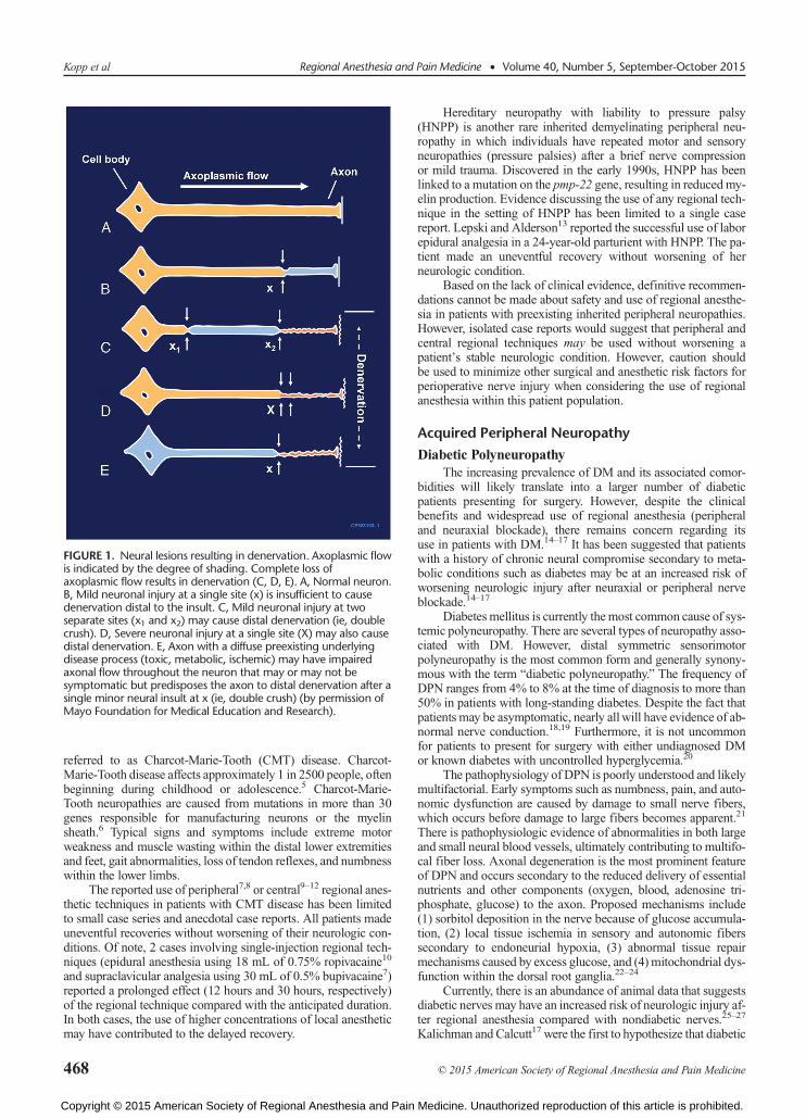
FIGURE 1. Neural lesions resulting in denervation. Axoplasmic flowis indicated by the degree of shading. Complete loss ofaxoplasmic flow results in denervation (C, D, E). A, Normal neuron.B, Mild neuronal injury at a single site (x) is insufficient to causedenervation distal to the insult. C, Mild neuronal injury at twoseparate sites (x1 and x2) may cause distal denervation (ie, doublecrush). D, Severe neuronal injury at a single site (X) may also causedistal denervation. E, Axon with a diffuse preexisting underlyingdisease process (toxic, metabolic, ischemic) may have impairedaxonal flow throughout the neuron that may or may not besymptomatic but predisposes the axon to distal denervation after asingle minor neural insult at x (ie, double crush) (by permission ofMayo Foundation for Medical Education and Research).
Kopp et al Regional Anesthesia and Pain Medicine • Volume 40, Number 5, September-October 2015
referred to as Charcot-Marie-Tooth (CMT) disease. Charcot-Marie-Tooth disease affects approximately 1 in 2500 people, oftenbeginning during childhood or adolescence.5 Charcot-Marie-Tooth neuropathies are caused from mutations in more than 30genes responsible for manufacturing neurons or the myelinsheath.6 Typical signs and symptoms include extreme motorweakness and muscle wasting within the distal lower extremitiesand feet, gait abnormalities, loss of tendon reflexes, and numbnesswithin the lower limbs.
The reported use of peripheral7,8 or central9–12 regional anes-thetic techniques in patients with CMT disease has been limitedto small case series and anecdotal case reports. All patients madeuneventful recoveries without worsening of their neurologic con-ditions. Of note, 2 cases involving single-injection regional tech-niques (epidural anesthesia using 18 mL of 0.75% ropivacaine10
and supraclavicular analgesia using 30 mL of 0.5% bupivacaine7)reported a prolonged effect (12 hours and 30 hours, respectively)of the regional technique compared with the anticipated duration.In both cases, the use of higher concentrations of local anestheticmay have contributed to the delayed recovery.
468
Copyright © 2015 American Society of Regional Anesthesia and Pain
Hereditary neuropathy with liability to pressure palsy(HNPP) is another rare inherited demyelinating peripheral neu-ropathy in which individuals have repeated motor and sensoryneuropathies (pressure palsies) after a brief nerve compressionor mild trauma. Discovered in the early 1990s, HNPP has beenlinked to a mutation on the pmp-22 gene, resulting in reduced my-elin production. Evidence discussing the use of any regional tech-nique in the setting of HNPP has been limited to a single casereport. Lepski and Alderson13 reported the successful use of laborepidural analgesia in a 24-year-old parturient with HNPP. The pa-tient made an uneventful recovery without worsening of herneurologic condition.
Based on the lack of clinical evidence, definitive recommen-dations cannot be made about safety and use of regional anesthe-sia in patients with preexisting inherited peripheral neuropathies.However, isolated case reports would suggest that peripheral andcentral regional techniques may be used without worsening apatient’s stable neurologic condition. However, caution shouldbe used to minimize other surgical and anesthetic risk factors forperioperative nerve injury when considering the use of regionalanesthesia within this patient population.
Acquired Peripheral Neuropathy
Diabetic PolyneuropathyThe increasing prevalence of DM and its associated comor-
bidities will likely translate into a larger number of diabeticpatients presenting for surgery. However, despite the clinicalbenefits and widespread use of regional anesthesia (peripheraland neuraxial blockade), there remains concern regarding itsuse in patients with DM.14–17 It has been suggested that patientswith a history of chronic neural compromise secondary to meta-bolic conditions such as diabetes may be at an increased risk ofworsening neurologic injury after neuraxial or peripheral nerveblockade.14–17
Diabetes mellitus is currently the most common cause of sys-temic polyneuropathy. There are several types of neuropathy asso-ciated with DM. However, distal symmetric sensorimotorpolyneuropathy is the most common form and generally synony-mous with the term “diabetic polyneuropathy.” The frequency ofDPN ranges from 4% to 8% at the time of diagnosis to more than50% in patients with long-standing diabetes. Despite the fact thatpatients may be asymptomatic, nearly allwill have evidence of ab-normal nerve conduction.18,19 Furthermore, it is not uncommonfor patients to present for surgery with either undiagnosed DMor known diabetes with uncontrolled hyperglycemia.20
The pathophysiology of DPN is poorly understood and likelymultifactorial. Early symptoms such as numbness, pain, and auto-nomic dysfunction are caused by damage to small nerve fibers,which occurs before damage to large fibers becomes apparent.21
There is pathophysiologic evidence of abnormalities in both largeand small neural blood vessels, ultimately contributing to multifo-cal fiber loss. Axonal degeneration is the most prominent featureof DPN and occurs secondary to the reduced delivery of essentialnutrients and other components (oxygen, blood, adenosine tri-phosphate, glucose) to the axon. Proposed mechanisms include(1) sorbitol deposition in the nerve because of glucose accumula-tion, (2) local tissue ischemia in sensory and autonomic fiberssecondary to endoneurial hypoxia, (3) abnormal tissue repairmechanisms caused by excess glucose, and (4) mitochondrial dys-function within the dorsal root ganglia.22–24
Currently, there is an abundance of animal data that suggestsdiabetic nerves may have an increased risk of neurologic injury af-ter regional anesthesia compared with nondiabetic nerves.25–27
Kalichman and Calcutt17 were the first to hypothesize that diabetic
© 2015 American Society of Regional Anesthesia and Pain Medicine
Medicine. Unauthorized reproduction of this article is prohibited.
Regional Anesthesia and Pain Medicine • Volume 40, Number 5, September-October 2015 RA and Preexisting Neurologic Disease
nerve fibers may be more susceptible to local anesthetic neuro-toxicity for 2 reasons: (1) the nerve is more susceptible to injurybecause of chronic ischemic hypoxia and (2) the nerves are ex-posed to larger concentrations of local anesthetics because of adecreased perineural blood flow. More recently, these findingswere supported with both animal and clinical data. Lirk and col-leagues 28 used Zucker diabetic fatty rats exposed to hyperglyce-mia to demonstrate that, although the overall neuronal survivaldifference was low, in vitro local anesthetic neurotoxicity wasmore pronounced in neurons from diabetic animals. The authorsalso reported that preexisting subclinical neuropathy led tosubstantial prolongation of the block duration in vivo. Kroinand colleagues26 also reported that the duration of sciatic nerveblock with lidocaine 1% or ropivacaine 0.5% was longer instreptozotocin-induced diabetic rats compared with nondiabeticrats, and that block duration actually correlated with nerve fiberdegeneration. In a subsequent study, the same authors alsoconcluded that, with continuous glycemic control, diabetic ratshad a block duration that was similar to nondiabetic rats and40 minutes shorter than rats without glycemic control.25 Interest-ingly, acute glycemic control did not lessen the nerve block dura-tion, suggesting that diabetic neuropathy is not rapidly reversedwithin this animal model. Currently, it is unclear whether the re-sults from animal studies using experimentally induced hypergly-cemia can be used to make recommendations about patients withlong-standing DM.29
Although animal studies have consistently found that dia-betic nerves are more sensitive to local anesthetics and potentiallymore susceptible to neural injury, it is unclear whether diabetic pa-tients have a higher incidence of neurologic injury after regionalanesthesia.17,25,26,30 There is limited clinical data suggesting thatthe success of peripheral nerve blockade (supraclavicular brachialplexus) may be higher in diabetic patients independent of otherpredictors of success (eg, bodymass index) compared with nondi-abetic patients.31,32 Gebhard and colleagues30 propose several the-ories for this finding, including (1) a higher sensitivity of diabeticnerve fibers to local anesthetics, (2) possible unknown intraneuralpenetration before injection, and (3) preexistingDPNwith accom-panying decreased sensation. Preexisting pathology has long beenreported to play a role in the development of postoperative neuro-logic dysfunction.33–35 A recent case report described a persistentpostoperative femoral neuropathy after discontinuing a femoralnerve catheter in a patient with a preexisting subclinical diabeticneuropathy that was undiagnosed preoperatively.36
In patients with DM, a decreased sensitivity to electricalstimulation combinedwith diminished sensory function and an in-creased sensitivity to local anesthetic toxicity may increase therisk of intraneural injection during peripheral nerve blockadeusing a peripheral nerve stimulator.37–39 Currently, there is a lackof clinical evidence suggesting that the use of ultrasound guidanceis safer than peripheral nerve stimulation within the general popu-lation.40,41 However, this lack of clinical benefit may be less clearfor diabetic patients. For example, there are a limited number ofanimal and clinical studies that suggest ultrasound guidance maybe a more desirable method of neural localization in diabetic pa-tients. Animal studies have shown that low-threshold electricalstimulation may not offer protection from intraneural injection inthe presence of hyperglycemia. Rigaud and colleagues42 demon-strated that all needle insertions within a hyperglycemic dogmodel resulted in intraneural injection (6 of 6); whereas onlyone (1 of 18) intraneural injection occurred among control dogs.Sites and colleagues39 also concluded that ultrasound guidancemay be a preferred method of neural localization in diabetic pa-tients after failing to elicit a motor response or paresthesia in 2 pa-tients undergoing sciatic nerve blockade using peripheral nerve
© 2015 American Society of Regional Anesthesia and Pain Medicine
Copyright © 2015 American Society of Regional Anesthesia and Pain
stimulation. The authors describe a very weak motor response inboth diabetic patients with a stimulating current of more than2.4 mA despite perineural placement of the stimulating needleusing ultrasound guidance. Another potential application of ultra-sound technology is the ability to use the cross-sectional area of aperipheral nerve to identify a clinical or subclinical peripheralneuropathy; a diagnosis that historically would require complexnerve conduction studies.43,44
Findings of spinal cord involvement in diabetic patientssuggest that the same or similar mechanism of injury may affectnot only peripheral nerves but also neural elements within the cen-tral neuraxis as well.45,46 Using magnetic resonance imaging,Selvarajah and colleagues47 described early central nervous sys-tem involvement consisting of a significant reduction in spinalcord cross-sectional area in patients with both subclinical and clin-ically detectable diabetic peripheral neuropathy. A case report of adiabetic patient experiencing a persistent lower-extremity neurop-athy after what appeared to be an uneventful epidural analgesia re-inforces concerns that diabetic patients may be at an increased riskof neurologic injury after neuraxial anesthesia.48 A retrospectivereview also evaluated neurologic complications in patients withpreexisting peripheral sensorimotor neuropathy or DPN who sub-sequently underwent neuraxial anesthesia or analgesia.22 Of the567 patients studied, 2 (0.4%; 95% confidence interval [CI],0.1%–1.3%) experienced new or progressive postoperative neuro-logic deficits when compared with preoperative findings. The au-thors concluded that, although the risk of severe postoperativeneurologic injury among diabetic patients is rare, it appears tobe higher than that reported for the general population. Althoughthe neuraxial technique could not be definitively implicated asthe primary cause of the neurologic insult, it may have been a con-tributing factor among patients with preexisting neural compro-mise. Echevarria and colleagues have also reported faster onsettimes, a longer duration of maximal block levels, and slower re-gression times of spinal anesthesia in diabetic patients comparedwith nondiabetic patients.49
In summary, patients with DPN likely have neural elementsthat are more sensitive to the effects of local anesthetic. As a result,diabetic peripheral nerves may be more susceptible to subsequentinjury from local anesthetic toxicity or ischemic insults. Ulti-mately, the decision to use regional anesthesia within diabetic pa-tients should be made on an individual basis after a thoroughdiscussion with the patient regarding the potential risks and bene-fits of the technique. Consideration should be given to decreasingthe concentration or total dose of local anesthetic for both periph-eral and neuraxial techniques,50 particularly in profoundly symp-tomatic patients. Furthermore, the use of ultrasound guidancemay facilitate perineural needle placement and the use of lower lo-cal anesthetic volumes in diabetic patients; although definitivedata ensuring increased safety with ultrasound guidance arecurrently lacking.51 Decreasing the concentration or dose of localanesthetic or eliminating epinephrine additives should also beconsidered given that diabetic nerves are already at risk of neuralischemia and infarction because of changes within the endoneuralmicrovasculature.52
Chemotherapy-Induced NeuropathyChemotherapy-induced peripheral neuropathy (CIPN) is a
frequent side effect of several commonly used chemotherapeuticagents. It is a dose-limiting side effect that occurs in approxi-mately 30% to 40% of patients.53 The exact mechanism of injuryis unclear, although damage to microtubules, interference withmicrotubule-based axonal transport, mitochondrial disruption,and cytotoxic effects on DNA are all possible mechanisms.53,54
469
Medicine. Unauthorized reproduction of this article is prohibited.
Kopp et al Regional Anesthesia and Pain Medicine • Volume 40, Number 5, September-October 2015
The neurotoxicity depends on the agent used, the duration ofadministration, and the cumulative dose received. Cisplatin,oxaliplatin, and carboplatin characteristically induce a purelysensory painful peripheral neuropathy, whereas vincristine,paclitaxel, and suramin tend to induce a mixed sensorimotorneuropathy with or without involvement of the autonomic ner-vous system.55 Symptoms are often in the “glove and stocking”distribution and consist of pain or paresthesias. Patients at risk ofdeveloping CIPN include those with preexisting neural damagesecondary to DM, excessive alcohol use, or an inherited periph-eral neuropathy. In general, a prolonged period of regenerationis required to restore neurologic function, with incomplete recov-ery being the most common outcome.54–56 However, patients whorecover from CIPN are at an increased risk of developing progres-sive neuropathic symptoms if exposed to additional neurotoxicagents. Local anesthetics are potentially neurotoxic, and cautionshould be used when deciding whether to perform regional anes-thesia in patients who have received chemotherapeutic agentsknown to cause CIPN. It is not uncommon for patients to have asubclinical neuropathy that only presents after a second neuro-logic insult, such as a peripheral or neuraxial block.16
INFLAMMATORY NEUROPATHIES
Guillain-Barré SyndromeGuillain-Barré syndrome is an acute, inflammatory, demye-
linating polyneuropathy characterized by areflexia and diffuse as-cending neuromuscular paralysis. The etiology of GBS is unclear,although infection, pregnancy, vaccinations, immunosuppression,systemic illnesses, and transfusion have all been proposed as po-tential triggers.57 The degree and distribution of paralysis are var-iable and can include sensory nerve, cranial nerve, and autonomicnervous system involvement. Symptoms peak approximately 2 to4 weeks after the initial onset, with most patients experiencingprolonged recovery. Unfortunately, many patients experiencemoderate-to-severe neurological impairment for years after theinitial diagnosis.
There are several reports of GBS occurring in the postopera-tive period after a variety of surgical procedures and types ofanesthetics.58–60 However, case reports of regional anesthesiause in patients with GBS are generally limited to the obstetricpopulation.61–64 Some patients with GBSmay have autonomic in-stability and subsequently experience an exaggerated response toneuraxial blockade,63 whereas other patients exhibit a normal re-sponse to neuraxial anesthesia.61,64 Although there have been re-ports of successful neuraxial anesthesia in parturients with GBS,the theoretical concern of local anesthetics interacting with pe-ripheral myelin or direct nerve trauma cannot be ignored.21 Thereis some evidence to suggest that epidural anesthesia may precipi-tate or reactivate GBS hours to weeks after surgery.58,65,66 How-ever, it is difficult to determine if this is caused by the effects ofthe epidural, the natural progression of the disease, the surgicalprocedure, or the stress response related to surgery.
Although it has been suggested that acute neuronal inflam-mation may be a relative contraindication to regional anesthesia,existing data provide little information regarding the safety ofneuraxial anesthesia or peripheral nerve blockade in patients withGBS.21 Ultimately, the decision to perform regional anesthesiashould be made on an individual basis after a thorough discussionwith the patient regarding the potential risks and benefits.
Postsurgical Inflammatory NeuropathyRecently, neurologists have become aware that an autoim-
mune or inflammatory process may be the cause of severe
470
Copyright © 2015 American Society of Regional Anesthesia and Pain
postoperative neurologic deficits. Staff and colleagues67 recentlydescribed a series of 33 patients who developed postsurgical in-flammatory neuropathy (PSIN) within 30 days of surgery. The di-agnosis was confirmed in most patients after a peripheral nervebiopsy. Postsurgical inflammatory neuropathy is believed to bean idiopathic immune-mediated response to a physiologic stresssuch as an infectious process, a vaccination, or a surgical proce-dure.67 The condition may present as focal, multifocal, or diffuseneurologic deficits in the setting of a negative radiographic imag-ing. Complicating the diagnosis, the onset of neurologic deficitsmay not be apparent during the immediate postoperative period;and the deficits may be in an anatomic distribution remote fromthe surgical site or regional anesthetic technique. Risk factors orpotential triggers for PSIN include malignancy, DM, tobaccouse, systemic infection, volatile anesthetic use, and recent bloodtransfusion.67 Suppression of the immune response with pro-longed high-dose corticosteroids or intravenous immunoglobulinis the current treatment of choice. The goal of treatment is to suf-ficiently blunt the inflammatory response to allow for axonalregeneration. Fortunately, most patients improve with currenttreatment recommendations, with pain and sensory deficits im-proving before the motor deficits.67
The degree towhich inflammatorymechanisms play a role inpostoperative neurologic dysfunction is unknown and poorlycharacterized particularly within the anesthesia literature.68 As aresult, anesthesia providers and surgeons rarely consider this po-tential etiology of nerve injury when evaluating patients with post-operative deficits. This is problematic because the commonapproach of watchful waiting and conservative management willnot be effective in patients with PSIN. Rather, PSIN is a clinicalcondition that must be suspected early in the disease process sothat a definitive diagnosis can be obtained (nerve biopsy) and ag-gressive immunotherapy can be initiated to potentially improveneurologic outcome.67
CENTRAL NERVOUS SYSTEM DISORDERSHistorically, neuraxial anesthesia techniques have not been
offered to patients with preexisting neurologic disorders of thecentral nervous system (MS, PPS, ALS) for fear of worseningneurologic outcome.69–72 In fact, many historians believe thatthe recommendation by Dripps and Vandam70 in 1956 to avoid re-gional anesthesia in patients with preexisting neurologic disordershas impacted clinical management for nearly half a century. Sev-eral theoretical mechanisms have been proposed based on thedouble-crush phenomenon, including neurologic injury fromneedle- or catheter-induced trauma, local anesthetic neurotoxicity,and neural ischemia caused by local anesthetic additives. However,the avoidance of regional anesthesia within this patient populationmay also be caused by physician and patient biases or potentialmedicolegal concerns. There are several confounding factors (age,body habitus, surgical trauma, tourniquet times and pressures, posi-tioning, anesthetic technique) that make it difficult to determine theetiology of worsening postoperative neurologic deficits.
A recent review evaluated 139 patients with a history of oneor more central nervous system disorders that subsequentlyunderwent a neuraxial anesthetic technique.71 Preoperative neuro-logic disorders included primarily PPS, MS, ALS, and traumaticspinal cord injury. In contrast to the findings of Dripps andVandam several decades ago, the authors identified no new orworsening postoperative neurologic deficits (0.0%; 95% CI,0.0%–0.3%) within their patient cohort. This was despite the factthat 74% of the patients reported active neurologic symptoms(paresthesias, dysesthesias, hyperreflexia) or sensorimotor deficitsduring the immediate preoperative period and subsequently
© 2015 American Society of Regional Anesthesia and Pain Medicine
Medicine. Unauthorized reproduction of this article is prohibited.
Regional Anesthesia and Pain Medicine • Volume 40, Number 5, September-October 2015 RA and Preexisting Neurologic Disease
received standard doses of local anesthetics. Two smaller reviewsin parturients receiving smaller doses of local anesthetic for laboranalgesia have reported similar results.73,74
Clearly, further investigations with more patients are neededto make definitive recommendations. However, the current datasuggest that the decision to perform neuraxial anesthesia in pa-tients with preexisting central nervous system disorders be basedon the risks and benefits for each individual patient. Some authorshave postulated that the neurologic risk may be higher in patientswho have progressive neurologic deficits when compared withthose patients with chronic stable sensorimotor symptoms thathave not changed during the course of several months or years.
Multiple SclerosisMultiple sclerosis is an inflammatory autoimmune disorder
of the central nervous systemwith a lifetime risk of 1 in 400, mak-ing it the most common debilitating neurologic disease in youngadults.75 It is a chronic degenerative disease characterized by focaldemyelination within the spinal cord and brain. The demyelin-ation results in a fluctuating conduction block that causes a classic“waxing and waning” of symptoms that is characteristic of the dis-ease. Signs and symptoms include sensory or motor deficits, dip-lopia or vision loss, bowel or bladder dysfunction, and ataxia. Theprecise etiology is unclear; however, a combination of genetic riskfactors and environmental factors likely plays a role. Twenty-fivepercent of MS patients are essentially asymptomatic, and theiractivities of daily living are unaffected. However, up to 15% ofpatients may become severely disabled, with significant sensori-motor deficits within a short period.76
Several factors common to surgery can negatively impact thedisease process, including hyperpyrexia, emotional stress, and in-fection.77 The mechanism of worsening neurologic function in pa-tients with MS is unclear and may occur coincidentally within thepostoperative period independent of the anesthetic technique. Ev-idence regarding the risk of regional anesthesia in patients withMS is limited. Despite some evidence for demyelination of the pe-ripheral nerves in MS, peripheral nerve blockade has traditionallybeen considered safe.78 However, a recent report of severe bra-chial plexopathy after an ultrasound-guided interscalene blockhas raised the concern that a segment of MS patients may havesubclinical peripheral neuropathy.79 Several investigators havedemonstrated evidence of axonal demyelinating peripheral lesions(sensory > motor) in patients with MS.80–82 Misawa and col-leagues81 demonstrated that peripheral demyelination may occurin 5% of MS patients, whereas Pogorzelski and colleagues80 re-port peripheral demyelination may occur in up to 47% of patients.Similarly, Sarova-Pinhas and colleagues82 describe nerve conduc-tion abnormalities in up to 14.7% of peripheral nerves within MSpatients compared with only 2.4% of nerves within the generalpopulation. Despite this evidence, the overall incidence and clini-cal relevance of this underlying peripheral neuropathy remain un-defined in the setting of performing peripheral nerve blockade inpatients with MS.
In contrast to peripheral nerve blockade, the potential risk ofnew or progressive neurologic deficits in MS patients after spinalanesthesiawas first described in 1937. Critchley83 described 3 pa-tients with “disseminated (multiple) sclerosis” that experiencedworsening of symptoms after spinal anesthesia. The author con-cluded that “spinal anesthesia may be a precipitating agent in theevolution of disseminated (multiple) sclerosis.” Several subsequentstudies demonstrated similar outcomes with the development ofnew or worsening neurologic deficits or a higher likelihood ofsymptom exacerbation after spinal anesthesia.69,72,84,85 In contrast,a more recent study demonstrated no new or worsening neurologic
© 2015 American Society of Regional Anesthesia and Pain Medicine
Copyright © 2015 American Society of Regional Anesthesia and Pain
symptoms after spinal anesthesia in 35 MS patients undergoinga variety of surgical procedures.22
The safety of epidural anesthesia and analgesia in MS pa-tients has been focused almost exclusively within the obstetricpopulation, which may not accurately represent the nonpregnantMS patient. Pregnancy is frequently associated with a decreasein disease relapses, whereas the postpartum period is often associ-ated with an increased risk of relapse. The transition from cellularimmunity to humoral immunity required for the mother's immunesystem to tolerate the fetus is thought to be protective during preg-nancy.73 However, as cell-mediated immunity rebounds during thepostpartum period, patients will often experience transient wors-ening of neurologic symptoms that could be falsely attributed torecent regional anesthetic techniques.
Confavreux and colleagues73 have performed one of the fewprospective studies evaluating risk factors associated with diseaserelapse during the postpartum period. They concluded that epidu-ral analgesia during labor and delivery did not contribute to ahigher risk of relapse compared with patients not receivingneuraxial techniques. Similarly, Kuczkowski86 found no associa-tion between any form of obstetric regional analgesia and theworsening of MS symptoms among obstetric patients. Epiduralanesthesia and analgesia have traditionally been recommendedover spinal anesthesia in MS patients because the concentrationof local anesthetic in the white matter of the spinal cord is onefourth the level after epidural injection compared with intrathecalinjection.87 It is believed that the lack of myelin may leave the spi-nal cord susceptible to the neurotoxic effects of local anes-thetics.87 Although definitive studies on the pharmacologicaleffects of local anesthetic concentrations and doses are lacking,many recommend limiting neuraxial local anesthetic doses andconcentrations to the lowest level possible. There is some evi-dence that lidocaine can reversibly worsen symptoms of MS.88
This is thought to occur when sodium channels in demyelinatedareas are blocked enough to unmask lesions that would otherwisebe below the level of clinical detection. With regard to the obstet-ric patient, the risk of neuraxial anesthesia or analgesia needs to beweighed against the increased risk of general anesthesia. A recentsurvey demonstrated that 99% of obstetric anesthesiologistswould perform neuraxial anesthesia for an emergency cesareandelivery in an MS patient after carefully weighing the potentialrisks and benefits.89
In summary, there remains little conclusive evidence to sup-port or refute the use of regional anesthesia in patients with MS.Peripheral nerve blockade has not been definitively shown tobe harmful in the setting of MS and, therefore, should not beconsidered an absolute contraindication. In contrast, given thatdemyelinated fibers may bemore prone to the toxic effects of localanesthetics, epidural anesthesia and analgesia may be consideredsafer than spinal anesthesia techniques. However, reducing the lo-cal anesthetic concentration and total dose to the lowest effectivelevel(s) may be prudent for both peripheral and neuraxial block-ade. All decisions regarding the use of regional anesthesia and an-algesia in patients with MS need to be made after carefulconsideration of the potential risks and benefits. Regardless ofthe anesthetic technique chosen, patients should be informedabout the risk of new or worsening neurologic symptoms duringthe postoperative period because of exposure to multiple exacer-bating factors.
Postpolio SyndromePostpolio syndrome refers to new-onset neurologic symp-
toms that develop several years after an acute poliomyelitis infec-tion. The onset of new or progressive symptoms may occur up to
471
Medicine. Unauthorized reproduction of this article is prohibited.
Kopp et al Regional Anesthesia and Pain Medicine • Volume 40, Number 5, September-October 2015
30 years after the initial episode of poliomyelitis. PPS affects an-terior horn cells within the anterior portion of the spinal cordand is, therefore, considered a lower motor neuron disorder.90 Ini-tial symptoms include muscle weakness, fatigue, gait instability,joint pain, and muscle atrophy within muscle groups that werepreviously affected by the disease. Sensory deficits are generallynot characteristic of the syndrome and are only observed if a sec-ondary disorder is present (ie, compression radiculopathy or diskherniation). The muscle effects of PPS are thought to be relatedto an ongoing process of denervation and reinnervation that ulti-mately ends when denervation is no longer compensated forby reinnervation.90
Postpolio syndrome is the most prevalent motor neuron dis-ease in North America. Furthermore, because acute poliomyelitiscontinues to occur in developing countries, PPS will likely remainan anesthetic concern for years to come.21 It is not uncommon forpatients with PPS to require orthopedic procedures; therefore, it isimportant to determine the safety of regional anesthetic techniquesunder these clinical circumstances. Although patients with PPShave fewer motor neurons than normal, it is difficult to knowwhether remaining motor neurons are more susceptible to thetoxic effects of local anesthetics. There have been no reports ofworsening neurologic status after neuraxial anesthesia with nor-mal doses of tetracaine and bupivacaine in patients withPPS.91,92 However, this does not imply that regional anesthetictechniques are without risk.93 As with all patients, the potentialrisk of regional anesthesia must be balanced against the disadvan-tages of general anesthesia, including a hypersensitivity to seda-tive or opioid medications, risk of muscle relaxant use, and therisk of hypoventilation and aspiration. The largest series of pa-tients with PPS (n = 79) undergoing neuraxial anesthesia or anal-gesia demonstrated no worsening of neurologic symptoms duringthe postoperative period.71 However, the paucity of clinical dataon this topic prevents clear recommendations from beingmade re-garding the safety of neuraxial anesthesia or peripheral nerveblockade in patients with PPS. Ultimately, the decision to use re-gional anesthesia should be made on an individual basis after athorough discussion of the potential risks and benefits with eachpatient. Given the increased sensitivity to opioid and sedativemedications within this patient subgroup, these medicationsshould always be used with caution.
Amyotrophic Lateral SclerosisAmyotrophic lateral sclerosis is a common form of motor
neuron disease characterized by adult-onset degeneration of boththe upper and lower motor neurons. Unfortunately, in the majorityof patients, death from respiratory failure occurs within a fewyears of disease onset.94
The existing evidence, albeit limited, has not supportedthe fear that neuraxial or peripheral blockade will exacerbatepreexisting symptoms in ALS patients.65,95–99 However, given thepotential for worsening respiratory failure after general anesthesiabecause of the use of muscle relaxants and opioid medications,the ability to avoid airway manipulation may be considered a ben-efit within this high-risk patient population. Regardless of theanesthetic technique, the possibility of postoperative respiratoryor neurologic deterioration is quite high in patients with ALS.Ultimately, the decision to use regional anesthesia should bemade on an individual basis after a thorough discussion of thepotential risks and benefits with each patient.
SPINAL CANAL PATHOLOGYSpine and spinal canal pathology has been proposed as a
potential risk factor for neurologic complications after neuraxial
472
Copyright © 2015 American Society of Regional Anesthesia and Pain
blockade. Several mechanisms of injury have been proposed, in-cluding an ischemic or compressive effect after the injection oflarge volumes of local anesthetic into a relatively confined space(ie, epidural anesthesia) as well as local anesthetic neurotoxicity(ie, spinal anesthesia). Although the precise mechanism(s) of in-jury remain unclear, there are several isolated case reports andlarge case series that are believed to support these hypotheses.
Spinal Stenosis and Lumbar Disk DiseaseSpinal stenosis occurs as age-related changes within the in-
tervertebral disks and facet joints result in narrowing of the spinalcanal or neural foramina. Changes include disk degeneration,facet joint hypertrophy, osteophyte formation, and infolding ofthe ligamentum flavum. The precise mechanism by which spinalnerve root compression results in signs or symptoms of spinal ste-nosis is not completely understood.100 Classic symptoms includeback and leg radicular pain that significantly worsens with exten-sion and is alleviated with flexion. Preexisting spinal stenosisor compressive lumbar disk disease has been proposed as a poten-tial risk factor for neurologic complications after a neuraxial(spinal or epidural) technique. Proposed mechanisms of injuryinclude mechanical trauma,101,102 local anesthetic neurotoxic-ity,103,104 ischemia,105–107 or a multifactorial etiology.108,109 Path-ophysiologically, patients with spinal stenosis have a reductionin the diameter of the spinal canal, resulting in less anatomic spacefor fluid collections such as blood or local anesthetic. As a result,small quantities of fluid may result in significant increases in pres-sure around the neuraxis that would otherwise have no clinical ef-fect in a widely patent spinal canal.
Two relatively large case series and several case reports havebeen published that suggest undiagnosed spinal stenosis may be arisk factor for neurologic complications after neuraxial block-ade.101,103,105,108,110 The majority of cauda equina cases involvedepidural analgesia, which may suggest an ischemic component(mechanical compression of the cord by the infusing local anes-thetic) to the injury.106 Hebl and colleagues108 performed a retro-spective review of patients with preexisting spinal stenosis orlumber disk disease with and without a history of prior spinal sur-gery and concluded that this cohort of patients was at an increasedrisk for the development or worsening of neurologic deficits whencompared with the general population undergoing a neuraxialtechnique. In addition, patients with more than one neurologicdiagnosis (eg, spinal stenosis, compressive radiculopathy, pre-existing peripheral neuropathy) appeared to have an even higherrisk of injury. Moen and colleagues103 also performed a largeepidemiologic survey in Sweden that revealed similar trends. Dur-ing a 10-year study period, 1,260,000 spinal anesthetics and450,000 epidural blocks were evaluated. Overall, the authors iden-tified 127 serious complications, including 85 (67%) patients withpermanent injuries. Although 14 patients had preexisting spinalstenosis, 13 (93%) of these were diagnosed in the postoperativeperiod during the evaluation of the neurologic deficit. The authorsconcluded that the incidence of severe anesthesia-related compli-cations may not be as low as previously reported, and preexistingspinal canal pathologymay be a “neglected risk factor.” Finally, al-though patients with prior spine surgery may have an increasedrisk of paraplegia after transforaminal epidural steroid injec-tions,111,112 no similar risk has been found in patients after neu-raxial anesthesia or analgesia.
In summary, although it appears that patients with spinal ste-nosis or compressive lumbar disk disease may be at increased riskof neurologic complications after neuraxial blockade, the exist-ing literature fails to provide a direct comparison of surgical pa-tients with similar spinal pathology undergoing general anesthesia.
© 2015 American Society of Regional Anesthesia and Pain Medicine
Medicine. Unauthorized reproduction of this article is prohibited.
Regional Anesthesia and Pain Medicine • Volume 40, Number 5, September-October 2015 RA and Preexisting Neurologic Disease
Therefore, it is unclear whether the higher incidence of neurol-ogic complications is caused by surgical factors, the anesthetictechnique, the natural progression of the disease process, or a com-bination of these factors.
Neural Tube DefectsNeural tube defects are congenital anomalies of neural
development that primarily affect the cranium or spine. Clinicalmanifestations vary widely and include cranial defects (eg, anen-cephaly, exencephaly, encephalocele), open spinal dysraphisms,and closed spinal dysraphisms. Open spinal dysraphisms, oftenreferred to as spina bifida cystica, occur at a frequency of 0.5 to8 cases per 1000 live births and include conditions in which neuraltissue is exposed to the external environment.113 The most com-mon open spinal dysraphisms are meningocele (exposed menin-ges) and meningomyelocele (exposed meninges and spinal cordtissue). In contrast, closed spinal dysraphisms are characterizedby unexposed neural tissue with abnormalities ranging from iso-lated defects in the fusion of the posterior vertebral column (ie,spina bifida occulta) to more serious spinal cord malformationssuch as diastematomyelia (split cord malformations), tethered spi-nal cord syndrome, and dural ectasia (lumbosacral widening orcaudal displacement of the dural sac).114 The etiology of neuraltube defects are believed to be multifactorial, with both geneticand environmental factors equally implicated.115
Open spinal dysraphisms are commonly treated with surgicalintervention during the early neonatal period. Clinical outcomesmay vary from no neurologic sequelae to sensorimotor deficits,lower extremity paraplegia, and bowel and bladder dysfunction.Four anecdotal case reports have been described in the literaturein which epidural analgesia116,117 or spinal anesthesia118,119 hasbeen used in parturients during labor and delivery with a historyof spina bifida cystica and subsequent surgical correction. In allbut 1 case, the authors describe extensive cranial spread of localanesthetic and dense neural blockade with normal or reduceddoses of local anesthetic. Limited spread of local anesthetic cau-dad to the site of surgical intervention was also noted. None ofthe patients experienced an inadvertent dural puncture, postduralpuncture headache, or new or progressive neurologic dysfunctionafter the regional technique. Tidmarsh and May120 have also de-scribed the use of epidural analgesia in four parturients who previ-ously underwent meningomyelocele repair during infancy.Clinical outcomes included extensive cranial spread of local anes-thetic (n = 1), poor sacral analgesia (n = 1), and successful epidu-ral analgesia (n = 2). The authors cautioned that performingneuraxial techniques within this patient population can be techni-cally challenging, with an increased risk of inadvertent duralpuncture and unpredictable local anesthetic spread.120 If neuraxialanesthesia or analgesia is performed under these clinical circum-stances, it is recommended that the site of needle insertion occursat a level above the original lesion because of limitations in localanesthetic spread.116
Spina bifida occulta is a common closed spinal dysraphismthat is thought to be a normal variant of vertebral column devel-opment. Studies report an incidence of 10% to 24% within thegeneral population.114 Isolated spina bifida occulta involves thefailure of a single-level vertebral arch (usually the lamina) fromfusing, with no clinical signs or symptoms. There is no externallesion, and the spinal cord and meninges are not involved. Theuse of regional anesthesia in parturients with isolated spina bifidaocculta has been reported but is limited to anecdotal case reports121
and small case series.120,122 Within this collection of 11 re-ported cases, successful epidural analgesia was achieved withnormal doses of local anesthetic without extensive cranialspread of local anesthetic, sacral sparing, or adverse neurologic
© 2015 American Society of Regional Anesthesia and Pain Medicine
Copyright © 2015 American Society of Regional Anesthesia and Pain
sequelae. One patient experienced technical difficulties dur-ing block placement, including the elicitation of a transientparesthesia and inadvertent dural puncture.121 If neuraxialanesthesia or analgesia is performed in patients with isolatedspina bifida occulta, it is generally recommended that thesite of needle insertion occur at a level above the vertebralabnormality.116
In contrast to patients with isolated spina bifida, complexspina bifida may occur in conjunction with more severe closedspinal dysraphisms. Patients with spina bifida and (a) associatedcutaneous manifestations (eg, hairy patch, subcutaneous lipoma,skin sinus), (b) involvement of more than one lamina, (c) neuro-logic symptoms, or (d) associated bowel or bladder dysfunctioncommonly have more severe coexisting conditions such as teth-ered spinal cord syndrome or diastematomyelia.123 Under theseclinical circumstances, neuraxial techniques should be consid-ered contraindicated because neurologic complications havebeen reported after spinal,124 epidural,125 and combined spinal-epidural126 techniques in patients with previously unrecognizedtethered spinal cord syndrome and/or diastematomyelia.
Dural ectasia is the abnormal widening or ballooning of thedural sac, most commonly within the lumbosacral region of thespinal cord. It is common among patients with Marfan syndrome,occurring in 63% to 92% of cases.127,128 Dural ectasia is alsoknown to occur in patients with Patau syndrome (trisomy 13),129
Ehlers-Danlos syndrome, neurofibromatosis type I, and ankylos-ing spondylitis.130 Several case reports have described inadvertentdural puncture during caudal anesthesia129,131,132 and incompletespinal anesthesia133 in patients with dural ectasia.
In summary, neural tube defects encompass a wide range ofspinal cord malformations, ranging from asymptomatic single-level vertebral canal abnormalities (ie, spina bifida occulta) tomeningomyelocele with paraplegia after surgical repair. Given thewide spectrum of clinical abnormalities, the varied risk, and thepaucity of clinical data for any one diagnosis, definitive recommen-dations cannot be made regarding the safety of neuraxial anesthesiaor analgesia in patients with neural tube defects. However, it is clearthat regional anesthesia should be avoided in patients with docu-mented tethered spinal cord syndrome, diastematomyelia, or spinabifida with associated cutaneous lesions, multilevel vertebral bodyinvolvement, neurologic deficits, or bowel or bladder dysfunction.
The neuroanatomy of all other neural tube defects (eg, isolatedspina bifida occulta, prior meningo-myelocele repair) should beclearly documented with radiographic imaging before neuraxial an-esthesia or analgesia is considered. If radiographic imaging canexclude the coexistence of complex closed spinal dysraphisms(eg, tethered spinal cord, diastematomyelia) within these patients,then regional anesthesia may be considered after a comprehensiverisk/benefit discussion with the patient, highlighting the risk oftechnical difficulties, extensive cephalad spread of local anes-thetic, sacral sparing, inadvertent dural puncture, and neurologicinjury. If neuraxial anesthesia or analgesia is performed underthese clinical circumstances, it is recommended that the site ofneedle insertion occurs at a level above the vertebral abnormalityor site of prior surgical repair.116
RECOMMENDATIONSThe following recommendations (Table 1) are intended to
encourage quality patient care, although observing them cannotguarantee any specific patient outcome. Their value should ulti-mately be determined by those who use them. The recommenda-tions are subject to revision from time to time, as warranted bythe evolution of technology, scientific evidence, and clinical prac-tice. Importantly, the recommendations address only the issue of
473
Medicine. Unauthorized reproduction of this article is prohibited.
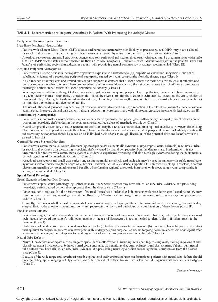
TABLE 1. Recommendations: Regional Anesthesia in Patients With Preexisting Neurologic Disease
Peripheral Nervous System DisordersHereditary Peripheral Neuropathies• Patients with Charcot-Marie-Tooth (CMT) disease and hereditary neuropathy with liability to pressure palsy (HNPP) may have a clinicalor subclinical evidence of a preexisting peripheral neuropathy caused by neural compromise from the disease state (Class I).
•Anecdotal case reports and small case series suggest that both peripheral and neuraxial regional techniques may be used in patients with stableCMTor HNPP disease states without worsening their neurologic symptoms. However, a careful discussion regarding the potential risks andbenefits of performing regional anesthesia in patients with preexisting neural compromise is strongly recommended (Class III).
Acquired Peripheral Neuropathies• Patients with diabetic peripheral neuropathy or previous exposure to chemotherapy (eg, cisplatin or vincristine) may have a clinical orsubclinical evidence of a preexisting peripheral neuropathy caused by neural compromise from the disease state (Class I).
• An abundance of animal data and limited clinical data support the concern that diabetic nerves are more sensitive to local anesthetics andperhaps more susceptible to injury. Therefore, peripheral and neuraxial blockade may theoretically increase the risk of new or progressiveneurologic deficits in patients with diabetic peripheral neuropathy (Class II).
• When regional anesthesia is thought to be appropriate in patients with acquired peripheral neuropathy (eg, diabetic peripheral neuropathyor chemotherapy-induced neuropathy), consideration should be given to modify the anesthetic technique (ie, decreasing the concentration oflocal anesthetic, reducing the total dose of local anesthetic, eliminating or reducing the concentration of vasoconstrictors such as epinephrine)to minimize the potential additive risk (Class II).
• The use of ultrasound guidance may facilitate (a) perineural needle placement and (b) a reduction in the total dose (volume) of local anestheticadministered. However, clinical data demonstrating a reduction in neurologic injury with ultrasound guidance are currently lacking (Class II).
Inflammatory Neuropathies• Patients with inflammatory neuropathies such as Guillain-Barré syndrome and postsurgical inflammatory neuropathy are at risk of new orworsening neurologic deficits during the postoperative period regardless of anesthetic technique (Class II).
•Neural compromise secondary to acute neuronal inflammation may be a relative contraindication to regional anesthesia. However, the existingliterature can neither support nor refute this claim. Therefore, the decision to perform neuraxial or peripheral nerve blockade in patients withinflammatory neuropathies should be made on an individual basis after a thorough discussion of the potential risks and benefits with thepatient (Class III).
Central Nervous System Disorders• Patients with central nervous system disorders (eg, multiple sclerosis, postpolio syndrome, amyotrophic lateral sclerosis) may have clinicalor subclinical evidence of a preexisting neurologic deficit caused by neural compromise from the disease state. Furthermore, it is notuncommon for patients with central nerve system disorders to experience worsening of their neurologic symptoms during the postoperativeperiod regardless of the anesthetic technique (Class I).
• Anecdotal case reports and small case series suggest that neuraxial anesthesia and analgesia may be used in patients with stable neurologicsymptoms without worsening their neurologic deficits. However, definitive evidence supporting this practice is lacking. Therefore, a carefuldiscussion regarding the potential risks and benefits of performing regional anesthesia in patients with preexisting neural compromise isstrongly recommended (Class II).
Spinal Canal PathologySpinal Stenosis or Lumbar Disk Disease• Patients with spinal canal pathology (eg, spinal stenosis, lumbar disk disease) may have clinical or subclinical evidence of a preexistingneurologic deficit caused by neural compromise from the disease state (Class I).
• Large case series suggest that the performance of neuraxial anesthesia and analgesia in patients with preexisting spinal canal pathology mayresult in new or worsening neurologic symptoms. However, definitive evidence suggesting an increased risk of neurologic complications islacking (Class II).
•Currently, it is unclear whether the development of new or worsening neurologic symptoms after neuraxial anesthesia or analgesia is caused bysurgical factors, the anesthetic technique, the natural progression of the spinal pathology, or a combination of these factors (Class II).
Previous Spine Surgery• Prior spine surgery is not a contraindication to the performance of neuraxial anesthesia or analgesia. However, before performing a regionaltechnique, a review of the patient's radiologic imaging or the use of fluoroscopy is recommended to identify the optimal approach to theneuraxis (Class I).
• Under most clinical circumstances, spinal anesthesia may be (a) technically easier to perform and (b) more reliable (ie, higher success rates)than epidural techniques in patients who have previously undergone spine surgery. Patients undergoing neuraxial anesthesia or analgesia aftera previous spine surgery do not appear to be at higher risk of new or progressive neurologic deficits (Class II).
Neural Tube Defects• Neural tube defects encompass a wide range of spinal cord malformations, including both open (eg, meningocele, meningomyelocele) andclosed (eg, spina bifida occulta, tethered spinal cord syndrome, diastematomyelia, dural ectasia) spinal dysraphisms. Patients with neuraltube defects may have clinical or subclinical evidence of a preexisting neurologic deficit caused by neural compromise from the diseasestate (Class I).
• Because of the wide range and severity of possible spinal cord and vertebral column malformations, patients with neural tube defects shouldundergo radiographic imaging to fully evaluate and define the extent of their disease state before considering neuraxial anesthesia or analgesia(Class II).
Continued next page
Kopp et al Regional Anesthesia and Pain Medicine • Volume 40, Number 5, September-October 2015
474 © 2015 American Society of Regional Anesthesia and Pain Medicine
Copyright © 2015 American Society of Regional Anesthesia and Pain Medicine. Unauthorized reproduction of this article is prohibited.
TABLE 1. (Continued)
• Anecdotal case reports and small case series suggest that the performance of neuraxial anesthesia and analgesia in patients with complexclosed spinal dysraphisms (ie, tethered spinal cord syndrome or diastematomyelia) may result in new or progressive neurologic symptoms.However, definitive evidence suggesting an increased risk of neurologic complications is lacking (Class II).
•Anecdotal case reports and small case series suggest that neuraxial anesthesia and analgesia may be used in patients with isolated spina bifidaocculta (without associated tethered spinal cord syndrome or diastematomyelia) without an increased risk of neurologic injury. However,definitive evidence supporting this practice is lacking. Therefore, a careful discussion regarding the potential risks (technical difficulties,unpredictable local anesthetic spread, inadvertent dural puncture, neural injury) and benefits of performing regional anesthesia in patientswith isolated spina bifida occulta is strongly recommended (Class II).
TABLE 2. Strength of Recommendations
ClassificationI Animal and/or human evidence and/or general agreement of
expert opinion supports the effectiveness and usefulnessof the recommendation.
II The weight of conflicting evidence and/or the weight of expertopinion supports the usefulness of the recommendation.
III The usefulness of the recommendation is limited by absent orconflicting evidence and/or divergent expert opinion.
This classification system is significantly modified from the AmericanCollege of Cardiology/American Heart Association construct for classify-ing strength of evidence.134
Regional Anesthesia and Pain Medicine • Volume 40, Number 5, September-October 2015 RA and Preexisting Neurologic Disease
regional anesthesia in patients with preexisting peripheral andneurologic disorders.
The recommendation classification scheme (Table 2) isa modification from the American College of Cardiology/American Heart Association construct for classifying strengthof evidence.134
REFERENCES1. Upton AR, McComas AJ. The double crush in nerve entrapment
syndromes. Lancet. 1973;2:359–362.
2. Osterman AL. The double crush syndrome.Orthop Clin North Am. 1988;19:147–155.
3. Neal JM, Bernards CM, Hadzic A, et al. ASRA Practice Advisory onNeurologic Complications in Regional Anesthesia and Pain Medicine.Reg Anesth Pain Med. 2008;33:404–415.
4. Jacob AK, Kopp SL. Regional anesthesia in the patient with preexistingneurologic disorders. Adv Anesth. 2011;29:1–18.
5. Skre H. Genetic and clinical aspects of Charcot-Marie-Tooth's disease.Clin Genet. 1974;6:98–118.
6. Saporta AS, Sottile SL, Miller LJ, Feely SM, Siskind CE, Shy ME.Charcot-Marie-Tooth disease subtypes and genetic testing strategies.Ann Neurol. 2011;69:22–33.
7. Bui AH, Marco AP. Peripheral nerve blockade in a patientwith Charcot-Marie-Tooth disease. Can J Anaesth. 2008;55:718–719.
8. Dhir S, Balasubramanian S, Ross D. Ultrasound-guided peripheralregional blockade in patients with Charcot-Marie-Tooth disease: a reviewof three cases. Can J Anaesth. 2008;55:515–520.
9. Fernandez Perez AB, Quesada Garcia C, Rodriguez Gonzalez O, BesadaEstevez JC. Obstetric epidural analgesia, a safe choice in a patient withCharcot-Marie-Tooth disease [in Spanish]. Rev Esp Anestesiol Reanim.2011;58:255–256.
10. Schmitt HJ, Muenster T, Schmidt J. Central neural blockade inCharcot-Marie-Tooth disease. Can J Anaesth. 2004;51:1049–1050.
© 2015 American Society of Regional Anesthesia and Pain Medicine
Copyright © 2015 American Society of Regional Anesthesia and Pain
11. Sugai K, Sugai Y. Epidural anesthesia for a patient withCharcot-Marie-Tooth disease, bronchial asthma and hypothyroidism[in Japanese].Masui. 1989;38:688–691.
12. Tanaka S, Tsuchida H, Namiki A. Epidural anesthesia for a patientwith Charcot-Marie-Tooth disease, mitral valve prolapse syndromeand 2nd degree AV block [in Japanese]. Masui. 1994;43:931–933.
13. Lepski GR, Alderson JD. Epidural analgesia in labour for a patient withhereditary neuropathy with liability to pressure palsy. Int J Obstet Anesth.2001;10:198–201.
14. Horlocker TT, O'Driscoll SW, Dinapoli RP. Recurring brachial plexusneuropathy in a diabetic patient after shoulder surgery and continuousinterscalene block. Anesth Analg. 2000;91:688–690.
15. Waters JH, Watson TB, Ward MG. Conus medullaris injury followingboth tetracaine and lidocaine spinal anesthesia. J Clin Anesth. 1996;8:656–658.
16. Hebl JR, Horlocker TT, Pritchard DJ. Diffuse brachial plexopathy afterinterscalene blockade in a patient receiving cisplatin chemotherapy: thepharmacologic double crush syndrome. Anesth Analg. 2001;92:249–251.
17. Kalichman MW, Calcutt NA. Local anesthetic–induced conduction blockand nerve fiber injury in streptozotocin-diabetic rats. Anesthesiology.1992;77:941–947.
18. Dyck PJ, Kratz KM, Karnes JL, et al. The prevalence by staged severity ofvarious types of diabetic neuropathy, retinopathy, and nephropathy in apopulation-based cohort: the Rochester Diabetic Neuropathy Study.Neurology. 1993;43:817–824.
19. Ross MA. Neuropathies associated with diabetes. Med Clin North Am.1993;77:111–124.
20. Centers for Disease Control and Prevention. 2011 National Diabetes FactSheet. Atlanta, GA: Centers for Disease Control and Prevention, USDepartment of Health and Human Services; 2011.
21. Lirk P, Birmingham B, Hogan Q. Regional anesthesia in patients withpreexisting neuropathy. Int Anesthesiol Clin. 2011;49:144–165.
22. Hebl JR, Kopp SL, Schroeder DR, Horlocker TT. Neurologiccomplications after neuraxial anesthesia or analgesia in patients withpreexisting peripheral sensorimotor neuropathy or diabeticpolyneuropathy. Anesth Analg. 2006;103:1294–1299.
23. Krishnan AV, Kiernan MC. Altered nerve excitability properties inestablished diabetic neuropathy. Brain. 2005;128(pt 5):1178–1187.
24. Sinnreich M, Taylor BV, Dyck PJ. Diabetic neuropathies. Classification,clinical features, and pathophysiological basis.Neurologist. 2005;11:63–79.
25. Kroin JS, Buvanendran A, Tuman KJ, Kerns JM. Safety of localanesthetics administered intrathecally in diabetic rats. Pain Med.2012;13:802–807.
26. Kroin JS, Buvanendran A, Williams DK, et al. Local anesthetic sciaticnerve block and nerve fiber damage in diabetic rats. Reg Anesth PainMed.2010;35:343–350.
27. Williams BA. Toward a potential paradigm shift for the clinical care ofdiabetic patients requiring perineural analgesia: strategies for using thediabetic rodent model. Reg Anesth Pain Med. 2010;35:329–332.
475
Medicine. Unauthorized reproduction of this article is prohibited.
Kopp et al Regional Anesthesia and Pain Medicine • Volume 40, Number 5, September-October 2015
28. Lirk P, Flatz M, Haller I, et al. In Zucker diabetic fatty rats, subclinicaldiabetic neuropathy increases in vivo lidocaine block duration but not invitro neurotoxicity. Reg Anesth Pain Med. 2012;37:601–606.
29. Williams BA, Murinson BB. Diabetes mellitus and subclinicalneuropathy: a call for new paths in peripheral nerve block research.Anesthesiology. 2008;109:361–362.
30. Gebhard RE, Nielsen KC, Pietrobon R,Missair A,Williams BA. Diabetesmellitus, independent of body mass index, is associated with a “highersuccess” rate for supraclavicular brachial plexus blocks. Reg Anesth PainMed. 2009;34:404–407.
31. Cuvillon P, Reubrecht V, Zoric L, et al. Comparison of subgluteal sciaticnerve block duration in type 2 diabetic and non-diabetic patients. Br JAnaesth. 2013;110:823–830.
32. Sertoz N, Deniz M, Ayanoglu H. Relationship between glycosylatedhemoglobin level and sciatic nerve block performance in diabetic patients.Foot & Ankle Intl. 2013;34:85–90.
33. Alvine FG, Schurrer ME. Postoperative ulnar-nerve palsy. Are therepredisposing factors? J Bone Joint Surg Am. 1987;69:255–259.
34. Chaudhry V, Glass JD, Griffin JW. Wallerian degeneration in peripheralnerve disease. Neurol Clin. 1992;10:613–627.
35. Selander D, Edshage S, Wolff T. Paresthesiae or no paresthesiae?Nerve lesions after axillary blocks. Acta Anaesthesiol Scand.1979;23:27–33.
36. Blumenthal S, Borgeat A, Maurer K, et al. Preexisting subclinicalneuropathy as a risk factor for nerve injury after continuous ropivacaineadministration through a femoral nerve catheter. Anesthesiology. 2006;105:1053–1056.
37. Bigeleisen PE. Nerve puncture and apparent intraneural injection duringultrasound-guided axillary block does not invariably result in neurologicinjury. Anesthesiology. 2006;105:779–783.
38. Lok C, Kirk P. Problems performing a sciatic nerve block in an amputee.Anaesthesia. 2003;58:289–290.
39. Sites BD, Gallagher J, Sparks M. Ultrasound-guided popliteal blockdemonstrates an atypical motor response to nerve stimulation in 2 patientswith diabetes mellitus. Reg Anesth Pain Med. 2003;28:479–482.
40. Liu SS, Ngeow JE, Yadeau JT. Ultrasound-guided regional anesthesiaand analgesia: a qualitative systematic review. Reg Anesth Pain Med.2009;34:47–59.
41. Sites BD, Taenzer AH, Herrick MD, et al. Incidence of local anestheticsystemic toxicity and postoperative neurologic symptoms associated with12,668 ultrasound-guided nerve blocks: an analysis from a prospectiveclinical registry. Reg Anesth Pain Med. 2012;37:478–482.
42. Rigaud M, Filip P, Lirk P, Fuchs A, Gemes G, Hogan Q. Guidance ofblock needle insertion by electrical nerve stimulation: a pilot study ofthe resulting distribution of injected solution in dogs. Anesthesiology.2008;19:473–478.
43. Lucchetta M, Pazzaglia C, Granata G, Briani C, Padua L. Ultrasoundevaluation of peripheral neuropathy in POEMS syndrome.Muscle Nerve.2011;44:868–872.
44. Riazi S, Bril V, Perkins BA, et al. Can ultrasound of the tibial nerve detectdiabetic peripheral neuropathy? A cross-sectional study. Diabetes Care.2012;35:2575–2579.
45. Eaton SE, Harris ND, Rajbhandari SM, et al. Spinal-cord involvement indiabetic peripheral neuropathy. Lancet. 2001;358:3536.
46. Varsik P, Kucera P, Buranova D, Balaz M. Is the spinal cord lesion rarein diabetes mellitus? Somatosensory evoked potentials and centralconduction time in diabetes mellitus. Med Sci Monit. 2001;7:712–715.
47. Selvarajah D, Wilkinson ID, Emery CJ, et al. Early involvement of thespinal cord in diabetic peripheral neuropathy. Diabetes Care. 2006;29:2664–2669.
48. Al-Nasser B. Toxic effects of epidural analgesia with ropivacaine 0.2% ina diabetic patient. J Clin Anesth. 2004;16:220–223.
476
Copyright © 2015 American Society of Regional Anesthesia and Pain
49. Echevarria M, Hachero A, Martinez A, Ramallo E, Garcia-Bernal D,RamosM, Fernandez A. Spinal anesthesiawith 0.5% isobaric bupivacainein patients with diabetes mellitus: the influence of CSF composition onsensory and motor block. Eur J Anaesthesiol. 2008;25:1014–1019.
50. Drasner K. Local anesthetic neurotoxicity: clinical injury and strategiesthat may minimize risk. Reg Anesth Pain Med. 2002;27:576–580.
51. Koscielniak–Nielsen ZJ. Ultrasound-guided peripheral nerve blocks: whatare the benefits? Acta Anaesthesiol Scand. 2008;52:727–737.
52. Williams BA, Murinson BB, Grable BR, Orebaugh SL. Futureconsiderations for pharmacologic adjuvants in single-injection peripheralnerve blocks for patients with diabetes mellitus. Reg Anesth Pain Med.2009;34:445–457.
53. Pachman DR, Barton DL, Watson JC, Loprinzi CL.Chemotherapy-induced peripheral neuropathy: prevention andtreatment. Clin Pharmacol Ther. 2011;90:377–387.
54. Peters CM, Jimenez-Andrade JM, Kuskowski MA, Ghilardi JR, MantyhPW. An evolving cellular pathology occurs in dorsal root ganglia,peripheral nerve and spinal cord following intravenous administration ofpaclitaxel in the rat. Brain Res. 2007;1168:46–59.
55. Quasthoff S, Hartung HP. Chemotherapy-induced peripheral neuropathy.J Neurol. 2002;249:9–17.
56. Pignata S, De Placido S, Biamonte R, et al. Residual neurotoxicityin ovarian cancer patients in clinical remission after first-linechemotherapy with carboplatin and paclitaxel: the MulticenterItalian Trial in Ovarian cancer (MITO-4) retrospective study.BMC Cancer. 2006;6:5.
57. Pithadia AB, Kakadia N. Guillain-Barré syndrome (GBS). PharmacolRep. 2010;62:220–232.
58. Bamberger PD, Thys DM. Guillain-Barré syndrome in a patient withpancreatic cancer after an epidural-general anesthetic. Anesth Analg.2005;100:1197–1199.
59. Gautier PE, Pierre PA,VanObbergh LJ, Van SteenbergeA. Guillain-Barrésyndrome after obstetrical epidural analgesia. Reg Anesth. 1989;14:251–252.
60. Heyworth BE, Fabricant PD, Pizzurro MM, Beksac B, Salvati EA.Guillain-Barré syndrome mimicking nerve injury after total hiparthroplasty. HSS J. 2011;7:286–289.
61. Alici HA, Cesur M, Erdem AF, Gursac M. Repeated use of epiduralanaesthesia for caesarean delivery in a patient with Guillain-Barrésyndrome. Int J Obstet Anesth. 2005;14:269–270.
62. McGrady EM. Management of labour and delivery in a patient withGuillain-Barré syndrome. Anaesthesia. 1987;42:899.
63. Perel A, Reches A, Davidson JT. Anaesthesia in the Guillian-Barrésyndrome. A case report and recommendations. Anaesthesia. 1977;32:257–260.
64. Vassiliev DV, Nystrom EU, Leicht CH. Combined spinal and epiduralanesthesia for labor and cesarean delivery in a patient with Guillain-Barrésyndrome. Reg Anesth Pain Med. 2001;26:174–176.
65. Otsuka N, Igarashi M, Shimodate Y, Nakabayashi K, Asano M, NamikiA. Anesthetic management of two patients with amyotrophic lateralsclerosis (ALS) [in Japanese]. Masui. 2004;53:1279–1281.
66. Steiner I, Argov Z, Cahan C, Abramsky O. Guillain-Barré syndrome afterepidural anesthesia: direct nerve root damage may trigger disease.Neurology. 1985;35:1473–1475.
67. Staff NP, Engelstad J, Klein CJ, et al. Post-surgical inflammatoryneuropathy. Brain. 2010;133:2866–2880.
68. Ahn KS, Watson JC, Scott KP, Trousdale RT, Hebl JR. Postsurgicalinflammatory neuropathy. Reg Anesth Pain Med. 2011;36:403–405.
69. Bamford C, Sibley W, Laguna J. Anesthesia in multiple sclerosis.Can J Neurol Sci. 1978;5:41–44.
© 2015 American Society of Regional Anesthesia and Pain Medicine
Medicine. Unauthorized reproduction of this article is prohibited.
Regional Anesthesia and Pain Medicine • Volume 40, Number 5, September-October 2015 RA and Preexisting Neurologic Disease
70. Dripps RD, Vandam LD. Exacerbation of pre-existing neurologic diseaseafter spinal anesthesia. N Engl J Med. 1956;255:843–849.
71. Hebl JR, Horlocker TT, Schroeder DR. Neuraxial anesthesia andanalgesia in patients with preexisting central nervous system disorders.Anesth Analg. 2006;103:223–228, table of contents.
72. Keschner M. The effect of injuries and illness on the course ofmultiple sclerosis. Res Publ Assoc Res Nerv Ment Dis.1950;28:533–547.
73. Confavreux C, Hutchinson M, Hours MM, Cortinovis-Tourniaire P,Moreau T. Rate of pregnancy-related relapse in multiple sclerosis.Pregnancy in Multiple Sclerosis Group. N Engl J Med. 1998;339:285–291.
74. Crawford JS. Epidural analgesia for patients with chronic neurologicaldisease. Anesth Analg. 1983;62:621–622.
75. Noseworthy JH, Lucchinetti C, Rodriguez M, Weinshenker BG. Multiplesclerosis. N Engl J Med. 2000;343:938–952.
76. Compston A, Coles A. Multiple sclerosis. Lancet. 2002;359:1221–1231.
77. Korn-Lubetzki I, Kahana E, Cooper G, Abramsky O. Activity ofmultiple sclerosis during pregnancy and puerperium. Ann Neurol.1984;16:229–231.
78. PollockM, Calder C, Allpress S. Peripheral nerve abnormality in multiplesclerosis. Ann Neurol. 1977;2:41–48.
79. Koff MD, Cohen JA, McIntyre JJ, Carr CF, Sites BD. Severe brachialplexopathy after an ultrasound-guided single-injection nerve block fortotal shoulder arthroplasty in a patient with multiple sclerosis.Anesthesiology. 2008;108:325–328.
80. Pogorzelski R, Baniukiewicz E, Drozdowski W. Subclinical lesions ofperipheral nervous system in multiple sclerosis patients [in Polish].Neurol Neurochir Pol. 2004;38:257–264.
81. Misawa S, Kuwabara S, Mori M, Hayakawa S, Sawai S, Hattori T.Peripheral nerve demyelination in multiple sclerosis. Clin Neurophysiol.2008;119:1829–1833.
82. Sarova-Pinhas I, Achiron A, Gilad R, Lampl Y. Peripheral neuropathy inmultiple sclerosis: a clinical and electrophysiologic study. Acta NeurolScand. 1995;91:234–238.
83. Critchley EP. Multiple sclerosis initially presenting as facial palsy.Aviat Space Environ Med. 2004;75:1001–1004.
84. Hammes E. Neurological complications associated with spinal anesthesia(Eight cases). Minn Med. 1943;36:339–345.
85. Stenuit J, Marchand P. Sequelae of spinal anesthesia [in French].Acta Neurol Psychiatr Belg. 1968;68:626–635.
86. Kuczkowski KM. Labor analgesia for the parturient with neurologicaldisease: what does an obstetrician need to know? Arch Gynecol Obstet.2006;274:41–46.
87. Warren TM, Datta S, Ostheimer GW. Lumbar epidural anesthesia in apatient with multiple sclerosis. Anesth Analg. 1982;61:1022–1023.
88. Sakurai M, Mannen T, Kanazawa I, Tanabe H. Lidocaine unmasks silentdemyelinative lesions in multiple sclerosis. Neurology. 1992;42:2088–2093.
89. Drake E, DrakeM, Bird J, Russell R. Obstetric regional blocks for womenwith multiple sclerosis: a survey of UK experience. Int J Obstet Anesth.2006;15:115–123.
90. Gonzalez H, Olsson T, Borg K. Management of postpolio syndrome.Lancet Neurol. 2010;9:634–642.
91. Bordes J, Gaillard PE, Lacroix G, Palmier B. Spinal anaesthesia guided bycomputed tomography scan in a patient with severe post-polio sequelae.Br J Anaesth. 2010;105:702–703.
92. Higashizawa T, Sugiura J, Takasugi Y. [Spinal anesthesia in a patient withhemiparesis after poliomyelitis]. Masui. 2003;52:1335–1337.
93. Lambert DA, Giannouli E, Schmidt BJ. Postpolio syndrome andanesthesia. Anesthesiology. 2005;103:638–644.
© 2015 American Society of Regional Anesthesia and Pain Medicine
Copyright © 2015 American Society of Regional Anesthesia and Pain
94. Pratt AJ, Getzoff ED, Perry JJ. Amyotrophic lateral sclerosis: update andnew developments. Degener Neurol Neuromuscul Dis. 2012;2012:1–14.
95. Chen LK, Chang Y, Liu CC, HouWY. Epidural anesthesia combinedwithpropofol sedation for abdominal hysterectomy in a patient withamyotrophic lateral sclerosis–a case report. Acta Anaesthesiol Sin. 1998;36:103–106.
96. Hara K, Sakura S, Saito Y, Maeda M, Kosaka Y. Epidural anesthesia andpulmonary function in a patient with amyotrophic lateral sclerosis. AnesthAnalg. 1996;83:878–879.
97. Hobaika AB, Neves BS. Combined spinal-epidural block in a patient withamyotrophic lateral sclerosis: case report. Rev Bras Anestesiol. 2009;59:206–209.
98. Kitoh T, Kobayashi K, Ina H, et al. Effects of lumbar sympatheticganglion block for a patient with amyotrophic lateral sclerosis (ALS).J Anesth. 2006;20:109–112.
99. Kochi T, Oka T, Mizuguchi T. Epidural anesthesia for patients withamyotrophic lateral sclerosis. Anesth Analg. 1989;68:410–412.
100. Katz JN, Harris MB. Clinical practice. Lumbar spinal stenosis.N Engl J Med. 2008;358:818–825.
101. Stambough JL, Stambough JB, Evans S. Acute cauda equina syndromeafter total knee arthroplasty as a result of epidural anesthesia and spinalstenosis. J Arthroplasty. 2000;15:375–379.
102. Tetzlaff JE, Dilger JA,Wu C, SmithMP, Bell G. Influence of lumbar spinepathology on the incidence of paresthesia during spinal anesthesia.Reg Anesth Pain Med. 1998;23:560–563.
103. Moen V, Dahlgren N, Irestedt L. Severe neurological complications aftercentral neuraxial blockades in Sweden 1990–1999. Anesthesiology. 2004;101:950–959.
104. Yuen EC, Layzer RB, Weitz SR, Olney RK. Neurologic complications oflumbar epidural anesthesia and analgesia. Neurology. 1995;45:1795–1801.
105. de Seze MP, Sztark F, Janvier G, Joseph PA. Severe and long-lastingcomplications of the nerve root and spinal cord after central neuraxialblockade. Anesth Analg. 2007;104:975–979.
106. Hooten WM, Hogan MS, Sanemann TC, Maus TJ. Acute spinalpain during an attempted lumbar epidural blood patch incongenital lumbar spinal stenosis and epidural lipomatosis.Pain Physician. 2008;11:87–90.
107. Usubiaga JE, Wikinski JA, Usubiaga LE. Epidural pressure and itsrelation to spread of anesthetic solutions in epidural space. Anesth Analg.1967;46:440–446.
108. Hebl JR, Horlocker TT, Kopp SL, Schroeder DR. Neuraxial blockade inpatients with preexisting spinal stenosis, lumbar disk disease, or priorspine surgery: efficacy and neurologic complications. Anesth Analg.2010;111:1511–1519.
109. Horlocker TT. Neuraxial blockade in patients with spinal stenosis:between a rock and a hard place. Anesth Analg. 2010;110:13–15.
110. Kubina P, Gupta A, OscarssonA,AxelssonK, BengtssonM. Two cases ofcauda equina syndrome following spinal-epidural anesthesia. Reg Anesth.1997;22:447–450.
111. Houten JK, Errico TJ. Paraplegia after lumbosacral nerve root block:report of three cases. Spine J. 2002;2:70–75.
112. Huntoon MA, Martin DP. Paralysis after transforaminal epiduralinjection and previous spinal surgery. Reg Anesth Pain Med.2004;29:494–495.
113. Saitsu H, Yamada S, Uwabe C, Ishibashi M, Shiota K. Developmentof the posterior neural tube in human embryos. Anat Embryol (Berl).2004;209:107–117.
114. Kaplan KM, Spivak JM, Bendo JA. Embryology of the spineand associated congenital abnormalities. Spine J.2005;5:564–576.
477
Medicine. Unauthorized reproduction of this article is prohibited.
Kopp et al Regional Anesthesia and Pain Medicine • Volume 40, Number 5, September-October 2015
115. Detrait ER, George TM, Etchevers HC, Gilbert JR, Vekemans M,Speer MC. Human neural tube defects: developmentalbiology, epidemiology, and genetics. Neurotoxicol Teratol.2005;27:515–524.
116. Vaagenes P, Fjacrestad I. Epidural block during labour in a patient withspina bifida cystica. Anaesthesia. 1981;36:299–301.
117. Altamimi Y, Pavy TJG. Epidural analgesia for labour in a patientwith a neural tube defect. Anaesth Intensive Care. 2006;34:816–819.
118. Nuyten F, Gielen M. Spinal catheter anaesthesia for cesarean sectionin a patient with spina bifida. Anaesthesia. 1990;45:846–847.
119. Broome IJ. Spinal anaesthesia for caesarean section in a patient with spinabifida cystica. Anaesth Intensive Care. 1989;17:377–379.
120. Tidmarsh MD, May AE. Epidural anaesthesia and neural tube defects.Intl J Obstet Anesth. 1998;7:111–114.
121. McGrady EM, Davis AG. Spina bifida occulta and epidural anaesthesia.Anaesthesia. 1988;43:867–869.
122. Rosaeg OP, Yarnell RW, Lindsay MP. The obstetrical anaesthesiaassessment clinic: a review of six years experience. Can J Anaesth.1993;40:346–356.
123. Ali L, Stocks GM. Spina bifida, tethered cord and regional anaesthesia.Anaesthesia. 2005;60:1149–1150.
124. Wenger M, Hauswirth CB, Brodhage RP. Undiagnosed adultdiastematomyelia associated with neurological symptoms followingspinal anaesthesia. Anaesthesia. 2001;56:764–767.
125. Morgenlander JC, Redick LF. Spinal dysraphism and epidural anesthesia.Anesthesiology. 1994;81:783–785.
126. Xue JX, Li B, Lan F. Accidental conus medullaris injury followingcombined epidural and spinal anesthesia in a pregnant woman with
478
Copyright © 2015 American Society of Regional Anesthesia and Pain
unknown tethered cord syndrome. Chin Med J (Engl). 2013;126:1188–1189.
127. Pyeritz RE, Fishman EK, Bernhardt BA, Siegelman SS. Dural ectasia is acommon feature of the Marfan syndrome. Am J Hum Genet. 1988;43:726–732.
128. Fattori R, Nienaber CA, Descovich B, et al. Importance of dural ectasia inphenotypic assessment of Marfan's syndrome. Lancet. 1999;354:910–913.
129. Cohen IT. Caudal block complication in a patient with trisomy 13.Paediatr Anaesth. 2006;16:213–215.
130. Liu CC, Lin YC, Lo CP, Chang TP. Cauda equina syndrome and duralectasia: raremanifestations in chronic ankylosing spondylitis.Br J Radiol.2011;84:123–125.
131. Rishimugesh Kanna P, Sekar C, Prasad ShettyA, Rajasekaran S. Transientparaplegia due to accidental intrathecal bupivacaine infiltration followingpre-emptive analgesia in a patient with missed sacral dural ectasia. Spine(Phila Pa 1976). 2010;35:1444–1446.
132. Visoiu M, Lichtenstein S. 25 Years of experience, thousands of caudalblocks, and no dural puncture. What happened today? Paediatr Anaesth.2012;22:304–305.
133. Lacassie HJ, Millar S, Leithe LG, et al. Dural ectasia: a likely cause ofinadvertent spinal anaesthesia in two parturients with Marfan's syndrome.Br J Anaesth. 2005;94:500–504.
134. Eagle KA, Berger PB, Calkins H, et al. ACC/AHA guideline update forperioperative cardiovascular evaluation for noncardiac surgery: executivesummary: a report of the American College of Cardiology/AmericanHeart Association Task Force on Practice Guidelines (Committee toUpdate the 1996 Guidelines on Perioperative Cardiovascular Evaluationfor Noncardiac Surgery). J Am Coll Cardiol. 2002;39:542–553.
© 2015 American Society of Regional Anesthesia and Pain Medicine
Medicine. Unauthorized reproduction of this article is prohibited.