regard chiasnia, - british journal of...
TRANSCRIPT

276 W. 0. LODGE
BITEMPORAL HEMIANOPIA*+BY
W. 0. LODGEHALIFAX
"If you will see a pageant truly play'd,..Go hence a little and I shall conduct you,If you will mark it."(Shakespeare. As You Like it. III.4.55).
Loss of the outer halves of the fields of vision is a defect which hasan interesting background of natural history.The amphioxus, a primitive vertebrate, has pigment spots which
serve for eyes, and to make a ripple in its throat, a ciliated organwhich is the forerunner of the anterior lobe of the pituitary. In thesightless larva of the lamprey, a median pore at the top of the headcommunicates by a duct with the hypophysis. In the fish Bichir,of the Nile, the communication between the hypophysis and themouth is still patent. Evidently, the anterior lobe of the pituitarywas once an exocrine gland.With regard to the optic chiasnia, total decussation is the rule
in the non-mammalian vertebrates. In the herring, one optic nervepasses through a hole in the other. The eyes of owls are. directedforward, yet decussation is complete. ln mammals a proportionof fibres are uncrossed, in accordance with the degree of frontalityof vision; thus a few fibres in rabbits, about an eighth in horses, aquarter in dogs, a third in cats, and half in man are uncrossed. Inthe embryos of primates. the eyes swing forward early in foetallife.
If in the.craniopharyngeal duct or Rathke's pouch, aberrantenamel cells or salivary acini persist, and proliferate, only the un-crossed fibres of the optic chiasma survive to tell the tale. Pituitaryadenomas is yet a commoner cause of bitemporal hemianopia..Adenomas of the three main types of cell each produce distinctivesyndromes. The olivary eminence is a common site for the growthof meningioma of varying grades of malignancy.Usually the onset.of bitemporal hemianopia is insidious, and
unaccompanied by papilloedema. Patients seldom complain in som-any words of restriction of the visual fields or volunteer otherrelevant information. More commonly than not, in the variouspituitary syndromes, the frequency of which I estimate at 1 in
* Read on Saturday, May 26, 1945, at a meeting of the North of EnglandOphthalmological Society in Bradford. Microscopic sections, skiagrams andvisual field charts were shown with the aid of an epidiascope.
+ Received for publication, September 11, 1945,
on 20 June 2018 by guest. Protected by copyright.
http://bjo.bmj.com
/B
r J Ophthalm
ol: first published as 10.1136/bjo.30.5.276 on 1 May 1946. D
ownloaded from
..f
'-NN
.9....%-..
N.
i.
I.1
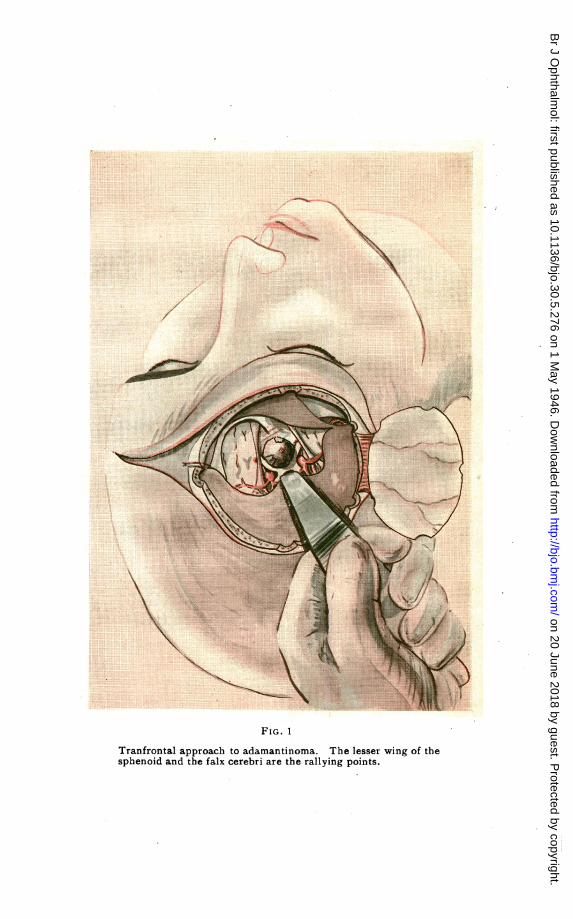
FIG. 1
Tranfrontal approach to adamantinoma. The lesser wing of thesphenoid and the falx cerebri are the rallying points.
on 20 June 2018 by guest. Protected by copyright.
http://bjo.bmj.com
/B
r J Ophthalm
ol: first published as 10.1136/bjo.30.5.276 on 1 May 1946. D
ownloaded from

.~~~~~~~~~~~~~~~~~~~1-f..
.. , . , / }~~~~~~~~g
FIG. 2.
Bilateral paranasal route to intrasellar tumours. Incisionbetween the angular veins. The ethmoidal and optic fora-mina bound the field of operation superiorly.
on 20 June 2018 by guest. Protected by copyright.
http://bjo.bmj.com
/B
r J Ophthalm
ol: first published as 10.1136/bjo.30.5.276 on 1 May 1946. D
ownloaded from

BITEMPORAL HEMIANOPIA 277-
10,000 ophthalmic cases, the presenting symptom is defectivle v'isionover a long period -of time. When a diagnosis has been made, andperhaps established by X-ray examination, the clinical picture isusually clear; for example, of secondary amenorrhoea due tochromophobe adenoma, of acromegaly, infantilism, basophilism orSimond's cachexia.
Pituitary facies depend mainly on skeletal changes, but it is note-worthy that the muscles of the face arise in connection with thehyoid arch and that their adaptation to expression- of the emotionsis more or less peculiar to' the primates. Recognition of subtlechanges produced by endocrine disorders should put us on ourguard. Familiarity with the age incidende of the various types ishelpful, though a tumour anlage may be latent for many years.Evans and Browder in one case had occasion to split the chiasma
in the sagittal plane and divide the anterior communicating artery,in order to gain access to a cystic tumour. Visual acuity of 6/12was retained. Traquair has doubted the supposed bilateral inner-vation of the macula. Pressure on the chiasma concerns the postcalcarine sulci rather than the eye, which may appear perfectlynormal. When present, however, the hemianopic pupillary reac-tion is in my experience of the utmost value, because it providesobjective confirmation.
Let us now consider three individual cases.
Case 1.-A case of adamantinomaResume'.-Eunice G., female, aged 15 years, was found to be
suffering from bilateral 15pafilloedetna and Frohlich's syndrome.Investigation led to a diagnosis of adamantinoma. The tumourwas subsequently removed by a trans-frontal opieration.
Eunice G., aged 15 years, was found at a routine school inspection, to be suffering from bilateral papilloedema. She was referredto the Royal Halifax Infirmary on January 19, 1945, as an emer-gency. The vision had been failing for eighteen months with morerapid deterioration during the past few days (? acute internalhydrocephalus), and there had been headaches and vomiting.Prior to this, she had felt well and was at the top of the class.On examination, Eunice was adipose and infantile; menstrua-
tion had never occurred. Only the inferior nasal quadrants of thefields of vision were intact. The optic discs were swollen and oede-matous and surrounded by exudate and flame-shaped haemor-rhages. There were no other physical signs in the central nervoussystem2 the Babinski reflex was planti-flexor. The Wassermannreaction was subsequently found to be negative.X-ray examination showed an enlarged sella turcica. The pos-
terior clinoids were missing. There was abnormal calcificationwithin and around the sella turcica.
on 20 June 2018 by guest. Protected by copyright.
http://bjo.bmj.com
/B
r J Ophthalm
ol: first published as 10.1136/bjo.30.5.276 on 1 May 1946. D
ownloaded from

#~~~~~~~~~~~~
W. 0. LOIDGE
On January- 24, after pre-medication with 1/100 atropine, 11grains of nembutal and 1/150 of hyoscine, anaesthesia was comrmenced with open ether and continued with nitrous oxide andoxygen only, through an intra-tracheal tube. The trans-frontalapproach was facilitated by, undeveloped frontal sinuses. Afterinfiltration by 075 per cent. procain solution wi.th adrenalin, acoronal incision was made and a temporal osteoplastic flap wasreflected on the right side. Haemostasis was-secured by diathermy.Before opening the dura, the lateral ventricles were emptied bvcisternal puncture. The olfactory bulbs were visible, but owingto the oblique approach, it was not until the faIx cerebri was definedthat orientation became possible. Presently, the optic chiasmacame into view. The anterior cerebral and ophthalmic arteriescould be discerned. The chiasma was pre-fixed, but it was pos-sible to elevate it sufficiently to expose the sella turcica, where a-coralliform tumour with scintillating crystals embedded in it cameinto view (see Fig. 1) and was seized with a pair of ring forceps.As the tumour was calcified and could not be crushed, it seemed atfirst impossible to extricate it without severing one optic nerve, butwith perseverance and a kind of " axis traction," it was eventuallydelivered without that sacrifice. The tumour was the size of acherry and seemed fully to account for the defective fields of vision.No further calcification, was to be seen in the region exposed; itwasthoughtbetternot to probe towards the infundibulum and thirdventricle. The wound was closed in layers, the bone flap beingsecured in position by a suture passed through special drill holes;the customary stainless steel wire sutures were in this case omitted.Blood plasma was administered intravenously.
After operation, the vision improved, and the patient recoveredsufficiently to be shown at a meeting of the Leeds and West RidingMedico-Chirurgical. Society, but she succumbed to recurrence ofthe tumour in June, 1945.
Histology'A microscopic section of the tissue removed is available. It has
been decalcified and stained under the direction of Professor M. J.Stewart who reports as follows:
The great bulk of this tumour is densely calcified and evenossified, but enough soft tissue is present to show its true nature.It is a typical adamantinoma showing beautifully the characteristicmyxoid degeneration of the rete malpighii of the squamous epi-thelium."Adamantinoma or ameloblastoma usually occurs in adolescence.
Appearing in the cleft between the pituitary lobes-, it is of slowgrowth, but may resemble in beh'aviour basal celled carcinoma.
278
on 20 June 2018 by guest. Protected by copyright.
http://bjo.bmj.com
/B
r J Ophthalm
ol: first published as 10.1136/bjo.30.5.276 on 1 May 1946. D
ownloaded from

BITEMPORAL HEMIANOPIA
Case 2.- 'Meningioma of Olivary eminence of sphenoid.Removal. Resuniption of work. Death from
recurrence twelve months later
J.W., aged 35 years, core tnaker, was a man of short stature with'somewhat leonine features and'spatulate fingers. He was the fatherof three children, the youngest eight years of age. In August, 1941,he complained of failing vision. He had had no headaches and novomiting.On examination, vision in the left eye was 6/24 with complete
temporal hemianopia, and acuity in the right eye was less than 6/60owing to a central scotoma, without any peripheral hemianopia.The optic discs were normal except for slight pallor, and nothingabnormal was discovered on examination of the central nervoussystem.An X-ray examination showed erosion of the posterior clinoid
processes and' flattening rather than expansion of the sella turcica.The patient did not consent to operation until November, 1941.
On November 19, 1941, after pre-medication with scopolamine and'morphine, the scalp was infiltrated with novocaine and adrenaline.The patient complained of intolerable itching of the nose and face,for which reason nitrous oxide and oxygen was administered. Afrontal osteoplastic flap was reflected downwards -and outwards'.After puncture of the lateral ventricle, the dura mater was openedalong the lesser wing of the sphenoid. On lifting up the left frontallobe of the brain, a tumour with the optic nerves stretched across itbecame visible. The tumour, which was attached to the olivaryeminence, was about the size of a pigeon's egg.By careful maniputations, it was brought fully into view. With,
a little coaxing, it alinost delivered itself. The-wound was closed,using the central drill hole for drainage for the first twenty-fourhours.On the following morning, the patient was restless and the pulse
rate 120, but he was conscious, -and asked when he would be ableto go home.Thankto o efficient nursing, he made a good recovery.By January, 1942, the visual acuity had improved to right 6/60.left 6/6, and he was contemplating resumption of work.On histological examination by Professor M. J. Stewart, the
tumour proved to be a highly cellular meningioma.The patient made a good recovery and resumed war production
work at Chatham dockyard; but he succumbed to a recurrencewhich caused mental aberration a year later.'
Case 3.-A case of chromophobe adenomaIn 1934, Mrs. H., aged 28 years, who had had amenorrhoea for
eight years, was found to have -bitemporal hemianopia and an
279
on 20 June 2018 by guest. Protected by copyright.
http://bjo.bmj.com
/B
r J Ophthalm
ol: first published as 10.1136/bjo.30.5.276 on 1 May 1946. D
ownloaded from

W. 0. LODGE
enlarged sella turcica. A paranasal operation was performed andradon inserted, as described in the-Brit. Med. 1J., Vol. II, p. 1,257,1936.A temporal island appeared in the right visual field and the left
became normal. Mrs. H., who had adopted a baby in 1934, couldplay tennis.
In 1939' papilloedema developed and bitemporal- hemianopiarecurred. A modified Chiari operation was performed and thetumour removed; a slide is available showin'g a chromophobeadenoma, areas of which have undergone atrophy.'When last seen about a year ago, there was not much vision in
the right eye, but the left was normal. The patient was able toaccompany her husband to London on visits occasionally and leadquite a normal life.My approach to intrasellar tumours is a modification of the para-
nasal route of -Chiari (1911). Bilateral external rhinotomy is car-ried out through a median incision-between the angular veins, asshown in the accompanying plate (see Fig. 2). When the body ofthe sphenoid has been distended by a large adenoma, the tumourcan be excochleated with less risk, in my opinion, than by the trans-frontal route. In the absence of sinusitis, this bilateral approachthrough the ethmoidal and sphenoidal sinuses allows manipula-tion or aspiration through one side and inspection through theother. As the brain is not exposed, it is a comparatively minoroperation.The lateral walls of the orbits are at right angles, the medial walls
parallel. Only the lightest retraction of the orbital contents is per-missible, however, if retinal thrombosis is to be avoided. 'Thirty-eight variations in the relationships of the optic nerves to thesphenoidal cells have been described by Onodi. An imaginaryline connecting the anterior and posterior ethmoidal foramina, onthe inner wall of the orbit, marks the upper boundary of this opera-tion of access. The incision, being well supported, heals with animperceptible scar, and without any tendency to webbing of theorbital margin.
ConclusionsCasualties at this crossroads-so to speak-between vision and
organised life have been discussed. It would seem that the- visualpathway is not the major one.
Normally, a hand held up to the forehead as in saliuting is insight of one eye but out of sight of the other; the shadowy mono-cular image is projected across the chiasma to the anterior third ofthe lateral geniculate body and thence to the antero-inferior extrem-ity of the post-calcarine sulcus. I trust that this simple preliminary
280
on 20 June 2018 by guest. Protected by copyright.
http://bjo.bmj.com
/B
r J Ophthalm
ol: first published as 10.1136/bjo.30.5.276 on 1 May 1946. D
ownloaded from

QUININE AMBLYOPIA 281
test will not only reassure the reader, but also be found of servicein busy clinics.
BIBLIOGRAPHY
EVANS, J. N. and BROWDER, J.-Problem of split macula; study of visual fields.Arch. Ophthal. Chicago, Vol. XXXI, pp. 43-53, January, 1944.
RATHKE, H.-Arch. f. A nat. Physiol. u. Wiss. Med., p. 227, 1839.SOUTTAR, H. S.-New methods of surgical access to the brain. Brit. Med. Jl.,
Vol. XXV, p. 2; Vol. XXVIII, p. 295.TRAgUAIR, H. M.-An introduction to clinical perimetry. Henry Kimpton,
London. P. 66, 1927TDE BEER, G. R.-The comparative anatomy, histology and development of the
pituitary body. Oliver and Boyd, Edinburgh. 1926.WALLS, G. L.-The vertebrate eye. Cranbrook Institute of Science, Bulletin No.
19. Michigan. August, 1942.ONODI, A.-The optic nerve and accessory sinuses of the nose. Baillidre, Tindall
and Cox. London. 1910.
QUININE AMBLYOPIABY
A. BISHAYEGYPT
THE two last years which I spent in Kom-Ombo of Upper Egypt,a district infected with a severe epidemic of malignant malaria, hadgiven me one of the most wonderful chances to study the subject ofquinine amblyopia, and it would have been very difficult undernormal circumstances to give such an account in such a short time.During this period I had seen in my h-pspital and private practice,seven cases of quinine amblyopia, which is relatively a large numberof such cases for an ophthalmologist to see in two years. Dealingwith these seven cases, I have observed some very interesting pointsin examination, diagnosis and treatment, which I am glad to stateto my colleagues.
, SymptomatologyFrom the accompanying table we can conclude(1) The age of the patient does not matter in the case.(2) The amount of the drug (quiinine) must not necessarily be
big as said by de Schweinitz, because out of my seven cases only twotook a large dose of the drug, while the other five developed theiramblyopia by taking ordinary doses; but the outstanding feature isthat all of them took the quinine on an empty stomach. Thus inmy opinion due to these observations, it is not only large doses ofquinine that lead to amblyopia,. but it is some kind of sensitiveness
* Received for publication, October 18, 1945.
on 20 June 2018 by guest. Protected by copyright.
http://bjo.bmj.com
/B
r J Ophthalm
ol: first published as 10.1136/bjo.30.5.276 on 1 May 1946. D
ownloaded from