referral and management guidelines for breast cancers … docs/new... · 2015-04-15 · referral...
TRANSCRIPT

1
August 2012
Date for guideline review: June 2014
Produced by the North Trent Breast Cancer Group
Referral and Management Guidelines for Breast Cancers within North Trent

2
CONTENTS North Tent Cancer Network Breast NSSG Membership List 26th March 2012 ........... 4
Core Members of the NSSG .................................................................................. 4 Members of the NSSG .......................................................................................... 5
1. REFERRAL FROM PRIMARY CARE ................................................................ 9 Teenagers and Young Adults Diagnosed with Breast Cancer .......................... 10
2. ASSESSMENT ................................................................................................ 11 2.1 Introduction .............................................................................................. 11
2.1.1 Breast Imaging Guidelines ...................................................................... 11 2.1.2 Tissue Sampling Guidelines ................................................................... 15
2.2 Pre-Operative Work-Up ........................................................................... 16 2.2.1 Diagnosis ......................................................................................... 16 Triple Assessment - imaging, core biopsy, clinical examination ....................... 16 2.2.2 Pre-treatment Investigations............................................................. 16
2.3 Pathology Reporting ................................................................................ 17 2.4 Staging .................................................................................................... 18
3. PSYCHOLOGICAL CARE, ASSESSMENT AND INTERVENTION ............. 19 Holistic Needs Assessment ............................................................................. 19
4. SURGICAL TREATMENT OF THE BREAST .................................................. 20 4.1 Introduction .............................................................................................. 20 4.2 Indications for Mastectomy ...................................................................... 20 4.3 Relative Indications for Mastectomy ......................................................... 20 4.4 Indications for Breast Conservation ......................................................... 20 4.5 Breast Reconstruction .............................................................................. 21
4.5.1 Guidelines for Immediate Breast Reconstruction .................................... 21 4.5.1.1 Morbid Obesity BMI >35 ...................................................................... 21 4.5.1.2 Patients Requiring Postoperative Radiotherapy ................................... 22 4.5.1.3 Delayed Reconstruction ....................................................................... 22
4.6 Axillary Staging and Treatment ................................................................ 23 4.6.1 Axillary Disease Management ................................................................. 23 4.6.2 Sentinel Node Biopsy ............................................................................. 23 4.6.3 Titanium Clips ......................................................................................... 24 4.6.4 Shoulder Mobility .................................................................................... 24 4.6.5 Lymphoedema Management ............................................................ 24
5. RADIOTHERAPY ............................................................................................ 25 5.1 Introduction .............................................................................................. 25 5.2 Referral for Consideration Of Radiotherapy ............................................. 26
5.2.1 Treatment of Invasive Carcinoma Following Wide Local Excision ........... 26 5.2.2 Treatment of Invasive Carcinoma after Mastectomy ............................... 26 5.2.3 Radiotherapy to the Ipsilateral Supraclavicular Fossa ............................. 26 5.2.4 Treatment to the Axilla ............................................................................ 26 5.2.5 Treatment of Ductal Carcinoma In-Situ (DCIS) Following Wide Local Excision ........................................................................................................... 27 Role of an RT boost to tumour bed: ................................................................. 27
6. PROGNOSIS IN BREAST CANCER ............................................................... 28 BCM = breast cancer mortality estimate without adjuvant therapy ........................... 29 7. SYSTEMIC THERAPY .................................................................................... 30
7.1 Adjuvant Systemic Therapy ..................................................................... 30 7.1.1 Early Breast Cancer Adjuvant Chemotherapy Guidelines ....................... 31 7.1.2 Clinical Guidelines for the Use of Adjuvant Trastuzumab (Herceptin®) ... 33
7.4 Primary Medical Therapy ......................................................................... 37 7.4.1 Neoadjuvant Chemotherapy Policy ......................................................... 37 7.4.2 Neoadjuvant endocrine therapy .............................................................. 40

3
7.4.2 Neoadjuvant Chemotherapy Patient Pathway ......................................... 42 7.5 Treatment for Advanced/Metastatic Disease ............................................ 42 7.5 Treatment for Advanced/Metastatic Disease ............................................ 43
7.5.1 Locally Advanced Breast Cancer ............................................................ 43 7.5.2 Metastatic Breast Cancer ........................................................................ 44 a) Endocrine treatment .................................................................................... 44 b) Chemotherapy ............................................................................................. 45 c) Biological therapy ........................................................................................ 46
Organ Specific Problems in Advanced Breast Cancer ......................................... 46 a) Loco-regional recurrence ............................................................................. 46 b) Bone metastases ......................................................................................... 46 c) Metastatic Spinal Cord Compression ........................................................... 47 d) Liver metastases ......................................................................................... 47 e) Lung metastases ......................................................................................... 47 f) Brain metastases .......................................................................................... 47 g) Pleural effusions .......................................................................................... 48 h) Ascites ......................................................................................................... 48
8. FOLLOW UP ................................................................................................... 49 8.1 Clinical Follow Up .................................................................................... 49
Follow Up protocol for Patients Treated with Curative Intent ............................ 49 8.2 Organisation of Follow Up ........................................................................ 51 8.3 Follow Up Mammography ........................................................................ 51 8.4 Management of Menopausal Symptoms .................................................. 52 8.5 Monitoring Bone Health ........................................................................... 52 8.6 Trials ........................................................................................................ 52
9. FAMILY HISTORY SERVICES ........................................................................ 53 9.1 Genetic Screening ................................................................................... 53
10. Management of Sarcoma of the Breast ............................................................. 54 Appendix A .............................................................................................................. 56
Available agents .................................................................................................. 56 Appendix B .............................................................................................................. 58
Royal College of Pathologists Data Set ............................................................... 58 Appendix C ............................................................................................................. 59
North Trent Cancer Network Teenage and Young Adult Referral Pathway (16-24 years) .................................................................................................................. 59
Appendix D ............................................................................................................. 60 Referral Form for Patients with Metastatic Spinal Cord Compression .................. 60

4
North Tent Cancer Network Breast NSSG Membership List 26
th March 2012
Core Members of the NSSG Chair: Miss Clare Rogers, Consultant Surgeon Deputy Chair: Miss Julia Dicks, Consultant Surgeon
MDT Leads: Barnsley Mr. Soumen Ghosh, Consultant Surgeon Chesterfield Mr. Steve Holt, Consultant Surgeon Doncaster & Bassetlaw Mr. Olumuyiwa Olubowale, Consultant Surgeon Rotherham Miss Pushpa Dudani, Consultant Surgeon Sheffield Dr. Christine Ingram, Consultant Radiologist Oncology Professor Rob Coleman, Professor of Medical Oncology
Deputy Dr. Matthew Hatton, Consultant Clinical Oncologist Histopathology Professor Tim Stephenson, Consultant Histopathologist
Deputy Dr. Anju Verghese, Consultant Histopathologist Imaging Dr. Christine Ingram, Consultant Radiologist
Deputy Dr. Sue Varkey, Consultant Radiologist
NSSG Named Lead for Service Improvement Mr. Inder Kumar, Consultant Surgeon Nurse Representatives Fiona Armitage, CNS Barbara Langdale, CNS Deputy Nurse Representative Alison Allen, CNS NSSG Named Lead for Information for Patients & Carer Alison Allen, CNS Patient Representatives Jacqueline Baker Diane Drabble
NSSG Named Lead for User Issues Barbara Langdale, CNS

5
Members of the NSSG Barnsley Ms. Julia Dicks, Consultant Breast Surgeon Mr. Soumen Ghosh, Consultant Breast Surgeon Mr. Olyinke, Staff Grade Surgeon Dr. Caroline Lee, Consultant Clinical Oncologist Dr. Jhulan Biswas, Consultant Histopathologist Dr Praveen Somarajan, Consultant Radiologist Dr D Ward, Consultant Radiologist Dr D Monaghan, Consultant Radiologist Ms. Alison Allen, Breast Clinical Nurse Specialist Ms. Jane Dolman, Breast Clinical Nurse Specialist Chesterfield Mr. Steve Holt, Consultant Surgeon (Ex Chair) Mr. David Chadwick, Consultant Surgeon Miss Iman Azmy, Consultant Surgeon Dr. Omar Din, Consultant Clinical Oncologist Dr. David Brooks, Consultant in Palliative Care Dr. Sarah Parnacott, Consultant Palliative Medicine Dr. Deirdre McKenna, Consultant Histopathologist Dr. Peter Shephard, Consultant Radiologist Dr. Ashleigh Genever, Consultant Breast Radiologist Dr. Anna Ford, Consultant Breast Radiologist Ms. Helen Scott, Breast Care Sister Ms. Joanne Doncaster, Breast Care Sister Ms. Jackie Conway, Breast Care Sister Ms. Julia Buxton, Departmental Practitioner Ms. Ami Pashley, Health Care Assistant Doncaster & Bassetlaw Dr. S Ramakrishnan, Consultant Oncologist Dr. Kate Dunn, Consultant Oncologist Mr. Kadappa Kolar, Consultant Surgeon Mr. Nazar Kazzazi, Consultant Surgeon Mr. Olumuyiwa Olubowale, Consultant Surgeon Miss Clare Rogers, Consultant Surgeon Mr. G M Wattoo, Associate Specialist Surgeon Miss Asma Husain, Staff Grade Surgeon Mr. Channegowda Navin, Staff Grade Surgeon Dr. Anju Verghese, Consultant Histopathologist Dr. D Ward, Consultant Radiologist Dr. D Monaghan, Consultant Radiologist Dr. Praveen Somarajan, Consultant Radiologist Mrs. Barbara Langdale, Breast Clinical Nurse Specialist Mrs. Christina Dyer, Breast Clinical Nurse Specialist Ms. Susan Rousell, Breast Clinical Nurse Specialist Ms. Jenny Burden, Breast Clinical Nurse Specialist Mrs. Carole Robinson, Breast Clinical Nurse Specialist Ms. Teresa Hull, Breast Clinical Nurse Specialist Mrs. Pauline Foulstone, Lead Nurse

6
Rotherham Miss Pushpa Dudani, Consultant Surgeon/Breast MDT Lead Mr. Mohamad Al-Gailani, Consultant Surgeon Mr Inder Kumar, Consultant Surgeon Dr. Ali Hussain, Consultant Histopathologist Dr Anju Nijhawan, Lead Pathologist for Breast Miss Gazalla Safdar, Associate Specialist for Breast Dr. Sujatha Varkey, Consultant Radiologist Dr. Fraser Cooke, Consultant Radiologist Ms. Michelle Fletcher, Breast Clinical Nurse Specialist Ms. Ann Parkin, Breast Clinical Nurse Specialist Ms. Stephanie Beard, Breast Clinical Nurse Specialist Ms. Carol Barrass, Superintendent Radiographer
Sheffield (including Weston Park) Ms. Lynda Wyld, Consultant Surgeon Prof Malcolm Reed, Consultant Surgeon in Oncology Mr. Stan Kohlhardt, Consultant Surgeon Mrs. Vidya Chandran, Consultant Surgeon Dr. Shirley Crawford, Breast Clinician Dr. Carol Shawcross, Breast Clinician Dr Claire Mills, Breast Clinician Mr. Chris Caddy, Consultant Plastic Surgeon Mr. David Dujon, Consultant Plastic Surgeon Dr. Tim Stephenson, Consultant Histopathologist Dr. Asha Dube, Consultant Histopathologist Dr. Simon Cross, Consultant Histopathologist Dr. Patricia Vergani, Consultant Histopathologist Professor Rob Coleman, Professor of Medical Oncology Dr. Martin Robinson, Clinical Director, Oncology Services Dr. Sundareswaran Ramakrishnan, Consultant Clinical Oncologist Dr. Om Purohit, Consultant Clinical Oncologist Dr Matthew Winter, Consultant Clinical Oncologist Dr. Matthew Hatton, Consultant Clinical Oncologist Dr. Kathleen Dunn, Consultant Clinical Oncologist Dr. Omar Din, Consultant Clinical Oncologist Dr. Christine Ingram, Consultant Radiologist Dr. Ingrid Jolley, Consultant Radiologist Dr Olga Hatsiopoulou, Consultant Radiologist Ms. Fiona Armitage, Clinical Nurse Specialist Ms. Lesley Bruce, Research Manager Dr. Bernadette Foran, Consultant Clinical Oncologist Sister Alison Gray, Breast Clinical Nurse Specialist Sister Kath Flint, Breast Clinical Nurse Specialist Sister Debbie Stanton, Breast Clinical Nurse Specialist Sister Angela Oakley, Breast Clinical Nurse Specialist Jo Beaumont, Breast Clinical Nurse Specialist Kath McLachlan, Breast Clinical Nurse Specialist (Lead) Dr. Jo Statham, Consultant Clinical Psychologist Lisa Dransfield, Breast Services Manager Cath Jones, Superintendent Radiographer/Programme Manager Hayley Williams, Cancer Manager Representative from North Trent Higher Surgical Training Scheme (one per meeting)

7
North Trent Research Nurses Mrs Suzanne Horwood Ms. Kay Nicholson Research Network Ms. Lesley Bruce, Research Network Lead Service Improvement Facilitator Vacancy Advice also provided by: Dr. J Cook, Consultant in Clinical Genetics, Sheffield Children’s Hospital
North Trent Cancer Network Linda Robinson, Administrator Simon Kendrick, Audit Lead Judith Bird, Lead Nurse
8
PREFACE These guidelines have been drawn up by the North Trent Breast Cancer Group for the North Trent Cancer Network. Guidelines for diagnosis, surgical management, multidisciplinary teams and acceptable waiting times are already contained in the British Association of Surgical Oncology (BASO) guidelines for the treatment of Breast Cancer. These are accepted in full for North Trent and have not, therefore, been duplicated here. These guidelines are intended to represent reasonable good practice for the management of patients with breast cancer in North Trent. They are intended as a guide to good practice and not to be applied as a clinical protocol. Whilst the guidelines have been agreed by the relevant clinicians, the pace of the detailed application of the guidelines will depend on local circumstances and the relative priority that any necessary developments are given by the commissioners for each local cancer unit. Version 1 August 1998; Version 2 June 2000; Version 3 June 2001, Version 4 April 2004, Version 5 June 2004. Version 6/7 2004-5, version 8/9 late 2005, Version 10 2006 Version 11July 2009; Version 12 April 2010.
Notes to the guidelines:
NETWORK AUDIT Audit of the Breast Screening Service is well established throughout the Trent region. Network Audit activity commenced in 1999 and will continue to examine clinical practice and service delivery in line with the requirements of the Trent and National Accreditation processes. Trent Cancer Registry defined a Breast Cancer Core Dataset, which was approved by each of the Units and the Centre. Data collection on all breast cancer patients was used for network audit activity. Units will be required to collect this data to comply with national requirements and future cancer accreditation.
9
1. REFERRAL FROM PRIMARY CARE Extracts from 'Referral Guidelines for Suspected Cancer NHS Executive' March 2000 and ‘Best Practice Diagnostic Guidelines for Patients Presenting with Breast Symptoms’ Nov 2010 Key Points
Incidence: Approximately 40 000 new cases p.a. in England and Wales Breast
Breast Cancer is the commonest malignancy to affect women.
Age: Incidence increases with age. 5% of cases occur before 40 years and only 2% before 35 years
A GP with a list size of 2000 patients can expect to see one or two new patients with breast cancer per year, but will see a considerably larger number of women with benign breast problems
Other breast problems include: Diffuse nodularity: common in all age groups up to 50 years Fibroadenoma: peak age range 20 - 30 years Cysts: peak age range 40 - 60 years Breast pain mastalgia: pain alone is a very uncommon presentation of breast
cancer Presenting features of symptomatic cases of breast cancer: Lump 90% Painful lump 20% Nipple change 10% Nipple discharge 3% Skin contour change 5% Note: The guidelines for urgent referral of patients with suspected breast cancer in this document are based on those set out in 'Guidelines for Referral of Patients with Breast Problems' second edition 1999 prepared by Joan Austoker and Robert Mansel under the auspices of the NHS Breast Screening Programme and the Cancer Research Campaign. Breast Cancer: Guidelines for Urgent Referral
Patients with a discrete lump in the appropriate age group (e.g. age >30)
Signs which are highly suggestive of cancer such as: o Ulceration o Skin nodule o Skin distortion o Nipple eczema o Recent nipple retraction or distortion (< 3 months) o Unilateral nipple discharge which stains clothes

10
Conditions that require referral - but not necessarily urgently
Discrete lump in a younger woman (e.g. age < 30 years)
Asymmetrical nodularity that persists at review after menstruation
Abscess
Persistently refilling or recurrent cyst
Intractable pain not responding to reassurance, simple measures such as wearing a well supporting bra and common Drugs
Nipple Discharge Age < 50 with bilateral discharge sufficient to stain clothes
Age < 50 with bloodstained discharge
Age > 50 with any nipple discharge Current Department of Health guidelines are to see all breast referrals within 2 weeks. For patients with suspected cancer, the decision to treat, to date of first treatment, should be within 31 days. The date of GP referral to the date of first treatment should be within 62 days.
Teenagers and Young Adults Diagnosed with Breast Cancer
All patients between the ages 16 and 24 diagnosed with breast cancer should be referred to the TYA MDT for discussion. Patients aged 16-18 should be referred to the Sheffield Teaching Hospitals (STH) site specific MDT for treatment. Patients over 18 may be given a choice of where they would like to be treated. See Appendix C for referral pathway.
Age at Diagnosis for Adult Breast Cancer Proportions presenting in different age groups <40 40-49 50-59 60-69 70-79 80+ 5 15 23 24 17 16 %
Source: Office of National Statistics 1992 Cancer Statistics Registrations Series MBI No. 37 published 2006
Estimates of the Numbers of Urgent Referrals (Adults)
Benign: New E&W DGH * DGH * Malignant Cancers Urgent per annum per week Referrals 15:1 16,000 256,000 1020 20 *Based on a DGH serving a population of 200,000
11
2. ASSESSMENT
2.1 Introduction The following guidelines supplement those that are contained in the BASO guidelines on diagnosis and surgical intervention for patients with Breast Cancer.
2.1.1 Breast Imaging Guidelines
Diagnosis of breast disease should occur in a multidisciplinary setting using the principles of triple assessment: clinical assessment, imaging, and core biopsy. This is best achieved in designated breast clinics in which both radiologists and surgeons work together. Direct access from GPs and other physicians for breast imaging is not recommended, without triple assessment. Triple assessment clinics should:
Provide rapid access with facility to identify and prioritise those with a high suspicion of malignancy
Be organised to ensure that all necessary diagnostic procedures are completed at the initial visit. Where not possible, imaging and core biopsy should ideally be performed and reported within three to five working days
Imaging should precede a needle aspiration or tissue sample procedure except in special circumstances. Imaging should be performed only where there is a clear clinical indication to do so. Inappropriate requests should be monitored and subject to audit. (Guidance on screening and symptomatic breast imaging, Royal College of Radiologists, June 2003). These network guidelines are designed to indicate the minimum standards to be achieved by individual units. Each unit may produce more detailed guidelines, which will be tailored to their particular circumstances. All units involved in the NHSBSP will already be subject to QA assessment of their practice and are expected to be compliant with these screening standards. Women who do not need always need imaging: 1 Breast Pain alone is not an indication for imaging (except focal breast pain) 2 Bilateral pain and symmetrical nodularity 3 Symmetrical nodularity alone Women under 25 Focal lumps should have either clinical core biopsy if deemed necessary or ultrasound +/- core biopsy of focal masses.

12
Women age 25 - <40 Ultrasound is the first examination of choice. All solid lesions should be subject to tissue sampling (core biopsy). Mammography is indicated in strongly suspicious cases and in all cases found to be malignant on biopsy, to exclude other incidental lesions. Women age 40 + Mammography plus ultrasound of any lumps. Core biopsy of all solid lesions. Imaging of the axilla All women with breast cancer should have ultrasound of the axilla and biopsy of any lymph nodes which show:
Localized bulge
Loss of node hilum
Complete loss of morphology
Cortical thickness >3mm Advanced breast cancer in the elderly It is only necessary to undertake tests that will affect management. This may in many cases be limited to a clinical core biopsy to test ER status. If size of lesion needs to be monitored because of primary endocrine therapy, an individual decision should be made on each patient to decide if clinical examination, mammographic or ultrasound measurement is most appropriate. Large or advanced breast cancer These patients may be candidates for neoadjuvant chemotherapy. Full investigation should be undertaken to determine the extent of disease, which may include breast MR and metastatic screening. It may be appropriate to insert a marker in the breast to mark the site of the tumour, if the intent of neoadjuvant therapy is breast conservation. This is best introduced after two cycles of chemotherapy, and is only necessary if the tumour is responding to treatment. It is only useful if there is a probability that there will be no detectable tumour to localise at the time of surgery. More detailed discussion of the management of neoadjuvant chemotherapy patients is provided in a separate section. Men with breast lumps The vast majority of these cases are due to gynaecomastia. Asymmetrical gynaecomastia does not require imaging. Focal lumps in the breast area are usually amenable to clinical core biopsy alone. Imaging is not always indicated. Follow-up mammography If follow-up mammography is delegated to the breast imaging department, clear protocols should be in place for assessment of any abnormality and for referral on to the surgical clinic and MDT meeting.

13
‘High-Risk’ follow-up Women with a diagnosis of Radial Scar should have excision biopsy to exclude cancer. Women with atypical ductal hyperplasia, lobular neoplasia, columnar cell change with atypia or multiple intra-ductal papillomata should have a risk assessment taking into account age and family history. The follow-up protocol should usually include yearly mammography between ages 40 and 50. Women with a family history of breast cancer should be managed according to NICE Clinical Guideline 41 (see below). Women found to have ‘risk lesions’ within the NHSBSP should continue with three-yearly screening mammography. There is no evidence that enhanced screening of these women has any effect on mortality. Family history screening For women satisfying the NICE criteria for moderate risk, mammographic screening should be carried out to the standards laid out by the NICE publication, i.e. annually from age 40, and subsequently within the NHSBSP. Very high risk patients should have MRI screening as per NICE Clinical guideline 41. Women who are known to be definite gene carriers may have mammograms performed every 18 months after age 50. Women who are definite carriers of the p53 gene mutation should not have X ray mammography but should be offered annual MRI screening from age 20 to age 70. http://www.nice.org.uk/guidance/CG41 Metastatic screening Formal staging investigations should only be done if this will definitely affect the primary treatment of the disease. In the vast majority of cases, local control will be more important. Screening of asymptomatic patients should be avoided as far as possible. Metastatic screening must not be allowed to delay the first therapeutic intervention. If indicated, a ‘metastatic screen’ should include:
CT chest and abdomen
Isotope bone scan (refer to local protocol, may not be required)
Blood tests (LFTS, Ca, FBC) OR:
Chest x-ray
Liver ultrasound
Isotope bone scan
Blood tests (LFTS, Ca, FBC) CT is indicated in cases where there may be involvement of supraclavicular nodes/ heavy axillary lymph node burden. Indications for a metastatic screen include clinical suspicion of metastases and locally advanced disease (T3, T4, N1 or above, inflammatory change). Post operative staging is required for patients with heavy lymph node involvement i.e. four or more positive nodes following axillary staging.

14
Indications for MRI scanning 1. Women with high risk family history of breast cancer (according to NICE Clinical
Guideline 41) 2. Consider pre-operative MRI in women with lobular cancer who would prefer
breast conserving surgery, to assess the tumour size (NICE Clinical Guideline 80, Early and Locally Advanced Breast Cancer, 2009).
3. If there is a discrepancy regarding the extent of disease after triple assessment, in order to plan treatment.
4. Pre- and post- neoadjuvant chemotherapy according to local protocols. 5. To investigate the integrity of breast implants (non-contrast). Indications for PET scanning
1. Assessment of suspected malignant infiltration in patients with a painful brachial plexopathy and known breast carcinoma where conventional imaging is equivocal and the result of the PET CT would make a significant difference to patient outcome
2. For more accurate staging in patients with potentially operable advanced disease to exclude occult metastases when conventional imaging is equivocal or indeterminate
3. For assessment of suspected recurrent disease when conventional imaging is equivocal or negative
All cases to be referred for PET CT only after discussion in a Breast MDT Referral to a different MDT in the Network or outside Network referral Following Primary care referral into the named receiving hospital most patients receive all their care within that unit. Very rarely there are referrals to a different MDT within the network but if this is required the procedure that will be followed is via the established NTCN inter trust transfer policy. If patients request out of network referral for other treatment such as radiotherapy, these patients are referred to the appropriate MDT for treatment and discussion there

15
2.1.2 Tissue Sampling Guidelines
Breast assessment should follow the principles of triple assessment. Discrete focal lesions should be subjected to core biopsy. In some circumstances FNA may be regarded as sufficient. For impalpable lesions, core biopsy should be carried out under image guidance by an appropriately trained person. For palpable lesions, either a radiological or clinical core biopsy may be appropriate, bearing in mind that concordance may be more difficult to achieve with clinical cores than with image assisted biopsy (allowing accurate targeting of the suspect area). In selected elderly unfit patients with advanced disease, a clinical core biopsy alone may yield sufficient information to start treatment, by confirming the diagnosis and giving the ER status. Punch biopsy is the most appropriate method in very superficial lesions and for nipple abnormalities such as suspected Paget’s disease. Lesions which do not need pathological confirmation, because of their pathognomonic appearance on imaging, include: 1 Typical calcified fibroadenomata. 2 Small lipomata entirely within a fatty nodule. 3 Fat necrosis in which fat has been aspirated from an oil cyst. 4 Fibroadenolipomata. 5 Typical benign lymph nodes.

16
2.2 Pre-Operative Work-Up
2.2.1 Diagnosis
Triple Assessment - imaging, core biopsy, clinical examination
All 3 may not be indicated if clinically locally advanced.
Preoperative diagnosis, without excision biopsy, is the ideal management
Mammography and/or ultrasound, core biopsy and clinical examination should be viewed together
Results should be considered by the MDT (and if necessary the patient seen in the joint oncology clinic) before proceeding to definitive surgery and for discussion of adjuvant treatment
Excision biopsies should be avoided if possible but may be necessary when Triple Assessment is inconclusive or non-concordant
2.2.2 Pre-treatment Investigations
Clinical stage of disease Staging investigations
Pre-invasive [in situ] / Primary operable breast cancer
Histological diagnosis of breast cancer Bilateral mammography USS +/- Core biopsy or FNA axilla Full blood count Urea & electrolytes Liver function tests Serum calcium Consider MRI for Lobular Ca suitable for breast conservation
Locally advanced breast cancer As above + CT chest and abdomen Isotope bone scan (refer to local protocols) or Liver ultrasound scan & Chest XR
Clinically metastatic breast cancer Histological diagnosis if possible
Discuss further investigations in MDT
Investigate according to individual symptoms and treatment plan
Definitions
Pre-invasive breast cancer i.e. ductal carcinoma in situ Primary operable breast cancer
Lump ≤ 5 cm, no evidence of inflammatory changes in the breast and no clinically malignant lymphadenopathy.
Clinically locally advanced breast cancer
Lump > 5 cm, presence of peau d’orange, inflammatory skin changes, deep fixation, or obvious malignant fixed/ matted lymphadenopathy.
Clinically metastatic breast cancer
e.g. pathological fracture, pleural effusion, hepatomegaly neurological symptoms or signs, distant lymphadenopathy (including ipsilateral supraclavicular fossa).
17
2.3 Pathology Reporting The Royal College of Pathologists cancer dataset, ER status and HER 2 status should be recorded routinely for all patients undergoing surgery (see Appendix B). HER 2 status should not be tested routinely in patients aged 80 or over but testing may be requested following MDT discussion, if a positive result would change management. If a patient is not fit for Trastuzumab therapy, HER 2 status should not be requested and the decision should be discussed at the MDT meeting. HER 2 status should be considered in patients with metastatic disease for whom HER 2 status is not previously available. ER status of all invasive cancers should be assessed locally by immunohistochemistry and reported according to the Allred quick score:
0-2 negative
3-4 weakly positive
5-8 strongly positive
ER status of DCIS may also be assessed if clinically requested. 10% or higher nuclear staining of any intensity in the DCIS is considered positive, although the exact % of nuclei staining should also be stated in case the threshold is revised nationally. HER 2 should be assessed by referral to STH (RHH site) for immunohistochemistry and performance of FISH in equivocal cases. For both ER and HER 2, the diagnostic needle core is the preferred specimen. PR status may be assessed by local immunohistochemistry in clinically selected ER negative cases (NICE Clinical Guideline 80, 2009). For this, the tumour excision is preferable to the needle core as PR expression can be very heterogeneous. The minimum requirement for a level 2 cancer centre is that data should be searchable i.e. recorded in a synoptic format which is easier to read and better for data completeness.

18
2.4 Staging The TNM Classification is recommended and this is commonly simplified into stage groupings. Primary tumour size is usually based on clinical and or mammographic assessment. The stage groupings can take account of pathological tumour size: Stage 1 Primary 2 cm or less (T1) no nodes (N0) no mets (M0) Stage 2 Primary tumour 2 – 5 cm (T2) or primary tumour more than 5 cm with no
chest wall or skin involvement (T3) or mobile ipsilateral axillary node involvement (N1) or a combination of the above T and N categories.
Stage 3 Anything more, short of distant metastases. (T4 and/or N > 1, but always
M0). Chest wall or skin involvement is T4. Stage 4 Distant mets. (But see below for those in whom it is thought reasonable to
investigate for this). SCF node involvement is classed as distant metastases (Stage 4).
TNM CLINICAL CLASSIFICATION
T Primary Tumour
Tx cannot be assessed
T0 no evidence of primary
Tis ca in situ
T1mic micro invasion
T1 Tumour 2cm or less in greatest dimension
T2 Tumour more than 2cm but not more than 5cm in greatest dimension
T3 Tumour more than 5cm in greatest dimension
T4 Tumour of any size with direct extension to chest wall or skin
N Regional Lymph Nodes
NX Regional lymph nodes cannot be assessed (e.g. previously removed)
N0 No regional lymph nodes metastasis
N1 Metastasis to moveable ipsilateral axillary node(s)
N2 Metastasis to moveable ipsilateral axillary node(s) fixed to one another or
to other structures
N3 Metastasis to ipsilateral internal mammary lymph node(s)
M Distant Metastasis
MX Distant metastasis cannot be assessed
M0 No distant metastasis
M1 Distant metastasis (Ref: TNM Classification of Malignant Tumours, 6th Edition, - UICC).
19
3. PSYCHOLOGICAL CARE, ASSESSMENT AND INTERVENTION
All members of the breast cancer clinical team should have completed an accredited communications skills training programme. All patients with breast cancer should:
- be assigned to a named breast care nurse specialist who will support them throughout diagnosis, treatment and follow-up (known as the patient’s ‘Key Worker’.)
- be offered prompt access to specialist psychological support and, where appropriate, psychiatric services. (NICE Clinical Guideline 80, 2009)
Holistic Needs Assessment
This has been developed from a key recommendation of the NICE guidance on Improving Supportive and Palliative Care to help patients and healthcare professionals to identify and address the needs of cancer patients and should be undertaken at key points within the patient pathway. Holistic Needs Assessment consists of: An approved assessment tool; for example -SPARC (Sheffield Profile for
Assessment and Referral for Care) questionnaire
Conversation
Action planning
More information can be found at: http://www.northtrentcancernetwork.nhs.uk/media/13185/final%20hna%20launch%20letter%20-%201st%20june%202009.pdf
20
4. SURGICAL TREATMENT OF THE BREAST
4.1 Introduction Wherever possible, patients should be offered a choice between breast conservation surgery and mastectomy. Where mastectomy is to be performed, breast reconstruction should be offered if appropriate. With regard to breast reconstruction and oncoplastic techniques, each unit will develop its own method of service provision and guidelines. (See below) The guidelines given below are based on current evidence and practice in the UK. However, guidelines are not rigid and patient variables and choices are of the utmost importance in planning individualised surgical treatment.
4.2 Indications for Mastectomy
Patient choice
Tumour >5cm (on clinical examination unless there is a clear discrepancy between this and the size on pre operative imaging)
Multi focal disease in more than one quadrant of the breast
Contra indication to radiotherapy
Failed breast conservation surgery e.g. local recurrence or positive margins after wide local excision where further wide local excision is not feasible)
4.3 Relative Indications for Mastectomy
Where breast conservation is unlikely to result in an acceptable cosmetic outcome e.g. larger tumour in a small breast
Where radiotherapy may be associated with a high risk of complications.
Central breast cancer. It is generally accepted that adequate margins are more difficult to achieve with central breast tumours and that central wide local excision may be associated with a relatively poor cosmetic outcome. However, in many cases of central tumours an adequate excision and good cosmetic can be achieved by a central wide local excision.
4.4 Indications for Breast Conservation
Patient Choice
Operable tumour up to 5cm diameter
Operable multi focal tumour restricted to single breast quadrant
No contraindications to radiotherapy
Larger tumours may be treated by breast conservation surgery when combined with oncoplastic procedures such as rotation of local breast parenchymal tissue, therapeutic mammoplasty or latissimus dorsi mini-flap
Following neoadjuvant chemotherapy or hormonal therapy specifically aimed at reducing tumour size. See section 7
Intra-operative specimen radiography is mandatory for impalpable lesions requiring radiological localisation, and recommended for all wide local

21
excision procedures. (BASO 2009)
For Invasive cancer a margin of 1mm should be adequate.
For DCIS a minimum of 2mm radial margin of excision is recommended, with pathological examination to NHSBSP reporting standards. (NICE 2009)
4.5 Breast Reconstruction Breast reconstruction can be performed as a primary procedure at the time of mastectomy or wide local excision or can be performed as a delayed procedure again after mastectomy or conservation surgery with a poor cosmetic outcome. Breast Surgeons may perform breast reconstruction either alone or in collaboration with the reconstructive surgeons (Mr. C Caddy, Mr. D Dujon, Mr. M Brotherston, Mr. D Ralston)
4.5.1 Guidelines for Immediate Breast Reconstruction
Immediate breast reconstruction may reduce the psychological morbidity of mastectomy. Contraindications There are few absolute contraindications to immediate breast reconstruction. It is important, however, that realistic expectations about outcome are ensured prior to surgery, to avoid dissatisfaction and negative psychological outcome post-surgery. For some women, a delayed breast reconstruction may be considered to allow more time to make fully informed choices/decisions. The following list of factors has been shown to be associated with poor results from primary reconstruction and if present these patients would be better served by considering a delayed breast reconstruction.
1. Significant co morbidity 2. Insulin dependent diabetes 3. Peripheral vascular disease 4. Chronic obstructive airways disease 5. Ischaemic heart disease
These conditions may not render the patient unfit for mastectomy, but may be a contraindication to the more major procedures associated with latissimus dorsi, myocutaneous flap and implant replacement. An anaesthetic opinion should be sought at an early stage in such patients.
4.5.1.1 Morbid Obesity BMI >35
There is evidence that anaesthetic and wound complications are significantly increased in these patients.

22
4.5.1.2 Patients Requiring Postoperative Radiotherapy
Post operative radiotherapy may be advised after mastectomy if there is a combination of a grade 3 tumour with 3 or more nodes, if the posterior margin is involved, if the tumour is inflammatory, or T4 or if the tumour is greater than 5cm in size. In these women, delayed reconstruction may be preferred to immediate if an implant based option is to be utilized. In women who request immediate reconstruction, some indication of whether post operative chest wall radiotherapy may be advised can be achieved by requesting a grade on the pre-operative core biopsy and by undertaking an axillary node sample or sentinel node biopsy prior to undertaking definitive surgery. When a decision has been made pre-operatively or intra-operatively that locally advanced disease will require postoperative radiotherapy, consideration should be given to a delayed reconstruction. This should be discussed with clinical oncologists and where appropriate reconstructive surgeons. Locally advanced disease or poor prognosis breast cancer should not be regarded as an absolute contra-indication to breast reconstruction.
4.5.1.3 Delayed Reconstruction
It is most important that patients considering primary or delayed reconstruction have adequate support and are fully informed regarding the likely cosmetic outcome and the risk of complications. It is important that patients’ expectations are realistic in order to avoid dissatisfaction.

23
4.6 Axillary Staging and Treatment Virtually all patients treated surgically for invasive breast cancer require staging of the axilla.
4.6.1 Axillary Disease Management
This depends on the risk of progression
Risk of Axillary Disease
Treatment
Involved Lymph Node:
Positive clinical core biopsy/ FNA from a palpable or radiologically suspicious axillary node
Axillary node clearance
Unknown Risk:
USS and clinical exam of axilla normal
Sentinel node biopsy or axillary node sampling Axillary node clearance at patient request
4.6.2 Sentinel Node Biopsy
Sentinel node biopsy is a less invasive way of staging the axilla. Patients found to have negative sentinel nodes require no further treatment to the axilla. Those with
positive sentinel nodes (micro and macrometastases) should be offered further
treatment to the axilla(treat patients with isolated tumour cells as node negative as
per NICE CG 80). Sentinel node biopsy using radiocolloid and blue dye is the preferred method of staging lower risk patients. Training and initial audit is through the Royal College of Surgeons of England New Start programme. If sentinel node mapping fails i.e. no radiocolloid or blue dye reaches the axillary nodes, the surgeon should proceed with a four node sample. Centres offering intra-operative sentinel node assessment can offer axillary clearance at the same operation for patients whose sentinel node(s) shows metastases. There is recent evidence to suggest that axillary clearance may not be necessary for all node positive patients having breast conservation. Axillary clearance should only be omitted as part of a clinical trial. (American College of Surgeons Oncology Group. Locoregional Recurrence After Sentinel Lymph Node Dissection with or without Axillary Dissection in Patients with Sentinel Lymph node metastases Annals of Surgery 2010;252(3):426-433)

24
4.6.3 Titanium Clips
Surgical clips should be inserted in the excision cavity and axillary clearance site to facilitate treatment planning for radiotherapy. Axillary clip(s) are to be positioned at the apex of the axilla, and two clips are inserted into the perimeter of the excision cavity at each margin (superior, lateral, inferior, medial and deep). Surgical clips may interfere slightly with scattering effect on MR images.
4.6.4 Shoulder Mobility
Guidelines written in association with physiotherapists should be given to patients undergoing surgery in order to facilitate shoulder mobility. Patients with pre-existing shoulder problems should be identified and measures taken to limit adverse effects. This can be achieved by providing instructions on functional exercises, which, ideally, should start the day after surgery. Referral to the physiotherapy department should be made in patients with persistent reduction in arm and shoulder mobility after breast cancer treatment.
4.6.5 Lymphoedema Management
Patients should be informed of the risks of developing lymphoedema post breast cancer surgery, especially if axillary clearance is to take place; this must include information and advice on how to prevent infection/trauma that may exacerbate lymphoedema. Leaflets and access to lymphoedema services should be readily available for patients with early breast cancer.
25
5. RADIOTHERAPY
5.1 Introduction Below is the list of referral guidelines for consideration of radiotherapy. In addition, referral should take into consideration inclusion in clinical trials like SUPREMO in which radiotherapy is a treatment option.
A number of factors are relative contra-indications to radiotherapy and should be discussed with a clinical oncologist pre-operatively
Poor performance status, mental or physical frailty, vertigo, significant respiratory or cardiovascular impairment may make administration of radiotherapy inappropriate
Local anatomical factors such as poor shoulder movements, deformed chest wall, chronic intertrigo or large, pendulous breasts may make the practical administration of radiotherapy difficult or undesirable
Systemic disorders such as auto-immune disease may render the administration of radiotherapy hazardous
It should be noted that:
1. The dose/fractionation for radiotherapy will take into account type of surgery, pathology, breast size and the presence of a prosthesis.
2. When planning radiotherapy imaging of the heart will be used to minimise myocardial radiation.
26
5.2 Referral for Consideration Of Radiotherapy
5.2.1 Treatment of Invasive Carcinoma Following Wide Local Excision
All patients with invasive carcinoma should be considered for radiotherapy to the breast when clear circumferential resection margins have been confirmed pathologically. For those under the age of 50 or close surgical margins, a boost to the tumour bed may be indicated. These patients may also be suitable for the IMPORT High study.
5.2.2 Treatment of Invasive Carcinoma after Mastectomy
Chest wall radiotherapy is estimated to give a constant relative risk reduction of local recurrence, of approximately 50%, in all situations and patients with invasive carcinoma should be considered for radiotherapy to the chest wall when
1. > 5cm (T3) primary tumour. 2. Positive resection margins of the mastectomy specimen. 3. Skin involvement – including skin tethering and peau d’ orange. 4. Four or more positive lymph nodes 5. Other factors indicating an increased chance of local recurrence are vascular
invasion, Grade III tumour and 1 – 3 nodes positive. The role of radiotherapy for these factors is being explored in the SUPREMO study and radiotherapy could be offered at clinician's discretion.
5.2.3 Radiotherapy to the Ipsilateral Supraclavicular Fossa
This should be considered in patients with > 4 involved nodes. .
5.2.4 Treatment to the Axilla
This should be considered in patients who have a positive axillary node sample or sentinel node and when surgical clearance has not been performed in patients with invasive disease.

27
5.2.5 Treatment of Ductal Carcinoma In-Situ (DCIS) Following Wide Local Excision
Following the EBCTCG meta-analysis in 2010, an improvement in ipsilateral local recurrence (invasive and non-invasive) was seen despite any prognostic factor (15.2% absolute reduction in any ipsilateral breast event, HR 0.46). The greatest benefit was seen in women over the age of 50 years, though age under 50 is still an important adverse prognostic factor.
Therefore all patients should be referred to a clinical oncologist post wide local excision for discussion regarding the benefits of adjuvant radiotherapy, compared to the risks. It would be appropriate to omit radiotherapy in “low risk” group, in which all the following are present: Small size (i.e. <15mm) Clear margins (>2mm) Non-high/intermediate grade No necrosis
Role of an RT boost to tumour bed:
This should not be considered routinely. Suitable patients may be entered into the BIG 3-08 trial evaluating the role of hypofractionation and boost in DCIS.
28
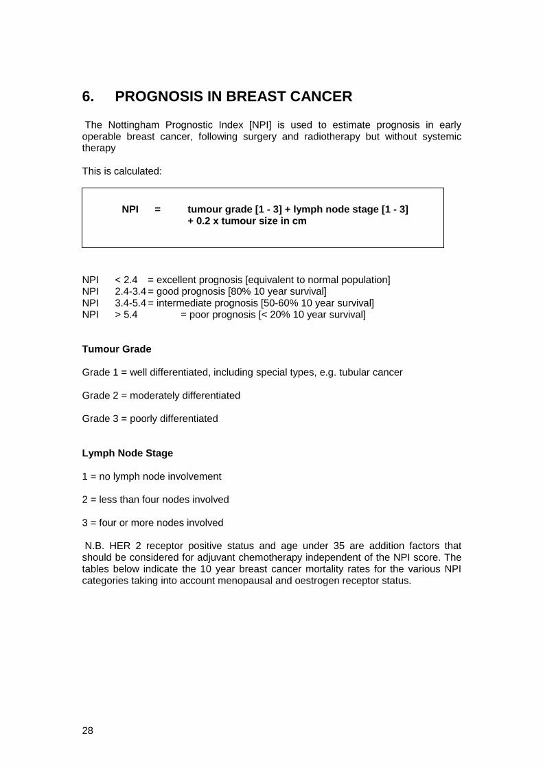
6. PROGNOSIS IN BREAST CANCER The Nottingham Prognostic Index [NPI] is used to estimate prognosis in early operable breast cancer, following surgery and radiotherapy but without systemic therapy This is calculated:
NPI < 2.4 = excellent prognosis [equivalent to normal population] NPI 2.4-3.4 = good prognosis [80% 10 year survival] NPI 3.4-5.4 = intermediate prognosis [50-60% 10 year survival] NPI > 5.4 = poor prognosis [< 20% 10 year survival] Tumour Grade Grade 1 = well differentiated, including special types, e.g. tubular cancer Grade 2 = moderately differentiated Grade 3 = poorly differentiated Lymph Node Stage 1 = no lymph node involvement 2 = less than four nodes involved 3 = four or more nodes involved N.B. HER 2 receptor positive status and age under 35 are addition factors that should be considered for adjuvant chemotherapy independent of the NPI score. The tables below indicate the 10 year breast cancer mortality rates for the various NPI categories taking into account menopausal and oestrogen receptor status.
NPI = tumour grade [1 - 3] + lymph node stage [1 - 3] + 0.2 x tumour size in cm

29
Pre-menopausal women
ER + ve ER - ve
Excellent risk (<2.4) – 3% 10yr BCM Very good risk (2.4-3.39) –8-9% 10yr BCM Good risk (3.4-4.39) –13-17% 10yr BCM Intermediate risk (4.4-5.39) –33% 10yr BCM Poor risk (>5.4) – 52% 10yr BCM
Postmenopausal women
ER+ ve ER- ve Excellent risk (<2.4) –3% 10yr BCM Very Good risk (2.41-3.4) –8% 10yr BCM Good risk (3.4-4.39) – 17% 10yr BCM Intermediate risk (4.4.-5.39) –30% 10yr BCM Poor risk (>5.4) –55% 10yr BCM
BCM = breast cancer mortality estimate without adjuvant therapy
Excellent risk (<2.4) – 6% 10yr BCM
Very good risk (2.4-3.39) – 14% 10yr BCM Good risk (3.4-4.39) – 30% 10yr BCM Intermediate risk (4.4-5.39) –33-44% 10yr BCM Poor risk (>5.4) – 60% 10yr BCM
Excellent risk (<2.4) – 6% 10yr BCM Very Good risk (2.4-3.39) – 11% 10yr BCM Good risk (3.4-4.39) –17%-29% 10 yr BCM
Intermediate risk (4.4-5.39) – 32-44% BCM
Poor risk (>5.4) –58% 10yr BCM

30
7. SYSTEMIC THERAPY
7.1 Adjuvant Systemic Therapy A combination of the Nottingham Prognostic Index (NPI) and receptor (ER + /or PR, HER 2) status is now used to guide the selection of adjuvant treatments. From the tables on the next two pages it is clear that age, HR, and the NPI define those patients who require adjuvant systemic therapy and whether this is endocrine therapy, chemotherapy or both. N.B. ER negative patients should only be considered HR negative if both ER and (if measured) PR are negative. ER Status using the Allred Score
0-2 negative
3-4 weakly positive
5-8 strongly positive Endocrine treatment is indicated for all patients with an Allred score of 3 or above. The threshold for chemotherapy differs between weakly and strongly positive tumours.

31
7.1.1 Early Breast Cancer Adjuvant Chemotherapy Guidelines
Adjuvant Treatment Decision Protocol Premenopausal women
ER RICH ER POOR ER- (H >100; Allred ≥6) (H 50 -100; Allred 3-5) (H <50; Allred <3)
Excellent risk (<2.4) (10) Excellent risk (<2.4) (5)
- TAM only or nil - TAM only or nil
Very good risk (2.4-3.39) (45) Very good risk (2.4-3.39) (10)
- TAM +/- OS - TAM +/- OS
Good risk (3.4-4.39) (60) Good risk (3.4-4.39) (15)
- TAM +/- OS - TAM +/- OS
- FEC x 4 then Herceptin if HER 2+ - FEC x 6
- FEC x 6 then Herceptin if HER 2+
Intermediate risk (4.4-5.39) (50) Intermediate risk (4.4-5.39) (15)
- TAM +/- OS - TAM +/- OS
- FEC if HER2- - FEC x 6 if HER2-
- TCarbo x 6 + Herceptin if HER2+ - TCarbo x 6 + Herceptin if HER2+
Poor risk (>5.4) (35) Poor risk (>5.4) (10)
- OS + TAM - OS + TAM
- FEC or TCyclo or TAC if HER2- - TAC x 6
- TCarbo x 6 + Herceptin if HER2+ - TCarbo x 6 + Herceptin if HER2+ Risk score = Nottingham Prognostic Index
+ 1 if age ≤35. Epi-Vinorelbine may be substituted for FEC in women with fertility concern
Excellent risk (<2.4) (5)
- No treatment
Very good risk (2.4-3.39) (20)
- No treatment if HER 2-
- FEC x 4 then Herceptin if HER 2+
Good risk (3.4-4.39) (25)
- FEC x 6
- FEC x 6 then Herceptin if HER 2+
Intermediate risk (4.4-5.39) (30)
- TCyclo x 6
- TCarbo x 6 + Herceptin if HER2+
Poor risk (>5.4) (25)
- TAC x 6
- TAC x 6 then Herceptin if HER 2+

32
Adjuvant Treatment Decision Protocol
Postmenopausal women
ER RICH ER POOR ER -ve
(H >100; Allred ≥6) (H 50 -100; Allred 3-5) (H <50; Allred <3)
Excellent risk (<2.4) (30) Excellent risk (<2.4) (5)
- TAM only or nil - TAM only or nil
Very Good risk (2.41-3.4) (100) Very Good risk (2.41-3.4) (10)
- TAM - AI switch - TAM – AI switch
Good risk (3.4-4.39) (90) Good risk (3.4-4.39) (15)
- AI for 5 years - AI for 5 years
- FEC x 4 then Herceptin if HER 2+ - FEC x 6 + Herceptin if HER 2+
Intermediate risk (4.4.-5.39) (80) Intermediate risk (4.4.-5.39) (10)
- AI for 5 years - AI for 5 years
- FEC x 6 if HER2- - FEC x 6 if HER2-
- FEC x 6 then Herceptin if HER2+ - FEC x 6 then Herceptin if HER2+
Poor risk (>5.4) (50) Poor risk (>5.4) (10)
- AI for 5 years - AI for 5 years
- FEC x 6 if HER2- or - FEC x 6 if HER2- or
- TAC x 6 (under age 60) - TAC (under age 60)
- TCyclo x 6 (over age 60) - TCyclo x 6 (over age 60)
- TCarbo x 6 + Herceptin if HER2+ - TCarbo x 6 + Herceptin if HER2+
Figures in ( ) are the approximate numbers of patients in each prognostic group presenting across North Trent each year.
Excellent risk (<2.4) (5)
- No treatment
Very good risk (2.4-3.39) (20)
- No treatment
- FEC x 4 then Herceptin if HER 2+
Good risk (3.4-4.39) (40)
- FEC x 6
- FEC x 6 then Herceptin if HER 2+
Intermediate risk (4.4-5.39) (45)
- FEC x 6
- FEC x 6 then Herceptin if HER2+
Poor risk (>5.4) (40)
- TAC x 6 (under age 60)
- TCyclo x 6 (over age 60)
- TCarbo x 6 + Herceptin if HER 2+
33
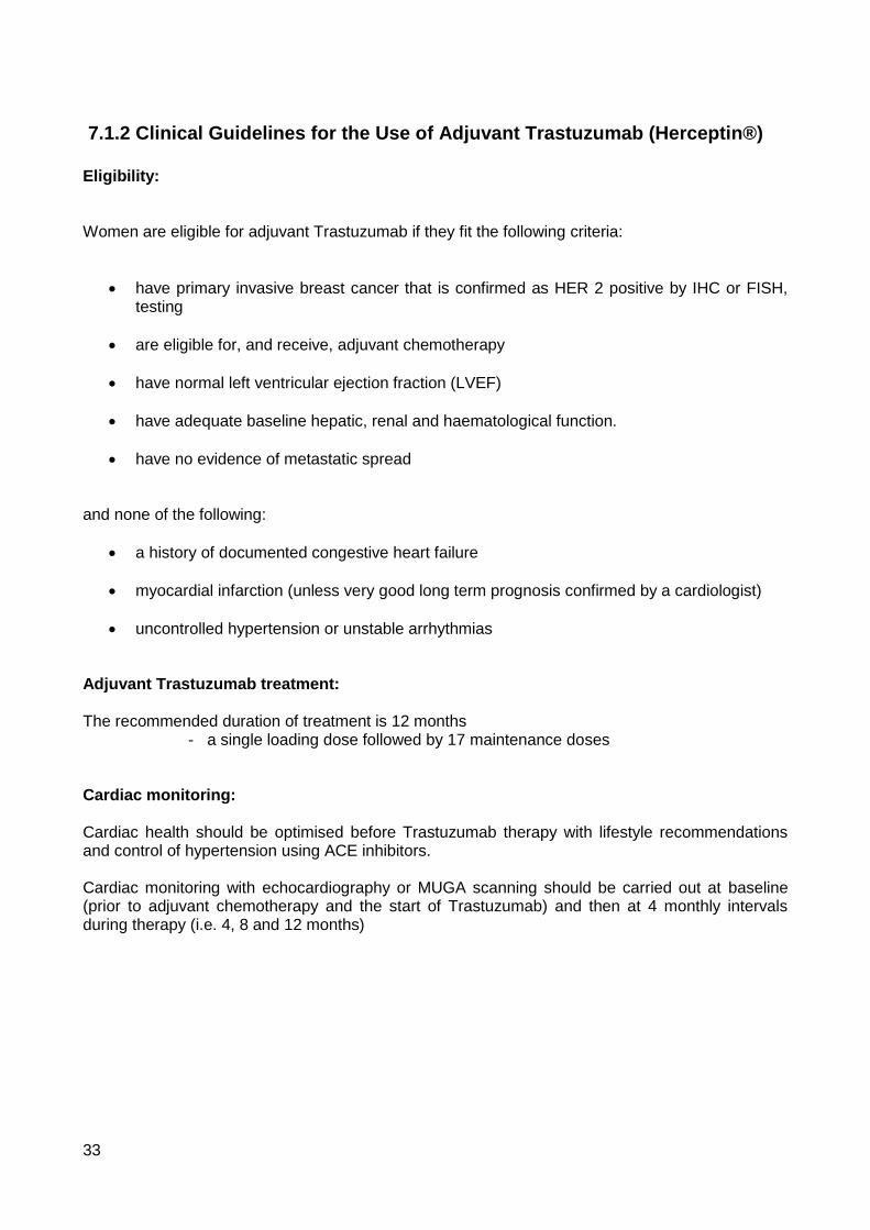
7.1.2 Clinical Guidelines for the Use of Adjuvant Trastuzumab (Herceptin®)
Eligibility: Women are eligible for adjuvant Trastuzumab if they fit the following criteria:
have primary invasive breast cancer that is confirmed as HER 2 positive by IHC or FISH, testing
are eligible for, and receive, adjuvant chemotherapy
have normal left ventricular ejection fraction (LVEF)
have adequate baseline hepatic, renal and haematological function.
have no evidence of metastatic spread and none of the following:
a history of documented congestive heart failure
myocardial infarction (unless very good long term prognosis confirmed by a cardiologist)
uncontrolled hypertension or unstable arrhythmias Adjuvant Trastuzumab treatment: The recommended duration of treatment is 12 months
- a single loading dose followed by 17 maintenance doses Cardiac monitoring: Cardiac health should be optimised before Trastuzumab therapy with lifestyle recommendations and control of hypertension using ACE inhibitors. Cardiac monitoring with echocardiography or MUGA scanning should be carried out at baseline (prior to adjuvant chemotherapy and the start of Trastuzumab) and then at 4 monthly intervals during therapy (i.e. 4, 8 and 12 months)

34
Guidance for stopping treatment in the event of reduced cardiac function is as follows: Patients who develop symptomatic cardiac dysfunction should have Trastuzumab discontinued and be referred to a cardiologist. Asymptomatic patients should be treated according to LVEF results with UK recommendations employing a ‘traffic light’ system. Green LVEF above lower limit of normal (LLN) – usually 50% No signs / symptoms CHF Trastuzumab related fall in LVEF < 10 % Amber LVEF between LLN and 40% No signs / symptoms CHF Or Trastuzumab related fall in LVEF > 10 % Red LVEF between < 40% Signs / symptoms CHF Amber and red are indications to consider ACE inhibitors and referral to cardiology with deferral of treatment until green light is given by repeat LVEF assessment. If criteria for continuation are met, resume Trastuzumab. If 2 consecutive ‘holds’ or a total of 3 ‘holds’ occur, discontinue Trastuzumab. Other monitoring: It is recommended that clinical review, FBC, U&E and LFTs should be performed at baseline and three monthly with at least annual oncology follow up for 5 years after Trastuzumab. Management of the over 70s ER + ve Aromatase inhibitors or Tamoxifen depending on NPI score ER - ve Consider FEC x 4-6 if fit and high risk No systemic treatment in all other situations Her2+ (ER- or ER+ and node positive) - Trastuzumab should be considered if fit for chemotherapy. Patients with significant cardiac history Echocardiography should be performed before treatment. Patients with impaired ventricular function may be offered cyclophosphamide/ docetaxel (or CMF classical iv x 6 courses), as an alternative.

35
Young patients and fertility For patients who have a strong desire to preserve their fertility classical CMF is highly likely to cause ovarian failure. AC is less damaging but probably the least toxic yet effective treatment is epirubicin and vinorelbine. Alopecia Alopecia is inevitable with anthracyclines but may be reduced by scalp cooling. This should be offered to all patients receiving anthracyclines or docetaxel where available. Scalp cooling is not suitable for paclitaxel treatments.

36
7.1.3 Early Breast Cancer Adjuvant Endocrine Therapy Guidelines Background information Endocrine treatments are a very important part of adjuvant post-operative therapy for women with oestrogen receptor (ER) positive breast cancer. Meta-analysis of the many trials of adjuvant endocrine treatments have indicated that these treatments reduce the annual odds of relapse by around one half and are responsible for a significant proportion of the declining death rate from breast cancer. ER / PR status is measured on all patients at diagnosis to ensure appropriate use of endocrine treatments. Endocrine treatments are rarely of any benefit in ER negative disease. Confirm post-menopausal status prior to prescribing aromatase inhibitors. Adjuvant treatment Pre-menopausal women:
Initiate Tamoxifen for five years.
Goserelin (LHRH analogue): monthly depot injections for two years are an appropriate alternative to chemotherapy for some patients (see section 7.1.1).
For pre-menopausal women receiving adjuvant chemotherapy there is no clear evidence that an LHRH analogue adds to the benefits of chemotherapy + Tamoxifen
Post-menopausal women:
Aromatase inhibitors (AI) are superior to Tamoxifen in the adjuvant setting and should constitute all or part of the adjuvant endocrine programme. Tamoxifen alone for five years is only considered the treatment of choice for patients with excellent or very good prognosis disease. Mindful of the costs of adjuvant AI therapy and the implications for follow-up of monitoring bone health, we recommend:
Tamoxifen for 2-3 years followed by an AI (usually Exemestane or Anastrozole) to complete 5 years treatment.
An AI (usually Anastrozole or Letrozole) from the outset for 5 years in women at high risk of early recurrence (e.g. HER 2 positive or PgR negative and node positive), or with a recent history of thrombo-embolic disease.
An AI (usually Letrozole) should also be considered for extended adjuvant treatment in patients considered at continuing significant risk for relapse (e.g. node positive) who are completing a previously planned 5 year course of Tamoxifen.
See section for advice on monitoring of bone health.

37
7.4 Primary Medical Therapy Patients with large operable tumours who do not wish to opt for mastectomy can be considered for primary medical therapy. This will usually be chemotherapy but those known to be ER positive may receive endocrine therapy. Discuss referrals with local oncologist. Where patients are unfit for general anaesthetic, consider local anaesthetic excision. In patients with an oestrogen receptor positive tumour unfit for, or refusing any type of surgery, an aromatase inhibitor is the first line treatment of choice. Patients who require liquid medication can have Tamoxifen syrup. Patients unable to take or comply with oral medication can have monthly Fulvestrant by injection (maximum ten patients in North Trent).
7.4.1 Neoadjuvant Chemotherapy Policy
Introduction Large randomised phase III studies and recent meta-analyses of patients with operable breast cancer have shown that there is no significant difference between neoadjuvant therapy and adjuvant therapy in terms of survival and overall disease progression1-3. However, neoadjuvant chemotherapy may often provide the opportunity for less extensive surgery to obtain local control. Consequently, in addition to treatment of locally advanced disease, neoadjuvant treatment has become a standard option for primary operable disease in patients who wish to potentially avoid mastectomy4. The logistical organisation of neoadjuvant chemotherapy is associated with increased complexities and requires careful multi-disciplinary planning. Aims of neoadjuvant chemotherapy
1. Increase breast conservation rate 2. Achieve operability in inoperable disease or to facilitate surgery in locally advanced disease
e.g. heavily clinically involved axillary lymph nodes (N2) or visible nodes measurable on U/S
3. Early introduction of systemic therapy as treatment of occult systemic micrometastatic disease e.g. in those with ER -ve or HER 2+ve disease
4. Translational research: evaluate chemosensitivity and tumour response to new drugs / schedules in vivo – using surrogate end-points (e.g. pCR) and biomarker assessment
Patient Selection Neoadjuvant chemotherapy: preferred treatment choice for:
1. Downstaging in patients favouring breast conserving surgery that is not possible (or would result in suboptimal cosmetic outcome) who would be expected to be candidates for adjuvant chemotherapy
2. Patients with tumours that express markers of a good response to chemotherapy (low or absent HR status, high grade)
3. Node positive, HER 2+ve patients (as addition of Trastuzumab can significantly increase pathological complete response – up to approximately 50%)
4. Inoperable and inflammatory breast cancer 5. Locally advanced tumours of difficult operability to facilitate surgery

38
Patient identification and pathway The patient is discussed in the weekly breast MDT and a multi-disciplinary management plan formulated re: diagnosis, suitability for neoadjuvant chemo, local surgical options and need for a metastatic screen. There should be a clear upfront surgical plan for the patient following neoadjuvant chemotherapy. All patients can be considered for breast conservation except those with multi-focal disease at presentation or widespread DCIS in association with invasive disease. The patient should have a surgical consultation, followed by oncology referral. Defining Selection Criteria For Breast Conserving Surgery (BCS) Following Neoadjuvant Chemotherapy Optimal selection criteria for Breast Conservation after neoadjuvant chemotherapy is still focus of research. Results from the M. D. Anderson (the largest single institution cohort of BCS after neoadjuvant chemotherapy) demonstrated that BCS is a safe and effective alternative to mastectomy for appropriately selected patients treated with neoadjuvant chemotherapy, even those with T3 or T4 disease5. Three post-chemotherapy factors independently associated with increased rate of local regional recurrence are multifocal or break-up pattern of residual disease, residual post treatment disease larger than 2 cm, and lymphovascular space invasion. Patients with any of the following factors after neoadjuvant chemotherapy will not be suitable for BCS
1. residual clinically assessed T size > 3cm 2. residual skin oedema 3. direct skin involvement or chest wall fixation 4. diffuse microcalcifications 5. multicentric disease 6. contraindications to use of radiotherapy
Pre-treatment work-up Depending on the indication, a metastatic screen (CT chest/ abdo, bone scan) is not mandatory in all patients selected neoadjuvant chemotherapy, but would be recommended in T3 /T4 disease or axillary lymph node involvement.
FBC / biochemistry (U+E. LFT, calcium)
Physical examination breast / axilla
Photographs of T4 tumours (if possible)
MRI breast in those patients being considered for breast conservation surgery
In those patients where axillary dissection is not indicated (i.e. not locally advanced disease), pre-chemotherapy sentinel lymph node biopsy should be considered when baseline axillary U/S is clear
Staging scans where appropriate

39
Assessment of response
Clinical assessment following each cycle
U/S guided insertion of markers clips for tumour bed localisation at surgery: to be performed after 1-2 cycles of chemotherapy in all patients if felt appropriate (as even in mastectomy specimens a clip helps localisation of the tumour bed)
Post-treatment MRI in those patients being considered for breast conserving surgery (this should be requested by the oncologist when patient attends for penultimate cycle of chemotherapy. The MRI will be discussed at the MDT. MDT discussion to take place 2 weeks following the last chemotherapy visit.
Chemotherapy
Current regime used is FE100C x 3, followed by 3 cycles of docetaxel, given 3-weekly. Patients considered unlikely to be able to tolerate this intensive therapy will be offered FE100C x 6
Note for patients with HER 2 positive tumours, trastuzumab (3 weekly) is now started concurrently with docetaxel (safety with concurrent anthracycline still under evaluation)
No trial has shown that additional chemotherapy in adjuvant setting in patients with residual invasive disease following primary anthracycline-taxane is of benefit. Therefore additional chemotherapy will not be given
Insufficient early response predicts a poorer prognosis. Progression on neoadjuvant chemotherapy is rare (<3%), and patients can be considered to switch to an alternate regimen or proceed to surgery
Local treatment
MDT review 2 weeks following attendance for final cycle of neoadjuvant chemotherapy (with prior repeat MRI if still considered for BCS)
Surgical review to determine definitive surgical procedure
Definitive surgical procedure approx. 4-6 weeks following completion of final cycle of chemotherapy
RT cannot replace surgery as sole method of breast conservation in patients with complete clinical and radiological response (thought to someway explain the small increased loco-regional recurrence rates in the neoadjuvant versus adjuvant chemotherapy trials). Surgery is always needed, even in cases of complete clinical response due to significant chance (up to 33%) of residual invasive tumour in breast
Axilla
Axillary clearance is standard procedure in patients with T3, N1 disease
Sentinel LN biopsy prior to commencement of neoadjuvant chemotherapy should be considered in those patients where axillary dissection is not necessarily indicated (in patients with clinical or radiological N0)
Sentinel LN biopsy following neoadjuvant chemotherapy is not recommended Radiotherapy
Following BCS
Post-mastectomy chest wall radiotherapy unless contraindicated
SCF radiotherapy if node positive or locally advanced at diagnosis

40
Histology
Need grading of tumour on diagnostic core
Standardised approach to assessment of breast cancer specimen from neoadjuvant chemotherapy
Definition of pathological Complete Response (pCR), as the current most reliable surrogate marker of long-term outcome, requires standardisation
Ideally definition of pCR should include absence of both invasive tumour in breast and axilla Breast care nurse key-worker Local Breast care nurse will remain patient key-worker throughout neoadjuvant treatment. If particular issues arise, to liaise with Jo Beaumont so patient can be seen at WPH chemotherapy assessment appointments
7.4.2 Neoadjuvant endocrine therapy
Neoadjuvant endocrine therapy is an option to downstage ER +ve (Her2 -ve) tumours to facilitate surgery or breast conserving surgery in post-menopausal patients considered for neoadjuvant therapy but who are thought to not be suitable for, or refuse, adjuvant chemotherapy e.g. patients with inoperable or large tumour size and co-morbidities. Suitable patients would include post-menopausal patients with lower grade ER+ve (particularly those with strong ER receptor expression, Allred score 6-8) and HER2 negative tumours with large / locally advanced breast cancer who are not suitable for breast conserving surgery at presentation and wish to pursue the possibility of potential down-staging. This approach may also be appropriate for patients for whom immediate surgery is not possible.
Pre-treatment work-up (as per neoadjuvant chemotherapy protocol)
Physical examination breast / axilla
Photographs of T4 tumours (if possible)
MRI breast in those patients being considered for breast conservation surgery
Pre-treatment axillary node staging
Staging scans where appropriate
Treatment should be started with Letrozole for a period of 4-6 months. Assessment of response
Clinical assessment every 4-8 weeks (preferably by the same clinician)
U/S repose assessment after 2-3 months and guided insertion of markers clips for tumour bed localisation at surgery
Post-treatment MRI in those patients being considered for breast conserving surgery during months 4-6
Surgical and Radiotherapy loco-regional management should follow as per section 7.4.1 neoadjuvant chemotherapy

41
References 1. Fisher et el. Effect of preoperative chemotherapy on the outcome of women with operable breast cancer. J Clin Onc 1998; 16:2672-85 2. Mauri et al. Neoadjuvant versus adjuvant systemic treatment in breast cancer; a meta-analysis. JNCI 2005, 97; 3: 188-194 3. Mieog et al. Neoadjuvant chemotherapy for operable breast cancer. Brit J Surgery 2007;94:1189-1200 4. Kaufmann et al. Recommendations from an international expert panel on the use of neoadjuvant (primary) systemic treatment of operable breast cancer: new perspectives 2006. Annals of Oncology 2007; 18:1927-1934 5. Chen et al. Breast-conserving therapy after neoadjuvant chemotherapy: The M.D. Anderson Cancer Center experience. J Clin Onc 2004; 22:2303-2312 Definitions
Primary operable breast cancer: lump 5cm, no evidence of inflammatory changes in the breast and no clinically malignant lymphadenopathy Locally advanced breast cancer: lump >5cm, presence of peau d’orange, inflammatory skin changes, deep fixation, or obvious malignant fixed / matted lymphadenopathy

42
7.4.2 Neoadjuvant Chemotherapy Patient Pathway
MDT MDT decision for neoadjuvant chemotherapy according to patient selection criteria
Identification of those patients suitable for potential breast conservation surgery Identification of those patients in whom definitive surgery is mastectomy (see policy)
Oncology referral generated Breast MRI requested for those patients suitable for potential BCS
Metastatic screen if indicated
Metastatic screen if indicated (LABC, heavy node involvement) using CT thorax / abdomen and bone scan
Surgical review
Neoadjuvant chemotherapy as per local policy Consider ongoing clinical trials Trastuzumab if HER 2+ve (with taxane) Response assessment Clinical assessment following each cycle Marker insertion U/S guided marker insertion of marker clips after 1-2 cycles of chemotherapy
Oncology review and work-up
Post treatment MRI in those being considered for BCS – to be
requested at penultimate cycle
MDT review 2 weeks following final cycle of chemotherapy
Surgical review
Definitive local surgical and axillary management 4-6 weeks after completion of final chemotherapy. Schedule between any ongoing 3 weekly Herceptin treatments
MDT review
Further surgery if required RT to breast/chest wall + SCF unless known to be node -ve Endocrine treatment if ER+ Herceptin if HER2 +
Axilla - If axillary dissection not necessarily indicated, consider sentinel node biopsy pre-chemo if axilla clinically or radiological N0
Histological assessment to include standardised definition of path CR

43
7.5 Treatment for Advanced/Metastatic Disease
7.5.1 Locally Advanced Breast Cancer
General principles: Patients with T4 tumours have a poor prognosis and local recurrence is usual after surgery alone. Management of these patients should be decided in the multidisciplinary breast clinic. The probability of overt metastases at diagnosis is quite high and therefore staging of these patients should include evaluation of the supraclavicular fossa, a bone scan, and either CT chest and liver, or liver ultrasound and a chest X-ray. Appropriate pre-operative systemic therapy followed, in the absence of distant metastases, by local treatment is indicated. Systemic therapy: Systemic therapy for locally advanced disease improves local control and has a modest effect on distant metastases and survival. Chemotherapy (+/- Trastuzumab) followed by endocrine treatment (if HR positive) is recommended except for those patients who are unfit for chemotherapy who are best treated by endocrine treatment alone (usually Letrozole for 3-4 months) then review local therapy options. Local therapy: There is insufficient randomised trial evidence to indicate whether surgery, radiotherapy or a combination of the two should be routinely recommended. In general, patients showing a good response to systemic treatment should be considered for surgery +/- radiotherapy, while those responding poorly have a poor prognosis and are best managed with radiotherapy in the first instance.

44
7.5.2 Metastatic Breast Cancer
General principles Advanced breast cancer is incurable. Treatments should be aimed primarily at symptom relief and maintaining quality of life. Modest improvements in survival from systemic therapy are generally acknowledged to occur, and in randomised trials, some of the newer treatment approaches have shown significant improvements in survival over traditional endocrine and cytotoxic treatments. Hormone and HER 2 receptor status should be used to guide treatment decisions. Clinical and appropriate imaging tests should be performed before and at occasional intervals during treatment to monitor response to treatment. Sufficient data should be collected to enable effective audit of the results of palliative treatments. Follow-up should be co-ordinated by the oncologist with access to the range of hospital and community based support services. Where possible, consideration should be given to treatment within properly constructed clinical trials. The Cancer Centre should co-ordinate a range of studies in advanced breast cancer to cover most aspects of systemic management. National guidance on the treatment of metastatic breast cancer endorsed by the NCRI breast cancer Clinical Studies Group can be found on line at: http://ncrndev.org.uk/csg/MBCGuidance.pdf and complements the NICE guidance avaialble at: http://www.nice.org.uk/nicemedia/pdf/CG81NICEGuideline.pdf
a) Endocrine treatment
Background information Endocrine treatments are a very important part of palliative treatment of advanced metastatic disease for women with oestrogen receptor (ER) positive breast cancer. ER / PR status is measured on all patients at diagnosis to ensure appropriate use of endocrine treatments. When ER status is unknown a request for retrospective determination of ER status should sought. Endocrine treatments are rarely of any benefit in ER negative disease. Check menstrual status prior to prescribing aromatase inhibitors. First line treatment, pre-menopausal patients: Tamoxifen + Goserelin Permanent ovarian ablation with radiotherapy or surgery usually recommended after a few months treatment to avoid long term commitment to monthly injections. First line endocrine treatment, post-menopausal women:
Aromatase inhibitor - Letrozole or Anastrozole. Consider Tamoxifen if aromatase inhibitors are contra-indicated.
Second line treatment:
Tamoxifen for progression on an aromatase inhibitor
Aromatase inhibitor for progression on Tamoxifen

45
Subsequent endocrine treatment:
Exemestane, Fulvestrant and Megestrol Acetate are all appropriate treatments for patients showing repeated endocrine sensitivity.
For women unable to take oral medication or in whom compliance is compromised by concomitant disability (e.g. dementia) Fulvestrant given by monthly intramuscular injection may be considered. Protocol for Prescribing Fulvestrant as 3rd Line Treatment: Patients with metastatic breast cancer, 3rd or subsequent line, who have responded to previous hormonal treatment (SD/PR/CR for greater than 6 months) where the alternative would be chemotherapy. WPH consultants prescribe via Chemocare to allow audit of usage. Other prescribers should follow local guidelines.
b) Chemotherapy
Chemotherapy may provide useful palliation for patients who are refractory to endocrine treatments. Chemotherapy should only be prescribed by specialist non-surgical oncologists, appropriately supported by chemotherapy nurse specialists, expert pharmacy and laboratory support. Treatment should be administered in designated day-care facilities or on an oncology ward. Patients should receive regimen-specific, written information about their own treatment, likely side-effects and appropriate contacts for advice. Clear protocols for the management of patients with chemotherapy complications, especially neutropenic sepsis, should be established that enable rapid admission to appropriate facilities. Inpatient support for chemotherapy complications should be available from a specialist multidisciplinary team that has expertise in solid tumour chemotherapy. Participation in clinical trials is encouraged. A wide variety of chemotherapeutic agents are used in metastatic disease. A review of randomised controlled trials does not reveal any clearly superior regimen, although side-effect profiles vary. The choice of regimen will depend on the extent of disease, performance status and wishes of the patient. Response is most likely with the first line of chemotherapy treatment with efficacy declining rapidly thereafter. Recent chemotherapy developments in advanced breast cancer include the introduction of the taxanes, (docetaxel, paclitaxel), capecitabine, vinorelbine gemcitabine and platinum agents (cisplatin, carboplatin). The role of these agents in advanced breast cancer has been reviewed by NICE. Appropriate patients should be initially offered palliative chemotherapy with a taxane, then capecitabine or vinorelbine used at relapse. The breast team should be able to audit the outcome of treatment and record the reasons a patient was either not offered or did not receive taxane treatment.

46
c) Biological therapy
Trastuzumab (Trastuzumab®, Roche) is a recombinant humanised monoclonal antibody that specifically targets the HER 2 protein. Trastuzumab has NICE approval for use in patients with metastatic breast cancer (MBC) who have tumours that over express HER 2. Trastuzumab when used in combination with chemotherapy (taxanes, capecitabine, vinorelbine) is more effective than chemotherapy alone for the treatment of MBC. However this only applies to patients with a breast tumour that over expresses HER 2 at the 3+ level when evaluated by immunohistochemistry, or with evidence of mRNA over-expression for HER 2 by fluorescence in situ hybridisation (FISH) testing. Trastuzumab is associated with congestive heart failure in patients receiving anthracycline based chemotherapy and is therefore usually given in combination with a taxane.
Organ Specific Problems in Advanced Breast Cancer
a) Loco-regional recurrence
The frequency of local recurrence is dependent on both the extent and initial treatment of the primary lesion. Recurrences within the conserved breast are best managed by surgery. Isolated hest wall recurrence should be removed and if not treated previously, the chest wall treated by radiotherapy. More extensive locoregional recurrence should be managed systemically. Salvage surgery for chest wall disease may be appropriate in carefully selected cases. Radiotherapy is useful for bleeding lesions. Management of recurrent disease in the axilla is difficult. Systemic therapy, surgery and or radiotherapy may be required to prevent uncontrolled disease and brachial plexopathy.
b) Bone metastases
Skeletal involvement usually causes pain, and may result in long bone fractures, vertebral collapse and hypercalcaemia. Pain relief should be given according to the analgesic ladder. NSAIDs are particularly effective. Radiotherapy is the treatment of choice for localised areas of pain. A short course of radiotherapy (1-5 fractions) is usually effective. Longer fractionation regimens may be indicated for solitary metastases or following internal fixation of pathological fracture. Wide-field irradiation or radioisotope treatment may occasionally be necessary. Prophylactic orthopaedic surgery is recommended to prevent fracture through lytic lesions in the proximal femora and humeri. Surgery may be appropriate for destructive lesions in the spine and to relieve the pain of spinal instability. Spinal cord compression requires high dose corticosteroids and emergency access to MR imaging. A referral pathway should be established to enable a same day consultation between a specialist spinal surgeon and a clinical oncologist to decide on the relative merits of surgery or radiotherapy. Bone metastases respond relatively well to endocrine treatment but chemotherapy may be more hazardous than usual due to poor bone marrow reserve. Bisphosphonate treatment reduces skeletal complications by 30-50%, reducing the requirements for radiotherapy and orthopaedic surgery. Intravenous bisphosphonate treatment may also relieve bone pain. Zoledronic acid is the
47
most effective bisphosphonate. Treatment should continue for as long as skeletal disease remains an important clinical problem for the patient. Bisphosphonates should continue even when a skeletal event occurs or the disease progresses. Biochemical markers (NTX) may be helpful for defining the schedule of treatment. Isotope bone scans must not be used to monitor progress of disease, as the osteoblastic response to healing of metastases can mimic progressive disease. Intravenous dehydration and bisphosphonate treatment is recommended for the acute management of hypercalcaemia.
c) Metastatic Spinal Cord Compression
Patients at risk of metastatic spinal cord compression should be given written information informing them of the signs and what they should do if they are concerned. Breast Cancer Care produce a helpful information leaflet which can be found at: http://www2.breastcancercare.org.uk/sites/default/files/Secondary%20breast%20cancer%20in%20the%20bone.pdf Sheffield teaching hospitals and Doncaster and Bassetlaw Hospitals have in house information leaflets. In all cases of suspected metastatic spinal cord compression, the NTCN guidelines should be followed and referral for management be made on the NTCN referral form http://www.northtrentcancernetwork.nhs.uk/Downloads/Chemotherapy-Guidelines/NTCN%20MSCC%20referral%20proformapage%202.pdf (Appendix D).
d) Liver metastases
Liver metastases, especially when associated with disordered liver function, indicate a very poor prognosis. Endocrine response is less common and in those patients where systemic treatment is still considered appropriate, chemotherapy is preferred. Toxicity following chemotherapy may be exacerbated when liver function is impaired. Great caution is required in treating such patients. The newer agents such as docetaxel may be more effective than conventional cytotoxic agents. Corticosteroids and NSAIDs provide good relief of liver capsule pain.
e) Lung metastases
Lung metastases have a poor prognosis. Endocrine response is less common in those patients with diffuse lymphangitic disease. Where systemic treatment is still considered appropriate, chemotherapy is preferred. Physical therapy, relaxation techniques, oxygen, opioids, benzodiazepines, corticosteroids and diuretics may provide symptomatic relief.
f) Brain metastases
Symptomatic brain metastases require high dose corticosteroids for symptom relief. Radiotherapy may provide worthwhile palliation. Brain metastases may respond to systemic treatments but this is unpredictable. Surgical resection of solitary brain metastases is only rarely recommended. Meningeal infiltration responds poorly to intrathecal treatment and this cannot be generally recommended. Cranial +/- spinal irradiation may provide useful palliation.

48
g) Pleural effusions
Pleural effusions should be aspirated for immediate symptomatic relief. Tube drainage to dryness followed by intrapleural administration of a sclerosant to effect a pleurodesis is recommended for recurrent effusions.
h) Ascites
Malignant ascites should be drained for symptomatic relief. Appropriate systemic therapy should be prescribed. There is no established role for intraperitoneal treatment.

49
8. FOLLOW UP
8.1 Clinical Follow Up There is little evidence that routine clinical follow up after the treatment reduces mortality. However, we believe that some hospital based follow-up is still desirable, particularly following breast conservation surgery where there is a significant rate of local recurrence which will only be detected by subtle changes found on clinical examination. Each locality within the Network has different mechanisms for follow up but all patients are seen by appropriately trained medical staff and identified personnel. This may be Consultant Breast Surgeon or Oncologist involved in their care, a trained staff or associate specialist grade doctor, a GP clinical assistant or a specialist nurse (Breast Care nurse, Nurse Practitioner or Nurse Consultant). Follow up is performed by the following personnel across the network: Barnsley Consultant or Specialist Registrar in Surgery, Consultant or Specialist Registrar in
Oncology, Staff Grade Doctor or Specialist Nurse Rotherham Consultant Surgeon, Consultant Oncologist, Associate Specialist or Staff Grade
Doctor Chesterfield High risk patients post chemotherapy, Consultant or Specialist Registrar in
Oncology. Other patients, Consultant or Specialist Registrar in Surgery , Staff Grade Doctor
or Specialist Nurse Doncaster Consultant or Specialist Registrar in Surgery, Consultant or Specialist Registrar in
Oncology, Staff Grade Doctor or Specialist Nurse, Supervised junior doctor Sheffield Post Chemotherapy, Consultant or Specialist Registrar in Oncology Other patients, Consultant or Specialist Registrar in Surgery or GP with Special
Interest in Breast Disease
Follow Up protocol for Patients Treated with Curative Intent
Following completion of active treatment, or six months after surgery (whichever is later), the patient will attend a ‘discharge to follow up’ consultation. A Breast Care nurse will perform a health needs assessment and a clinician or nurse practitioner will perform a physical examination and ensure that follow up mammography and DEXA scanning (as appropriate) has been arranged. Further support and information will be provided as required. If the patient is suitable for reduced clinical follow up, they will be seen according to the schedule below at 3 and 5 years. Switching will be performed at the year 3 visit, if recommended at the visit to discuss adjuvant therapy post surgery. If the patient is not suitable for reduced follow up (e.g. excessive anxiety, ongoing symptoms, difficulty self managing due to co-morbdities) the patient will enter the traditional follow up pathway:
Moderate and poor prognostic groups on a 6 monthly basis for 2 years and then annually up to 5 years
Excellent and good prognostic groups on an annual basis up to 5 years

50
Yes
Yes
No Yes
Holistic Needs Assessment and
Care Plan
Patient has self managed follow up Patient has discharge interview and ongoing
rapid access to breast care team
Holistic Needs Assessment and
Care Plan
Year 3 Follow Up Appointment
Clinical AssessmentHolistic Needs Assessment and
Care Plan
Year 5 Follow Up Appointment
Clinical AssessmentHolistic Needs Assessment &
Discharge Plan
Patient/GP
concern
Patient/GP
concern
No
Follow up appointments
as required
Follow up appointments
as required
Su
rviv
ors
hip
Year 1
mammogram
Year 2
mammogram
Year 3
mammogram
Year 4
Mammogram
Year 5
mammogram
Pre
p fo
r
Fo
llow
Up
Su
pp
ort
Alte
rna
tive
Th
era
py
Exe
rcis
e &
Nu
triti
on
Be
ne
fits
Ad
vice
Ed
uca
tion
Fa
st T
rack
to
Bre
ast
Te
am
or
Re
ferr
al f
or
Re
con
stru
ctio
n
Patients aged >50 discharged to NHS BSP
Patients aged <50 continue on annual mammographic surveillance until she reaches age of NHS BSP
Active treatment
completed
Is patient suitable for
self management?
Follow UpMammographic Surveillance
•All females except bilateral mastectomy
Tre
atm
en
t
North Trent Cancer Network Breast Cancer Follow Up Pathway
Patient/GP
concern
Patient has individualised follow up due
to:
•Complex physical or psychological co-morbidity
•Ongoing treatment related complications
•Clinical trial protocol requirement
No

51
Patients should be informed of the arrangements for follow-up after surgery and adjuvant treatment and given a written explanation of the signs and symptoms that should be reported at any time.
Patients are offered open access for early review if they have concerns e.g. new lump, lymphoedema etc. Access should be via the Breast Care Nurse or Nurse Practitioner who will arrange an appropriate appointment within 2 weeks
Patients should follow an agreed, written care plan, recorded by a named healthcare professional (or professionals). A copy should be sent to the GP and a copy given to the patient. It should include:
designated named healthcare professionals
dates for review of any adjuvant therapy
details of surveillance mammography
contact details for immediate referral to specialist care, and
contact details for support services, for example, support for patients with lymphoedema
8.2 Organisation of Follow Up Location
Secondary Care Breast Clinic, staffed according to the schedule above
Primary Care Follow up in primary care following agreement between the patient, General Practitioner and the treating Consultants
Responsibility for advising the patient on adjuvant hormonal therapy and on their therapeutic options after completion of 5 years therapy falls with the reviewing clinician. Where a clear plan has not already been agreed following initial treatment, this will be discussed with the Consultant Surgeon or Oncologist. Rapid access to specialist follow up should be available via the Breast Care Nurse or Nurse Practitioner for patients with concerns. The Breast Care Nurse will arrange the appropriate out patient appointment with Consultant medical staff. All patients are made aware of the open access policy.
8.3 Follow Up Mammography 1. The reviewing clinician is responsible for arranging imaging surveillance and follow up.
Annual bilateral mammography for 5 years
Women under the age of 50 at discharge from clinical follow up should be offered annual mammography until the age of 50
Women should revert to the NHS breast-screening programme after 5 years if still appropriate.
In certain very high risk individuals consideration may be given to MRI screening
Patients with a strong family history of breast cancer should be followed up according to the NICE family history guidelines

52
8.4 Management of Menopausal Symptoms The SSRI antidepressants paroxetine and fluoxetine may be offered to women with breast cancer for relieving menopausal symptoms, particularly hot flushes, but not to those taking Tamoxifen. Clonidine and venlafaxine should only be offered to treat hot flushes in women with breast cancer after they have been fully informed of the significant side effects. It should be noted that these medications are not licensed for post-menopausal symptoms, therefore, informed consent must be obtained.
8.5 Monitoring Bone Health Patients undergoing a premature menopause (post chemotherapy or ovarian suppression therapy) or receiving adjuvant aromatase inhibitors should be assessed for bone loss by: DEXA scan during the first 6 months of treatment and at 12-24 month intervals thereafter. Advice on lifestyle (stopping smoking, limiting alcohol intake, encouraging weight bearing exercise) should be offered. Calcium and vitamin D supplements should be encouraged. Women with or developing osteoporosis should receive treatment with a bisphosphonates. Referral to a bone specialist is advised. More detailed Guidelines are available at: http://ncrndev.org.uk/index.php?option=com_content&task=view&id=115&Itemid=254
8.6 Trials See North Trent Cancer Research Network (NTCRN) Annual Report http://ntcrn.group.shef.ac.uk/top/trials.html

53
9. FAMILY HISTORY SERVICES
9.1 Genetic Screening NICE has issued guidelines relating to the management of women at increased familial risk of breast cancer. These guidelines list criteria for women suitable to be managed in primary care (GP), secondary care (family history clinics) and tertiary care (Genetics services). These criteria are adhered to by this group. Women categorized as at near population risk should be reassured and managed in primary care. Women at moderate and high risk should be offered an appointment to discuss risk and management strategies. Women at moderate risk may be offered annual mammograms between age 40 and 50 and then revert to the NHS BSP. Lifestyle advice about self examination, weight, exercise, HRT etc should be given. Women at high risk may be eligible for referral for genetic counselling and gene testing, should be offered annual mammograms between age 40 and 50 and if very high risk may also be offered MRI screening. Very high risk women, where gene carriage is present or likely, may be counselled about and offered prophylactic mastectomy. These women may also require referral to gynaecology to discuss prophylactic oophorectomy. Prior to prophylactic surgery, psychological counselling, formalized risk assessment and discussion of reconstructive options must be provided. http://www.nice.org.uk/guidance/CG41
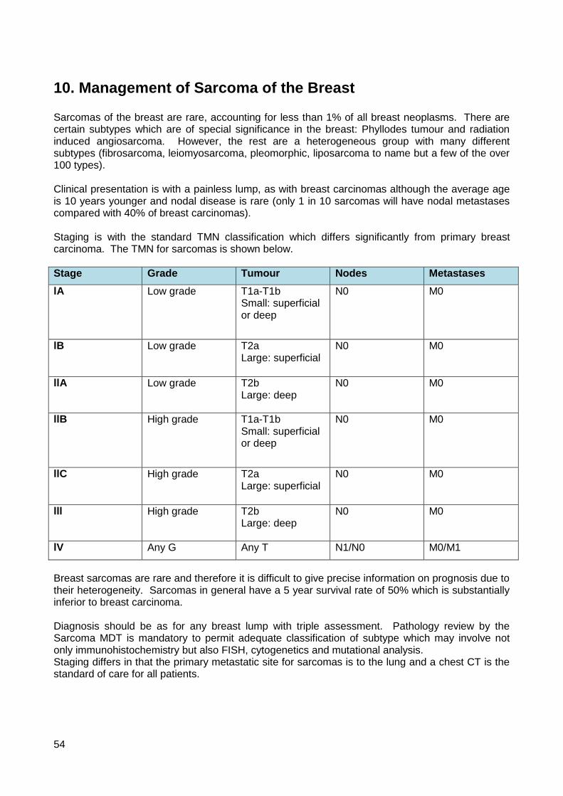
54
10. Management of Sarcoma of the Breast Sarcomas of the breast are rare, accounting for less than 1% of all breast neoplasms. There are certain subtypes which are of special significance in the breast: Phyllodes tumour and radiation induced angiosarcoma. However, the rest are a heterogeneous group with many different subtypes (fibrosarcoma, leiomyosarcoma, pleomorphic, liposarcoma to name but a few of the over 100 types). Clinical presentation is with a painless lump, as with breast carcinomas although the average age is 10 years younger and nodal disease is rare (only 1 in 10 sarcomas will have nodal metastases compared with 40% of breast carcinomas). Staging is with the standard TMN classification which differs significantly from primary breast carcinoma. The TMN for sarcomas is shown below.
Stage Grade Tumour Nodes Metastases
lA Low grade T1a-T1b Small: superficial or deep
N0 M0
lB Low grade T2a Large: superficial
N0 M0
llA Low grade T2b Large: deep
N0 M0
llB High grade T1a-T1b Small: superficial or deep
N0 M0
llC High grade T2a Large: superficial
N0 M0
lll High grade T2b Large: deep
N0 M0
lV Any G Any T N1/N0 M0/M1
Breast sarcomas are rare and therefore it is difficult to give precise information on prognosis due to their heterogeneity. Sarcomas in general have a 5 year survival rate of 50% which is substantially inferior to breast carcinoma. Diagnosis should be as for any breast lump with triple assessment. Pathology review by the Sarcoma MDT is mandatory to permit adequate classification of subtype which may involve not only immunohistochemistry but also FISH, cytogenetics and mutational analysis. Staging differs in that the primary metastatic site for sarcomas is to the lung and a chest CT is the standard of care for all patients.

55
Treatment has traditionally been with mastectomy and radiotherapy if the tumour is greater than 5 cm or high grade (trojani grade 3). No nodal surgery is indicated. There may be a role for breast conserving surgery if the tumour size is small enough and clear margins are obtainable but many sarcomas tend to be large. Post operatively, there is rarely a place for chemotherapy as most sarcomas, except for some rare subtypes such as Ewings, Rhabdomyosarcomas or osteosarcomas, all of which are not usually seem in the breast, are chemoresistant. Follow up also differs in that in addition to standard breast follow up with mammography after breast conservation, chest CT should be performed every 4-6 months. Pulmonary metastatectomy for limited lung disease may be curative in 40% of cases and is therefore not futile as it is in women with lung metastases from breast carcinoma.

56
Appendix A
Available agents Common side effects are listed for each of the Drugs below. For a complete listing of potential side effects and detailed information on indications, cautions, contraindications, dosage and formulations refer to the current edition of the British National Formulary. Tamoxifen: 20 mg once daily by mouth. Pre and post-menopausal. Adjuvant use currently discontinued after 5 years. Small risk of endometrial cancer with prolonged usage. Side effects: hot flushes, vaginal discharge. Anastrozole (Arimidex™): 1 mg once daily by mouth. Post menopausal only. Slightly more effective than Tamoxifen, particularly in those previously exposed to adjuvant Tamoxifen. Side effects: hot flushes. Letrozole (Femara™): 2.5 mg once daily by mouth. Post menopausal only. Clearly superior to Tamoxifen for all sub-groups with advanced breast cancer. Side effects: hot flushes, bone loss. Exemestane (Aromasin™): 25 mg once daily by mouth. Post menopausal only. Useful second/third line treatment after failure of Tamoxifen or Letrozole/ Anastrozole. Side effects: hot flushes, bone loss. Megestrol Acetate (Megace™): 160 mg once daily by mouth. Useful third line treatment. Side effects: weight gain, fluid retention, vaginal bleeding on withdrawal. Medroxyprogesterone acetate (Farlutal™, Provera™) 200-500 mg twice daily by mouth. Occasionally used as third line treatment. No advantages over Megestrol Acetate. Side effects: weight gain, fluid retention, vaginal bleeding on withdrawal. Aminoglutethimide (Orimeten™): 250–500 mgs twice daily by mouth. Post menopausal only. Rarely used, rather non-selective drug. Side effects: rash, dizziness, drowsiness, nausea. Requires concomitant hydrocortisone replacement (20 mg bd). Goserelin (Zoladex™): 3.6 mg subcutaneously by a monthly depot injection. Pre-menopausal only. Effectively suppresses ovarian function. Usually given in combination with Tamoxifen. Side effects: injection site pain, hot flushes, menopausal symptoms, bone loss. Fulvestrant (Faslodex™): 250mg deep intra-muscular injection every 4 weeks. Third line treatment following agreement with Oncologist. Primary treatment where the patient is unable to take oral medication. Side effects: hot flushes, injection site pain, thromboembolism

57
Trastuzumab (Herceptin®, Roche): A loading dose of 4mg/kg IV is required over at least 1 hour, followed by 2mg/kg weekly over 30 minutes. Patients should be pre-medicated with Dexamethasone 8mg IV and Chlorpheniramine 10mg IV. Other agents such as androgens and oestrogens are now only very rarely considered appropriate.

58
Appendix B
Royal College of Pathologists Data Set
http://www.rcpath.org/Resources/RCPath/Migrated%20Resources/Documents/N/NHSBreastCanHistoMinData.pdf

59
Appendix C
North Trent Cancer Network Teenage and Young Adult Referral Pathway (16-24 years)
Adobe Acrobat Document
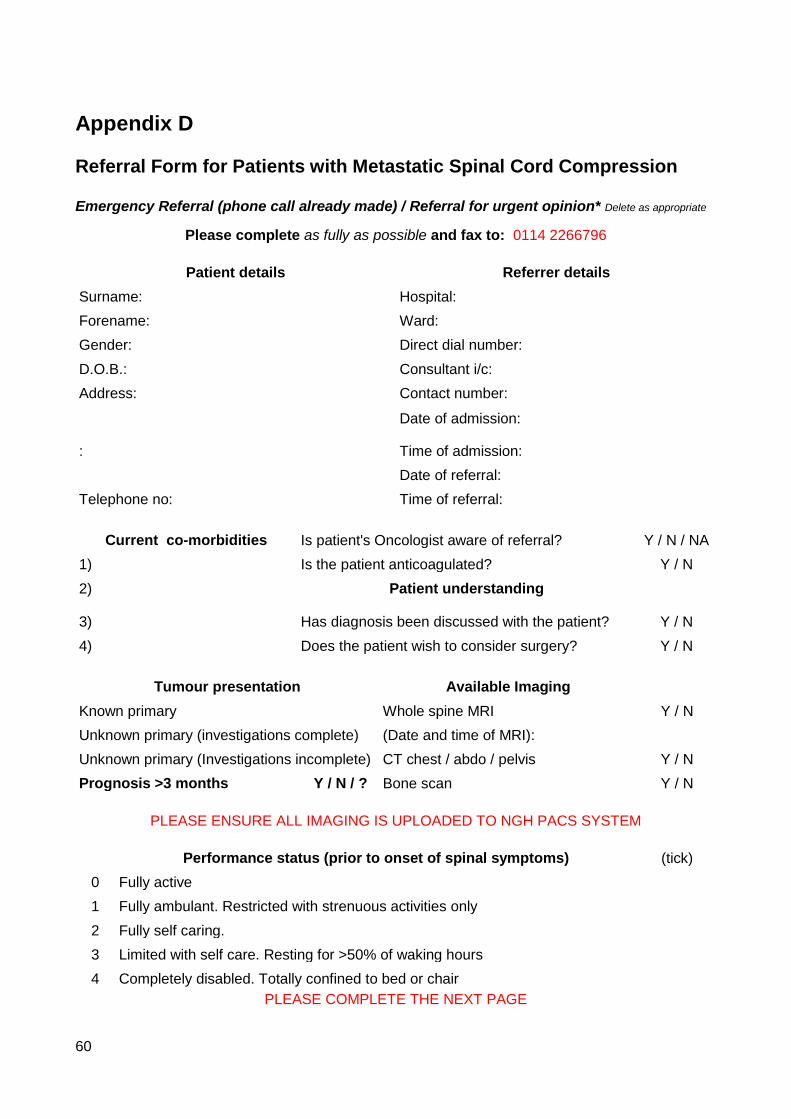
60
Appendix D
Referral Form for Patients with Metastatic Spinal Cord Compression Emergency Referral (phone call already made) / Referral for urgent opinion* Delete as appropriate
Please complete as fully as possible and fax to: 0114 2266796
Patient details Referrer details
Surname: Hospital:
Forename: Ward:
Gender: Direct dial number:
D.O.B.: Consultant i/c:
Address: Contact number:
Date of admission:
: Time of admission:
Date of referral:
Telephone no: Time of referral:
Current co-morbidities Is patient's Oncologist aware of referral? Y / N / NA
1) Is the patient anticoagulated? Y / N
2) Patient understanding
3) Has diagnosis been discussed with the patient? Y / N
4) Does the patient wish to consider surgery? Y / N
Tumour presentation Available Imaging
Known primary Whole spine MRI Y / N
Unknown primary (investigations complete) (Date and time of MRI):
Unknown primary (Investigations incomplete) CT chest / abdo / pelvis Y / N
Prognosis >3 months Y / N / ? Bone scan Y / N
PLEASE ENSURE ALL IMAGING IS UPLOADED TO NGH PACS SYSTEM
Performance status (prior to onset of spinal symptoms) (tick)
0 Fully active
1 Fully ambulant. Restricted with strenuous activities only
2 Fully self caring.
3 Limited with self care. Resting for >50% of waking hours
4 Completely disabled. Totally confined to bed or chair
PLEASE COMPLETE THE NEXT PAGE

61
Patient name: D.O.B.:
Primary tumour site Pain Symptoms
Breast Prostate Renal Pain Y / N since (date):
Lung Myeloma Lymphoma Level / location:
Thyroid GIT Urothelial Type Non-specific Mechanical Neuralgic
Uterine/Cx Melanoma Pattern Nocturnal Diurnal Constant
Other (specify): Analgesia Minor Major
Date of diagnosis: VAS Score: __ / 10
Primary treatment: Neurological symptoms
Describe:
Adjuvant treatment Current walking status
1) XRT to spinal met Y / N Normal
2) Unsteady since (date):
3) Non-ambulant since (date):
Metastases Continence
Extra spinal bone mets Y / N Urinary incontinence Y / N since (date):
Visceral mets Y / N Faecal incontinence Y / N since (date):
Liver Lung Anal tone Normal Reduced Absent
Brain Adrenal Perineal sensation Normal Reduced Absent
Lymph nodes Other Catheter tug Felt Not felt N/A
Sensation
Normal Reduced Absent
Most distal dermatome with normal sensation:
Power
Most distal myotome with normal power
MRC grade of weakest muscle(s)
Details of clinician to be responsible for ongoing care of the patient following surgery
Name: Contact number:
If you wish to discuss a referral please contact the on-call spinal surgeon on 0114 2715244 during office hours or otherwise via the NGH switchboard (0114 2434343)
Other relevant Information: