reference: bioi. bull. 166: 310;327. (april, -...
TRANSCRIPT
Reference: Bioi. Bull. 166: 310...;327. (April, 1984)
THE FUNCTIONS OF NEMATOCYSTS IN PREY CAPTURE BY EPIPELAGIC SIPHONOPHORES (COELENTERATA, HYDROZOA)'
JENNIFER E. PURCELU
Woods Hole Oceanographic Institution, Woods Hole, Massachusetts 02543
ABSTRACT
The nematocysts of 24 siphonophore species were examined by light and scanning electron microscopy (SEM) for differences that could relate to differences in the sizes and types of prey captured. The siphonophore species in the suborder Calycophorae had 4-30 microbasic mastigophores (0.7-18.0 ~-tl volume), and 50-2000 smaller homotrichous anisorhizas in uncoiled nematocyst batteries. The physonect siphonophore species had 4-120 stenoteles or micro basic mastigophores ( 1. 8-40.7 ~-tl volume), and 150-20,500 smaller homotrichous anisorhizas in coiled nematocyst batteries. The sizes of crustacean prey (primarily copepods) captured by species in both suborders increased with increasing nematocyst size and numbers. Examination by SEM of captured, but uningested prey showed that the heavily-spined threads of these nematocysts adhered to the prey surface, and primarily entangled the prey. In contrast, the tentacles of siphonophores in the suborder Cystonectae, which includes Physalia physalis, have only isorhizas of 1.0-18.0 ~-tl volume with and without small spines on the threads. These nematocysts penetrate the soft-bodied prey (mostly fish larvae) of these siphonophores, but apparently do not penetrate or entangle hard-bodied prey. Thus prey capture by siphonophores differs with the sizes, numbers, and types of nematocysts present in each species. The possible functions of nematocyst batteries in tentacle spreading, and luring of large zooplankton prey are discussed.
INTRODUCTION .
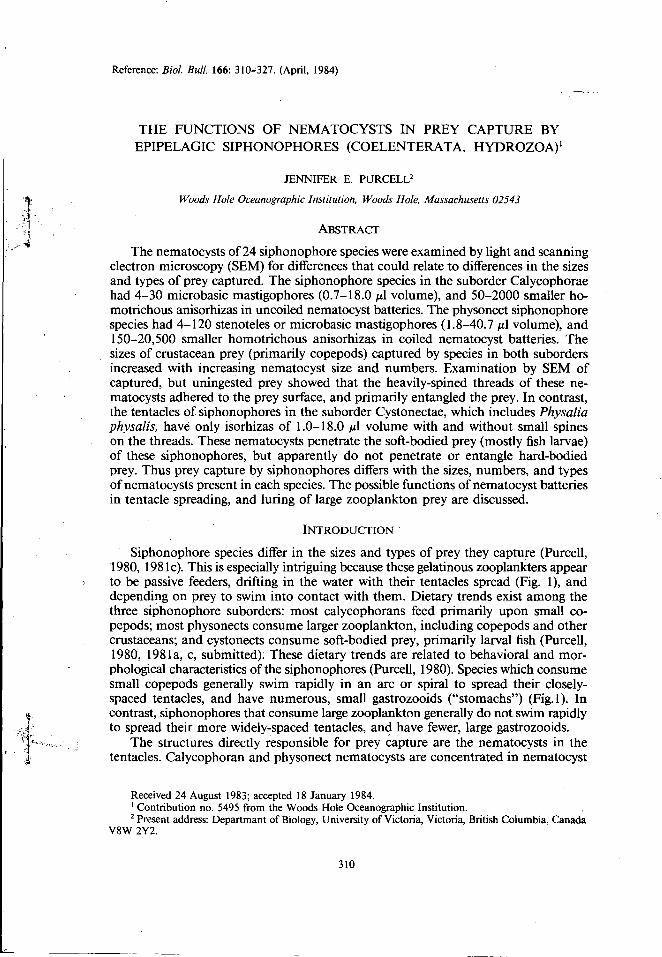
Siphonophore species differ in the sizes and types of prey they capture (Purcell, 1980, 1981c). This is especially intriguing because these gelatinous zooplankters appear to be passive feeders, drifting in the water with their tentacles spread (Fig. 1), and depending on prey to swim into contact with them. Dietary trends exist among the three siphonophore suborders: most calycophorans feed primarily upon small copepods; most physonects consume larger zooplankton, including copepods and other crustaceans; and cystonects consume soft-bodied prey, primarily larval fish (Purcell, 1980, 1981 a, c, submitted): These dietary trends are related to behavioral and morphological characteristics of the siphonophores (Purcell, 1980). Species which consume small copepods generally swim rapidly in an arc or spiral to spread their closelyspaced tentacles, and have numerous, small gastrozooids ("stomachs") (Fig.1 ). In contrast, siphonophores that consume large zooplankton generally do not swim rapidly to spread their more widely-spaced tentacles, and have fewer, large gastrozooids.
The structures directly responsible for prey capture are the nematocysts in the tentacles. Calycophoran and physonect nematocysts are concentrated in nematocyst
Received 24 August 1983; accepted 18 January 1984. 1 Contribution no. 5495 from the Woods Hole Oceanographic Institution. . 2 Present address: Departmant of Biology, University of Victoria, Victoria, British Columbia, Canada
V8W 2Y2.
310
NEMA TOCYST FUNCTIONS lN PREY CAPTURE 311
FIGURE I . Sulculeolaria quadrivalvis (Suborder Calycophorae) in feeding posture with tentacles spread. The tentacles appear as a string of dots, which are the nematocyst batteries. n = Nectophores (swimming bells), g = Chain of gastrozooids (stomachs). Scale = 2 em. Photograph by J. M. King.
batteries, one battery on each of several side branches (tentilla) of the tentacles. Nematocyst batteries consist of a cnidoband with many nematocysts, several larger nematocysts along the cnidoband, an elastic ligament, and I or 2 terminal filaments. The battery nematocysts fire as a unit when the terminal filaments are pulled and
312 J. E. PURCELL
stretch the elastic ligament. Cystonect nematocysts are not found in these complex batteries, but may be concentrated in clumps or bands in the tentacles.
Nematocysts are classified according to the structure of the thread, which is coiled within the nematocyst capsule and everts upon discharge (reviewed in Mariscal, 1974). Historical works on siphonopbore nematocysts have dealt primarily with systematic classification and the processes of nematocyst formation and discharge (Chun, 1891 , 1892; lwanzoff, 1896; Schneider, 1899, 1900; Weill, 1934; Russell, 1938, 1939; Werner, 1965). More recently, sipbonophore nematocysts have been used to elucidate the process of nematocyst development (Carre, 1972, 1974a, b, c; Carre and Carre, 1973; Skaer, 1973).
Different functions have been ascribed to some of the various types of coelenterate nematocysts based on the structure of the thread, or on correlation of certain nematocyst types with specific functions. For example, an anacrophore is called a glutinant because the thread is closed at the tip while other types are presumed to be penetrants because the thread is open at the tip (Mariscal, 1974); holotrichous isorhizas in the acroraghi and catch tentacles of sea anemones presumably penetrate the victims of inter- and intraspecific agonistic interactions (Francis, 1973; Purcell, 1977). The discharge ofnematocysts on prey organisms has been investigated only in Hydra; Tappe ( 1909), Ewer ( 1947), and Tardent and Holstein ( 1982) found that prey were penetrated by stenoteles and were entangled by desmonemes. Observations in Schwartz et a/. ( 1983) suggest that a particular cladoceran species, which was not captured often by Hydra spp., might not be penetrated by the nematocysts of Hydra, might not stimulate the nematocysts to fire, or might be immune to the toxins. The present paper summarizes the characteristics of the nematocyst batteries and nematocysts of the three siphonophore suborders, and examines differences in the nematocysts that may mediate the differential prey capture seen in several siphonophore species.
MATERIALS AND METHODS
Prey size analysis
Siphonophores were collected in jars by SCUBA divers at 5-25 m depth primarily in the Gulf of California, the Gulf Stream, and the Sargasso Sea. Details of the methods, and of the locations and dates for collection of most siphonophores used in analysis of prey size are given in Purcell {1981c). Additional dietary data came from specimens collected during August, 1981 in the North Atlantic along the 25°N parallel, and during February and May, 1982 in the Sargasso Sea (approximately 30°77-78'W and 34°N 73°W, respectively), and in the Gulf Stream (approximately 32°N 78°W to 36°N 72°W and 33°N 77°W to 36°N 73°W). The cephalothorax length of copepods was measured using an optical micrometer at 25-400X magnification.
Microscopy
Siphonophores used for scanning electron microscopy (SEM) were collected primarily in the North Atlantic during August, 1981 , as above. When the siphonophores had assumed a feeding posture with tentacles spread in 4 I clear plastic containers aboard ship, active copepods, chaetognaths, or fish larvae from plankton tows were released individually near the tentacles by pipette. The captured prey were retrieved with forceps after the prey swam into contact with the siphonophore tentacles, but before they could be ingested. Specimens were placed in 2% gluteraldebyde in sea
NEMA TOCYST FUNCTIONS rN PREY CAPTURE 313
water for I h, postfixed in I% Os04 in sea water for I h, transferred at I 0 min intervals into 30%, 50%, and 70% ethanol and stored at 4°C. Ethanol dehydration was followed by critical-point drying with C02 or Freon in a Tousimas sam-dry-780 or a Bowmar SPC-900 cri tical-point drying chamber. Specimens were mounted on stubs, rotarycoated with gold in a Denton DSM-5 or an International Scientific Instruments (lSI) PS-2 vacuum evaporator, and examined in a JEOLCO V3 or an lSI AJpha-9 scanning electron microscope in order to examine the effect of nematocysts on prey. Small pieces of Nitex screen were swept through extended tentacles and treated as above, in order to examine the thread structure of discharged nematocysts. One or 2 copepods, and I screen were examined for each siphonophore species.
Siphonophores used for light microscopy first were relaxed in MgCI2 in sea water until the tentacles no longer contracted when touched, and then preserved in 5% formalin. The length, width, and depth of nematocyst batteries were measured at 40-400X magnification on a compound microscope with an optical micrometer. The maximum length and width of undischarged nematocysts from disrupted batteries were measured at 400-IOOOX. The numbers of anisorhizas (haplonemes) were approximated from the surface area (length X width) of the cnidoband divided by the maximum cross-sectional area of the anisorhizas; the depth of the cnidoband corresponds to their length. The large microbasic mastigophores and stenoteles (heteronemes) were counted, and their volumes were estimated according to the formula for the volume of an elipsoid, 4ab2/ 3, where a = length/2, and b = diameter/ 2. The dimensions of at least 5 nematocysts of each type were measured for each specimen, and 2-6 specimens were examined in each species. The measurements are from one specimen, or are maximum and minimum values where substantial differences existed among several specimens within a species.
Terminology
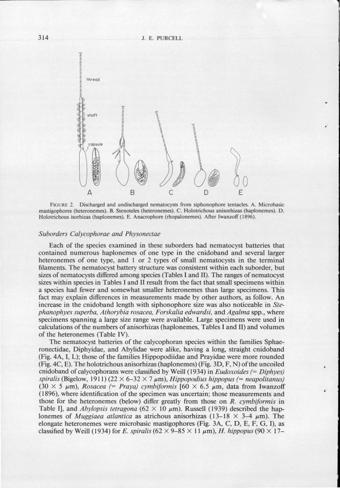
Nematocyst classification and terminology used herein is as presented by Mariscal ( 1974) based on the structure of the nematocyst thread (Fig. 2). Simply, haplonemes are nematocysts without a well-defined shaft at the base of the thread; isorhizas have threads of equal diameter throughout, while the base of the thread of an anisorhiza is slightly dilated; holotrichous indicates that spines occur along the length of the thread, and atrichous threads have no spines. Heteronemes have a well-defined shaft; microbasic mastigophores have a short shaft of equal diameter throughout; stenoteles have a short shaft of unequal diameter, with 3 well-developed spines. Desmonemes, acrophores, and anacrophores have threads with closed tips and no spines.
REsULTS
Measurements and photographs ofnematocyst batteries and nematocysts are presented for the 3 siphonophore suborders: Calycophorae (Table I, Fig. 3), Physonectae (Table II, Fig. 4), and Cystonectae (Table III, Fig. 5). Each table and figure has been organized by siphonophore family, and arranged in order of increasing nematocyst and nematocyst battery sizes, as far as was possible. Differences in corresponding nematocyst dimensions in the tables and figures may be due to effects of preservation, and to a 20% reduction in length and diameter in capsules of discharged nematocysts (Tardent and Holstein, 1982). The batteries and nematocysts were similar enough among species of each family that only representative illustrations are given for each family in Figures 3, 4, and 5.
314 J . E. PURCELL
lhr~od
A 8 c D E
FIGURE 2. Discharged and undischarged nematocysts from siphonophore tentacles. A. Microbasic mastigophores (heteronemes). B. Stenoteles (heteronemes). C. Holotrichous anisorhizas (haplonemes). D. Holotrichous isorhizas (haplonemes). E. Anacrophore (rhopalonemes). After lwanzoff ( 1896).
Suborders Calycophorae and Physonectae
Each of the species examined in these suborders had nematocyst batteries that contained numerous haplonemes of one type in the cnidoband and several larger heteronemes of one type, and I or 2 types of small nematocysts in the terminal filaments. The nematocyst battery structure was consistent within each suborder, but sizes of nematocysts differed among species (Tables I and II). The ranges of nematocyst sizes within species in Tables I and II result from the fact that small specimens within a species had fewer and somewhat smaller heteronemes than large specimens. This fact may explain differences in measurements made by other authors, as follow. An increase in the cnidoband length with siphonophore size was also noticeable in Stephanophyes superba, Athorybia rosacea, Forskalia edwardsi, and Aga/ma spp., where specimens spanning a large size range were available. Large specimens were used in calculations of the numbers ofanisorruzas (haplonemes, Tables I and II) and volumes of the heteronemes (Table IV).
The nematocyst batteries of the calycophoran species within the families Sphaeronectidae, Diphyidae, and Abylidae were alike, having a long, straight cnidoband (Fig. 4A, I, L); those of the families Hippopodijdae and Prayidae were more rounded (Fig. 4C, E). The holotricbous anisorhizas (haplonemes) (Fig. 30, F, N) of the uncoiled cnidoband of calycophorans were classified by Weill ( 1934) in Eudoxoides (= Diphyes) spiralis (Bigelow, 1911) (22 X 6-32 X 7 J.Lm), Hippopodius hippopus (= neapolitanus) (30 X 5 J.LID), Rosacea (= Praya) cymbiformis [60 X 6.5 J.LID, data from lwanzoff ( 1896), where identification of the specimen was uncertain; those measurements and those for the heteronemes (below) differ greatly from those on R . cymbiformis in Table 1], and Aby/opsis tetragona (62 X I 0 J.Lm). Russell ( 1939) described the baplonernes of Muggiaea atlantica as atrichous anisorhizas ( 13-18 X 3--4 J.Lm). The elongate heteronemes were microbasic mastigophores (Fig. 3A, C, D, E, F, G, 1), as classified by Weill ( 1934) for E. spiral is (62 X 9-85 X II J.Lm), H. hippopus (90 X 17-
..
,...-------------:-----...... ...-- - --..... ~---~------~--~-----~ --- - ...
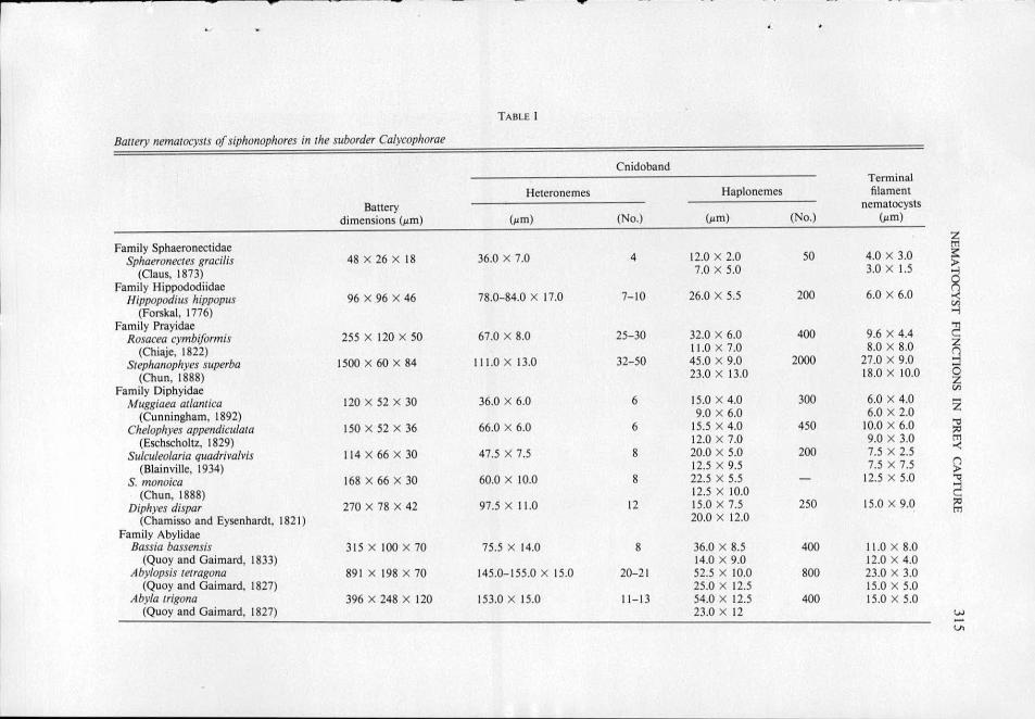
TABLE I
Bauery nematocysts of siphonophores in the suborder Calycophorae
Cnidoband Terminal
Heteronemes Haplonemes filament
Battery nematocysts
dimensions (~m) (~m) (No.) (~tm) (No.) (~tm)
Family Sphaeronectidae ~ Sphaeronectes gracilis 48 X 26 X 18 36.0 X 7.0 4 12.0 X 2.0 50 4.0 X 3.0 >
(Claus, 1873) 7.0 X 5.0 3.0 X 1.5
a Family Hippododiidae Hippopodius hippopr1s 96 X 96 X 46 78.0-84.0 X 17.0 7- 10 26.0 X 5.5 200 6.0 X 6.0
(Forskal, 1776) ~ Family Prayidae
~ Rosacea cymbiformis 255 X 120 X 50 67.0 X 8.0 25-30 32.0 X 6.0 400 9.6 X 4.4
(Chiaje, 1822) 11.0 X 7.0 8.0 X 8.0 Q Srephanophyes superba 1500 X 60 X 84 11 1.0 X 13.0 32-50 45.0 X 9.0 2000 27.0 X 9.0
(Chun, 1888) 23.0 X 13.0 18.0 X 10.0 6 z Family Diphyidae tn
Muggiaea atlantica 120 X 52 X 30 36.0 X 6.0 6 15.0 X 4.0 300 6.0 X 4 .0 z (Cunningham, 1892) 9.0 X 6.0 6.0 X 2.0
Che/ophyes appendiculata 150 X 52 X 36 66.0 X 6.0 6 15.5 X 4.0 450 10.0 X 6.0 .., ?'
(Eschscholtz, 1829) 12.0 X 7.0 9.0 X 3.0 tTl
Sulculeo/aria quadriva/vis 114 X 66 X 30 47.5 X 7.5 8 20.0 X 5.0 200 7.5 X 2.5 -<
(Biainville, 1934) 12.5 X 9.5 7.5 X 7.5 () >
S. monoica 168 X 66 X 30 60.0 X 10.0 8 22.5 X 5.5 - 12.5 X 5.0 2 (Chun, 1888) 12.5 X 10.0
Diphyes dispar 270 X 78 X 42 97.5 X 11.0 12 15.0 X 7.5 250 15.0 X 9 .0 ;:o
(Chamisso and Eysenhardt, 182 1) 20.0 X 12.0 tTl
Family Abylidae Bassia bassensis 315 X 100 X 70 75.5 X 14.0 8 36.0 X 8.5 400 11.0 X 8.0
(Quoy and Gaimard, 1833) 14.0 X 9.0 12.0 X 4.0 Aby/opsis tetragona 89 1 X 198 X 70 145.0-155.0 X 15.0 20-2 1 52.5 X 10.0 800 23.0 X 3.0
(Quoy and Gaimard, 1827) 25.0 X 12.5 15.0 X 5.0 Abyla trigona 396 X 248 X 120 153.0 X 15.0 11 - 13 54.0 X 12.5 400 15.0 X 5.0
(Quoy and Gaimard, 1827) 23.0 X 12 w -Vl
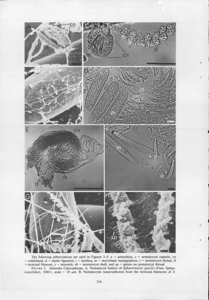
The following abbreviations are used in Figures 3-5: a = anisorhiza, c = nematocyst capsule, en = cnidoband, el = elastic ligament, i = isorhiza, m = microbasic mastigophore, t = oematocyst thread, tf = terminal filament, s = stenotele, sh = oematocyst shafi , and sp = spines on oematocyst thread.
FlGURE 3. Suborder Calycophorae. A. ematocyst battery of Sphaeronectes gracilis (Fam. Sphaeronectidae), IOOOX, scale = 10 llm. B. ematocysts (anacrophores) from the terminal filaments of S.
316
EMA TOCYST FUNCTIO S IN PREY CAPTURE 317
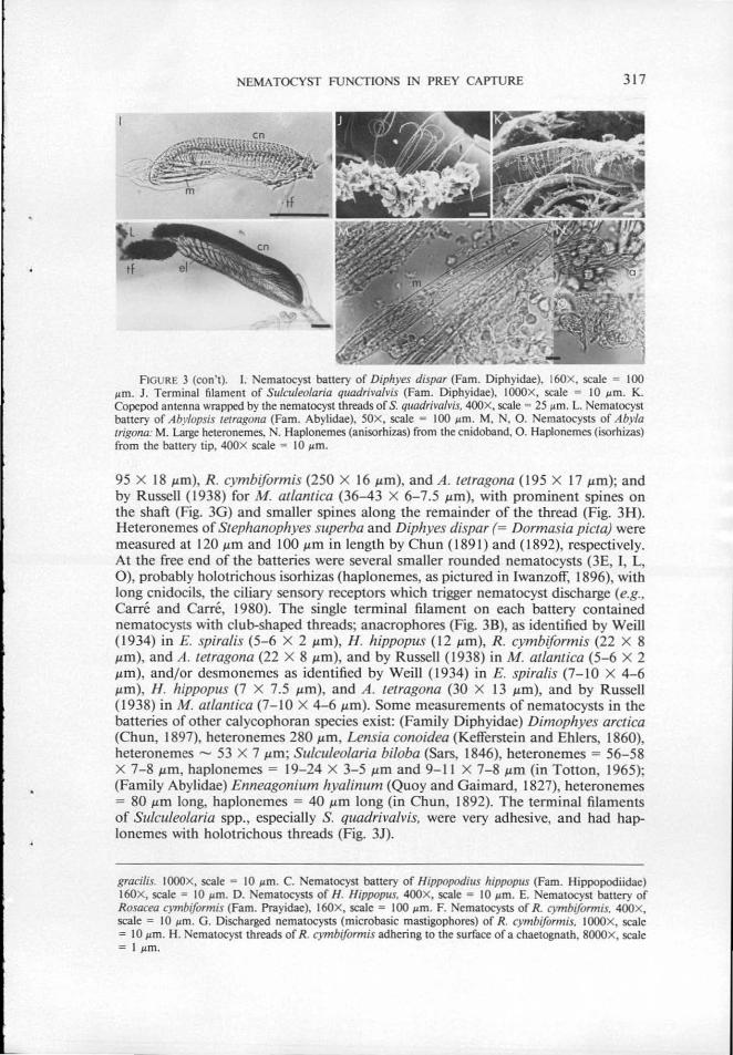
FIGURE 3 (con't). I. ematocyst battery of Diphyes dispar (Fam. Diphyidae), 160X, scale = 100 I'm. J . Terminal filament of Sulculeolaria quadrivafvis (Fam. Diphyidae), IOOOX, scale = 10 I'm. K. Copepod antenna wrapped by the nematocyst threads of S. quadrivalvis, 400X, scale = 25 I'm. L. Nematocyst bartery of Abylopsis tetragona (Fam. Abylidae), SOX, scale = 100 I'm. M, N, 0 . Nematocysts of Abyla trigona: M. Large heteronemes, N. Haplonemes (anisorhizas) from the cnidoband, 0. Haplonemes (isorhizas) from the battery tip, 400X scale = 10 I'm.
95 X 18 J.Lffi), R. cymbiformis (250 X 16 J.Lm), and A. tetragona ( 195 X 17 J.Lm); and by Russell ( 1938) for M atlantica (36-43 X 6-7.5 J.Lffi), with prominent spines on the shaft (Fig. 3G) and smaller spines along the remainder of the thread (Fig. 3H). Heteronemes of Stephanophyes superba and Diphyes dispar (= Dormasia picta) were measured at 120 J.Lm and 100 J.Lm in length by Chun (1891) and ( 1892), respectively. At the free end of the batteries were several smaller rounded nematocysts (3E, I, L, 0 ), probably holotrichous isorhizas (haplonemes, as pictured in lwanzoff, 1896), with long cnidocils, the ciliary sensory receptors which trigger nematocyst discharge (e.g., Carre and Carre, 1980). The single terminal filament on each battery contained nematocysts with club-shaped threads; anacrophores (Fig. 3B), as identified by Weill ( 1934) in E. spiralis (5- 6 X 2 J.Lm), H. hippopus ( 12 J.Lffi), R. cymbiformis (22 X 8 1-lm), and A. tetragona (22 X 8 1-Lm), and by Russell ( 1938) in M. atlantica (5- 6 X 2 1-lm), and/or desmonemes as identified by Weill ( 1934) in E. spiral is (7-1 0 X 4- 6 /-LID), H. hippopus (1 X 7.5 1-lm), and A . tetragona (30 X 13 J.LID), and by Russell (1938) in M. atlantica (7-10 X 4-6 J.Lm). Some measurements ofnematocysts in the batteries of other calycophoran species exist: (Family Diphyidae) Dimophyes arctica (Chun, 1897), heteronemes 280 /Jm , Lensia conoidea (Kefferstein and Ehlers, 1860), heteronemes - 53 X 7 J.Lm; Sulculeolaria biloba (Sars, 1846), heteronemes = 56- 58 X 7-8 /-Lm, haplonemes = 19- 24 X 3-5 J.Lm and 9- 11 X 7-8 J.LID (in Totton, 1965); (Family Abylidae) Enneagonium hyalinum (Quoy and Gaimard, 1827), heteronemes = 80 1-Lm long, haplonemes = 40 1-lm long (in Chun, 1892). The terminal filaments of Sulculeolaria spp., especially S. quadrivalvis, were very adhesive, and had haplonemes with holotrichous threads (Fig. 3J).
gracilis. !OOOX, scale = 10 I'm. C. ematocyst battery of Hippopodius hippopus (Fam. Hippopodiidae) 160X, scale = 10 I'm. D. ematocystS of H. Hippopus. 400X, scale = 10 I'm. E. ematocyst battery of Rosacea cymbiformis (Fam. Prayidae), 160X, scale = 100 I'm. F. NematocystS of R. cymbiformis. 400X, scale = 10 I'm. G. Discharged nematocysts (microbasic mastigophores) of R. cymbiformis, IOOOX, scale = 10 I'm. H. ematocyst threads of R. cymbiformis adhering to the surface of a chaetognath, 8000X, scale = I I'm.
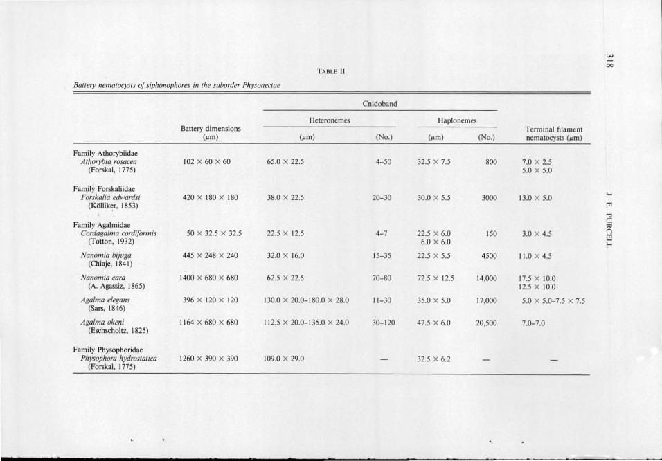
w -00 TABLE II
Bauery nernatocysts of siphonophores in the suborder Physonectae
Cnidoband
Heteronemes Haplonemes Battery dimensions Terminal filament
{Jlm) (~tm) (No.) (~tm) (No.) nematocysts (~tm)
Family Athorybiidae Athorybia rosacea 102 X 60 X 60 65.0 X 22.5 4-50 32.5 X 7.5 800 7.0 X 2.5
(Forskal, 1775) 5.0 X 5.0
Family Forskaliidae :-Forskalia edwardsi 420 X 180 X 180 38.0 X 22.5 20-30 30.0 X 5.5 3000 13.0 X 5.0
(Kolliker, 1853) !"" "0
Family Agalmidae ~ Cordaga/rna cordiforrnis 50 X 32.5 X 32.5 22.5 X 12.5 4-7 22.5 X 6.0 150 3.0 X 4.5 Q
(Totton, 1932) 6.0 X 6.0 r r-Nanornia bijuga 445 X 248 X 240 32.0 X 16.0 15-35 22.5 X 5.5 4500 11.0 X 4.5
(Chiaje, 1841)
Nanomia cara 1400 X 680 X 680 62.5 X 22.5 70-80 72.5 X 12.5 14,000 17.5 X 10.0 (A. Agassiz, 1865) 12.5 X 10.0
Agalma e/egans 396 X 120 X 120 130.0 X 20.0-180.0 X 28.0 11- 30 35.0 X 5.0 17,000 5.0 X 5.0-7.5 X 7.5 (Sars, 1846)
Agalma okeni 1164 X 680 X 680 11 2.5 X 20.0-135.0 X 24.0 30-120 47.5 X 6.0 20,500 7.0-7.0 (Eschscholtz, 1825)
Family Physophoridae Physophora hydrostatica 1260 X 390 X 390 109.0 X 29.0 - 32.5 X 6.2
(Forskal, 1775)
NEMATOCYST FUNCflONS IN PREY CAPTURE 319
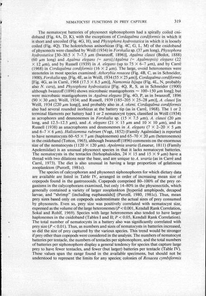
The nernatocyst batteries of physonect siphonophores had a spirally coiled cnidoband (Fig. 4A, D, K), with the exceptions of Cordaga/ma cordiform is in which it is short and uncoiled (Fig. 4G, H), and Physophora hydrostatica in which it is loosely coiled (Fig. 4Q). The holotrichous anisorhizas (Fig. 4C, G, L, M) of the cnidoband ofphysonects were classified by Weill (1934) in Forskalia sp. (37 .urn long), Physophora hydrostatica [30-30.5 X 7-7.5 .urn (lwanzoff, 1896)], Agalma clausi (Bedot, 1888) (60 .urn long) and Aga/ma elegans (= sarsi)/Agalma (= Aga/mopsis) e/egans (22 X 12 .urn), and by Russell (1939) in A. elegans (up to 75 X 6-7 .urn), and by Carre (1968) in Cordagalma cordiformis (16 X 2 .urn). The large, ovoid heteronernes were stenoteles in most species examined: Athorybia rosacea (Fig. 4B, C, as in Schneider, 1900), Forskalia spp. [Fig. 4E, as in Weill, 1934 (55 X 25 .urn)], Cordagalma cordiform is [Fig. 4G, as in Carre, 1968 ( 17.5 X 8.5 .urn)], Namomia bijuga (Fig. 4L, N, probably also N. cara), and Physophora hydrostatica [Fig. 4Q, R, S, as in Schneider (1900) although lwanzoff(l896) shows microbasic mastigophores- 100-150 .urn long]; but were rojcrobasic mastigophores in Agalma elegans [Fig. 40, P, as in lwanzoff, 1896 (90 X 30 .urn); Weill, 1934; and Russell, 1939 ( 185-205 X 25-28 .urn)], A. clausi [in Weill, 1934 (220 .urn long)], and probably also in A. okeni. Cordagalma cordiformis also had several rounded isorhizas at the battery tip (as in Carre, 1968). The l or 2 terminal ftlaments per battery had 1 or 2 nematocyst types, classified in Weill ( 1934) as acrophores and desmonemes in Forskalia sp. (15 X 7.5 .urn), A. clausi (20 .urn long, and 12.5- 12.5 .urn), and A. elegans (21 X 15 .urn and I 0 X 10 .urn), and in Russell ( 1938) as anacrophores and desmonemes in A. e/egans (7 X 2-20 X 5 .urn and 6-7 X 6 .urn). Halistemma rubrum (Vogt , 1852) (Family Agalrojdae) is reported to have nematocysts 60-1')5 X 7 .urn (haplonemes) and 65-70 X 20 .urn (heteronemes) in the cnidoband (Totton, 1965), although lwanzoff ( 1896) comments on the enormous size of the nematocysts ( 1120 X 120 .urn). Apolemia uvaria (Lesueur, 181 I) (Family Apolemiidae) is an unusual physonect species in that it lacks nematocyst batteries. The nematocysts in the tentacles {birhophaloides, 24 X 15 and 15 X 9 .urn) have a thread with two dilations near the base, and are unique to A. uvaria (as in Carre and Carre, 1973). The diet is also unusual in having a large proportion of gelatinous zooplankton (Purcell, 1981 c).
The species of calcyophoran and physonect siphonophores for which dietary data are available are listed in Table IV, arranged in order of increasing mean size of copepods found in the gastrozooids. Copepods comprised 80-100% of the prey organisms in the calycophorans examined, but only 14-80% in the physonectids, which generally contained a variety of larger zooplankton [hyperiid amphipods, decapod larvae, and "shrimp" (including euphausiids)] (Purcell, 1980, 1981 c). Thus, mean prey sizes based only on copepods underestimate the actual sizes of prey consumed by pbysonects. Even so, prey size was positively correlated with nematocyst size, expressed as the volume of the large heteronemes (P < 0.00 I, Kendall Rank Correlation, Sakal and Rohlf, 1969). Species with large heteronemes also tended to have larger haplonemes in the cnidoband (Tables I and IT; P < 0.05, Kendall Rank Correlation). The total number of nematocysts in a battery also was significantly correlated with prey size (P < 0.0 I). Thus, as numbers and sizes of nematocysts in batteries increased, so did the size of prey captured by the various species. This trend would be stronger if prey other than copepods were considered in the analysis. The numbers of nematocyst batteries per tentacle, the numbers of tentacles per siphonophore, and the total numbers of batteries per siphonophore display a general tendency for species that capture large prey to have fewer tentacles, and fewer (but larger) batteries per tentacle (Table IV). These values span the range found in the available specimens, but should not be understood to represent the limits for any species; colonies of R osacea cymbiformis
320 J. E. PURCELL
FIGURE 4 . Suborder Physonectae. A. Nematocyst battery of Athorybia rosacea (Fam. Athorybiidae), IOOX, scale = 100 1-1m. B. Nematocyst shaft of a stenotele from A. rosacea, 2000X, scale = 10 1-1m . C. Nematocysts of A. rosacea, 400X, scale = 10 1-1m. D. ematocyst battery of Forska/ia edwardsi (Fam. Forskaliidae), 40X, scale = 100 1-1m. E. Nematocyst (stenotele) of F. edwardsi. IOOOX, scale = 10 1-1m. F. Copcpod captured by F. edwardsi, IOOX, scale = 100 1-1m. G. Nematocyst battery ofCordaga/ma cordiform is (Fam. AgaJmidae), 200X, scale = 25 1-1m. H. Nematocyst battery of C. cordiformis. IOOOX, scale = 10 1-1m. I. Nematocyst threads of C. cordiformis adhering to a copcpod, IOOOX, sca.le = 10 I'm. J. Uncaptured copcpod, SOX, scale = 100 1-1m.
and Stephanophyes superba, in particular, reach much greater lengths, and therefore have many more tentacles.
The effect of caJycophoran and physonect nematocysts on prey was dramaticthe nematocyst threads wrapped and entangled copepod prey (Fig. 3K; 4F, M). Nematocyst threads with and without spines adhered to the surfaces of copepods, chaetognaths, and Nitex fibers (Fig. 3B, G, H , J, K; 41, M). The spines along the length of the threads were affixed to the prey surface wherever they were in contact. The flattened threads of Cordagalma cordiformis nematocysts were unlike the tubular threads of other species; the tips of these unspined threads clearly adhered to crustaceans (Fig. 41). Penetration of the exoskeleton by the nematocyst threads was not obvious in any of the copepods prepared for SEM. Threads may have entered joints in the exoskeleton in a few cases, such as when the stenoteles of Nanomia bijuga oriented along the cephalothorax/ abdominal joint of a copepod (Fig. 4N). Nematocyst threads penetrated, as well as adhered to soft-bodied prey.
EMA TOCYST FUNCfiONS lN PREY CAPTURE 32 1
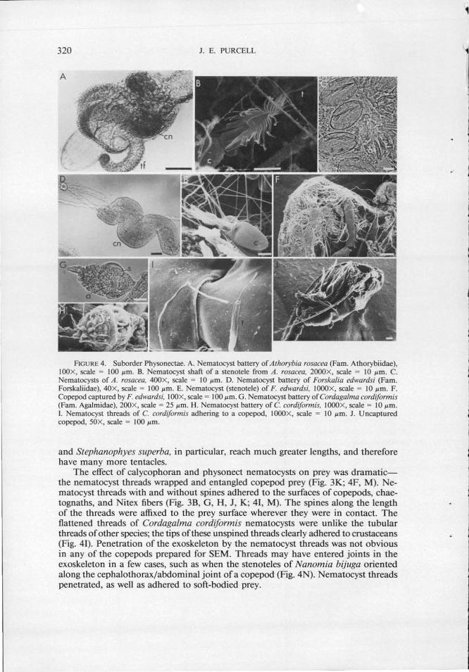
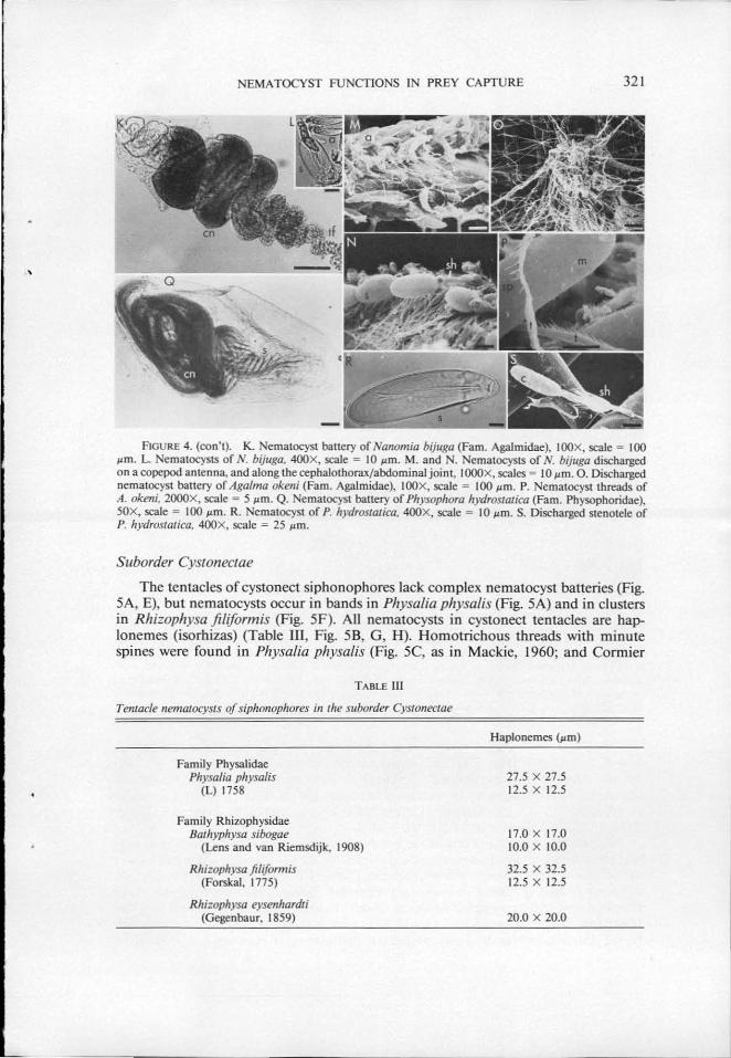
f'IGURE 4. (con't). K. Nematocyst battery of Nanomia bijuga (Fam. Agalmidae), IOOX, scale = 100 ~tm. L ematocysts of N. bijuga, 400X, scale = 10 ~tm. M. and . Nematocysts of N. bijuga discharged on a cope pod antenna, and along the cephalothorax/abdominal joint, I OOOx, scales = I 0 ~tm. 0. Discharged nematocyst battery of Agalma okeni (Fam. Agalmidae), IOOX, scale = 100 ~tm. P. Nematocyst threads of A. okeni, 2000X, scale = 5 ~tm. Q. ematocyst battery of Physophora hydrostatica (Fam. Physophoridae), SOX, scale = 100 ~tm. R. ematocyst of P. hydrostaJica, 400X, scale = 10 ~tm. S. Discharged stenotele of P. hydrostatica, 400X, scale = 25 ~tm.
Suborder Cystonectae
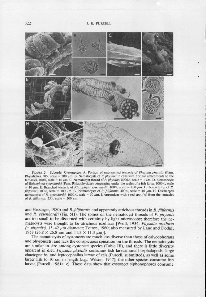
The tentacles of cystonect siphonophores lack complex nematocyst batteries (Fig. SA, E), but nematocysts occur in bands in Physalia physalis (Fig. SA) and in clusters in Rhizophysa fi/iformis (Fig. SF). All nematocysts in cystonect tentacles are haplonemes (isorhizas) (Table III, Fig. SB, G, H). Homotrichous threads with minute spines were found in Physalia physalis (Fig. SC, as in Mackie, 1960; and Cormier
TABLE lll
Tentacle nematocysts of siphonophores in the suborder Cysronectae
Family Physalidae Physalia physalis
(L) 1758
Family Rhizophysidae Bathyphysa sibogae
(Lens and van Riemsdijk, 1908)
Rhizophysa filiform is (Forskal, 1775)
Rhizoplrysa eysenhardti (Gegenbaur, 1859)
Haplonemes (!lm)
27.5 X 27.5 12.5 X 12.5
17.0 X 17.0 10.0 X 10.0
32.5 X 32.5 12.5 X 12.5
20.0 X 20.0
322 J. E. PURCELL
FIGURE 5. Suborder Cystonectae. A. Portion of unbranched tentacle of Physa/ia physalis (Fam. Physalidae), SOX, scale = 200 1-1m. B. Nematocysts of P. physalis in ceUs with fibriUar attachments to the tentacles, 400X, scale= 10 pm. C. Nematocyst thread of P. physalis, 8000X, scale = I 1-1m. D. Nematocyst of Rhizophysa eysenhardti (Farn. Rhizophysidae) penetrating under the scales of a fish larva, I OOOX, scale = 10 1-1m. E. Branched tentacle of Rhizophysa eysenhardti, IOOX, scale = 100 I'm. F. Tentacle tip of R . filiformis, IOOX, scale = 100 pm. G . Nematocysts of R. filiformis, 400X, scale = 10 1-1m. H. Discharged nematocyst of R . eysenhardti, IOOOX, scale= 10 1-1m. I. Appendage with a red spot (rs) from the tentacles of R . filiformis, 25X, scale = 200 1-1m.
and Hessinger, 1980) and R .filiformis, and apparently atrichous threads in R .filiformis and R. eysenhardti (Fig. 5H). The spines on the nematocyst threads of P. physalis are too small to be discerned with certainty by light microscopy; therefore the nematocysts were thought to be atrichous isorhizas [Weill, 1934, Physalia arethusa {= physalis}, 15-42 J.Lm diameter; Totton, 1960; also measured by Lane and Dodge, 1958 (26.8 X 26.8 J.Lffi and 11 .3 X 11 .3 J.Lm)].
The nematocysts of cystonects are much less diverse than those of calycophorans and physonects, and lack the conspicuous spination on the threads. The nematocysts are similar in size among cystonect species (Table III), and there is little diversity apparent in diet. Physalia physalis consumes fish larvae, small cephalopods, large chaetognaths, and leptocephali us larvae of eels (Purcell, submitted), as well as some larger fish to 10 em in length (e.g., Wilson, 1947); the other species consume fish larvae (Purcell, 1981a, c). Those data show that cystonect siphonophores consume
,

NEMA TOCYST FUNCTIONS IN PREY CAPTURE 323
TABLE IV
Calycophoran (C) and physonect (P) siphonophores listed in order of increasing prey size (copepods only)
Mean Large Batteries prey nematocyst Nematocysts per
Sub- size volume per battery tentacle Tentacles Number of
Species order (mm) (~tl)* (no.)* (no.) (no.) batteries
Muggiaea atlantica c 0.36 0.68 306 20 20-30 400-600
Sphaeronectes gracilis c 0.36 0.92 54 25-30 20-60 500-1800 Che/ophyes appendiculata c 0.42 1.24 456 25-30 10-20 250-600 Cordagalma cordiformis p 0.45 1.84 157 60-70 20-30 1200-2100 Sulculeolaria quadrivalvis c 0.57 1.40 208 80-90 85-520 6800-46,800
Bassia bassensis c 0.79 7.74 408 5-10 5-15 25-150 Athorybia rosacea p 0.84 17.22 850 120 1-10 120-1200 Hippopodius hippopus c 0.86 5.32 210 30-100 10-20 300-2000 Rosacea cymbiformis c 0.98 2.25 430 80-90 10-100 800-9000 Diphyes dispar c 0.99 6.46 262 40-50 90-180 3600-9000 Abyla trigona c 1.10 18.02 413 10-20 10-20 100-400
Forska/ia edwardsi p 1.17 9.94 3030 10-20 10-320 100-6400 Nanomia bijuga p 1.18 4.29 4535 15-20 15-20 225-400
Aga/ma e/egans p 1.33 27.21-73.85 17,030 12-50 5-30 60-150 Stephanophyes super/Ja c 1.52 9.82 2050 30-40 10-30 300-1200 Aga/ma okeni p 1.97 40.69 20,620 15-35 2-9 30-315
• Calculated from values in Table I and II.
only prey lacking hard exoskeletons. Their nematocysts clearly penetrate soft-bodied prey (Fig. 5D). No discharged nematocysts penetrated or adhered to a shrimp (Leander sp.) that was thrust repeatedly into the tentacles of P. physalis. That result could be due to failure of the nematocysts to discharge or failure to affect the prey.
DISCUSSION
The large majority of the nematocysts in the battery complex of calycophorans and physonects appear to adhere to and entangle crustaceans, their principle prey. The threads of nematocysts in the terminal filaments (anacrophores and desmonemes) have closed, rounded ends. Anisorhizas, by far the most numerous nematocysts of the cnidobands, had spines along the length of the threads which adhered to all surfaces. The long, spined shaft of micro basic mastigophores also adhered to surfaces. To date, only stenoteles in Hydra have been shown to penetrate crustacean exoskeleton (Tappe, 1909; Ewer, 1947; Tardent and Holstein, 1982). The stenoteles of Hydra are forcefully ejected from the tentacle and penetrate crustacean exoskeleton presumably by the impact of the three sty lets, which form a "pointed arrowhead" (Tardent and Holstein, 1982). Mariscal ( 1974) cited his unpublished results that showed penetration of prey by Anthozoan microbasic p-mastigophores and basitrichous isorhizas, but did not specify the prey used. Most siphonophore nematocysts do not appear to penetrate crustacean exoskeleton. Only physonect siphonophores have penetrating stenoteles, but adhering anisorhizas outnumbered stenoteles in the cnidobands of physonects by 20-40:1 (C. cordiformis) to -200:1 (Nanomia spp.).
These results suggest that most nematocysts of calycophoran and physonect siphonophores function to entangle crustacean prey. The nematocyst threads clearly adhere to surfaces, however the mechanism of adhesion is unknown. Anthozoan nematocysts adhere to glass slides and to cleaned gastropod shells (e.g., Sandberg et a/., 1971; Mariscal, 1972). Similarly, tentaculate ctenophores rely only upon a glue
324 J. E. PURCELL
produced by colloblasts on the tentacles to adhere to, and capture, the same crustacean zooplankton prey (Franc, 1978).
The fact that the threads of most of these siphonophore nematocysts appear not to penetrate the prey raises questions about their toxicity. The toxins of siphonophore nematocysts have been examined only for Physalia (Cystonectae), and it has not been possible to isolate single types of nematocysts (e.g., Lane and Dodge, 1958; Burnett and Calton, 1977). Therefore, it is unknown if the nematocyst types of other siphonophores contain toxin. It is also unknown if nematocyst toxins are a liquid in the capsule which is expelled through the thread upon discharge (like a syringe) or if the toxins are associated with the surface of the thread such that they contact the prey all along its discharged length (see Tardent and Holstein, 1982). The above results suggest that very little toxin would be injected directly into the copepod. Perhaps toxin is released into the water immediately surrounding the prey or over the surface of the prey and is taken up through the respiratory surfaces, pores such as chemoreceptors, or thin exoskeleton at the joints in sufficient quantity to narcotize or kill the prey. The extent to which the experimental prey were entangled by nematocyst threads seemed adequate to immobilize them (Fig. 4F).
Cystonect siphonophores lack nematocysts with heavily-spined or club-shaped threads that adhere to the crustacean prey captured by calycophoran and physonect siphonophores. Therefore, cystonects may be unable to entangle crustaceans and may be limited to prey types that can be penetrated by their nematocysts. The isorhizas in cystonect tentacles may be unable to penetrate the exoskeleton of crustaceans; the isorhizas are firmly anchored in the tentacle (Cormier and Hessinger, 1980), and have no structure for puncturing comparable to the sty lets of stenoteles. The results of this study cannot exclude the possibility that cystonect nematocysts, are stimulated to discharge only by soft-bodied prey, perhaps by a mucus coating, which is lacking in crustaceans. Nematocysts of other coelenterates discharge upon a combination of chemical and mechanical stimulation (e.g., Mariscal, 1974), and the stimulus can be quite specific in some cases (e.g., Francis, 1973; Purcell, 1977). It is also possible that the toxins of cystonect nematocysts are not effective on crustaceans. However, toxin extracted from the nematocysts of Physalia (the only siphonophore thus tested) killed both crabs and fish when it was injected (Lane and Dodge, 1958; Burnett and Calton, 1977).
The above data show that there are marked differences in the .tentacle nematocysts of calycophoran and physonect siphonophores, and those of cystonect siphonophores, as well as marked differences in diet. The evidence suggests that the different structure of the nematocyst threads could contribute to the dietary differences; the heavilyspined threads of calycophoran and physonect nematocysts may promote entanglement of crustacean prey, while the simple threads of cystonect nematocysts may promote penetration of soft-bodied prey.
Larger prey were captured by calycophoran and physonect siphonophore species with larger nematocysts. Nematocysts oflarger volumes could contain longer threads, which presumably could be more effective in entangling prey. Nematocyst discharge may be due to an increase in the internal pressure of the capsule, caused by an influx of water in response to osmotic changes in the fluid of the capsule (Lubbock and Amos, 1981; Lubbock et a/., 1981 ). The force generated for discharge could be proportional to the volume of the capsule (greater for rounded nematocysts), or to the surface-to-volume ratio of the capsule (greater for elongate nematocysts) if the reactions were surface-mediated. This idea should be testable using methods of Tardent and Holstein ( 1982); presently, it is only speculation.
The nematocyst batteries of calycophoran and physonect siphonophores apparently
NEMA TOCYST FUNCTIONS IN PREY CAPTURE 325
function in two other capacities. The swimming activity of most siphonophores in these suborders serves to spread their tentacles in a 3-dimensional array (Mackie and Boag, 1963; Biggs, 1977) (Fig. 1 ). In Muggiaea atlantica, tentacle extension appears to be due to drag on the tentacles (Purcell and LaBarbera, unpub. results). Battery diameter is greater than tentacle diameter, hence the nematocyst batteries would increase drag at the ends of the tentacle branches and cause the tentacles to be drawn out. Thus, nematocyst batteries may aid in tentacle extension, enabling the spread of the tentacles to be greater and the tentacles finer (and therefore less conspicuous to prey) than possible with tentacles of uniform thickness.
Some information on morphology, behavior, and diet suggested that the nematocyst batteries of two siphonophore species may resemble small zooplankters and act to lure larger, predatory zooplankton into the siphonophore tentacles; the batteries of Agalma okeni resemble copepods, and some of Athorybia rosacea resemble fish larvae (Purcell, 1980). Further observations suggest that "lures" may occur in other siphonophore species. Two spots within the nematocyst batteries of Nanomia cara fluoresced when excited at 460-470 nm under an epifluorescence microscope (Purcell, unpubl. results), strongly suggesting that these spots are bioluminescent (see Morin and Reynolds, 1969). The batteries of A. okeni, A. elegans, N. bijuga, and Forskalia sp. did not fluoresce. Luminescent batteries may serve to attract prey at night or at depth, as may the luminescent lures (esca) of midwater anglerfish (Pietsch, 1974). The nematocyst batteries of Athorybia Iucida reportedly resemble larvaceans in their houses actively pumping water (J. Trent, pers. comm.). The nematocyst batteries of Physophora hydrostatica began to vibrate upon the addition to the water of reduced glutathione (an inducer of feeding responses in hydrozoans, e.g., Lenhoff and Schneiderman, 1959) (Purcell, unpub. results); motion of the lures of other organisms presumably serves to attract potential prey by visual or vibrational stimuli (Pietsch and Grobecker, 1978; Purcell, 1980). There are structures on the tentacles of Rhizophysa filiformis that lack nematocysts, but which have a red central spot that might attract the fish larvae prey of that species (Fig. 51). These observations suggest that luring of prey by nematocyst batteries may be an important and wide-spread phenomenon, and that morphological and behavioral mechanisms underlying prey selection are unexpectedly sophisticated.
ACKNOWLEDGMENTS
Sincere thanks go to R. Petty (University of California, Santa Barbara) and S. Honjo (Woods Hole Oceanographic Institution) for use of the SEM facilities; W. K. Fitt and V. C. Asper for assistance in SEM sample preparation and scope operation; C. L. Kitting for help with light photomicroscopy; and to the several divers who collected many of the siphonophores. R. D. Burke, G. 0. Mackie, L. P. Madin, and R. N. Mariscal made valuable suggestions on the manuscript. This research was supported by a Woods Hole Oceanographic Institution postdoctoral fellowship to the author, by the Dept. of Commerce, NOAA, office of Sea Grant, under grant #NA80-AA-D-00077 (R/B-44) to L. P. Madin and the author, and by NSF grant #OCE 81-24441 to L. P. Madin.
LITERATURE CITED
BIGGS, D. C. 1977. Field studies of fishing, feeding, and digestion in siphonophores. Mar. Behav. Physiol. 4: 261-274.
BuRNETT, J. W., AND G. J. CALTON. 1977. The chemistry and toxicology of some venomous pelagic coelenterates. Toxicon IS: 177-196.

326 J. E. PURCELL
CARRE, C. 1968. Description d'un siphonophore Agalmidae, Cordaga/ma cordiformis Totton, 1932. Beaufortia 16: 79-86.
CARRE, C., AND D. CARRE. 1973. Etude du cnidome et de Ia cnidogenese chez Apo/emia uvaria (Lesuer, 1811) (Siphonophore physonecte). Exp. Cell Res. 81: 237-249.
CARRE, D. 1972. Etude du developpement des cnidocystes dans le gastrozoids de Muggiaea kochi (Will, 1844) (Siphonophore calycophore). C. R. Acad. Sci. Paris 275: 1263-1266.
CARRE, D. 1974a. Formation, migration et maturation des nematoblastes et des nematocystes chez les siphonophores. I. Mise en evidence et formation des clones de nematocystes. Ann. Embryo/. Morphog. 7: 205-218.
CARRE, D. 1974b. Formation, migration et maturation des nematoblastes et des nematocystes chez les siphonophores. II. Migration. Ann. Embryo/. Morphog. 7: 221-232.
CARRE, D. 1974c. Formation, migration et maturation des nematoblastes et des nematocystes chez les siphonophores. III. Maturation des nematoblastes et des nematocystes. Ann. Embryo/. Morphog. 7: 233-242.
CARRE, D., AND C. CARRE. 1980. On triggering and control of cnidocyst discharge. Mar. Behav. Physiol. 7: 109-117.
CHUN, C. 1891. Die Canarischen Siphonophoren I. Stephanophyes superba und die Familie der Stephanophyiiden. Abh. Senckenb. Naturforsch. Ges. XVI: 1-69.
CHUN, C. 1892. Die Canarischen Siphonophoren II. Die Monophyiden. Abh. Senckenb. Naturforsch. Ges. XVIII: 81-168.
CORMIER, S. M., AND D. A. HESSINGER. 1980. Cellular basis for tentacle adherence in the Portuguese man-of-war (Physalia physalis). Tissue Cell12: 713-721. ·
EWER, R. F. 1947. On the functions and mode of action of the nematocysts of hydra. Proc. Zoo/. Soc. Land 117: 365-376.
FRANC, J-M. 1978. Organization and function of ctenophore colloblasts: an ultrastructural study. Bioi. Bull. 155: 527-541.
FRANCIS, L. 1973. Intraspecific aggression and its effect on the sea anemone Anthop/eura elegantissima and some related sea anemones. Bioi. Bull. 144: 73-92.
IWANZOFF, N. 1896. Ueber den Bau, die Wirkungsweise und die Entwickelung der Nesselkapseln der Coelenteraten. Bull. Soc. Nat. Mouscou 2: 323-355.
LANE, C. E., AND E. DoDGE. 1958. The toxicity of Physalia nematocysts. Bioi. Bull. 115: 219-226. LENHOFF, H. M., AND H. A. SCHNEIDERMAN. 1959. The chemical control of feeding in the Portuguese
man-of-war, Physalia physalis L. and its bearing on the evolution of the cnidaria. Bioi. Bull. 116: 452-460.
LUBBOCK, R., AND W. B. AMOS. 1981. Removal of bound calcium from nematocyst contents causes discharge. Nature 290: 500-501.
LUBBOCK, R., B. L. GUPTA, AND T. A. HALL. 1981. Novel role of calcium in exocytosis: mechanism of nematocyst discharge as shown by x-ray microanalysis. Proc. Nat/. Acad Sci. 78: 3624-3628.
MACKIE, G. 0. 1960. Studies on Physalia physalis (L.). Part 2. Behavior and histology. Discovery Rep. 30: 369-408.
MACKIE, G. 0., AND D. A. BOAG. 1963. Fishing, feeding and digestion in siphonophores. Pubbl. Stn. Zoo/. Napoli 33: 178-196.
MARISCAL, R.N. 1972. The nature of the adhesion to shells of the symbiotic sea anemone Calliactis tricolor (Leseur). J. Exp. Mar. Bioi. Eco/. 8: 217-224.
MARISCAL, R. N. 1974. Nematocysts. Pp. 129-178 in Coelenterate Biology, Reviews and New Perspectives, L. Muscatine and H. M. Lenhoff, eds. Academic Press, New York.
MORIN, J. G., AND G. T. REYNOLDS. 1969. Fluorescence and time distribution of photon emission of bioluminescent photocytes in Obelia genicu/ata. Bioi. Bull. 137: 410.
PIETSCH, T. W. 1974. Osteology and relationships ofCeratoid anglerfishes of the family Oneirodidae, with a review of the genus Oneiroides Liitken. Nat. Hist. Mus. Los Angeles C. Sci. Bull. 18: 1-113.
PIETSCH, T. W., AND D. B. GROBECKER. 1978. The compleat angler: aggressive mimicry in an Anntennariid anglerfish. Science 201: 369-370.
PURCELL, J. E. 1977. Aggressive function and induced development of catch tentacles in the sea anemone Metridium senile (Coelenterata, Actiniaria). Bioi. Bull. 153: 355-368.
PuRCELL, J. E. 1980. Influence ofsiphonophore behavior upon their natural diets: evidence for aggressive mimicry. Science 209: 1045-1047.
PuRCELL, J. E. 1981 a. Feeding ecology of Rhizophysa eysenhardti, a siphonophore predator of fish larvae. Limnol. Oceanogr. 26: 424-432.
PuRCELL, J. E. 1981 b. Selective predation and. caloric consumption by the siphonophore Rosacea cymbiformis in nature. Mar. Bioi. 63: 283-294.
PuRCELL, J. E. 198lc. Dietary composition and die! feeding patterns of epipelagic siphonophores. Mar. Bioi. 65: 83-90.
\~
NEMATOCYST FUNCTIONS IN PREY CAPTURE 327
PURCELL, J. E. submitted. Predation on fish larvae by Physalia, the Portuguese man of war. RussELL, F. S. 1938. On the nematocysts ofhydromedusae. J. Mar. Bioi. Assoc. U.K. 23: 145-165. RussELL, F. S. 1939. On the nematocysts ofhydromedusae. II. J. Mar. Bioi. Assoc. U.K. 23: 347-359. SANDBERG, D. M., P. KANCIRUK, AND R. N. MARISCAL. 1971. Inhibition of nematocyst discharge correlated
with feeding in a sea anemone, Calliactis 'tricolor (Leseur). Nature 232: 263-264. SCHNEIDER, K. C. 1899. Mittheilungen uber Siphonophoren IV. Nesselknopfe. Arb. Zoo/. Inst. Wien 11:
65-116, 4 pis. SCHNEIDER, K. C. 1900. Mittheilungen uber Siphonophoren. V. Nesselzellen. Arb. Zoo/. Inst. Wien 12:
133-143. SKAER, J. J. 1973. The secretion and development of nematocysts in a siphonophore. J. Cell Sci. 13: 371-
393. SaKAL, R. R., AND J. J. ROHLF. 1969. Biometry. W. H. Freeman and Co., San Francisco. 766 pp. SCHWARTZ, S. S., B. J. HANN, AND P. D. N. HERBERT. 1983. The feeding ecology of Hydra and possible
implications in the structuring of pond zooplankton communities. Bioi. Bull. 164: 136-142. TARDENT, P., AND T. HoLSTEIN. 1982. Morphology and morphodynamics of the stenotele nematocyst of
Hydra attenuata Pall. (Hydrozoa, Cnidaria). Cell Tissue Res. 224: 269-290. TaPPE, 0. 1909. Uber die Wirkungsweise der Nesselkapseln von Hydra. Zoo/. Anz. 33: 798-805. TOTTON, A. K. 1960. Studies on Physa/ia physalis (L.). I. Natural history and morphology. Discovery
Rep. 30: 301-367. ToTTON, A. K. 1965. A synopsis of the Siphonophora. British Museum (Natural History), London. 230
pp. WEILL, R. 1934. Contribution a I' etude des Cnidaires et leurs nematocystes. I. Recherches sur les nematocystes
(morphologie, physiologie, developpement). Trav. Stat. Zoo/. Wimereux 10 and 11: 701 pp. WERNER, B. 1965. Die Nesselkapseln der Cnidaria, mit besonderer Berucksichtigung der Hydroida. He/go/.
·Wiss. Meeresunters. 12: 1-39. WILSON, D. P. 1947. The Portuguese man-of-war Physa/ia physalis L. in British and adjacent seas. J. Mar.
Bioi. Assoc. U. K. 27: 139-172.