red eye - cumedicine.cu.edu.eg/beta1/images/stories/food/red eye.pdf · red eye having a red eye is...
TRANSCRIPT
Red Eye
Having a red eye is often a disturbing symptom for the patient and an alarming sign for the physician. From a clinical standpoint, the complaint may be of acute onset and short duration or of chronic nature and long protracted or recurrent course. When the eye appears red this often means that the blood vessels in the conjunctiva, sclera or episclera are dilated and engorged. This dilatation and engorgement is called “injection”. We need to differentiate between two types of injection: conjunctival injection and ciliary injection. Conjunctival injection is associated with conjunctival pathology especially conjunctivitis whereas ciliary injection is more serious and indicates inflammation of the episclera, sclera, cornea, iris or ciliary body.
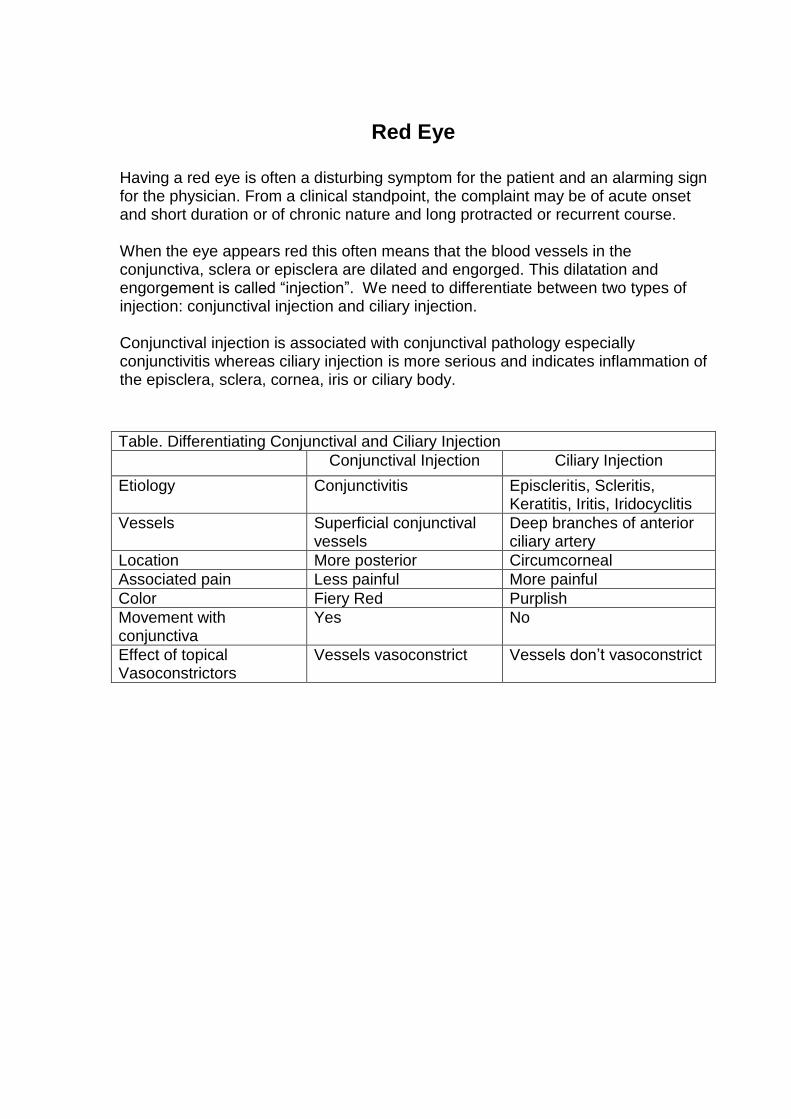
Table. Differentiating Conjunctival and Ciliary Injection
Conjunctival Injection Ciliary Injection
Etiology Conjunctivitis Episcleritis, Scleritis, Keratitis, Iritis, Iridocyclitis
Vessels Superficial conjunctival vessels
Deep branches of anterior ciliary artery
Location More posterior Circumcorneal
Associated pain Less painful More painful
Color Fiery Red Purplish
Movement with conjunctiva
Yes No
Effect of topical Vasoconstrictors
Vessels vasoconstrict Vessels don’t vasoconstrict
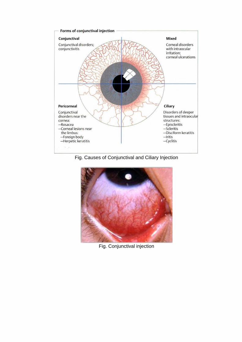
Fig. Causes of Conjunctival and Ciliary Injection
Fig. Conjunctival injection
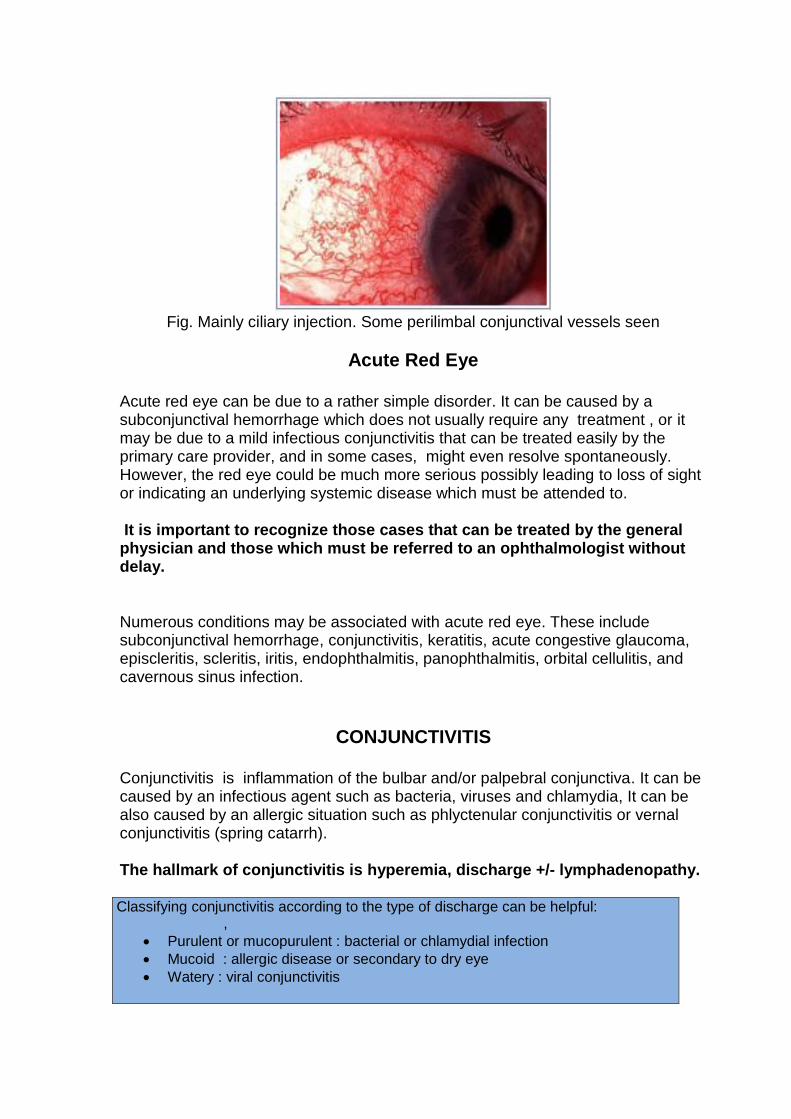
Fig. Mainly ciliary injection. Some perilimbal conjunctival vessels seen
Acute Red Eye
Acute red eye can be due to a rather simple disorder. It can be caused by a subconjunctival hemorrhage which does not usually require any treatment , or it may be due to a mild infectious conjunctivitis that can be treated easily by the primary care provider, and in some cases, might even resolve spontaneously. However, the red eye could be much more serious possibly leading to loss of sight or indicating an underlying systemic disease which must be attended to. It is important to recognize those cases that can be treated by the general physician and those which must be referred to an ophthalmologist without delay.
Numerous conditions may be associated with acute red eye. These include subconjunctival hemorrhage, conjunctivitis, keratitis, acute congestive glaucoma, episcleritis, scleritis, iritis, endophthalmitis, panophthalmitis, orbital cellulitis, and cavernous sinus infection.
CONJUNCTIVITIS
Conjunctivitis is inflammation of the bulbar and/or palpebral conjunctiva. It can be caused by an infectious agent such as bacteria, viruses and chlamydia, It can be also caused by an allergic situation such as phlyctenular conjunctivitis or vernal conjunctivitis (spring catarrh).
The hallmark of conjunctivitis is hyperemia, discharge +/- lymphadenopathy.
Classi Classifying conjunctivitis according to the type of discharge can be helpful: ,
Purulent or mucopurulent : bacterial or chlamydial infection
Mucoid : allergic disease or secondary to dry eye
Watery : viral conjunctivitis

Acute bacterial conjunctivitis
It is inflammation of the conjunctiva caused by gram positive bacteria as Staphylococci and Streptococci pneumonia and viridans and gram negative bacteria as Haemophilus influenza, Neisseria gonorrhoeae, Pseudomonas aeruginosa and Moraxella Clinical Picture: 1. Lid edema. 2. Hyperemia of the conjunctiva ( injection) 3. Mucopurulent or purulent discharge
Fig. Acute bacterial conjunctivitis with purulent discharge
Fate and complications:
Spontaneous cure may occur within 2 weeks without treatment, and in a much shorter time with treatment.
May turn into chronic bacterial conjunctivitis.
Secondary keratitis with possible corneal ulceration (central, marginal or even ring ulcers).
Management: A. Prevention: Bacterial conjunctivitis is very contagious. Therefore, standard infection control measures should be followed. B. Treatment:
1. Antibiotics a. Broad spectrum antibiotic drops. Commonly used ones include the following: i. Norfloxacin and ciprofloxacin are quinolone antibiotics with broad spectrum activity and low toxicity. ii. Other antibiotics include chloramphenicol, gentamicin & tobramycin. ( It is noteworthy that some of the topical antibiotics which are not used systemically due to their systemic side effects are excellent choices for topical use due to their broad spectrum,
effectiveness and excellent tolerability)
They are instilled 4-6 times daily ( sometimes more frequently in the more severe
situations with the additional benefit of the rinsing effect for the profuse discharge) b. Antibiotic ointments—Ointments provide higher concentration of antibiotic for a longer period of time than drops. They are used at bedtime to be effective during sleep.
c. Systemic antibiotics are indicated only in exceptional situations such as gonorrheal and chlamydial infections. Note: 1) No bandage whenever there is discharge as it accumulates the discharge and allows more multiplication of the organism. 2) Culture and sensitivity studies are NOT routinely requested in simple uncomplicated bacterial conjunctivitis.
Neonatal conjunctivitis or ophthalmia neonatorum
It is a preventable disease occurring in newborn babies. (See pediatric diseases)
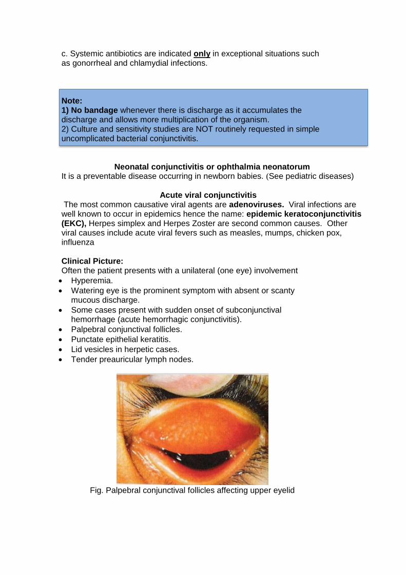
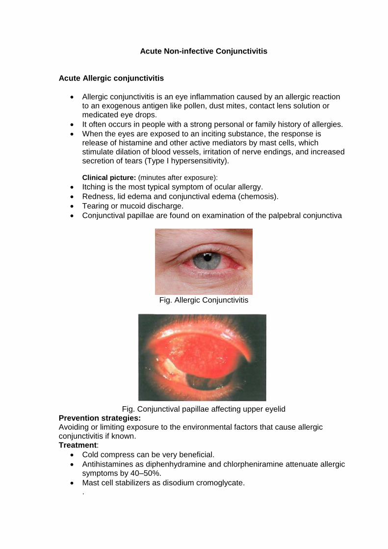
Acute viral conjunctivitis The most common causative viral agents are adenoviruses. Viral infections are well known to occur in epidemics hence the name: epidemic keratoconjunctivitis (EKC), Herpes simplex and Herpes Zoster are second common causes. Other viral causes include acute viral fevers such as measles, mumps, chicken pox, influenza Clinical Picture: Often the patient presents with a unilateral (one eye) involvement
Hyperemia.
Watering eye is the prominent symptom with absent or scanty mucous discharge.
Some cases present with sudden onset of subconjunctival hemorrhage (acute hemorrhagic conjunctivitis).
Palpebral conjunctival follicles.
Punctate epithelial keratitis.
Lid vesicles in herpetic cases.
Tender preauricular lymph nodes.
Fig. Palpebral conjunctival follicles affecting upper eyelid
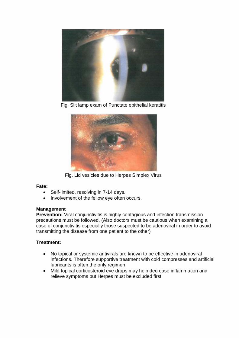
Fig. Slit lamp exam of Punctate epithelial keratitis
Fig. Lid vesicles due to Herpes Simplex Virus
Fate:
Self-limited, resolving in 7-14 days.
Involvement of the fellow eye often occurs. Management Prevention: Viral conjunctivitis is highly contagious and infection transmission precautions must be followed. (Also doctors must be cautious when examining a case of conjunctivitis especially those suspected to be adenoviral in order to avoid transmitting the disease from one patient to the other) Treatment:
No topical or systemic antivirals are known to be effective in adenoviral infections. Therefore supportive treatment with cold compresses and artificial lubricants is often the only regimen
Mild topical corticosteroid eye drops may help decrease inflammation and relieve symptoms but Herpes must be excluded first
Acute Non-infective Conjunctivitis
Acute Allergic conjunctivitis
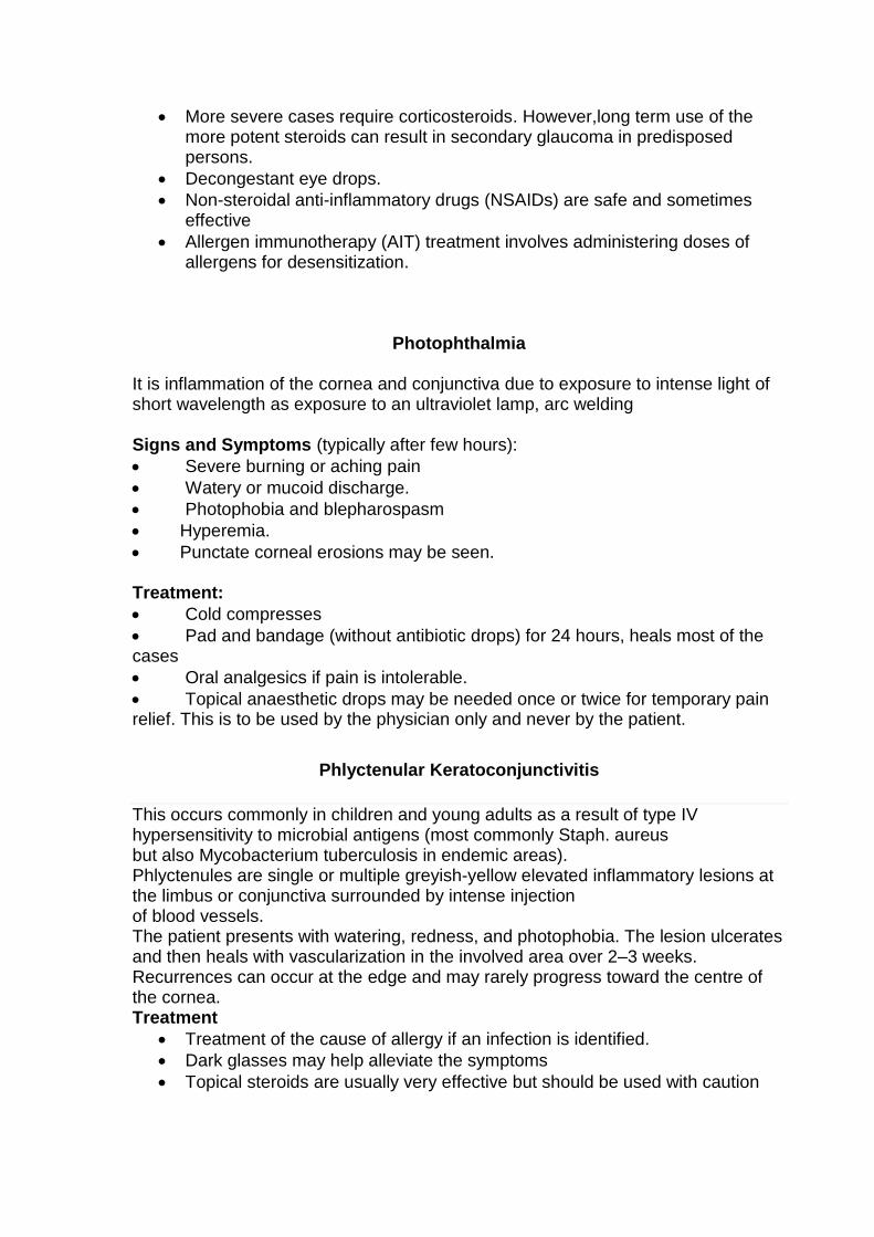
Allergic conjunctivitis is an eye inflammation caused by an allergic reaction to an exogenous antigen like pollen, dust mites, contact lens solution or medicated eye drops.
It often occurs in people with a strong personal or family history of allergies.
When the eyes are exposed to an inciting substance, the response is release of histamine and other active mediators by mast cells, which stimulate dilation of blood vessels, irritation of nerve endings, and increased secretion of tears (Type I hypersensitivity). Clinical picture: (minutes after exposure):
Itching is the most typical symptom of ocular allergy.
Redness, lid edema and conjunctival edema (chemosis).
Tearing or mucoid discharge.
Conjunctival papillae are found on examination of the palpebral conjunctiva
Fig. Allergic Conjunctivitis
Fig. Conjunctival papillae affecting upper eyelid
Prevention strategies: Avoiding or limiting exposure to the environmental factors that cause allergic conjunctivitis if known. Treatment:
Cold compress can be very beneficial.
Antihistamines as diphenhydramine and chlorpheniramine attenuate allergic symptoms by 40–50%.
Mast cell stabilizers as disodium cromoglycate. .
More severe cases require corticosteroids. However,long term use of the more potent steroids can result in secondary glaucoma in predisposed persons.
Decongestant eye drops.
Non-steroidal anti-inflammatory drugs (NSAIDs) are safe and sometimes effective
Allergen immunotherapy (AIT) treatment involves administering doses of allergens for desensitization.
Photophthalmia
It is inflammation of the cornea and conjunctiva due to exposure to intense light of short wavelength as exposure to an ultraviolet lamp, arc welding Signs and Symptoms (typically after few hours):
Severe burning or aching pain
Watery or mucoid discharge.
Photophobia and blepharospasm
Hyperemia.
Punctate corneal erosions may be seen. Treatment:
Cold compresses
Pad and bandage (without antibiotic drops) for 24 hours, heals most of the cases
Oral analgesics if pain is intolerable.
Topical anaesthetic drops may be needed once or twice for temporary pain relief. This is to be used by the physician only and never by the patient.
Phlyctenular Keratoconjunctivitis
This occurs commonly in children and young adults as a result of type IV hypersensitivity to microbial antigens (most commonly Staph. aureus but also Mycobacterium tuberculosis in endemic areas). Phlyctenules are single or multiple greyish-yellow elevated inflammatory lesions at the limbus or conjunctiva surrounded by intense injection of blood vessels. The patient presents with watering, redness, and photophobia. The lesion ulcerates and then heals with vascularization in the involved area over 2–3 weeks. Recurrences can occur at the edge and may rarely progress toward the centre of the cornea. Treatment
Treatment of the cause of allergy if an infection is identified.
Dark glasses may help alleviate the symptoms
Topical steroids are usually very effective but should be used with caution
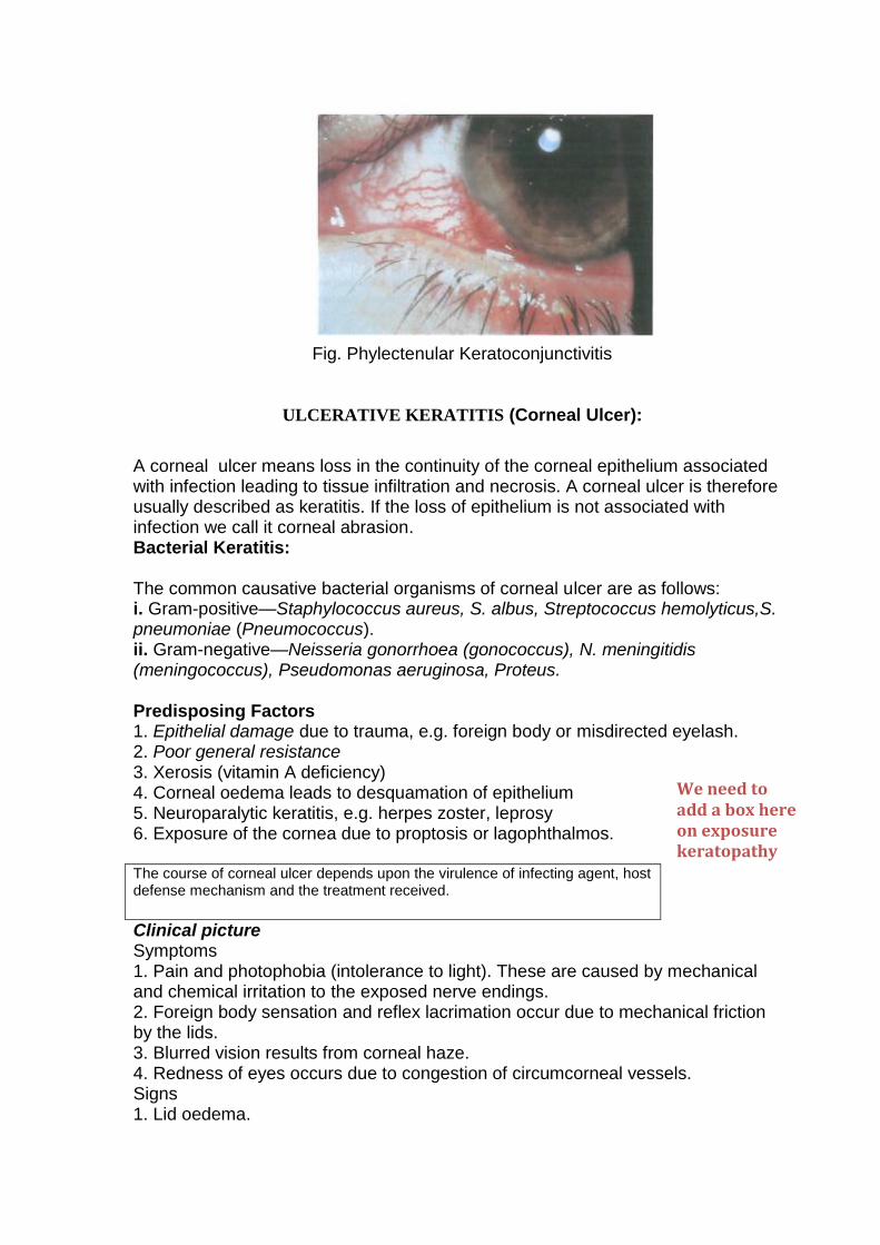
Fig. Phylectenular Keratoconjunctivitis
ULCERATIVE KERATITIS (Corneal Ulcer):
A corneal ulcer means loss in the continuity of the corneal epithelium associated with infection leading to tissue infiltration and necrosis. A corneal ulcer is therefore usually described as keratitis. If the loss of epithelium is not associated with infection we call it corneal abrasion. Bacterial Keratitis: The common causative bacterial organisms of corneal ulcer are as follows: i. Gram-positive—Staphylococcus aureus, S. albus, Streptococcus hemolyticus,S. pneumoniae (Pneumococcus). ii. Gram-negative—Neisseria gonorrhoea (gonococcus), N. meningitidis (meningococcus), Pseudomonas aeruginosa, Proteus. Predisposing Factors 1. Epithelial damage due to trauma, e.g. foreign body or misdirected eyelash. 2. Poor general resistance 3. Xerosis (vitamin A deficiency) 4. Corneal oedema leads to desquamation of epithelium 5. Neuroparalytic keratitis, e.g. herpes zoster, leprosy 6. Exposure of the cornea due to proptosis or lagophthalmos. The course of corneal ulcer depends upon the virulence of infecting agent, host defense mechanism and the treatment received.
Clinical picture Symptoms 1. Pain and photophobia (intolerance to light). These are caused by mechanical and chemical irritation to the exposed nerve endings. 2. Foreign body sensation and reflex lacrimation occur due to mechanical friction by the lids. 3. Blurred vision results from corneal haze. 4. Redness of eyes occurs due to congestion of circumcorneal vessels. Signs 1. Lid oedema.
We need to add a box here on exposure keratopathy
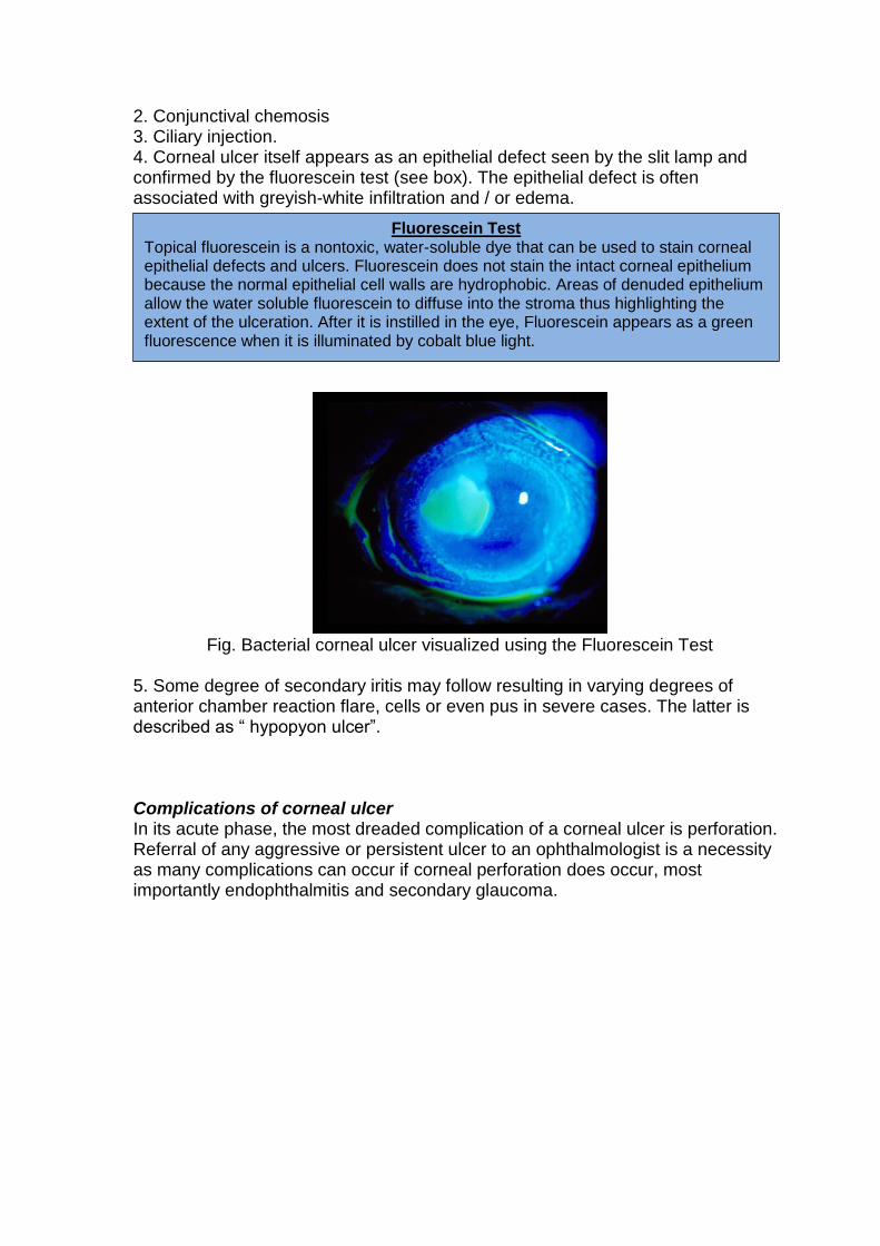
2. Conjunctival chemosis 3. Ciliary injection. 4. Corneal ulcer itself appears as an epithelial defect seen by the slit lamp and confirmed by the fluorescein test (see box). The epithelial defect is often associated with greyish-white infiltration and / or edema.
Fig. Bacterial corneal ulcer visualized using the Fluorescein Test
5. Some degree of secondary iritis may follow resulting in varying degrees of anterior chamber reaction flare, cells or even pus in severe cases. The latter is described as “ hypopyon ulcer”. Complications of corneal ulcer In its acute phase, the most dreaded complication of a corneal ulcer is perforation. Referral of any aggressive or persistent ulcer to an ophthalmologist is a necessity as many complications can occur if corneal perforation does occur, most importantly endophthalmitis and secondary glaucoma.
Fluorescein Test Topical fluorescein is a nontoxic, water-soluble dye that can be used to stain corneal epithelial defects and ulcers. Fluorescein does not stain the intact corneal epithelium because the normal epithelial cell walls are hydrophobic. Areas of denuded epithelium allow the water soluble fluorescein to diffuse into the stroma thus highlighting the extent of the ulceration. After it is instilled in the eye, Fluorescein appears as a green fluorescence when it is illuminated by cobalt blue light.
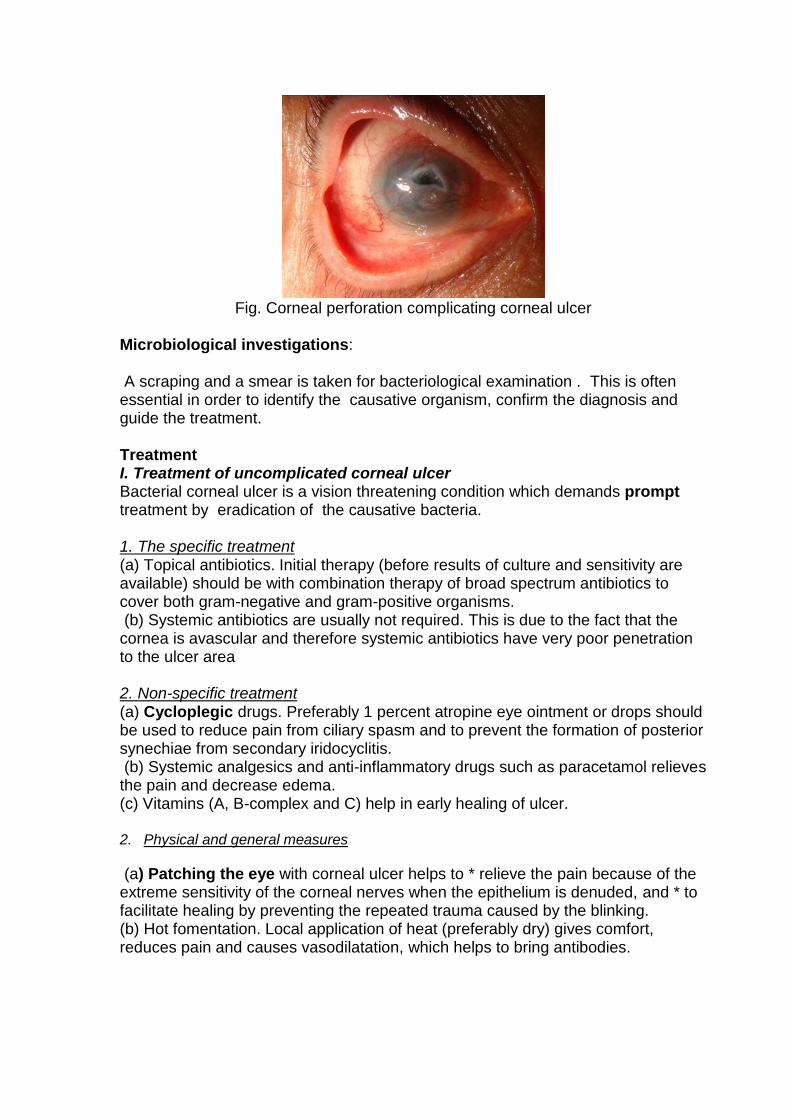
Fig. Corneal perforation complicating corneal ulcer
Microbiological investigations: A scraping and a smear is taken for bacteriological examination . This is often essential in order to identify the causative organism, confirm the diagnosis and guide the treatment. Treatment I. Treatment of uncomplicated corneal ulcer Bacterial corneal ulcer is a vision threatening condition which demands prompt treatment by eradication of the causative bacteria. 1. The specific treatment (a) Topical antibiotics. Initial therapy (before results of culture and sensitivity are available) should be with combination therapy of broad spectrum antibiotics to cover both gram-negative and gram-positive organisms. (b) Systemic antibiotics are usually not required. This is due to the fact that the cornea is avascular and therefore systemic antibiotics have very poor penetration to the ulcer area 2. Non-specific treatment (a) Cycloplegic drugs. Preferably 1 percent atropine eye ointment or drops should be used to reduce pain from ciliary spasm and to prevent the formation of posterior synechiae from secondary iridocyclitis. (b) Systemic analgesics and anti-inflammatory drugs such as paracetamol relieves the pain and decrease edema. (c) Vitamins (A, B-complex and C) help in early healing of ulcer. 2. Physical and general measures
(a) Patching the eye with corneal ulcer helps to * relieve the pain because of the extreme sensitivity of the corneal nerves when the epithelium is denuded, and * to facilitate healing by preventing the repeated trauma caused by the blinking. (b) Hot fomentation. Local application of heat (preferably dry) gives comfort, reduces pain and causes vasodilatation, which helps to bring antibodies.
Viral Keratitis
Most of the viruses tend to affect the epithelium of both the conjunctiva and cornea, hence the typical viral lesions constitute the viral keratoconjunctivitis. Common viral infections include herpes simplex keratitis, herpes zoster ophthalmicus and adenovirus keratitis.
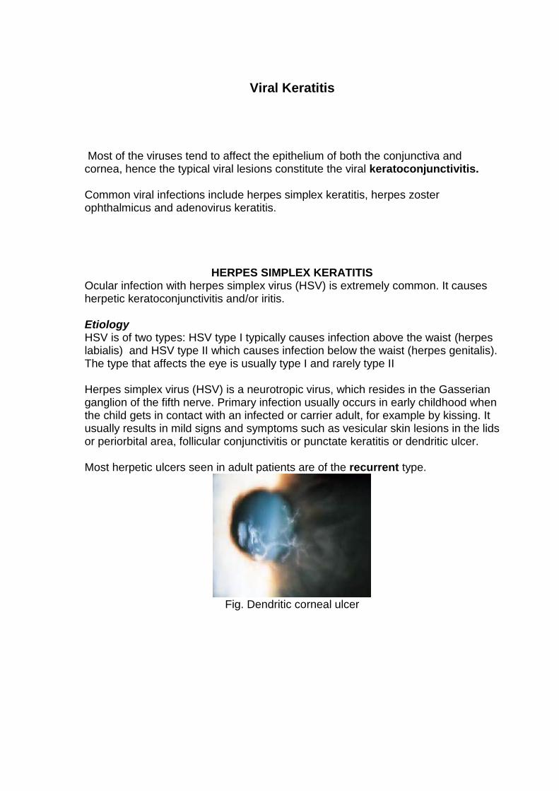
HERPES SIMPLEX KERATITIS Ocular infection with herpes simplex virus (HSV) is extremely common. It causes herpetic keratoconjunctivitis and/or iritis. Etiology HSV is of two types: HSV type I typically causes infection above the waist (herpes labialis) and HSV type II which causes infection below the waist (herpes genitalis). The type that affects the eye is usually type I and rarely type II Herpes simplex virus (HSV) is a neurotropic virus, which resides in the Gasserian ganglion of the fifth nerve. Primary infection usually occurs in early childhood when the child gets in contact with an infected or carrier adult, for example by kissing. It usually results in mild signs and symptoms such as vesicular skin lesions in the lids or periorbital area, follicular conjunctivitis or punctate keratitis or dendritic ulcer. Most herpetic ulcers seen in adult patients are of the recurrent type.
Fig. Dendritic corneal ulcer
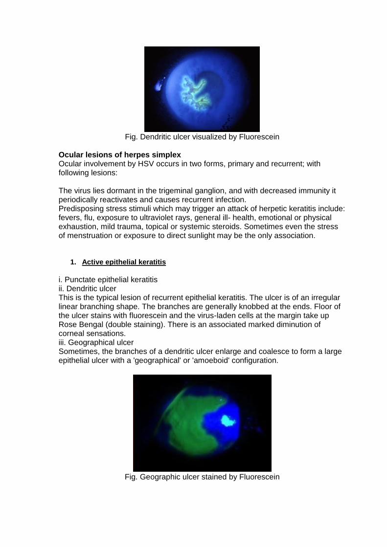
Fig. Dendritic ulcer visualized by Fluorescein
Ocular lesions of herpes simplex Ocular involvement by HSV occurs in two forms, primary and recurrent; with following lesions: The virus lies dormant in the trigeminal ganglion, and with decreased immunity it periodically reactivates and causes recurrent infection. Predisposing stress stimuli which may trigger an attack of herpetic keratitis include: fevers, flu, exposure to ultraviolet rays, general ill- health, emotional or physical exhaustion, mild trauma, topical or systemic steroids. Sometimes even the stress of menstruation or exposure to direct sunlight may be the only association.
1. Active epithelial keratitis
i. Punctate epithelial keratitis ii. Dendritic ulcer This is the typical lesion of recurrent epithelial keratitis. The ulcer is of an irregular linear branching shape. The branches are generally knobbed at the ends. Floor of the ulcer stains with fluorescein and the virus-laden cells at the margin take up Rose Bengal (double staining). There is an associated marked diminution of corneal sensations. iii. Geographical ulcer Sometimes, the branches of a dendritic ulcer enlarge and coalesce to form a large epithelial ulcer with a 'geographical' or 'amoeboid' configuration.
Fig. Geographic ulcer stained by Fluorescein
2. Stromal keratitis i. Disciform keratitis (a central disc of persistent corneal edema) ii. Diffuse stromal necrotic keratitis ( more extensive involvement of corneal stroma)
3. Herpetic iridocyclitis
Treatment I. Specific treatment 1. Topical antiviral, ocular acyclovir 5 times/day, until epithelial healing has occurred. This should normally be discontinued after 10–14 days in order to avoid toxic keratopathy. If there are signs of toxicity, an alternative to topical treatment is oral antiviral treatment. 2. Epithelial debridement may help. It is performed by gently wiping the surface of the dendrite with a cellulose sponge. II. Non-specific supportive therapy and physical and general measures are same as for bacterial corneal ulcer.
HERPES ZOSTER OPHTHALMICUS
Herpes zoster Ophthalmicus is an acute infection of Gasserian ganglion of the fifth cranial nerve by the varicella-zoster virus (VZV). Etiology Varicella -zoster virus. It is neurotropic in nature. Mode of infection. The infection is contracted in childhood, which may manifest as chickenpox. The virus then remains dormant in the sensory ganglion of trigeminal nerve. It is thought that, with depressed cellular immunity, the virus reactivates. Clinical features * In herpes zoster ophthalmicus, frontal nerve is more frequently affected than the lacrimal and nasociliary nerves. * The Hutchinson's rule, which implies that ocular involvement is frequent if the side or tip of nose presents vesicles (cutaneous involvement of nasociliary nerve). * Lesions of herpes zoster are strictly limited to one side of the midline of head. Clinical phases of H. zoster ophthalmicus are: i. Acute, which may totally resolve. ii. Chronic, which may persist for years. iii. Relapsing, where the acute or chronic lesions reappear sometimes years later.
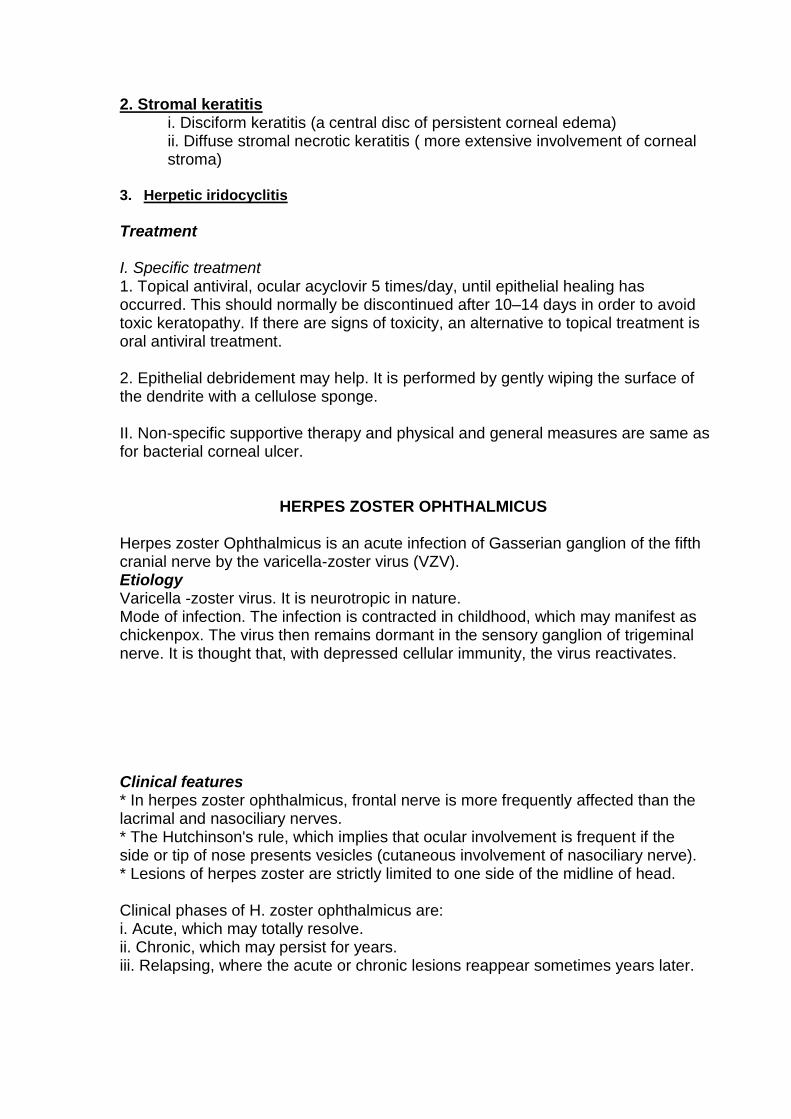
A. General features. The onset of illness is sudden with fever, malaise and severe neuralgic pain along the course of the affected nerve. B. Cutaneous lesions in area of distribution of the involved nerve. Redness followed by vesicle formation then pustules, which subsequently burst to become crusting ulcers. When crusts are shed, sometimes permanent pitted scars are left.
Fig. Skin lesions due to Herpes Zoster Virus respecting the midline with
involvement of the tip of the nose.
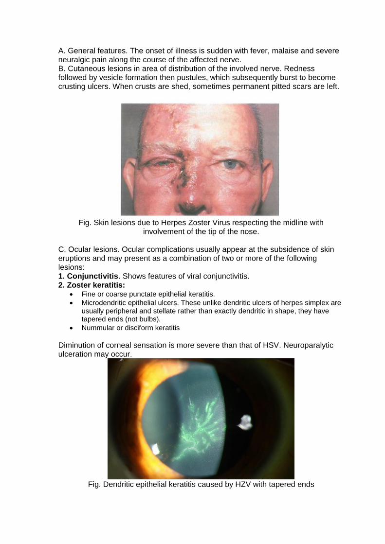
C. Ocular lesions. Ocular complications usually appear at the subsidence of skin eruptions and may present as a combination of two or more of the following lesions: 1. Conjunctivitis. Shows features of viral conjunctivitis. 2. Zoster keratitis:
Fine or coarse punctate epithelial keratitis.
Microdendritic epithelial ulcers. These unlike dendritic ulcers of herpes simplex are usually peripheral and stellate rather than exactly dendritic in shape, they have tapered ends (not bulbs).
Nummular or disciform keratitis
Diminution of corneal sensation is more severe than that of HSV. Neuroparalytic ulceration may occur.
Fig. Dendritic epithelial keratitis caused by HZV with tapered ends
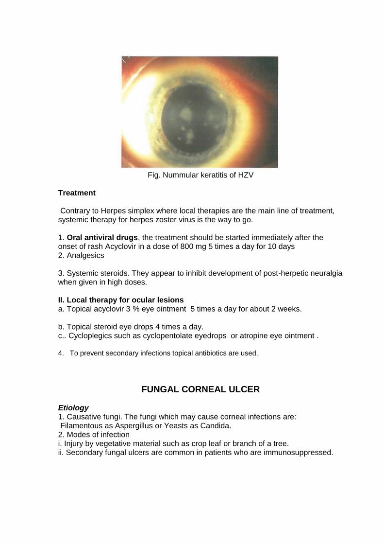
Fig. Nummular keratitis of HZV
Treatment Contrary to Herpes simplex where local therapies are the main line of treatment, systemic therapy for herpes zoster virus is the way to go. 1. Oral antiviral drugs, the treatment should be started immediately after the onset of rash Acyclovir in a dose of 800 mg 5 times a day for 10 days 2. Analgesics 3. Systemic steroids. They appear to inhibit development of post-herpetic neuralgia when given in high doses. II. Local therapy for ocular lesions a. Topical acyclovir 3 % eye ointment 5 times a day for about 2 weeks. b. Topical steroid eye drops 4 times a day. c.. Cycloplegics such as cyclopentolate eyedrops or atropine eye ointment . 4. To prevent secondary infections topical antibiotics are used.
FUNGAL CORNEAL ULCER Etiology 1. Causative fungi. The fungi which may cause corneal infections are: Filamentous as Aspergillus or Yeasts as Candida. 2. Modes of infection i. Injury by vegetative material such as crop leaf or branch of a tree. ii. Secondary fungal ulcers are common in patients who are immunosuppressed.
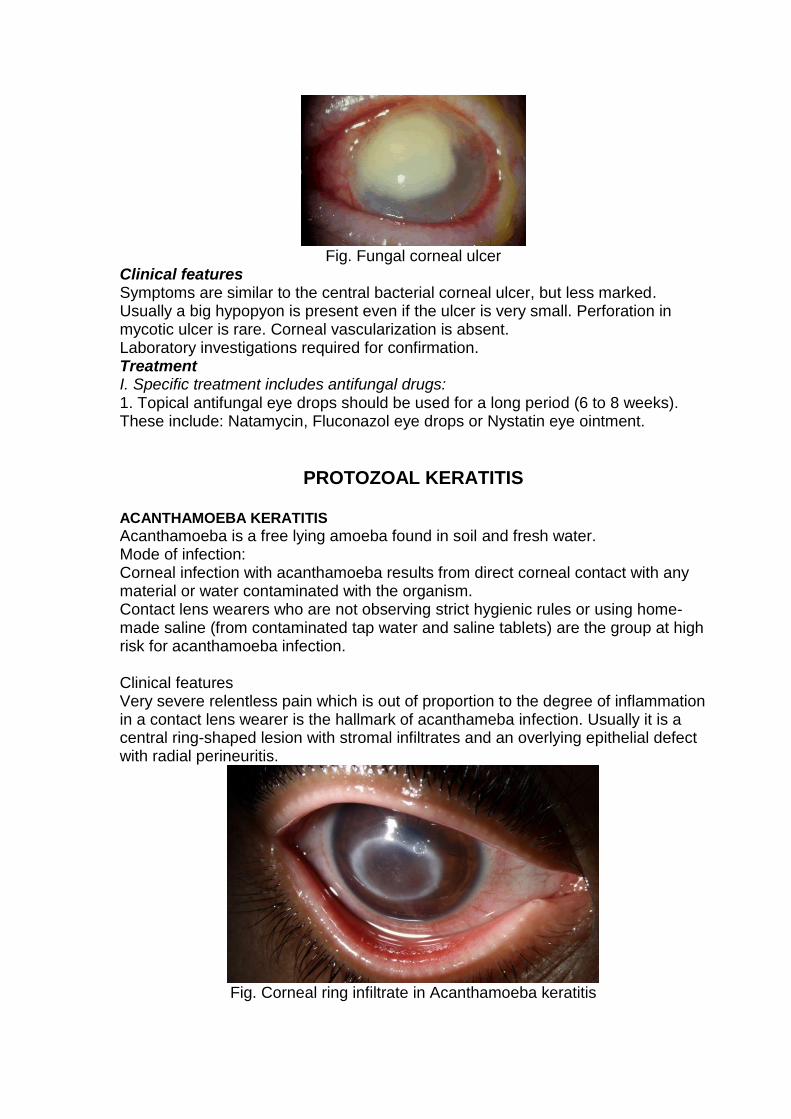
Fig. Fungal corneal ulcer
Clinical features Symptoms are similar to the central bacterial corneal ulcer, but less marked. Usually a big hypopyon is present even if the ulcer is very small. Perforation in mycotic ulcer is rare. Corneal vascularization is absent. Laboratory investigations required for confirmation. Treatment I. Specific treatment includes antifungal drugs: 1. Topical antifungal eye drops should be used for a long period (6 to 8 weeks). These include: Natamycin, Fluconazol eye drops or Nystatin eye ointment.
PROTOZOAL KERATITIS
ACANTHAMOEBA KERATITIS
Acanthamoeba is a free lying amoeba found in soil and fresh water. Mode of infection: Corneal infection with acanthamoeba results from direct corneal contact with any material or water contaminated with the organism. Contact lens wearers who are not observing strict hygienic rules or using home-made saline (from contaminated tap water and saline tablets) are the group at high risk for acanthamoeba infection. Clinical features Very severe relentless pain which is out of proportion to the degree of inflammation in a contact lens wearer is the hallmark of acanthameba infection. Usually it is a central ring-shaped lesion with stromal infiltrates and an overlying epithelial defect with radial perineuritis.
Fig. Corneal ring infiltrate in Acanthamoeba keratitis
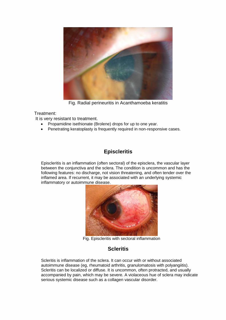
Fig. Radial perineuritis in Acanthamoeba keratitis
Treatment: It is very resistant to treatment.
Propamidine isethionate (Brolene) drops for up to one year.
Penetrating keratoplasty is frequently required in non-responsive cases.
Episcleritis
Episcleritis is an inflammation (often sectoral) of the episclera, the vascular layer between the conjunctiva and the sclera. The condition is uncommon and has the following features: no discharge, not vision threatening, and often tender over the inflamed area. If recurrent, it may be associated with an underlying systemic inflammatory or autoimmune disease.
Fig. Episcleritis with sectoral inflammation
Scleritis
Scleritis is inflammation of the sclera. It can occur with or without associated autoimmune disease (eg, rheumatoid arthritis, granulomatosis with polyangiitis). Scleritis can be localized or diffuse. It is uncommon, often protracted, and usually accompanied by pain, which may be severe. A violaceous hue of sclera may indicate serious systemic disease such as a collagen vascular disorder.
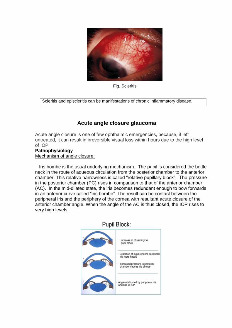
Fig. Scleritis
Scleritis and episcleritis can be manifestations of chronic inflammatory disease.
Acute angle closure glaucoma:
Acute angle closure is one of few ophthalmic emergencies, because, if left untreated, it can result in irreversible visual loss within hours due to the high level of IOP. Pathophysiology Mechanism of angle closure: Iris bombe is the usual underlying mechanism. The pupil is considered the bottle neck in the route of aqueous circulation from the posterior chamber to the anterior chamber. This relative narrowness is called “relative pupillary block”. The pressure in the posterior chamber (PC) rises in comparison to that of the anterior chamber (AC). In the mid-dilated state, the iris becomes redundant enough to bow forwards in an anterior curve called “iris bombe”. The result can be contact between the peripheral iris and the periphery of the cornea with resultant acute closure of the anterior chamber angle. When the angle of the AC is thus closed, the IOP rises to very high levels.
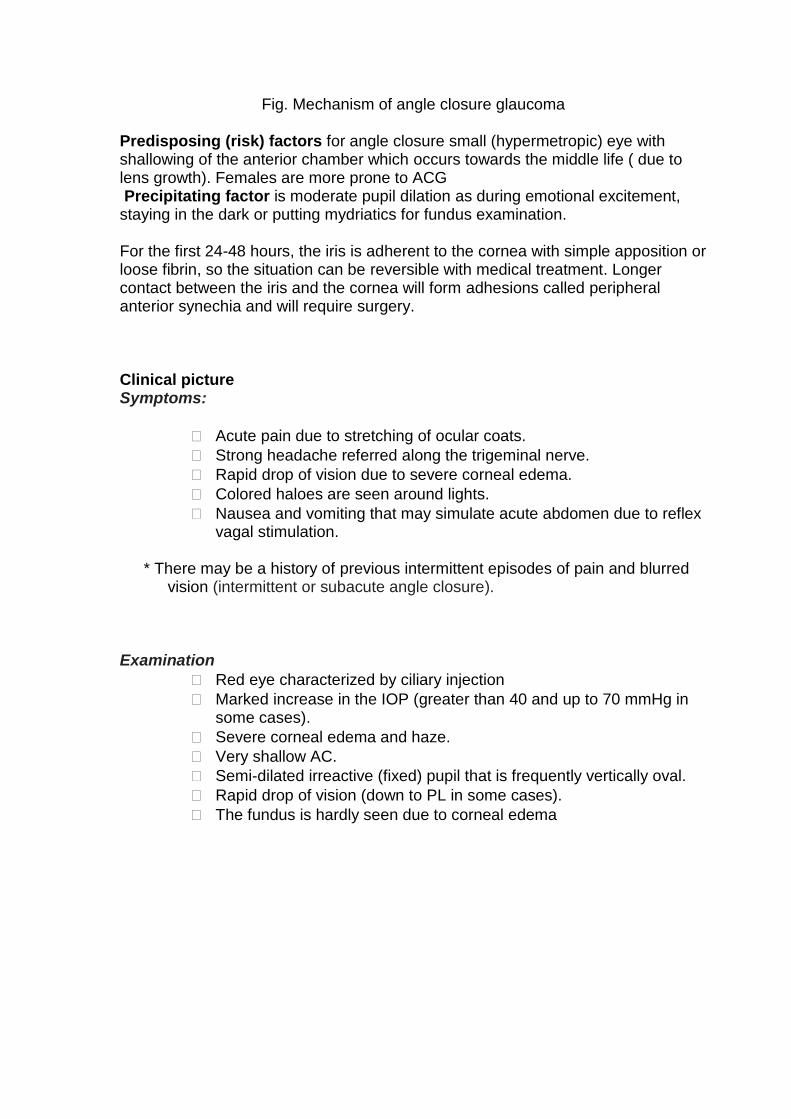
Fig. Mechanism of angle closure glaucoma Predisposing (risk) factors for angle closure small (hypermetropic) eye with shallowing of the anterior chamber which occurs towards the middle life ( due to lens growth). Females are more prone to ACG Precipitating factor is moderate pupil dilation as during emotional excitement, staying in the dark or putting mydriatics for fundus examination. For the first 24-48 hours, the iris is adherent to the cornea with simple apposition or loose fibrin, so the situation can be reversible with medical treatment. Longer contact between the iris and the cornea will form adhesions called peripheral anterior synechia and will require surgery. Clinical picture Symptoms:
Acute pain due to stretching of ocular coats.
Strong headache referred along the trigeminal nerve.
Rapid drop of vision due to severe corneal edema.
Colored haloes are seen around lights.
Nausea and vomiting that may simulate acute abdomen due to reflex vagal stimulation.
* There may be a history of previous intermittent episodes of pain and blurred
vision (intermittent or subacute angle closure). Examination
Red eye characterized by ciliary injection
Marked increase in the IOP (greater than 40 and up to 70 mmHg in some cases).
Severe corneal edema and haze.
Very shallow AC.
Semi-dilated irreactive (fixed) pupil that is frequently vertically oval.
Rapid drop of vision (down to PL in some cases).
The fundus is hardly seen due to corneal edema
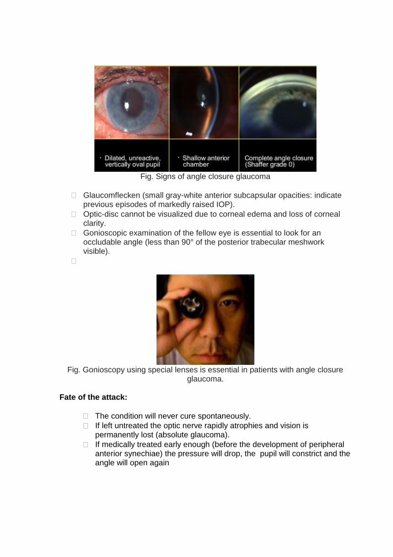
Fig. Signs of angle closure glaucoma
Glaucomflecken (small gray-white anterior subcapsular opacities: indicate previous episodes of markedly raised IOP).
Optic-disc cannot be visualized due to corneal edema and loss of corneal clarity.
Gonioscopic examination of the fellow eye is essential to look for an occludable angle (less than 90° of the posterior trabecular meshwork visible).
Fig. Gonioscopy using special lenses is essential in patients with angle closure
glaucoma.
Fate of the attack:
The condition will never cure spontaneously.
If left untreated the optic nerve rapidly atrophies and vision is permanently lost (absolute glaucoma).
If medically treated early enough (before the development of peripheral anterior synechiae) the pressure will drop, the pupil will constrict and the angle will open again
Treatment The definitive treatment of acute angle-closure glaucoma (AACG) is surgical. The initial treatment is, however, medical aiming at IOP reduction, suppression of inflammation, and reversal of angle closure in order to provide favorable conditions for surgery. 1- Hospitalization. 2- Hyperosmotic agents: Mode of action—These agents increase the plasma tonicity or osmolality to draw water out of the eyes. This results in lowering the intraocular pressure. The most commonly used is intravenous Mannitol (20%)—1-2 g/kg body weight. A 20% solution is given over 30-40 minutes 3- Carbonic anhydrase inhibitors: as oral tablets or intravenous injection ( if available)
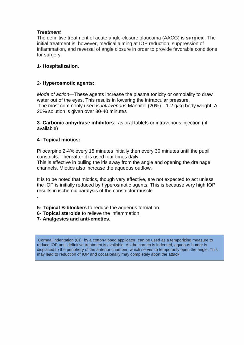
4- Topical miotics: Pilocarpine 2-4% every 15 minutes initially then every 30 minutes until the pupil constricts. Thereafter it is used four times daily. This is effective in pulling the iris away from the angle and opening the drainage channels. Miotics also increase the aqueous outflow. It is to be noted that miotics, though very effective, are not expected to act unless the IOP is initially reduced by hyperosmotic agents. This is because very high IOP results in ischemic paralysis of the constrictor muscle . 5- Topical B-blockers to reduce the aqueous formation. 6- Topical steroids to relieve the inflammation. 7- Analgesics and anti-emetics. Corneal indentation (CI), by a cotton-tipped applicator, can be used as a temporizing measure to reduce IOP until definitive treatment is available. As the cornea is indented, aqueous humor is displaced to the periphery of the anterior chamber, which serves to temporarily open the angle. This may lead to reduction of IOP and occasionally may completely abort the attack.
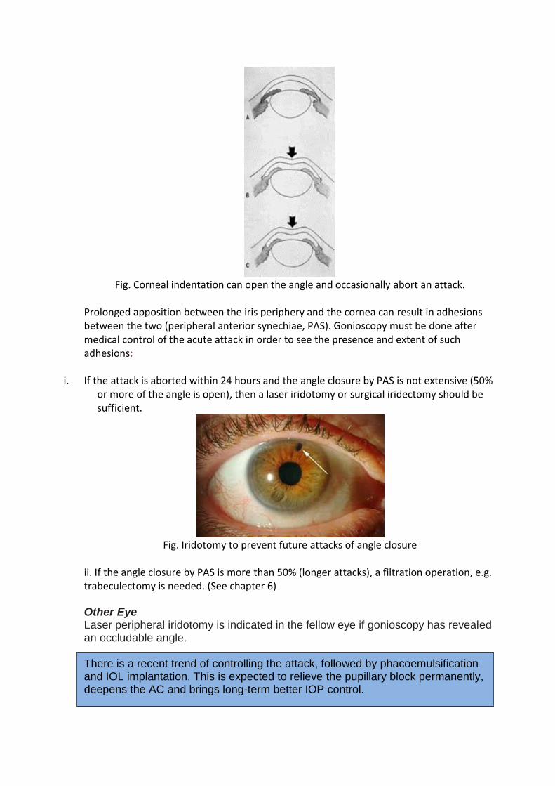
Fig. Corneal indentation can open the angle and occasionally abort an attack.
Prolonged apposition between the iris periphery and the cornea can result in adhesions between the two (peripheral anterior synechiae, PAS). Gonioscopy must be done after medical control of the acute attack in order to see the presence and extent of such adhesions:
i. If the attack is aborted within 24 hours and the angle closure by PAS is not extensive (50% or more of the angle is open), then a laser iridotomy or surgical iridectomy should be sufficient.
Fig. Iridotomy to prevent future attacks of angle closure
ii. If the angle closure by PAS is more than 50% (longer attacks), a filtration operation, e.g. trabeculectomy is needed. (See chapter 6) Other Eye Laser peripheral iridotomy is indicated in the fellow eye if gonioscopy has revealed an occludable angle. There is a recent trend of controlling the attack, followed by phacoemulsification and IOL implantation. This is expected to relieve the pupillary block permanently, deepens the AC and brings long-term better IOP control.

Acute Iridocyclitis (Anterior Uveitis)
Uveitis is defined as inflammation of the uveal tract, which is further subdivided into anterior, intermediate and posterior components. The anterior part is composed of the iris and ciliary body. Uveitis may be triggered by genetic, traumatic, immune, or infectious mechanisms. However, it is commonly idiopathic (in about 50% of cases). Inflammatory cells and exudates and/or toxins pour into the narrow spaces of the eye: anterior chamber, posterior chamber, retrolental space and anterior vitreous causing the clinical picture and complications.
Aetiology Non-infectious *Idiopathic (most common).
Juvenile idiopathic arthritis :Human leucocyte antigen (HLA B27-associated sero-negative arthritis
Sarcoidosis cause granulomatous uveitis
Behcet’s disease: an autoimmune disease commonly seen in young and middle age males associated with orogenital ulcers, arthritis, thrombophlebitis in addition to ocular inflammation (ant. and post. uveitis with possible hypopyon, vitritis, retinal vasculitis and papillopathy)
Vogt Koyanagi Harada (VKH) syndrome: multisystem disorder with panuveitis, exudative retinal detachment, sunset glow fundus, vitiligo, poliosis, and neurological manifestations including headache and possible deafness.
Secondary to some ocular causes such as traumatic and hypermature cataract.
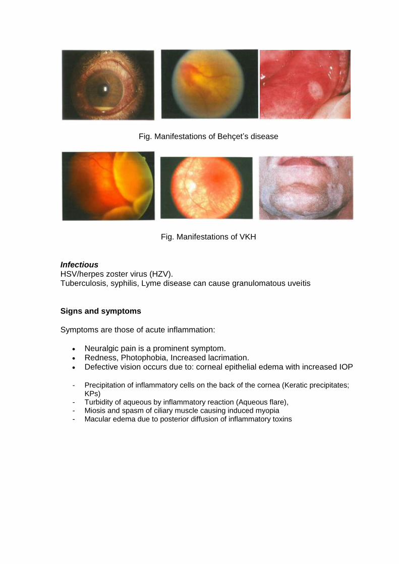
Fig. Manifestations of Behçet’s disease
Fig. Manifestations of VKH
Infectious HSV/herpes zoster virus (HZV). Tuberculosis, syphilis, Lyme disease can cause granulomatous uveitis
Signs and symptoms
Symptoms are those of acute inflammation:
Neuralgic pain is a prominent symptom. Redness, Photophobia, Increased lacrimation. Defective vision occurs due to: corneal epithelial edema with increased IOP
- Precipitation of inflammatory cells on the back of the cornea (Keratic precipitates; KPs)
- Turbidity of aqueous by inflammatory reaction (Aqueous flare), - Miosis and spasm of ciliary muscle causing induced myopia - Macular edema due to posterior diffusion of inflammatory toxins
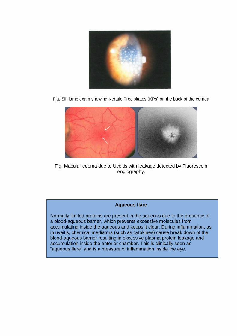
Fig. Slit lamp exam showing Keratic Precipitates (KPs) on the back of the cornea
Fig. Macular edema due to Uveitis with leakage detected by Fluorescein Angiography.
Aqueous flare
Normally limited proteins are present in the aqueous due to the presence of a blood-aqueous barrier, which prevents excessive molecules from accumulating inside the aqueous and keeps it clear. During inflammation, as in uveitis, chemical mediators (such as cytokines) cause break down of the blood-aqueous barrier resulting in excessive plasma protein leakage and accumulation inside the anterior chamber. This is clinically seen as “aqueous flare” and is a measure of inflammation inside the eye.
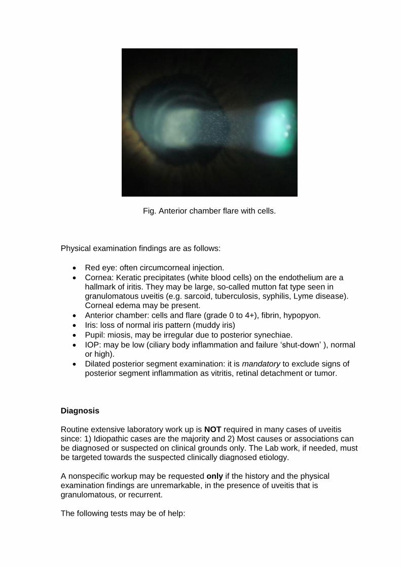
Fig. Anterior chamber flare with cells.
Physical examination findings are as follows:
Red eye: often circumcorneal injection.
Cornea: Keratic precipitates (white blood cells) on the endothelium are a hallmark of iritis. They may be large, so-called mutton fat type seen in granulomatous uveitis (e.g. sarcoid, tuberculosis, syphilis, Lyme disease). Corneal edema may be present.
Anterior chamber: cells and flare (grade 0 to 4+), fibrin, hypopyon.
Iris: loss of normal iris pattern (muddy iris)
Pupil: miosis, may be irregular due to posterior synechiae.
IOP: may be low (ciliary body inflammation and failure ‘shut-down’ ), normal or high).
Dilated posterior segment examination: it is mandatory to exclude signs of posterior segment inflammation as vitritis, retinal detachment or tumor.
Diagnosis
Routine extensive laboratory work up is NOT required in many cases of uveitis since: 1) Idiopathic cases are the majority and 2) Most causes or associations can be diagnosed or suspected on clinical grounds only. The Lab work, if needed, must be targeted towards the suspected clinically diagnosed etiology.
A nonspecific workup may be requested only if the history and the physical examination findings are unremarkable, in the presence of uveitis that is granulomatous, or recurrent.
The following tests may be of help:
Complete blood cell (CBC) count Erythrocyte sedimentation rate (ESR) Antinuclear antibody (ANA) Venereal disease research laboratory (VDRL) and Rapid plasma reagin test
(RPR) Purified protein derivative (PPD) Lyme antibody titre HLA ( such as B27 and B5) testing Chest radiography (to assess for sarcoidosis or tuberculosis) Infectious workup (eg, HIV, toxoplasmosis), depending on the presentation.
If left untreated, acute anterior uveitis can lead to serious complications:
Uveitic glaucoma, cells and exudates in the anterior chamber can clog the angle.
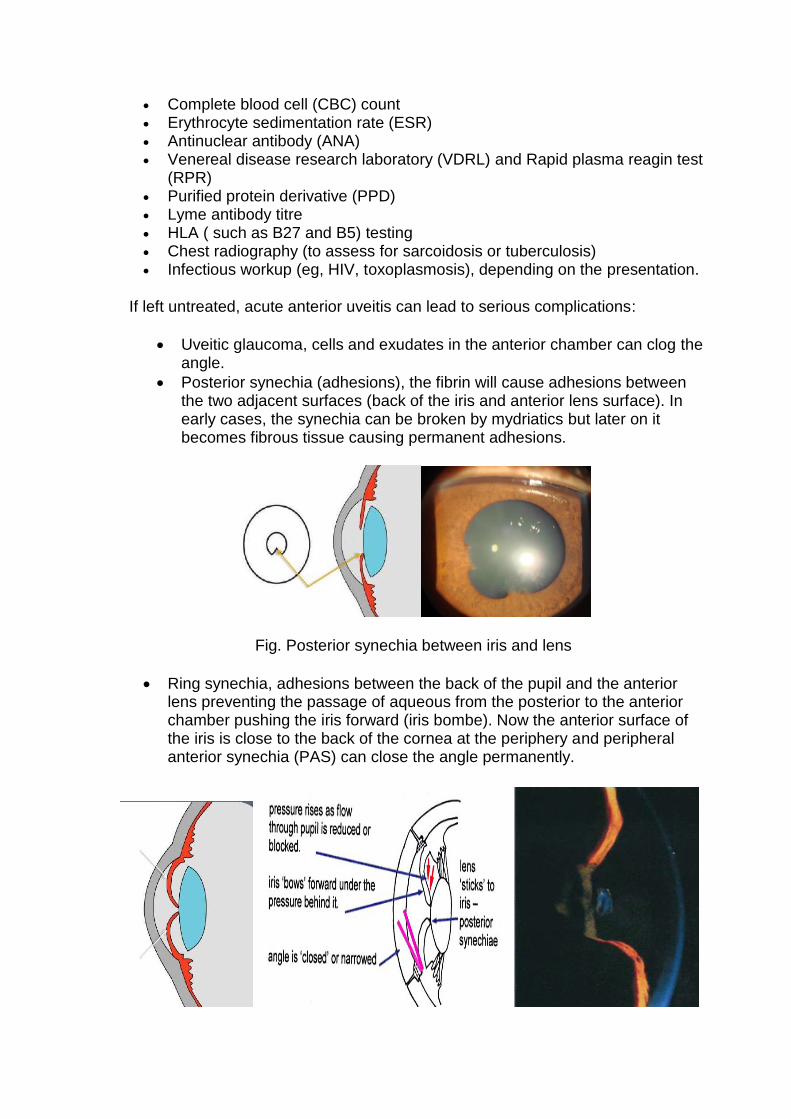
Posterior synechia (adhesions), the fibrin will cause adhesions between the two adjacent surfaces (back of the iris and anterior lens surface). In early cases, the synechia can be broken by mydriatics but later on it becomes fibrous tissue causing permanent adhesions.
Fig. Posterior synechia between iris and lens
Ring synechia, adhesions between the back of the pupil and the anterior lens preventing the passage of aqueous from the posterior to the anterior chamber pushing the iris forward (iris bombe). Now the anterior surface of the iris is close to the back of the cornea at the periphery and peripheral anterior synechia (PAS) can close the angle permanently.
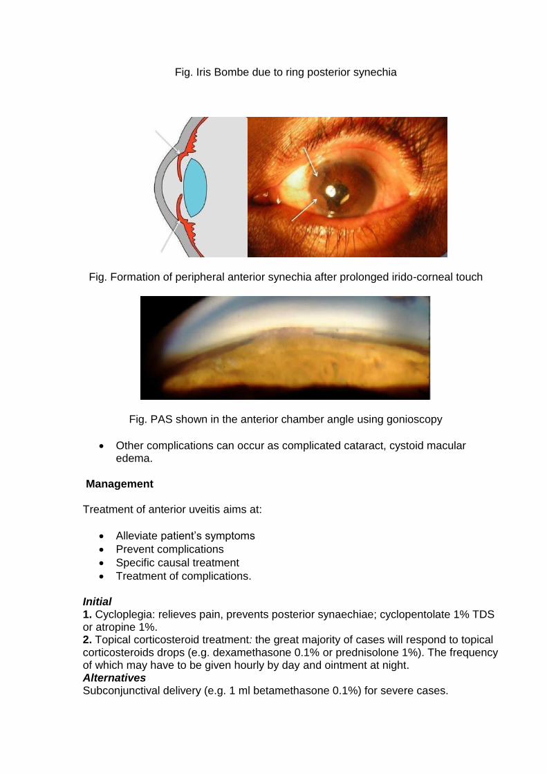
Fig. Iris Bombe due to ring posterior synechia
Fig. Formation of peripheral anterior synechia after prolonged irido-corneal touch
Fig. PAS shown in the anterior chamber angle using gonioscopy
Other complications can occur as complicated cataract, cystoid macular edema.
Management
Treatment of anterior uveitis aims at:
Alleviate patient’s symptoms
Prevent complications
Specific causal treatment
Treatment of complications.
Initial 1. Cycloplegia: relieves pain, prevents posterior synaechiae; cyclopentolate 1% TDS or atropine 1%. 2. Topical corticosteroid treatment: the great majority of cases will respond to topical corticosteroids drops (e.g. dexamethasone 0.1% or prednisolone 1%). The frequency of which may have to be given hourly by day and ointment at night. Alternatives Subconjunctival delivery (e.g. 1 ml betamethasone 0.1%) for severe cases.
Periocular and systemic steroids: very rarely indicated. 3. Ocular hypotensive treatment: if necessary. 4. Appropriate antimicrobial treatment in infectious cases. Later Gradual tapering of topical steroid treatment is necessary to avoid recurrence/rebound of inflammation, four weeks taper may be suitable in most cases. Some require extended therapy with topical steroid eye drops once twice daily.
Secondary glaucoma in cases of uveitis can occur due to: Cellular trabecular block, 2) Steroid response, 3) Acute angle closure due to iris bombe,4) Synechial angle closure and 5) Neovascularization in rubeosis.
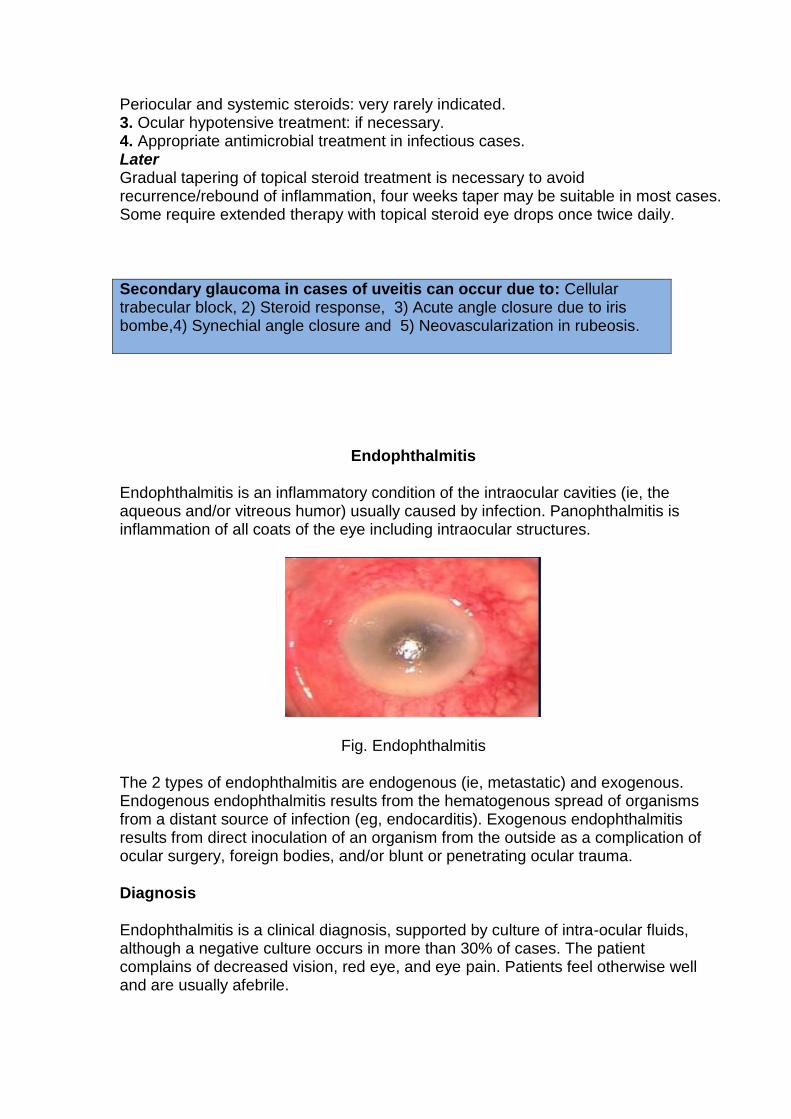
Endophthalmitis
Endophthalmitis is an inflammatory condition of the intraocular cavities (ie, the aqueous and/or vitreous humor) usually caused by infection. Panophthalmitis is inflammation of all coats of the eye including intraocular structures.
Fig. Endophthalmitis
The 2 types of endophthalmitis are endogenous (ie, metastatic) and exogenous. Endogenous endophthalmitis results from the hematogenous spread of organisms from a distant source of infection (eg, endocarditis). Exogenous endophthalmitis results from direct inoculation of an organism from the outside as a complication of ocular surgery, foreign bodies, and/or blunt or penetrating ocular trauma.
Diagnosis
Endophthalmitis is a clinical diagnosis, supported by culture of intra-ocular fluids, although a negative culture occurs in more than 30% of cases. The patient complains of decreased vision, red eye, and eye pain. Patients feel otherwise well and are usually afebrile.
On physical examination, vision is decreased, the conjunctiva is injected, and a hypopyon is present in most cases. There are white blood cells in the aqueous humor and vitreous humor, so the view of the retina is hazy. The differential diagnosis is sterile intra-ocular inflammation. This may occur as a reaction to surgery, and is typically greatest on the first postoperative day, whereas endophthalmitis usually occurs on day 2 or later.
These cases are medical emergencies, as delay in treatment may result in permanent vision loss.
Treatment:
Systemic antibiotics alone are not effective in treating endophthalmitis because of poor intraocular penetration. Intravitreal antibiotics (vancomycin plus ceftazidime) are given after a sample of vitreous is taken. Whereas initial therapy is empirical, subsequent injections may be tailored to culture results. Vitrectomy is the line of treatment and is resorted to in more the more severe cases especially those with vision of HM or less.
Orbital Cellulitis
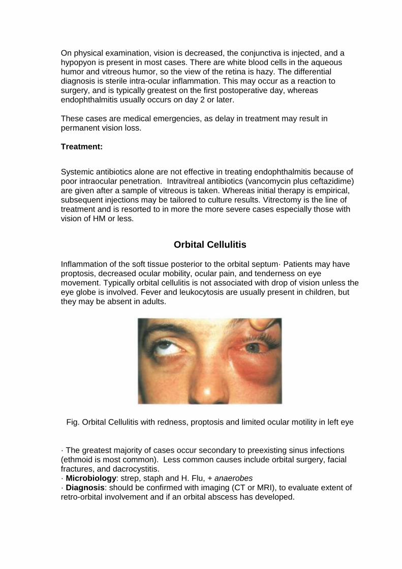
Inflammation of the soft tissue posterior to the orbital septum· Patients may have proptosis, decreased ocular mobility, ocular pain, and tenderness on eye movement. Typically orbital cellulitis is not associated with drop of vision unless the eye globe is involved. Fever and leukocytosis are usually present in children, but they may be absent in adults.
Fig. Orbital Cellulitis with redness, proptosis and limited ocular motility in left eye
· The greatest majority of cases occur secondary to preexisting sinus infections (ethmoid is most common). Less common causes include orbital surgery, facial fractures, and dacrocystitis. · Microbiology: strep, staph and H. Flu, + anaerobes · Diagnosis: should be confirmed with imaging (CT or MRI), to evaluate extent of retro-orbital involvement and if an orbital abscess has developed.
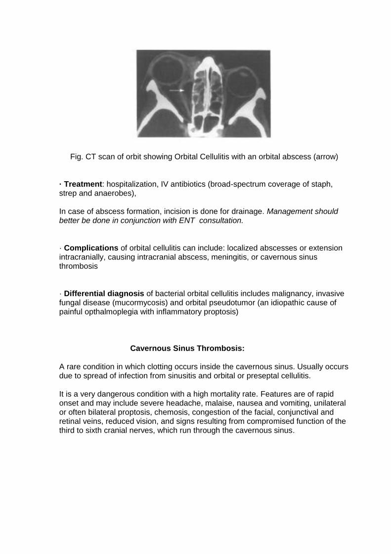
Fig. CT scan of orbit showing Orbital Cellulitis with an orbital abscess (arrow)
· Treatment: hospitalization, IV antibiotics (broad-spectrum coverage of staph, strep and anaerobes),
In case of abscess formation, incision is done for drainage. Management should better be done in conjunction with ENT consultation.
· Complications of orbital cellulitis can include: localized abscesses or extension intracranially, causing intracranial abscess, meningitis, or cavernous sinus thrombosis
· Differential diagnosis of bacterial orbital cellulitis includes malignancy, invasive fungal disease (mucormycosis) and orbital pseudotumor (an idiopathic cause of painful opthalmoplegia with inflammatory proptosis)
Cavernous Sinus Thrombosis:
A rare condition in which clotting occurs inside the cavernous sinus. Usually occurs due to spread of infection from sinusitis and orbital or preseptal cellulitis.
It is a very dangerous condition with a high mortality rate. Features are of rapid onset and may include severe headache, malaise, nausea and vomiting, unilateral or often bilateral proptosis, chemosis, congestion of the facial, conjunctival and retinal veins, reduced vision, and signs resulting from compromised function of the third to sixth cranial nerves, which run through the cavernous sinus.
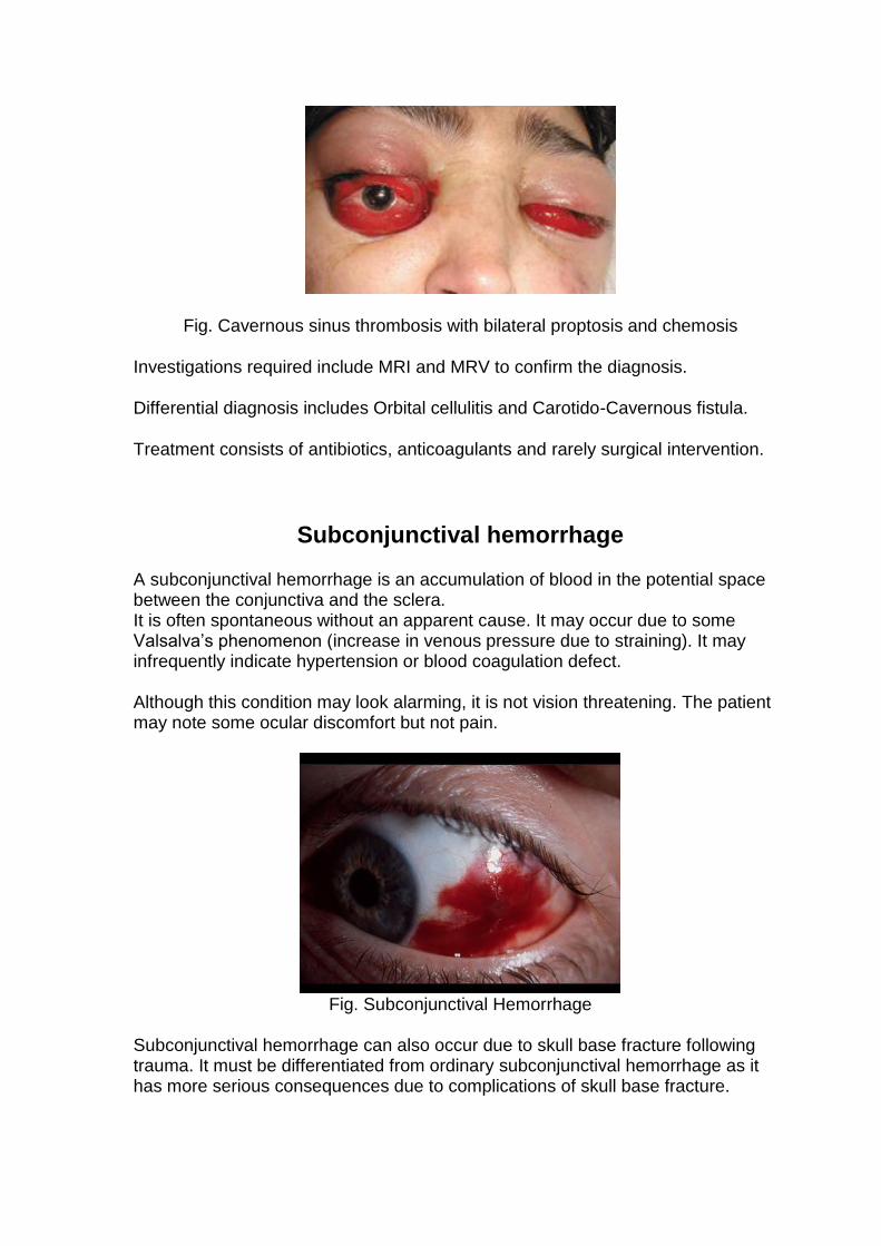
Fig. Cavernous sinus thrombosis with bilateral proptosis and chemosis
Investigations required include MRI and MRV to confirm the diagnosis.
Differential diagnosis includes Orbital cellulitis and Carotido-Cavernous fistula.
Treatment consists of antibiotics, anticoagulants and rarely surgical intervention.
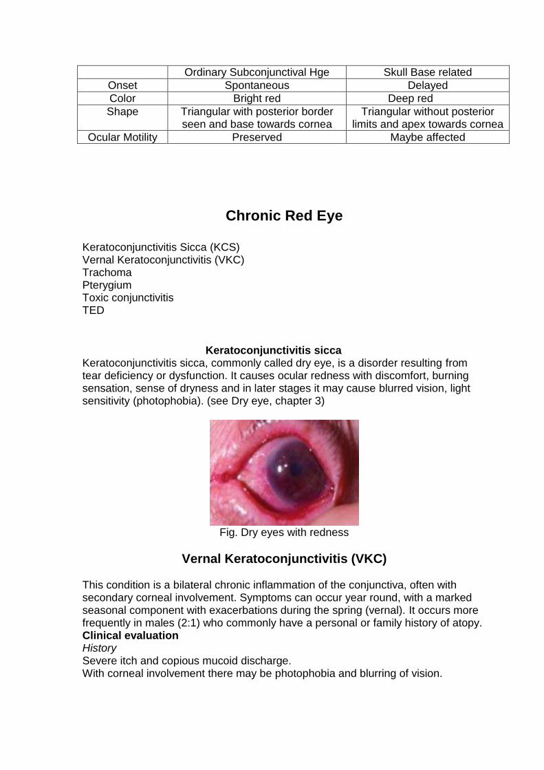
Subconjunctival hemorrhage
A subconjunctival hemorrhage is an accumulation of blood in the potential space between the conjunctiva and the sclera. It is often spontaneous without an apparent cause. It may occur due to some Valsalva’s phenomenon (increase in venous pressure due to straining). It may infrequently indicate hypertension or blood coagulation defect. Although this condition may look alarming, it is not vision threatening. The patient may note some ocular discomfort but not pain.
Fig. Subconjunctival Hemorrhage
Subconjunctival hemorrhage can also occur due to skull base fracture following trauma. It must be differentiated from ordinary subconjunctival hemorrhage as it has more serious consequences due to complications of skull base fracture.
Ordinary Subconjunctival Hge Skull Base related
Onset Spontaneous Delayed
Color Bright red Deep red
Shape Triangular with posterior border seen and base towards cornea
Triangular without posterior limits and apex towards cornea
Ocular Motility Preserved Maybe affected
Chronic Red Eye
Keratoconjunctivitis Sicca (KCS) Vernal Keratoconjunctivitis (VKC) Trachoma Pterygium Toxic conjunctivitis TED
Keratoconjunctivitis sicca
Keratoconjunctivitis sicca, commonly called dry eye, is a disorder resulting from tear deficiency or dysfunction. It causes ocular redness with discomfort, burning sensation, sense of dryness and in later stages it may cause blurred vision, light sensitivity (photophobia). (see Dry eye, chapter 3)
Fig. Dry eyes with redness
Vernal Keratoconjunctivitis (VKC) This condition is a bilateral chronic inflammation of the conjunctiva, often with secondary corneal involvement. Symptoms can occur year round, with a marked seasonal component with exacerbations during the spring (vernal). It occurs more frequently in males (2:1) who commonly have a personal or family history of atopy. Clinical evaluation History Severe itch and copious mucoid discharge. With corneal involvement there may be photophobia and blurring of vision.
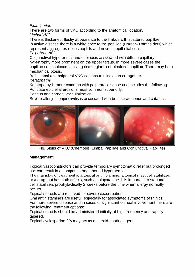
Examination There are two forms of VKC according to the anatomical location. Limbal VKC There is thickened, fleshy appearance to the limbus with scattered papillae. In active disease there is a white apex to the papillae (Horner–Trantas dots) which represent aggregates of eosinophils and necrotic epithelial cells. Palpebral VKC. Conjunctival hyperaemia and chemosis associated with diffuse papillary hypertrophy more prominent on the upper tarsus. In more severe cases the papillae can coalesce to giving rise to giant ‘cobblestone’ papillae. There may be a mechanical ptosis. Both limbal and palpebral VKC can occur in isolation or together. Keratopathy Keratopathy is more common with palpebral disease and includes the following. Punctate epithelial erosions most common superiorly. Pannus and corneal vascularization. Severe allergic conjunctivitis is associated with both keratoconus and cataract.
Fig. Signs of VKC (Chemosis, Limbal Papillae and Conjunctival Papillae)
Management Topical vasoconstrictors can provide temporary symptomatic relief but prolonged use can result in a compensatory rebound hyperaemia. The mainstay of treatment is a topical antihistamine, a topical mast cell stabilizer, or a drug that has both effects, such as olopatadine. It is important to start mast cell stabilizers prophylactically 2 weeks before the time when allergy normally occurs. Topical steroids are reserved for severe exacerbations. Oral antihistamines are useful, especially for associated symptoms of rhinitis. For more severe disease and in cases of significant corneal involvement there are the following treatment options: Topical steroids should be administered initially at high frequency and rapidly tapered. Topical cyclosporine 2% may act as a steroid-sparing agent..
Trachomatous Conjunctivitis (Egyptian Ophthalmia) Trachomatous conjunctivitis is a chronic inflammatory condition. Trachoma is the world’s leading cause of preventable blindness of infectious origin. It is endemic in Egypt. Among children under 5, prevalence of active trachoma infections can be 60 percent or more. Etiology Trachoma is a chronic keratoconjunctivitis caused by the gram-negative organism, Chlamydia trachomatis that contains both DNA and RNA. It produces intracellular basophilic inclusion bodies in epithelial cells. It is susceptible to tetracycline, erythromycin and sulfonamides. No solid immunity occurs so recurrences are common. Pathology:
The organism is epitheliotropic.
Two phases of the disease process exist. However both phases may coexist in the same patient.
− Active phase: follicular or papillary conjunctivitis − Cicatricial ( healing) phase: conjunctival scarring alone, if mild to
moderate, tends to be asymptomatic. Severe scarring causes many complications.
Clinical Picture: Symptoms 1. Mild irritation and foreign body sensation is often present. 2. Itching is a not an uncommon complaint. 3. In chronic stage, cornea is involved causing pain, lacrimation and photophobia. Signs: WHO classification: World Health Organization identified five stages in trachoma:
Trachomatous follicles TF: beginning of the infection with 5 or more follicles, on the inner surface of the upper palpebral conjunctiva, visible with magnification.
Trachomatous inflammation TI: eye is irritated and highly infectious, with a thickening or swelling of the upper eyelid. Papillary hypertrophy and inflammatory thickening of the upper tarsal conjunctiva obscuring more than half the deep tarsal vessels
Trachomatous scarring TS: presence of scarring in tarsal conjunctiva which often appear as white lines when examined with magnification.
Trachomatous trichiasis TT: lashes turn in and rub and scratch the cornea.
Corneal opacity CO: continual inflammation and scratching from rubbing lashes lead to clouding of the cornea with decreased vision.
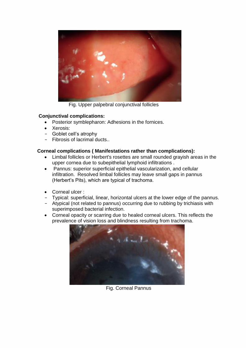
Fig. Upper palpebral conjunctival follicles
Conjunctival complications:
Posterior symblepharon: Adhesions in the fornices.
Xerosis: − Goblet cell’s atrophy − Fibrosis of lacrimal ducts..
Corneal complications ( Manifestations rather than complications):
Limbal follicles or Herbert's rosettes are small rounded grayish areas in the upper cornea due to subepithelial lymphoid infiltrations .
Pannus: superior superficial epithelial vascularization, and cellular infiltration. Resolved limbal follicles may leave small gaps in pannus (Herbert’s Pits), which are typical of trachoma.
Corneal ulcer : − Typical: superficial, linear, horizontal ulcers at the lower edge of the pannus. − Atypical (not related to pannus) occurring due to rubbing by trichiasis with
superimposed bacterial infection.
Corneal opacity or scarring due to healed corneal ulcers. This reflects the prevalence of vision loss and blindness resulting from trachoma.
Fig. Corneal Pannus
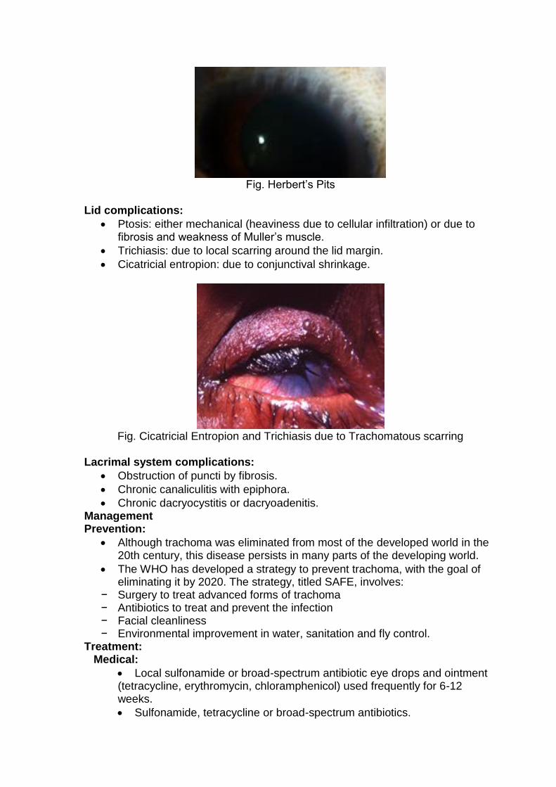
Fig. Herbert’s Pits
Lid complications:
Ptosis: either mechanical (heaviness due to cellular infiltration) or due to fibrosis and weakness of Muller’s muscle.
Trichiasis: due to local scarring around the lid margin.
Cicatricial entropion: due to conjunctival shrinkage.
Fig. Cicatricial Entropion and Trichiasis due to Trachomatous scarring
Lacrimal system complications:
Obstruction of puncti by fibrosis.
Chronic canaliculitis with epiphora.
Chronic dacryocystitis or dacryoadenitis. Management Prevention:
Although trachoma was eliminated from most of the developed world in the 20th century, this disease persists in many parts of the developing world.
The WHO has developed a strategy to prevent trachoma, with the goal of eliminating it by 2020. The strategy, titled SAFE, involves:
− Surgery to treat advanced forms of trachoma − Antibiotics to treat and prevent the infection − Facial cleanliness − Environmental improvement in water, sanitation and fly control.
Treatment: Medical:
Local sulfonamide or broad-spectrum antibiotic eye drops and ointment (tetracycline, erythromycin, chloramphenicol) used frequently for 6-12 weeks.
Sulfonamide, tetracycline or broad-spectrum antibiotics.
Oral azithromycin Surgical: .
Picking of PTDs.
Surgery for complications such as trichiasis and entropion
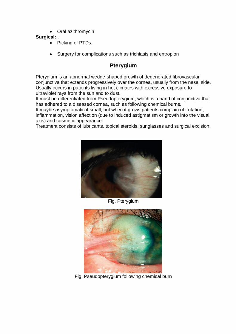
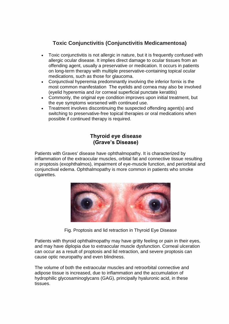
Pterygium
Pterygium is an abnormal wedge-shaped growth of degenerated fibrovascular conjunctiva that extends progressively over the cornea, usually from the nasal side. Usually occurs in patients living in hot climates with excessive exposure to ultraviolet rays from the sun and to dust. It must be differentiated from Pseudopterygium, which is a band of conjunctiva that has adhered to a diseased cornea, such as following chemical burns. It maybe asymptomatic if small, but when it grows patients complain of irritation, inflammation, vision affection (due to induced astigmatism or growth into the visual axis) and cosmetic appearance. Treatment consists of lubricants, topical steroids, sunglasses and surgical excision.
Fig. Pterygium
Fig. Pseudopterygium following chemical burn
Toxic Conjunctivitis (Conjunctivitis Medicamentosa)
Toxic conjunctivitis is not allergic in nature, but it is frequently confused with allergic ocular disease. It implies direct damage to ocular tissues from an offending agent, usually a preservative or medication. It occurs in patients on long-term therapy with multiple preservative-containing topical ocular medications, such as those for glaucoma.
Conjunctival hyperemia predominantly involving the inferior fornix is the most common manifestation The eyelids and cornea may also be involved (eyelid hyperemia and /or corneal superficial punctate keratitis)
Commonly, the original eye condition improves upon initial treatment, but the eye symptoms worsened with continued use.
Treatment involves discontinuing the suspected offending agent(s) and switching to preservative-free topical therapies or oral medications when possible if continued therapy is required.
Thyroid eye disease (Grave’s Disease)
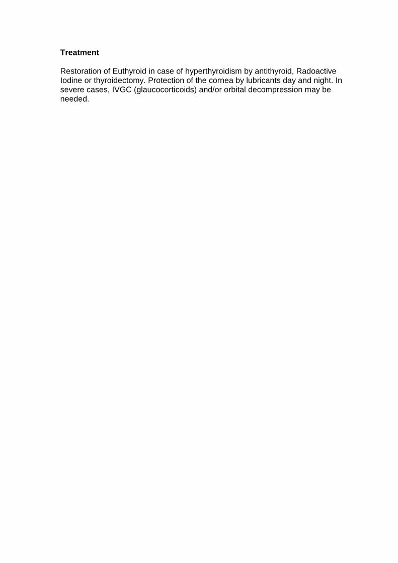
Patients with Graves' disease have ophthalmopathy. It is characterized by inflammation of the extraocular muscles, orbital fat and connective tissue resulting in proptosis (exophthalmos), impairment of eye-muscle function, and periorbital and conjunctival edema. Ophthalmopathy is more common in patients who smoke cigarettes.
Fig. Proptosis and lid retraction in Thyroid Eye Disease
Patients with thyroid ophthalmopathy may have gritty feeling or pain in their eyes, and may have diplopia due to extraocular muscle dysfunction. Corneal ulceration can occur as a result of proptosis and lid retraction, and severe proptosis can cause optic neuropathy and even blindness.
The volume of both the extraocular muscles and retroorbital connective and adipose tissue is increased, due to inflammation and the accumulation of hydrophilic glycosaminoglycans (GAG), principally hyaluronic acid, in these tissues.

Fig. CT scan showing enlargement of extraocular muscles in Thyroid Eye Disease
Physical examination of the eyes of a patient with Graves' ophthalmopathy should include:
Inspection of the conjunctiva and periorbital tissue, looking for conjunctival injection and edema (chemosis) and periorbital edema.
Determination of the extent to which the upper and lower lids can be closed, because failure of apposition promotes dryness and ulceration of the cornea.
Assessment of the range of motion of the eyes. Impairment of extraocular muscle function is often evident by an inability to achieve or maintain convergence.
Objective measurements of the degree of proptosis, using an exophthalmometer.
Visual acuity and color vision should be assessed by simple reading tests and color charts, and visual fields should be evaluated by confrontation. If any evidence of impairment is obtained, the patient should be evaluated by an ophthalmologist.
Assessment of severity — The type of examination described above provides the basis for the routine evaluation of most patients. A classification of the eye changes of Graves' disease developed by the American Thyroid Association has become widely used. The first letters of each category constitute the mnemonic NO SPECS — NO connotes absence or mild degree of involvement; SPECS the more serious degrees of involvement.
The severity therefore ranges from 0 to VI.
Class 0 — No symptoms or signs Class I — Only signs, no symptoms (eg, lid retraction, stare, lid lag) Class II — Soft tissue involvement Class III — Proptosis Class IV — Extraocular muscle involvement Class V — Corneal involvement Class VI — Sight loss (optic nerve involvement)
Treatment
Restoration of Euthyroid in case of hyperthyroidism by antithyroid, Radoactive Iodine or thyroidectomy. Protection of the cornea by lubricants day and night. In severe cases, IVGC (glaucocorticoids) and/or orbital decompression may be needed.