recurrence and progression in meningiomas: the clonal ... · oncology · neuropathology ·...
TRANSCRIPT

Neurology · Neurosurgery · Medical Oncology · Radiotherapy · Paediatric Neuro-
oncology · Neuropathology · Neuroradiology · Neuroimaging · Nursing · Patient Issues
THE EUROPEAN ASSOCIATION OF
NEUROONCOLOGY
Volume 3 (2013) // Issue 3 // e-ISSN 2224-3453
Member of the
Homepage:Homepage:
www.kup.at/journals/eano/index.html
Online Database Featuring Author, Key Word and
Full-Text Search
Online Database Featuring Author, Key Word and
Full-Text Search
Recurrence and Progression in
Meningiomas: The Clonal Cytogenetic
Evolution of a Benign Human Tumour
Ketter R, Oertel J, Linsler S
Urbschat S
European Association of
NeuroOncology Magazine 2013; 3 (3)
105-111
EUR ASSOC NEUROONCOL MAG 2013; 3 (3)
The Genetic Progression Score of Meningioma
105
Received on April 21, 2013; accepted on May 27, 2013; Pre-Publishing Online onJuly 18, 2013
From the Department of Neurosurgery, Saarland University, Homburg/Saar, GermanyCorrespondence to: Ralf Ketter, MD, Department of Neurosurgery, Saarland Uni-versity, Kirrberger Straße, 66421 Homburg/Saar, Germany;e-mail: [email protected]
Recurrence and Progression in Meningiomas:The Clonal Cytogenetic Evolution of a
Benign Human TumourRalf Ketter, Joachim Oertel, Stefan Linsler, Steffi Urbschat
Abstract: Meningiomas are mostly benign tu-mours that originate from the coverings of brainand spinal cord. Only a minority of cases showprogression to an anaplastic tumour (WHO gradesII and III). Multiple and familial cases are rare andmostly associated with (hereditary) neurofibroma-tosis 2 (NF2). Meningiomas show an unexpectedlyhigh recurrence rate. Also, completely removedlow-grade tumours can recur. On a cytogeneticlevel, meningiomas are the best-studied tumoursin humans. The majority of high-grade but only aminority of low-grade meningiomas show loss ofmerlin, a cytoskeleton-cytoplasm-linker protein.Merlin is the product of the NF2 gene located onchromosome 22. A second tumour suppressor geneon chromosome 22 on 22q12.3 is the gene for thetissue inhibitor of metalloproteinase 3 (TIMP3),which appears to be involved in meningioma pro-
gression and a high-grade meningioma pheno-type. In contrast to other solid tumours, progres-sion of meningiomas is correlated with increasinghypodiploidy, showing characteristic clonal evolu-tions that mostly include chromosomes 14, 18,and 19 and, more rarely, 6 and 10. Structural aber-rations are rare, except for the loss of the shortarm of one chromosome 1, which appears to bethe decisive step for anaplastic growth. A bio-statistical approach has been proposed, using anoncogenetic tree model that estimates the mostlikely cytogenetic pathways of meningioma pa-tients in terms of accumulation of chromosomechanges in tumour cells. The genetic progressionscore (GPS) estimates the genetic status of a tu-mour as progression in the corresponding tumourcells along this model. High GPS values are highlycorrelated with early recurrence of meningiomas
(p < 10–4). This correlation holds true even whenpatients are stratified by WHO grade. Tumour lo-cation also has an impact on genetic progression.Clinical relevance of the GPS is demonstratedwith respect to origin, WHO grade, and recurrenceof the tumour. As a quantitative measure, the GPSallows a more precise assessment of the progno-sis of meningiomas than categorical cytogeneticmarkers based on single chromosomal aberra-tions. Comparative histochemical and molecularcytogenetic studies point to the alkaline phos-phatase gene (ALPL, liver-bone-kidney type) lo-cated on 1p36.1–p34 as a candidate tumour sup-pressor gene. Eur Assoc NeuroOncol Mag2013; 3 (3): 105–11.
Key words: meningioma, recurrence, deletion of1p, chromosomes, genetic progression
Introduction
Meningiomas are derived from the arachnoidal cap cells ofthe leptomeninges, the soft coverings of the brain and spinalcord. Although their matrix tissue constitutes < 5 % of the in-tracranial and intraspinal masses, meningiomas are estimatedto constitute between 13 % and 26 % of the primary tumourswithin the CNS. Most meningiomas are sporadic, slowlygrowing benign tumours and correspond histologically toWHO grade I. However, certain histological subtypes andalso a minority of common-type meningiomas show a moreaggressive biological behaviour and are associated with anincreased risk of recurrence and an unfavourable prognosis.Corresponding to their localization, meningiomas of the spi-nal cord tend to be significantly more benign than those of thebrain basis, which, in turn, are more benign than meningio-mas of the brain convexity [1, 2]. The current WHO classifica-tion of brain tumours [1] distinguishes 3 grades of meningio-mas: the common type (WHO grade I), the atypical or inter-mediate type (WHO grade II), and the anaplastic (WHO gradeIII) meningioma.
For appropriate treatment of tumour patients, prediction of timeuntil death or time to progression after initial treatment is animportant task. Due to many clinical, topographical, radiologi-cal, and surgical factors, histology is not solely decisive forprognosis [3], although mitotic activity, cellular pleomorphism
with prominent nucleoli and micronecrosis, and focally raisedcell density have been discussed as indicators of a poorer prog-nosis [4]. A major challenge is the identification of geneticprognostic markers that better reflect tumour biology.
Multiple occurrences are rare events, observed in only about2 % of cases. Frequently, multiple meningiomas occur inpatients with neurofibromatosis 2 (NF2), an autosomallydominant tumour syndrome, in combination with vestibularschwannomas and ependymomas. A polyclonal origin mustbe supposed in these cases.
In non-NF2 cases, molecular genetic studies demonstrate acommon clonal origin of multiple or recurrent meningiomas [5,6]. The development of familial (multiple) meningiomas withinthe same or different generations has so far been reported in< 20 families. Pathologic and genetic data on these familiesfirst suggested a biologically variant type of NF2 with the de-velopment of meningiomas only but no schwannomas. Mo-lecular studies on a few cases indeed excluded the involvementof the NF2 gene although a chromosome 22 was missing. Alter-natively, homozygous inactivation of a different tumour sup-pressor gene on the same chromosome was proposed [7].
The meningioma is one of the cytogenetically best-studiedsolid tumours. The characteristic and most frequent chromo-somal aberration in meningiomas is monosomy 22 [8], which,however, seems not to be relevant for prognosis as an isolatedanomaly. The progression from common-type to atypical andanaplastic meningioma is characterized by 2 different cytoge-netic events: Firstly, further loss of up to 6 other chromo-somes, with a mostly typical pattern of clonal evolution, and,secondly, partial or complete loss of the short arm of one chro-mosome 1 [3, 9–15].
For personal use only. Not to be reproduced without permission of Krause & Pachernegg GmbH.
The Genetic Progression Score of Meningioma
106 EUR ASSOC NEUROONCOL MAG 2013; 3 (3)
The identification of pathogenetic pathways in humantumours is one of the main challenges in molecular oncology.For many tumour types, genetic events defined by somaticchromosome alterations or gene mutations, respectively, areknown to accumulate over time in the course of the disease.Based on these findings, genetic changes associated withchromosome instability are believed to play an important rolein both tumourigenesis and tumour progression in meningio-mas. There is a vast amount of literature on linking single ge-netic alterations to survival [16], but only few efforts havebeen made to construct more complex and comprehensivemarkers. Zang [15] described a first model of clonal evolutionin meningiomas based on conventional cytogenetic studies.Data of the cytogenetic findings of 394 meningiomas led to anempirical model of meningioma progression.
Cytogenetic analyses show that numerical and structuralchromosome changes with pronounced hypodiploidy, orrarely hyperdiploidy [17], and especially deletion of the shortarm of a chromosome 1, are accompanied by more aggressivebiological characteristics [3, 9–15, 18].
By using a mathematical model for estimating the most likelycytogenetic pathways in meningiomas, a model for tumour re-currence could be established. In this model, each tumour isrepresented by the genetic events that have occurred in thetumour. The most important difference of the mathematicalmodel compared to the “hand-crafted” model [15] is that itallows an objective assignment of the estimated time to recur-rence for a single tumour based on its genetic status.
Incidence, Materials, and Statistics
Patient PopulationWe performed a retrospective study on 661 patients (482women and 179 men) with meningiomas operated on at theDepartment of Neurosurgery, Saarland University, betweenJanuary 1973 and April 2005. Average age of the overall pa-tient population was 57.3 years (SD ± 12.8 years). Averageage of the female patients was 57.6 years (SD ± 12.3 years),average age of the male patients was 56.7 years (SD ± 14.1years).
Clinical Variables
The clinical variables investigated comprise patient genderand age, tumour location and histology, and the completenessof tumour extirpation.
LocationWe formed 9 groups depending on meningioma location:(1) convexity, (2) parasagittal region, (3) tuberculum sellae,(4) olfactory groove, (5) sphenoid wing ridge, (6) posteriorcranial fossa, (7) tentorium, (8) ventricular, and (9) spinalchannel. The classification was carried out on the basis of thepreoperative CT and NMR.
Tumour ExtirpationComplete surgical extirpation of the tumour was defined asSimpson grades I and II corresponding to a macroscopicallycomplete tumour resection with bipolar coagulation of thedura insertion.
Tumour HistologyThe 661 cases investigated comprised 465 common-type(WHO grade I), 156 intermediate-type (WHO atypical men-ingioma, grade II), and 14 anaplastic meningiomas (WHOgrade III). Meningioma grade was assessed by a combinedhistological and morphometric approach on routinely HE andKi-67/Feulgen-stained, formalin-fixed, paraffin-embeddedtissue sections [12].
Cell Culture and Cytogenetic Preparation
Cell cultures from 661 meningioma biopsies and chromo-some preparations with GTC banding were carried out ac-cording to standard procedures. Many tumours were charac-terized by multiple patterns of cytogenetic aberrations. Intotal, 1068 clonal patterns were observed in the 661 tumours.
Histochemical Detection of ALPL
The histochemical findings shown in Figure 1 were perform-ed on frozen sections as described by Niedermayer et al [3].
Follow-up
Patients were investigated in the neurosurgical outpatient de-partment of the Saarland University, either within the frame-work of routine follow-up or because of the appearance ofneurological symptoms. Recurrence was evaluated as newevidence of a tumour in CT or NMR after previous completeextirpation (Simpson grades I and II). The Simpson grade IIwas established on the basis of the operation report and thepostoperative CT or NMR investigation. Average follow-uptime was 40.3 months (41.5 months for female patients and37.2 months for male patients).
Statistical Analyses
Oncogenetic Tree ModelsMixtures of oncogenetic trees were used to describe the or-dered accumulation of genetic aberrations during tumour pro-gression. In a single oncogenetic tree, vertices represent ge-netic events, and edges between vertices represent transitionsbetween the events. Each edge is associated with the probabil-ity that the successor event will occur, given the predecessorevent has already occurred. In the model, genetic events areassumed to be non-reversible, thus the disease process can befully described by the accumulation of genetic aberrations. Inthe mixture model, more than one tree component is esti-mated [18].
Genetic Progression ScoreIn oncogenetic tree models, consecutive genetic aberrationsare associated with corresponding conditional transitionprobabilities. These probabilities can be converted to averagewaiting times by assuming Poisson processes for the occur-rence of aberrations (see Rahnenführer et al [19] for details).Formally, waiting time associated to an edge of the tree withcorresponding conditional probability p is given by (1–p)/pmultiplied with a scaling factor that is typically set to 1. Thegenetic progression score (GPS) of a tumour then is defined asthe average waiting time of its pattern of genetic aberrations,given the underlying tree mixture model. Tumours with fewaberrations that appear early in the model receive low GPSvalues; tumours with many late aberrations in the model areassociated with high GPS values. For 221 out of the 661 pa-
EUR ASSOC NEUROONCOL MAG 2013; 3 (3)
The Genetic Progression Score of Meningioma
107
tients, more than one cytogenetic pattern was detected. Inthese cases, the GPS of the tumour was defined as the highestGPS of all clones found in the tumour.
Clinical Factors of Meningioma Prognosis
Grading of meningiomas has always been controversial. Ob-viously, the biological behaviour of meningiomas cannot beaccounted for by histological parameters alone [14, 15]. In1979, Zülch stated that it is not the histological grading whichis most crucial for the rate of recurrence of meningiomas, butprimarily the completeness of extirpation [20]. There isagreement in the literature that radical surgical extirpation iscorrelated with a good prognosis [21, 22].
It is well known that females are affected far more frequentlyby meningiomas than males [20, 21]. This observation wasconfirmed in our previous study [12, 14, 15] with a ratio of2.67:1. In particular, in the 49 spinal tumours investigated, thefemale sex was overrepresented (87 %). This sex ratio wasshifted when the tumours were broken down by karyotype. Inthe GPS group 2 (GPS ≥ 6.39), ie, in tumours with a pro-nounced aberration of the karyotype with deletion 1p, wefound a sex distribution of 1.81:1.
Cytogenetic Aspects of Meningiomas
Localization of Meningiomas
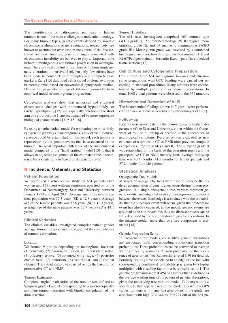
Frequency and behaviour of meningiomas at different intracraniallocations were first discussed in 1922 [23]. A correlation betweentumour localization and malignancy has repeatedly been observed.Meningiomas of the spinal cord tend to be significantly moreoften benign than those of the brain basis, which, in turn, aremore often benign than meningiomas of the brain convexity [1,2]. In 1980, it was shown that tumours located at the base of theskull typically contain cells with 46 chromosomes, whereas men-ingiomas located at the convexity show significant numbers ofchromosomal aberrations [15]. In spinal tumours, almost exclu-sively a 22-monosomic karyotype was detected. It was alreadystriking at that time that meningiomas which recurred showedsignificantly more chromosomal aberrations and a preference ofthe convexity. These findings are also in line with the results of alarge series of meningioma patients (n > 9000 cases), where be-nign meningiomas were more frequently located at the skull thanmalignant meningiomas (p < 0.02) (Figure 2) [2].
Cytogenetic Findings and Histomorphology
Among histopathologically prognostic parameters, mitoticactivity is the most important one. As the cut-offs of mitotic
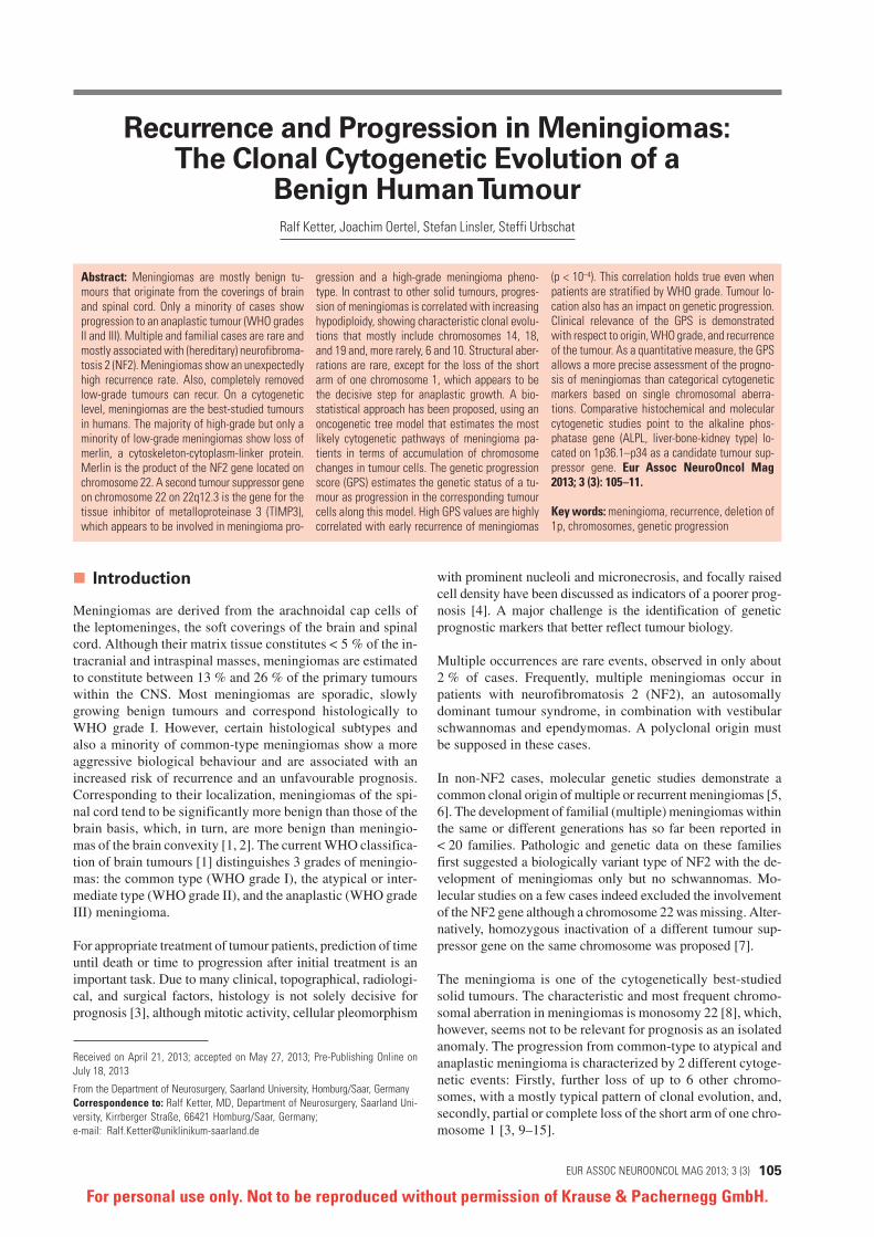
Figure 1. Expression pattern of alkaline phosphatase in meningiomas of different grade (liver-bone-kidney type, ALPL, located at 1p36.1–p34). In the rows, parallel sectionsof meningiomas are shown. The upper row (A–C) shows a WHO grade-I, the middle row (D–F) a grade-II, and the lower row (G–I) a grade-III meningioma.The first column shows haematoxylin and eosin stainings, the second column histochemical alkaline phosphatase reactivity, and the third column immunohistochemicallocalization of the alkaline phosphatase protein. Histochemical and immunohistochemical stainings show a very good correlation. In the grade-I meningioma, all tumourcells are positive; in the grade-II tumour, clearly delineated areas are spared, and in the grade-III meningioma only the endothelial cells of the blood vessels are positive, thetumour cells being completely negative histochemically as well as immunohistochemically. Loss of ALPL expression is highly correlated with FISH results on the terminal 1pdeletion. All sections were photographed with a 20× objective. Reprinted with permission from [Zang KD. Meningioma: a cytogenetic model of a complex benign humantumor, including data on 394 karyotyped cases. Cytogenet Cell Genet 2001; 93: 207–20.] © 2001 Karger Publishers, Basel, Switzerland.
The Genetic Progression Score of Meningioma
108 EUR ASSOC NEUROONCOL MAG 2013; 3 (3)
activity (MI) are defined for each grade by the WHO classifica-tion of brain tumours and because MI can be applied as the solegrading criterion, the reliable and reproducible assessment of MIis crucial for appropriate risk stratification. In 2007, we classified661 meningiomas according to their genetic progression scores(GPS values) [18]. We found a high correlation between cytoge-netic findings and histomorphology: higher genetic progressionscores correlated highly significantly with higher histologicalgrades (p < 10–10, Fisher’s exact test). This result is importantsince in the literature no clear correlation between histologicalgrading and the rate of recurrence in meningiomas has been re-ported [9, 24, 25]. In our study, no clear distinction between thehistological grades I and II with respect to tumour recurrencecould be shown, either (Figure 1). However, WHO grade-IIImeningiomas show a statistically significant correlation with ear-lier tumour recurrence.
Expression of the Alkaline Phosphatase ALPL in
MeningiomasMore than 4 decades ago, Osske and Jänisch [26] reporteddifferences in the expression of ALPL in meningiomas. How-ever, no clear correlation with tumour grade or subtype wasfound. It could been shown that there is a strong homogeneousexpression of this enzyme in low-grade meningiomas, with in-creasing patchwork-like patterns of expression loss in anaplasticmeningiomas [15]. ALPL is located on the short arm of chromo-some 1 (1p34–1, p36.1). In a more detailed study using inter-phase FISH on frozen sections of native meningioma cells [3,27], the autors were able to show a complete correlation betweenlosses in 1p and reductions in the amount and activity of alkalinephospatase (Figure 1). As the activity of alkaline phosphatase caneasily be detected histochemically and is highly correlated withthe presence of 1p, this prognostically relevant parameter is alsoavailable to non-cytogeneticists. The data speak in favour of atumour suppressor gene function of ALPL; however, the mecha-nism of inactivation of the second allele is not yet explained.
Oncogenetic Tree Models
The basis for the oncogenetic tree mixture models are singleoncogenetic trees as introduced by Desper et al [28] that can
be used to estimate the most likely pathogenetic routes intumours from observed subsets of genetic events. These mod-els are of high explanatory power, but often only for a portionof the analyzed tumour samples. A subset of genetic events isonly represented by this tree model if for any event in this sub-set all precursor events in the tree also belong to the subset.All other subsets of events are assigned likelihood zero. VonHeydebreck et al [29] propose to include additional hiddenevents in the tree and to model genetic events as leaves in thetree. This method trades feasibility of maximum-likelihoodestimation of oncogenetic trees with reduced interpretabilitydue to the introduction of hidden events.
We introduced mixture models of the single oncogenetic treesas used in Desper et al [28]. In these mixture models, one treecomponent is restricted to have a star-like topology, repre-senting independence between genetic events. Owing to thestarlike component, every combination of genetic events isrepresented in the model. The oncogenetic tree mixture mod-els combine interpretability of the trees of Desper et al [28]with an appropriate probabilistic framework.
Relevance of Gender for Genetic Progression
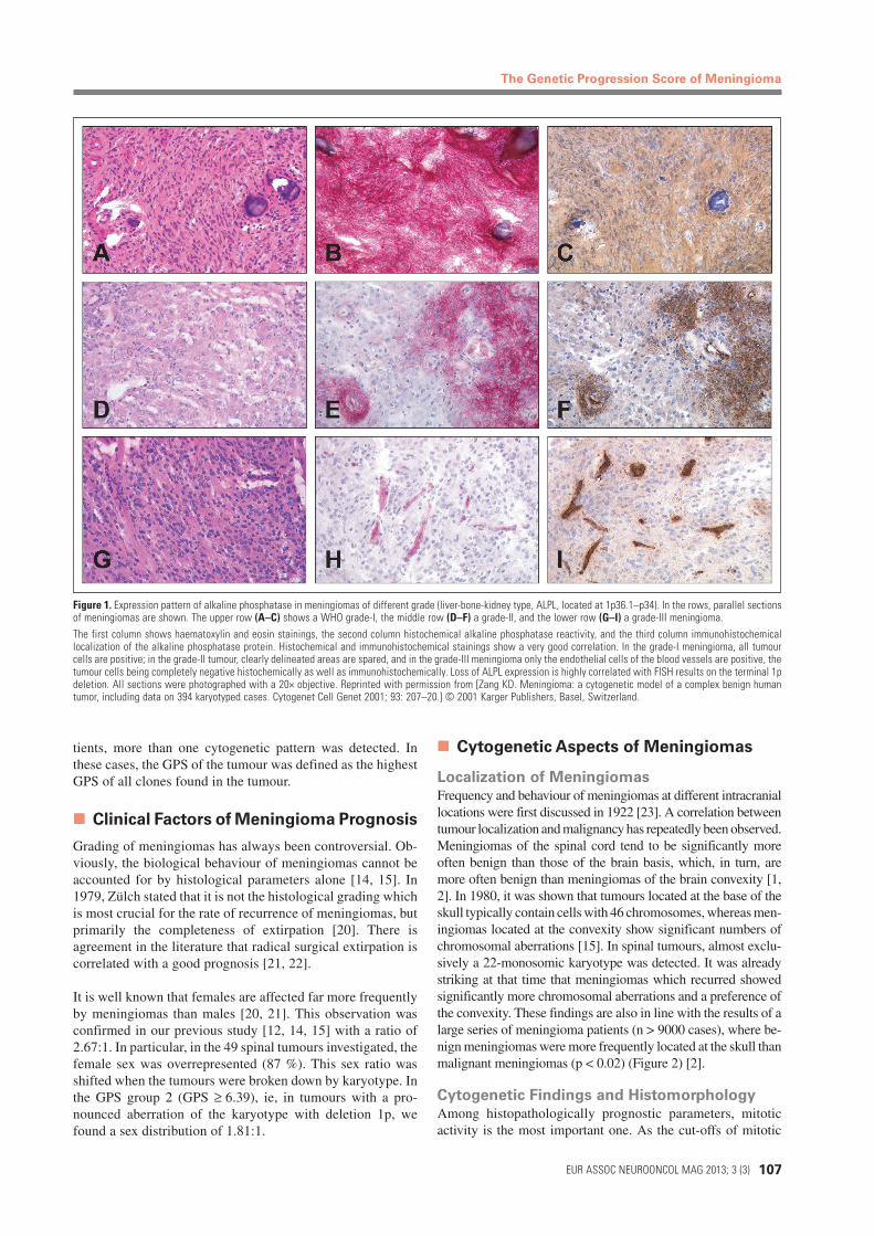
ModelsBreaking down the oncogenetic tree by gender, there is a re-versal concerning chromosomes 14 and 1p– in the male popu-lation. For males, monosomy 14 is estimated to be an earlierevent followed by the deletion of the short arm of one chro-mosome 1. Monosomy 14 has been found to be associatedwith aggressive behaviour of meningiomas [14, 24, 25, 30,31]. However, in the literature the loss of chromosome 14 hasnever been correlated with gender. It is well known that fe-males predominate over males with a ratio of 2.67:1 when itcomes to the overall incidence of meningiomas.
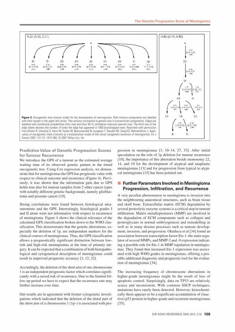
It could be shown that the deletion of the short arm of one chro-mosome 1 has to be regarded as a more valid cytogenetic pa-rameter than monosomy 14 for the prediction of tumour recur-rence, particularly because all anaplastic meningiomas in ourseries displayed a deletion of chromosome 1p (Figure 3).
Figure 2. Bar plots for chromosomal aberrations (1p–, –14) und recurrence [Rec] according to brain regions. Red color indicates convexity, green color skull base, and bluecolor spinal tumours. Reprinted with permission from [Ketter R, Rahnenführer J, Henn W, Kim YJ, Feiden W, Steudel WI, Zang KD, Urbschat S. Correspondence of tumorlocalization with tumor recurrence and cytogenetic progression in meningiomas. Neurosurgery 2008; 62 (1): 61–9; discussion 69–70]. Promotional and commercial use of thematerial in print, digital or mobile device format is prohibited without the permission from the publisher Lippincott Wiliams & Wilkins. Please contact [email protected] forfurther information.
EUR ASSOC NEUROONCOL MAG 2013; 3 (3)
The Genetic Progression Score of Meningioma
109
Predictive Value of Genetic Progression Scores
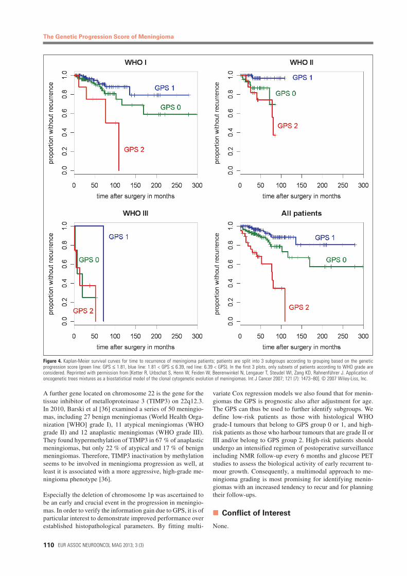
for Tumour RecurrenceWe introduce the GPS of a tumour as the estimated averagewaiting time of its observed genetic pattern in the timedoncogenetic tree. Using Cox regression analysis, we demon-strate that for meningiomas the GPS has prognostic value withrespect to clinical outcome and recurrence (Figure 4). Previ-ously, it was shown that the information gain due to GPSholds true also for tumour samples from 2 other cancer typeswith notably different genetic backgrounds, namely glioblas-toma and prostate cancer [19].
Strong correlations were found between histological mea-surements and the GPS. Interestingly, histological grades Iand II alone were not informative with respect to recurrenceof meningioma. Figure 4 shows the clinical relevance of thecalculated GPS classification broken down to the WHO clas-sification. This demonstrates that the genetic aberrations, es-pecially the deletion of 1p, are independent markers for theclinical courses of meningiomas. Thus, the GPS classificationallows a prognostically significant distinction between low-risk and high-risk meningiomas at the time of primary sur-gery. It can be expected that a combination of both histopatho-logical and cytogenetical description of meningiomas couldresult in improved prognostic accuracy [3, 12, 32].
Accordingly, the deletion of the short arm of one chromosome1 is an independent prognostic factor which correlates signifi-cantly with a raised risk of recurrence. Due to the limited fol-low-up period we have to expect that the recurrence rate mayfurther increase over time.
Our results are in agreement with former cytogenetic investi-gations which indicated that the deletion of the distal part ofthe short arm of a chromosome 1 (1p–) is associated with pro-
Figure 3. Oncogenetic tree mixture model for the development of meningiomas. Both mixture components are labelledwith their weight in the upper left corner. Tree vertices correspond to genetic loss in conventional cytogenetics. Edges arelabelled with conditional probabilities (first row) and their 95-% confidence intervals (second row). The third row of theedge labels denotes the number of times the edge has appeared in 1000 bootstrapped trees. Reprinted with permissionfrom [Ketter R, Urbschat S, Henn W, Feiden W, Beerenwinkel N, Lengauer T, Steudel WI, Zang KD, Rahnenführer J. Appli-cation of oncogenetic trees mixtures as a biostatistical model of the clonal cytogenetic evolution of meningiomas. Int JCancer 2007; 121 (7): 1473–80]. © 2007 Wiley-Liss, Inc.
gression in meningiomas [3, 10–14, 27, 33]. After initialspeculation on the role of 1p deletion for tumour recurrence[10], the importance of this aberration beside monosomy 22,14, and 10 for the development of atypical and anaplasticmeningiomas [13] and for progression from typical to atypi-cal meningioma [15] has been pointed out.
Further Parameters Involved in Meningioma
Progression, Infiltration, and Recurrence
A very peculiar phenomenon in meningioma is invasion intothe neighbouring anatomical structures, such as brain tissueand skull bone. Extracellular matrix (ECM) degradation byseveral proteolytic enzyme systems is a critical step in tumourinfiltration. Matrix metalloproteases (MMP) are involved inthe degradation of ECM components such as collagen andproteoglycans in normal embryogenesis and remodelling aswell as in many disease processes such as tumour develop-ment, invasion, and progression. Okuducu et al [34] found anassociation between transcription factor Ets-1, the main regu-lator of several MMPs, and MMP-2 and -9 expression indicat-ing a possible role for Ets-1 in MMP regulation in meningio-mas. They found that increased Ets-1 expression was associ-ated with high WHO grades in meningiomas, offering a pos-sible additional diagnostic and prognostic tool for the evalua-tion of meningiomas [34].
The increasing frequency of chromosome aberrations inhigher-grade meningiomas might be the result of loss ofapoptotic control. Surprisingly, data on TP53 are relativelyscarce and inconsistent. With common SSCP techniques,mutations have rarely been detected. However, histochemi-cally there appears to be a significant accumulation of (inac-tive) p53 protein in higher-grade and recurrent meningiomas[35].
The Genetic Progression Score of Meningioma
110 EUR ASSOC NEUROONCOL MAG 2013; 3 (3)
A further gene located on chromosome 22 is the gene for thetissue inhibitor of metalloproteinase 3 (TIMP3) on 22q12.3.In 2010, Barski et al [36] examined a series of 50 meningio-mas, including 27 benign meningiomas (World Health Orga-nization [WHO] grade I), 11 atypical meningiomas (WHOgrade II) and 12 anaplastic meningiomas (WHO grade III).They found hypermethylation of TIMP3 in 67 % of anaplasticmeningiomas, but only 22 % of atypical and 17 % of benignmeningiomas. Therefore, TIMP3 inactivation by methylationseems to be involved in meningioma progression as well, atleast it is associated with a more aggressive, high-grade me-ningioma phenotype [36].
Especially the deletion of chromosome 1p was ascertained tobe an early and crucial event in the progression in meningio-mas. In order to verify the information gain due to GPS, it is ofparticular interest to demonstrate improved performance overestablished histopathological parameters. By fitting multi-
Figure 4. Kaplan-Meier survival curves for time to recurrence of meningioma patients; patients are split into 3 subgroups according to grouping based on the geneticprogression score (green line: GPS ≤ 1.81, blue line: 1.81 < GPS ≤ 6.39, red line: 6.39 < GPS). In the first 3 plots, only subsets of patients according to WHO grade areconsidered. Reprinted with permission from [Ketter R, Urbschat S, Henn W, Feiden W, Beerenwinkel N, Lengauer T, Steudel WI, Zang KD, Rahnenführer J. Application ofoncogenetic trees mixtures as a biostatistical model of the clonal cytogenetic evolution of meningiomas. Int J Cancer 2007; 121 (7): 1473–80]. © 2007 Wiley-Liss, Inc.
variate Cox regression models we also found that for menin-giomas the GPS is prognostic also after adjustment for age.The GPS can thus be used to further identify subgroups. Wedefine low-risk patients as those with histological WHOgrade-I tumours that belong to GPS group 0 or 1, and high-risk patients as those who harbour tumours that are grade II orIII and/or belong to GPS group 2. High-risk patients shouldundergo an intensified regimen of postoperative surveillanceincluding NMR follow-up every 6 months and glucose PETstudies to assess the biological activity of early recurrent tu-mour growth. Consequently, a multimodal approach to me-ningioma grading is most promising for identifying menin-giomas with an increased tendency to recur and for planningtheir follow-ups.
Conflict of Interest
None.
EUR ASSOC NEUROONCOL MAG 2013; 3 (3)
The Genetic Progression Score of Meningioma
111
References:1. Louis DN, Scheithauer BW, Budka H, et al.Meningiomas. In Kleihues P, Cavenee WK(eds). Pathology and Genetics. Tumors of theNervous System. IARC, Lyon, 2000; 176–84.2. Ketter R, Rahnenführer J, Henn W, et al.Correspondence of tumor localization withtumor recurrence and cytogenetic progres-sion in meningiomas. Neurosurgery 2008;62: 61–9.3. Niedermayer I, Feiden W, Henn W, et al.Loss of alkaline phosphatase activity inmeningiomas: a rapid histochemical tech-nique indicating progression-associated de-letion of a putative tumor suppressor geneon the distal part of the short arm of chro-mosome 1. J Neuropathol Exp Neurol 1997;56: 879–86.4. Takeuchi H, Kubota T, Kabuto M, et al.Prediction of recurrence in histologically be-nign meningiomas: proliferating cell nuclearantigen and Ki-67 immunohistochemicalstudy. Surg Neurol 1997; 48: 501–6.5. von Deimling A, Larson J, WellenreutherR, et al. Clonal origin of recurrent meningio-mas. Brain Pathol 1999; 9: 645–50.6. Zhu JJ, Maruyama T, Jacoby LB, et al.Clonal analysis of a case of multiple mo-lecular genetic approaches: pathology casereport. Neurosurgery 1999; 45: 409–16.7. Ruttledge MH, Sarrazin J, Rangrartnam S,et al. Evidence for the complete inactivationon the NF2 gene in the majority of sporadicmeningiomas. Nature Genet 1994; 6: 180–4.8. Zang KD, Singer H. Chromosomal consti-tution of meningiomas. Nature 1967; 216:84–5.9. Perry A, Gutmann DH, Reifenberger G.Molecular pathogenesis of meningiomas.J Neurooncol 2004; 70: 183–202.10. Bello MJ, de Campos JM, Kusak ME, etal. Allelic loss of 1p is associated with tumor
progression of meningiomas. Genes Chromo-somes Cancer 1994; 9: 296–8.
11. Henn W, Cremerius U, Heide G, et al.Monosomy 1p is correlated with enhancedin vivo glucose metabolism in meningiomas.Cancer Genet Cytogenet 1995; 79: 144–8.
12. Kolles H, Niedermayer I, Schmitt C, et al.Triple approach for diagnosis and grading ofmeningiomas: histology, morphometry ofKi-67/Feulgen stainings, and cytogenetics.Acta Neurochir (Vienna) 1995; 137: 174–81.
13. Simon M, von Deimling A, Larson JJ, etal. Allelic loss on chromosomes 14, 10 and 1in atypical and malignant meningiomas: agenetic model of meningioma progression.Cancer Res 1995; 55: 4696–701.
14. Ketter R, Henn W, Niedermayer I, et al.Predictive value of progression-associatedchromosomal aberrations for the prognosisof meningiomas: a retrospective study of198 cases. J Neurosurg 2001; 95: 601–7.
15. Zang KD. Meningioma: a cytogeneticmodel of a complex benign human tumor,including data on 394 karyotyped cases.Cytogenet Cell Genet 2001; 93: 207–20.
16. Jiang F, Desper R, Papadimitriou CH, etal. Construction of evolutionary tree modelsfor renal cell carcinoma from comparativegenomic hybridization data. Cancer Res 2000;60: 6503–9.
17. Ketter R, Kim YJ, Storck S, et al. Hyper-diploidy defines a distinct cytogenetic entityof meningiomas. J Neurooncol 2007; 83:213–21.
18. Ketter R, Urbschat S, Henn W, et al. Ap-plication of oncogenetic trees mixtures as abiostatistical model of the clonal cytogeneticevolution of meningiomas. Int J Cancer 2007;121: 1473–80.
19. Rahnenführer R, Beerenwinkel N, SchulzWA, et al. Estimating cancer survival and
clinical outcome based on genetic tumorprogession scores. Bioinformatics 2005; 21:2438–46.
20. Zülch KJ. Histological typing of tumorsof the central nervous system. World HealthOrganisation, Geneva, 1979.
21. Adegbite AB, Khan MI, Paine KWE, et al.The recurrence of intracranial meningiomasafter surgical treatment. J Neurosurg 1983;58: 51–6.
22. Mirimanoff RO, Dosoretz DE, LinggoodRM, et al. Meningioma. Analysis of recur-rence and progression following neurosurgi-cal resection. J Neurosurg 1985; 62: 18–24.
23. Cushing H. The cranial hyperostoses pro-duced by meningeal endotheliomas. ArchNeurol Psychiat 1922; 8: 139–52.
24. Perry A, Jenkins RB, Dahl RJ, et al. Cyto-genetic analysis of aggressive meningiomas.Cancer 1996; 77: 2567–73.
25. Maíllo A, Orfao A, Sayagués JM, et al.New classification scheme for the prognos-tic stratification of meningioma on the basisof chromosome 14 abnormalities, patientage, and tumor histopathology. J Clin Oncol2003; 21: 3285–95.
26. Osske G, Jänisch W. Zur Histochemie derMeningeome. Acta Neuropathol 1967; 9:290–7.
27. Mueller P, Henn W, Niedermayer I, et al.Deletion of chromosome 1p and loss of ex-pression of alkaline phosphatase indicateprogression of meningiomas. Clin Cancer Res1999; 5: 3569–77.
28. Desper R, Jiang F, Kallioniemi OP, et al.Inferring tree models for oncogenesis fromcomparative genome hybridization data.J Comput Biol 1999; 6: 37–51.
29. von Heydebreck A, Gunawan B, Fuzesi L.Maximum likelihood estimation of oncoge-
netic tree models. Biostatistics 2004; 5: 545–56.30. Sayagués JM, Tabernero MD, Maíllo A,et al. Intratumoral patterns of clonal evolu-tion in meningiomas as defined by multi-colour interphase fluorescence in situ hybrid-ization (FISH): is there a relationship betweenhistopathologically benign and atypical/ana-plastic lesions? J Mol Diagn 2004; 6: 316–25.31. Menon GM, Rutter JL, von Sattel JP, etal. Frequent loss of chromosome 14 in atypi-cal and malignant meningioma: identifica-tion of a putative “tumor progression” locus.Oncogene 1997; 14: 611–6.32. Perry A, Stafford SL, Scheithauer BW, etal. Meningioma grading: an analysis of his-tologic parameters. Am J Surg Pathol 1997;21: 1455–65.33. Mueller P, Henn W, Niedermayer I, et al.Deletion of chromosome 1p and loss of ex-pression of alkaline phosphatise indicateprogression of meningiomas. Clin CancerRes 1999; 5: 3569–77.34. Okuducu AF, Zils U, Michaelis SA, et al.Increased expression of avian erythroblasto-sis virus E26 oncogene homolog 1 in WorldHealth Organization grade 1 meningiomas isassociated with an elevated risk of recurrenceand is correlated with the expression of itstarget genes matrix metalloproteinase-2 andMMP-9. Cancer 2006; 15: 1365–72.35. Kamai Y, Watanabe M, Nakayama T, etal. Prognostic significance of p53 and p21immunoreactivity and tumor mikronecrosisfor recurrence of meningiomas. J Neuro-Onkol 2000; 46: 205–13.36. Barski D, Wolter M, Reifenberger G, etal. Hypermethylation and transcriptionaldownregulation of the TIMP3 gene is asso-ciated with allelic loss on 22q12.3 and ma-lignancy in meningiomas. Brain Pathol 2010;20: 623–31.