recent advances in pulmonary hypertension -...
TRANSCRIPT
Recent Advances in Pulmonary Hypertension
1820
There has been an increased recognition of pulmonary hypertension (PH) in clinical practice over the past 30
years. It is likely that this rise in PH diagnoses is attribut-able to multiple factors, including increased awareness by clinicians, the routine use of diagnostic tools such as Doppler echocardiography, and the availability and marketing of the many PH-specific drugs.1 Although the increased awareness of the disease should be viewed as a favorable development, the unintended consequences include overdiagnosis, mis-classification, and at times indiscriminate use of expensive, PH-specific drugs. In this review, we address the epidemi-ology, prognosis, and updated clinical classification of PH. We then focus on the diagnostic approach to the patient with suspected PH with an emphasis placed on highlighting key pearls for the clinician to consider. Finally, we review an evolving conceptual framework of viewing the pulmonary vasculature and the right ventricle as a single, coupled car-diopulmonary unit.
Epidemiology and PrognosisThe exact prevalence of PH in both the United States and the world is not known. The most common cause of PH in the United States is left heart failure (including both heart failure with preserved and reduced systolic function) and, depending on the definition of PH used, PH may be present in upwards of 83% of patients with heart failure.2 Pulmonary arterial hyper-tension (PAH) on the other hand remains a generally rare disease with estimates of prevalence ranging from between 5 to 15 cases per 1 million adults.3,4 Clinical practice suggests that there has been an evolution in the phenotype of patients referred to specialty centers for the diagnosis and manage-ment of PH of all types, including PAH. Registries have provided important information about the epidemiology and changes in the PAH phenotype that have been observed over the past decades. These include changes in age, sex, comor-bidities, and survival. One explanation may be the increased awareness for PAH in the modern management era, as effec-tive therapies are now available.1 Because primary or idio-pathic PH (IPAH) was considered a rare disease that affected young women at the time of the initial National Institutes of Health (NIH) registry,5 it is likely that older patients and men were often not considered for the diagnosis at that time. Other factors potentially contributing to biased enrollment included a lack of familiarity of PH among nonexperts in the commu-nity and unavailability of widespread screening tools such as
Doppler echocardiography. On the other hand, despite a per-ceived increased recognition of the disease in recent years, the time between patient-reported onset of symptoms and a PAH diagnosis remains considerably delayed.6
The NIH registry conducted in the 1980s of incident PAH cases demonstrated that patients were predominantly young (mean age of 36 at presentation) and female (1.7:1) and had idiopathic, familial, or anorexigen-associated PAH. The observed 1-, 3-, and 5-year survival of 67%, 45%, and 37%, respectively represented the natural history of the dis-ease untreated, because no therapies for PAH existed at that time.5,7 The patients in the modern PAH registries are older (mean age at presentation ranging from 48–53 years old) and have better survival rates, but the contemporary cohorts are predominated by prevalent cases of PAH, which introduces a survivor bias.4,8 The French PAH registry enrolled 674 newly and previously diagnosed patients over a 1-year period commencing in October 2002, with patients treated with the currently available therapies. The estimated 1- and 3-year survival rates of the subgroup of patients with IPAH, familial PAH, or anorexigen-associated PAH for whom 3-year follow-up results were reported were 82.9% and 58.2%, respectively. The modestly improved survival compared with the original NIH registry likely reflects better treatment, but may also reflect a predominance of prevalent PAH cases. Regardless, it also demonstrated that the mortality in PAH remains unac-ceptably high (Figure 1). Similar to the French registry, the Registry to EValuate Early And Long-term pulmonary arte-rial hypertension disease management (REVEAL) Registry from the United States also reports an older patient population (mean age 53 years) but found a higher proportion of women (4.1:1) compared with the French and original NIH registries (1.9:1 and 1.7:1, respectively). One-year survival in this pre-dominantly prevalent PAH cohort was 91%.8,9
Classification of Pulmonary HypertensionThe modern classification for PH was established in 1998.10 The intention of the classification was to group patients who appeared to share common mechanisms of disease. What emerged was a schema that classifies PH diagnoses into 5 distinct groups: PAH (Group 1); PH secondary to left heart disease (Group 2); PH secondary to lung disease (Group 3); chronic thromboembolic PH (Group 4); and PH secondary to unclear or multifactorial mechanisms (Group 5). Since then, modifications to that classification have been made every 5
Clinical Diagnosis of Pulmonary HypertensionJonathan D. Rich, MD; Stuart Rich, MD
(Circulation. 2014;130:1820-1830.)© 2014 American Heart Association, Inc.
Circulation is available at http://circ.ahajournals.org DOI: 10.1161/CIRCULATIONAHA.114.006971
From the Department of Medicine, Division of Cardiology, Northwestern University Feinberg School of Medicine, Chicago, IL (J.D.R.); and the Department of Medicine, Section of Cardiology, University of Chicago Pritzker School of Medicine, Chicago, IL (S.R.).
Correspondence to Jonathan D. Rich, MD, Assistant Professor of Medicine, Northwestern University Feinberg School of Medicine, 676 North St. Clair, Chicago, IL 60611. E-mail [email protected]
by guest on July 13, 2018http://circ.ahajournals.org/
Dow
nloaded from

Rich and Rich Diagnosis of Pulmonary Hypertension 1821
years.11,12 Although the 5 Groups of PH have remained the same, some of the lesser understood subgroups have been reassigned to different categories. The most recent changes to the classification system were made during the 5th World Symposium of PH in Nice in 201313 (Table 1). The main change was to withdraw persistent PH of the newborn from Group 1 because this entity carries more differences than similarities with the other PAH subgroups. Thus, in the cur-rent classification, persistent PH of the newborn is now des-ignated number 1". Additional changes include moving PH associated with chronic hemolytic anemia from Group 1 PAH to Group 5 (unclear/multifactorial mechanism). It was also decided to add specific items related to pediatric PH to cre-ate a comprehensive, common classification for both adults and children. As a result, congenital or acquired left-heart inflow/outflow obstructive lesions and congenital cardiomy-opathies have been added to Group 2, and segmental PH has been added to Group 5. Also, over the last 5 years, new drugs have been identified and listed as probable or suspected risk factors for PAH.
Contrary to popular belief, the classification scheme for PH was never intended to serve as a guide to direct therapy, because important differences exist in the subgroups within each Group. Nonetheless, the current PAH-specific therapies have been given approval for the entire class of Group 1 PAH by the regulatory authorities.14
Clinical Assessment of the Patient With Suspected Pulmonary Hypertension
HistoryBecause the earliest symptoms in patients with PAH are manifest with exercise, PAH can have an insidious presen-tation, but usually includes dyspnea with exertion.15 This should be distinguished from a complaint of fatigue, which though also a nonspecific symptom, is not suggestive of PAH in the absence of shortness of breath. With the onset of right ventricular failure, lower extremity edema or complaints of abdominal distention or early satiety secondary to venous congestion are characteristic. Angina is also common, likely reflecting demand ischemia from impaired coronary blood
flow to a markedly hypertrophied right ventricle (RVH).16 As cardiac output becomes fixed and eventually falls, patients may have episodes of syncope or near-syncope, an ominous symptom requiring prompt medical attention. Patients with PH related to left ventricular systolic or diastolic dysfunc-tion, the most common cause of PH, may have orthopnea or paroxysmal nocturnal dyspnea. Hemoptysis is relatively uncommon in patients with PH and may be associated with
Figure 1. Kaplan–Meier survival estimates in the combined population of patients with pulmonary arterial hypertension (PAH; black line). When the predictive modeling approach of the National Institutes of Health (NIH) registry was used, the esti-mated survival (gray line) was 10% lower than what was actually observed. Adapted from Humbert et al4 with permission of the publisher. Copyright ©2010, Lippincott Williams & Wilkins.
Table 1. Updated Classification of Pulmonary Hypertension
1. Pulmonary arterial hypertension
1.1 Idiopathic PAH
1.2 Heritable PAH
1.2.1 BMPR2
1.2.2 ALK-1, ENG, SMAD9, CAV1, KCNK3
1.2.3 Unknown
1.3 Drug and toxin induced
1.4 Associated with:
1.4.1 Connective tissue disease
1.4.2 HIV infection
1.4.3 Portal hypertension
1.4.4 Congenital heart diseases
1.4.5 Schistosomiasis
1' Pulmonary veno-occlusive disease or pulmonary capillary hemangiomatosis
1'' Persistent pulmonary hypertension of the newborn (PPHN)
2. Pulmonary hypertension due to left heart disease
2.1 Left ventricular systolic dysfunction
2.2 Left ventricular diastolic dysfunction
2.3 Valvular disease
2.4 Congenital/acquired left heart inflow/outflow tract obstruction and congenital cardiomyopathies
3. Pulmonary hypertension due to lung diseases or hypoxia
3.1 Chronic obstructive pulmonary disease
3.2 Interstitial lung disease
3.3 Other pulmonary diseases with mixed restrictive and obstructive pattern
3.4 Sleep-disordered breathing
3.5 Alveolar hypoventilation disorders
3.6 Chronic exposure to high altitude
3.7 Developmental lung diseases
4. Chronic thromboembolic pulmonary hypertension (CTEPH)
5. Pulmonary hypertension with unclear multifactorial mechanisms
5.1 Hematologic disorders: chronic hemolytic anemia, myeloproliferative disorders, splenectomy
5.2 Systemic disorders: sarcoidosis, pulmonary histiocytosis, lymphangioleiomyomatosis
5.3 Metabolic disorders: glycogen storage disease, Gaucher disease, thyroid disorders
5.4 Others: tumoral obstruction, fibrosing mediastinitis, chronic renal failure, segmental PH.
BMPR indicates bone morphogenic protein receptor type II; CAV1, caveolin-1; ENG, endoglin; HIV, human immunodeficiency virus; and PAH, pulmonary arterial hypertension.
Adapted from Simonneau et al13 with permission of the publisher. Copyright ©2013, Elsevier.
by guest on July 13, 2018http://circ.ahajournals.org/
Dow
nloaded from
1822 Circulation November 11, 2014
underlying thromboembolism and pulmonary infarction, and with advanced mitral stenosis.
Physical ExaminationBedside cardiovascular findings reliably reflect the severity of the PH and right heart failure if the examination is done care-fully.15 Some of these bedside pearls include the following:
● Vital signs: Blood pressure is frequently low but toler-ated, representing what has been referred to as warm shock. Peripheral vasodilation is increased by the use of vasodilator drugs. An increased resting heart rate from baseline without another explanation is a sensitive sign of impending or overt decompensated right ventricular (RV) failure and should alert the clinician that adjust-ments of treatments are needed.
● Pulse oximetry: The resting arterial oxygen saturation in PAH can vary from normal to low. One distinction between Eisenmenger syndrome and other forms of PAH is the presence of cyanosis. Pulse oximetry at rest and with exercise is a useful way to uncover a missed intracardiac shunt because marked desaturation with exercise in IPAH is not typically observed in early stages of the disease. Patients with bidirectional shunt-ing may have normal resting pulse oximetry which will fall during exercise, reflecting shunt reversal. The mag-nitude of the fall may be helpful in deciding how to intervene.
● Jugular venous pressure and waveform: Jugular venous pressure (JVP) is commonly elevated, with an increased ‘a’ wave from loss of RV compliance which accompanies RVH. Attention to the effects of breathing will allow one to detect an adaptive, compensated state where the JVP collapses with inspiration, from a maladaptive, decom-pensated state where the JVP does not collapse or even increases with inspiration (Kussmaul’s sign). As the RV dilates, the JVP may change waveform and reflect an elevated ‘v’ wave from worsening tricuspid regurgita-tion. In this scenario, the jugular venous pulsation will often be stronger and may be confused with the carotid arterial pulsation. Assessment of carotid volumes should be routinely performed because it is a useful surrogate of stroke volume.
● Palpation and auscultation: A parasternal RV heave is often palpable at the left lower sternal border while the closure of the pulmonic valve (palpable P2) is approxi-mately at the level of the left second intercostal space. The intensity of the P2 is a representation of the PA sys-tolic pressure. A right-sided S4 should parallel the ‘a’ wave of the jugular venous waveform. A right-sided S3 is uncommon unless the patient is in a shock-like state. The murmur of tricuspid regurgitation becomes increas-ingly apparent when the RV dilates. Its loudness is affected both by the malcoaptation of the TV leaflets and the contractility of the RV, with accentuation of the mur-mur occurring with inspiration. Pulmonic insufficiency may be appreciated in patients with a dilated main pul-monary artery, which usually represents longstanding elevated pressures, most prominent in congenital heart disease, long-term survivors from effective therapies, or chronic thromboembolic PH.
● Abdominal and peripheral examination: When right heart failure occurs, manifestations of elevated right sided filling pressures are often readily apparent by physical examination, which includes the presence of hepatomegaly, ascites, and lower extremity edema. A palpable liver may be present in the setting of severe tricuspid regurgitation. The presence of clubbing is not typical of IPAH but may be present in Eisenmenger syn-drome and hypoxic lung disease.
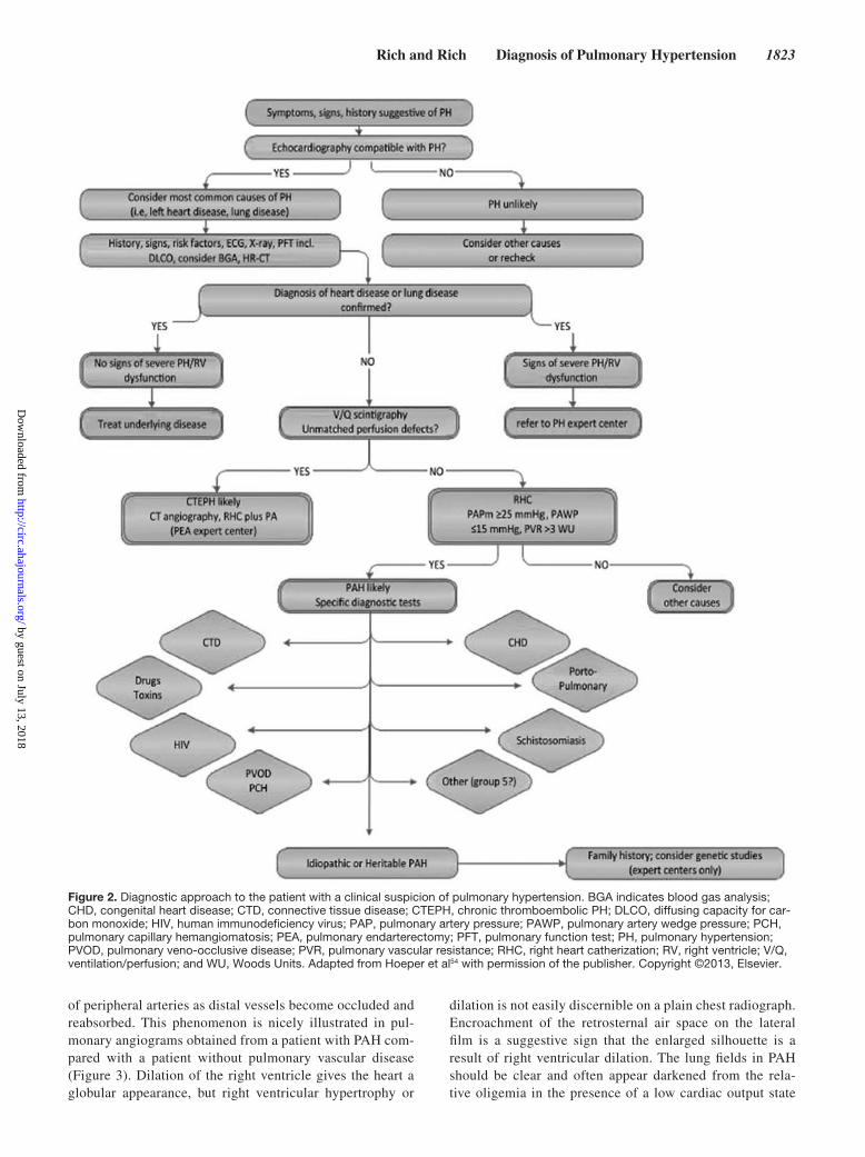
Even though the physical examination can provide a wealth of information about the presence and severity of PH, there is an increasing reliance on diagnostic tests to do the same. Although comprehensive reviews of the diagnostic testing evaluation have been published,17,18 some helpful clues and pearls from these tests are listed below. Figure 2 provides a generalized algorithmic approach to the diagnostic evaluation of the patient with possible PH.
Laboratory TestsIn IPAH, the complete blood count is normal. If chronic arte-rial oxygen desaturation exists, a reactive polycythemia should be present. Thus, patients with a normal pulse oximetry at rest and polycythemia should be retested with exercise. An elevated platelet count (or polycythemia from any cause) is a risk factor for PH, whereas a low platelet count may reflect an associated autoimmune disease, hypersplenism, or even occult cirrhosis with associated portopulmonary hypertension. A his-tory of arterial or venous thrombosis warrants thrombophilia screening including antiphospholipid antibodies, such as lupus anticoagulant and anticardiolipin antibodies. Measurement of brain natriuretic peptide (BNP) levels is reasonable as BNP levels are often elevated in patients with PH, loosely cor-relate with the severity of RVH, and are predictive of clini-cal outcomes.19,20 Specifically, BNP values <50 (NT-proBNP <300) are markers of enhanced survival, whereas BNP >180 (NTproBNP >1500) has been associated with worse survival.21 Uric acid levels are elevated in patients with PH and inversely correlate with pulmonary vascular resistance. Although the mechanism is uncertain, it may relate both to overproduction and impaired uric acid excretion caused by the low cardiac out-put state and tissue hypoxia.22 There is an increased incidence of thyroid disease in patients with PAH, which can be confused with symptoms of RV failure. Consequently, it is advised that thyroid function tests be monitored serially in all patients.23 Screening for antinuclear antibodies is important because all of the connective tissue diseases have been associated with the development of PAH. Although scleroderma is the leading cause of death in patients with PAH secondary to connective tissue diseases, PAH has also been described in patients with systemic lupus erythematosus, mixed connective tissue dis-ease, polymyositis, dermatomyositis, rheumatoid arthritis, and Sjogren’s syndrome. Because connective tissue diseases may have an insidious onset and a slowly progressive course, early recognition of the symptoms of PH may be difficult.24
Chest RadiographyA chest radiograph will typically show enlargement of the main pulmonary artery and its major branches, with pruning
by guest on July 13, 2018http://circ.ahajournals.org/
Dow
nloaded from
Rich and Rich Diagnosis of Pulmonary Hypertension 1823
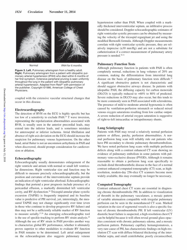
of peripheral arteries as distal vessels become occluded and reabsorbed. This phenomenon is nicely illustrated in pul-monary angiograms obtained from a patient with PAH com-pared with a patient without pulmonary vascular disease (Figure 3). Dilation of the right ventricle gives the heart a globular appearance, but right ventricular hypertrophy or
dilation is not easily discernible on a plain chest radiograph. Encroachment of the retrosternal air space on the lateral film is a suggestive sign that the enlarged silhouette is a result of right ventricular dilation. The lung fields in PAH should be clear and often appear darkened from the rela-tive oligemia in the presence of a low cardiac output state
Figure 2. Diagnostic approach to the patient with a clinical suspicion of pulmonary hypertension. BGA indicates blood gas analysis; CHD, congenital heart disease; CTD, connective tissue disease; CTEPH, chronic thromboembolic PH; DLCO, diffusing capacity for car-bon monoxide; HIV, human immunodeficiency virus; PAP, pulmonary artery pressure; PAWP, pulmonary artery wedge pressure; PCH, pulmonary capillary hemangiomatosis; PEA, pulmonary endarterectomy; PFT, pulmonary function test; PH, pulmonary hypertension; PVOD, pulmonary veno-occlusive disease; PVR, pulmonary vascular resistance; RHC, right heart catherization; RV, right ventricle; V/Q, ventilation/perfusion; and WU, Woods Units. Adapted from Hoeper et al54 with permission of the publisher. Copyright ©2013, Elsevier.
by guest on July 13, 2018http://circ.ahajournals.org/
Dow
nloaded from
1824 Circulation November 11, 2014
coupled with the extensive vascular structural changes that occur in this disease.
ElectrocardiographyThe detection of RVH on the ECG is highly specific but has too low of a sensitivity to exclude PAH.25 T wave inversion, representing the repolarization abnormalities associated with RVH, is usually seen in the anterior precordial leads, may extend into the inferior leads, and is sometimes mistaken for anteroseptal or inferior ischemia. Atrial fibrillation and absence of right axis deviation on the ECG should increase the suspicion for pulmonary venous hypertension. On the other hand, atrial flutter is not an uncommon arrhythmia in PAH and when discovered, should prompt consideration for cardiover-sion or ablation.
EchocardiographyEchocardiography usually demonstrates enlargement of the right ventricle and atrium with normal or small left ventricu-lar dimensions. Right ventricular size and function may be difficult to measure precisely echocardiographically, but the position and curvature of the interventricular septum provide an indication of right ventricular afterload. Echocardiographic findings that portend a poor prognosis include presence of a pericardial effusion, a markedly diminished left ventricular cavity, and RV dysfunction.26 Tricuspid annular plane systolic excursion (TAPSE) is frequently impaired and its baseline value is predictive of PH survival, yet, interestingly, the mea-sured TAPSE may not change significantly over time (even in those who continue to develop progressive RV failure) and thus may not be a sufficiently reliable marker of RV function to measure serially.27,28 An emerging echocardiographic tool is the use of speckle-tracking to perform RV strain analysis.29 Although the use of RV strain in PAH provides another way to characterize global RV performance, whether it ultimately proves superior to other modalities to evaluate RV function in PAH remains to be determined. Left atrial enlargement on the echocardiogram also suggests pulmonary venous
hypertension rather than PAH. When coupled with a mark-edly thickened interventricular septum, an infiltrative process should be considered. Doppler echocardiographic estimates of right ventricular systolic pressures can be obtained by measur-ing the velocity of the tricuspid regurgitant jet and using the modified Bernoulli formula. Although Doppler measurements correlate with right ventricular systolic pressure, they are rel-atively imprecise (±30 mm Hg) and are not a substitute for catheterization if a correct measurement of pulmonary artery pressure is needed.30,31
Pulmonary Function TestsAlthough pulmonary function in patients with PAH is often completely normal, reductions in lung volumes of 20% are common, making the differentiation from interstitial lung disease on the basis of pulmonary function tests difficult.32 A significant obstructive pattern is not characteristic and should suggest obstructive airways disease. In patients with idiopathic PAH, the diffusing capacity for carbon monoxide (DLCO) is typically reduced to ≈60% to 80% of predicted. Severe reductions in DLCO may also occur, but this tends to be more commonly seen in PAH associated with scleroderma. The presence of mild to moderate arterial hypoxemia is often caused by ventilation-perfusion mismatch or reduced mixed venous oxygen saturations resulting from low cardiac output. A severe reduction of arterial oxygen saturation is suggestive of right-to-left intracardiac or intrapulmonary shunts.
Lung ScintigraphyPatients with PAH may reveal a relatively normal perfusion pattern or diffuse, patchy, perfusion abnormalities. A nor-mal perfusion lung scan will reliably rule out patients who have PH secondary to chronic pulmonary thromboembolism. We have noted perfusion lung scans with multiple perfusion defects along with a contrast enhanced CT scan showing no evidence of pulmonary embolism in some patients with pul-monary veno-occlusive disease (PVOD). Although it remains reasonable to obtain a perfusion lung scan specifically to exclude distal thromboembolic disease in the face of a normal contrast enhanced computed tomorgraphy (CT) scan, as high-resolution, modern-day 256-slice CT scanners become more widely available, this may eventually no longer be necessary.
Computed TomographyContrast enhanced chest CT scans are essential in diagnos-ing chronic thromboembolic PH. In addition to visualization of thrombi in the pulmonary vasculature, a mosaic pattern of variable attenuation compatible with irregular pulmonary perfusion can be seen in the nonenhanced CT scan. Marked variation in the size of segmental vessels is also a specific fea-ture of chronic thromboembolic PH. If left ventricular (LV) diastolic heart failure is suspected, a high-resolution chest CT can be helpful because it will often reveal ground-glass opac-ities consistent with chronic pulmonary edema, and a mosaic perfusion pattern. Pulmonary capillary hemangiomatosis, a very rare cause of PH, has characteristic findings on high-res-olution CT scan with diffuse bilateral thickening of the inter-lobular septa, and small centrilobular, poorly circumscribed,
Normal IPAH for 6 months
Figure 3. Left, Pulmonary arteriogram from a healthy adult. Right, Pulmonary arteriogram from a patient with idiopathic pul-monary arterial hypertension (IPAH) who died within 6 months of her first symptom. Extensive peripheral pruning is seen uniformly throughout the lung in the patient with idiopathic pulmonary arterial hypertension. Adapted from Reid62 with permission of the publisher. Copyright ©1986, American College of Chest Physicians.
by guest on July 13, 2018http://circ.ahajournals.org/
Dow
nloaded from
Rich and Rich Diagnosis of Pulmonary Hypertension 1825
nodular opacities. Pulmonary capillary hemangiomatosis may occur in isolation or as an overlapping syndrome with PVOD.33 High-resolution CT is also helpful to diagnose inter-stitial lung disease. It has a high degree of specificity, but its relatively low sensitivity is often underappreciated. Patients with PAH without coexisting lung disease should have nor-mal lung parenchyma. Thus, although the CT scan tends to under-represent the extent of the disease, the presence of any interstitial abnormality should suggest that some degree of interstitial lung disease is underlying the PH. A high-resolu-tion CT scan of the chest is also a useful means of detecting emphysema and may occasionally demonstrate emphysema even in patients with little or no abnormality detected by pul-monary function tests.34 In such instances, one should suspect the possibility of mixed interstitial and obstructive lung dis-ease, particularly in the presence of a reduced DLCO.
Cardiac MRIAlthough echocardiography is the mainstay in the imaging of the right heart in clinical practice, advances in cardiac MRI (CMR) technology have led to the development of more pre-cise techniques for the assessment of hemodynamics in the pulmonary circulation and identification of right ventricular morphological changes. CMR is now regarded as the refer-ence standard in the assessment of RV structure and function via the measurement of RV volumes and ejection fraction, which makes CMR an attractive modality for serial follow-up in PAH management to determine treatment response.35 In addition, CMR offers the possibility to quantify regurgitant volumes, use delayed gadolinium enhancement as a marker for focal scar burden, and to assess myocardial strain, coro-nary perfusion, and pulmonary pulsatility.36 One recent study was able to predict survival in patients with PAH based on the RV ejection fraction computed from CMR.37 A more recent multicenter study demonstrated the feasibility of using CMR to track changes in RV size and function in response to 12 months of PAH-specific therapy. Taken together, these
findings hold promise for the increased use of CMR protocols in both clinical trials and practice.38
Positron Emission Tomography ScanningAlthough not an accepted standard test to diagnose PH, non-invasive molecular imaging using positron emission tomog-raphy (PET) offers the potential for monitoring cellular and biochemical events in otherwise inaccessible tissue. Lung parenchymal glucose uptake, measured by 18FDG-PET, has been reported to be increased in IPAH patients compared with healthy controls.39 The observation of inter-/intravariability in lung 18FDG measurements in IPAH patients aligns with our current understanding of the disease, both in terms of the mor-phological heterogeneity within individual lungs and between lungs from IPAH patients. 18FDG-PET merits further evalua-tion as a clinical tool to deep phenotype patients with IPAH in studies exploring efficacy and dose-response relationships of new PAH targeted treatments. Additionally, the assessment of RV uptake of FDG and changes in FDG uptake with disease progression or response to therapy may develop as a clinically useful tool to track RV metabolism.40,41
Exercise TestingThe use of a symptom-limited exercise test should be part of the evaluation of patients with PH. The 6-minute walk test is commonly used in clinical trials as an end point for effi-cacy of therapy in patients with PH. It has been correlated with workload, heart rate, oxygen saturation, and dyspnea response. However, it has many drawbacks that include the fact that anthropometric factors such as gait speed, age, weight, muscle mass, and length of stride can affect the test results. In addition, any encouragement from the tester can cause large variations in the result.42 Treadmill testing using the Naughton-Balke protocol avoids many of these limitations and provides an estimate of work because the program cre-ates incremental increases in work of 1 metabolic equivalent at 2-minute stages.43 Cardiopulmonary exercise testing using
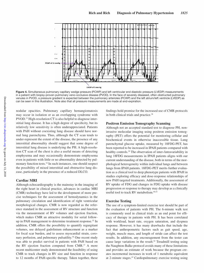
Figure 4. Simultaneous pulmonary capillary wedge pressure (PCWP) and left ventricular end diastolic pressure (LVEDP) measurements in a patient with biopsy-proven pulmonary veno-occlusive disease (PVOD). In the face of severely diseased, often obstructed pulmonary venules in PVOD, a pressure gradient is expected between the pulmonary arterioles (PCWP) and the left atrium/left ventricle (LVEDP) as can be seen in this illustration. Note also that all pressure measurements are made at end-expiration.
by guest on July 13, 2018http://circ.ahajournals.org/
Dow
nloaded from
1826 Circulation November 11, 2014
either a treadmill or an upright bicycle with measurements of gas exchange has the potential to grade the severity of exercise limitation in patients with PH noninvasively and can be per-formed serially to follow changes in functional capacity that often accompany disease progression.44
Hemodynamic Assessment of PHCardiac CatheterizationIn addition to confirming the diagnosis of PAH and excluding other PH causes, cardiac catheterization also establishes the severity of disease and allows an assessment of prognosis.7,45 By definition, patients with PAH should have a low or nor-mal pulmonary capillary wedge pressure (PCWP). Because this is a critical measurement in distinguishing a patient with PAH from one with pulmonary venous hypertension, qual-ity measures must be established in the cardiac catheteriza-tion laboratory to ensure that correct values are obtained. Pressures should not be determined by the electronically integrated mean pressure from the laboratory’s computer displayed on the catheterization laboratory monitor, because these measurements ignore respiratory influences and may lead to erroneous measurements.46 Instead, measurements of all pressures should be properly made at end-expiration to avoid incorporating negative intrathoracic pressures. Direct measurement of LV end-diastolic pressure is advised when a reproducible wedge pressure cannot be obtained.47 In cer-tain cases, administering a volume load of saline may reveal abnormal LV filling pressures that had previously normalized in patients with mild states of dehydration and unmask the presence of pulmonary venous hypertension.48 Interestingly, the PCWP in patients with PVOD is often found to be normal. This is likely attributable to the heterogeneity among cases of PVOD, differences in procedural practices, and a lack of consideration of PVOD in the differential diagnosis. In cases in which there is a suspicion of PVOD, we advocate simul-taneous PCWP and LV end-diastolic pressure measurements with the PCWP measurements obtained from multiple lung segments. Unfortunately, this is not frequently done in many clinical practices, which underscores the benefit of referral of these complex cases to highly specialized, experienced cen-ters. If PVOD is present, one may find an elevated PCWP to LV end-diastolic pressure ratio in some segments, with nearly equal values in others49 (Figure 4).
The diagnosis of PH relies on establishing an elevation in pulmonary artery pressure above normal. The upper limit of normal for pulmonary artery mean pressure at rest is 19 mm Hg. However, this assumes that there are no abnormalities in downstream pressures of the left atrium or left ventricle, or an increased cardiac output. That is why a patient can have PH from the standpoint of an elevated pulmonary artery pressure, but normal pulmonary vascular resistance. Parameters for normal pulmonary arterial systolic pressure derived by echo-cardiographic Doppler studies suggest that the upper limit of normal of pulmonary arterial systolic pressure in the general population may be higher than previously appreciated.50
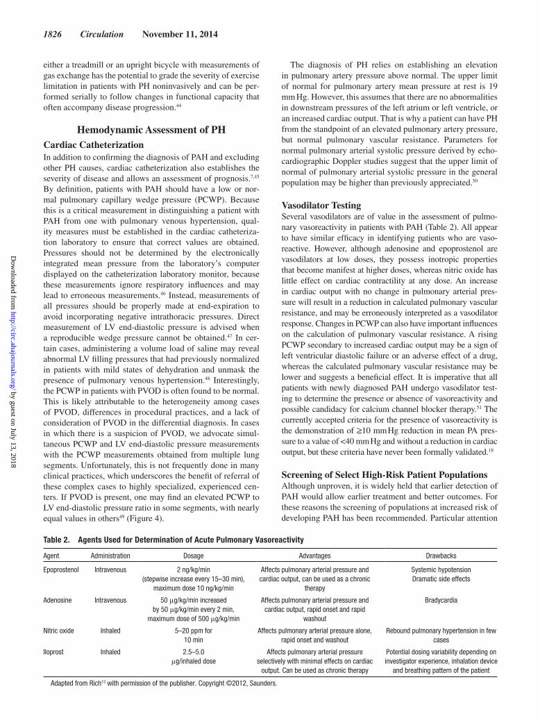
Vasodilator TestingSeveral vasodilators are of value in the assessment of pulmo-nary vasoreactivity in patients with PAH (Table 2). All appear to have similar efficacy in identifying patients who are vaso-reactive. However, although adenosine and epoprostenol are vasodilators at low doses, they possess inotropic properties that become manifest at higher doses, whereas nitric oxide has little effect on cardiac contractility at any dose. An increase in cardiac output with no change in pulmonary arterial pres-sure will result in a reduction in calculated pulmonary vascular resistance, and may be erroneously interpreted as a vasodilator response. Changes in PCWP can also have important influences on the calculation of pulmonary vascular resistance. A rising PCWP secondary to increased cardiac output may be a sign of left ventricular diastolic failure or an adverse effect of a drug, whereas the calculated pulmonary vascular resistance may be lower and suggests a beneficial effect. It is imperative that all patients with newly diagnosed PAH undergo vasodilator test-ing to determine the presence or absence of vasoreactivity and possible candidacy for calcium channel blocker therapy.51 The currently accepted criteria for the presence of vasoreactivity is the demonstration of ≥10 mm Hg reduction in mean PA pres-sure to a value of <40 mm Hg and without a reduction in cardiac output, but these criteria have never been formally validated.18
Screening of Select High-Risk Patient PopulationsAlthough unproven, it is widely held that earlier detection of PAH would allow earlier treatment and better outcomes. For these reasons the screening of populations at increased risk of developing PAH has been recommended. Particular attention
Table 2. Agents Used for Determination of Acute Pulmonary Vasoreactivity
Agent Administration Dosage Advantages Drawbacks
Epoprostenol Intravenous 2 ng/kg/min(stepwise increase every 15–30 min),
maximum dose 10 ng/kg/min
Affects pulmonary arterial pressure and cardiac output, can be used as a chronic
therapy
Systemic hypotensionDramatic side effects
Adenosine Intravenous 50 μg/kg/min increasedby 50 μg/kg/min every 2 min,
maximum dose of 500 μg/kg/min
Affects pulmonary arterial pressure and cardiac output, rapid onset and rapid
washout
Bradycardia
Nitric oxide Inhaled 5–20 ppm for10 min
Affects pulmonary arterial pressure alone, rapid onset and washout
Rebound pulmonary hypertension in few cases
Iloprost Inhaled 2.5–5.0μg/inhaled dose
Affects pulmonary arterial pressure selectively with minimal effects on cardiac
output. Can be used as chronic therapy
Potential dosing variability depending on investigator experience, inhalation device
and breathing pattern of the patient
Adapted from Rich13 with permission of the publisher. Copyright ©2012, Saunders.
by guest on July 13, 2018http://circ.ahajournals.org/
Dow
nloaded from
Rich and Rich Diagnosis of Pulmonary Hypertension 1827
has been given to patients with the scleroderma spectrum of diseases, advanced liver disease, and a family history of heri-table PAH.
Because of the high prevalence of PH in patients with scleroderma spectrum of diseases, which is a leading cause of death in these patients, recent guidelines have recommended regular screening by echocardiography. In an attempt to estab-lish the best way to screen these patients, the Evidence-based detection of pulmonary arterial hypertension in systemic scle-rosis (DETECT) study developed the first evidence-based detection algorithm for PAH in the scleroderma patients with-out clinical signs and symptoms.52 A 2-step, internally vali-dated detection algorithm for clinical practice was created. The first step included the clinical assessment for the presence of telangiectasia, anticentromere antibodies, pulmonary func-tion test, and DLCO measurements, an ECG, and NT-proBNP and uric acid levels. For those with a high composite score, the second step included echocardiography and consideration of right heart catherization in patients with abnormal findings. This resulted in a 97% sensitivity and 35% specificity for the diagnosis of PAH.
The incidence of portopulmonary hypertension in patients with advanced liver disease has been reported to be between 2% to 9%. Although low, the coexistence of PAH in these
patients will dictate their candidacy for liver transplantation. Although not prospectively validated, current portopulmo-nary hypertension screening recommendations endorsed by the American Association for the Study of Liver Disease are that every patient considered for liver transplant have a screening transthoracic echocardiogram to assess for PH and, if suggestive, a confirmatory right heart catheterization.53
The genetic testing of relatives of patients with heritable PAH is controversial.54 Genetic testing will identify presymp-tomatic carriers of PAH-causing mutations who are at high risk of developing PAH. However, because of incomplete penetrance of mutations in PAH-predisposing genes, it is cur-rently not possible to identify which carriers of a mutation will develop PAH.55 In addition, there are no proven effective inter-ventions or medications available to prevent the onset of PAH in mutation carriers. Thus, although genetic testing in relatives will identify mutation noncarriers who have no increased risk of the heritable disease, it will also identify mutation carriers who will have to live wondering whether or when they will develop the disease. Given that the most common presenting symptom of PAH is dyspnea with exertional activity, which is also a feature of normalcy, we agree with the argument that has been made by others, that the relatives are likely better off not knowing their genetic status than living a lifetime of worry.
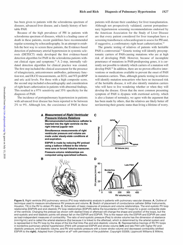
Figure 5. Right ventricle (RV) pulmonary venous (PV) loop relationship analysis in patients with pulmonary vascular disease. A, Outline of technique used to measure simultaneous RV pressure and volume. B, Sketch of placement of conductance catheter (Millar Instruments, Houston, TX) in the RV to obtain PV data. C, Schematic of basic measures of pressure and volume relationships. The end-systolic PV loop relationship (ESPVR) and end-diastolic PV loop relationships (EDPVR) define the boundaries of the PV loops for a given contractile state of the ventricle. Changing the preload (as shown with dashed lines) or afterload will change the shape and position of the loops, but the end-systolic and end-diastolic points will always fall on the ESPVR and EDPVR. This is the reason why the ESPVR and EDPVR are used as load-independent measures of contractility. The ratio of end-systolic pressure (Pes) to stroke volume has the dimension of elastance (mm Hg/mL) and is called the arterial elastance (Ea) because it is linked to the afterload, which is determined by the arterial system and is represented in the PV loop by the slope of the line that links Pes and end-diastolic volume. D, Representative tracings of RV PV loops in borderline pulmonary arterial hypertension (PAH) with preserved ESPVR and stroke volume (left) and late PAH with higher RV end-diastolic pressure, end-diastolic volume, and RV end-systolic pressure with a lower stroke volume and decreased contractility (shifted ESPVR to the right). Adapted from Champion et al56 with permission of the publisher. Copyright ©2009, Lippincott Williams & Wilkins.
by guest on July 13, 2018http://circ.ahajournals.org/
Dow
nloaded from
1828 Circulation November 11, 2014
Ventriculoarterial Coupling and the Cardiopulmonary UnitThe clinical assessment of PH requires an evaluation of the status of both the pulmonary vasculature and the right ven-tricle, and such characterizations should ideally be made and considered as an integrated cardiopulmonary unit (see reviews in references 56 and 57 for an in depth discussion of this topic). Indeed, the RV and PA are inextricably linked or coupled both in health and in disease. In normal people, the RV pumps into a high capacitance pulmonary arterial circuit. Under these circumstances, the oscillatory components of arterial load are generally minor and therefore expression of RV after-load purely as a mean pulmonary vascular resistance is usu-ally adequate. In the setting of pulmonary vascular diseases such as PAH, however, the RV is confronted with a diseased pulmonary vascular bed that imposes a severely heightened afterload, including both resistive and pulsatile components, which ultimately determine the work of the RV. Recent studies have found that the steady or resistive component accounts for approximately 77% of total hydraulic RV power and that the remaining 23% is used for the pulsatile compo-nent.57 Moreover, studies have shown that elevated PCWP can decrease the resistance–compliance time constant in the pul-monary system, thus enhancing net RV afterload by elevating pulsatile relative to resistive load.58 The ability of the RV to adapt over time to such changes in loading conditions essen-tially determines prognosis in PAH. The use of high-fidelity micromanometer catheters to generate pressure-volume loops allows for the measurements of RV elastance (Ees; an index of contractility) and arterial elastance (Ea; an index of afterload), their coupling ratios (Ees/Ea), and changes in coupling with disease progression or in response to treatment (Figure 5). The optimal coupling of the RV and its afterload, where RV mechanical and energetic efficiency is close to maximal, is when the Ees/Ea is ≈1.5 to 2.0. Thus, isolated increases is Ea (increased vascular resistance), or decreases in Ees (decreased RV contractility), decreases the Ees/Ea ratio and thus lowers ventricular–vascular coupling efficiency.59 Unfortunately, it remains somewhat tedious to routinely apply many of these techniques in the clinical setting. To overcome some of the inherent complexities of generating pressure–volume loops to determine Ees and Ea, respectively, some investigators have validated and now use the single-beat method, both invasively and noninvasively using CMR.60,61 The future incorporation of these and other more sophisticated measurements of ventricu-lovascular coupling into translational research, clinical trials, and ultimately clinical practice appears promising.
ConclusionsPAH remains a challenging disease. Because it is uncommon, it is difficult for physicians to develop sufficient experience and expertise unless they are part of a specialty PH Center. The cause can be multifactorial, making it difficult to know the dominant cause in some patients and thus requires a metic-ulous approach to arrive at the correct diagnosis. Also, the RV has traditionally been difficult to study because of its unique geometry and the lack of simple, reliable, and quantifiable noninvasive assessments. The emergence of CMR and other
innovative modalities such as PET and measurements of ven-triculoarterial coupling are encouraging developments to more comprehensively assess and follow RV function. However, cost limitations and the lack of routine availability of these tests outside of specialized, tertiary care centers may con-tinue to limit their routine implementation. For these reasons we believe that a careful and detailed physical examination will continue to provide the clinician with useful information about the severity and response to therapy in most patients.
DisclosuresDr Jonathan D. Rich reports receiving speaker fees from Bayer. Dr Stuart Rich has no disclosures.
References 1. McGoon MD, Benza RL, Escribano-Subias P, Jiang X, Miller DP, Peacock
AJ, Pepke-Zaba J, Pulido T, Rich S, Rosenkranz S, Suissa S, Humbert M. Pulmonary arterial hypertension: epidemiology and registries. J Am Coll Cardiol. 2013;62(25 Suppl):D51–D59.
2. Lam CS, Roger VL, Rodeheffer RJ, Borlaug BA, Enders FT, Redfield MM. Pulmonary hypertension in heart failure with preserved ejection fraction: a community-based study. J Am Coll Cardiol. 2009;53:1119–1126.
3. Ling Y, Johnson MK, Kiely DG, Condliffe R, Elliot CA, Gibbs JS, Howard LS, Pepke-Zaba J, Sheares KK, Corris PA, Fisher AJ, Lordan JL, Gaine S, Coghlan JG, Wort SJ, Gatzoulis MA, Peacock AJ. Changing demograph-ics, epidemiology, and survival of incident pulmonary arterial hyperten-sion: results from the pulmonary hypertension registry of the United Kingdom and Ireland. Am J Respir Crit Care Med. 2012;186:790–796.
4. Humbert M, Sitbon O, Chaouat A, Bertocchi M, Habib G, Gressin V, Yaïci A, Weitzenblum E, Cordier JF, Chabot F, Dromer C, Pison C, Reynaud-Gaubert M, Haloun A, Laurent M, Hachulla E, Cottin V, Degano B, Jaïs X, Montani D, Souza R, Simonneau G. Survival in patients with idiopathic, familial, and anorexigen-associated pulmonary arterial hypertension in the modern management era. Circulation. 2010;122:156–163.
5. Rich S, Dantzker DR, Ayres SM, Bergofsky EH, Brundage BH, Detre KM, Fishman AP, Goldring RM, Groves BM, Koerner SK. Primary pulmonary hypertension. A national prospective study. Ann Intern Med. 1987;107:216–223.
6. Strange G, Gabbay E, Kermeen F, Williams T, Carrington M, Stewart S, Keogh A. Time from symptoms to definitive diagnosis of idiopathic pul-monary arterial hypertension: The delay study. Pulm Circ. 2013;3:89–94.
7. D’Alonzo GE, Barst RJ, Ayres SM, Bergofsky EH, Brundage BH, Detre KM, Fishman AP, Goldring RM, Groves BM, Kernis JT. Survival in patients with primary pulmonary hypertension. Results from a national prospective registry. Ann Intern Med. 1991;115:343–349.
8. Badesch DB, Raskob GE, Elliott CG, Krichman AM, Farber HW, Frost AE, Barst RJ, Benza RL, Liou TG, Turner M, Giles S, Feldkircher K, Miller DP, McGoon MD. Pulmonary arterial hypertension: baseline char-acteristics from the REVEAL Registry. Chest. 2010;137:376–387.
9. Benza RL, Miller DP, Barst RJ, Badesch DB, Frost AE, McGoon MD. An evaluation of long-term survival from time of diagnosis in pulmonary arte-rial hypertension from the REVEAL Registry. Chest. 2012;142:448–456.
10. Primary pulmonary hypertension executive summary from the world sym-posium. 1998.
11. Simonneau G, Galiè N, Rubin LJ, Langleben D, Seeger W, Domenighetti G, Gibbs S, Lebrec D, Speich R, Beghetti M, Rich S, Fishman A. Clinical classification of pulmonary hypertension. J Am Coll Cardiol. 2004;43(12 Suppl S):5S–12S.
12. Simonneau G, Robbins IM, Beghetti M, Channick RN, Delcroix M, Denton CP, Elliott CG, Gaine SP, Gladwin MT, Jing ZC, Krowka MJ, Langleben D, Nakanishi N, Souza R. Updated clinical classification of pulmonary hypertension. J Am Coll Cardiol. 2009;54(1 Suppl):S43–S54.
13. Simonneau G, Gatzoulis MA, Adatia I, Celermajer D, Denton C, Ghofrani A, Gomez Sanchez MA, Krishna Kumar R, Landzberg M, Machado RF, Olschewski H, Robbins IM, Souza R. Updated clinical classification of pul-monary hypertension. J Am Coll Cardiol. 2013;62(25 Suppl):D34–D41.
14. Rich S. What is pulmonary arterial hypertension? Pulm Circ. 2012;2:271–272.
15. Rich S. Pulmonary hypertension. In: Bonow R, Mann D, Zipes D, Libby P, eds. Braunwald’s heart disease: A textbook of cardiovascular medicine. 2012:1696–1718.
by guest on July 13, 2018http://circ.ahajournals.org/
Dow
nloaded from
Rich and Rich Diagnosis of Pulmonary Hypertension 1829
16. van Wolferen SA, Marcus JT, Westerhof N, Spreeuwenberg MD, Marques KM, Bronzwaer JG, Henkens IR, Gan CT, Boonstra A, Postmus PE, Vonk-Noordegraaf A. Right coronary artery flow impairment in patients with pulmonary hypertension. Eur Heart J. 2008;29:120–127.
17. Galiè N, Hoeper MM, Humbert M, Torbicki A, Vachiery JL, Barbera JA, Beghetti M, Corris P, Gaine S, Gibbs JS, Gomez-Sanchez MA, Jondeau G, Klepetko W, Opitz C, Peacock A, Rubin L, Zellweger M, Simonneau G; ESC Committee for Practice Guidelines (CPG). Guidelines for the diagnosis and treatment of pulmonary hypertension: the Task Force for the Diagnosis and Treatment of Pulmonary Hypertension of the European Society of Cardiology (ESC) and the European Respiratory Society (ERS), endorsed by the International Society of Heart and Lung Transplantation (ISHLT). Eur Heart J. 2009;30:2493–2537.
18. McLaughlin VV, Archer SL, Badesch DB, Barst RJ, Farber HW, Lindner JR, Mathier MA, McGoon MD, Park MH, Rosenson RS, Rubin LJ, Tapson VF, Varga J; American College of Cardiology Foundation Task Force on Expert Consensus Documents; American Heart Association; American College of Chest Physicians; American Thoracic Society, Inc; Pulmonary Hypertension Association. ACCF/AHA 2009 expert consensus document on pulmonary hypertension a report of the American College of Cardiology Foundation Task Force on Expert Consensus Documents and the American Heart Association developed in collaboration with the American College of Chest Physicians; American Thoracic Society, Inc.; and the Pulmonary Hypertension Association. J Am Coll Cardiol. 2009;53:1573–1619.
19. Rich JD, Thenappan T, Freed B, Patel AR, Thisted RA, Childers R, Archer SL. QTc prolongation is associated with impaired right ventricular func-tion and predicts mortality in pulmonary hypertension. Int J Cardiol. 2013;167:669–676.
20. Souza R, Bogossian HB, Humbert M, Jardim C, Rabelo R, Amato MB, Carvalho CR. N-terminal-pro-brain natriuretic peptide as a haemody-namic marker in idiopathic pulmonary arterial hypertension. Eur Respir J. 2005;25:509–513.
21. Benza RL, Miller DP, Gomberg-Maitland M, Frantz RP, Foreman AJ, Coffey CS, Frost A, Barst RJ, Badesch DB, Elliott CG, Liou TG, McGoon MD. Predicting survival in pulmonary arterial hypertension: insights from the Registry to Evaluate Early and Long-Term Pulmonary Arterial Hypertension Disease Management (REVEAL). Circulation. 2010;122:164–172.
22. Voelkel MA, Wynne KM, Badesch DB, Groves BM, Voelkel NF. Hyperuricemia in severe pulmonary hypertension. Chest. 2000;117:19–24.
23. Li JH, Safford RE, Aduen JF, Heckman MG, Crook JE, Burger CD. Pulmonary hypertension and thyroid disease. Chest. 2007;132:793–797.
24. Mathai SC, Hassoun PM. Pulmonary arterial hypertension in connective tissue diseases. Heart Fail Clin. 2012;8:413–425.
25. Ahearn GS, Tapson VF, Rebeiz A, Greenfield JC Jr. Electrocardiography to define clinical status in primary pulmonary hypertension and pulmo-nary arterial hypertension secondary to collagen vascular disease. Chest. 2002;122:524–527.
26. Raymond RJ, Hinderliter AL, Willis PW, Ralph D, Caldwell EJ, Williams W, Ettinger NA, Hill NS, Summer WR, de Boisblanc B, Schwartz T, Koch G, Clayton LM, Jöbsis MM, Crow JW, Long W. Echocardiographic pre-dictors of adverse outcomes in primary pulmonary hypertension. J Am Coll Cardiol. 2002;39:1214–1219.
27. Forfia PR, Fisher MR, Mathai SC, Housten-Harris T, Hemnes AR, Borlaug BA, Chamera E, Corretti MC, Champion HC, Abraham TP, Girgis RE, Hassoun PM. Tricuspid annular displacement predicts survival in pulmo-nary hypertension. Am J Respir Crit Care Med. 2006;174:1034–1041.
28. Mauritz GJ, Kind T, Marcus JT, Bogaard HJ, van de Veerdonk M, Postmus PE, Boonstra A, Westerhof N, Vonk-Noordegraaf A. Progressive changes in right ventricular geometric shortening and long-term survival in pulmo-nary arterial hypertension. Chest. 2012;141:935–943.
29. Fine NM, Chen L, Bastiansen PM, Frantz RP, Pellikka PA, Oh JK, Kane GC. Outcome prediction by quantitative right ventricular function assessment in 575 subjects evaluated for pulmonary hypertension. Circ Cardiovasc Imaging. 2013;6:711–721.
30. Fisher MR, Forfia PR, Chamera E, Housten-Harris T, Champion HC, Girgis RE, Corretti MC, Hassoun PM. Accuracy of Doppler echocardiog-raphy in the hemodynamic assessment of pulmonary hypertension. Am J Respir Crit Care Med. 2009;179:615–621.
31. Rich JD, Shah SJ, Swamy RS, Kamp A, Rich S. Inaccuracy of Doppler echocardiographic estimates of pulmonary artery pressures in patients with pulmonary hypertension: implications for clinical practice. Chest. 2011;139:988–993.
32. McGoon M, Gutterman D, Steen V, Barst R, McCrory DC, Fortin TA, Loyd JE; American College of Chest Physicians. Screening, early detec-tion, and diagnosis of pulmonary arterial hypertension: ACCP evidence-based clinical practice guidelines. Chest. 2004;126(1 Suppl):14S–34S.
33. Frazier AA, Franks TJ, Mohammed TL, Ozbudak IH, Galvin JR. From the Archives of the AFIP: pulmonary veno-occlusive disease and pulmonary capillary hemangiomatosis. Radiographics. 2007;27:867–882.
34. Omori H, Fujimoto K, Katoh T. Computed-tomography findings of emphysema: correlation with spirometric values. Curr Opin Pulm Med. 2008;14:110–114.
35. Benza R, Biederman R, Murali S, Gupta H. Role of cardiac magnetic resonance imaging in the management of patients with pulmonary arterial hypertension. J Am Coll Cardiol. 2008;52:1683–1692.
36. Peacock AJ, Vonk Noordegraaf A. Cardiac magnetic resonance imaging in pulmonary arterial hypertension. Eur Respir Rev. 2013;22:526–534.
37. Freed BH, Gomberg-Maitland M, Chandra S, Mor-Avi V, Rich S, Archer SL, Jamison EB Jr, Lang RM, Patel AR. Late gadolinium enhance-ment cardiovascular magnetic resonance predicts clinical worsening in patients with pulmonary hypertension. J Cardiovasc Magn Reson. 2012;14:11.
38. Peacock AJ, Crawley S, McLure L, Blyth K, Vizza CD, Poscia R, Francone M, Iacucci I, Olschewski H, Kovacs G, Vonk Noordegraaf A, Marcus JT, van de Veerdonk MC, Oosterveer FP. Changes in right ventricular function measured by cardiac magnetic resonance imaging in patients receiving pulmonary arterial hypertension-targeted therapy: the EURO-MR study. Circ Cardiovasc Imaging. 2014;7:107–114.
39. Zhao L, Ashek A, Wang L, Fang W, Dabral S, Dubois O, Cupitt J, Pullamsetti SS, Cotroneo E, Jones H, Tomasi G, Nguyen QD, Aboagye EO, El-Bahrawy MA, Barnes G, Howard LS, Gibbs JS, Gsell W, He JG, Wilkins MR. Heterogeneity in lung (18)FDG uptake in pulmonary arterial hypertension: potential of dynamic (18)FDG positron emission tomogra-phy with kinetic analysis as a bridging biomarker for pulmonary vascular remodeling targeted treatments. Circulation. 2013;128:1214–1224.
40. Oikawa M, Kagaya Y, Otani H, Sakuma M, Demachi J, Suzuki J, Takahashi T, Nawata J, Ido T, Watanabe J, Shirato K. Increased [18F]fluorodeoxyglucose accumulation in right ventricular free wall in patients with pulmonary hypertension and the effect of epoprostenol. J Am Coll Cardiol. 2005;45:1849–1855.
41. Piao L, Fang YH, Cadete VJ, Wietholt C, Urboniene D, Toth PT, Marsboom G, Zhang HJ, Haber I, Rehman J, Lopaschuk GD, Archer SL. The inhibition of pyruvate dehydrogenase kinase improves impaired car-diac function and electrical remodeling in two models of right ventricu-lar hypertrophy: resuscitating the hibernating right ventricle. J Mol Med (Berl). 2010;88:47–60.
42. Salzman SH. The 6-min walk test: clinical and research role, technique, coding, and reimbursement. Chest. 2009;135:1345–1352.
43. Gomberg-Maitland M, Huo D, Benza RL, McLaughlin VV, Tapson VF, Barst RJ. Creation of a model comparing 6-minute walk test to metabolic equivalent in evaluating treatment effects in pulmonary arterial hyperten-sion. J Heart Lung Transplant. 2007;26:732–738.
44. Sun XG, Hansen JE, Oudiz RJ, Wasserman K. Exercise pathophysi-ology in patients with primary pulmonary hypertension. Circulation. 2001;104:429–435.
45. Sandoval J, Bauerle O, Palomar A, Gómez A, Martínez-Guerra ML, Beltrán M, Guerrero ML. Survival in primary pulmonary hypertension. Validation of a prognostic equation. Circulation. 1994;89:1733–1744.
46. Ryan JJ, Rich JD, Thiruvoipati T, Swamy R, Kim GH, Rich S. Current practice for determining pulmonary capillary wedge pressure predisposes to serious errors in the classification of patients with pulmonary hyperten-sion. Am Heart J. 2012;163:589–594.
47. Halpern SD, Taichman DB. Misclassification of pulmonary hypertension due to reliance on pulmonary capillary wedge pressure rather than left ventricular end-diastolic pressure. Chest. 2009;136:37–43.
48. Robbins IM, Hemnes AR, Pugh ME, Brittain EL, Zhao DX, Piana RN, Fong PP, Newman JH. High prevalence of occult pulmonary venous hypertension revealed by fluid challenge in pulmonary hypertension. Circ Heart Fail. 2014;7:116–122.
49. Case records of the Massachusetts General Hospital. Weekly clinico-pathological exercises. Case 21–1986. Recent development of pulmonary hypertension seven years after an aortic valve replacement. N Engl J Med. 1986;314:1435–1445
50. McQuillan BM, Picard MH, Leavitt M, Weyman AE. Clinical cor-relates and reference intervals for pulmonary artery systolic pres-sure among echocardiographically normal subjects. Circulation. 2001;104:2797–2802.
by guest on July 13, 2018http://circ.ahajournals.org/
Dow
nloaded from
1830 Circulation November 11, 2014
51. Rich S, Kaufmann E, Levy PS. The effect of high doses of calcium-chan-nel blockers on survival in primary pulmonary hypertension. N Engl J Med. 1992;327:76–81.
52. Coghlan JG, Denton CP, Grünig E, Bonderman D, Distler O, Khanna D, Müller-Ladner U, Pope JE, Vonk MC, Doelberg M, Chadha-Boreham H, Heinzl H, Rosenberg DM, McLaughlin VV, Seibold JR; DETECT study group. Evidence-based detection of pulmonary arterial hyper-tension in systemic sclerosis: the DETECT study. Ann Rheum Dis. 2014;73:1340–1349.
53. Krowka MJ. Portopulmonary hypertension. Semin Respir Crit Care Med. 2012;33:17–25.
54. Hoeper MM, Bogaard HJ, Condliffe R, Frantz R, Khanna D, Kurzyna M, Langleben D, Manes A, Satoh T, Torres F, Wilkins MR, Badesch DB. Definitions and diagnosis of pulmonary hypertension. J Am Coll Cardiol. 2013;62(25 Suppl):D42–D50.
55. Soubrier F, Chung WK, Machado R, Grünig E, Aldred M, Geraci M, Loyd JE, Elliott CG, Trembath RC, Newman JH, Humbert M. Genetics and genomics of pulmonary arterial hypertension. J Am Coll Cardiol. 2013;62(25 Suppl):D13–D21.
56. Champion HC, Michelakis ED, Hassoun PM. Comprehensive invasive and noninvasive approach to the right ventricle-pulmonary circulation unit: state of the art and clinical and research implications. Circulation. 2009;120:992–1007.
57. Vonk-Noordegraaf A, Haddad F, Chin KM, Forfia PR, Kawut SM, Lumens J, Naeije R, Newman J, Oudiz RJ, Provencher S, Torbicki A, Voelkel NF, Hassoun PM. Right heart adaptation to pulmonary arterial hypertension: phys-iology and pathobiology. J Am Coll Cardiol. 2013;62(25 Suppl):D22–D33.
58. Tedford RJ, Hassoun PM, Mathai SC, Girgis RE, Russell SD, Thiemann DR, Cingolani OH, Mudd JO, Borlaug BA, Redfield MM, Lederer DJ, Kass DA. Pulmonary capillary wedge pressure augments right ventricular pulsatile loading. Circulation. 2012;125:289–297.
59. Naeije R, Chesler N. Pulmonary circulation at exercise. Compr Physiol. 2012;2:711–741.
60. Brimioulle S, Wauthy P, Ewalenko P, Rondelet B, Vermeulen F, Kerbaul F, Naeije R. Single-beat estimation of right ventricular end-systolic pressure-volume relationship. Am J Physiol Heart Circ Physiol. 2003;284:H1625–H1630.
61. Klotz S, Hay I, Dickstein ML, Yi GH, Wang J, Maurer MS, Kass DA, Burkhoff D. Single-beat estimation of end-diastolic pressure-volume rela-tionship: a novel method with potential for noninvasive application. Am J Physiol Heart Circ Physiol. 2006;291:H403–H412.
62. Reid LM. Structure and function in pulmonary hypertension: new percep-tions. Chest. 1986;90:279–288.
KEY WORDS: diagnosis ◼ pulmonary hypertension ◼ right ventricle ◼ vasculature
by guest on July 13, 2018http://circ.ahajournals.org/
Dow
nloaded from
Jonathan D. Rich and Stuart RichClinical Diagnosis of Pulmonary Hypertension
Print ISSN: 0009-7322. Online ISSN: 1524-4539 Copyright © 2014 American Heart Association, Inc. All rights reserved.
is published by the American Heart Association, 7272 Greenville Avenue, Dallas, TX 75231Circulation doi: 10.1161/CIRCULATIONAHA.114.006971
2014;130:1820-1830Circulation.
http://circ.ahajournals.org/content/130/20/1820World Wide Web at:
The online version of this article, along with updated information and services, is located on the
http://circ.ahajournals.org//subscriptions/
is online at: Circulation Information about subscribing to Subscriptions:
http://www.lww.com/reprints Information about reprints can be found online at: Reprints:
document. Permissions and Rights Question and Answer this process is available in the
click Request Permissions in the middle column of the Web page under Services. Further information aboutOffice. Once the online version of the published article for which permission is being requested is located,
can be obtained via RightsLink, a service of the Copyright Clearance Center, not the EditorialCirculationin Requests for permissions to reproduce figures, tables, or portions of articles originally publishedPermissions:
by guest on July 13, 2018http://circ.ahajournals.org/
Dow
nloaded from