the g-protein coupled receptor cmklr1/chemr23: studies on...
TRANSCRIPT

LUND UNIVERSITY
PO Box 117221 00 Lund+46 46-222 00 00
The G-protein coupled receptor CMKLR1/ChemR23: Studies on gene regulation,receptor ligand activation, and HIV/SIV co-receptor function
Mårtensson, Ulrika
Published: 2005-01-01
Link to publication
Citation for published version (APA):Mårtensson, U. (2005). The G-protein coupled receptor CMKLR1/ChemR23: Studies on gene regulation,receptor ligand activation, and HIV/SIV co-receptor function Elsevier
General rightsCopyright and moral rights for the publications made accessible in the public portal are retained by the authorsand/or other copyright owners and it is a condition of accessing publications that users recognise and abide by thelegal requirements associated with these rights.
• Users may download and print one copy of any publication from the public portal for the purpose of private studyor research. • You may not further distribute the material or use it for any profit-making activity or commercial gain • You may freely distribute the URL identifying the publication in the public portalTake down policyIf you believe that this document breaches copyright please contact us providing details, and we will removeaccess to the work immediately and investigate your claim.
An academic dissertation
The G-protein coupled receptor CMKLR1/ChemR23:
Studies on gene regulation, receptor ligand activation, and
HIV/SIV co-receptor function
by Ulrika E. A. Mårtensson
Division of Molecular Neurobiology
Department of Physiological Sciences
Faculty of Medicine
Lund University, Sweden
With the approval of the Faculty of Medicine, Lund University, to be presented for public
examination at the BioMedical Center (BMC), Segerfalksalen,
April 2, 2005, at 9.15.
Faculty opponent
Associate Professor Håkan Melhus
Department of Medical Sciences
Uppsala University, Sweden


To my parents, in memoria
4
Ulrika E. A. Mårtensson Division of Molecular Neurobiology Department of Physiological Sciences Faculty of Medicine Lund University BMC A12 Tornavägen 10 SE-221 84 Lund Sweden http://www.mphy.lu.se/mnb Email: [email protected]
Printed by KFS, Lund, Sweden © Ulrika E. A. Mårtensson ISBN 91-85439-24-X

5
TABLE OF CONTENTS
ABBREVIATIONS ..................................................................................... 7LIST OF PUBLICATIONS ....................................................................... 9BACKGROUND ....................................................................................... 11
The G-protein coupled receptor family..........................................................................11
Signal transduction through the cell membrane and the role of G-proteins ...................12
Regulation of gene transcription ....................................................................................14
The transcriptional regulatory region of a gene .......................................................15
A general model for regulation of a gene..................................................................16
Retinoic acid receptors nuclear transcription factors ...........................................17
Sp1 ............................................................................................................................18
NFY ...........................................................................................................................19
Involvement of GPCRs in immunodeficiency virus infection of human cells...............19
Human and simian deficiency virus ..........................................................................20
HIV and SIV infection of host cells ...........................................................................22
GPCRs as targets for immunodeficiency viruses ......................................................23
Studies of the chemoattractant-like receptor CMKLR1/ChemR23................................24
CMKLR1/ChemR23 ..................................................................................................24
The natural ligand, TIG2/chemerin ..........................................................................26
AIMS OF THE STUDY............................................................................ 27METHODOLOGY ................................................................................... 28
Cell lines and receptor expression (I-IV) .......................................................................28
Southern blot analysis (I) ...............................................................................................29
Northern blot analysis (I-IV)..........................................................................................29
Rapid amplification of cDNA ends (5´-RACE) (I, II) ...................................................29
Genome walking (I, II) ..................................................................................................29
5´ Deletions of promoter regions (I, II)..........................................................................30
Site-directed mutagenesis (II) ........................................................................................30
Luciferase reporter assay (I, II)......................................................................................31
Electrophoretic mobility shift assay (I, II) .....................................................................31
Real-time reverse transcription PCR (II) .......................................................................33
Cloning of mouse wild-type- and FLAG-TIG2/chemerin (III) ......................................33
HFF11 reporter assay (III) .............................................................................................34
Phosphoinositide hydrolysis assay (III) .........................................................................34
Flow cytometric analysis (IV)........................................................................................35
6
Constructions of EGFP tagged receptors and receptor hybrids (IV)..............................35
Confocal microscopy (IV) .............................................................................................36
Amplification of virus isolates (IV) ...............................................................................36
Virus detection in cell culture supernatants by enzyme-linked immunosorbent assay or
by measuring reverse transcriptase activity (IV)............................................................36
Virus infection of NP-2 cells (IV)..................................................................................37
RESULTS AND DISCUSSION ............................................................... 39Genomic organization of CMKLR1/ChemR23 in mouse, and the regulatory mechanism
behind receptor expression (Paper I and II) ...................................................................39
Genomic organization of mouse cmklr1a and b........................................................39
Promoter analysis of mouse cmklr1a and b ..............................................................40
Activation of mouse CMKLR1 using C-terminal peptides of the mouse TIG2/chemerin
ligand (Paper III)............................................................................................................43
HIV/SIV co-receptor function of CMKLR1/ChemR23 (Paper IV) ...............................44
CONCLUSIONS ....................................................................................... 46POPULÄRVETENSKAPLIG SAMMANFATTNING......................... 48ACKNOWLEDGEMENTS...................................................................... 50REFERENCES.......................................................................................... 52APPENDIX (PAPERS I - IV) .................................................................. 62
ABBREVIATIONS
7
ABBREVIATIONS
AIDS Acquired immunodeficiency syndrome
ATRA All-trans retinoic acid
BAC Bacterial artificial chromosome
PBMCs Peripheral blood mononuclear cells
BrdU-triphosphate Bromo-deoxyuridine-triphosphate
CMKLR1 chemoattractant-like receptor 1
CHO-K1 cells Chinese hamster ovary cells
DAG Diacylglycerol
EGFP Enhanced green fluorescent protein
ELISA Enzyme-linked immunosorbent assay
EMSA Electrophoretic mobility shift assay
Env Envelope
GDP guanosine diphosphate
gp120 Glycoprotein 120
gp41 Glycoprotein 41
GPCR G-protein coupled receptor
GTP guanosine triphosphate
GTF General transcription factors
HEK293 cells Human embryonic kidney cells
HeLa cells Human cervix carcinoma cells derived from Henrietta Lack
HIV Human immunodeficiency virus
IP3 Inositol 1,4,5 triphosphate
LiCl Lithium chloride
mRNA messenger RNA
NFY Nuclear factor for Y-box
NTPs nucleotide triphosphates
PTX Pertussis toxin

ABBREVIATIONS
8
PHA Phytohemagglutinin
PLC Phospholipase C
PIP2 Phosphatidylinositol 4,5-bisphosphate
Pol II RNA polymerase II
PtIns Phosphatidylinositols
5´ RACE 5´ Rapid amplification of cDNA ends
RARs Retinoic acid receptors
RT assay Reverse transcriptase activity assay
RXRs Retinoid X receptors
Sp Specificity protein
Sp1 Specificity protein 1
SIV Simian immunodeficiency virus
SIVmac SIV isolated from macaque
SIVsm SIV isolated from sooty mangabey
TAFs TBP Associated Factors
TBP TATA binding protein
TIG2 Tazarotene-induced gene 2

LIST OF PUBLICATIONS
9
LIST OF PUBLICATIONS
This thesis is based on the following papers, which will be referred to in the text by their
Roman numerals (I-IV).
I. Genomic organization and promoter analysis of the gene encoding the mouse
chemoattractant-like receptor, CMKLR1.
Ulrika E. A. Mårtensson, Christer Owman, Björn Olde
Gene 328:167-176 (2004)
II. The mouse chemerin receptor gene, mcmklr1, utilizes alternative promoters for
transcription and is regulated by all-trans retinoic acid.
Ulrika E. A. Mårtensson, Jesper Bristulf, Christer Owman and Björn Olde
Gene; in press
III. C-terminal domains of the TIG2/chemerin ligand, required for activation of its
receptor CMKLR1/ChemR23, differ in mouse and human.
Ulrika E. A. Mårtensson, Knut Kotarsky, Niclas E. Nilsson, Christer Owman and
Björn Olde
Manuscript
IV. Characterization of the human chemerin receptor ChemR23/CMKLR1 as
co-receptor for human and simian immunodeficiency virus infection, and
identification of virus-binding receptor domains.
Ulrika E. A. Mårtensson, Eva-Maria Fenyö, Björn Olde and Christer Owman
Manuscript

10
BACKGROUND
11
BACKGROUND
The G-protein coupled receptor family
In all higher organisms, there is a need for intercellular communication. Cells synthesize
and release signalling molecules (ligands), which produce a specific response only in the
target cells that have a receptor for that ligand. There are three major families of cell
surface receptors involved in signal transmission: the G-protein coupled receptors
(GPCRs), the ion-channel receptors, and the enzyme-linked receptors. GPCRs constitute
by far the largest and most diverse family of cell surface receptors. Based on structural and
functional characteristics, this genetic superfamily is divided into five subfamilies
rhodopsin, glutamate, adhesion, frizzled/taste and secretin (1). GPCRs are activated by a
variety of ligand stimuli, including neurotransmitters, chemoattractants, hormones, growth
factors, odorants, pheromones and light. The superfamily of GPCRs constitutes an
important portion of the genome. Indeed, it has been estimated that as much as 1% of all
genes in fruit fly (Drosophila melanogaster), 5% in nematodes (Caenorhabditis elegans)
and 2% in humans (Homo
sapiens) are coding for GPCRs
(2-5). Although the ligands for
these receptors differ
dramatically in size from large
proteins to metal ions, the basic
receptor structure is the same
with seven highly conserved
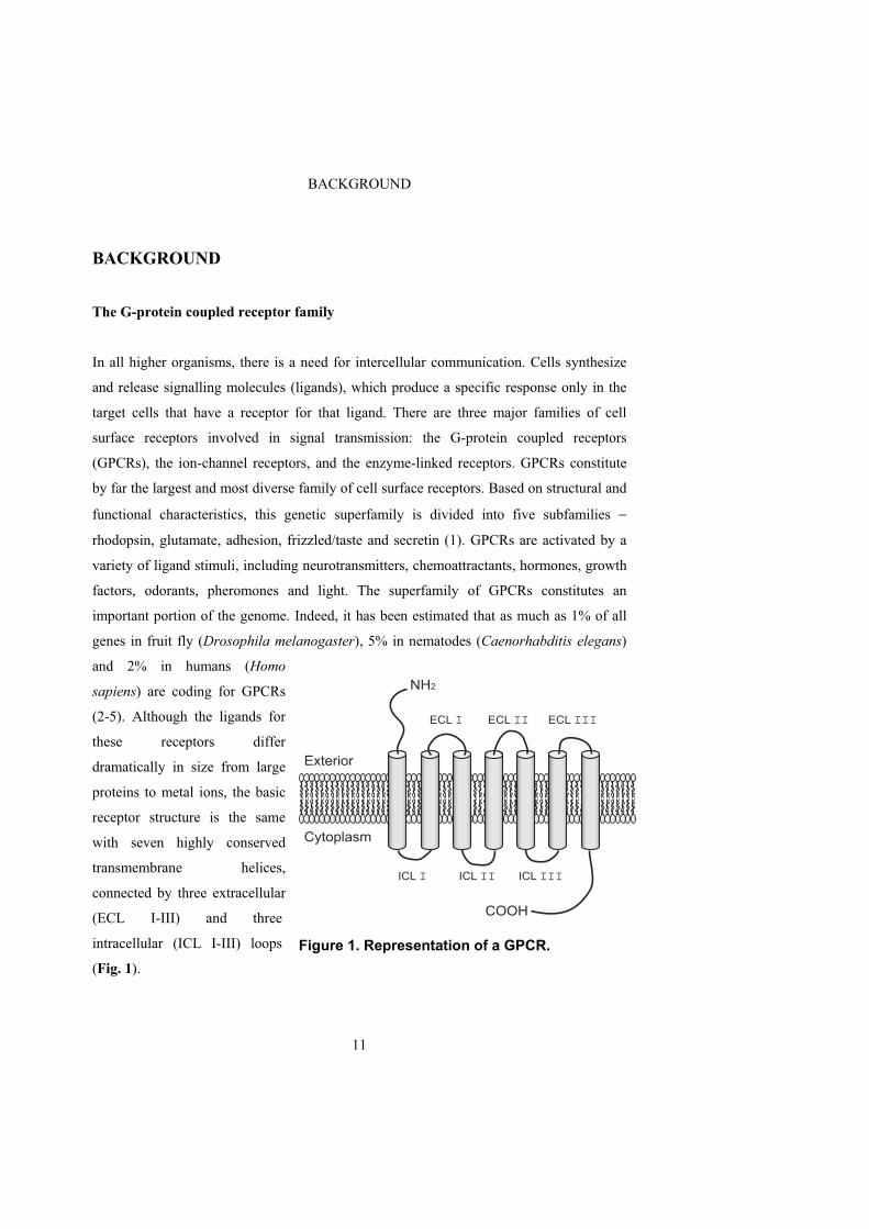
transmembrane helices,
connected by three extracellular
(ECL I-III) and three
intracellular (ICL I-III) loops
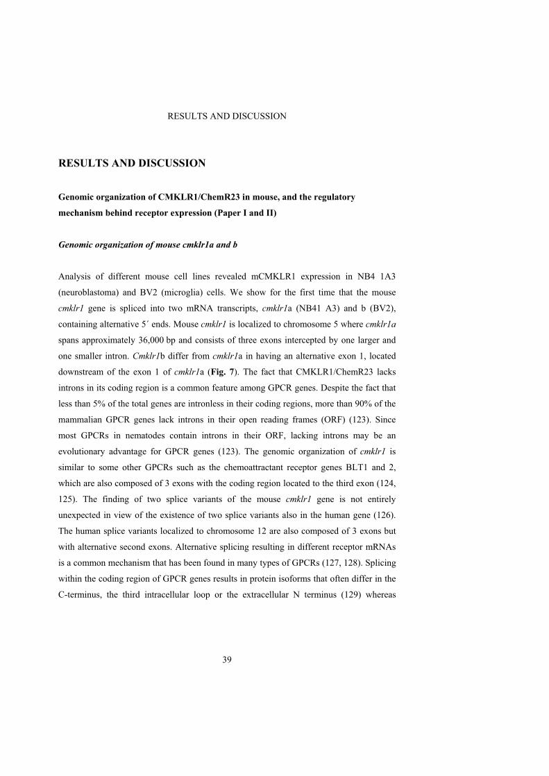
(Fig. 1).
Figure 1. Representation of a GPCR.
NH2
COOH
Exterior
Cytoplasm
ECL I ECL II ECL III
ICL I ICL II ICL III
BACKGROUND
12
The important function of GPCRs in the body is emphasized by the fact that they are
remarkably well conserved during evolution. For example, the muscarinic receptor of C.
elegans displays 51% amino acid sequence homology with its orthologue in D.
melanogaster and 42-44% with its human orthologue (in the alignment, a highly variable
sequence in ICLIII is excluded) (6). A three-dimensional receptor structure would have
been desirable in order to understand how ligand binding to a GPCR leads to
conformational changes and transmission of a signal from the outside to the inside of the
cell. Unfortunately, transmembrane proteins are difficult to crystallize, the main reason
being that GPCRs contain highly hydrophobic regions. Another problem is the difficulty in
obtaining the large quantities of pure receptor protein needed for crystallization. So far, the
only three-dimensional structure of a GPCR described is the crystallized structure of the
inactive state of bovine rhodopsin (7). The availability of the rhodopsin crystal information
has greatly facilitates the construction of models of other GPCRs. However, the
construction of realistic GPCR models is time-consuming and requires biological,
pharmacological and biophysical data for verification.
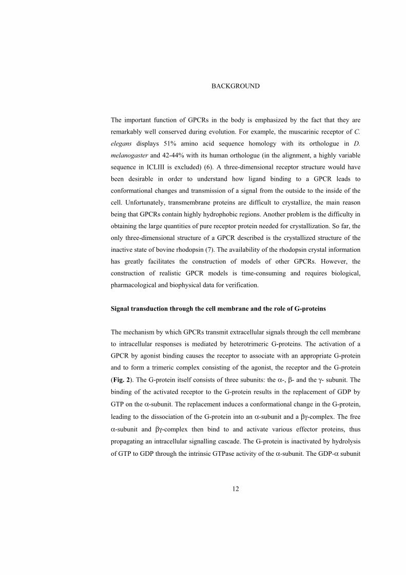
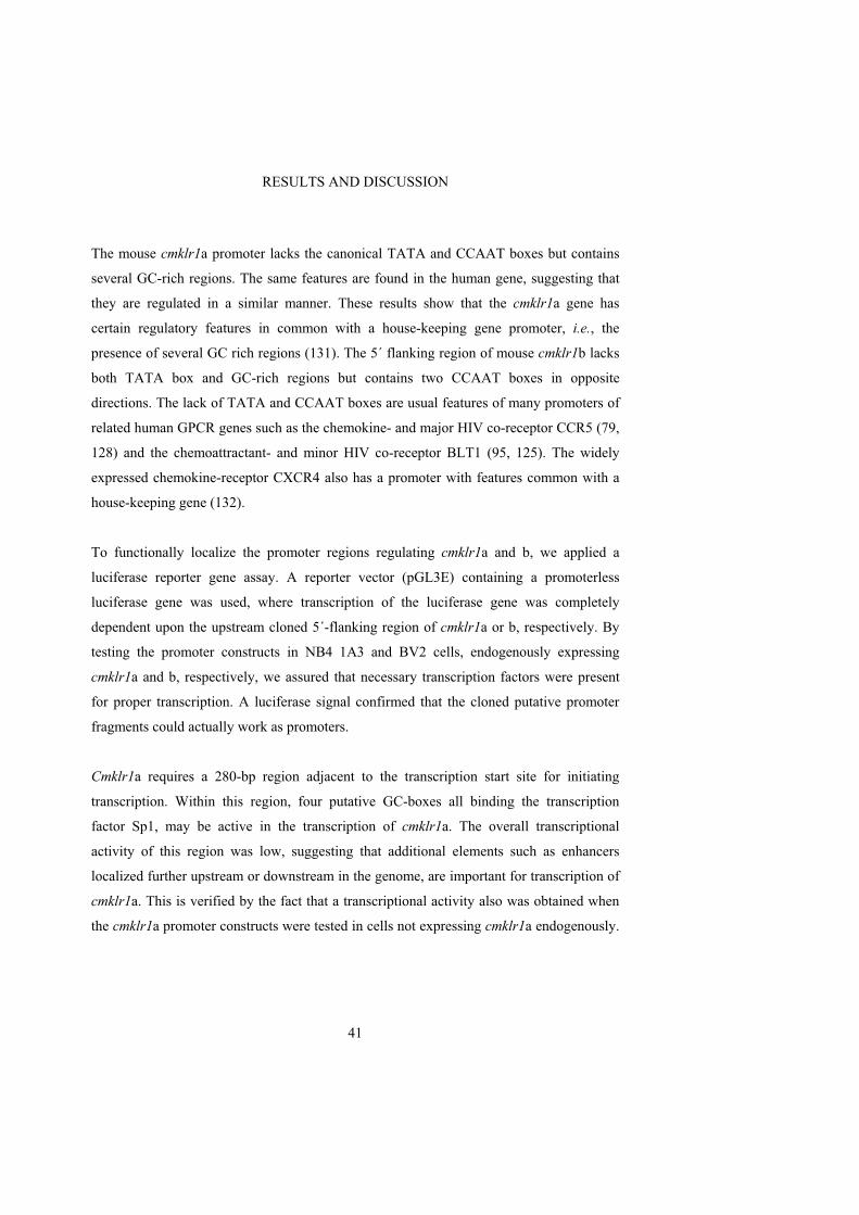
Signal transduction through the cell membrane and the role of G-proteins
The mechanism by which GPCRs transmit extracellular signals through the cell membrane
to intracellular responses is mediated by heterotrimeric G-proteins. The activation of a
GPCR by agonist binding causes the receptor to associate with an appropriate G-protein
and to form a trimeric complex consisting of the agonist, the receptor and the G-protein
(Fig. 2). The G-protein itself consists of three subunits: the -, - and the - subunit. The
binding of the activated receptor to the G-protein results in the replacement of GDP by
GTP on the -subunit. The replacement induces a conformational change in the G-protein,
leading to the dissociation of the G-protein into an -subunit and a -complex. The free
-subunit and -complex then bind to and activate various effector proteins, thus
propagating an intracellular signalling cascade. The G-protein is inactivated by hydrolysis
of GTP to GDP through the intrinsic GTPase activity of the -subunit. The GDP- subunit
BACKGROUND
13
then reassociates with the -complex to re-establish the trimeric G-protein, which is then
ready for another round of receptor activation (Fig. 2).
In mammals, more than twenty different -subunits have been described. These are
organized into four subfamilies, G s, G i/o, G q/11, and G 12/13, based on structural and
functional homologies (8-10). For a long time, it was believed that the -subunit of the
trimeric G-protein alone was responsible for the regulation of all effectors. It has now
become clear that also the -complex is important in the regulation of effector proteins
(11). In mammals, the -complex can be combined from five subtypes of -subunits and
twelve subtypes of -subunits (12).
The four classes of G-protein -subunits interact with different specific effector proteins:
G q/11 activates PLC (13), which catalyses the hydrolysis of PIP2 into IP3 and DAG, which
in turn leads to the release of intracellular stored Ca2+. G s stimulates whereas G i inhibits
adenylyl cyclase leading to an increase and a decrease, respectively in cAMP (14-16).
Figure 2. GPCR activation.
αGTP
βαGDP
Exterior
Cytoplasm
β
γ
Ligand activation
GTP GDP
αGDP
β
γ Pi
Reassociation
Dissociation
γ
Effectors
Ion channels
Enzymes
Intracellular messengers
αGTP
β
γ
BACKGROUND
14
G 12/13 activates different pathways such as the effector protein phospholipase D (17). The
-complex stimulates a diversity of effector proteins including PLC (18) and certain
isoforms of adenylyl cyclase (19, 20).
The bacterial toxin, pertussis toxin (PTX), produced by Bordetella pertussis, has been
useful in the characterization of G-protein signalling pathways since it interferes with the
G i pathway whereas other G-protein pathways are unaffected. Pertussis toxin acts through
ADP ribosylation of the G i class, thereby uncoupling the receptor from the G-protein
(21).
Regulation of gene transcription
In order to investigate how GPCR expression is regulated, we need to study gene
transcription, which takes place in the nucleus.
The genetic material is stored in genes, which are nucleotide sequences forming stretches
of deoxyribonucleic acids, DNA, the universal genetic material. In the eukaryotic cell,
DNA is located in the nucleus where it is wrapped around histone proteins, forming
complexes called nucleosomes. Strings of nucleosomes are coiled up and folded into a
highly condensed chromatin structure. The chromatin is further organized into larger units
of up to thousands of kilobases in length called chromosomes. The total amount of genes
in the human DNA has been estimated to approximately 25,000, where the total number of
GPCR genes amounts to approximately 800 (22). Each gene provides the instructions for
synthesizing a unique protein that has a specialized function in the cell. In order to transfer
the information residing in a gene into a protein, the cell uses two processes: transcription
and translation. Transcription occurs in the nucleus where the transcription machinery
copies the gene into messenger RNA (mRNA), the template for protein synthesis. The
mRNA is subsequently transported out of the nucleus and into the cytoplasm where it is
translated into a protein. The whole process of transforming genetic information into a
BACKGROUND
15
protein is known as expression. When and where a gene will be expressed is regulated
primarily at the level of transcription but also partly by mRNA processing and translation.
Transcription is dependent on the accessibility of the DNA. For a gene to be transcribed,
the chromatin structure must be unpacked so that the transcription machinery can get
access to the DNA. This is performed either by acetylation (23, 24), and/or
phosphorylation of the NH2-terminal of the histones (23).
The transcriptional regulatory region of a gene
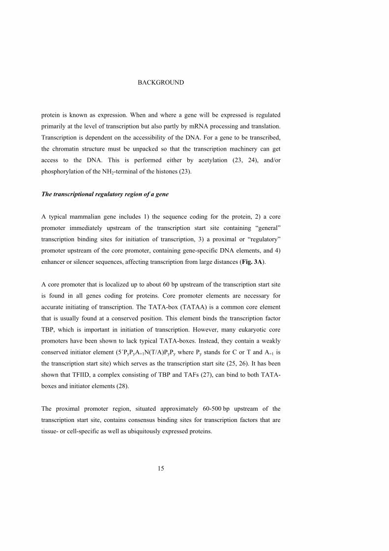
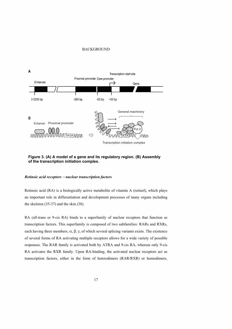
A typical mammalian gene includes 1) the sequence coding for the protein, 2) a core
promoter immediately upstream of the transcription start site containing “general”
transcription binding sites for initiation of transcription, 3) a proximal or “regulatory”
promoter upstream of the core promoter, containing gene-specific DNA elements, and 4)
enhancer or silencer sequences, affecting transcription from large distances (Fig. 3A).
A core promoter that is localized up to about 60 bp upstream of the transcription start site
is found in all genes coding for proteins. Core promoter elements are necessary for
accurate initiating of transcription. The TATA-box (TATAA) is a common core element
that is usually found at a conserved position. This element binds the transcription factor
TBP, which is important in initiation of transcription. However, many eukaryotic core
promoters have been shown to lack typical TATA-boxes. Instead, they contain a weakly
conserved initiator element (5´PyPyA+1N(T/A)PyPy where Py stands for C or T and A+1 is
the transcription start site) which serves as the transcription start site (25, 26). It has been
shown that TFIID, a complex consisting of TBP and TAFs (27), can bind to both TATA-
boxes and initiator elements (28).
The proximal promoter region, situated approximately 60-500 bp upstream of the
transcription start site, contains consensus binding sites for transcription factors that are
tissue- or cell-specific as well as ubiquitously expressed proteins.

BACKGROUND
16
Enhancers and silencers are sequences that are found thousands of base pairs upstream,
downstream or even within the gene that they regulate (29). These regulatory regions
affect transcription by binding the same kind of transcription factors as the promoters.
Proteins that bind to enhancers/silencers also have themselves binding sites for the
transcription factors that are assembled at the proximal promoter of the gene. This
arrangement gives rise to large complexes bridging and forcing the DNA to make a loop
and thus activating or repressing the target gene (29, 30).
A general model for regulation of a gene
RNA polymerase II (Pol II) transcribes genes coding for proteins. For a gene to be
transcribed, a cascade of events occurs that eventually leads to transcription. First, the
opening up of the chromatin structure makes the regulatory region of the gene accessible
for binding combinations of transcription factors. The bound transcription factors
subsequently interact with the general transcription machinery recruiting it to the core
promoter (31). The general machinery is then stepwise assembled on the core promoter
forming the transcription-initiation complex. In short, TFIID initially binds the TATA-box
or the initiator element, which is then followed by sequential recruitment of GTF, Pol II,
co-activators and co-repressors (32-34) (Fig. 3B). The assembly of the transcription-
initiation complex induces separation of the two DNA strands, and the presence of NTPs
initiates transcription by starting RNA synthesis. The transcription factors then dissociate
from the promoter, and a complex consisting of Pol II and certain transcription factors
move along the DNA template strand, causing the synthesis of the mRNA. Termination of
transcription occurs at a special stop sequence in the DNA (AATAAA).
BACKGROUND
17
Retinoic acid receptors nuclear transcription factors
Retinoic acid (RA) is a biologically active metabolite of vitamin A (retinol), which plays
an important role in differentiation and development processes of many organs including
the skeleton (35-37) and the skin (38).
RA (all-trans or 9-cis RA) binds to a superfamily of nuclear receptors that function as
transcription factors. This superfamily is composed of two subfamilies: RARs and RXRs,
each having three members, , , , of which several splicing variants exists. The existence
of several forms of RA activating multiple receptors allows for a wide variety of possible
responses. The RAR family is activated both by ATRA and 9-cis RA, whereas only 9-cis
RA activates the RXR family. Upon RA-binding, the activated nuclear receptors act as
transcription factors, either in the form of heterodimers (RAR/RXR) or homodimers,
Figure 3. (A) A model of a gene and its regulatory region. (B) Assembly of the transcription initiation complex.
B
TATA
Enhancer Proximal promoter
TBP TAFS
GTF
GTFGTF
GTF
GTF
GTF
Pol II
Transcription initiation complex
General machinery
TATA

BACKGROUND
18
(RXR/RXR), by binding specific retinoic acid elements (RAREs) in the promoter and
thereby regulating the transcription of the target gene (39). The final signalling cascade is
more complex since the transcription factors can interact and be regulated by multiple co-
activators and/or co-suppressors (40). In addition to binding directly to RAREs, the nuclear
receptors also bind other transcription factors thereby indirectly regulate gene transcription
from other motifs in the DNA (41, 42).
Sp1
Sp1 belongs to a family of structurally related transcriptional proteins (43). The Sp family
binds specific G-rich DNA elements such as the GC-box (GGGGCGGGG) and the
GT/CACCC-box (GGTGTGGGG) located in promoters and enhancers of both house-
keeping and tissue-specific genes. All Sp members contain a C-terminal DNA-binding
domain consisting of three zinc fingers. Sp1 is ubiquitously expressed (44) whereas the
expression of other members is either ubiquitous or more cell specific (45). Sp1 is an
activator of transcription (46), whereas other Sp-family members repress Sp1-mediated
transcription (47). Constitutive expression of many genes is dependent upon Sp proteins,
which are able to interact with proteins associated with the basal transcription machinery
such as TBP (48) and TAFs (49). Sp protein activation can also be cell- or tissue-specific
(45, 50, 51) depending on differences in Sp protein expression and competition in binding
between the Sp proteins to the GC-rich domains. The final effect on expression of different
genes can be either synergistic or antagonistic depending on which of the Sp family
proteins that bind to the promoter and which other DNA-dependent or DNA-independent
transcription factors that are involved.

BACKGROUND
19
NFY
The transcription factor NFY binds to the CCAAT-box (5´YYRRCCAATCAG3´ or
5´CTGATTGGYYRR3´ where Y=pyrimidines and R= purines), which is a transcription
factor-binding element common in cell-cycle regulated (52), developmentally and tissue-
specific (53, 54), house-keeping and inducible promoters (55, 56). The CCAAT box is
usually located 60-100 bp upstream of the transcription start site. In addition to NFY,
several other nuclear proteins are able to bind to CCAAT boxes including NF1, C/EBP and
CP2. On the other hand, while the other CCAAT binding transcription factors are rather
promiscuous in their binding to the consensus motif, NFY is the only factor that requires
the intact CCAAT sequence as well as specific flanking sequences (57). NFY is a trimeric
protein composed of three subunits, A, B and C, where all three subunits are needed for
binding to the CCAAT box (58). All subunits contain sites also for protein-protein
interactions. Oligomerization of the NFY protein with other transcription factors or co-
activators results in positive cooperativity reflected in an increased affinity of NFY for the
CCAAT box (59, 60). It has also been shown that NFY is able to increase the affinity of
closely located transcription factors for their DNA elements (61, 62). In addition, NFY has
been shown to interact with TBP in the transcriptional machinery (63).
Involvement of GPCRs in immunodeficiency virus infection of human cells
Human immunodeficiency virus (HIV) is the cause of the severe disease called acquired
immunodeficiency syndrome (AIDS). HIV infects and kills cells of the human immune
system thereby destroying the body’s ability to fight infections. Because of the impaired
immune system, the infected individual becomes susceptible to non-pathogenic virus,
fungi or bacteria (opportunistic infections), which eventually leads to death. HIV is passed
between humans mainly through sexual contact (exposure to semen or vaginal fluids) or by
blood-to-blood contact (through needles or syringes).
BACKGROUND
20
The AIDS epidemic in the world is continuously growing. By the end of 2004 39 million
people was estimated to live with HIV worldwide including 25.4 million people infected in
Sub-Saharan Africa, which is the worst area affected (64).
Human and simian deficiency virus
HIV and the closely related simian immunodeficiency virus (SIV) in monkeys are viruses
that belong to the lentivirus subgroup of the retroviridae family. HIV and SIV are envelope
viruses that store their genetic material as RNA. Upon infection of a host, the viral RNA is
transcribed into DNA by the viral reverse transcriptase. Viral DNA becomes incorporated
into the host DNA, where it is replicated together with the host genes. The majority of the
HIV viruses integrate their DNA into the genome of activated immune cells, and new
viruses are immediately produced. However, if the infected immune cells are in a latent
state (which may last for a long time) they produce little or no virus. Upon proper stimuli,
the cells are activated leading to an increase in viral production and dissemination.
HIV is divided into two types, HIV-1 and HIV-2. The transmission route is the same for
both types but they differ in their geographic distribution and pathogenicity.
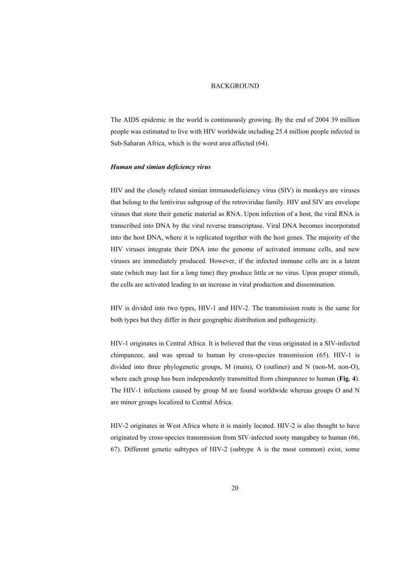
HIV-1 originates in Central Africa. It is believed that the virus originated in a SIV-infected
chimpanzee, and was spread to human by cross-species transmission (65). HIV-1 is
divided into three phylogenetic groups, M (main), O (outliner) and N (non-M, non-O),
where each group has been independently transmitted from chimpanzee to human (Fig. 4).
The HIV-1 infections caused by group M are found worldwide whereas groups O and N
are minor groups localized to Central Africa.
HIV-2 originates in West Africa where it is mainly located. HIV-2 is also thought to have
originated by cross-species transmission from SIV-infected sooty mangabey to human (66,
67). Different genetic subtypes of HIV-2 (subtype A is the most common) exist, some

BACKGROUND
21
probably appeared by independent transmissions from sooty mangabey to human (68)
(Fig. 4). HIV-2 is less pathogenic than HIV-1, which may be due to a better human
immune response to HIV-2 replication (69). This leads to a low viral load (70), a slower
progression to disease (71), and a lower rate of transmission (72). The phylogenetic tree in
Figure 4 shows that HIV-2 is more closely related to SIV than to HIV-1.
The SIV family of retroviruses is composed of several distinct branches originating from
different simian species spread throughout Africa (Fig. 4). SIV infections result in
Figure 4. A phylogenetic tree illustrating the relationship between HIV-1, HIV-2 and SIV (SIVMM; SIV of sooty mangabeys or macaques experimentally infected with SIVMM). The pol gene of the different viruses was used for the alignment.
SIV CPZ P.t.t.
SIV-AGM
AC
GF B D
H J
HIV-1 N group
Gab
US Cam3
SIV CPZ P.t.s.
HIV-1 O groupSIV L'HOEST
SIVSUN
SIVMND
GRI
VER
TANSAB
SIVSYKHIV-2BHIV-2A
SIVMM
0.10
HIV-2, SIVMM
HIV-1, CPZ
HIV-1 M group

BACKGROUND
22
different host responses depending on the type of monkey and virus. The African monkeys
(e.g. sooty mangabeys) are natural hosts for SIV and upon natural infection they are
infected but do not develop disease (73). Experimental infections with SIVmac239 (of
macaque origin) cause an outcome similar to natural infection in the sooty managbey
whereas a disease similar to AIDS is developed in Asian monkeys (macaques) (74). The
close relationship between HIV and SIV has resulted in the usage of SIV-infected
monkeys, such as the Asian monkey (macaques), as a suitable model for studying the
pathogenesis of AIDS (75).
HIV and SIV infection of host cells
Usually, HIV and SIV isolates invade host cells that express the cell surface receptor CD4
and certain G-protein coupled receptors, so-called “co-receptors”. During viral entry, the
envelope (Env) glycoprotein subunit 120 (gp120) initially binds to the host cell CD4
receptor. The CD4 binding leads to conformational changes in gp120 that enables the virus
to bind to the co-receptor. The co-receptor binding elicits further changes in gp120 that
exposes the viral Env glycoprotein subunit 41 (gp41), which penetrates the host cell
membrane. This allows for fusion between viral and host membranes and subsequent viral
entry (76-78) (Fig. 5).

BACKGROUND
23
GPCRs as targets for immunodeficiency viruses
Both HIV and SIV isolates use GPCRs as co-receptors. The chemokine receptors CXCR4
and CCR5 are the major co-receptors in vivo (79, 80). HIV can use either or both CCR5
and CXCR4, whereas CCR5 is the main co-receptor for SIV isolates (81). HIV-1 uses
CCR5 in the early stages of infection, whereas CXCR4 is used mainly in the later phases
of infection (82). In addition, there are a large number of other GPCRs that can function as
minor co-receptors for some HIV and SIV strains in vitro, but their in vivo significance is
not yet clear. Many of these additional receptors are chemokine receptors like CCR1 (83,
84), CCR2b (85, 86), CCR3 (86, 87), CCR4 (88), CCR8 (89), CCR9, CXCR2 (90),
CXCR5 (91), CXCR6/STRL33/Bonzo (86, 92, 93), and CX3CR1 (89, 94),
chemoattractant/-like receptors such as BLTR (95) and ChemR23/CMKLR1 (96), the
angiotensin-like receptor APJ (97, 98), orphan receptors such as GPR1 (99, 100),
BOB/GPR15 (92, 100), or RDC1 (101) as well as viral chemokine receptor like CMV-
US28, expressed in cytomegalovirus (CMV)-infected cells (102). HIV-2 and SIV are more
promiscuous than HIV-1. A broad range of co-receptors is often used by HIV-2 (84, 86,
Figure 5. HIV/SIV binding and entry into immune cells.
CD4 binding
Co-receptor binding
Fusion peptid insertionMembrane fusionVirion
gp41gp120
v3CD4
Cytoplasm
BACKGROUND
24
88, 91) and SIV isolates (86, 103, 104) for cellular entry whereas few HIV-1 isolates are
promiscuous in their co-receptors usage (96, 105, 106).
Studies of the chemoattractant-like receptor CMKLR1/ChemR23
CMKLR1/ChemR23
The body’s immune response against micro-organisms and other foreign agents depends
upon the trafficking of the immune cells. This trafficking is regulated by chemical
mediators, which activate certain GPCRs on the immune cells causing the cells to migrate
towards the mediator concentration gradient. This process is called chemotaxis and the
involved receptors belong to the GPCR subfamily of “classical leukocyte chemoattractants
receptors”, exemplified by the receptors for complement factor, N-formyl peptide, and
leukotriene B4 (107).
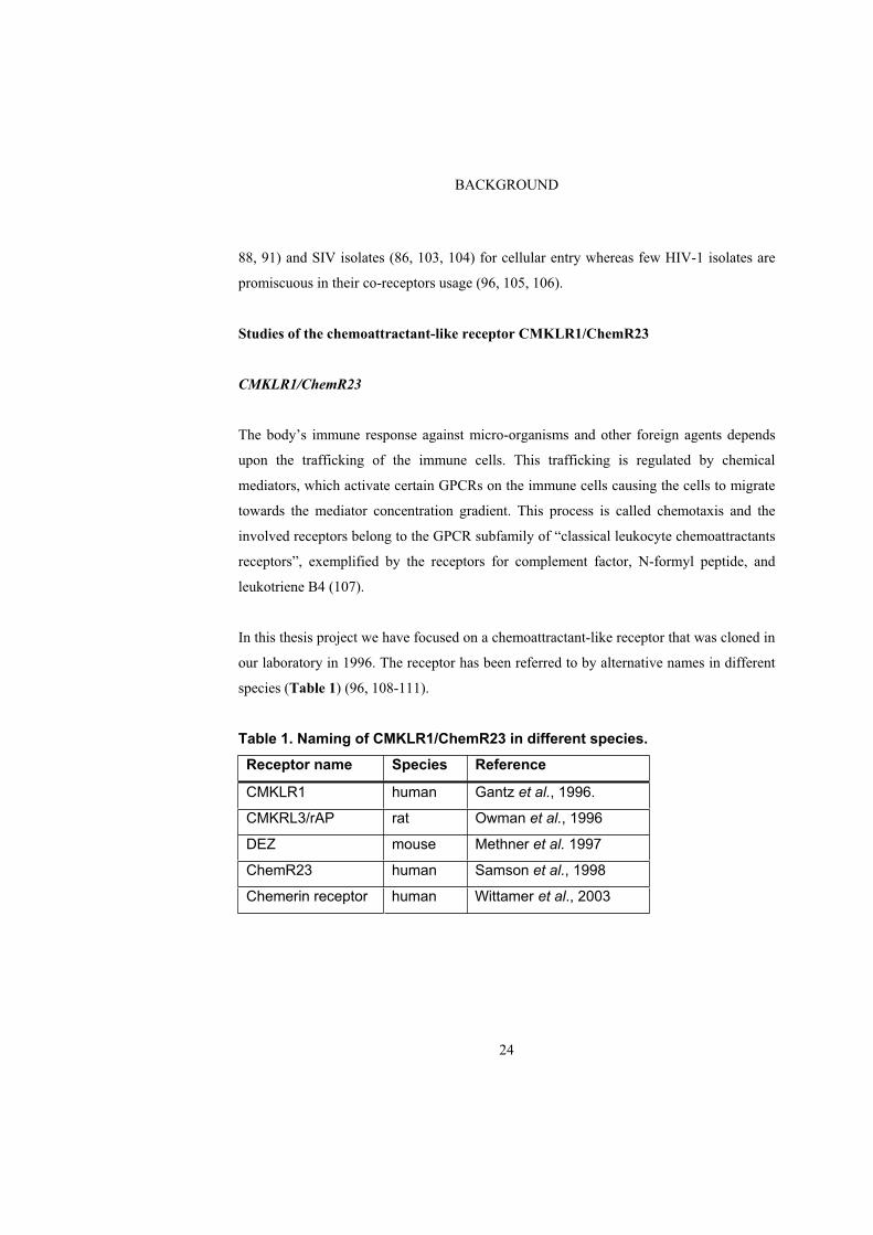
In this thesis project we have focused on a chemoattractant-like receptor that was cloned in
our laboratory in 1996. The receptor has been referred to by alternative names in different
species (Table 1) (96, 108-111).
Table 1. Naming of CMKLR1/ChemR23 in different species.
Receptor name Species Reference
CMKLR1 human Gantz et al., 1996.
CMKRL3/rAP rat Owman et al., 1996
DEZ mouse Methner et al. 1997
ChemR23 human Samson et al., 1998
Chemerin receptor human Wittamer et al., 2003

BACKGROUND
25
We describe the receptor under the name CMKLR1/ChemR3. It displays high homology to
other chemoattractant-like receptors as shown in Figure 6. CMKLR1/ChemR23 is
expressed in immune cells such as macrophages and dendritic cells (96), and it has a
pathophysiological role as one of the minor co-receptors involved in HIV-1/SIV infection
of human CD4+ cells (96). The receptor has also been suggested to be involved in osseous
and cartilage development (110).
Figure 6. A dendrogram illustrating the evolutionary relationship based on similarities in the amino acid sequences, between human CMKLR1/ChemR23 and other receptors.
C3aRC5aRfMLPxRfMLPyRfMLPRChemR23LTB4RCCR2aCCR2bCCR5CCR3CCR1CCR4CCR8V28U 94888U 95626GPR5CXCR2CXCR1CXCR3CCR6CCR7STRL33PPR1CXCR4GPR15
BACKGROUND
26
The natural ligand, TIG2/chemerin
CMKLR1/ChemR23 has been classified as an “orphan” receptor during the major part of
the graduate research work described here in. In 2003, the human receptor was “de-
orphanized” when two independent research groups isolated the natural ligand from human
inflammatory fluids (111) and from hemofiltrate (112). The identified natural ligand is a
143 amino acid residue long protein, previously known as TIG2 (“tazarotene-induced gene
2”), and was tentatively named “chemerin” (111). The ligand is secreted as the precursor
“pro-chemerin” which, upon proteolytic cleavage removing six to nine amino acids in the
C-terminal end, becomes able to activate CMKLR1/ChemR23 (111, 112). In fact, the
molecule itself was known before, under the name TIG2, a gene implicated in dermal
physiology where it was suggested to be involved in keratinocyte differentiation and the
skin disorder psoriasis (113). Tazarotene, a synthetic RA analogue (114), used for
treatment of psoriasis, has been shown to up-regulate the TIG2 gene (113).
Chemerin/TIG2 also seems to play a role in the mechanisms of bone modelling (115).
The identification of the natural ligand to CMKLR1/ChemR23 now makes it possible to
explore the physiological relevance of this receptor. The role of CMKLR1/ChemR23 in
chemotaxis could be confirmed when it was revealed that stimulation of immature
dendritic cells and macrophages with TIG2/chemerin induces chemotaxis (111).

AIMS OF THE STUDY
27
AIMS OF THE STUDY
The aims of this project were to:
1) Characterize the genomic structure of CMKLR1/ChemR23 in mouse for
comparison with the human sequence.
2) Study the regulation of receptor gene expression in mouse.
3) Investigate whether mouse TIG2/chemerin activates CMKLR1/ChemR23 in
mouse.
4) Analyse if C-terminal peptides of mouse TIG2/chemerin can activate mouse
CMKLR1/ChemR23.
5) Elucidate the importance of CMKLR1/ChemR23 as a co-receptor for human and
simian immunodeficiency virus (HIV and SIV).
6) Identify the extracellular regions of the receptor needed for virus binding and
entry into human immune cells.

METHODOLOGY
28
METHODOLOGY
This section gives a short summary and comments on the methods used in the papers
included in this thesis. The paper where each method has been applied is indicated in
roman letters. For more detailed instructions; see paper I-IV; Materials and methods.
Cell lines and receptor expression (I-IV)
NB4 1A3 cells (mouse neuroblastoma), endogenously expressing mouse cmklr1a and BV2
cells (mouse microglia), endogenously expressing cmklr1b, were used in the promoter
studies. In these experiments, 3T3 clone A31 cells (mouse embryonic fibroblast), which
does not express cmklr1 endogenously, was used as negative control.
The HeLa-based reporter cell line, HFF11, stably expressing the reporter plasmid pcFUS3
was constructed by Kotarsky et al. (116). HFF11 cells, stably expressing mouse and
human CMKLR1/ChemR23 were constructed for studies of receptor activation. HFF11
cells, expressing the reporter plasmid but no receptor (HFF11-sham) functioned as
negative control.
The cell line HEK293 (human embryonic kidney) containing an effective apparatus for
protein synthesis was used for expression of mouse wild-type chemerin. HEK293 cells and
CHO-K1 cells (chinese hamster ovary) were used for expression of FLAG-tagged mouse
TIG2/chemerin.
NP-2 cells (human glioma), stably expressing the human CD4 receptor, alone or in
combination with CCR5, CXCR4, CCR3, FC-4b, hCMKLR1, rCMKLR1, hCMKLR1-
EGFP, rCMKLR1-EGFP, rCMKLR1.hECL2-EGFP or rCMKLR1.hNterm-EGFP were
constructed for studying human CMKLR1/ChemR23 as a co-receptor for HIV-1, HIV-2
and SIV or to map CMKLR1/ChemR23 domains important for virus binding. NP-2 cells

METHODOLOGY
29
expressing the CD4 receptor alone, was used as negative control in the infection
experiments.
Southern blot analysis (I)
Southern blotting was used for isolation of the genomic region containing the mcmklr1
gene. A mouse bacterial chromosome (BAC) containing the mcmklr1 gene was digested
with different enzymes and southern blot was performed according to standard procedures
using a radioactively labelled probe containing the coding region of mcmklr1.
Northern blot analysis (I-IV)
Expression of receptor transcript in different cell lines was confirmed by northern blot
analysis. Total RNA was isolated by the guanidinium isothiocyanate method (117) and
mRNA was selected using a commercial kit. The northern blot was performed according to
standard procedures using a radioactively labelled probe containing the coding DNA
region of the receptor to be analysed.
Rapid amplification of cDNA ends (5´-RACE) (I, II)
The transcription start sites of cmklr1a and b were revealed by 5´-RACE. Total RNA and
mRNA were isolated as for northern blot analysis. cDNA synthesis and the following PCR
amplification of the 5´ cDNA end including the transcription start site were performed
using the MarathonTM cDNA amplification method.
Genome walking (I, II)
Genome walking was applied for the genomic mapping of mouse cmklr1a and b and for
obtaining the promoter regions of cmklr1a and b. The genome walking method is suitable
METHODOLOGY
30
for amplifying DNA fragments that starts in a known sequence and extends into the
unknown adjacent genomic DNA. Genomic walking was performed using the Universal
Genome walker kit.
5´ Deletions of promoter regions (I, II)
5´ Deletions of the cmklr1a and cmklr1b promoter regions cloned into the pGL3-Enhancer
plasmid were performed according to the Erase-A-Base system developed by Henikoff
(118). The promoter plasmids were linearized with MluI and KpnI resulting in one end
with 5´overhang and one with 3´overhang. After purification, the linearized fragments
were digested in their 5´ends with exonuclease III. The digestions were performed for
different lengths of time whereafter the reactions were stopped by adding S1 Nuclease (the
Nuclease digested the single stranded ends and the low pH in the buffer stopped the
exonuclease III activity). The Nuclease activity was inactivated by heating whereafter
Klenow DNA polymerase was added to blunt the ends. The plasmids were finally re-
ligated with DNA ligase.
Site-directed mutagenesis (II)
To establish if both CCAAT binding sites found in the cmklr1b promoter were functionally
important for transcription, the elements were separately mutated. The mutations were
performed using the site-directed mutagenesis method (QuickChangeTM). Primers
containing the desired mutation were used in a PCR reaction using a plasmid containing
the cmklr1b promoter region as template. After PCR amplification, treatment with DpnI,
an enzyme that specifically cleaves methylated DNA, digested the parental DNA
(methylated in E. coli) and selected for the mutated non-methylated plasmid. Mutated
plasmids were transformed into competent E. coli, and positive clones were identified by
PCR screening using primers with the mutated element in the 3´end.
METHODOLOGY
31
Luciferase reporter assay (I, II)
We applied a dual-luciferase reporter assay to confirm that the isolated putative promoter
regions of cmklr1a and b possessed functional promoter activity. The dual luciferase
reporter assay, based on transient transfection of cells, makes it possible to measure the
activity of the two luciferases Firefly and Renilla in the same sample allowing for
simultaneous promoter activity measurement (Firefly) and determination of transfection
efficiency (Renilla). The enzyme activities are distinguished since they require different
substrates and function at different pHs. In the presence of the substrates Beetle Luciferine
and Coelenterazine, bioluminescent reactions occur catalysed by firefly and Renilla
luciferases, respectively. Obtained luminescence signals are measured in a BMG Lumistar
microplate luminometer.
In order to determine the transcriptional activities of cmklr1a and b, the putative promoter
fragments were sub-cloned in front of the firefly luciferase gene using the reporter vector,
pGL3-Enhancer. We used the pRL-TK vector, containing a herpes simplex virus
thymidine kinase promoter in front of the Renilla luciferase, as control vector to
compensate for differences in transfection efficiency.
The day before transfection, cells were seeded in white 96-well tissue culture plates.
Luciferase constructs were co-transfected with pRL-TK as internal control. Forty-two
hours after transfection, the cells were harvested in reporter lysis buffer. The dual-
luciferase assay was applied to measure Firefly and Renilla luciferase activities using a
luminometer.
Electrophoretic mobility shift assay (I, II)
Electrophoretic mobility shift assay (EMSA) is a widely used method for studying gene
regulation and determining protein-DNA interactions. The method is based on the fact that
METHODOLOGY
32
DNA that is bound to proteins (like transcription factors) migrates more slowly than free
DNA when run on a non-denaturing polyacrylamide gel. The migration rate of the DNA is
decreased or “shifted” upon protein binding. When an antibody also is present, for
identification of the bound protein, the migration rate is further decreased or “super-
shifted”. EMSA is used to identify the sequence-specific DNA-binding transcription factor
in nuclear extracts and, in combination with mutagenesis, to identify the important binding
motif within the gene’s regulatory region.
We used EMSA to investigate if transcription factors were able to bind the G-rich DNA
elements in the cmklr1a promoter and the two identified CCAAT boxes in the cmklr1b
promoter, and also to identify the nature of the binding proteins. The affinities of the
transcription factors for the DNA elements were titrated by using increasing amounts of
unlabelled probes containing the putative transcription binding elements. The specific
proteins binding the DNA elements were identified using unlabelled DNA probes with or
without mutations in the transcription-binding element, and by the use of antibodies.
Nuclear extracts containing transcription factors were prepared from cell lines
endogenously expressing cmklr1a (NB4 1A3) and cmklr1b (BV2), essentially as described
by Andrew and Faller (119). Synthetic radioactively labelled DNA oligonucleotides
containing the putative transcription binding elements were used as probes. Binding was
performed at 25 C and DNA-protein complexes were resolved on a non-denaturing
polyacrylamide gel. After electrophoresis, the gel was dried and exposed to X-ray film. For
super-shift assays, either antibody (Sp1) and extract were incubated at 25 C before
addition of labelled probe or labelled probe and extract were incubated before addition of
antibody (NFY-A or NFY-B).
METHODOLOGY
33
Real-time reverse transcription PCR (II)
Expression of cmklr1a and b was analysed by real-time reverse transcription PCR (real-
time RT-PCR) in different tissues and in NB4 1A3 and BV2 cells with/without pre-
treatment with ATRA. 3T3 clone A31 cells were used as negative control. The real-time
PCR was performed in a LightCycler system using Sybr green, a dye that binds to double
stranded DNA, for monitoring DNA synthesis. Primers specific to mcmklr1a and b were
used in the PCR reaction. Copy-numbers of mcmklr1a and b were normalised to the copy-
number of the house-keeping gene (2)-microglobulin.
Cloning of mouse wild-type- and FLAG-TIG2/chemerin (III)
To clone mouse wild-type chemerin, total RNA was isolated from mouse liver by the
guanidinium isothiocyanate method (117). mRNA was prepared using a commercial kit
and cDNA synthesized using the PCR-based first-strand synthesis system. Wild-type
chemerin was amplified by PCR according to standard procedures using specific chemerin
primers and the liver cDNA as template. Amplified wild-type chemerin was cloned into
the expression vector, pEAK12.
In order to optimise TIG2/chemerin expression and simplify the purification process, the
proximal part of the mouse TIG2/chemerin was replaced with a synthetic (mouse Igk V-
J2-C) secretion signal, a TEV (tobacco etch virus)-recognition site and a FLAG epitope.
The TEV site was included for possible removal of the FLAG-tag from the protein using
TEV protease. Chemerin without the endogenous secretion signal was amplified by PCR
using chemerin specific primers. The amplified product was ligated to a forward and a
reversed oligo containing the synthetic secretion signal sequence, the sequence of the
TEV-recognition site and the FLAG epitope sequence. The amplified FLAG-chemerin was
ligated into the expression vector, pEAK12.
METHODOLOGY
34
HFF11 reporter assay (III)
We used the HFF11 reporter cell line (116) for studying human and mouse
CMKLR1/ChemR23 activation. Activation of different signalling pathways leads to
activation of different transcription factors that can affect genes driven by promoters
containing responsive elements for these transcription factors. HFF11 utilizes a reporter
construct based on a luciferase gene driven by a multifunctional promoter including
responsive elements for different transcription factors such as NF- B, STAT and AP-1.
Upon binding of transcription factors to the promoter, luciferase is expressed. The
luciferase activity is measured in a luminometer. The HFF11 reporter cell line is suitable
for studying activation of receptors that signal primarily through G q, G i/o, and G 12/13.
Consequently, it is a suitable system for studying activation of CMKLR1/ChemR23 since
the human receptor has been described to signal through G i/o.
On day one, HFF11 cells, either sham-transfected or transfected to express mouse or
human CMKLR1/ChemR23, were seeded into a white 96-well plate. On day 3, the
medium was changed to serum-free medium. On day 4, different activators were added to
the wells and the cells were further incubated. After 7 hours, cells were harvested in lysis
buffer and the plate assayed in a BMG Lumistar Galaxy microplate luminometer using a
luciferase kit.
Phosphoinositide hydrolysis assay (III)
Activation of human and mouse CMKLR1/ChemR23 was also studied using a
phosphoinositide hydrolysis assay (PI assay) by monitoring the formation of inositol
phosphates.
Phosphatidylinositols (PtIns) are components of the cell membrane and are important in
signal transduction. By incorporating myo- 3H -inositol into PtIns in the membrane, it is
METHODOLOGY
35
possible to monitor signal transduction and receptor activation. When a receptor is
activated, G q of G q-coupled receptors or of G i-coupled receptors activates PLC in
the membrane, which catalyses the hydrolysis of PIP2 into IP3 and DAG. IP3 is then
stepwise dephosphorylated. By using LiCl to block further dephosphorylation into myo-
inositol, the accumulation of radioactively labelled inosithol phosphate may be monitored.
Cells were pre-labelled with myo- 3H -inositol for 20 h. Stimulation with RA or PTX were
performed for 16 h. At the day of analysis, medium containing myo- 3H -inositol was
removed, the plates washed and further incubated in medium containing LiCl with or
without C-terminal peptides for 30 min. After incubation, medium was removed, and the
cells were lysed in formic acid. 3H -Inositol phosphates were isolated by extraction and
anion exchange chromatography and counted in a Beckham LS6500 liquid scintillation
counter.
Flow cytometric analysis (IV)
Stable expression of recombinant receptors (CCR5, CXCR4, CMKLR1/ChemR23, CCR3,
FC-4b) in NP-2.CD4 cells was verified by flow cytometric analysis using the FACS
Calibur flow cytometer (Becton-Dickinson).
Constructions of EGFP tagged receptors and receptor hybrids (IV)
To define extracellular receptor domains important for the virus interaction with
CMKLR1/ChemR23 we applied a hybrid receptor model. This was based upon the fact
that the rat receptor, although having high amino acid identity to human
CMKLR1/ChemR23, is inefficient as viral co-receptor. Since no antibody is available to
confirm rat receptor expression, we tagged the receptors and receptor hybrids with EGFP.
EGFP was fused in-frame to the C-terminus of human and rat CMKLR1/ChemR23,
respectively, using a stepwise PCR procedure according to a modified protocol (120) of
METHODOLOGY
36
the single-overlap extension method (121). The same method was applied to replace the N-
terminal and the second extracellular loop of the rat CMKLR1/ChemR23-EGFP with the
corresponding human sequences.
Confocal microscopy (IV)
Confocal microscopy (Zeiss LSM510) was used to verify that the EGFP-tagged
recombinant receptors, stably expressed in NP-2.CD4 cells, were expressed at the cell
surface. Cells stained with anti-CD4 mAb were used as positive control.
Amplification of virus isolates (IV)
Virus stocks of HIV-1, HIV-2 and SIV were prepared by infection of phytohemagglutinin
(PHA)-stimulated peripheral blood mononuclear cells (PBMCs) mixed from two blood
donors. Donor PBMCs were isolated by separation of buffy coats on a Ficoll gradient.
Pooled PBMCs were infected with virus in RPMI medium containing 10% FCS, 5 U/ml of
interleukin 2, 2 g/ml polybrene, 50 U/ml penicillin and 50 g/ml streptomycin. Cell-free
supernatants were harvested at day 8, 10, and 12, assayed for virus antigen content and
stored at -80 C until used.
Virus detection in cell culture supernatants by enzyme-linked immunosorbent assay
or by measuring reverse transcriptase activity (IV)
Enzyme-linked immunosorbent assay (ELISA) or reverse transcriptase activity (RT assay)
was performed to detect virus antigens in cell culture supernatants after virus infection.
The presence of HIV-1 was monitored by detection of p24 core antigen using a p24 ELISA
(Vironostika HIV-1 Antigen, Biomérieux) based on the “sandwich” principle. ELISA
plates were coated with antibodies (murine monoclonal) against the p24 core antigen.

METHODOLOGY
37
Disruption buffer was added to the wells to verify that all viruses were to be disrupted.
Cell culture supernatants containing p24 antigens together with positive (p24 core antigen)
and negative (human serum not containing p24 antigen) controls were added to the wells
and the plate was incubated to allow formation of complexes between antibody and
antigen. The wells were washed and p24 antibodies (human) coupled to horseradish
peroxidase were added to the wells and the plate was further incubated allowing the
secondary antibody to bind to the previously formed complex. After a final wash the
tetramethylbenzidine substrate was added to the wells producing a blue colour that turned
yellow upon stopping the reaction with sulfuric acid. The amount of p24 in the wells was
proportional to the amount of produced colour, which could be measured photometrically
at an absorbance of 450 nm.
The presence of HIV-2 and SIV were monitored either by p26 core antigen detection using
an in-house p26 ELISA (122) or by measurement of virus reverse transcriptase activity
using the Cavidi HS-kit Lenti RT. To measure reverse transcriptase activity, plates coated
with poly-rA were used as template. By adding oligo-dT primer and the BrdU-triphospate
substrate, reverse transcriptase was able to synthesize a new DNA strand, which could be
detected by addition of a monoclonal anti-BrdU antibody coupled to alkaline phosphatase.
After washing, the chromogenic substrate para-nitro-phenyl phosphate was added to the
wells and the colour produced was measured photometrically at an absorbance of 405 nm.
Virus infection of NP-2 cells (IV)
Virus infection studies were performed to elucidate the importance of CMKLR1/ChemR23
as a co-receptor for HIV-1, HIV-2 and SIV and to map CMKLR1/ChemR23 domains
important for virus binding.
Two days before infection, NP-2.CD4 cells alone or in combination with human
CMKLR1/ChemR23, rat CMKLR1/ChemR23, CCR5, CXCR4, CCR3, FC-4b,
hCMKLR1-EGFP, rCMKLR1-EGFP, rCMKLR1.hECL2-EGFP or rCMKLR1.hNterm-

METHODOLOGY
38
EGFP were seeded into 48-well plates. At the time of infection, medium was removed and
virus was added to the wells in 200 l medium containing polybrene. Two hours after
infection, medium with polybrene was added to a total volume of 500 l/well. After
overnight incubation, cells were washed and medium without polybrene was added to
each well. Twelve days after infection, medium was sampled from each well for detection
of viral antigen. Cells were also evaluated microscopically for syncytia formation.
RESULTS AND DISCUSSION
39
RESULTS AND DISCUSSION
Genomic organization of CMKLR1/ChemR23 in mouse, and the regulatory
mechanism behind receptor expression (Paper I and II)
Genomic organization of mouse cmklr1a and b
Analysis of different mouse cell lines revealed mCMKLR1 expression in NB4 1A3
(neuroblastoma) and BV2 (microglia) cells. We show for the first time that the mouse
cmklr1 gene is spliced into two mRNA transcripts, cmklr1a (NB41 A3) and b (BV2),
containing alternative 5´ ends. Mouse cmklr1 is localized to chromosome 5 where cmklr1a
spans approximately 36,000 bp and consists of three exons intercepted by one larger and
one smaller intron. Cmklr1b differ from cmklr1a in having an alternative exon 1, located
downstream of the exon 1 of cmklr1a (Fig. 7). The fact that CMKLR1/ChemR23 lacks
introns in its coding region is a common feature among GPCR genes. Despite the fact that
less than 5% of the total genes are intronless in their coding regions, more than 90% of the
mammalian GPCR genes lack introns in their open reading frames (ORF) (123). Since
most GPCRs in nematodes contain introns in their ORF, lacking introns may be an
evolutionary advantage for GPCR genes (123). The genomic organization of cmklr1 is
similar to some other GPCRs such as the chemoattractant receptor genes BLT1 and 2,
which are also composed of 3 exons with the coding region located to the third exon (124,
125). The finding of two splice variants of the mouse cmklr1 gene is not entirely
unexpected in view of the existence of two splice variants also in the human gene (126).
The human splice variants localized to chromosome 12 are also composed of 3 exons but
with alternative second exons. Alternative splicing resulting in different receptor mRNAs
is a common mechanism that has been found in many types of GPCRs (127, 128). Splicing
within the coding region of GPCR genes results in protein isoforms that often differ in the
C-terminus, the third intracellular loop or the extracellular N terminus (129) whereas

RESULTS AND DISCUSSION
40
splicing in the uncoding region affects gene regulation thereby increasing the diversity and
complexity of the receptor.
Promoter analysis of mouse cmklr1a and b
Since mouse cmklr1a and b utilize alternative exon 1, they utilize different start sites for
transcription and are consequently transcribed from alternative promoters. The use of
multiple promoters is an important mechanism among GPCR genes, which contribute to
diversity and complexity in regulation of gene expression (130). The utilization of multiple
promoters makes it possible for a gene to be expressed at different stages in development,
specifically in certain cells or tissues, or to respond differently to a specific stimulus.
Figure 7. Genomic organization of the mouse cmklr1 gene and the alternative splicing variants mcmklr1a and b.
RESULTS AND DISCUSSION
41
The mouse cmklr1a promoter lacks the canonical TATA and CCAAT boxes but contains
several GC-rich regions. The same features are found in the human gene, suggesting that
they are regulated in a similar manner. These results show that the cmklr1a gene has
certain regulatory features in common with a house-keeping gene promoter, i.e., the
presence of several GC rich regions (131). The 5´ flanking region of mouse cmklr1b lacks
both TATA box and GC-rich regions but contains two CCAAT boxes in opposite
directions. The lack of TATA and CCAAT boxes are usual features of many promoters of
related human GPCR genes such as the chemokine- and major HIV co-receptor CCR5 (79,
128) and the chemoattractant- and minor HIV co-receptor BLT1 (95, 125). The widely
expressed chemokine-receptor CXCR4 also has a promoter with features common with a
house-keeping gene (132).
To functionally localize the promoter regions regulating cmklr1a and b, we applied a
luciferase reporter gene assay. A reporter vector (pGL3E) containing a promoterless
luciferase gene was used, where transcription of the luciferase gene was completely
dependent upon the upstream cloned 5´-flanking region of cmklr1a or b, respectively. By
testing the promoter constructs in NB4 1A3 and BV2 cells, endogenously expressing
cmklr1a and b, respectively, we assured that necessary transcription factors were present
for proper transcription. A luciferase signal confirmed that the cloned putative promoter
fragments could actually work as promoters.
Cmklr1a requires a 280-bp region adjacent to the transcription start site for initiating
transcription. Within this region, four putative GC-boxes all binding the transcription
factor Sp1, may be active in the transcription of cmklr1a. The overall transcriptional
activity of this region was low, suggesting that additional elements such as enhancers
localized further upstream or downstream in the genome, are important for transcription of
cmklr1a. This is verified by the fact that a transcriptional activity also was obtained when
the cmklr1a promoter constructs were tested in cells not expressing cmklr1a endogenously.

RESULTS AND DISCUSSION
42
This indicates that the promoter fragment is lacking elements for regulatory tissue
selectivity.
A proximal and a distal region seem to be important for transcription of cmklr1b. The
proximal promoter region includes two CCAAT boxes. However, site-directed
mutagenesis separately within these elements revealed that only the forward CCAAT
element binding the transcription factor NFY contribute to transcription of cmklr1b. Since
the cmklr1b promoter constructs resulted in transcription activity also in cells not
expressing cmklr1b endogenously, the proximal region seems to be controlled by a
common mechanism, whereas the cell specificity may reside elsewhere in the genome.
When cmklr1a and b expression was investigated in different mouse organs (skeletal
muscle, spleen, brain, kidney, liver, lung and heart), we found that cmklr1a is
constitutively expressed in organs such as heart and lung, whereas cmklr1b expression is
generally too low for reliable detection. The low detection levels of cmklr1b may be due to
the fact that it is expressed only in a specific cell population, too few in number to give a
proper signal, or that this transcript requires stimulation for transcription. We confirmed
the later alternative by showing that cmklr1b is strongly up-regulated by ATRA, whereas
cmklr1a is unaffected. A RARE element within the analysed cmklr1b promoter region
could not be identified indicating that the element may be located further upstream or that
ATRA works through an indirect mechanism affecting other transcription factors. Example
of such other transcription factors are AP-1 and GATA-2, proteins that have been
described to interact with ATRA (41, 42) and for which binding sites have been found in
the promoter.
The emerging picture of the regulation of the cmklr1 gene indicates a bi-functional mode
of action including a basal and an inducible, possibly tissue- or cell- specific transcription.
The results from the functional experiments with ATRA emphasises the potential
involvement of the receptor in, e.g. bone modelling. An interesting notion is that ATRA

RESULTS AND DISCUSSION
43
up-regulates cmklr1b expression just as the synthetic RA derivate, tazarotene up-regulates
the ligand TIG2/chemerin. In addition, cmklr1 has shown to be expressed during bone
development (110), where RA also has proved to play an important role (36).
Activation of mouse CMKLR1 using C-terminal peptides of the mouse
TIG2/chemerin ligand (Paper III)
We have searched for the natural ligand for human CMKLR1/ChemR23 by expressing the
receptor in a HFF11 reporter cell system in order to monitor receptor activation (116). We
tested a variety of different substances, and conditioned media from different cell lines, but
without success. In 2003, two independent research groups identified the chemotactic
protein TIG2/chemerin as the natural ligand for human CMKLR1/ChemR23. (111, 112).
The ligand is secreted as a precursor, “pro-chemerin”, which upon proteolytic digestion in
the C-terminus becomes capable of activating CMKLR1/ChemR23 (111, 112). Using
peptides corresponding to various parts of the processed form of TIG2/chemerin, the active
region important for human receptor activation was mapped to the C-terminus (133).
Applying the HFF11 reporter cells expressing CMKLR1/ChemR23, we could confirm that
a C-terminal peptide of the processed human ligand activates human CMKLR1/ChemR23.
We also showed that receptor activation is inhibited by PTX, further verifying that the
receptor signals through G i.
Since we have been studying human as well as mouse CMKLR1/ChemR3, we also wanted
to investigate if the mouse orthologue to human TIG2/chemerin can activate mouse
CMKLR1/ChemR23, and if the C-terminus is the important region for receptor activation
also, in mouse. We cloned and expressed mouse TIG2/chemerin in HEK293 cells.
Conditioned medium containing TIG2/chemerin was found to activate mouse
CMKLR1/ChemR23 expressed in HFF11 reporter cells, although to a lower degree than
for the human receptor. In an attempt to obtain purified ligand, mouse TIG2/chemerin with
a fused N-terminal FLAG-tag was expressed in HEK293 and CHO-K1 cells under a strong

RESULTS AND DISCUSSION
44
promoter. The FLAG-tagged fusion protein was purified using ANTI-FLAG M2 affinity
gel, but the amounts obtained were too small for use. The difficulty in expressing
TIG2/chemerin has been experienced also by other investigators both when using CHO-K1
cells (134) and E. coli (135). We could show that C-terminal peptides of mouse
TIG2/chemerin activate the mouse receptor, though to a lower extent than for the human
receptor. These results indicate that the peptide domains necessary for receptor activation
differ for human and mouse TIG2/chemerin. A possibility is that the mouse receptor
requires a longer active peptide, including further upstream amino acid residues, than does
the human receptor. Another possibility is that the mouse receptor responds less well than
the human receptor to activation with TIG2/chemerin. This difference has also been
reported when comparing orthologues of other receptors (136-138).
HIV/SIV co-receptor function of CMKLR1/ChemR23 (Paper IV)
It is important to gain knowledge about the type of HIV and SIV that use a certain co-
receptor for cellular entry and to investigate the receptor epitopes responsible for the
interaction between receptor and virus. This will give an opportunity to design molecules
that may intervene with the virus particle during its entry into and infection of the human
cell, without disturbing the natural importance of the receptor in the immune system.
We have investigated the importance of CMKLR1/ChemR23 as co-receptor for HIV-1,
HIV-2, and SIV primary isolates by expressing the receptor in human astroglia (NP-2)
cells. HIV-1 isolates of genetic subtype B and D utilizes CMKLR1/ChemR23 for infection
whereas the more promiscuous HIV-2 (84, 90) and SIV isolates use CMKLR1/ChemR23
to a wider extent for infection. Among certain better-characterized co-receptors, the HIV-1
co-receptor function of CMKLR1/ChemR23 resembles that of the chemokine receptor,
CCR3. The FC-4b receptor chimera, which is a hybrid between CCR5 and CXCR4, has
proved useful in elucidating certain evolutionary aspects of HIV-1 co-receptor use. This

RESULTS AND DISCUSSION
45
chimera was therefore included in the present co-receptor characterization. However, it did
not reveal any further distinct feature of CMKLR1/ChemR23 co-receptor function.
The general picture of the structural requirements for co-receptor function indicates that
multiple extracellular epitopes are involved in virus binding (139-143). To define
extracellular receptor domains important for the virus interaction with
CMKLR1/ChemR23, we applied a hybrid receptor model. This was based upon the fact
that the rat receptor, although having high amino acid identity to human
CMKLR1/ChemR23, is inefficient as viral co-receptor. When we “humanized” the rat
receptor to include either the human N-terminus or the second extracellular loop, exposure
to HIV-1, HIV-2 and SIV resulted in efficient infection. HIV-1 and HIV-2 showed
preference for the N-terminus and the second extracellular loop, whereas SIV was
primarily dependent on the second extracellular loop for infection. The observation that the
receptor domains important for virus binding differ for different virus isolates makes it
harder to inhibit the HIV-infection at the receptor level.
CONCLUSIONS
46
CONCLUSIONS
In order to increase the knowledge of the previously identified orphan chemoattractant-like
G-protein coupled receptor CMKLR1/ChemR23, we have focused our interest on various
genetic and molecular biological aspects of this receptor as well as on its role as an
immunodeficiency viral co-receptor. In this thesis work, the following conclusions were
obtained:
Two transcripts of mouse cmklr1 have been identified, cmklr1a and b, that contain
alternative exon 1. The gene comprises three exons, intercepted by one larger and one
smaller intron. The first and second exons contain untranslated sequence, while the coding
region is localized to the third exon. Gene splicing into two variants also occurs in the
human gene.
Mouse cmklr1a and b utilize alternative promoters for transcription. The transcription
factor Sp1 is important for transcription of cmklr1a, whereas NFY is required for
transcription of cmklr1b. ATRA strongly up-regulates cmklr1b, whereas cmklr1a is
unaffected. The emerging picture of the regulation of the mouse cmklr1 gene indicates a
bi-functional mode of action, including a basal action through cmklr1a and an inducible,
possibly tissue- or cell-specific, transcription through cmklr1b. The functional experiments
with ATRA indicate a potential involvement of the receptor in, e.g. bone modelling.
The mouse chemotactic protein TIG2/chemerin activates mouse CMKLR1/ChemR23 but
to a lower degree than the human receptor. A peptide corresponding to the C-terminus of
the processed TIG2/chemerin in human activates the human receptor, whereas
corresponding mouse peptides activate the mouse receptor to a lower extent. This indicates
either that the peptide domain necessary for receptor activation differ for human and
mouse TIG2/chemerin or that the maximal response of the mouse receptor is lower than
the human.
CONCLUSIONS
47
The human CMKLR1/ChemR23 receptor functions as a minor co-receptor for viral entry
into human CD4+ immune cells. Select HIV-1 isolates use the receptor for cellular entry
whereas the receptor usage of HIV-2 and SIV is more general. The receptor domains
important for virus interaction differ for HIV-1/HIV-2 and SIV, which should be taken into
consideration when designing molecules that inhibit the HIV-infection at the receptor
level.
POPULÄRVETENSKAPLIG SAMMANFATTNING
48
POPULÄRVETENSKAPLIG SAMMANFATTNING
Popularized summary in Swedish
För att en organism ska fungera är det nödvändigt att alla de celler som bygger upp
organismen kommunicerar med varandra. Denna kommunikation sker genom att speciella
proteiner på cellens yta, s.k. receptorer, tar emot signaler från andra celler. G-
proteinkopplade receptorer (GPCR) är den vanligaste typen av receptorer. Då en
signalmolekyl binder till en GPCR ändrar den form vilket medför att den kan binda ett G-
protein på insidan av cellmembranet. G-proteinet förmedlar signalen vidare till
effektorproteiner inne i cellen och ett cellsvar uppkommer. GPCRs medverkar i de flesta
fysiologiska funktioner som t.ex. immunförsvar eller uppfattning av ljus och lukt. GPCRs
är involverade i många sjukdomar t.ex. HIV/AIDS där viruset använder sig av G-protein
kopplade receptorer för att ta sig in i kroppens immunförsvarsceller. Av de närmare 800
GPCRs som finns i vår kropp är dock cirka 160 fortfarande okända, dvs man vet ej deras
funktion i kroppen eller signalmolekylerna som aktiverar dem.
Vi har fokuserat på en GPCR som kallas CMKLR1/ChemR23. Receptorn är strukturellt lik
andra receptorer involverade i immunförsvaret. CMKLR1/ChemR23 uttrycks bl.a. i ben,
brosk och i immunförsvarsceller men dess fysiologiska roll i kroppen är nästan helt okänd.
I sjukdomssammanhang har CMKLR1/ChemR23 visat sig ha betydelse vid HIV-infektion
där viruset binder till receptorn för att sedan ta sig in i kroppens immunförsvarsceller.
Signalmolekylen som aktiverar CMKLR1/ChemR23 har under större delen av mitt
avhandlingsarbete varit okänd men nyligen identifierades molekylen, ”TIG2” eller
”chemerin”, i inflammatoriska vätskor och i blodfiltrat. TIG2/chemerin är liksom dess
receptor aktiv i inflammatoriska processer och verkar också ha en funktion i benbildning.
I syfte att öka kunskapen om denna länge okända receptor har vi studerat
CMKLR1/ChemR23 i mus och människa. Med hjälp av molekylärbiologiska metoder har
vi kartlagt hur receptorgenen i mus ser ut i jämförelse med människa. Receptorgenen i mus
POPULÄRVETENSKAPLIG SAMMANFATTNING
49
kan se ut på två olika sätt och systemet som reglerar receptorns bildning kan också se ut på
två olika sätt beroende på i vilken cell receptorn bildas.
I en studie av hur signalmolekylen TIG2/chemerin interagerar med CMKLR1/ChemR23
kan vi visa att mus TIG2/chemerin aktiverar musreceptorn i lägre grad än i människa.
Vi kan visa att HIV-1, HIV-2 och SIV (motsvarigheten till HIV i apa) binder
CMKLR1/ChemR23 för att sedan infektera människans immunförsvarsceller. Av ett antal
olika testade HIV-1 stammar är det endast två stammar som använder CMKLR1/ChemR23
för infektion. Flera HIV-1 isolat som använder den etablerade receptorn CCR3 kan utnyttja
CMKLR1/ChemR23. HIV-2 och SIV använder CMKLR1/ChemR23 mer generellt. Vi har
kunnat konstatera att vid infektion binder de olika virustyperna olika extracellulära
receptorregioner.

ACKNOWLEDGEMENTS
50
ACKNOWLEDGEMENTS
I am very grateful for all invaluable help and support I have received during my PhD
period.
I would specially like to thank:
Christer Owman and Björn Olde, my supervisors and mentors, for guiding me through this
scientific education and for sharing your great knowledge. Thanks for all your support, and
for always finding time for me, even in the middle of your vacation.
Joanna Daszkiewicz-Nilsson and Margareta Pusch for your excellent technical assistance,
and for friendship and genuine concern.
All present and former friends and collegues in the lab: Al Sabirsh, Annika Pettersson,
Caroline Gustafsson, Dong-Soo Kang, Erik Flodgren, Fredrik Leeb-Lundberg, Jenny
Eklund, Jesper Bristulf, Johan Enquist, Katarina Danielson, Knut Kotarsky, Kristina
Ryberg, Liselotte Antonsson, Marie Ingemarsson, Niclas Nilsson, Ulf Karlsson, Ulla-Britt
Andersson, Ylva Tryselius and Åke Boketoft. I wish to thank all of you for creating such
an enjoyable and inspiring athmosphere in the lab!
A special thanks to Erik for always sharing your computer knowledge generously with me,
to Niclas for your help with illustrations to this book and to Fredrik for critically reading
through my thesis.
The group of Eva-Maria Fenyö for providing virus isolates and valuable discussions. A
special thanks to Elzbieta Vinzic for excellent experimental help.
All my wonderful friends for always being there for me and for sharing so much fun.
Oscar’s family for all the nice moments we have together.

ACKNOWLEDGEMENTS
51
My family. My parents Evy and Walfrid, for your endless love -in my heart you are always
with me. My sister Helena, and Tobias, for your constant support, care and encouragement
and Oscar, for your patience, unlimited support and love. I could never have made it
without you!
This work was supported by the Foundation for Strategic Research (SSF, Inflammation
Research Program), The Swedish Research Council (Project No. 05680), GS
Development, Alfred Österlund´s Foundation, the Crafoord Foundation, and the Royal
Physiographic Society.

REFERENCES
52
REFERENCES
1. Fredriksson, R., M. C. Lagerstrom, L. G. Lundin, and H. B. Schioth. 2003. The G-protein-coupled receptors in the human genome form five main families. Phylogenetic analysis, paralogon groups, and fingerprints. Mol Pharmacol 63:1256-72.
2. Bockaert, J., and J. P. Pin. 1999. Molecular tinkering of G protein-coupled receptors: an evolutionary success. Embo J 18:1723-9.
3. Bargmann, C. I. 1998. Neurobiology of the Caenorhabditis elegans genome. Science 282:2028-33.
4. Adams, M. D., S. E. Celniker, R. A. Holt, C. A. Evans, J. D. Gocayne, P. G. Amanatides, S. E. Scherer, P. W. Li, R. A. Hoskins, R. F. Galle, et al. 2000. The genome sequence of Drosophila melanogaster. Science 287:2185-95.
5. Lander, E. S., L. M. Linton, B. Birren, C. Nusbaum, M. C. Zody, J. Baldwin, K. Devon, K. Dewar, M. Doyle, W. FitzHugh, et al. 2001. Initial sequencing and analysis of the human genome. Nature 409:860-921.
6. Hwang, J. M., D. J. Chang, U. S. Kim, Y. S. Lee, Y. S. Park, B. K. Kaang, and N. J. Cho. 1999. Cloning and functional characterization of a Caenorhabditis elegans muscarinic acetylcholine receptor. Receptors Channels 6:415-24.
7. Palczewski, K., T. Kumasaka, T. Hori, C. A. Behnke, H. Motoshima, B. A. Fox, I. L. Trong, D. C. Teller, T. Okada, R. E. Stenkamp, et al. 2000. Crystal structure of rhodopsin: a G protein-coupled receptor. Science 289:739-45.
8. Hepler, J. R., and A. G. Gilman. 1992. G proteins. Trends Biochem Sci 17:383-7.
9. Simon, M. I., M. P. Strathmann, and N. Gautam. 1991. Diversity of G proteins in signal transduction. Science 252:802-8.
10. Strathmann, M. P., and M. I. Simon. 1991. G alpha 12 and G alpha 13 subunits define a fourth class of G protein alpha subunits. Proc Natl Acad Sci U S A 88:5582-6.
11. Clapham, D. E., and E. J. Neer. 1997. G protein beta gamma subunits. Annu Rev Pharmacol Toxicol 37:167-203.
12. Schwindinger, W. F., and J. D. Robishaw. 2001. Heterotrimeric G-protein betagamma-dimers in growth and differentiation. Oncogene 20:1653-60.
13. Smrcka, A. V., J. R. Hepler, K. O. Brown, and P. C. Sternweis. 1991. Regulation of polyphosphoinositide-specific phospholipase C activity by purified Gq. Science 251:804-7.
14. Rodbell, M., L. Birnbaumer, S. L. Pohl, and H. M. Krans. 1971. The glucagon-sensitive adenyl cyclase system in plasma membranes of rat liver. V. An obligatory role of guanylnucleotides in glucagon action. J Biol Chem 246:1877-82.
15. Bokoch, G. M., T. Katada, J. K. Northup, M. Ui, and A. G. Gilman. 1984. Purification and properties of the inhibitory guanine nucleotide-binding regulatory component of adenylate cyclase. J Biol Chem 259:3560-7.
REFERENCES
53
16. Katada, T., J. K. Northup, G. M. Bokoch, M. Ui, and A. G. Gilman. 1984. The inhibitory guanine nucleotide-binding regulatory component of adenylate cyclase. Subunit dissociation and guanine nucleotide-dependent hormonal inhibition. J Biol Chem 259:3578-85.
17. Plonk, S. G., S. K. Park, and J. H. Exton. 1998. The alpha-subunit of the heterotrimeric G protein G13 activates a phospholipase D isozyme by a pathway requiring Rho family GTPases. J Biol Chem 273:4823-6.
18. Boyer, J. L., G. L. Waldo, and T. K. Harden. 1992. Beta gamma-subunit activation of G-protein-regulated phospholipase C. J Biol Chem 267:25451-6.
19. Tang, W. J., and A. G. Gilman. 1991. Type-specific regulation of adenylyl cyclase by G protein beta gamma subunits. Science 254:1500-3.
20. Taussig, R., L. M. Quarmby, and A. G. Gilman. 1993. Regulation of purified type I and type II adenylylcyclases by G protein beta gamma subunits. J Biol Chem 268:9-12.
21. Ui, M. 1990. Pertussis Toxin as a vaulable Probe for G-protein Involvement in Signal Transduction. American Society of Microbiology, Washington DC:45-77.
22. Takeda, S., S. Kadowaki, T. Haga, H. Takaesu, and S. Mitaku. 2002. Identification of G protein-coupled receptor genes from the human genome sequence. FEBS Lett 520:97-101.
23. Cheung, P., C. D. Allis, and P. Sassone-Corsi. 2000. Signaling to chromatin through histone modifications. Cell 103:263-71.
24. Wolffe, A. P. 1996. Histone deacetylase: a regulator of transcription. Science 272:371-2.
25. Lo, K., and S. T. Smale. 1996. Generality of a functional initiator consensus sequence. Gene 182:13-22.
26. Smale, S. T., and D. Baltimore. 1989. The "initiator" as a transcription control element. Cell 57:103-13.
27. Goodrich, J. A., and R. Tjian. 1994. TBP-TAF complexes: selectivity factors for eukaryotic transcription. Curr Opin Cell Biol 6:403-9.
28. Kaufmann, J., and S. T. Smale. 1994. Direct recognition of initiator elements by a component of the transcription factor IID complex. Genes Dev 8:821-9.
29. Blackwood, E. M., and J. T. Kadonaga. 1998. Going the distance: a current view of enhancer action. Science 281:61-3.
30. Rippe, K., P. H. von Hippel, and J. Langowski. 1995. Action at a distance: DNA-looping and initiation of transcription. Trends Biochem Sci 20:500-6.
31. Kollmar, R., and P. J. Farnham. 1993. Site-specific initiation of transcription by RNA polymerase II. Proc Soc Exp Biol Med 203:127-39.
32. Orphanides, G., T. Lagrange, and D. Reinberg. 1996. The general transcription factors of RNA polymerase II. Genes Dev 10:2657-83.
33. Roeder, R. G. 1996. The role of general initiation factors in transcription by RNA polymerase II. Trends Biochem Sci 21:327-35.
34. Goodrich, J. A., G. Cutler, and R. Tjian. 1996. Contacts in context: promoter specificity and macromolecular interactions in transcription. Cell 84:825-30.
REFERENCES
54
35. Weston, A. D., L. M. Hoffman, and T. M. Underhill. 2003. Revisiting the role of retinoid signaling in skeletal development. Birth Defects Res C Embryo Today 69:156-73.
36. Underhill, T. M., A. V. Sampaio, and A. D. Weston. 2001. Retinoid signalling and skeletal development. Novartis Found. Symp. 232:171-85; discussion 185-8.
37. Hoffman, L. M., A. D. Weston, and T. M. Underhill. 2003. Molecular mechanisms regulating chondroblast differentiation. J Bone Joint Surg Am 85-A Suppl 2:124-32.
38. Fisher, G. J., and J. J. Voorhees. 1996. Molecular mechanisms of retinoid actions in skin. Faseb J 10:1002-13.
39. Chambon, P. 1996. A decade of molecular biology of retinoic acid receptors. Faseb J 10:940-54.
40. Wei, L. N. 2003. Retinoid receptors and their coregulators. Annu Rev Pharmacol Toxicol 43:47-72.
41. Pfahl, M. 1993. Nuclear receptor/AP-1 interaction. Endocr. Rev. 14:651-8. 42. Tsuzuki, S., K. Kitajima, T. Nakano, A. Glasow, A. Zelent, and T. Enver.
2004. Cross talk between retinoic acid signaling and transcription factor GATA-2. Mol Cell Biol 24:6824-36.
43. Suske, G. 1999. The Sp-family of transcription factors. Gene 238:291-300. 44. Saffer, J. D., S. P. Jackson, and M. B. Annarella. 1991. Developmental
expression of Sp1 in the mouse. Mol Cell Biol 11:2189-99. 45. Supp, D. M., D. P. Witte, W. W. Branford, E. P. Smith, and S. S. Potter.
1996. Sp4, a member of the Sp1-family of zinc finger transcription factors, is required for normal murine growth, viability, and male fertility. Dev Biol 176:284-99.
46. Courey, A. J., and R. Tjian. 1988. Analysis of Sp1 in vivo reveals multiple transcriptional domains, including a novel glutamine-rich activation motif. Cell 55:887-98.
47. Hagen, G., S. Muller, M. Beato, and G. Suske. 1994. Sp1-mediated transcriptional activation is repressed by Sp3. Embo J 13:3843-51.
48. Emili, A., J. Greenblatt, and C. J. Ingles. 1994. Species-specific interaction of the glutamine-rich activation domains of Sp1 with the TATA box-binding protein. Mol Cell Biol 14:1582-93.
49. Chiang, C. M., and R. G. Roeder. 1995. Cloning of an intrinsic human TFIID subunit that interacts with multiple transcriptional activators. Science 267:531-6.
50. Kingsley, C., and A. Winoto. 1992. Cloning of GT box-binding proteins: a novel Sp1 multigene family regulating T-cell receptor gene expression. Mol Cell Biol 12:4251-61.
51. Hagen, G., S. Muller, M. Beato, and G. Suske. 1992. Cloning by recognition site screening of two novel GT box binding proteins: a family of Sp1 related genes. Nucleic Acids Res 20:5519-25.
52. Mantovani, R. 1998. A survey of 178 NF-Y binding CCAAT boxes. Nucleic Acids Res 26:1135-43.

REFERENCES
55
53. Berry, M., F. Grosveld, and N. Dillon. 1992. A single point mutation is the cause of the Greek form of hereditary persistence of fetal haemoglobin. Nature 358:499-502.
54. Ronchi, A., M. Berry, S. Raguz, A. Imam, N. Yannoutsos, S. Ottolenghi, F. Grosveld, and N. Dillon. 1996. Role of the duplicated CCAAT box region in gamma-globin gene regulation and hereditary persistence of fetal haemoglobin. Embo J 15:143-9.
55. Marziali, G., E. Perrotti, R. Ilari, U. Testa, E. M. Coccia, and A. Battistini.1997. Transcriptional regulation of the ferritin heavy-chain gene: the activity of the CCAAT binding factor NF-Y is modulated in heme-treated Friend leukemia cells and during monocyte-to-macrophage differentiation. Mol Cell Biol 17:1387-95.
56. Roy, B., and A. S. Lee. 1995. Transduction of calcium stress through interaction of the human transcription factor CBF with the proximal CCAAT regulatory element of the grp78/BiP promoter. Mol Cell Biol 15:2263-74.
57. Ronchi, A., M. Bellorini, N. Mongelli, and R. Mantovani. 1995. CCAAT-box binding protein NF-Y (CBF, CP1) recognizes the minor groove and distorts DNA. Nucleic Acids Res 23:4565-72.
58. Sinha, S., S. N. Maity, J. Lu, and B. de Crombrugghe. 1995. Recombinant rat CBF-C, the third subunit of CBF/NFY, allows formation of a protein-DNA complex with CBF-A and CBF-B and with yeast HAP2 and HAP3. Proc Natl Acad Sci U S A 92:1624-8.
59. Currie, R. A. 1997. Functional interaction between the DNA binding subunit trimerization domain of NF-Y and the high mobility group protein HMG-I(Y). J Biol Chem 272:30880-8.
60. Liberati, C., R. Sgarra, G. Manfioletti, and R. Mantovani. 1998. DNA binding of NF-Y: the effect of HMGI proteins depends upon the CCAAT box. FEBS Lett 433:174-8.
61. Wright, K. L., T. L. Moore, B. J. Vilen, A. M. Brown, and J. P. Ting. 1995. Major histocompatibility complex class II-associated invariant chain gene expression is up-regulated by cooperative interactions of Sp1 and NF-Y. J Biol Chem 270:20978-86.
62. Jackson, S. M., J. Ericsson, R. Mantovani, and P. A. Edwards. 1998. Synergistic activation of transcription by nuclear factor Y and sterol regulatory element binding protein. J Lipid Res 39:767-76.
63. Mantovani, R., U. Pessara, F. Tronche, X. Y. Li, A. M. Knapp, J. L. Pasquali, C. Benoist, and D. Mathis. 1992. Monoclonal antibodies to NF-Y define its function in MHC class II and albumin gene transcription. Embo J 11:3315-22.
64. UNAIDS/WHO. 2004. Global summary of the AIDS epidemic, December 2004. 65. Gao, F., E. Bailes, D. L. Robertson, Y. Chen, C. M. Rodenburg, S. F.
Michael, L. B. Cummins, L. O. Arthur, M. Peeters, G. M. Shaw, et al. 1999. Origin of HIV-1 in the chimpanzee Pan troglodytes troglodytes. Nature 397:436-41.

REFERENCES
56
66. Gao, F., L. Yue, A. T. White, P. G. Pappas, J. Barchue, A. P. Hanson, B. M. Greene, P. M. Sharp, G. M. Shaw, and B. H. Hahn. 1992. Human infection by genetically diverse SIVSM-related HIV-2 in west Africa. Nature 358:495-9.
67. Sharp, P. M., D. L. Robertson, and B. H. Hahn. 1995. Cross-species transmission and recombination of 'AIDS' viruses. Philos Trans R Soc Lond B Biol Sci 349:41-7.
68. Gao, F., L. Yue, D. L. Robertson, S. C. Hill, H. Hui, R. J. Biggar, A. E. Neequaye, T. M. Whelan, D. D. Ho, G. M. Shaw, et al. 1994. Genetic diversity of human immunodeficiency virus type 2: evidence for distinct sequence subtypes with differences in virus biology. J Virol 68:7433-47.
69. Gotch, F., S. N. McAdam, C. E. Allsopp, A. Gallimore, J. Elvin, M. P. Kieny, A. V. Hill, A. J. McMichael, and H. C. Whittle. 1993. Cytotoxic T cells in HIV2 seropositive Gambians. Identification of a virus-specific MHC-restricted peptide epitope. J Immunol 151:3361-9.
70. Simon, F., S. Matheron, C. Tamalet, I. Loussert-Ajaka, S. Bartczak, J. M. Pepin, C. Dhiver, E. Gamba, C. Elbim, J. A. Gastaut, et al. 1993. Cellular and plasma viral load in patients infected with HIV-2. Aids 7:1411-7.
71. Whittle, H., J. Morris, J. Todd, T. Corrah, S. Sabally, J. Bangali, P. T. Ngom, M. Rolfe, and A. Wilkins. 1994. HIV-2-infected patients survive longer than HIV-1-infected patients. Aids 8:1617-20.
72. Kanki, P. J., K. U. Travers, M. B. S, C. C. Hsieh, R. G. Marlink, N. A. Gueye, T. Siby, I. Thior, M. Hernandez-Avila, J. L. Sankale, et al. 1994. Slower heterosexual spread of HIV-2 than HIV-1. Lancet 343:943-6.
73. Rey-Cuille, M. A., J. L. Berthier, M. C. Bomsel-Demontoy, Y. Chaduc, L. Montagnier, A. G. Hovanessian, and L. A. Chakrabarti. 1998. Simian immunodeficiency virus replicates to high levels in sooty mangabeys without inducing disease. J Virol 72:3872-86.
74. Kaur, A., R. M. Grant, R. E. Means, H. McClure, M. Feinberg, and R. P. Johnson. 1998. Diverse host responses and outcomes following simian immunodeficiency virus SIVmac239 infection in sooty mangabeys and rhesus macaques. J Virol 72:9597-611.
75. Desrosiers, R. C., and D. J. Ringler. 1989. Use of simian immunodeficiency viruses for AIDS research. Intervirology 30:301-12.
76. Lapham, C. K., J. Ouyang, B. Chandrasekhar, N. Y. Nguyen, D. S. Dimitrov, and H. Golding. 1996. Evidence for cell-surface association between fusin and the CD4-gp120 complex in human cell lines. Science 274:602-5.
77. Trkola, A., T. Dragic, J. Arthos, J. M. Binley, W. C. Olson, G. P. Allaway, C. Cheng-Mayer, J. Robinson, P. J. Maddon, and J. P. Moore. 1996. CD4-dependent, antibody-sensitive interactions between HIV-1 and its co-receptor CCR-5. Nature 384:184-7.
78. Wu, L., N. P. Gerard, R. Wyatt, H. Choe, C. Parolin, N. Ruffing, A. Borsetti, A. A. Cardoso, E. Desjardin, W. Newman, et al. 1996. CD4-induced interaction of primary HIV-1 gp120 glycoproteins with the chemokine receptor CCR-5. Nature 384:179-83.
REFERENCES
57
79. Feng, Y., C. C. Broder, P. E. Kennedy, and E. A. Berger. 1996. HIV-1 entry cofactor: functional cDNA cloning of a seven-transmembrane, G protein-coupled receptor. Science 272:872-7.
80. Deng, H., R. Liu, W. Ellmeier, S. Choe, D. Unutmaz, M. Burkhart, P. Di Marzio, S. Marmon, R. E. Sutton, C. M. Hill, et al. 1996. Identification of a major co-receptor for primary isolates of HIV-1. Nature 381:661-6.
81. Marcon, L., H. Choe, K. A. Martin, M. Farzan, P. D. Ponath, L. Wu, W. Newman, N. Gerard, C. Gerard, and J. Sodroski. 1997. Utilization of C-C chemokine receptor 5 by the envelope glycoproteins of a pathogenic simian immunodeficiency virus, SIVmac239. J Virol 71:2522-7.
82. Choe, H. 1998. Chemokine receptors in HIV-1 and SIV infection. Arch Pharm Res 21:634-9.
83. Guillon, C., M. E. van der Ende, P. H. Boers, R. A. Gruters, M. Schutten, and A. D. Osterhaus. 1998. Coreceptor usage of human immunodeficiency virus type 2 primary isolates and biological clones is broad and does not correlate with their syncytium-inducing capacities. J Virol 72:6260-3.
84. McKnight, A., M. T. Dittmar, J. Moniz-Periera, K. Ariyoshi, J. D. Reeves, S. Hibbitts, D. Whitby, E. Aarons, A. E. Proudfoot, H. Whittle, et al. 1998. A broad range of chemokine receptors are used by primary isolates of human immunodeficiency virus type 2 as coreceptors with CD4. J Virol 72:4065-71.
85. Doranz, B. J., J. Rucker, Y. Yi, R. J. Smyth, M. Samson, S. C. Peiper, M. Parmentier, R. G. Collman, and R. W. Doms. 1996. A dual-tropic primary HIV-1 isolate that uses fusin and the beta-chemokine receptors CKR-5, CKR-3, and CKR-2b as fusion cofactors. Cell 85:1149-58.
86. Sol, N., F. Ferchal, J. Braun, O. Pleskoff, C. Treboute, I. Ansart, and M. Alizon. 1997. Usage of the coreceptors CCR-5, CCR-3, and CXCR-4 by primary and cell line-adapted human immunodeficiency virus type 2. J Virol 71:8237-44.
87. Choe, H., M. Farzan, Y. Sun, N. Sullivan, B. Rollins, P. D. Ponath, L. Wu, C. R. Mackay, G. LaRosa, W. Newman, et al. 1996. The beta-chemokine receptors CCR3 and CCR5 facilitate infection by primary HIV-1 isolates. Cell 85:1135-48.
88. Owen, S. M., D. Ellenberger, M. Rayfield, S. Wiktor, P. Michel, M. H. Grieco, F. Gao, B. H. Hahn, and R. B. Lal. 1998. Genetically divergent strains of human immunodeficiency virus type 2 use multiple coreceptors for viral entry. J Virol 72:5425-32.
89. Rucker, J., A. L. Edinger, M. Sharron, M. Samson, B. Lee, J. F. Berson, Y. Yi, B. Margulies, R. G. Collman, B. J. Doranz, et al. 1997. Utilization of chemokine receptors, orphan receptors, and herpesvirus-encoded receptors by diverse human and simian immunodeficiency viruses. J Virol 71:8999-9007.
90. Bron, R., P. J. Klasse, D. Wilkinson, P. R. Clapham, A. Pelchen-Matthews, C. Power, T. N. Wells, J. Kim, S. C. Peiper, J. A. Hoxie, et al. 1997. Promiscuous use of CC and CXC chemokine receptors in cell-to-cell fusion mediated by a human immunodeficiency virus type 2 envelope protein. J Virol 71:8405-15.

REFERENCES
58
91. Kanbe, K., N. Shimizu, Y. Soda, K. Takagishi, and H. Hoshino. 1999. A CXC chemokine receptor, CXCR5/BLR1, is a novel and specific coreceptor for human immunodeficiency virus type 2. Virology 265:264-73.
92. Deng, H. K., D. Unutmaz, V. N. KewalRamani, and D. R. Littman. 1997. Expression cloning of new receptors used by simian and human immunodeficiency viruses. Nature 388:296-300.
93. Liao, F., G. Alkhatib, K. W. Peden, G. Sharma, E. A. Berger, and J. M. Farber. 1997. STRL33, A novel chemokine receptor-like protein, functions as a fusion cofactor for both macrophage-tropic and T cell line-tropic HIV-1. J Exp Med 185:2015-23.
94. Reeves, J. D., A. McKnight, S. Potempa, G. Simmons, P. W. Gray, C. A. Power, T. Wells, R. A. Weiss, and S. J. Talbot. 1997. CD4-independent infection by HIV-2 (ROD/B): use of the 7-transmembrane receptors CXCR-4, CCR-3, and V28 for entry. Virology 231:130-4.
95. Owman, C., A. Garzino-Demo, F. Cocchi, M. Popovic, A. Sabirsh, and R. C. Gallo. 1998. The leukotriene B4 receptor functions as a novel type of coreceptor mediating entry of primary HIV-1 isolates into CD4-positive cells. Proc Natl Acad Sci U S A 95:9530-4.
96. Samson, M., A. L. Edinger, P. Stordeur, J. Rucker, V. Verhasselt, M. Sharron, C. Govaerts, C. Mollereau, G. Vassart, R. W. Doms, et al. 1998. ChemR23, a putative chemoattractant receptor, is expressed in monocyte-derived dendritic cells and macrophages and is a coreceptor for SIV and some primary HIV-1 strains. Eur. J. Immunol. 28:1689-700.
97. Edinger, A. L., T. L. Hoffman, M. Sharron, B. Lee, Y. Yi, W. Choe, D. L. Kolson, B. Mitrovic, Y. Zhou, D. Faulds, et al. 1998. An orphan seven-transmembrane domain receptor expressed widely in the brain functions as a coreceptor for human immunodeficiency virus type 1 and simian immunodeficiency virus. J Virol 72:7934-40.
98. Choe, H., M. Farzan, M. Konkel, K. Martin, Y. Sun, L. Marcon, M. Cayabyab, M. Berman, M. E. Dorf, N. Gerard, et al. 1998. The orphan seven-transmembrane receptor apj supports the entry of primary T-cell-line-tropic and dualtropic human immunodeficiency virus type 1. J Virol 72:6113-8.
99. Shimizu, N., Y. Soda, K. Kanbe, H. Y. Liu, A. Jinno, T. Kitamura, and H. Hoshino. 1999. An orphan G protein-coupled receptor, GPR1, acts as a coreceptor to allow replication of human immunodeficiency virus types 1 and 2 in brain-derived cells. J Virol 73:5231-9.
100. Farzan, M., H. Choe, K. Martin, L. Marcon, W. Hofmann, G. Karlsson, Y. Sun, P. Barrett, N. Marchand, N. Sullivan, et al. 1997. Two orphan seven-transmembrane segment receptors which are expressed in CD4-positive cells support simian immunodeficiency virus infection. J Exp Med 186:405-11.
101. Shimizu, N., Y. Soda, K. Kanbe, H. Y. Liu, R. Mukai, T. Kitamura, and H. Hoshino. 2000. A putative G protein-coupled receptor, RDC1, is a novel coreceptor for human and simian immunodeficiency viruses. J Virol 74:619-26.
REFERENCES
59
102. Pleskoff, O., C. Treboute, A. Brelot, N. Heveker, M. Seman, and M. Alizon.1997. Identification of a chemokine receptor encoded by human cytomegalovirus as a cofactor for HIV-1 entry. Science 276:1874-8.
103. Clapham, P. R., and R. A. Weiss. 1997. Immunodeficiency viruses. Spoilt for choice of co-receptors. Nature 388:230-1.
104. Marx, P. A., and Z. Chen. 1998. The function of simian chemokine receptors in the replication of SIV. Semin Immunol 10:215-23.
105. Edinger, A. L., T. L. Hoffman, M. Sharron, B. Lee, B. O'Dowd, and R. W. Doms. 1998. Use of GPR1, GPR15, and STRL33 as coreceptors by diverse human immunodeficiency virus type 1 and simian immunodeficiency virus envelope proteins. Virology 249:367-78.
106. Zhang, Y. J., T. Dragic, Y. Cao, L. Kostrikis, D. S. Kwon, D. R. Littman, V. N. KewalRamani, and J. P. Moore. 1998. Use of coreceptors other than CCR5 by non-syncytium-inducing adult and pediatric isolates of human immunodeficiency virus type 1 is rare in vitro. J Virol 72:9337-44.
107. Murphy, P. M. 1994. The molecular biology of leukocyte chemoattractant receptors. Annu Rev Immunol 12:593-633.
108. Gantz, I., Y. Konda, Y. K. Yang, D. E. Miller, H. A. Dierick, and T. Yamada.1996. Molecular cloning of a novel receptor (CMKLR1) with homology to the chemotactic factor receptors. Cytogenet Cell Genet 74:286-90.
109. Owman, C., C. Nilsson, and S. J. Lolait. 1996. Cloning of cDNA encoding a putative chemoattractant receptor. Genomics 37:187-94.
110. Methner, A., G. Hermey, B. Schinke, and I. Hermans-Borgmeyer. 1997. A novel G protein-coupled receptor with homology to neuropeptide and chemoattractant receptors expressed during bone development. Biochem. Biophys. Res. Commun. 233:336-42.
111. Wittamer, V., J. D. Franssen, M. Vulcano, J. F. Mirjolet, E. Le Poul, I. Migeotte, S. Brezillon, R. Tyldesley, C. Blanpain, M. Detheux, et al. 2003. Specific recruitment of antigen-presenting cells by chemerin, a novel processed ligand from human inflammatory fluids. J. Exp. Med. 198:977-85.
112. Meder, W., M. Wendland, A. Busmann, C. Kutzleb, N. Spodsberg, H. John, R. Richter, D. Schleuder, M. Meyer, and W. G. Forssmann. 2003. Characterization of human circulating TIG2 as a ligand for the orphan receptor ChemR23. FEBS Lett 555:495-9.
113. Nagpal, S., S. Patel, H. Jacobe, D. DiSepio, C. Ghosn, M. Malhotra, M. Teng, M. Duvic, and R. A. Chandraratna. 1997. Tazarotene-induced gene 2 (TIG2), a novel retinoid-responsive gene in skin. J Invest Dermatol 109:91-5.
114. Chandraratna, R. A. 1996. Tazarotene--first of a new generation of receptor-selective retinoids. Br J Dermatol 135 Suppl 49:18-25.
115. Adams, A. E., Y. Abu-Amer, J. Chappel, S. Stueckle, F. P. Ross, S. L. Teitelbaum, and L. J. Suva. 1999. 1,25 dihydroxyvitamin D3 and dexamethasone induce the cyclooxygenase 1 gene in osteoclast-supporting stromal cells. J Cell Biochem 74:587-95.

REFERENCES
60
116. Kotarsky, K., L. Antonsson, C. Owman, and B. Olde. 2003. Optimized reporter gene assays based on a synthetic multifunctional promoter and a secreted luciferase. Anal Biochem 316:208-15.
117. Chomczynski, P., and N. Sacchi. 1987. Single-step method of RNA isolation by acid guanidinium thiocyanate-phenol-chloroform extraction. Anal Biochem 162:156-9.
118. Henikoff, S. 1984. Unidirectional digestion with exonuclease III creates targeted breakpoints for DNA sequencing. Gene 28:351-9.
119. Andrews, N. C., and D. V. Faller. 1991. A rapid micropreparation technique for extraction of DNA-binding proteins from limiting numbers of mammalian cells. Nucleic Acids Res 19:2499.
120. Olde, B., A. Sabirsh, and C. Owman. 1998. Molecular mapping of epitopes involved in ligand activation of the human receptor for the neuropeptide, VIP, based on hybrids with the human secretin receptor. J Mol Neurosci 11:127-34.
121. Lefebvre, B., P. Formstecher, and P. Lefebvre. 1995. Improvement of the gene splicing overlap (SOE) method. Biotechniques 19:186-8.
122. Thorstensson, R., L. Walther, P. Putkonen, J. Albert, and G. Biberfeld. 1991. A capture enzyme immunoassay for detection of HIV-2/SIV antigen. J Acquir Immune Defic Syndr 4:374-9.
123. Gentles, A. J., and S. Karlin. 1999. Why are human G-protein-coupled receptors predominantly intronless? Trends Genet 15:47-9.
124. Yokomizo, T., K. Kato, K. Terawaki, T. Izumi, and T. Shimizu. 2000. A second leukotriene B(4) receptor, BLT2. A new therapeutic target in inflammation and immunological disorders. J Exp Med 192:421-32.
125. Kato, K., T. Yokomizo, T. Izumi, and T. Shimizu. 2000. Cell-specific transcriptional regulation of human leukotriene B(4) receptor gene. J Exp Med 192:413-20.
126. Mårtensson, U. E. A., C. Owman, and B. Olde. 2004. Genomic organization and promoter analysis of the gene encoding the mouse chemoattractant-like receptor, CMKLR1. Gene 328:167-76.
127. Nilsson, N. E., Y. Tryselius, and C. Owman. 2000. Genomic organization of the leukotriene B(4) receptor locus of human chromosome 14. Biochem Biophys Res Commun 274:383-8.
128. Mummidi, S., S. S. Ahuja, B. L. McDaniel, and S. K. Ahuja. 1997. The human CC chemokine receptor 5 (CCR5) gene. Multiple transcripts with 5'-end heterogeneity, dual promoter usage, and evidence for polymorphisms within the regulatory regions and noncoding exons. J Biol Chem 272:30662-71.
129. Kilpatrick, G. J., F. M. Dautzenberg, G. R. Martin, and R. M. Eglen. 1999. 7TM receptors: the splicing on the cake. Trends Pharmacol Sci 20:294-301.
130. Ayoubi, T. A., and W. J. Van De Ven. 1996. Regulation of gene expression by alternative promoters. Faseb J 10:453-60.
131. Kissonerghis, M., G. Daly, M. Feldmann, and Y. Chernajovsky. 1999. The murine p75 TNF receptor promoter region: DNA sequence and characterization of a cis-acting silencer. Mol Immunol 36:125-34.
REFERENCES
61
132. Wegner, S. A., P. K. Ehrenberg, G. Chang, D. E. Dayhoff, A. L. Sleeker, and N. L. Michael. 1998. Genomic organization and functional characterization of the chemokine receptor CXCR4, a major entry co-receptor for human immunodeficiency virus type 1. J Biol Chem 273:4754-60.
133. Wittamer, V., F. Gregoire, P. Robberecht, G. Vassart, D. Communi, and M. Parmentier. 2004. The C-terminal nonapeptide of mature chemerin activates the chemerin receptor with low nanomolar potency. J Biol Chem 279:9956-62.
134. Busmann, A., M. Walden, M. Wendland, C. Kutzleb, W. G. Forssmann, and H. John. 2004. A three-step purification strategy for isolation of hamster TIG2 from CHO cells: characterization of two processed endogenous forms. J Chromatogr B Analyt Technol Biomed Life Sci 811:217-23.
135. Nakashima, N., and T. Tamura. 2004. A novel system for expressing recombinant proteins over a wide temperature range from 4 to 35 degrees C. Biotechnol Bioeng 86:136-48.
136. Seifert, R., K. Wenzel-Seifert, T. Burckstummer, H. H. Pertz, W. Schunack, S. Dove, A. Buschauer, and S. Elz. 2003. Multiple differences in agonist and antagonist pharmacology between human and guinea pig histamine H1-receptor. J Pharmacol Exp Ther 305:1104-15.
137. Hess, J. F., J. A. Borkowski, T. Macneil, G. Y. Stonesifer, J. Fraher, C. D. Strader, and R. W. Ransom. 1994. Differential pharmacology of cloned human and mouse B2 bradykinin receptors. Mol Pharmacol 45:1-8.
138. Liu, J., Z. J. Lao, J. Zhang, M. T. Schaeffer, M. M. Jiang, X. M. Guan, L. H. Van der Ploeg, and T. M. Fong. 2002. Molecular basis of the pharmacological difference between rat and human bombesin receptor subtype-3 (BRS-3). Biochemistry 41:8954-60.
139. Zhou, N., Z. Luo, J. Luo, D. Liu, J. W. Hall, R. J. Pomerantz, and Z. Huang.2001. Structural and functional characterization of human CXCR4 as a chemokine receptor and HIV-1 co-receptor by mutagenesis and molecular modeling studies. J Biol Chem 276:42826-33.
140. Alkhatib, G., E. A. Berger, P. M. Murphy, and J. E. Pease. 1997. Determinants of HIV-1 coreceptor function on CC chemokine receptor 3. Importance of both extracellular and transmembrane/cytoplasmic regions. J Biol Chem 272:20420-6.
141. Atchison, R. E., J. Gosling, F. S. Monteclaro, C. Franci, L. Digilio, I. F. Charo, and M. A. Goldsmith. 1996. Multiple extracellular elements of CCR5 and HIV-1 entry: dissociation from response to chemokines. Science 274:1924-6.
142. Brelot, A., N. Heveker, O. Pleskoff, N. Sol, and M. Alizon. 1997. Role of the first and third extracellular domains of CXCR-4 in human immunodeficiency virus coreceptor activity. J Virol 71:4744-51.
143. Doranz, B. J., Z. H. Lu, J. Rucker, T. Y. Zhang, M. Sharron, Y. H. Cen, Z. X. Wang, H. H. Guo, J. G. Du, M. A. Accavitti, et al. 1997. Two distinct CCR5 domains can mediate coreceptor usage by human immunodeficiency virus type 1. J Virol 71:6305-14.