teco® hyaluronic acid
TRANSCRIPT
TECOmedical 1
TECO®Hyaluronic Acid
Hyaluronic Acid PLUSELISA
Catalogue No. TE 1018-2For Research Use Only
TE10
18-2
| 0
7/20
13 ©
TEC
Om
edic
al G
roup
Instructions for use English
2
TECOmedical AG
Headquarters TECOmedical Group
Gewerbestrasse 10
4450 Sissach
Switzerland
phone + 41(0)61 985 81 00
fax + 41(0)61 985 81 09
www.tecomedical.com
Technical ServicesGermany phone 0800 985 99 99
France phone 0800 100 437
Benelux phone +31(0)33 4951 473
Or contact our local representative in your country.
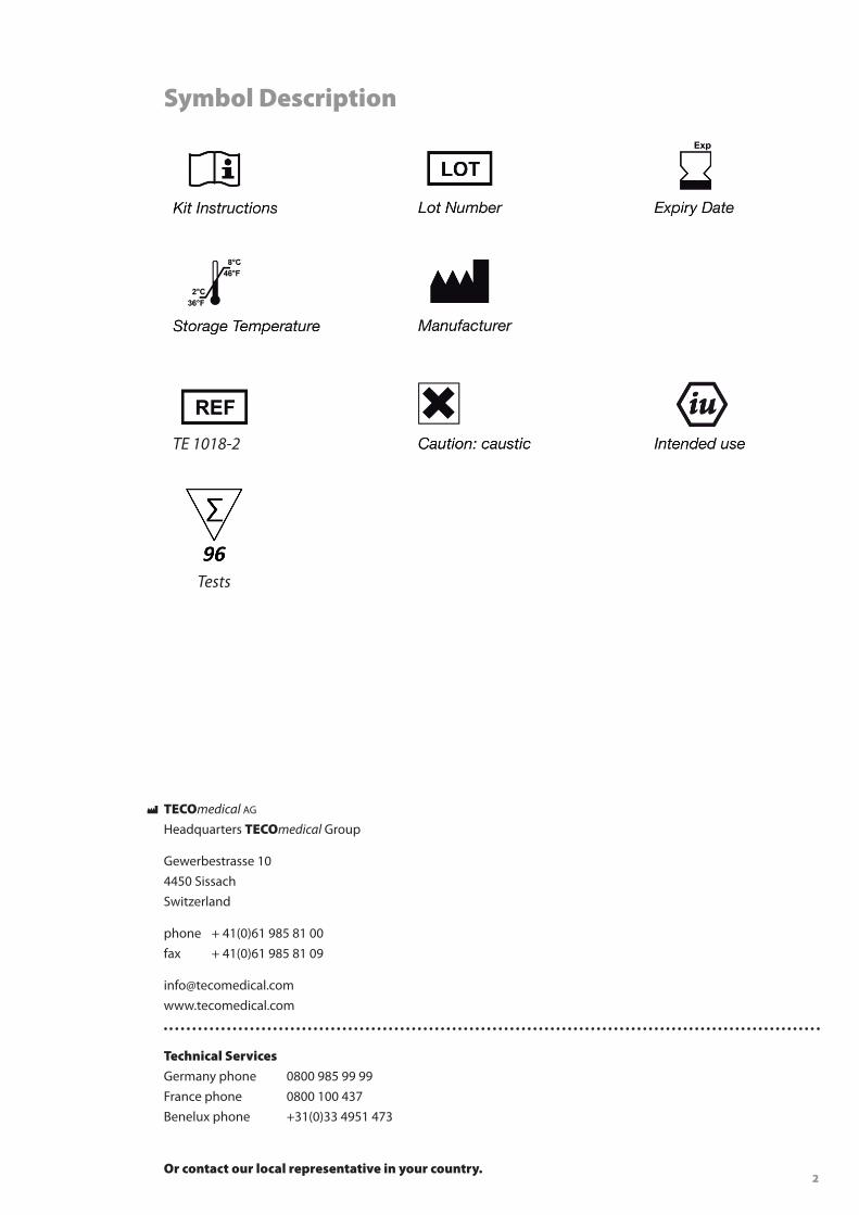
Symbol Description
TE 1018-2
Tests
TECOmedical 3
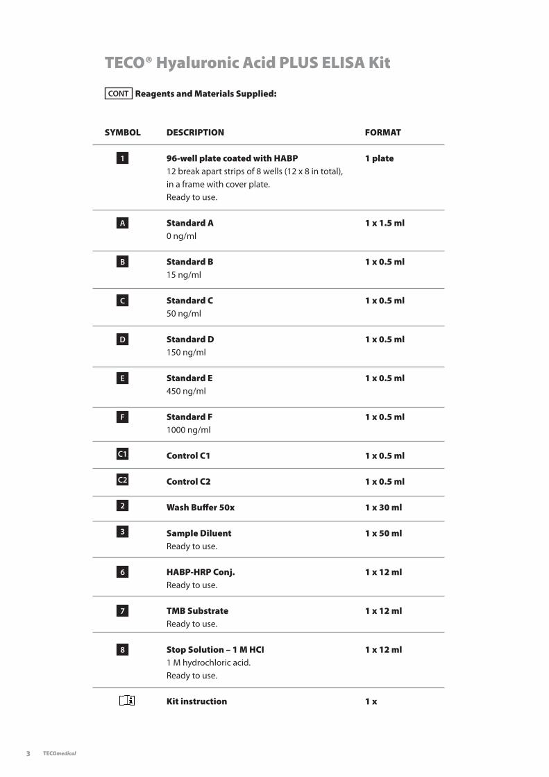
Symbol Description TECO® Hyaluronic Acid PLUS ELISA Kit
CONT Reagents and Materials Supplied:
SYMBOL DESCRIPTION FORMAT
1 96-well plate coated with HABP 1 plate 12 break apart strips of 8 wells (12 x 8 in total), in a frame with cover plate. Ready to use.
A Standard A 1 x 1.5 ml 0 ng/ml
B Standard B 1 x 0.5 ml 15 ng/ml
C Standard C 1 x 0.5 ml 50 ng/ml
D Standard D 1 x 0.5 ml 150 ng/ml
E Standard E 1 x 0.5 ml 450 ng/ml
F Standard F 1 x 0.5 ml 1000 ng/ml
C1 Control C1 1 x 0.5 ml
C2 Control C2 1 x 0.5 ml
2 Wash Buffer 50x 1 x 30 ml
3 Sample Diluent 1 x 50 ml Ready to use.
6 HABP-HRP Conj. 1 x 12 ml Ready to use.
7 TMB Substrate 1 x 12 ml Ready to use.
8 Stop Solution – 1 M HCI 1 x 12 ml 1 M hydrochloric acid. Ready to use.
Kit instruction 1 x
4
StorageStore kit at 2–8 °C. Do not freeze. Store unused reagents at 2–8 °C.
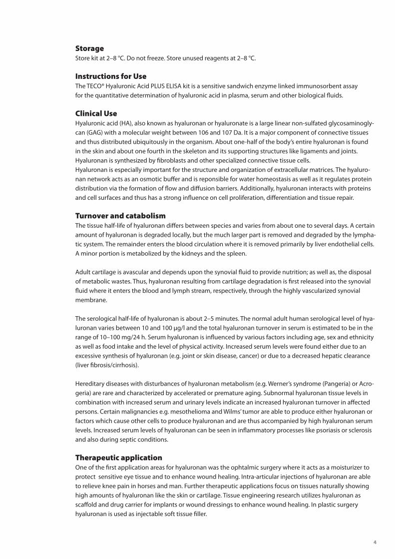
Instructions for UseThe TECO® Hyaluronic Acid PLUS ELISA kit is a sensitive sandwich enzyme linked immunosorbent assay for the quantitative determination of hyaluronic acid in plasma, serum and other biological fluids.
Clinical UseHyaluronic acid (HA), also known as hyaluronan or hyaluronate is a large linear non-sulfated glycosaminogly-can (GAG) with a molecular weight between 106 and 107 Da. It is a major component of connective tissues and thus distributed ubiquitously in the organism. About one-half of the body’s entire hyaluronan is foundin the skin and about one fourth in the skeleton and its supporting structures like ligaments and joints. Hyaluronan is synthesized by fibroblasts and other specialized connective tissue cells.Hyaluronan is especially important for the structure and organization of extracellular matrices. The hyaluro-nan network acts as an osmotic buffer and is reponsible for water homeostasis as well as it regulates protein distribution via the formation of flow and diffusion barriers. Additionally, hyaluronan interacts with proteins and cell surfaces and thus has a strong influence on cell proliferation, differentiation and tissue repair.
Turnover and catabolismThe tissue half-life of hyaluronan differs between species and varies from about one to several days. A certain amount of hyaluronan is degraded locally, but the much larger part is removed and degraded by the lympha-tic system. The remainder enters the blood circulation where it is removed primarily by liver endothelial cells. A minor portion is metabolized by the kidneys and the spleen.
Adult cartilage is avascular and depends upon the synovial fluid to provide nutrition; as well as, the disposal of metabolic wastes. Thus, hyaluronan resulting from cartilage degradation is first released into the synovial fluid where it enters the blood and lymph stream, respectively, through the highly vascularized synovial membrane.
The serological half-life of hyaluronan is about 2–5 minutes. The normal adult human serological level of hya-luronan varies between 10 and 100 µg/l and the total hyaluronan turnover in serum is estimated to be in the range of 10–100 mg/24 h. Serum hyaluronan is influenced by various factors including age, sex and ethnicity as well as food intake and the level of physical activity. Increased serum levels were found either due to an excessive synthesis of hyaluronan (e.g. joint or skin disease, cancer) or due to a decreased hepatic clearance (liver fibrosis/cirrhosis).
Hereditary diseases with disturbances of hyaluronan metabolism (e.g. Werner’s syndrome (Pangeria) or Acro-geria) are rare and characterized by accelerated or premature aging. Subnormal hyaluronan tissue levels in combination with increased serum and urinary levels indicate an increased hyaluronan turnover in affected persons. Certain malignancies e.g. mesothelioma and Wilms’ tumor are able to produce either hyaluronan or factors which cause other cells to produce hyaluronan and are thus accompanied by high hyaluronan serum levels. Increased serum levels of hyaluronan can be seen in inflammatory processes like psoriasis or sclerosis and also during septic conditions.
Therapeutic applicationOne of the first application areas for hyaluronan was the ophtalmic surgery where it acts as a moisturizer to protect sensitive eye tissue and to enhance wound healing. Intra-articular injections of hyaluronan are able to relieve knee pain in horses and man. Further therapeutic applications focus on tissues naturally showing high amounts of hyaluronan like the skin or cartilage. Tissue engineering research utilizes hyaluronan as scaffold and drug carrier for implants or wound dressings to enhance wound healing. In plastic surgery hyaluronan is used as injectable soft tissue filler.
TECOmedical 5
Hyaluronan as a biomarker
Joint diseaseHyaluronan is a main component of the cartilage matrix as well as the synovial fluid. Its viscoelastic properties are responsible for a proper joint function.
Proliferative synovial inflammation - which is a key feature of rheumatoid arthritis (RA) - results in the forced synthesis of hyaluronan; increasing both the synovial and serological level of hyaluronan. However, joint inflammation may also occur during other types of joint diseases, e.g. osteoarthritis (OA) or traumatic injury. In RA and OA patients, the concentration of hyaluronan correlates with the degree of joint inflammation and synovial proliferation, as well as, with the degree of joint space narrowing. Patients with higher initial values showed a more progressive course of disease. Therefore, serum hyaluronan could be used to study degenera-tive joint disease and to monitor disease progression, as well as, analyze the success of appropriate therapies. However, increased hyaluronan levels might indicate both forced hyaluronan synthesis due to synovial in-flammation and progressive cartilage degradation. Thus, hyaluronan values should be correlated with clinical or radiographic findings to clarify the disease focus.
Since RA patients, have shown markedly increased hyaluronan concentrations 0.5–2 hours after arising corresponding with a decrease in joint stiffness, valuable information might be obtained by sampling during the morning hours in these patients. Liver diseasesAs a result of permanent inflammation, most chronic liver diseases are characterised by fibrosis and cirrhosis causing a decreased capacity for hyaluronan clearance. The leading causes of chronic liver disease are viral infection (hepatitis B or C), and alcohol abuse. Patients with extensive liver fibrosis and cirrhosis show markedly increased serum levels of hyaluronan; with the progression of liver fibrosis being associated with an increase of serum hyaluronan. Serum hyaluronan could therefore be used to study patients with the risk of progressive fibrosis, as well as, to analyze the success of antifibrotic therapies. Additionally, serum hyaluronan is correlated with the rejection of liver transplants.
Tumor markerIn certain tumors, like prostate or breast cancer, the serological hyaluronan level seems to correlate with malignancy. Serum hyaluronan might therefore be used to analyze disease progression, as well as, patients responding to chemotherapy in certain tumors. In bladder cancer, the synthesis of hyaluronan is as-sociated with tumor angiogenesis and metastasis. Thus, increased urinary levels of hyaluronan may indicate the presence of bladder cancer regardless of tumor grade.
DiabetesSerum levels of hyaluronic acid were significantly higher in diabetic patients than in normal subjects and correlated with the levels of fasting plasma glucose and CRP and the body mass index (BMI). Diabetic complications, like retinopathy and nephropathy, were associated with higher hyaluronic acid serum levels. The hyaluronic acid level also correlated with the occurrence of diabetic angiopathy [6]. In overweight diabetes patients, hypertrophic fat cells showed an increased synthesis of hyaluronic acid causing a chronic inflammation of the adipose tissue [7].
Other applicationsHyaluronan concentrations have been also determined in various other sample types like perfusion and lavage fluids. Markedly increased hyaluronan levels were found in bronchoalveolar lavage fluids of patients with pulmonary inflammation like farmer’s lung, sarcoidosis or adult respiratory distress syndrome.
8
TECOmedical 7
Warnings and Precautions
This kit is intended for research use by professional persons only.Follow the instructions carefully.Observe expiration dates stated on the labels and the specified stability for reconstituted reagents. Refer to ”Materials Safety Data Sheet” for more detailed safety information.
Material of animal origin used in the preparation of this kit has been obtained from animals certified as healthy but these materials should be handled as potentially infectious.
Material of human origin used in the preparation of this kit has been tested and found non reactive for HIV-1 and HIV-2 as well as for HCV antibodies and HbsAg but should, nonetheless, be handled as potentially infectious.
TECOmedical AG is not liable for loss or harm caused by non-observance of the Kit instructions.
1 For research use only.2 Treat all specimen samples as potentially biohazardous material. Follow General Precautions when handling contents of this kit and any patient samples.3 Disposal of containers and unused contents should be done in accordance with federal and local regulatory requirements.4 Use the supplied reagents as an integral unit prior to the expiration date indicated on the package label.5 Store assay reagents as indicated.6 Do not use coated strips if pouch is punctured.7 Test each sample in duplicate.8 Use of multichannel pipettes or repeat pipettors is recommended to ensure the timely delivery of liquids.9 a. 1 M hydrochloric acid is caustic and can be harmfull for skin, eyes and mucosae. b. Handle TMB with care. Do not ingest. Avoid contact with skin, eyes, or clothing. Should there be any contact, wash with water. If ingested, call a physician.10 A mercury-free preservative is used. Incidental contact with or ingestion of buffer solutions may cause irritation of skin, eyes or mouth. Should there be any contact, wash with water. If ingested, call a physician.
8
Preparation of Reagents
1 HABP coated Microtiter Plate 12 break apart strips of 8 wells (96 in total) in a frame and sealed in a foil bag. Fit strip wells firmly into the frame. After opening, return any unused wells to the original foil package and seal. Store at 2–8 °C until expiration date. Cover for microtiter plates.
A Standards
F 6 vials of standard containing native hyaluronic acid. (0, 15, 50, 150, 450, and 1000 ng/ml).
Store at 2–8 °C until expiry date. C1 Control 1 1 vial of low control. Concentration see data sheet. Store at 2–8 °C until expiry date.
C2 Control 2 1 vial of high control. Concentration see data sheet. Store at 2–8 °C until expiry date.
2 Wash Buffer 50x 1 vial of 30 ml Wash Buffer concentrate. Dilute the 1:50 concentrate with deionized or distilled water up to 1500 ml. Store undiluted at 2–8 °C until expiration date. The diluted washing solution is stable for 4 weeks at 2–8 °C.
3 Sample Diluent 1 vial of 50 ml, ready to use. Store at 2–8 °C until expiration date.
6 HABP-HRP Conj. 1 vial of 12 ml, ready to use. Store at 2–8 °C until expiration date.
7 TMB Substrate 1 vial of 12 ml of H2O2 stabilized tetramethylbenzidine. Ready to use. Store at 2–8 °C until expiration date.
8 Stop Solution – 1 M HCI 1 vial of 12 ml of 1 M hydrochloric acid. Ready to use. Store at 2–8°C until expiration date.
till
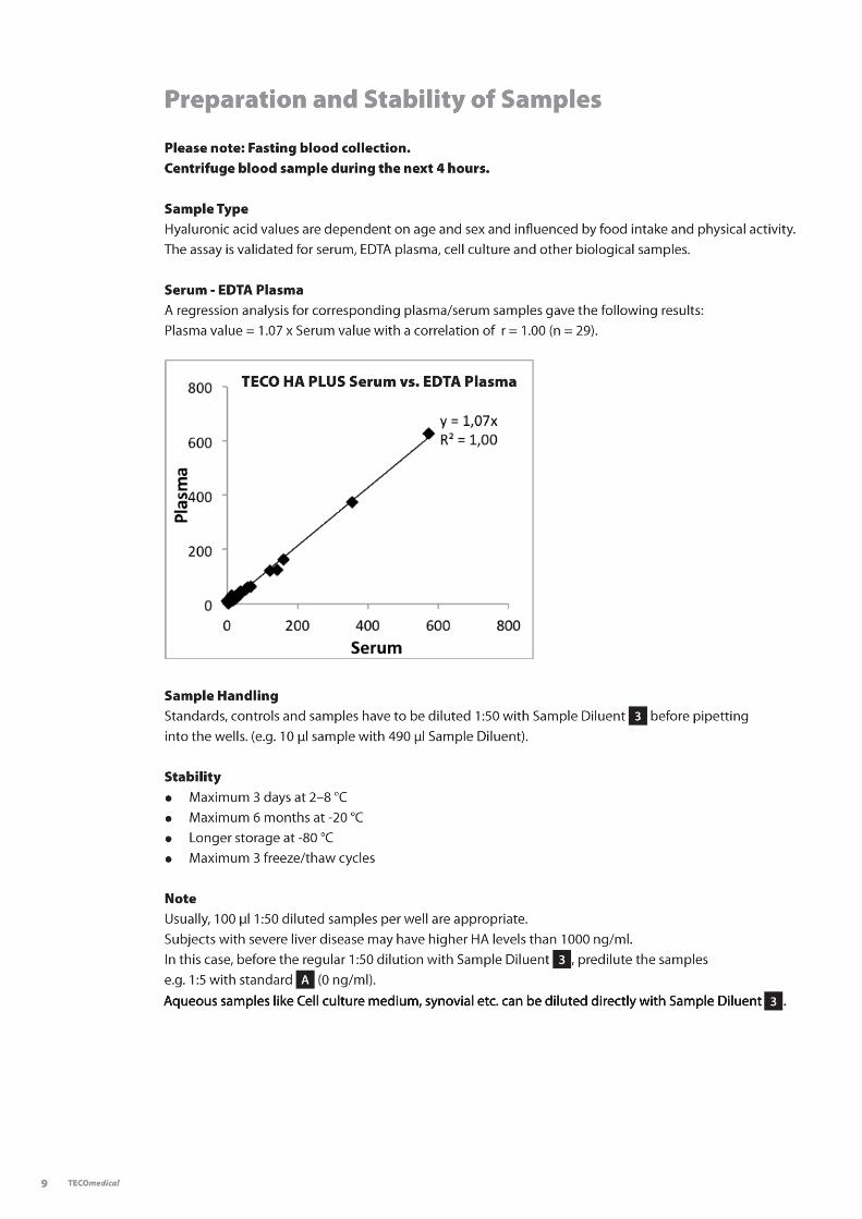
Aqueous samples like Cell culture medium, synovial etc. can be diluted directly with Sample Diluent .
10
Assay Procedure
All determinations (standards, controls and samples) should be assayed in duplicate. When performing the assay, the standards, controls and samples should be pipetted as fast as possible (<15 minutes).To avoid distortions due to differences in incubation times, HABP-HRP Conjugate, Substrate Solution and Stop Solution should be added to the plate in the same order and with the same time interval as the samples. A multichannel pipette is essential.
Allow all reagents to stand at room temperature (20–25 °C) for at least 30 minutes. During all incubation steps, plates should be sealed with the adhesive foil or a plastic cover. For light protection, incubate in a dark chamber or cover plate with aluminium foil.
1 Allocate the wells of the Microtiterplate 1 for standards, controls and samples.
2 Dilute standards ( A till F ), controls ( C1 and C2 ) and samples 1:50 with Sample Diluent 3 .
3 Pipette 100 µl of each diluted standards ( A till F), controls ( C1 and C2) and samples into the corresponding wells.
4 Cover the wells with a plastic cover and incubate the plate for 2h ± 5 min at room temperature (20–25 °C) on a shaker (500 rpm).
5 After incubation, aspirate the wells by using a plate washer or manually decant by inverting the plate. Wash the wells 3 times with 350 µl diluted Wash Buffer per well. After the last wash cycle tap the inverted wells on a dry absorbent surface to remove excess wash solution. The use of an automatic plate washer is recommended.
6 Following the last washing step, pipette 100 µl of the HABP-HRP Conjugate 6 in each well (multichannel pipette).
7 Cover the wells with a plastic cover and incubate the plate for 30 ± 5 min at room temperature (20–25 °C) on a shaker (500 rpm).
8 After incubation wash the wells 5 times with Wash Buffer as described in step 5.
9 Pipette 100 µl of the TMB Substrate Solution 7 in each well (multichannel pipette).
10 Incubate the plate for 30 min, in the dark, at room temperature (20–25 °C) on a shaker (500 rpm).
11 Stop the reaction by adding 100 µl of Stop Solution 8 (multichannel pipette). Measure the color reaction within 10 minutes at 450 nm (reference filter between 590–650 nm). If the extinction of the Std F exceeds 3.0, the measurement should be repeated at 405 nm.
Protocols for the different automatic ELISA systems are available.
TECOmedical 11
Result Analysis
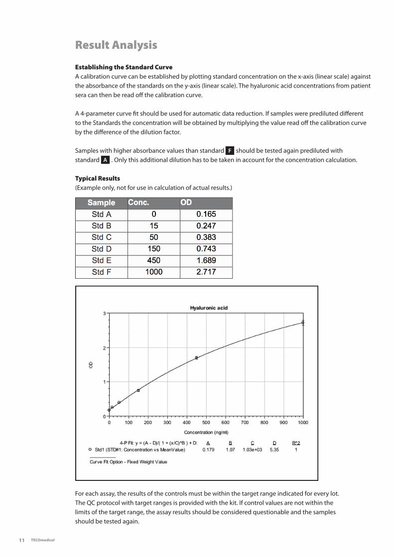
Establishing the Standard CurveA calibration curve can be established by plotting standard concentration on the x-axis (linear scale) against the absorbance of the standards on the y-axis (linear scale). The hyaluronic acid concentrations from patient sera can then be read off the calibration curve.
A 4-parameter curve fit should be used for automatic data reduction. If samples were prediluted different to the Standards the concentration will be obtained by multiplying the value read off the calibration curve by the difference of the dilution factor.
Samples with higher absorbance values than standard F should be tested again prediluted with standard A . Only this additional dilution has to be taken in account for the concentration calculation.
Typical Results(Example only, not for use in calculation of actual results.)
For each assay, the results of the controls must be within the target range indicated for every lot. The QC protocol with target ranges is provided with the kit. If control values are not within the limits of the target range, the assay results should be considered questionable and the samples should be tested again.
12
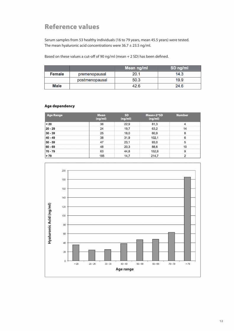
Reference values
Serum samples from 53 healthy individuals (16 to 79 years, mean 45.5 years) were tested. The mean hyaluronic acid concentrations were 36.7 ± 23.5 ng/ml.
Based on these values a cut-off of 90 ng/ml (mean + 2 SD) has been defined.
Age dependency
Hya
luro
nic
Aci
d (n
g/m
l)
Age Range Mean SD Mean+2*SD Number (ng/ml) (ng/ml) (ng/ml)
Age range
TECOmedical 13
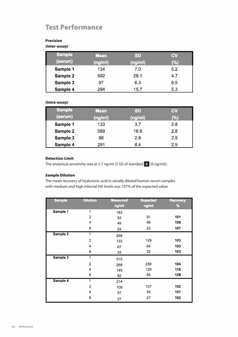
Test Performance
Precision(Inter-assay)
Detection LimitThe analytical sensitivity was at 2.7 ng/ml (3 SD of standard A (0 ng/ml)).
(Intra-assay)
Sample DilutionThe mean recovery of hyaluronic acid in serially diluted human serum samples with medium and high internal HA levels was 107% of the expected value.
14
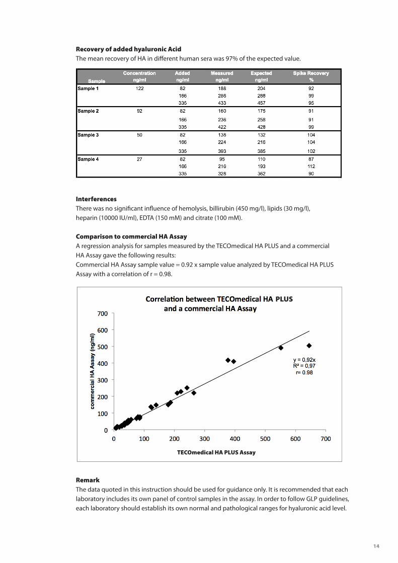
Recovery of added hyaluronic AcidThe mean recovery of HA in different human sera was 97% of the expected value.
InterferencesThere was no significant influence of hemolysis, billirubin (450 mg/l), lipids (30 mg/l), heparin (10000 IU/ml), EDTA (150 mM) and citrate (100 mM).
Comparison to commercial HA AssayA regression analysis for samples measured by the TECOmedical HA PLUS and a commercial HA Assay gave the following results: Commercial HA Assay sample value = 0.92 x sample value analyzed by TECOmedical HA PLUSAssay with a correlation of r = 0.98.
RemarkThe data quoted in this instruction should be used for guidance only. It is recommended that each laboratory includes its own panel of control samples in the assay. In order to follow GLP guidelines, each laboratory should establish its own normal and pathological ranges for hyaluronic acid level.
PLUS
TECOmedical HA PLUS Assay

TECOmedical 15
Note:
16
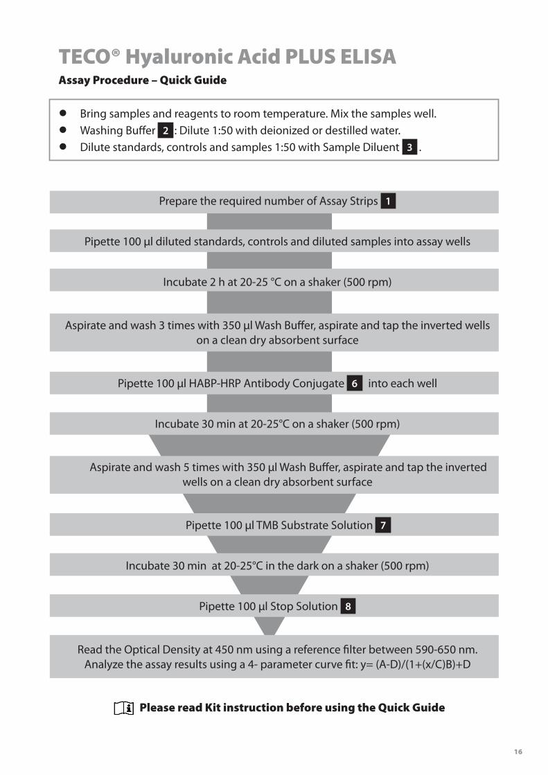
TECO® Hyaluronic Acid PLUS ELISAAssay Procedure – Quick Guide
• Bring samples and reagents to room temperature. Mix the samples well.
• Washing Buffer 2 : Dilute 1:50 with deionized or destilled water.
• Dilute standards, controls and samples 1:50 with Sample Diluent 3 .
Prepare the required number of Assay Strips 1
Pipette 100 µl diluted standards, controls and diluted samples into assay wells
Incubate 2 h at 20-25 °C on a shaker (500 rpm)
Aspirate and wash 3 times with 350 µl Wash Buffer, aspirate and tap the inverted wells on a clean dry absorbent surface
Pipette 100 µl HABP-HRP Antibody Conjugate 6 into each well
Incubate 30 min at 20-25°C on a shaker (500 rpm)
Aspirate and wash 5 times with 350 µl Wash Buffer, aspirate and tap the inverted wells on a clean dry absorbent surface
Pipette 100 µl TMB Substrate Solution 7
Incubate 30 min at 20-25°C in the dark on a shaker (500 rpm)
Pipette 100 µl Stop Solution 8
Read the Optical Density at 450 nm using a reference filter between 590-650 nm. Analyze the assay results using a 4- parameter curve fit: y= (A-D)/(1+(x/C)B)+D
Please read Kit instruction before using the Quick Guide