research article childhood septicemia in nepal...
TRANSCRIPT
Research ArticleChildhood Septicemia in Nepal: Documenting the BacterialEtiology and Its Susceptibility to Antibiotics
Shamshul Ansari,1 Hari Prasad Nepal,1 Rajendra Gautam,1 Sony Shrestha,1 Puja Neopane,1
Brihaspati Rimal,2 Fuleshwar Mandal,2 Safiur Rahman Ansari,3 and Moti Lal Chapagain1
1Department of Microbiology, Chitwan Medical College, Bharatpur, Chitwan, Nepal2Department of Biochemistry, Chitwan Medical College, Bharatpur, Chitwan, Nepal3Asian College for Advance Studies, Satdobato, Kathmandu, Nepal
Correspondence should be addressed to Shamshul Ansari; [email protected]
Received 31 October 2014; Revised 5 December 2014; Accepted 11 December 2014; Published 25 December 2014
Academic Editor: Maurizio Sanguinetti
Copyright © 2014 Shamshul Ansari et al. This is an open access article distributed under the Creative Commons AttributionLicense, which permits unrestricted use, distribution, and reproduction in any medium, provided the original work is properlycited.
Introduction. Children are among the most vulnerable population groups to contract illnesses.The varying microbiological patternof septicemia warrants the need for an ongoing review of the causative organisms and their antimicrobial susceptibility pattern.Therefore, the objective of this study was to document the bacterial etiology of childhood septicemia and its antibiotic susceptibilityprofile.Methods.Cross-sectional type of study in 1630 suspected patients was conducted at CMCTH from January 2012 toDecember2013. Blood samples were collected aseptically for culture. The organisms grown were identified by standard microbiologicalmethods recommended by American Society forMicrobiology (ASM) and subjected to antibiotic susceptibility testing bymodifiedKirby-Bauer disk diffusion method. Methicillin resistance was confirmed using cefoxitin and oxacillin disks methods. Results.Septicemia was detected in 172 (10.6%) cases. Among Gram-positive organisms, coagulase negative staphylococci (CoNS) wereleading pathogen and Acinetobacter spp. were leading pathogen among Gram-negative isolates. Vancomycin, teicoplanin, andclindamycin were the most effective antibiotics against Gram-positive isolates while amikacin was effective against Gram-positiveas well as Gram-negative isolates. Methicillin resistance was detected in 44.4% of Staphylococcus aureus. Conclusions. This studyhas highlighted the burden of bacterial etiology for septicemia among children in a tertiary care center of central Nepal.
1. Introduction
Presence of bacteria within the blood stream, continuous ortransient, is known as bacteremia, while the disseminationof bacteria throughout the body with evidence of systemicresponses towards microorganism with variable severity iscalled septicemia [1]. Septicemia is a common cause ofpediatricmorbidity andmortality [2].The highmortality ratevaries between 30 and 70 percent and depends on severalfactors including virulence of the pathogen and host factor[3, 4]. Bacteriological culture method to isolate the offendingpathogen remains the gold standardmethod for the diagnosisof bacteremia and septicemia [2, 5]. Organisms isolatedfrom the bloodstream of patients with sepsis vary from areato area [6]. In Nepal, the majority of the bacteremia and
septicemia cases are caused by a number of pathogens includ-ing coagulase negative staphylococci (CoNS), Staphylococcusaureus, Streptococcus spp., Enterobacter spp., Escherichia coli,Klebsiella pneumoniae, Salmonella spp., Acinetobacter spp.,Citrobacter spp., and Pseudomonas spp. [1, 7, 8].
The rational use of antibiotics for varyingmicrobiologicalpattern of septicemia in children warrants the need foran ongoing review of the causative organisms and theirantimicrobial susceptibility pattern [9, 10]. Uncontrolled useof various potent and broad-spectrum antibiotics has led toemergence of resistant strains which has become a majorproblem in various intensive care units. Therefore, under-standing of common pathogens and their drug sensitivitypattern in a specific region demands the correct use ofantibiotics. Due to constantly evolving antimicrobial resistant
Hindawi Publishing CorporationInternational Journal of MicrobiologyVolume 2014, Article ID 452648, 6 pageshttp://dx.doi.org/10.1155/2014/452648
2 International Journal of Microbiology
patterns, there is need for constant antimicrobial sensitiv-ity surveillance. This will help clinicians provide safe andeffective empirical therapies, develop rational prescriptionprograms and make policy decisions, and finally assess theeffectiveness of all [2].
The results of bacteriological cultures and antibioticsusceptibility tests method are time consuming that can takeabout 2–4 days, necessitating initial empirical treatment ofsuspected septicemia.Therefore, knowledge of the epidemio-logical and antimicrobial susceptibility patterns of commonpathogens in a given area helps to inform the choice ofantibiotics. In accordance with this, the present study wascarried out to document the bacterial etiology of septicemiaand its antibiotic sensitivity profile.
2. Materials and Methods
Across-sectional type of studywas carried out in bacteriologylaboratory of Chitwan Medical College Teaching Hospital (a600 bed hospital) in Narayani zone of central Nepal fromJanuary 2012 to December 2013.
2.1. Study Population. A total of 1630 patients during twoyears of study period between the ages of 1 month and 15years having clinical features suggestive of septicemia (fever,shortness in breath, weakness, drowsiness, irritability, etc.)were enrolled in this study.
2.2. Sample Collection. Two milliliter (mL) of blood samplesfrom early age children and 5mL of blood samples from lateage children were collected aseptically (clearing the skin with70% alcohol followed by 2% tincture of iodine) by clinicians,trained nurses, or laboratory staff using sterile syringe andneedle by venipuncture. Immediately the blood samples werecarefully transferred into the blood culture bottle containing18mL (if 2mL blood sample) or 45mL (if 5mL blood sample)of Brain Heart Infusion broth to maintain a ratio of 1 : 10 ofblood to broth. The blood culture bottles were labeled withthe patient’s name, age/sex, identification number, date, andtime of collection.
2.3. Bacteriological Processing. The Brain Heart Infusionbroth inoculated with blood was transported to the labo-ratory and incubated at 37∘C in aerobic condition. All thebottles were examined for turbidity, hemolysis, and pellicleformation and subcultures weremade on to sheep blood agar,chocolate agar, and MacConkey agar after overnight aerobicincubation. Blood agar and MacConkey agar plates wereincubated overnight at 37∘C in aerobic atmosphere whilechocolate agar plates were incubated overnight at 37∘C in 5%carbon dioxide (CO
2). Culture bottles were observed for tur-
bidity and blind subcultures were performed each day. Finalsubcultures were done on the 10th day before reporting neg-ative. Growth obtained was examined for colony and Gram-staining characteristics. Conventional biochemical tests wereperformed and identification of the organism was done byusing standard microbiological methods [11]. A purity plate
was employed to ensure that the inoculum used for thebiochemical tests was pure.
2.4. Antibiotic Susceptibility Testing. All the isolates grownwere subjected to antibiotic susceptibility testing by modi-fied Kirby-Bauer disk diffusion method in compliance withClinical and Laboratory Standards Institute (CLSI) guidelinesusing Mueller-Hinton agar standard media. The inhibitionzone standards for antimicrobial susceptibility were consid-ered from tables for interpretative zone diameters of CLSI[12].
Antibiotic disks (HiMedia Laboratories, Pvt. Limited,India) used were as follows: oxacillin (1 𝜇g), cefoxitin (30 𝜇g),vancomycin (30𝜇g), teicoplanin (30𝜇g), erythromycin(15 𝜇g), clindamycin (2𝜇g), penicillin G (10U), cephalexin(30 𝜇g), cotrimoxazole (25𝜇g), gentamicin (10 𝜇g), amikacin(30 𝜇g), ofloxacin (5 𝜇g), cefixime (5𝜇g), cefotaxime (30 𝜇g),ceftazidime (30 𝜇g), piperacillin (100 𝜇g), piperacillin-tazobactam (100/10 𝜇g), carbenicillin (100𝜇g), amoxycillin(10 𝜇g), and nalidixic acid (30 𝜇g).
Staphylococcus aureus ATCC 25923 and Escherichia coli25922were used as control organisms for antibiotic sensitivitytesting.
2.5. Identification of Methicillin Resistance in Staphylococcusaureus. Identification of methicillin resistant Staphylococcusaureus (MRSA) strains was carried out by using oxacillin(1 𝜇g) and cefoxitin (30 𝜇g) disks. Plates were incubated at35∘C. Plates containing oxacillin disk were read followinga 24-hour incubation period. The diameter of the zone ofinhibition (ZOI) of growth was recorded and interpretedas susceptible or resistant according to the criteria of CLSI.Staphylococcus aureus isolates were deemedmethicillin resis-tant when the ZOI was ≤10mm with the oxacillin disk or≤21mm with the cefoxitin disk [13].
2.6. Ethical Aspects. Verbal consent in local language forthis study was taken from the guardians of participatingchildren.This study was approved by the Institutional ReviewCommittee of Chitwan Medical College, Bharatpur, Nepal.
2.7. Data Analysis. Statistical analysis was performed usingSPSS-11.5 version. Association of septicemia with gender andtype of causative agents were assessed by using the chi-squaretest and others. 𝑃 values < 0.05 were considered statisticallysignificant.
3. Results
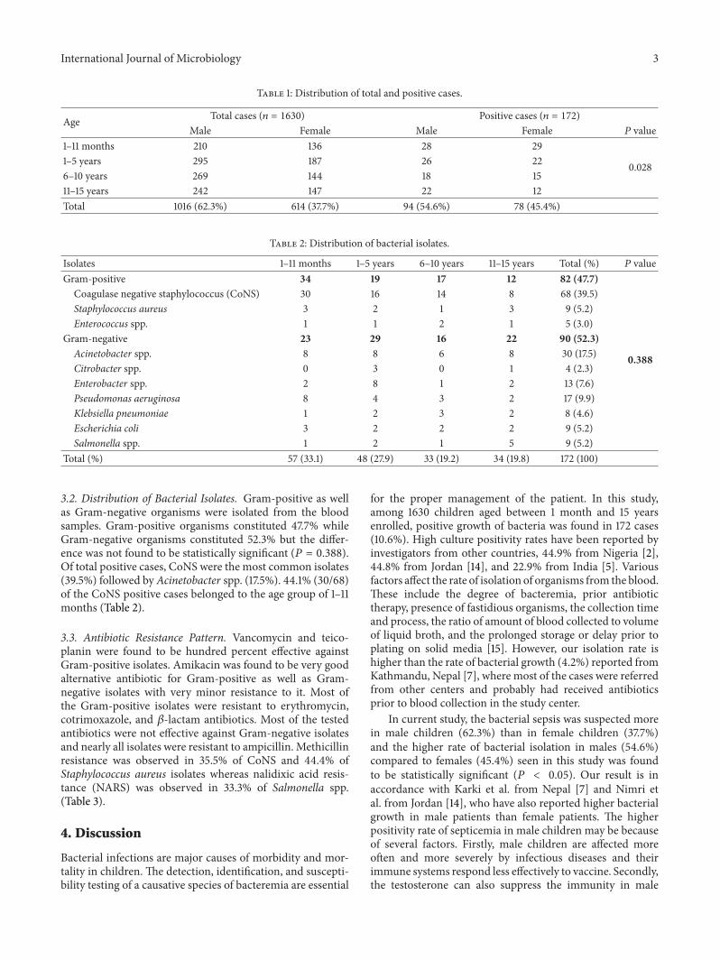
3.1. Age and Sex-Wise Distribution of Cases. During two yearsof study period, 1630 children aged between 1 month and 15years were enrolled, from whom positive growth of bacteriawas obtained in 172 cases (10.6%). Of total enrolled cases, 1016(62.3%) were males and 614 (37.7%) were females, whereas,out of total positive cases, 94 (54.6%) were males and 78(45.4%) were females which was found to be statisticallysignificant (𝑃 = 0.028). Suspected cases and positive caseswere more or less equal in each age group (Table 1).
International Journal of Microbiology 3
Table 1: Distribution of total and positive cases.
Age Total cases (𝑛 = 1630) Positive cases (𝑛 = 172)Male Female Male Female 𝑃 value
1–11 months 210 136 28 29
0.0281–5 years 295 187 26 226–10 years 269 144 18 1511–15 years 242 147 22 12Total 1016 (62.3%) 614 (37.7%) 94 (54.6%) 78 (45.4%)
Table 2: Distribution of bacterial isolates.
Isolates 1–11 months 1–5 years 6–10 years 11–15 years Total (%) 𝑃 valueGram-positive 34 19 17 12 82 (47.7)
0.388
Coagulase negative staphylococcus (CoNS) 30 16 14 8 68 (39.5)Staphylococcus aureus 3 2 1 3 9 (5.2)Enterococcus spp. 1 1 2 1 5 (3.0)
Gram-negative 23 29 16 22 90 (52.3)Acinetobacter spp. 8 8 6 8 30 (17.5)Citrobacter spp. 0 3 0 1 4 (2.3)Enterobacter spp. 2 8 1 2 13 (7.6)Pseudomonas aeruginosa 8 4 3 2 17 (9.9)Klebsiella pneumoniae 1 2 3 2 8 (4.6)Escherichia coli 3 2 2 2 9 (5.2)Salmonella spp. 1 2 1 5 9 (5.2)
Total (%) 57 (33.1) 48 (27.9) 33 (19.2) 34 (19.8) 172 (100)
3.2. Distribution of Bacterial Isolates. Gram-positive as wellas Gram-negative organisms were isolated from the bloodsamples. Gram-positive organisms constituted 47.7% whileGram-negative organisms constituted 52.3% but the differ-ence was not found to be statistically significant (𝑃 = 0.388).Of total positive cases, CoNS were the most common isolates(39.5%) followed by Acinetobacter spp. (17.5%). 44.1% (30/68)of the CoNS positive cases belonged to the age group of 1–11months (Table 2).
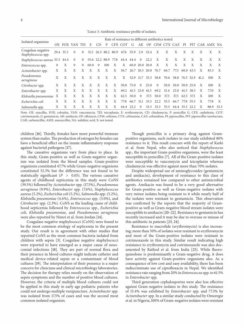
3.3. Antibiotic Resistance Pattern. Vancomycin and teico-planin were found to be hundred percent effective againstGram-positive isolates. Amikacin was found to be very goodalternative antibiotic for Gram-positive as well as Gram-negative isolates with very minor resistance to it. Most ofthe Gram-positive isolates were resistant to erythromycin,cotrimoxazole, and 𝛽-lactam antibiotics. Most of the testedantibiotics were not effective against Gram-negative isolatesand nearly all isolates were resistant to ampicillin. Methicillinresistance was observed in 35.5% of CoNS and 44.4% ofStaphylococcus aureus isolates whereas nalidixic acid resis-tance (NARS) was observed in 33.3% of Salmonella spp.(Table 3).
4. Discussion
Bacterial infections are major causes of morbidity and mor-tality in children. The detection, identification, and suscepti-bility testing of a causative species of bacteremia are essential
for the proper management of the patient. In this study,among 1630 children aged between 1 month and 15 yearsenrolled, positive growth of bacteria was found in 172 cases(10.6%). High culture positivity rates have been reported byinvestigators from other countries, 44.9% from Nigeria [2],44.8% from Jordan [14], and 22.9% from India [5]. Variousfactors affect the rate of isolation of organisms from the blood.These include the degree of bacteremia, prior antibiotictherapy, presence of fastidious organisms, the collection timeand process, the ratio of amount of blood collected to volumeof liquid broth, and the prolonged storage or delay prior toplating on solid media [15]. However, our isolation rate ishigher than the rate of bacterial growth (4.2%) reported fromKathmandu, Nepal [7], where most of the cases were referredfrom other centers and probably had received antibioticsprior to blood collection in the study center.
In current study, the bacterial sepsis was suspected morein male children (62.3%) than in female children (37.7%)and the higher rate of bacterial isolation in males (54.6%)compared to females (45.4%) seen in this study was foundto be statistically significant (𝑃 < 0.05). Our result is inaccordance with Karki et al. from Nepal [7] and Nimri etal. from Jordan [14], who have also reported higher bacterialgrowth in male patients than female patients. The higherpositivity rate of septicemia in male children may be becauseof several factors. Firstly, male children are affected moreoften and more severely by infectious diseases and theirimmune systems respond less effectively to vaccine. Secondly,the testosterone can also suppress the immunity in male
4 International Journal of Microbiology
Table 3: Antibiotic resistance profile of isolates.
Isolated organisms Rate of resistance to different antibiotics testedOX FOX VAN TEI E CD P CFX COT G AK OF CFM CTX CAZ PI PIT CAR AMX NA
Coagulase negativeStaphylococcus spp. 29.4 35.3 0 0 51.5 26.5 88.2 80.9 67.6 55.9 2.9 32.4 X X X X X X X X
Staphylococcus aureus 33.3 44.4 0 0 55.6 22.2 88.9 77.8 44.4 44.4 0 22.2 X X X X X X X XEnterococcus spp. 0 X 0 0 60.0 0 100 X X 60.0 20.0 20.0 X X X X X X X XAcinetobacter spp. X X X X X X X X 56.7 26.7 10.3 20.0 76.7 66.7 77.3 60.0 43.3 X 83.3 XPseudomonasaeruginosa X X X X X X X X X 52.9 11.7 35.3 58.8 70.6 58.8 76.5 52.9 41.2 100 X
Citrobacter spp. X X X X X X X X 50.0 75.0 0 25.0 0 50.0 50.0 50.0 25.0 X 100 XEnterobacter spp. X X X X X X X X 69.2 61.5 23.0 61.5 69.2 15.4 23.0 61.5 38.5 X 77.0 XKlebsiella pneumoniae X X X X X X X X 62.5 50.0 0 37.5 50.0 37.5 37.5 62.5 37.5 X 100 XEscherichia coli X X X X X X X X 77.8 66.7 11.1 33.3 22.2 55.5 66.7 77.8 55.5 X 77.8 XSalmonella spp. X X X X X X X X 44.4 22.2 0 33.3 33.3 55.5 44.4 55.5 22.2 X 88.9 33.3Note: OX: oxacillin, FOX: cefoxitin, VAN: vancomycin, TEI: teicoplanin, E: erythromycin, CD: clindamycin, P: penicillin G, CFX: cephalexin, COT:cotrimoxazole, G: gentamicin, AK: amikacin, OF: ofloxacin, CFM: cefixime, CTX: cefotaxime, CAZ: ceftazidime, PI: piperacillin, PIT: piperacillin-tazobactam,CAR: carbenicillin, AMX: amoxycillin, NA: nalidixic acid, X: not tested.
children [16]. Thirdly, females have more powerful immunesystem thanmales.The production of estrogen by females canhave a beneficial effect on the innate inflammatory responseagainst bacterial pathogens [17].
The causative organisms vary from place to place. Inthis study, Gram-positive as well as Gram-negative organ-ism was isolated from the blood samples. Gram-positiveorganisms constituted 47.7% while Gram-negative organismsconstituted 52.3% but the difference was not found to bestatistically significant (𝑃 > 0.05). The various causativeagents of childhood septicemia in this study were CoNS(39.5%) followed by Acinetobacter spp. (17.5%), Pseudomonasaeruginosa (9.9%), Enterobacter spp. (7.6%), Staphylococcusaureus (5.2%), Escherichia coli (5.2%), Salmonella spp. (5.2%),Klebsiella pneumoniae (4.6%), Enterococcus spp. (3.0%), andCitrobacter spp. (2.3%). CoNS as the leading cause of child-hood septicemia followed by Enterococcus spp., Escherichiacoli, Klebsiella pneumoniae, and Pseudomonas aeruginosawere also reported by Nimri et al. from Jordan [14].
Coagulase negative staphylococci (CoNS) were found tobe the most common etiology of septicemia in the presentstudy. Our result is in agreement with other studies thatreported CoNS as the most common bacteria isolated fromchildren with sepsis [3]. Coagulase negative staphylococciwere reported to have emerged as a major cause of noso-comial infections [18]. They are part of normal flora andtheir presence in blood cultures might indicate catheter andmedical device-related sepsis or a contaminant of bloodcultures [19]. The interpretation of their presence is a majorconcern for clinicians and clinical microbiology laboratories.The decision for therapy relies mostly on the observation ofsepsis symptoms and the number of positive blood cultures.However, the criteria of multiple blood cultures could notbe applied in this study in early age pediatric patients whocould not undergo multiple venipuncture. Acinetobacter spp.was isolated from 17.5% of cases and was the second mostcommon isolated organism.
Though penicillin is a primary drug against Gram-positive organisms, such isolates in our study exhibited 80%resistance to it. This result concurs with the report of Karkiet al. from Nepal, who also noticed that Staphylococcusspp., the important Gram-positive organisms, were the leastsusceptible to penicillin [7]. All of the Gram-positive isolateswere susceptible to vancomycin and teicoplanin whereasclindamycin was effective against more than 70% isolates.
Despite widespread use of aminoglycosides (gentamicinand amikacin), development of resistance to this class ofantibiotics remained low compared to other antimicrobialagents. Amikacin was found to be a very good alternativefor Gram-positive as well as Gram-negative isolates withvery minor isolates being resistant to it while nearly half ofthe isolates were resistant to gentamicin. This observationwas confirmed by the reports that the majority of Gram-positive as well as Gram-negative blood stream isolates weresusceptible to amikacin [20–22]. Resistance to gentamicin hasrecently increased and it may be due to overuse or misuse ofthis antibiotic in patients [23, 24].
Resistance to macrolide (erythromycin) is also increas-ing; more than 50% of isolates were resistant to erythromycinand most of the Gram-positive isolates were resistant tocotrimoxazole in this study. Similar result indicating highresistance to erythromycin and cotrimoxazole was also doc-umented by Rathod et al. from India [25]. While fluoro-quinolone is predominantly a Gram-negative drug, it doeshave activity against Gram-positive organisms also. As aconsequence of low cost and easy availability, there has beenindiscriminate use of ciprofloxacin in Nepal. We identifiedresistance rate ranging from20% inEnterococcus spp. to 61.5%in Enterobacter spp.
Third generation cephalosporins were also less effectiveagainst Gram-negative isolates in this study. The resistanceof 15.4% was exhibited by Enterobacter spp. and 77.3% byAcinetobacter spp. In a similar study conducted by Omoregieet al. inNigeria, 100%ofGram-negative isolateswere resistant
International Journal of Microbiology 5
to third generation cephalosporins [13]. Ceftriaxone andceftazidime are being used without laboratory guidance,especially as coverage antibiotic during surgery and as blindantibiotic in emergencies, which perhaps has resulted in bac-terial resistance to this drug. Amoxycillin was also not effec-tive against Gram-negative isolates, 77% to 100% of Gram-negative isolates were resistant to amoxycillin. Omoregie etal. from Nigeria have also observed that nearly all Gram-negative isolates were found to be resistant to amoxicillin [13].
Nowadays, the methicillin resistance in CoNS andStaphylococcus aureus (MRSA) is posing a great challengeto the treatment narrowing the regimen options for theseresistant bugs. Prior antibiotic use is the most common riskfactor for colonization and infection with MRSA. In thisstudy, 35.5% of CoNS and 44.4% of Staphylococcus aureusisolates were found to be resistant to methicillin. This resultcorroborates with 30.7% MRSA observed in children suffer-ing from bacteremia by Saravanan et al. in India [26]. Twodifferent methods were employed for the detection of MRSA.The cefoxitin disk method detected 44.4% of MRSA caseswhile the oxacillin disk method detected 33.3% of MRSA.According to CLSI the cefoxitin disk test is comparable tothe oxacillin disk test for the prediction of mecA-mediatedresistance to oxacillin [27]. The cefoxitin disk test is easier toread and thus is the preferredmethod. Besides, cefoxitin is aninducer of the mecA gene.
The emergence of quinolone resistance in the most com-mon Salmonella serotype worldwide is a serious public healthconcern. Resistance to nalidixic acid has been associated withreduced efficacy of fluoroquinolones such as ciprofloxacinand ofloxacin [28, 29]. In current study, 33.3% of Salmonellaspp. were nalidixic acid resistant (NARS).
There was no attempt to isolate anaerobic bacteria in thisstudy although they might have been the cause of bacteremiaand septicemia in some of the cases where no aerobic bacteriawere detected in blood cultures. Anaerobic bacteria werereported to constitute 18%of the total number of isolates fromblood [30].
5. Conclusions
This study has highlighted the burden of bacterial etiologyfor septicemia among children in a tertiary care center ofcentral Nepal. However, since the spectrum of pathogens,incidence of diseases, and antimicrobial susceptibility changeover time and places, the data should be monitored con-tinuously to allow an appropriate clinical response andhealthcare planning.This study highlights the variable natureof antibiotic susceptibility patterns. Therefore, it is advisableto continuously evaluate the resistance pattern of isolates soas to make a rational use of antibiotics.
The present study identified the burden of MRSA infec-tion among septicemic children. Regular monitoring ofantibiotic susceptibility pattern of MRSA and selection of adefinite antimicrobial agent may be helpful for reducing theincidence of MRSA infections in septicemia in children. Itis also important for clinicians to be aware of the existence
of the bacterial strains showing decreased fluoroquinolonesusceptibility.
Conflict of Interests
The authors declare that they have no conflict of interests.
Acknowledgments
The authors are deeply indebted to the children and theirparents for participating in this study. They also thankall the laboratory staff of the Bacteriology Department ofChitwan Medical College Teaching Hospital (CMCTH) fortheir kind support in the collection of data and performingthe necessary laboratory tests during the study.
References
[1] R. Chaudhary, S. Karmacharya, S. Shrestha et al., “Incidence ofBacteremia and Septicemia in patients attending in tertiary carecenter of Nepal,” Journal of Institute of Medicine, vol. 34, no. 3,pp. 32–38, 2012.
[2] M. M. Meremikwu, C. E. Nwachukwu, A. E. Asuquo, J. U.Okebe, and S. J. Utsalo, “Bacterial isolates fromblood cultures ofchildren with suspected septicaemia in Calabar, Nigeria,” BMCInfectious Diseases, vol. 5, article 110, 2005.
[3] R. P.Wenzel, M. R. Pinsk, R. J. Ulevitch, and L. Young, “Currentunderstanding of sepsis,”Clinical Infectious Diseases, vol. 22, no.3, pp. 407–412, 1996.
[4] A. P. Wheeler and G. R. Bernard, “Treating patients with severesepsis,”TheNew England Journal of Medicine, vol. 340, no. 3, pp.207–214, 1999.
[5] M. Sharma, N. Goel, U. Chaudhary, R. Aggarwal, and D. R.Arora, “Bacteraemia in children,” Indian Journal of Pediatrics,vol. 69, no. 12, pp. 1029–1032, 2002.
[6] J. Mugalu, M. K. Nakakeeto, S. Kiguli, and D. H. Kaddu-Mulindwa, “Aetiology, risk factors and immediate outcome ofbacteriologically confirmed neonatal septicaemia in Mulagohospital, Uganda,”African Health Sciences, vol. 6, no. 2, pp. 120–126, 2006.
[7] S. Karki, G. K. Rai, and R. Manandhar, “Bacteriological analysisand antibiotic sensitivity pattern of blood culture isolates inKanti children hospital,” Journal of Nepal Paediatric Society, vol.30, no. 2, pp. 94–97, 2010.
[8] R. Pradhan, U. Shrestha, S. C. Gautam et al., “Bloodstreaminfection among children presenting to a general hospitaloutpatient clinic in urban Nepal,” PLoS ONE, vol. 7, no. 10,Article ID e47531, 2012.
[9] N. Palikhe, “Prescribing pattern of antibiotics in PaediatricHospital of Kathmandu Valley,” Kathmandu University MedicalJournal, vol. 2, no. 5, pp. 6–12, 2004.
[10] M. Sharma, A. Yadav, S. Yadav, N. Goel, and U. Chaudhary,“Microbial profile of septicemia in children,” Indian Journal forthe Practising Doctor, vol. 5, pp. 9–10, 2008.
[11] I. Hd, Clinical Microbiology Procedures Handbook, ASM Press,Washington, DC, USA, 2nd edition, 2004.
[12] Clinical and Laboratory Standard Institute (CLSI), “Per-formance standards for antimicrobial susceptibility testing,”Wayne, Pa, USA, CLSI, M100-S16, 2006.
6 International Journal of Microbiology
[13] R. Omoregie, C. A. Egbe, H. O. Ogefere, I. Igbarumah, andR. E. Omijie, “Effects of gender and seasonal variation on theprevalence of bacterial septicemia among young children inBenin City, Nigeria,” Libyan Journal of Medicine, vol. 4, no. 3,pp. 107–109, 2009.
[14] L. F. Nimri,M. Rawashdeh, andM.M.Meqdam, “Bacteremia inchildren: etiologic agents, focal sites, and risk factors,” Journal ofTropical Pediatrics, vol. 47, no. 6, pp. 356–360, 2001.
[15] C. K. Shaw, P. Shaw, and A.Thapalial, “Neonatal sepsis bacterialisolates and antibiotic susceptibility patterns at a NICU in atertiary care hospital in western Nepal: a retrospective analysis,”Kathmandu University Medical Journal, vol. 5, no. 18, pp. 153–160, 2007.
[16] M.White, “Why do women have such stronger immune systemthan men?” 2014, http://www.psmag.com/navigation/health-and-behavior/girls-immune-systems-rule-boys-drool-73250/.
[17] Women have a more powerful immune system thanmen, 2009,http://phys.org/news161315351.html.
[18] N. B. Frebourg, S. Lefebvre, S. Baert, and J.-F. Lemeland,“PCR-based assay for discrimination between invasive andcontaminating Staphylococcus epidermidis strains,” Journal ofClinical Microbiology, vol. 38, no. 2, pp. 877–880, 2000.
[19] W. E. Kloos and T. L. Bannerman, “Update on clinical signifi-cance of coagulase-negative staphylococci,” Clinical Microbiol-ogy Reviews, vol. 7, no. 1, pp. 117–140, 1994.
[20] H. E. Akalin, M. Torun, and R. Alacam, “Aminoglycosideresistance patterns inTurkey,” Scandinavian Journal of InfectiousDiseases, vol. 20, no. 2, pp. 199–203, 1988.
[21] A. Japoni, S. Farshad, A. Alborzi et al., “Epidemiology andantibacterial susceptibility patterns of bloodstream infections,2001–2004: an experience with Bactec 9240 in Southern Iran,”Pakistan Journal of Biological Sciences, vol. 11, no. 3, pp. 422–427,2008.
[22] M. Barati, M. T. Taher, R. Abasi, M. M. Zadeh, M. Barati,and A. R. Shamshiri, “Bacteriological profile and antimicrobialresistance of blood culture isolates,” Iranian Journal of ClinicalInfectious Diseases, vol. 4, no. 2, pp. 87–95, 2009.
[23] A. Mohammad, “Bacteremia among Jordanian children atprincess Rahmahhospital: pathogens and antimicrobial suscep-tibility patterns,” Iranian Journal ofMicrobiology, vol. 2, no. 1, pp.22–26, 2010.
[24] S. Harbarth, P. Rohner, E. Safran, J. Garbino, R. Auckenthaler,and D. Pittet, “Resistance to amikacin and gentamicin amongGram-negative bloodstream isolates hospital between 1989 and1994,” Clinical Microbiology and Infection, vol. 4, no. 4, pp. 199–204, 1998.
[25] S. D. Rathod, P. V. Bhatia, P. H. Patel, J. D. Pethani, L. R. Patel,and B. Chauhan, “Bacteriological analysis and resistance pat-tern among various culture isolates from neonatal septicemiaat tertiary care hospital of Ahmedabad,” National Journal ofMedical Research, vol. 2, no. 4, pp. 466–469, 2012.
[26] M. Saravanan, A. Nanda, and T. Tesfaye, “Antibiotic susceptibil-ity pattern of methicillin resistant Staphylococcus aureus fromsepticemia suspected children in tertiary hospital in Hosur,South India,” American Journal of Microbiological Research, vol.1, no. 2, pp. 21–24, 2013.
[27] Clinical and Laboratory Standards Institute (CLSI), “Perfor-mance standards for antimicrobial susceptibility testing,” inProceedings of the 17th Informational Supplement, M100-S17,Clinical and Laboratory Standards (CLSI), Wayne, Pa, USA,2007.
[28] J. Wain, N. T. T. Hoa, N. T. Chinh et al., “Quinolone-resistantSalmonella typhi in Viet Nam:molecular basis of resistance andclinical response to treatment,” Clinical Infectious Diseases, vol.25, no. 6, pp. 1404–1410, 1997.
[29] K. Mølbak, D. L. Baggesen, F. M. Aarestrup et al., “An outbreakof multidrug-resistant, quinolone-resistant Salmonella entericaserotype typhimurium DT104,” The New England Journal ofMedicine, vol. 341, no. 19, pp. 1420–1425, 1999.
[30] D. E. Anuradha, K. Saraswathi, and A. Gogate, “Anaerobicbacteraemia: a review of 17 cases,” Journal of PostgraduateMedicine, vol. 44, no. 3, pp. 63–66, 1998.

Submit your manuscripts athttp://www.hindawi.com
Hindawi Publishing Corporationhttp://www.hindawi.com Volume 2014
Anatomy Research International
PeptidesInternational Journal of
Hindawi Publishing Corporationhttp://www.hindawi.com Volume 2014
Hindawi Publishing Corporation http://www.hindawi.com
International Journal of
Volume 2014
Zoology
Hindawi Publishing Corporationhttp://www.hindawi.com Volume 2014
Molecular Biology International
GenomicsInternational Journal of
Hindawi Publishing Corporationhttp://www.hindawi.com Volume 2014
The Scientific World JournalHindawi Publishing Corporation http://www.hindawi.com Volume 2014
Hindawi Publishing Corporationhttp://www.hindawi.com Volume 2014
BioinformaticsAdvances in
Marine BiologyJournal of
Hindawi Publishing Corporationhttp://www.hindawi.com Volume 2014
Hindawi Publishing Corporationhttp://www.hindawi.com Volume 2014
Signal TransductionJournal of
Hindawi Publishing Corporationhttp://www.hindawi.com Volume 2014
BioMed Research International
Evolutionary BiologyInternational Journal of
Hindawi Publishing Corporationhttp://www.hindawi.com Volume 2014
Hindawi Publishing Corporationhttp://www.hindawi.com Volume 2014
Biochemistry Research International
ArchaeaHindawi Publishing Corporationhttp://www.hindawi.com Volume 2014
Hindawi Publishing Corporationhttp://www.hindawi.com Volume 2014
Genetics Research International
Hindawi Publishing Corporationhttp://www.hindawi.com Volume 2014
Advances in
Virolog y
Hindawi Publishing Corporationhttp://www.hindawi.com
Nucleic AcidsJournal of
Volume 2014
Stem CellsInternational
Hindawi Publishing Corporationhttp://www.hindawi.com Volume 2014
Hindawi Publishing Corporationhttp://www.hindawi.com Volume 2014
Enzyme Research
Hindawi Publishing Corporationhttp://www.hindawi.com Volume 2014
International Journal of
Microbiology