presbyopia treatment refractive cataract surgery … · presbyopia treatment refractive cataract...
TRANSCRIPT

EUROTIMESESCRS
S U P P L E M E N T S E P T E M B E R 2 0 0 6
Merging Cataract and Refractive Surgery
Improving Visual Quality and Acuity with AdvancedCorneal and Lenticular Technologies
Inside this Issue:Presbyopia Treatment
Refractive Cataract Surgery
Laser Vision Correction
Pseudophakic LASIK

By Baha Toygar, MD
At a private eye hospital in Turkey, mycolleagues and I have performedapproximately 300 Tecnis Multifocal IOLimplantations. The Tecnis Multifocal is asilicone lens with an aspheric modifiedprolate anterior surface (just like the Tecnismonofocal), and a diffractive posteriorsurface. The optics diffract incoming light to
two focal points for near and distance vision, with about half thelight distributed to each. It is available in a wide dioptric range of 5D to 34 D.
We recently presented long-term (12-month) data on 37 eyes of 19patients with this IOL. The mean age was 48.4, with a fairly widerange (22-77 years). Twenty-five eyes were hyperopic, 6 myopic, and6 plano presbyopes.
Study ResultsIn the hyperopic eyes, the mean spherical equivalent (SE) improvedfrom 3.43 ± 2.25 D to 0.44 ± 0.42 D at 12 months. In the myopiceyes, the mean SE improved from -8.02 ± 5.83 D to 0.52 ± 0.42 D.
At 1 year, mean monocular uncorrected distance visual acuityimproved from 0.29 (20/69) preop to 0.64 (20/31) postop. Best-corrected acuity was unchanged at 0.85 (20/23).
Uncorrected near visual acuity was considerably enhanced, with89% of eyes achieving J3 or better and 81% of eyes reading J2.Withdistance correction, 95% of eyes could read J2 or better, including76% achieving J1 or better. About half the patients were testedbinocularly at 12 months, and all of them could read at least J2 withboth eyes.
We also tested intermediate vision at 60 cm in a subset of about halfthe patients. Seventy-eight percent of these patients achieved atleast J2 intermediate vision with both eyes open. Monocularly, 63%were J3 or better and nearly a third were J2 or better. This isreassuring. I had been concerned that the design of the TecnisMultifocal might not provide sufficient intermediate vision, but theacuity results were quite good and we had no complaints from thepatients in this study about difficulty with computers or otherintermediate tasks.
Visual acuity after Tecnis Multifocal implantation is stable andpredictable. There were no surgical complications in the study andno problems with centration. Two eyes underwent Nd:YAGcapsulotomy postop. One patient had an IOL exchange (both eyes).This patient, a dental technician who was a plano presbyope, hadgood distance acuity and J1 near vision postop but complained ofreflections off his dental instruments. We implanted Tecnismonofocal lenses instead and he is happy now.
Patient satisfaction in this study has been high. Ninety-five percentof patients told us they never wear spectacles, and almost all weresatisfied or very satisfied with the results.
Patients reported experiencing some glare and halos around lights.The vast majority ranked these symptoms as mild (1-2 on a scale of1 to 6). Three patients complained of more severe glare and halo (5-6). However, 94% said they had little or no difficulty seeing at night.
Our study demonstrates that the Tecnis Multifocal IOL effectivelycorrects far, intermediate, and near vision, reducing patientdependency on glasses. With appropriate patient selection andpreoperative biometry and IOL power calculations, outcomes arecomparable to LASIK. This is an excellent modality for refractivecorrection, especially in hyperopes and higher myopes who may notbe good candidates for laser refractive surgery.
Dr.Toygar is the director of the refractive surgery department at DünyaEye Hospital, Levent Branch, in Istanbul,Turkey. He has no financialrelationship with AMO. Contact him at +902123393900 [email protected].
Patients Highly Satisfied with Bilaterial Diffractive Multifocal IOLsResults at 1 year show Tecnis Multifocal provides excellent visual acuity and quality
2
Presbyopia Treatment
Multifocal IOL Pearls
■ Avoid plano presbyopes and low myopes for initial cases
■ Counsel patients appropriately
■ Use IOLMaster or immersion biometry
■ Use appropriate IOL power calculation formula
■ Make a careful capsulorrhexis
Table 1:12-Month Near and Intermediate Vision Results with TecnisMultifocal
≥ J5 ≥ J3 ≥ J2 ≥ J1
Monocular DCNVA 100% 94.6% 94.6% 75.7%
Monocular UCNVA 97.3% 89.2% 81.1% 51.4%
Binocular UCNVA (n=9) 100% 100% 100% 66.6%
Monocular UCIVA (n=19) 73.7% 63.2% 31.6% 0
Binocular UCIVA (n=9) 100% 78% 78% 0
“Ninety-five percent of patients told usthey never wear spectacles, and almostall were satisfied or very satisfied withthe results”

By Frank Goes, MD
Patients can achieve excellent near anddistance vision and spectacle independenceacross a range of distances with a diffractivelens, such as the Tecnis ZM900 Multifocal IOL.Recent studies show that patients can havegood reading vision at short distances andalso have excellent intermediate visionwithout compromising distance vision.
The TechnologyThe Tecnis Multifocal lens has a prolate surface that is designed tocompensate for spherical aberrations of the cornea and improvecontrast sensitivity.
The multifocal lens is indicated for cataract patients and visualcorrection of presbyopia.
Careful patient selection remains important. Patients can read atshort distances while maintaining excellent intermediate vision.Thisis achieved without compromising distance vision. Multifocality isindependent of pupil size.
Patient SelectionBy determining the patients’ desires for their vision and lifestyle, wecan best determine their needs. I ask all my patients how importantis it if they are spectacle free? I then discuss their lifestyle andoccupational activities. I make sure to ask them several keyquestions, such as: Do they play sports or do a great deal of reading?Do they do a lot of night driving or computer work? I also considertheir personalities, are they an obsessive person?
It is important to discuss the possibility of postoperative glare andhalos with each patient. Also, I never promise a patient that they willbe “free of spectacles.” Instead, I advise them that they will be “moreindependent with multifocals” and often say that in more than 90%of cases, they will be spectacle-free for both far and near.
Recent StudiesIn an average cornea eye model (with a 4-mm or 5-mm pupil), visualquality at near and far with the Tecnis™ ZM900 lens with a modifiedprolate surface was better than the ReSTOR multifocal lens with aspheric surface.
We conducted a study of 114 eyes of refractive lensectomy patientsfor correction of presbyopia and hyperopia (range +1.5D to +8D).The mean patient age at the time of surgery was 55 years. Eighty-four eyes of 42 patients were reviewed at three-month follow-up.Patients had a simultaneous bilateral outpatient surgery withtopical anesthesia.These patients presented with refractiveproblems, and some only had reading problems.
ResultsThe mean spherical equivalent (SE) before surgery was 4.21 D andpostop SE improved to 0.29 D. At three months, 40 out of the 42patients in the study indicated that they never used spectacles
afterwards. Only two patients needed spectacles for hobby orpersonal computer work at intermediate distances (-1.25 D glasses).
At three months, the distance uncorrected vision was a mean of20/25 monocular. Distance best corrected vision had a mean of20/20 monocular, but 14/84 eyes had LASIK as an enhancement.Thepossible need of LASIK enhancement should be discussed prior totreatment.
For near uncorrected vision, 95% of patients achieved Jaeger 1uncorrected monocular. For near with distance correction, 98% ofpatients reached Jaeger 1 distance corrected monocular.
Intermediate vision required for PC work is critical for many patientstoday. In this study, intermediate PC vision was excellent. Also at 50cm binocular and eventually distance corrected, 40% of patientscould read Jaeger 2 and another 52% could read Jaeger 3.
Patient FeedbackAmazingly, patients did not have any complaints at all concerningtheir distance vision. However, some patients did indicate that theyexperienced some side effects, which decreased over time.
A review of the six month follow-up data shows that some patientsstill mention seeing rings from time to time; but they are no longerdisturbed by the occurrence any more.
Concerning the three-month data, 16% of patient indicated minorsubjective complaints when asked, while 7% reported majorsubjective complaints.Two patients noted significant drivingproblems at night, but felt they could still drive.
All patients said they would recommend the lens and not onepatient considered explantation.
Future OutlookThe overall acceptance and patient satisfaction for this combinationprocedure was excellent. I performed simultaneous bilateral surgeryand topical anesthesia on an outpatient basis and the recuperationtime was extremely short.
Dr. Frank Jozef Goes is medical director of the Goes Eye Centre-International Representative in Antwerp, Belgium. He can be reached atTel. +.32.(0)3.2193925; Fax. +32. (0)3.2196667 or at [email protected]
Achieving Spectacle Independence with Bilateral Multifocal Lens ImplantationDiffractive IOLs can provide patients with excellent near and distance vision
3
Presbyopia Treatment
“At three months, 40 out of the 42patients in the study indicated that theynever used spectacles afterwards”
3 Month Visual Acuity Outcomes
Mean UCVA for Distance 20/25
Mean DCNVA 20/20
UCNVA 95% J1 or better
DCNVA 98% J1 or better
By Leonardo Akaishi, MD
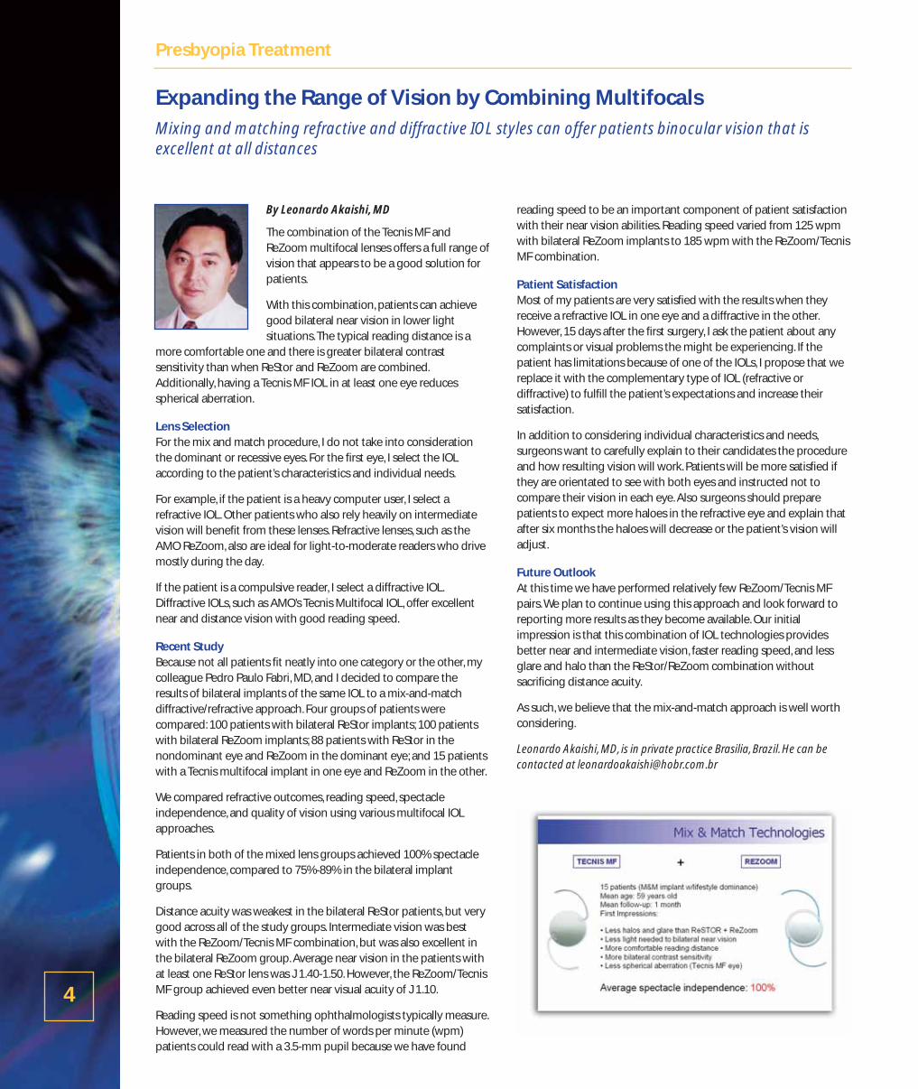
The combination of the Tecnis MF andReZoom multifocal lenses offers a full range ofvision that appears to be a good solution forpatients.
With this combination, patients can achievegood bilateral near vision in lower lightsituations.The typical reading distance is a
more comfortable one and there is greater bilateral contrastsensitivity than when ReStor and ReZoom are combined.Additionally, having a Tecnis MF IOL in at least one eye reducesspherical aberration.
Lens SelectionFor the mix and match procedure, I do not take into considerationthe dominant or recessive eyes. For the first eye, I select the IOLaccording to the patient’s characteristics and individual needs.
For example, if the patient is a heavy computer user, I select arefractive IOL. Other patients who also rely heavily on intermediatevision will benefit from these lenses. Refractive lenses, such as theAMO ReZoom, also are ideal for light-to-moderate readers who drivemostly during the day.
If the patient is a compulsive reader, I select a diffractive IOL.Diffractive IOLs, such as AMO’s Tecnis Multifocal IOL, offer excellentnear and distance vision with good reading speed.
Recent StudyBecause not all patients fit neatly into one category or the other, mycolleague Pedro Paulo Fabri, MD, and I decided to compare theresults of bilateral implants of the same IOL to a mix-and-matchdiffractive/refractive approach. Four groups of patients werecompared: 100 patients with bilateral ReStor implants; 100 patientswith bilateral ReZoom implants; 88 patients with ReStor in thenondominant eye and ReZoom in the dominant eye; and 15 patientswith a Tecnis multifocal implant in one eye and ReZoom in the other.
We compared refractive outcomes, reading speed, spectacleindependence, and quality of vision using various multifocal IOLapproaches.
Patients in both of the mixed lens groups achieved 100% spectacleindependence, compared to 75%-89% in the bilateral implantgroups.
Distance acuity was weakest in the bilateral ReStor patients, but verygood across all of the study groups. Intermediate vision was bestwith the ReZoom/Tecnis MF combination, but was also excellent inthe bilateral ReZoom group. Average near vision in the patients withat least one ReStor lens was J 1.40-1.50. However, the ReZoom/TecnisMF group achieved even better near visual acuity of J 1.10.
Reading speed is not something ophthalmologists typically measure.However, we measured the number of words per minute (wpm)patients could read with a 3.5-mm pupil because we have found
reading speed to be an important component of patient satisfactionwith their near vision abilities. Reading speed varied from 125 wpmwith bilateral ReZoom implants to 185 wpm with the ReZoom/TecnisMF combination.
Patient SatisfactionMost of my patients are very satisfied with the results when theyreceive a refractive IOL in one eye and a diffractive in the other.However, 15 days after the first surgery, I ask the patient about anycomplaints or visual problems the might be experiencing. If thepatient has limitations because of one of the IOLs, I propose that wereplace it with the complementary type of IOL (refractive ordiffractive) to fulfill the patient’s expectations and increase theirsatisfaction.
In addition to considering individual characteristics and needs,surgeons want to carefully explain to their candidates the procedureand how resulting vision will work. Patients will be more satisfied ifthey are orientated to see with both eyes and instructed not tocompare their vision in each eye. Also surgeons should preparepatients to expect more haloes in the refractive eye and explain thatafter six months the haloes will decrease or the patient’s vision willadjust.
Future OutlookAt this time we have performed relatively few ReZoom/Tecnis MFpairs.We plan to continue using this approach and look forward toreporting more results as they become available. Our initialimpression is that this combination of IOL technologies providesbetter near and intermediate vision, faster reading speed, and lessglare and halo than the ReStor/ReZoom combination withoutsacrificing distance acuity.
As such, we believe that the mix-and-match approach is well worthconsidering.
Leonardo Akaishi, MD, is in private practice Brasilia, Brazil. He can becontacted at [email protected]
Expanding the Range of Vision by Combining Multifocals Mixing and matching refractive and diffractive IOL styles can offer patients binocular vision that isexcellent at all distances
4
Presbyopia Treatment
By Ángel López Castro, MD
Mixing and matching the Tecnis Multifocal(MF) and ReZoom IOLs can maximizepatient vision at both near and fardistances.
I am implanting this combination in allcataract surgery or clear lens extractionpatients who are interested in discharging
the uses of glasses for distance and near vision. To date, I haveimplanted 40 patients with this combination. The majority ofthese patients are clear lens exchange patients.
My first mixed implants have achieved excellent distance vision.The ReZoom provides improved distance vision compared withthe Tecnis MF. Also, because of the optical design of both lenses,patients achieve better distance and near vision with differentpupil sizes. Therefore, patients’ maximize the range of vision indifferent light condition. The Tecnis MF is a superb IOL forreading with good light, but not as good in dim condition.Conversely, ReZoom is wonderful for distance in bright light andbetter for reading in dim light.
In my experience, it is better to implant the Tecnis MF firstbecause near vision rehabilitation is faster than with ReZoom. Todate, no patient regrets doing the second eye because the resultof the first. I always implant the ReZoom in the dominant eye.
ResultsPatients are reporting high levels of satisfaction with their vision.I will perform a LASIK enhancement if there is any residualdistance refraction. Therefore, for far vision there are no patientsusing correction, unless there is a reason not to recommend alaser procedure.
Patient SelectionFirst and foremost, patients must be interested in near visionwithout glasses. Also, patients who are eager to be spectacle-independent usually are more willing to accept the potentialvisual side effects of the IOLs. In cataract patients, goodcandidates are those with a quiet lifestyle, interested in nearvision without glasses, but who do not want to lose thesharpness of the monofocal IOLs for distance.
The ideal patient for PRELEX mix and match is a presbyopic orprepresbyopic hyperope (45 years or older), especially highhyperopes. Second best are high myopes (more than -6.00D),followed by emmetropic presbyopes, and then the least likelycandidates are low myopes.
PRELEX should be done with extreme caution in emmetropesand patients with minor refractive errors because of a highincidence of intolerable halos and secondary procedures.Informed consent should clearly state the frequency ofsecondary procedure, in particular IOL exchange, and excimerlaser touch up.
Photic Phenomena All my patients are advised about halos and glare. I explain thatphotic phenomena are normal and usually resolve way or declinewithin about three months. I note that they could persist and beuncomfortable. However, no patients have asked for explantationof the IOL.
I carefully explain realistic expectations for the procedure. Thisrequires more chair time in the office. Surgeons must know morethings about the patient’s lifestyle, visual needs, and personality. Itell each patient that it is better not to do the procedure if theycan not assume some photic phenomena, or if they are expectinga perfect vision, or if they are looking for perfection in near ordistance vision with poor light conditions.
All my patients who have been operated on are driving during dayand night without difficulties. The majority do not notice halos orglare at three months and can read without any prescription. Ialways inform them about the possibility of needing glasses forsome tasks that they considered as very tricky or in unusual lightconditions.
Recommendations Surgeons who are going to begin using both a refractive anddiffractive lens in the same patient must keep in mind that theselection of the patient is critical, and it is important to dischargethose who will be uncomfortable.
Also, it is crucial to get emmetropia and I recommend the use ofIOL Master biometry with The Haigis Formula. I also do arcuatekeratotomy at the end of the lens extraction. A LASIK or PRKtouch-up is necessary if there is any residual refraction remainingtwo months after lens surgery as any residual error will lead to anunsatisfied patient.
If you reckon the patient is happy, but is still complaining, you cansuggest removal of the IOL and exchange for a monofocalwarning them that they will lose the ability to read withoutglasses. I believe all of the patients would not accept that optionand would choose their present situation.
To date, I have not had any need for a lens exchange; andtherefore in my opinion it is an option worth considering for ourpresbyopic and cataract patients.
Ángel López Castro, MD, is with Laservision Clínica Oftalmológica inMadrid. He can be reached at +34914448230 or by email at:[email protected].
Key Considerations for Successful Mixing and Matching The differing IOLs’ designs allow some patients to achieve better distance and near vision,which increases range of vision in different light conditions
5
Presbyopia Treatment
“A LASIK or PRK touch-up is necessary ifthere is any residual refraction remainingtwo months after lens surgery as anyresidual error will lead to an unsatisfiedpatient”
By W. Bruce Jackson, MD
Long-term International/Canadian studyresults for the correction of hyperopicpresbyopia are excellent with 100% ofsubjects achieving simultaneous 20/25distance vision and J3 near vision at 12months. In the majority of cases, spectacleindependence is achieved and maintained.In addition, patients report a high level ofsatisfaction.
Treatment DesignThe treatment design uses a patented wavefront-guidedaspheric presbyopic ablation profile and VSS ablationtechnology is used to create subtle ablation shape changes tothe subject’s wavefront map. The central zone is steepened toprovide near vision and the peripheral zone is targeted fordistance vision.
The combination of the pupil-size dependent central zone, theperipheral zone, and the LASIK flap produces an aspheric curvethat is customized to the patients eye and expands the depth offocus.
ResultsIn the multicenter trial, 82 eyes of 49 hyperopic presbyopicsubjects received treatment. Preoperative refractions includemean manifest sphere of +1.66 D ± 0.60 (range +0.5 to +3.50 D),and cylinder: +0.43 D ± 0.35 (range +0.00 to +1.50 D). The meanpatient age was 56 ± 5 (range 47 to 68) and 68% were female.Follow up is available for 12 months.
Most patients received bilateral LASIK treatments with the AMOAdvanced CustomVue aspheric ablation treatment. This was apupil-size dependent presbyopic correction with no nomogramadjustment and no retreatments. The Amadeus microkeratomewas used in all cases.
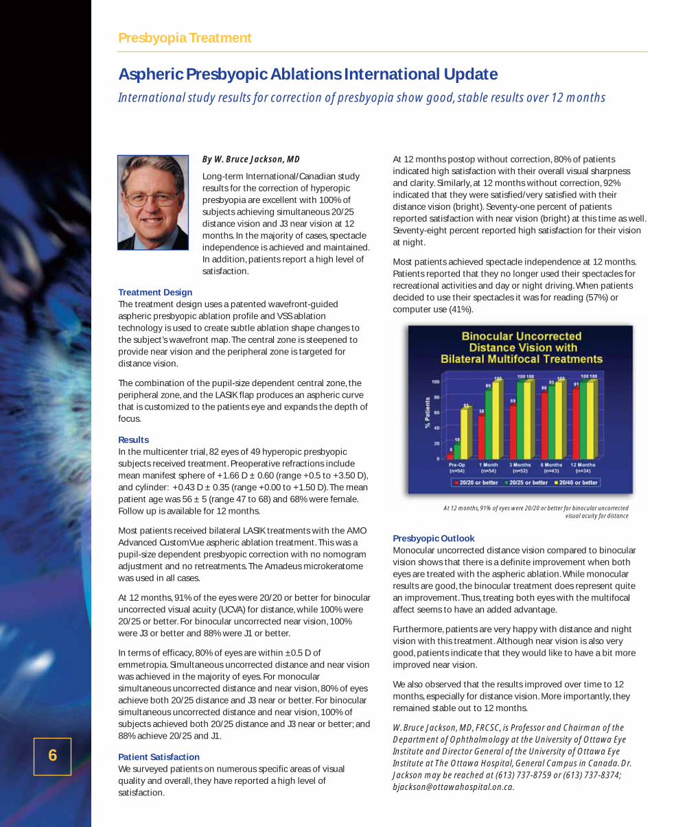
At 12 months, 91% of the eyes were 20/20 or better for binocularuncorrected visual acuity (UCVA) for distance, while 100% were20/25 or better. For binocular uncorrected near vision, 100%were J3 or better and 88% were J1 or better.
In terms of efficacy, 80% of eyes are within ±0.5 D ofemmetropia. Simultaneous uncorrected distance and near visionwas achieved in the majority of eyes. For monocularsimultaneous uncorrected distance and near vision, 80% of eyesachieve both 20/25 distance and J3 near or better. For binocularsimultaneous uncorrected distance and near vision, 100% ofsubjects achieved both 20/25 distance and J3 near or better; and88% achieve 20/25 and J1.
Patient SatisfactionWe surveyed patients on numerous specific areas of visualquality and overall, they have reported a high level ofsatisfaction.
At 12 months postop without correction, 80% of patientsindicated high satisfaction with their overall visual sharpnessand clarity. Similarly, at 12 months without correction, 92%indicated that they were satisfied/very satisfied with theirdistance vision (bright). Seventy-one percent of patientsreported satisfaction with near vision (bright) at this time as well.Seventy-eight percent reported high satisfaction for their visionat night.
Most patients achieved spectacle independence at 12 months.Patients reported that they no longer used their spectacles forrecreational activities and day or night driving. When patientsdecided to use their spectacles it was for reading (57%) orcomputer use (41%).
Presbyopic OutlookMonocular uncorrected distance vision compared to binocularvision shows that there is a definite improvement when botheyes are treated with the aspheric ablation. While monocularresults are good, the binocular treatment does represent quitean improvement. Thus, treating both eyes with the multifocalaffect seems to have an added advantage.
Furthermore, patients are very happy with distance and nightvision with this treatment. Although near vision is also verygood, patients indicate that they would like to have a bit moreimproved near vision.
We also observed that the results improved over time to 12months, especially for distance vision. More importantly, theyremained stable out to 12 months.
W. Bruce Jackson, MD, FRCSC, is Professor and Chairman of theDepartment of Ophthalmology at the University of Ottawa EyeInstitute and Director General of the University of Ottawa EyeInstitute at The Ottawa Hospital, General Campus in Canada. Dr.Jackson may be reached at (613) 737-8759 or (613) 737-8374;[email protected].
Aspheric Presbyopic Ablations International UpdateInternational study results for correction of presbyopia show good, stable results over 12 months
6
Presbyopia Treatment
At 12 months, 91% of eyes were 20/20 or better for binocular uncorrected visual acuity for distance
It has been speculated for many years thatlight exposure might play a role in thedevelopment of age-related maculardegeneration (AMD), but this relationshipremains unproven. The recent availabilityand ongoing development of visible light-blocking intraocular lenses emphasize theimportance of continuing research in thisarea.
Ophthalmologist-physicist Martin A. Mainster, PhD, MD, FRCOphth,became involved in this subject almost 30 years ago. As the debaterages on, he wanted clinicians to recognize how IOL chromophoresbalance retinal photoprotection with photoreception.
AMD and Retinal PhototoxicityThere are at least two forms of retinal phototoxicity: Blue-green andUV-blue. Blue-green phototoxicity is mediated by rhodopsin, thesame photopigment involved in scotopic vision. The second type ofretinal phototoxicity, UV-blue, increases with decreasingwavelength. In other words, UV radiation (100-400 nm) is morehazardous than violet light (400-440 nm), which in turn is morehazardous than blue light (440-500 nm). UV radiation is responsiblefor 67% of acute UV-blue phototoxicity in the part of the spectrumthat can reach the retina through an IOL, while violet light accountsfor 18% and blue light for 14%.
Articles Mainster wrote in 1978 about the potentially harmful effectsof UV radiation1, 2 eventually led to the inclusion of UV-blockingchromophores in nearly all IOLs on the market today. Recently, ithas been suggested that violet- and blue-blocking lenses (AcrySofNatural, Alcon Laboratories; AF-1, Hoya Corporation) may helpprevent AMD.
Visual Benefits of Blue Light Blue light is much more important for mesopic and scotopic visionthan it is for photopic vision. According to Mainster, blue lightprovides 35% of aphakic scotopic and 7% of photopic sensitivity.3
“Rod photoreceptors are responsible for scotopic and lowermesopic vision,” he said. “They contain the photopigmentrhodopsin, which has peak sensitivity near 500 nm, the borderbetween blue and green light. That explains why blue light is soimportant for scotopic vision.”
In older adults, declining photoreceptor populations cause scotopicthreshold and contrast sensitivity to decrease and dark adaptationto slow. In addition, age-related pupillary miosis reduces retinalillumination. “Impaired dark adaptation increases the risk of fallingin older adults, and falling increases the risk of debilitating injuries,costly long-term hospitalization and death,” Mainster said.
Cataract surgery can’t replace lost rod photoreceptors, but it doesincrease retinal illumination, particularly the blue light needed forvision in dim environments.
Health Benefits of Blue LightMelatonin is a potent free-radical scavenger with numerousneuroprotective, anti-cancer and anti-aging functions. Retinalganglion photoreceptors control suppression and secretion ofmelatonin using signals sent to the suprachiasmatic nucleus, thehuman body’s master biological clock. These retinal ganglion cellsexpress the blue-light sensitive photopigment melanopsin.According to Mainster,“Blue light is responsible for over 50% ofmelanopsin sensitivity. In comparison to standard UV-only blockingIOLs, blue-blockers reduce melatonin suppression efficiency by27–38%, depending on their dioptric power3.”
Age-related pupillary miosis and crystalline lens yellowing conspireto reduce older adults’ effective blue light exposure to one-tenththat of younger people. “Numerous clinical studies have shown therisks of disturbed circadian photoentrainment and the benefits ofoptimal rhythmicity,” Mainster said.
Weighing the TradeoffsIn the end, one has to consider the tradeoffs inherent in increasingretinal photoprotection. “UV radiation is potentially hazardous forthe retina. It’s not useful for vision, so it makes good sense to block itwith IOL chromophores,” said Mainster. But he isn’t satisfied withtradeoffs between photoprotection and photoreception made byblue-blocking IOLs.
In 1986, Dr. Mainster suggested blocking violet light in addition toUV to increase retinal photoprotection4,5. He recently showed thatviolet-blocking IOLs can provide the same level of UV-bluephotoprotection as blue-blockers but reduce scotopic andmelanopsin sensitivity loss by 50%.3
“I believe that it is important for older adults to have all the bluelight possible for retinal rod and ganglion cell photoreception,particularly since there is no clinical or experimental proof thatchronic exposure to environmental light harms the human retina,”Mainster said. “Cataract surgery that improves a patient’senvironmental blue light exposure helps assure their propercircadian photoentrainment and increase their chances of goodphysical and mental health.”
Dr. Mainster is the Luther L. Fry Professor and Vice Chairman of theDepartment of Ophthalmology at the University of Kansas MedicalSchool in Kansas City, Kansas. Contact him at [email protected].
References1. Mainster MA. Spectral transmittance of intraocular lenses and retinal damage from
intense light sources. Am J Ophthalmol 1978;85:167-70.2. Mainster MA. Solar retinitis, photic maculopathy and the pseudophakic eye. J Am
Intraocular Implant Soc 1978;4:84-6.3. Mainster MA. Violet and blue light blocking intraocular lenses: photoprotection versus
photoreception. Br J Ophthalmol 2006;90:784-92.4. Mainster MA.The spectra, classification, and rationale of ultraviolet-protective
intraocular lenses. Am J Ophthalmol 1986;102:727-32.5. Mainster MA. Intraocular lenses should block UV radiation and violet but not blue
light. Arch Ophthalmol 2005;123:550-5.
Photoprotection of Blue-Blocking IOLs Comes at a High PriceBlue light is important for mesopic and scotopic vision and critical for entraining biological clocks toenvironmental day-night rhythms
7
Refractive Cataract Surgery
By Ralph Chu, MD
The preliminary results from theprospective, randomized double-maskedtrial comparing the three FDA-approvedaspheric monofocal IOLs demonstrated thatthe Tecnis was the most effective inreducing spherical aberration to zero,regardless of the patient’s preoperativespherical aberration. For this study, our goal
was to compare the performance of these three aspheric opticdesigns that correct different values of spherical aberration.
The StudyWe evaluated the Tecnis, from Advanced Medical Optics, Inc., theAcrySof IQ lens from Alcon, and LI61AO from B&L.
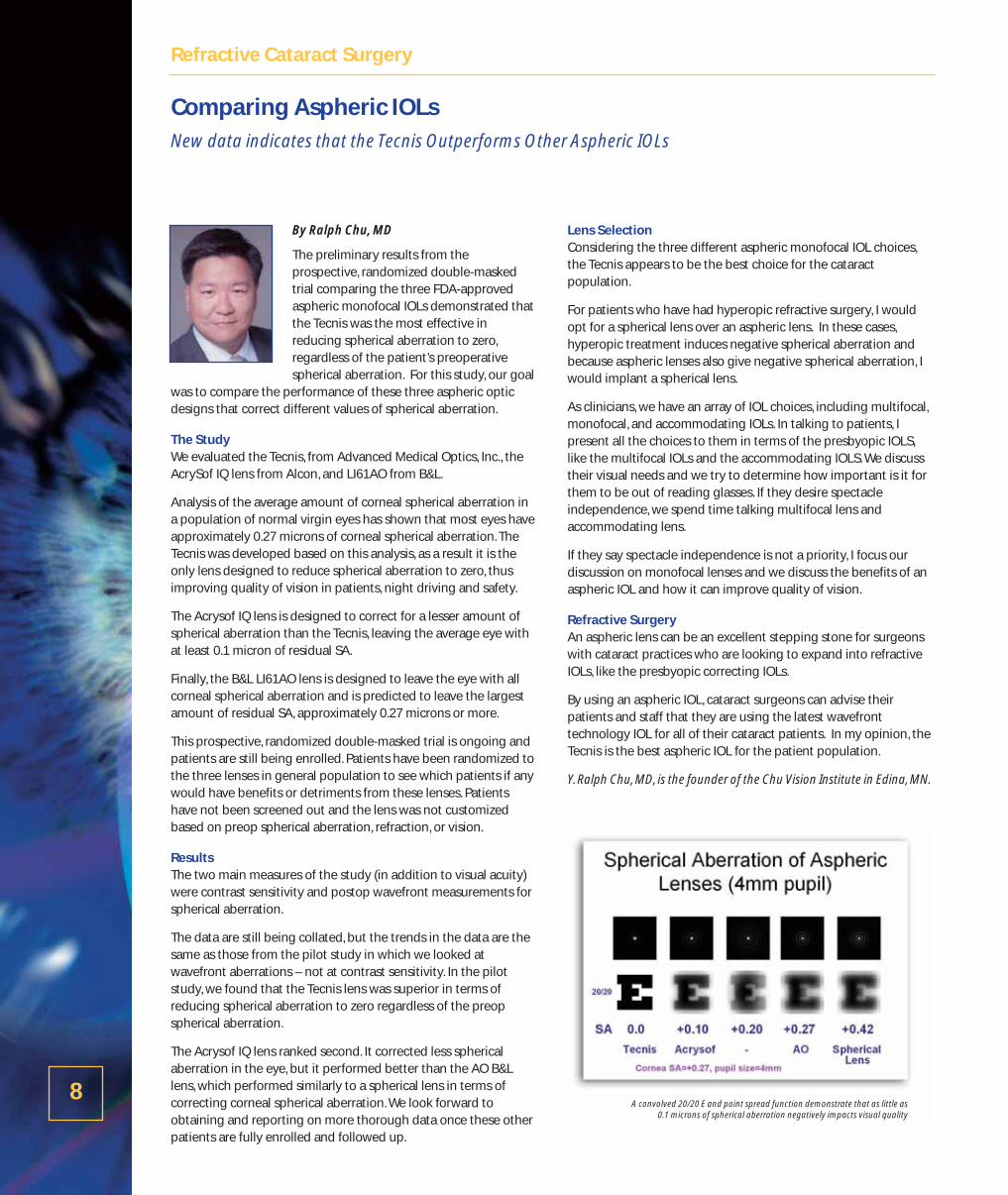
Analysis of the average amount of corneal spherical aberration ina population of normal virgin eyes has shown that most eyes haveapproximately 0.27 microns of corneal spherical aberration. TheTecnis was developed based on this analysis, as a result it is theonly lens designed to reduce spherical aberration to zero, thusimproving quality of vision in patients, night driving and safety.
The Acrysof IQ lens is designed to correct for a lesser amount ofspherical aberration than the Tecnis, leaving the average eye withat least 0.1 micron of residual SA.
Finally, the B&L LI61AO lens is designed to leave the eye with allcorneal spherical aberration and is predicted to leave the largestamount of residual SA, approximately 0.27 microns or more.
This prospective, randomized double-masked trial is ongoing andpatients are still being enrolled. Patients have been randomized tothe three lenses in general population to see which patients if anywould have benefits or detriments from these lenses. Patientshave not been screened out and the lens was not customizedbased on preop spherical aberration, refraction, or vision.
Results The two main measures of the study (in addition to visual acuity)were contrast sensitivity and postop wavefront measurements forspherical aberration.
The data are still being collated, but the trends in the data are thesame as those from the pilot study in which we looked atwavefront aberrations – not at contrast sensitivity. In the pilotstudy, we found that the Tecnis lens was superior in terms ofreducing spherical aberration to zero regardless of the preopspherical aberration.
The Acrysof IQ lens ranked second. It corrected less sphericalaberration in the eye, but it performed better than the AO B&Llens, which performed similarly to a spherical lens in terms ofcorrecting corneal spherical aberration. We look forward toobtaining and reporting on more thorough data once these otherpatients are fully enrolled and followed up.
Lens SelectionConsidering the three different aspheric monofocal IOL choices,the Tecnis appears to be the best choice for the cataractpopulation.
For patients who have had hyperopic refractive surgery, I wouldopt for a spherical lens over an aspheric lens. In these cases,hyperopic treatment induces negative spherical aberration andbecause aspheric lenses also give negative spherical aberration, Iwould implant a spherical lens.
As clinicians, we have an array of IOL choices, including multifocal,monofocal, and accommodating IOLs. In talking to patients, Ipresent all the choices to them in terms of the presbyopic IOLS,like the multifocal IOLs and the accommodating IOLS. We discusstheir visual needs and we try to determine how important is it forthem to be out of reading glasses. If they desire spectacleindependence, we spend time talking multifocal lens andaccommodating lens.
If they say spectacle independence is not a priority, I focus ourdiscussion on monofocal lenses and we discuss the benefits of anaspheric IOL and how it can improve quality of vision.
Refractive Surgery An aspheric lens can be an excellent stepping stone for surgeonswith cataract practices who are looking to expand into refractiveIOLs, like the presbyopic correcting IOLs.
By using an aspheric IOL, cataract surgeons can advise theirpatients and staff that they are using the latest wavefronttechnology IOL for all of their cataract patients. In my opinion, theTecnis is the best aspheric IOL for the patient population.
Y. Ralph Chu, MD, is the founder of the Chu Vision Institute in Edina, MN.
Comparing Aspheric IOLsNew data indicates that the Tecnis Outperforms Other Aspheric IOLs
8
Refractive Cataract Surgery
A convolved 20/20 E and point spread function demonstrate that as little as0.1 microns of spherical aberration negatively impacts visual quality

By Pablo Artal, PhD
My colleagues and I at the University ofMurcia, in Spain, have been looking at theinteractions between the optics of the eyeand quality of vision for many years. Froma number of experiments performed undertightly controlled conditions, we know twothings for certain. The first is that there is aclear correlation between poor optics and
poor performance. A highly aberrated eye simply cannotprovide high quality vision.
The second lesson we’ve learned more recently in a series ofstudies by Eloy Villegas, Encarna Alcon, and myself, however, runscounter to the conventional wisdom. In individuals with“normal” optics, we find no correlation between aberrations andvisual performance. Even those individuals with excellentvision—better than 20/15—do not have perfect optics.
With this research at our foundation, we have recently begunlooking specifically at spherical aberration (SA) to see if we coulddetermine an optimal value for this particular higher-orderaberration (HOA).
Spherical aberration is of particular importance, for severalreasons. First of all, we know that it changes continuously withage. Young subjects have a low value of SA but it alwaysincreases with age. SA is also one of the predominant HOAs inthe eye. It is rotationally symmetrical and, therefore, the easiestaberration to correct, from a technical standpoint.
When a conventional intraocular lens is implanted followingcataract surgery, it randomly adds to or subtracts from SA, whichis already elevated naturally in the older eye. In the last fewyears, several manufacturers have introduced asphericintraocular lenses that propose to deliberately cancel or partiallycompensate for the SA of the aging cornea in order to improvevisual performance.
SA in Young Subjects with Excellent VisionIn our most recent study, we evaluated SA in a group of youngsubjects with exceptional natural visual acuity (VA) of 20/15 to20/10. Subjects were nearly emmetropic (-1.0 to +1.0 D) withvery low astigmatism (< 0.5 D). The average age was 25.3 years.High-contrast VA was measured using a forced choice procedurethat is more robust than a standard Snellen eye chart. Wemeasured wavefront aberrations for different pupil diametersusing our own research prototype wavefront sensor.Accommodation was strictly controlled so that sphericalaberration data could be obtained from the eye in itsunaccommodated state.
All of the subjects had good—but not perfect—optics,reinforcing our previous findings that better-than-normal acuitydoes not require a perfectly flat wavefront.
The average magnitude of SA was small and was not correlatedwith visual acuity. In other words, the 20/10 eyes did not haveless SA than the 20/15 eyes in average. SA was correlated withage. Even in this young group, patients younger than 25 hadlower SA than those who were older than 25.
Across the entire group of 46 subjects, the average SA for a 5-mm pupil was slightly positive (0.032 ± 0.047 microns). For a 4-mm pupil the average SA was 0.017 ± 0.024 microns. Theaverage SA for a 5-mm pupil in the subgroup younger than 25was 0.02 ± 0.052 microns, which is not statistically different fromzero.
Trying to replicate the slight positive SA we found in our studyafter implantation of an IOL or laser surgery could becounterproductive. First of all, the SA present in these youngeyes was essentially zero. Secondly, lens implants almost neverhit a target exactly. A target of zero means that a small margin oferror may, in fact, result in a slight positive amount of SA. Thehigher the SA target, the greater the chance of inducing orleaving too much SA. Several years ago, we proposed that theoptimal target for SA in an older eye should be zero, and Ibelieve this study supports that recommendation.
Dr. Artal is Professor of Optics and lead researcher in the OpticsLaboratory of the Physics Department at the University of Murcia inMurcia, Spain. Contact him at +34-96-836-7224 or [email protected].
Relationship Between Spherical Aberration and Acuity is ComplexIn young patients with excellent vision, spherical aberration is not statistically different from zero and is not correlated with visual performance.
9
Refractive Cataract Surgery
According to our study results, young patients with naturally occurring supernormal vision have zero spherical aberration
“The higher the SA target, the greater thechance of inducing or leaving too much SA”
10
Laser Vision Correction
By Captain Steve Schallhorn, MD
The visual quality of wavefront-guidedLASIK is predicted to be better thanoptimized LASIK, according to results from a model we developed to simulate visualperformance.
From the model, we determined thatwavefront-guided laser vision correction
(LVC) would induce fewer aberrations than wavefront-optimizedLVC regardless of the preoperative higher order aberrations(HOAs). This includes patients with a low amount of preoperative HOA.
Differences in the Ablation ProfilesThe treatment basis of WFG LASIK is the optical path deviationderived from a wavefront aberrometer measurement. The opticalpath deviation, which can also be visualized as a wavefrontpattern, contains both the lower order (sphere and cylinder) andhigher order (coma, trefoil, spherical aberration, etc.) aberrationsof the entire ocular system. This is then used to create anappropriate ablation profile for the excimer laser. The ablationprofile can be extraordinarily complex dependent on theamount of higher order aberrations.
”Optimized” LASIK was designed to reduce the induction ofspherical aberration, the most commonly induced higher orderaberration after conventional LASIK. It does this with a specialaspheric ablation profile tailored to the amount of intendedsphere correction. It cannot treat pre-existing higher orderaberrations. The treatment basis is the same as conventionalLASIK, namely, sphere and cylinder.
The StudyMany investigators have shown that LASIK typically induceshigher HOA, most prominently spherical aberration, whilewavefront-guided (WFG) LASIK induces less higher orderaberrations. Analyzing WFG LASIK in greater detail shows thatpatients who have small amounts of preoperative higher orderaberrations tend to have an increase, whereas patients who havea larger amount (above 0.3 microns) tend to have a reduction.
Our model simulates visual performance of optimized andwavefront-guided LASIK, and assumed a perfect correction ofsphere and cylinder. Patterned after the dataset of conventionaland WFG LASIK, the induction or reduction of higher orderaberrations was modeled to the preoperative level of higherorder aberrations and the type of surgery.
We assumed the best case scenario that optimized surgeryinduced no spherical aberration, but that the induction orreduction of other higher order aberrations would be the sameas conventional.
For the simulation, we used a wavefront dataset (all 6-mmentrance pupil analysis) from patients before and one month
after conventional and wavefront-guided LASIK in a large samplesize at our center. We generated 10,000 random preoperativewavefront maps, model ‘eyes’, to simulate a normal population.
There is considerable population variability in the preop topostop change of higher order Zernike terms after any type ofsurgery. To model this variation, the simulated postoperativehigher order aberrations were randomly assigned to be withinone standard deviation of the mean change, dependent on thetype of surgery (optimize and WFG) and the level of preoperativehigher order aberrations.
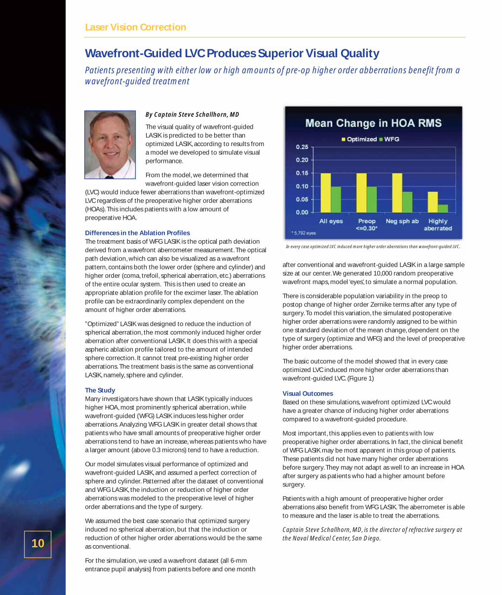
The basic outcome of the model showed that in every caseoptimized LVC induced more higher order aberrations thanwavefront-guided LVC. (Figure 1)
Visual OutcomesBased on these simulations, wavefront optimized LVC wouldhave a greater chance of inducing higher order aberrationscompared to a wavefront-guided procedure.
Most important, this applies even to patients with lowpreoperative higher order aberrations. In fact, the clinical benefitof WFG LASIK may be most apparent in this group of patients.These patients did not have many higher order aberrationsbefore surgery. They may not adapt as well to an increase in HOAafter surgery as patients who had a higher amount beforesurgery.
Patients with a high amount of preoperative higher orderaberrations also benefit from WFG LASIK. The aberrometer is ableto measure and the laser is able to treat the aberrations.
Captain Steve Schallhorn, MD, is the director of refractive surgery atthe Naval Medical Center, San Diego.
Wavefront-Guided LVC Produces Superior Visual Quality Patients presenting with either low or high amounts of pre-op higher order abberrations benefit from awavefront-guided treatment
In every case optimized LVC induced more higher order aberrations than wavefront-guided LVC.
By Mounir A. Khalifa, MD
I recently conducted a prospective studycomparing wavefront-guided customizedablation to wavefront-optimized ablations.
The study participants were 24 consecutiverefractive surgery patients with myopia ormyopic astigmatism.They were randomized toeither Group A (wavefront-guided) or Group B
(wavefront-optimized), with 12 patients (24 eyes) in each group.
All of the patients had LASIK. All patients were evaluated pre- andpost-operatively using the Visx WaveScan wavefront aberrometerand CSV-1000 contrast sensitivity testing. Patients whose post-operative WaveScan pupil diameter differed from the pre-operative pupil diameter were excluded to avoid any variations inhigher-order aberrations (HOAs) due to pupil size.
Group A was treated with custom wavefront-guided ablationsusing the Visx Advanced CustomVue platform (Advanced MedicalOptics) with Fourier analysis and iris registration. Group B wastreated with wavefront-optimized standard ablation using theWaveLight Allegretto laser. This laser performs a conventionalablation but it incorporates software that is supposed tocompensate for and neutralize induced spherical aberration.
Visual acuity, HOAs, and contrast sensitivity were evaluated atthree months. Paired t-tests with equal variance were used in thedata analysis.
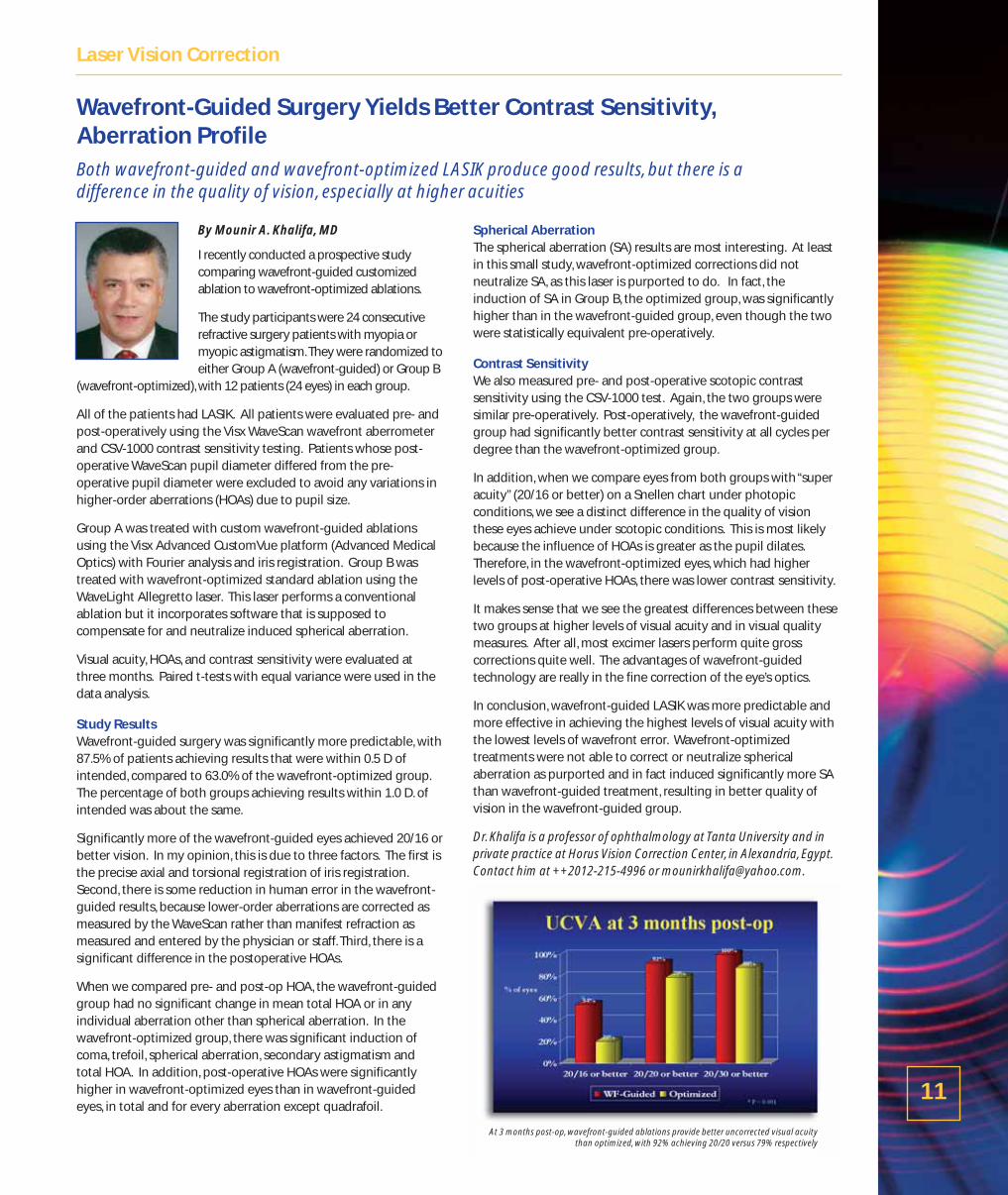
Study ResultsWavefront-guided surgery was significantly more predictable, with87.5% of patients achieving results that were within 0.5 D ofintended, compared to 63.0% of the wavefront-optimized group.The percentage of both groups achieving results within 1.0 D. ofintended was about the same.
Significantly more of the wavefront-guided eyes achieved 20/16 orbetter vision. In my opinion, this is due to three factors. The first isthe precise axial and torsional registration of iris registration.Second, there is some reduction in human error in the wavefront-guided results, because lower-order aberrations are corrected asmeasured by the WaveScan rather than manifest refraction asmeasured and entered by the physician or staff. Third, there is asignificant difference in the postoperative HOAs.
When we compared pre- and post-op HOA, the wavefront-guidedgroup had no significant change in mean total HOA or in anyindividual aberration other than spherical aberration. In thewavefront-optimized group, there was significant induction ofcoma, trefoil, spherical aberration, secondary astigmatism andtotal HOA. In addition, post-operative HOAs were significantlyhigher in wavefront-optimized eyes than in wavefront-guidedeyes, in total and for every aberration except quadrafoil.
Spherical AberrationThe spherical aberration (SA) results are most interesting. At leastin this small study, wavefront-optimized corrections did notneutralize SA, as this laser is purported to do. In fact, theinduction of SA in Group B, the optimized group, was significantlyhigher than in the wavefront-guided group, even though the twowere statistically equivalent pre-operatively.
Contrast SensitivityWe also measured pre- and post-operative scotopic contrastsensitivity using the CSV-1000 test. Again, the two groups weresimilar pre-operatively. Post-operatively, the wavefront-guidedgroup had significantly better contrast sensitivity at all cycles perdegree than the wavefront-optimized group.
In addition, when we compare eyes from both groups with “superacuity” (20/16 or better) on a Snellen chart under photopicconditions, we see a distinct difference in the quality of visionthese eyes achieve under scotopic conditions. This is most likelybecause the influence of HOAs is greater as the pupil dilates.Therefore, in the wavefront-optimized eyes, which had higherlevels of post-operative HOAs, there was lower contrast sensitivity.
It makes sense that we see the greatest differences between thesetwo groups at higher levels of visual acuity and in visual qualitymeasures. After all, most excimer lasers perform quite grosscorrections quite well. The advantages of wavefront-guidedtechnology are really in the fine correction of the eye’s optics.
In conclusion, wavefront-guided LASIK was more predictable andmore effective in achieving the highest levels of visual acuity withthe lowest levels of wavefront error. Wavefront-optimizedtreatments were not able to correct or neutralize sphericalaberration as purported and in fact induced significantly more SAthan wavefront-guided treatment, resulting in better quality ofvision in the wavefront-guided group.
Dr. Khalifa is a professor of ophthalmology at Tanta University and inprivate practice at Horus Vision Correction Center, in Alexandria, Egypt.Contact him at ++2012-215-4996 or [email protected].
Wavefront-Guided Surgery Yields Better Contrast Sensitivity,Aberration ProfileBoth wavefront-guided and wavefront-optimized LASIK produce good results, but there is a difference in the quality of vision, especially at higher acuities
11
Laser Vision Correction
At 3 months post-op, wavefront-guided ablations provide better uncorrected visual acuitythan optimized, with 92% achieving 20/20 versus 79% respectively
Refractive surgeons have increasingly realizedthe importance of accurate registration ofwavefront treatments to the cornea.
Iris registration, used by the Visx CustomVuesystem (AMO), relies on matching iris featuresin images taken at the time of measurementand treatment. The system then compensatesfor any cyclotorsion or pupil centroid shiftthat may have occurred between the initial
wavefront capture and the time of ablation.
Previously, Douglas D. Koch, MD, and Li Wang, MD, of Baylor Collegeof Medicine, used theoretical modeling to calculate the typicalamount of wavefront aberration induced in normal eyes without irisregistration. They looked at what happened to total, lower-order, andhigher-order RMS error with average mis-registrations—that is, a 2.1-degree counter-clockwise rotation or a horizontal pupil centroid shiftof 0.27 mm. They found that decentration of the treatment has amuch greater influence on residual higher-order aberration thanrotational shifts. The most commonly induced aberration from X-Ymis-registration was coma.
Julian Stevens, FRCS, was curious to see what the effects of mis-registration would be in the more highly aberrated therapeutic casesthat he often treats. “By definition, the effects of such mis-registration, both rotational and X-Y, potentially can be much greaterthan in normal eyes,”he said.
At this year’s ASCRS meeting in San Francisco, Stevens presented thechallenging case of an airline pilot whose vision had deteriorated tosuch an extent that he could no longer fly. The patient had bilateralcorneal grafts due to Fuchs’ corneal degeneration. His refraction was-5.17 -3.27 x 148 OD and -3.57 -4.38 x 168 OS. In addition to the veryhigh cylinder, he had huge (> 8.0 mm) pupils that exceeded thediameter of the corneal grafts. There were also some lenticularchanges.
Stevens planned a two-stage therapeutic treatment, beginning withphacoemulsification and lens surgery. He made a small, 5.0-mmcapsulorrhexis to act as a pupil stop, which avoided the poor opticsof the peripheral graft, and targeted a hyperopic result. The secondstep in the treatment would be wavefront-guided LASIK with Fourierreconstruction and iris registration.“The reason I target hyperopia inthese eyes is so that the subsequent laser ablation will be performedaround the periphery,”he said. “With all of the pulses in thatperipheral region you will get a more effective ablation.”
The treatment was carefully registered and the end result, followingbilateral IOL implantation and wavefront-guided LASIK, wasuncorrected visual acuity of 20/15 OD and 20/20 OS. The pilot, whowas able to fly again, was delighted.
Stevens used this patient’s preoperative point spread function (PSF)to demonstrate the effects of shifting the treatment slightly alongthe X-Y axis, as could happen in a treatment that did not compensatefor pupil centroid shift.
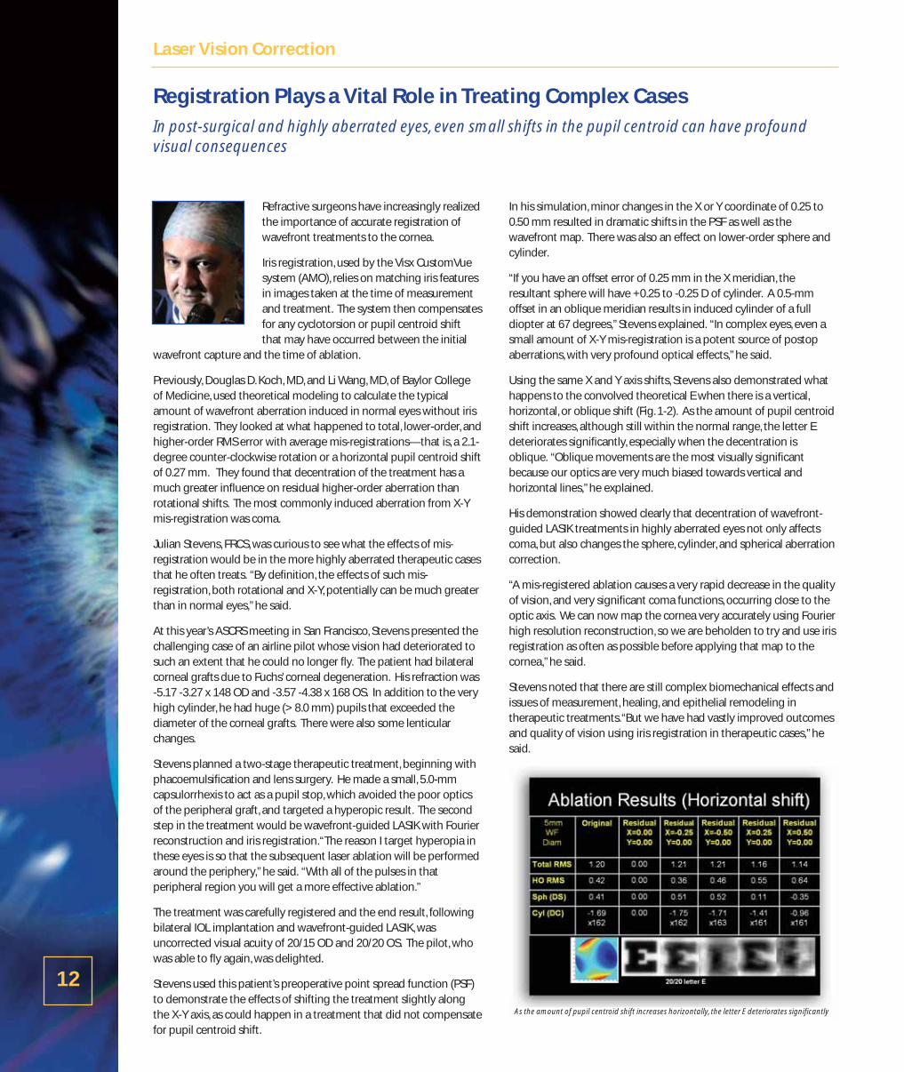
In his simulation, minor changes in the X or Y coordinate of 0.25 to0.50 mm resulted in dramatic shifts in the PSF as well as thewavefront map. There was also an effect on lower-order sphere andcylinder.
“If you have an offset error of 0.25 mm in the X meridian, theresultant sphere will have +0.25 to -0.25 D of cylinder. A 0.5-mmoffset in an oblique meridian results in induced cylinder of a fulldiopter at 67 degrees,”Stevens explained. “In complex eyes, even asmall amount of X-Y mis-registration is a potent source of postopaberrations, with very profound optical effects,”he said.
Using the same X and Y axis shifts, Stevens also demonstrated whathappens to the convolved theoretical E when there is a vertical,horizontal, or oblique shift (Fig. 1-2). As the amount of pupil centroidshift increases, although still within the normal range, the letter Edeteriorates significantly, especially when the decentration isoblique. “Oblique movements are the most visually significantbecause our optics are very much biased towards vertical andhorizontal lines,”he explained.
His demonstration showed clearly that decentration of wavefront-guided LASIK treatments in highly aberrated eyes not only affectscoma, but also changes the sphere, cylinder, and spherical aberrationcorrection.
“A mis-registered ablation causes a very rapid decrease in the qualityof vision, and very significant coma functions, occurring close to theoptic axis. We can now map the cornea very accurately using Fourierhigh resolution reconstruction, so we are beholden to try and use irisregistration as often as possible before applying that map to thecornea,”he said.
Stevens noted that there are still complex biomechanical effects andissues of measurement, healing, and epithelial remodeling intherapeutic treatments.“But we have had vastly improved outcomesand quality of vision using iris registration in therapeutic cases,”hesaid.
Registration Plays a Vital Role in Treating Complex CasesIn post-surgical and highly aberrated eyes, even small shifts in the pupil centroid can have profoundvisual consequences
12
Laser Vision Correction
As the amount of pupil centroid shift increases horizontally, the letter E deteriorates significantly
A study that evaluated the residualwavefront aberrations induced by thecyclotorsional rotation and pupil centroidshift demonstrated that, for normal eyeswith astigmatism less than 2D, the pupilcentroid shift feature provides greater visualbenefit than cyclotorsional rotation.However, for eyes with astigmatism greaterthan 2D, cyclotorsional rotation registration
provides comparable or greater visual benefit, according toDouglas D. Koch, MD, a professor and the Allen, Mosbacher, andLaw Chair in Ophthalmology, Cullen Eye Institute, Baylor College ofMedicine, in Houston, TX.
RegistrationIris registration permits verification of patient identification andintraoperative registration.“Most excimer lasers track horizontaland vertical movements of the pupil. The unique advantages ofthe new VISX IR system are cyclotorsional registration and pupilcentroid shift adjustment. Cyclotorsional registration is essential tooptimize outcomes,” he said, noting that the literature supportsthat cyclotorsional movement is 2º-5º on average, but can besignificantly higher in some cases.
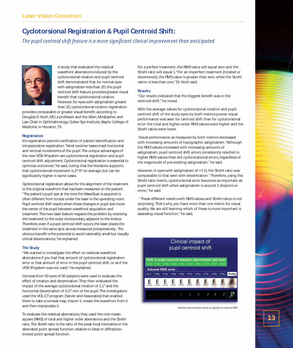
Cyclotorsional registration allows for the alignment of the treatmentto the original wavefront that has been measured on the patient.“The patient’s pupil size at the time the WaveScan is acquired isoften different from its size under the laser in the operating room.Pupil centroid shift results when these changes in pupil size movethe center of the pupil between wavefront acquisition andtreatment.This new laser feature negates this problem by orientingthe treatment to the outer iris boundary, adjacent to the limbus.Therefore, even if a pupil centroid shift occurs, the laser places thetreatment in the same spot as was measured preoperatively. Theobvious benefit is the potential to avoid ostensibly small but visuallycritical decentrations,” he explained.
The Study“We wanted to investigate the effect on residual wavefrontaberrations if you had that amount of cyclotorsional registrationerror or that amount of error in the pupil centroid shift, or as if theVISX IR system was not used,” he explained.
Corneas from 50 eyes of 50 subjects were used to evaluate theeffect of rotation and decentration. They then evaluated theimpact of the average cyclotorsional rotation of 2.1° and thehorizontal decentration of 0.27 mm of the pupil. The investigatorsused the VOL-CT program (Sarver and Associates) that enabledthem to take a corneal map, import it, create the wavefront from itand then manipulate it.
To evaluate the residual aberrations, they used the root-mean-square (RMS) of total and higher order aberrations and the Strehlratio. The Strehl ratio is the ratio of the peak focal intensities in theaberrated point spread function, relative to ideal or diffraction-limited point spread function.
For a perfect treatment, the RMS value will equal zero and theStrehl ratio will equal 1.“For an imperfect treatment (rotated ordecentered), the RMS value is greater than zero, while the Strehlration is less than one,” Dr. Koch said.
Results“Our results indicated that the biggest benefit was in thecentroid shift,” he noted.
With the average values for cyclotorsional rotation and pupilcentroid shift of the study eyes, by both metrics poorer visualperformance was seen for centroid shift than for cyclotorsionalerror: the total and higher-order RMS values were higher and theStrehl ratios were lower.
Visual performance as measured by both metrics decreasedwith increasing amounts of topographic astigmatism.“Althoughthe RMS values increased with increasing amounts ofastigmatism, pupil centroid shift errors consistently resulted inhigher RMS values than did cyclorotational errors, regardless ofthe magnitude of pre-existing astigmatism.” he said.
However, in eyes with astigmatism of >2 D, the Strehl ratio wascomparable to that seen with decentration.“Therefore, using theStrehl ratio metric, cyclotorsional error becomes as important aspupil centroid shift when astigmatism is around 2 diopters ormore,” he said.
“These different results with RMS values and Strehl ratios is notsurprising. That’s why you have more than one metric for visualquality. We are still learning which of these is more important isassessing visual function,” he said.
Cyclotorsional Registration & Pupil Centroid Shift:The pupil centroid shift feature is a more significant clinical improvement than anticipated
13
Laser Vision Correction
Ablation decentration results in significant induced RMS
Michael C. Knorz, MD
Cataract surgery is increasingly becoming arefractive procedure. More and more of ourlens replacement patients are interested in—or even demanding—spectacleindependence. As we know, new multifocalIOLs make this a real possibility, butemmetropia must be achieved for multifocallenses to provide optimal visual outcomes.
At our clinic, we now offer a bioptics package that includes lensreplacement surgery and wavefront-guided LASIK, if needed, to finetune the results.
For cataract or lens replacement patients there are two options. Thefirst is a standard monofocal lens, although in our practice this hasbeen almost entirely replaced by the aspheric Tecnis monofocal IOL.
The second option is to replace the crystalline lens with a multifocalIOL to correct presbyopia and provide good near and distance visionwithout any glasses. Currently, I implant the Tecnis Multifocal,ReZoom and ReStor multifocal lenses.
Implanting a multifocal IOL is essentially a promise of spectacleindependence to my patients. Emmetropia must be achieved.However, we all know that there is no way to guarantee anemmetropic outcome, even with very careful lens calculations.This iswhy we elect to perform LASIK in those patients in whomemmetropia is not achieved, about 15% of the cases. Patients witheven a little bit of residual refractive error postop will not be happywith their vision.
Wavefront Correction in PseudophakesIn our practice, we perform customized laser surgery on our laserrefractive surgery patients, using the Visx Star S4 laser with irisregistration.
A wavefront-driven ablation is desirable in pseudophakes for thesame reasons we do it in non-lensectomy patients. It allows us tocorrect not only the sphere and cylinder, but also any higher-orderaberrations (HOAs). It is even more important in pseudophakesbecause minor decentration of the IOL with respect to the visual axismay induce HOAs, specifically coma.
Initially I expected to encounter problems performing custom LASIKfollowing a multifocal implant, especially with capturing a wavefrontimage. In our experience, about 70% of eyes with multifocal implantscan be easily captured by the WaveScan aberrometer, compared tonearly 100% of “normal” eyes or even eyes with monofocal implants.
Even with a successful capture, I recommend performing awavefront-guided ablation only when the manifest refraction andWaveScan refraction match closely. Assuming that one encountersneither of these problems, there does not seem to be any problemwith the interaction of the multifocal implant and the treatmentpattern designed by the laser. I now have many patients with
multifocal IOL implants and subsequent wavefront-guided LASIK forfine-tuning.We have seen tremendous improvement in vision andfull spectacle independence with this approach.
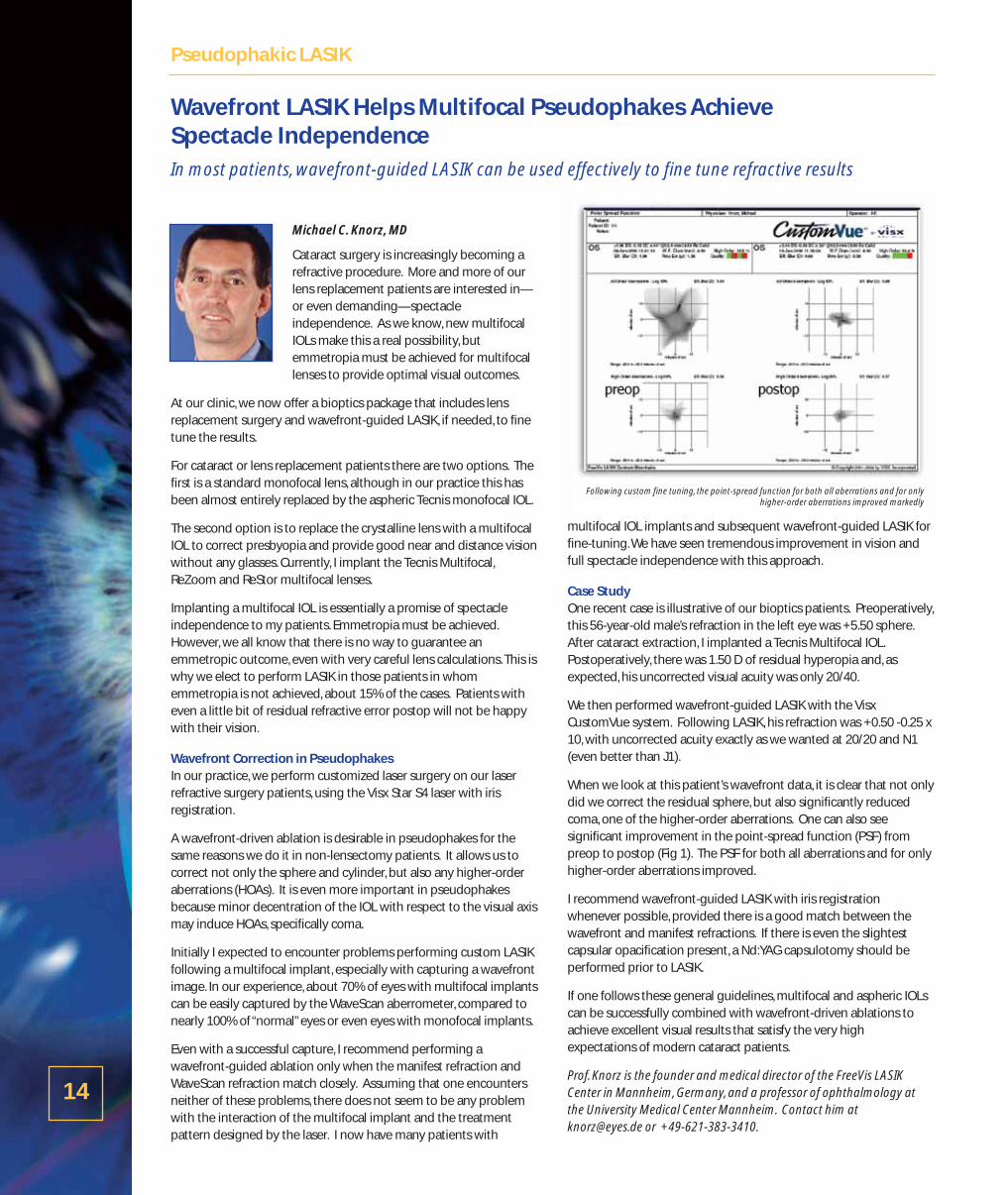
Case StudyOne recent case is illustrative of our bioptics patients. Preoperatively,this 56-year-old male’s refraction in the left eye was +5.50 sphere.After cataract extraction, I implanted a Tecnis Multifocal IOL.Postoperatively, there was 1.50 D of residual hyperopia and, asexpected, his uncorrected visual acuity was only 20/40.
We then performed wavefront-guided LASIK with the VisxCustomVue system. Following LASIK, his refraction was +0.50 -0.25 x10, with uncorrected acuity exactly as we wanted at 20/20 and N1(even better than J1).
When we look at this patient’s wavefront data, it is clear that not onlydid we correct the residual sphere, but also significantly reducedcoma, one of the higher-order aberrations. One can also seesignificant improvement in the point-spread function (PSF) frompreop to postop (Fig 1). The PSF for both all aberrations and for onlyhigher-order aberrations improved.
I recommend wavefront-guided LASIK with iris registrationwhenever possible, provided there is a good match between thewavefront and manifest refractions. If there is even the slightestcapsular opacification present, a Nd:YAG capsulotomy should beperformed prior to LASIK.
If one follows these general guidelines, multifocal and aspheric IOLscan be successfully combined with wavefront-driven ablations toachieve excellent visual results that satisfy the very highexpectations of modern cataract patients.
Prof. Knorz is the founder and medical director of the FreeVis LASIKCenter in Mannheim, Germany, and a professor of ophthalmology atthe University Medical Center Mannheim. Contact him [email protected] or +49-621-383-3410.
Wavefront LASIK Helps Multifocal Pseudophakes Achieve Spectacle IndependenceIn most patients, wavefront-guided LASIK can be used effectively to fine tune refractive results
14
Pseudophakic LASIK
Following custom fine tuning, the point-spread function for both all aberrations and for onlyhigher-order aberrations improved markedly
By David R. Hardten, MD
Multifocal IOLs have transformed cataractsurgery into a consumer-driven market.Patient expectations of surgery with thesepremium lenses are very high. If thepostoperative outcome isn’t exactly ontarget or if the patient finds that near visionis not what he or she had hoped, some formof enhancement may be necessary.
Of course, prevention is the best tactic. Good biometry, precisecalibration of the keratometer, and using the latest-generationIOL power calculation formulas are all important factors inachieving refractive accuracy and success with premium lensimplants. That said, there will always be some unexpectedoutcomes or cases where the target refraction needs to beadjusted postoperatively based on patient feedback.
For residual myopia or hyperopia, I prefer laser vision correctionas the most accurate way of enhancing the outcome for mostpatients. Piggyback IOLs should probably make up less than 5%of total enhancements. I rely on them primarily in cases of highspherical error without much residual cylinder. IOL exchangeshould be done even more rarely.
Correcting CylinderAt this point, we can’t correct astigmatism with multifocal IOLs. Iwill generally recommend trying to correct the cylinder at thetime of surgery with limbal relaxing incisions (LRIs) or astigmatickeratotomy (AK), with the caveat that we may need to do a laservision procedure afterwards. Although incisional techniques donot offer the same degree of predictability and accuracy as laservision correction, doing them in the OR at the time of surgery isconvenient and saves the patient from having a second surgicalprocedure.
If there is some cylinder remaining after lens implantation and apostoperative enhancement is necessary, I always prefer to usethe laser. Ideally, I will do an Advanced CustomVue procedurewith iris registration to most accurately treat the cylinder, but ifthe capsulorhexis is too small to get a 5.0-mm capture, or if thewavefront doesn’t match the refraction or is not compatible withthe uncorrected vision, then I plan for a standard treatment. Iprefer LASIK in most patients because of the quicker recovery, butif there are any corneal health issues, or if I made any LRI or AKincisions during the initial surgery, I opt for PRK.
Timing the Second InterventionIn a bilateral case I typically wait to enhance until I haveimplanted IOLs in both eyes, so that we know the balance of theoptics. If one eye is myopic and the other close to emmetropia,for example, the patient may be happy without any furthercorrection.
After that, I generally follow the same principles I do for laservision correction. If there is a large residual error that I know is
not going to resolve itself I will do an early enhancement. So inthe case of an unusual 2.0-D surprise, I may wait just a month orso for the vision to stabilize, and then perform a secondintervention so the patient doesn’t have to suffer for a longperiod of time. In some of these cases, I may opt for a lensexchange or piggyback IOL.
In most cases, however, the refractive result is within 1.0 D of theintended correction, and I generally wait 6 months beforeenhancing. The refraction may change again or the patient maybe able to adapt to the mild residual error. If there is any capsularfibrosis or haze I perform a Nd:YAG capsulotomy at around 4-5months postop, prior to laser vision correction.
Custom or Conventional? Whenever possible, I prefer to perform a wavefront-guidedenhancement after multifocal IOL implantation. Customizedcorrections offer better astigmatic targeting, especially with irisregistration, and the ability to address corneal higher orderaberrations.
However, it is important to make sure that the manifest and thewavefront refractions match. If there is any doubt, I perform aconventional ablation, making sure to push the plus and do acareful refraction.
In summary, surgeons who implant multifocal and other premiumIOLs are going to need to enhance the refractive outcome, atleast occasionally. In my opinion, laser vision correction providesthe most accurate method for addressing residual sphere andcylinder, but one should also be familiar with IOL exchange orpiggyback IOL implantation for cases in which the refractive erroris large or corneal surgery is contraindicated.
Dr. Hardten is a founding partner of Minnesota Eye Consultants, anddirector of its Clinical Research Department. Contact him at (612)813-3632 or [email protected].
Eliminating Refractive Error After Multifocal IOLIn most cases, laser vision enhancement is the most accurate, least invasive approach to correctingresidual sphere or cylinder after premium IOL implantation
15
Pseudophakic LASIK
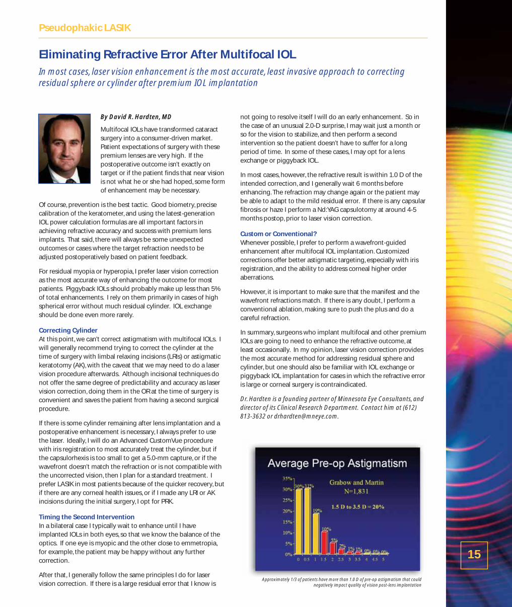
Approximately 1/3 of patients have more than 1.0 D of pre-op astigmatism that couldnegatively impact quality of vision post-lens implantation

Sponsored by