polar value analysis of low to moderate astigmatism with...
TRANSCRIPT
Research ArticlePolar Value Analysis of Low to Moderate Astigmatism withWavefront-Guided Sub-Bowman Keratomileusis
Pisong Yan,1 Zhiyu Du,1,2 and Yu Zhang1
1Medal Eye Institute, Chongqing, China2Department of Ophthalmology, Second Affiliated Hospital, Chongqing Medical University, Chongqing, China
Correspondence should be addressed to Zhiyu Du; [email protected]
Received 22 November 2016; Revised 9 March 2017; Accepted 28 March 2017; Published 2 August 2017
Academic Editor: Antonio Benito
Copyright © 2017 Pisong Yan et al. This is an open access article distributed under the Creative Commons Attribution License,which permits unrestricted use, distribution, and reproduction in any medium, provided the original work is properly cited.
Purpose. To evaluate the astigmatic outcomes of wavefront-guided sub-Bowman keratomileusis (WFG-SBK) for low to moderatemyopic astigmatism. Methods. This study enrolled 100 right eyes from 100 patients who underwent WFG-SBK for thecorrection of myopia and astigmatism. The polar value method was performed with anterior and posterior cornealastigmatism measured with Scheimpflug camera combined with Placido corneal topography (Sirius, CSO) and refractiveastigmatism preoperatively and 1 month, 3 months, and 6 months postoperatively. Results. Similar results for surgicallyinduced astigmatism (SIA) and error of the procedure in both anterior corneal astigmatism (ACA) and total ocularastigmatism (TOA). There was a minor undercorrection of the cylinder in both ACA and TOA. Posterior cornealastigmatism (PCA) showed no significant change. Conclusions. Wavefront-guided SBK could provide good astigmaticoutcomes for the correction of low to moderate myopic astigmatism. The surgical effects were largely attributed to theastigmatic correction of the anterior corneal surface. Posterior corneal astigmatism remained unchanged even after WFG-SBK for myopic astigmatism. Polar value analysis can be used to guide adjustments to the treatment cylinder alongside anomogram designed to optimize postoperative astigmatic outcomes in myopic WFG-SBK.
1. Introduction
Uncorrected astigmatism in most persons with healthy eyes,even when as low as 1.00 diopter (D), can lead to substantialreductions in visual performance [1, 2]. Therefore, in orderto achieve better visual performance through refractive sur-gery, it is crucial to accurately measure ocular astigmatismand precisely treat astigmatism by means of an excimer laserablation. Moreover, astigmatisms are vectors with defineddirections and magnitudes, and the quantitative analysis ofastigmatic change might have been eliminated importantinformation and might yield inconsistent results withoutaccounting for the polar nature of astigmatism [3]. Thepolar value method described by Næser [4] is an excellentway of understanding the precise change in the astigmaticcomponent of refractive surgery. To use this method, someprevious studies [5–7] assessed surgically induced astigma-tism, error of treatment, and change in the astigmatic
component after various refractive surgery. However, to ourknowledge, few studies have examined quantitative astig-matic outcomes in wavefront-guided sub-Bowman kerato-mileusis (WFG-SBK) [8, 9].
The current study was aimed to retrospectively assessquantitative astigmatic outcomes of corneal astigmatismof the anterior and posterior corneal surfaces as well asrefractive astigmatism after WFG-SBK for low to moderatemyopic astigmatism.
2. Materials and Methods
This retrospective observational study comprised 100 righteyes of 100 patients (45 men, 55 women; mean age23.63 years± 5.26 [SD], range: 18 to 37 years) who weretreated for myopic astigmatism using wavefront-guidedsub-Bowman keratomileusis (WFG-SBK). All proceduresoccurred between June 2012 and September 2015. Only
HindawiJournal of OphthalmologyVolume 2017, Article ID 5647615, 9 pageshttps://doi.org/10.1155/2017/5647615
the right eye from each patient was included in the studyto avoid the potential bias. The research was carried outaccording to the Declaration of Helsinki and the ethical stan-dards of the local ethics committee. All patients in the studywere healthy individuals who met the standard criteria forrefractive surgery. Exclusion criteria were any previousocular surgery, any corneal diseases, or medical conditionsthat could impair healing of the ocular surface, and centralcorneal thickness (CCT) whereby the postoperative thicknesswould be less than 250μm below the flap.
2.1. Surgical Technique. All surgeries were performed by thesame surgeon (ZYD), and all surgical procedures wereperformed with topical anesthesia. 76 patients underwentflap creation using the 60 kHz IntraLase FS femtosecond laser(Abbott Medical Optics (AMO), Santa Ana, California). Thefemtosecond laser created a 100μm thick flap, varying indiameter from 8.2 to 8.5mm, with a superior hinge. 24patients underwent flap creation using the Amadeus IImicrokeratome (Ziemer Group AG, Port, Switzerland). Themicrokeratome created a 120μm thick flap with 9mm diam-eter. Correction was based on preoperative objective refrac-tion. Target postoperative refraction was +0.25 to +0.50diopters (D) in all eyes. Stromal ablation was done with theVISX Star S4 IR excimer laser (Abbott Medical Optics(AMO), Santa Ana, California). On the basis of the dioptersof myopia, the optical zone was selected from 5.5mm to6.5mm with an up to 8.00mm blend zone. The averageintended ablation depth (AD) was 99.73± 27.25μm (range:47 to 166μm). The stromal bed was irrigated with balancedsalt solution after the excimer laser ablation to remove anydebris, and then the flap was repositioned.
After surgery, patients received ofloxacin 0.3% eye drops4 times daily and dexamethasone 0.1% eye drops 4 timesdaily for 1 week. Fuorometholone 0.1% eye drops wereapplied four times daily for 1 month. Carboxymethyl cellu-lose eye drops were given 4 times daily for 1 month.
2.2. Measurements and Analysis for Astigmatism. Patientswere examined preoperatively and 1, 3, and 6 monthspostoperatively. Uncorrected distance visual acuity andcorrected distance visual acuity were recorded, and objec-tive and manifest refraction tests were performed duringall follow-up visits.
Anterior and posterior corneal surface measurementswere achieved using the Scheimpflug camera combined withPlacido corneal topography (Sirius, Costruzione StrumentiOftalmici (CSO), Florence, Italy, software version 2.5). The4.0mm pupil wavefront refraction was obtained using aWaveScan aberrometer (Abbott Medical Optics Inc., soft-ware version 3.68). All measurements were performed bythe same experienced examiner (PSY) acting in line withthe manufacturer’s guidelines in all visits.
Anterior corneal astigmatism (ACA) and posterior cor-neal astigmatism (PCA) were identified as the differencebetween the anterior or posterior corneal surface power ofthe steepest (Ps) and flattest (Pf) meridians. For the purposeof this calculation, the axial curvature of the anterior and pos-terior corneal surface inside a 3.0mm circular zone centered
on the vertex is considered. The measured anterior cornealradii (r) are converted into power (P) using the formulaP= (n− 1)/r, where n is the corneal refractive index (1.376).Likewise, the measured posterior corneal radii are convertedinto power using the formula P= (1.336− 1.376)/r. Refractivedata were transformed from the vertex to the corneal planeand then further to polar values, as described by Næser [4].
The orientation of corneal astigmatism was definedas against-the-rule (ATR) astigmatism in the case of thesteepest corneal meridian between 0 to 30° and 150 to 180°,oblique astigmatism between 30 to 60° and 120 to 150°, andwith-the-rule (WTR) astigmatism between 60 and 120°.
As Naeser [4] described, any net astigmatism is fullycharacterized by two polar values, that is the meridional(AKP) and torsional (AKP+45) powers, where the formeris the power acting along a given meridian Φ (i.e., preopera-tively steeper meridian) and the latter is the force twisting theastigmatic direction out of that plane, thereby giving rise to acylinder rotation. For any given surgical meridian Φ, thesepolar values are given as
AKP =meridional power =M × cos 2 × a −Φ , 1
AKP + 45 = torsional power =M × sin 2 × a −Φ 2
Surgically induced astigmatism (SIA) vector is theamount and axis of astigmatic change caused by surgery,which expressed as polar values (AKP, AKP+45) are thedifference between the postoperative and preoperative polarvalues. The errors in treatment, AKPerror and AKP+45errorwere obtained by subtraction of the actual postoperativefrom the intended keratometry or refraction. A positiveAKPerror indicates an undercorrection and a negativeAKPerror an overcorrection. A positive value for AKP+45errorresults from an anticlockwise torque, while a clockwisetorque is revealed by a negative error. Furthermore, averagepolar values were converted to net cylinder format by meansof the following general equations:
M = AKP Φ 2 + AKP Φ + 45 2, 3
a = arctan M −AKP ΦAKP Φ + 45 +Φ 4
2.3. Statistical Analysis. All outcome data were recorded ina Microsoft Excel spreadsheet (Microsoft, Redmond,Washington, USA), and statistical calculations were per-formed using SPSS software version 23.0 for Windows (SPSSInc., Chicago, Illinois, USA). Data was described as mean± standard deviation (SD) and tested for normality usingthe Kolmogorov-Smirnov test. The repeated measuresanalysis of variance process of the general linear model inSPSS was used before comparing the preoperative and thepostoperative data. Statistics were performed as bivariateanalyses of the combined mean polar values with calcula-tion of 2-dimensional confidence ellipses and determinationof Hotelling T2, as previously detailed [10]. All P values weretwo-sided and were determined to be statistically significantwhen the values were less than 0.05.
2 Journal of Ophthalmology
3. Results
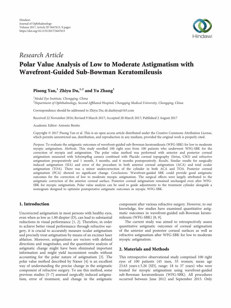
Mean preoperative subjective sphere and cylinder at cornealplane was −4.74± 1.98D (range: −9.33 to −0.74D) and−1.16± 0.80D (range: −4.49 to −0.06D), respectively. Meanpreoperative sphere and cylinder by WaveScan at cornealplane was −4.76± 2.02D (range: −9.38 to 0.03D) and −1.14± 0.82D (range: −4.54 to −0.06D), respectively. Preoperativeand postoperative visual and refractive outcomes weresummarized in Table 1. The distributions of preoperativeastigmatic components at the corneal plane is shown inTable 2, and 86% of eyes had preoperative astigmatic com-ponents at the corneal plane less than or equal to 2.0D.The spherically equivalent refraction was significantlyreduced by 6.20± 2.03D from the preoperative level of−6.12± 1.96D to the postoperative value of 0.08± 0.24D.Figure 1 shows attempted spherical equivalent refraction(SER) against achieved SER at 6 months postoperatively.The linear regression of the scattergram has a slope of1.03 and an intercept of 0.08.
3.1. Surgically Induced Corneal Astigmatism and Error ofTreatment in ACA. On the anterior corneal surface, thesteeper meridian was vertical in 90 eyes (90.0%), horizontalin 4 eyes (4.0%), and oblique in 6 eyes (6.0%). However, thesteepest posterior corneal meridian was vertically aligned inall eyes. The posterior corneal astigmatism was 0.25D orlower in 12 eyes (12%) and greater than or equal to 0.50D
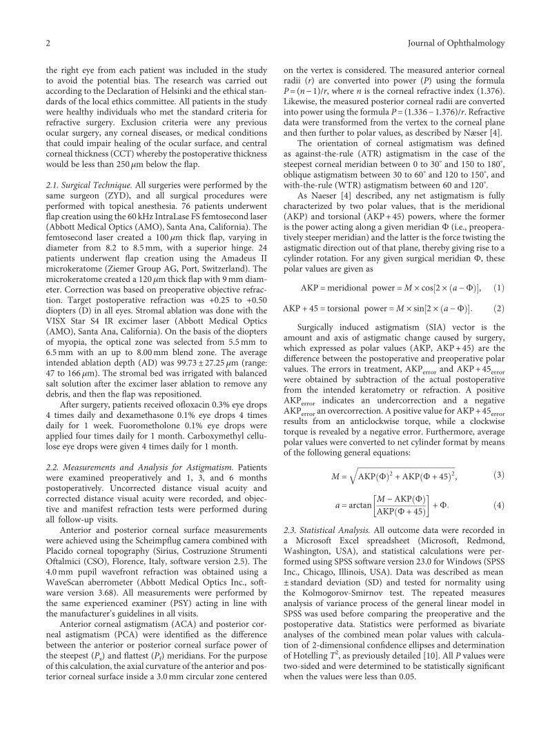
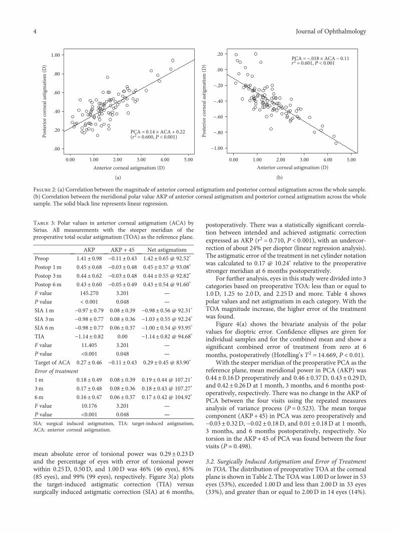
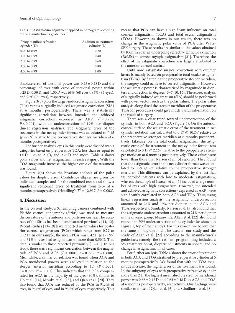
in 34 eyes (34%). Across the entire sample, a significant cor-relation was found between the magnitude of PCA and ACA(P < 0001, r = 0 775, r2 = 0 600) (Figure 2(a)). The equationthat best fits these data was PCA=0.14×ACA+0.22. A sim-ilar correlation was found when ACA and PCA meridionalpowers were analyzed in relation to the steeper anteriormeridian according to (1) (P < 0001, r = 0 775, r2 = 0 601)(Figure 2(b)). The equation that best fits these data wasPCA=−0.18×ACA− 0.11. Therefore, ACA was compen-sated by the PCA in the majority of eyes (94%).
With the steeper meridian of the preoperative TOA as thereference plane, all polar values of ACA before and after sur-gery are summarized in Table 3. As shown in Table 3, surgeryinduced a statistically significant flattening of the surgicalmeridian (F = 145 270, P < 0 001). There were a minimalinduced anticlockwise torque of 0.08± 0.39D, 0.08± 0.36D,and 0.06± 0.37D at 1, 3, and 6 months, respectively. Using(3) and (4) on the polar values, SIA may be expressed asthe net cylinder: 0.98± 0.56 @ 2.31°, 1.03± 0.55 @ 2.24°,and 1.00± 0.54 @ 3.95° at 1, 3, and 6 months, respectively.Given the mean intended postoperative astigmatism (targetof ACA-AKP), there was a significant undercorrection ofastigmatism (or error of treatment) with repeated measuresanalysis of variance (F = 10 176, P < 0 001). However,Table 3 shows the relative small mean and the large standarddeviations of error of the procedure, indicating a consider-able spread with a lot of positive and negative changes. At6 months, the mean absolute error of meridional power was0.40± 0.29D and the percentage of eyes with error ofmeridional power within 0.25D, 0.50D, and 1.00D was36% (36 eyes), 66% (66 eyes), and 97% (97 eyes), respectively.
Apart from the changes at the main meridian, there wasinduced astigmatism at the oblique meridian. Thus, the errorof target in the mean torque component (AKP+45) indi-cated a minor, but significant, anticlockwise torsion ofthe cylinder axis (F = 3 201, P = 0 048). At 6 months, the
Table 1: Refractive and visual outcomes (n = 100 eyes).
ParameterPreop
mean (SD)Postop 6mmean (SD)
P value
Sphere (D) (corneal plane) −4.76 (2.02) 0.53 (0.28) <0.001Cylinder (D) (corneal plane) −1.14 (0.82) −0.42 (0.36) <0.001SER (D) (corneal plane) −6.12 (1.96) 0.08 (0.24) <0.001UDVA (logMAR) 1.16 (0.33) −0.10 (0.06) <0.001CDVA (logMAR) −0.07 (0.05) −0.11 (0.07) >0.05Note: sphere and cylinder byWaveScan at corneal plane. SER = sphere +1/2∗
cylinder. Preop: preoperative visit; Postop 6m: 6 months postoperative visitafter surgery; SD: standard deviation; D: diopter; SER: spherical equivalentrefraction; UDVA: uncorrected distance visual acuity; CDVA: correcteddistance visual acuity.
Table 2: Distribution of preoperative astigmatic components(corneal plane, N = 100 eyes).
ParameterMagnitude (D) Percentage of eyes
Mean (SD)Median(range)
≤1.0 D 1.0 to2.0D
≥2.0D
TOA 1.14 (0.82)0.93
(0.06 to 4.54)53 33 14
ACA 1.54 (0.88)1.41
(0.09 to 4.35)31 44 25
PCA −0.44 (0.16) −0.43(−0.14 to −0.94) 100 0 0
TOA: total ocular astigmatism, ACA: anterior corneal astigmatism,PCA: posterior corneal astigmatism, D: diopter.
Attempted SER (D)
Ach
ieve
d SE
R (D
)
−12.00
−12.00
−10.00
−8.00
−6.00
−4.00
−2.00 Follow-up: 6 monthsn = 100 eyesy = 1.03x + 0.08r2 = 0.986
−10.00 −8.00 −6.00 −4.00 −2.00
Figure 1: Predictability of spherical equivalent refraction (SER) at 6months postoperatively. The area between two dotted lines is thepostoperative SER within ±0.50D. The solid grey line representslinear regression.
3Journal of Ophthalmology
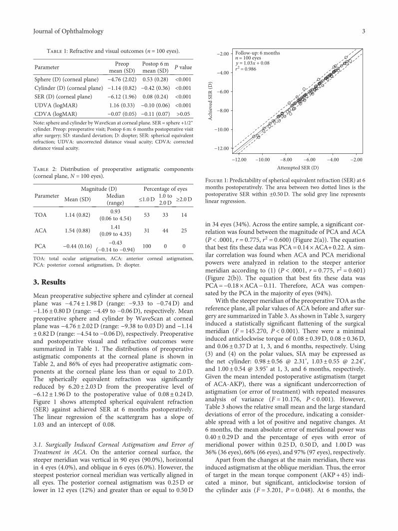
mean absolute error of torsional power was 0.29± 0.23Dand the percentage of eyes with error of torsional powerwithin 0.25D, 0.50D, and 1.00D was 46% (46 eyes), 85%(85 eyes), and 99% (99 eyes), respectively. Figure 3(a) plotsthe target-induced astigmatic correction (TIA) versussurgically induced astigmatic correction (SIA) at 6 months,
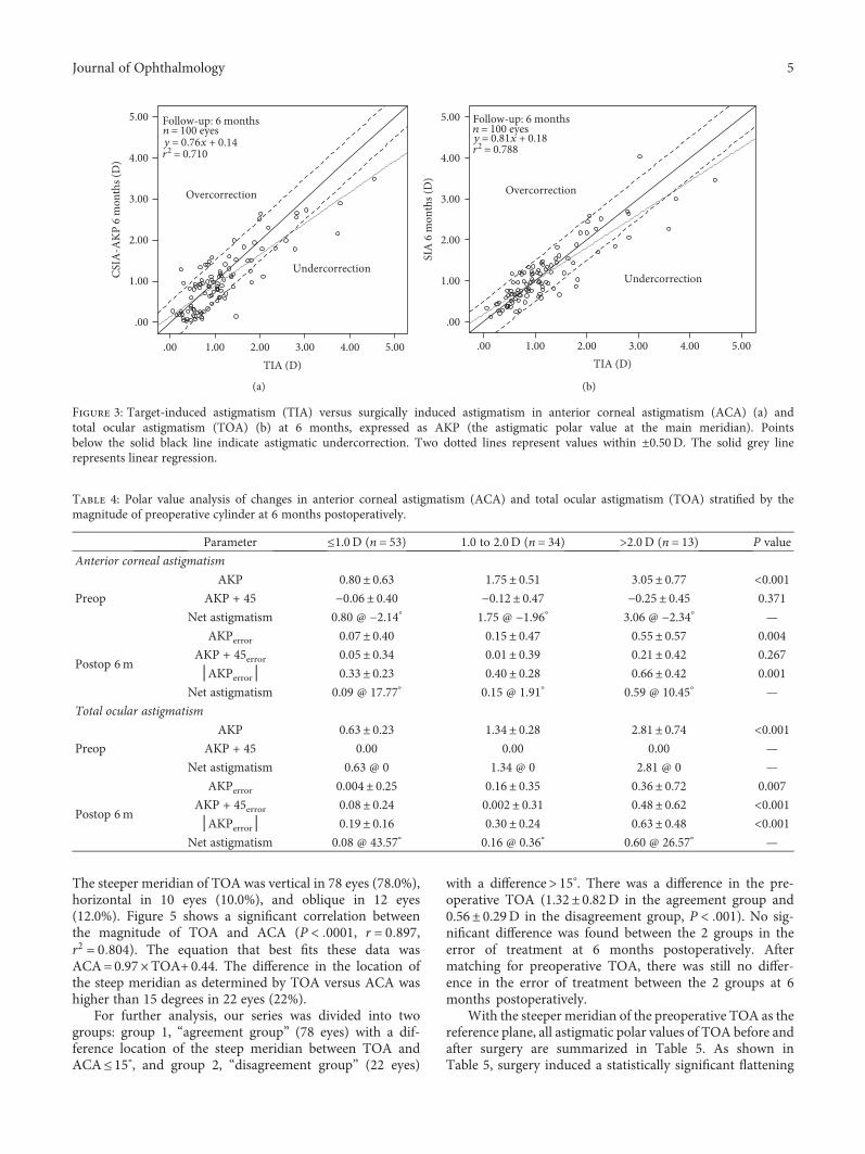
postoperatively. There was a statistically significant correla-tion between intended and achieved astigmatic correctionexpressed as AKP (r2 = 0 710, P < 0 001), with an undercor-rection of about 24% per diopter (linear regression analysis).The astigmatic error of the treatment in net cylinder notationwas calculated to 0.17 @ 10.24° relative to the preoperativestronger meridian at 6 months postoperatively.
For further analysis, eyes in this study were divided into 3categories based on preoperative TOA: less than or equal to1.0D, 1.25 to 2.0D, and 2.25D and more. Table 4 showspolar values and net astigmatism in each category. With theTOA magnitude increase, the higher error of the treatmentwas found.
Figure 4(a) shows the bivariate analysis of the polarvalues for dioptric error. Confidence ellipses are given forindividual samples and for the combined mean and show asignificant combined error of treatment from zero at 6months, postoperatively (Hotelling’s T2 = 14 669, P < 0 01).
With the steeper meridian of the preoperative PCA as thereference plane, mean meridional power in PCA (AKP) was0.44± 0.16D preoperatively and 0.46± 0.37D, 0.43± 0.29D,and 0.42± 0.26D at 1 month, 3 months, and 6 months post-operatively, respectively. There was no change in the AKP ofPCA between the four visits using the repeated measuresanalysis of variance process (P = 0 523). The mean torquecomponent (AKP+45) in PCA was zero preoperatively and−0.03± 0.32D, −0.02± 0.18D, and 0.01± 0.18D at 1 month,3 months, and 6 months postoperatively, respectively. Notorsion in the AKP+45 of PCA was found between the fourvisits (P = 0 498).
3.2. Surgically Induced Astigmatism and Error of Treatmentin TOA. The distribution of preoperative TOA at the cornealplane is shown in Table 2. The TOAwas 1.00D or lower in 53eyes (53%), exceeded 1.00D and less than 2.00D in 33 eyes(33%), and greater than or equal to 2.00D in 14 eyes (14%).
Anterior corneal astigmatism (D)0.00
Poste
rior c
orne
al as
tigm
atism
(D)
.00
.20
.40
.60
.80
1.00
1.00 2.00 3.00 4.00
PCA = 0.14 × ACA + 0.22(r2 = 0.600, P < 0.001)
5.00
(a)
Anterior corneal astigmatism (D)0.00
−1.00
−.80
−.60
Poste
rior c
orne
al as
tigm
atism
(D)
−.40
−.20
.00
.20PCA = −.018 × ACA − 0.11r2 = 0.601, P < 0.001
1.00 2.00 3.00 4.00 5.00
(b)
Figure 2: (a) Correlation between the magnitude of anterior corneal astigmatism and posterior corneal astigmatism across the whole sample.(b) Correlation between the meridional polar value AKP of anterior corneal astigmatism and posterior corneal astigmatism across the wholesample. The solid black line represents linear regression.
Table 3: Polar values in anterior corneal astigmatism (ACA) bySirius. All measurements with the steeper meridian of thepreoperative total ocular astigmatism (TOA) as the reference plane.
AKP AKP + 45 Net astigmatism
Preop 1.41± 0.98 −0.11± 0.43 1.42± 0.65 @ 92.52°
Postop 1m 0.45± 0.68 −0.03± 0.48 0.45± 0.57 @ 93.08°
Postop 3m 0.44± 0.62 −0.03± 0.48 0.44± 0.55 @ 92.82°
Postop 6m 0.43± 0.60 −0.05± 0.49 0.43± 0.54 @ 91.60°
F value 145.270 3.201 —
P value < 0.001 0.048 —
SIA 1m −0.97± 0.79 0.08± 0.39 −0.98± 0.56 @ 92.31°
SIA 3m −0.98± 0.77 0.08± 0.36 −1.03± 0.55 @ 92.24°
SIA 6m −0.98± 0.77 0.06± 0.37 −1.00± 0.54 @ 93.95°
TIA −1.14± 0.82 0.00 −1.14± 0.82 @ 94.68°
F value 11.405 3.201 —
P value <0.001 0.048 —
Target of ACA 0.27± 0.46 −0.11± 0.43 0.29± 0.45 @ 83.90°
Error of treatment
1m 0.18± 0.49 0.08± 0.39 0.19± 0.44 @ 107.21°
3m 0.17± 0.48 0.08± 0.36 0.18± 0.43 @ 107.27°
6m 0.16± 0.47 0.06± 0.37 0.17± 0.42 @ 104.92°
F value 10.176 3.201 —
P value <0.001 0.048 —
SIA: surgical induced astigmatism, TIA: target-induced astigmatism,ACA: anterior corneal astigmatism.
4 Journal of Ophthalmology
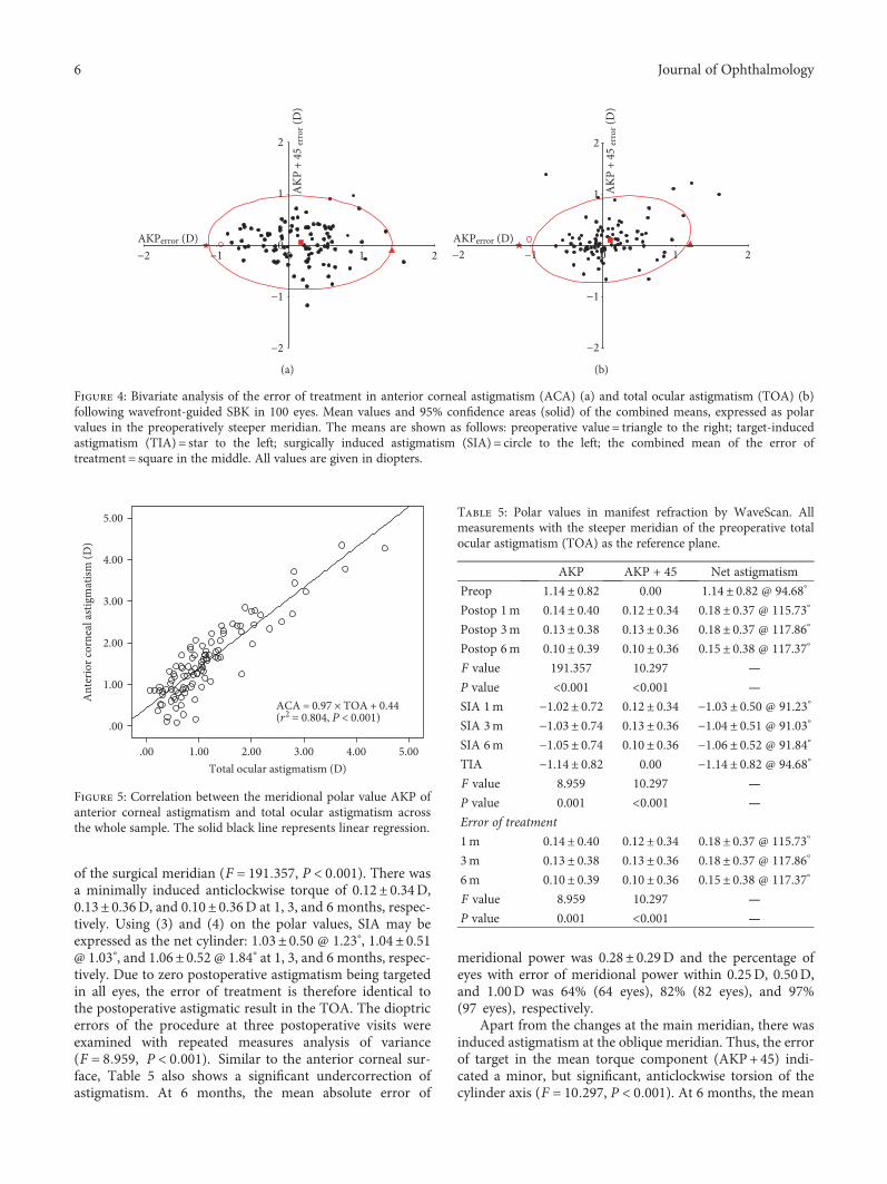
The steeper meridian of TOA was vertical in 78 eyes (78.0%),horizontal in 10 eyes (10.0%), and oblique in 12 eyes(12.0%). Figure 5 shows a significant correlation betweenthe magnitude of TOA and ACA (P < 0001, r = 0 897,r2 = 0 804). The equation that best fits these data wasACA=0.97×TOA+0.44. The difference in the location ofthe steep meridian as determined by TOA versus ACA washigher than 15 degrees in 22 eyes (22%).
For further analysis, our series was divided into twogroups: group 1, “agreement group” (78 eyes) with a dif-ference location of the steep meridian between TOA andACA≤ 15°, and group 2, “disagreement group” (22 eyes)
with a difference> 15°. There was a difference in the pre-operative TOA (1.32± 0.82D in the agreement group and0.56± 0.29D in the disagreement group, P < 001). No sig-nificant difference was found between the 2 groups in theerror of treatment at 6 months postoperatively. Aftermatching for preoperative TOA, there was still no differ-ence in the error of treatment between the 2 groups at 6months postoperatively.
With the steeper meridian of the preoperative TOA as thereference plane, all astigmatic polar values of TOA before andafter surgery are summarized in Table 5. As shown inTable 5, surgery induced a statistically significant flattening
TIA (D).00
.00
1.00
2.00
3.00
4.00
5.00
1.00 2.00 3.00 4.00
Undercorrection
Overcorrection
CSIA
-AKP
6 m
onth
s (D
)Follow-up: 6 monthsn = 100 eyesy = 0.76x + 0.14r2 = 0.710
5.00
(a)
TIA (D).00
SIA
6 m
onth
s (D
)
.00
1.00
2.00
3.00
4.00
5.00 Follow-up: 6 monthsn = 100 eyesy = 0.81x + 0.18r2 = 0.788
Overcorrection
1.00 2.00 3.00 4.00
Undercorrection
5.00
(b)
Figure 3: Target-induced astigmatism (TIA) versus surgically induced astigmatism in anterior corneal astigmatism (ACA) (a) andtotal ocular astigmatism (TOA) (b) at 6 months, expressed as AKP (the astigmatic polar value at the main meridian). Pointsbelow the solid black line indicate astigmatic undercorrection. Two dotted lines represent values within ±0.50D. The solid grey linerepresents linear regression.
Table 4: Polar value analysis of changes in anterior corneal astigmatism (ACA) and total ocular astigmatism (TOA) stratified by themagnitude of preoperative cylinder at 6 months postoperatively.
Parameter ≤1.0D (n = 53) 1.0 to 2.0D (n = 34) >2.0 D (n = 13) P value
Anterior corneal astigmatism
Preop
AKP 0.80± 0.63 1.75± 0.51 3.05± 0.77 <0.001AKP + 45 −0.06± 0.40 −0.12± 0.47 −0.25± 0.45 0.371
Net astigmatism 0.80 @ −2.14° 1.75 @ −1.96° 3.06 @ −2.34° —
Postop 6m
AKPerror 0.07± 0.40 0.15± 0.47 0.55± 0.57 0.004
AKP + 45error 0.05± 0.34 0.01± 0.39 0.21± 0.42 0.267
│AKPerror│ 0.33± 0.23 0.40± 0.28 0.66± 0.42 0.001
Net astigmatism 0.09 @ 17.77° 0.15 @ 1.91° 0.59 @ 10.45° —
Total ocular astigmatism
Preop
AKP 0.63± 0.23 1.34± 0.28 2.81± 0.74 <0.001AKP + 45 0.00 0.00 0.00 —
Net astigmatism 0.63 @ 0 1.34 @ 0 2.81 @ 0 —
Postop 6m
AKPerror 0.004± 0.25 0.16± 0.35 0.36± 0.72 0.007
AKP + 45error 0.08± 0.24 0.002± 0.31 0.48± 0.62 <0.001│AKPerror│ 0.19± 0.16 0.30± 0.24 0.63± 0.48 <0.001
Net astigmatism 0.08 @ 43.57° 0.16 @ 0.36° 0.60 @ 26.57° —
5Journal of Ophthalmology
of the surgical meridian (F = 191 357, P < 0 001). There wasa minimally induced anticlockwise torque of 0.12± 0.34D,0.13± 0.36D, and 0.10± 0.36D at 1, 3, and 6 months, respec-tively. Using (3) and (4) on the polar values, SIA may beexpressed as the net cylinder: 1.03± 0.50 @ 1.23°, 1.04± 0.51@ 1.03°, and 1.06± 0.52 @ 1.84° at 1, 3, and 6 months, respec-tively. Due to zero postoperative astigmatism being targetedin all eyes, the error of treatment is therefore identical tothe postoperative astigmatic result in the TOA. The dioptricerrors of the procedure at three postoperative visits wereexamined with repeated measures analysis of variance(F = 8 959, P < 0 001). Similar to the anterior corneal sur-face, Table 5 also shows a significant undercorrection ofastigmatism. At 6 months, the mean absolute error of
meridional power was 0.28± 0.29D and the percentage ofeyes with error of meridional power within 0.25D, 0.50D,and 1.00D was 64% (64 eyes), 82% (82 eyes), and 97%(97 eyes), respectively.
Apart from the changes at the main meridian, there wasinduced astigmatism at the oblique meridian. Thus, the errorof target in the mean torque component (AKP+45) indi-cated a minor, but significant, anticlockwise torsion of thecylinder axis (F = 10 297, P < 0 001). At 6 months, the mean
−2
−2AKPerror (D)
2
−1
1
2
−1 10
AKP
+ 4
5 er
ror (
D)
0
(a)
AKPerror (D)
AKP
+ 4
5 er
ror (
D)
−2
−2 2
−1
1
2
−1 100
(b)
Figure 4: Bivariate analysis of the error of treatment in anterior corneal astigmatism (ACA) (a) and total ocular astigmatism (TOA) (b)following wavefront-guided SBK in 100 eyes. Mean values and 95% confidence areas (solid) of the combined means, expressed as polarvalues in the preoperatively steeper meridian. The means are shown as follows: preoperative value = triangle to the right; target-inducedastigmatism (TIA) = star to the left; surgically induced astigmatism (SIA) = circle to the left; the combined mean of the error oftreatment = square in the middle. All values are given in diopters.
Total ocular astigmatism (D)
ACA = 0.97 × TOA + 0.44(r2 = 0.804, P < 0.001)
.00
.00
1.00
2.00
3.00
Ant
erio
r cor
neal
astig
mat
ism (D
)
4.00
5.00
1.00 2.00 3.00 4.00 5.00
Figure 5: Correlation between the meridional polar value AKP ofanterior corneal astigmatism and total ocular astigmatism acrossthe whole sample. The solid black line represents linear regression.
Table 5: Polar values in manifest refraction by WaveScan. Allmeasurements with the steeper meridian of the preoperative totalocular astigmatism (TOA) as the reference plane.
AKP AKP + 45 Net astigmatism
Preop 1.14± 0.82 0.00 1.14± 0.82 @ 94.68°
Postop 1m 0.14± 0.40 0.12± 0.34 0.18± 0.37 @ 115.73°
Postop 3m 0.13± 0.38 0.13± 0.36 0.18± 0.37 @ 117.86°
Postop 6m 0.10± 0.39 0.10± 0.36 0.15± 0.38 @ 117.37°
F value 191.357 10.297 —
P value <0.001 <0.001 —
SIA 1m −1.02± 0.72 0.12± 0.34 −1.03± 0.50 @ 91.23°
SIA 3m −1.03± 0.74 0.13± 0.36 −1.04± 0.51 @ 91.03°
SIA 6m −1.05± 0.74 0.10± 0.36 −1.06± 0.52 @ 91.84°
TIA −1.14± 0.82 0.00 −1.14± 0.82 @ 94.68°
F value 8.959 10.297 —
P value 0.001 <0.001 —
Error of treatment
1m 0.14± 0.40 0.12± 0.34 0.18± 0.37 @ 115.73°
3m 0.13± 0.38 0.13± 0.36 0.18± 0.37 @ 117.86°
6m 0.10± 0.39 0.10± 0.36 0.15± 0.38 @ 117.37°
F value 8.959 10.297 —
P value 0.001 <0.001 —
6 Journal of Ophthalmology
absolute error of torsional power was 0.25± 0.28D and thepercentage of eyes with error of torsional power within0.25D, 0.50D, and 1.00D was 68% (68 eyes), 85% (85 eyes),and 96% (96 eyes), respectively.
Figure 3(b) plots the target-induced astigmatic correction(TIA) versus surgically induced astigmatic correction (SIA)at 6 months, postoperatively. There was a statisticallysignificant correlation between intended and achievedastigmatic correction expressed as AKP (r2 = 0 788,P < 0 001), with an undercorrection of 19% per diopter(linear regression analysis). The astigmatic error of thetreatment in the net cylinder format was calculated to 0.15@ 22.69° relative to the preoperative stronger meridian at 6months postoperatively.
For further analysis, eyes in this study were divided into 3categories based on preoperative TOA: less than or equal to1.0D, 1.25 to 2.0D, and 2.25D and more. Table 4 showspolar values and net astigmatism in each category. With theTOA magnitude increase, the higher error of the treatmentwas found.
Figure 4(b) shows the bivariate analysis of the polarvalues for dioptric error. Confidence ellipses are given forindividual samples and for the combined mean and show asignificant combined error of treatment from zero at 6months, postoperatively (Hotelling’s T2 = 12 917, P = 0 002).
4. Discussion
In the current study, a Scheimpflug camera combined withPlacido corneal topography (Sirius) was used to measurethe curvature of the anterior and posterior cornea. The accu-racy of the Sirius has been demonstrated previously [11, 12].Recent studies [13–19] have reported mean values for poste-rior corneal astigmatism (PCA) which range from 0.29 to0.52D. In our sample, the mean PCA was 0.42D @ 179.95°
and 31% of eyes had astigmatism of more than 0.50D. Thisdata is similar to those reported previously [13–19]. In ourstudy, there was a significant correlation between the magni-tude of PCA and ACA (P < 0001, r = 0 775, r2 = 0 600).Meanwhile, a similar correlation was found when ACA andPCA meridional powers were analyzed in relation to thesteeper anterior meridian according to (1) (P < 0001,r = 0 775, r2 = 0 601). This indicates that the PCA compen-sated for ACA in the majority of the eyes (94%), similar toHo et al. [14], Miyake et al. [15], and Qian et al. [20]. Theyalso found that ACA was reduced by the PCA in 91.4% ofeyes, in 96.6% of eyes, and in 95.8% of eyes, respectively. This
means that PCA can have a significant influence on totalcorneal astigmatism (TCA) and total ocular astigmatism(TOA). However, as shown in our results, there was nochange in the astigmatic polar value of PCA after WFG-SBK surgery. These results are similar to the values obtainedby Kamiya et al. in undergoing refractive lenticule extraction(ReLEx) to correct myopic astigmatism [21]. Therefore, theeffect of the astigmatic correction was largely attributed tothe anterior corneal surface.
Until now, astigmatic surgical correction with excimerlasers is mainly based on preoperative total ocular astigma-tism (TOA). By flattening the preoperative steeper meridian,the surgery could achieve to correct astigmatism. However,the astigmatic power is characterized by magnitude in diop-ters and direction in degrees [3–7, 10, 16]. Therefore, analysisof surgically induced astigmatism (SIA) should be performedwith power vector, such as the polar values. The polar valueanalysis along fixed the steeper meridian of the preoperativeTOA for procedures could give much useful information onthe result of surgery.
There was a clear trend toward undercorrection of thecylinder in both ACA and TOA (Figure 3). On the anteriorcorneal surface, the astigmatic error of the treatment in netcylinder notation was calculated to 0.17 @ 10.24° relative tothe preoperative stronger meridian at 6 months postopera-tively. Likewise, on the total ocular astigmatism, the astig-matic error of the treatment in the net cylinder format wascalculated to 0.15 @ 22.69° relative to the preoperative stron-ger meridian at 6 months postoperatively. These values werelower than those that Ivarsen et al. [5] reported. They foundthat the astigmatic error in the net cylinder format was calcu-lated to 0.79 @ −7° relative to the preoperative strongermeridian. This difference can be explained by the fact thatwe enrolled patients with low to moderate astigmatism,whereas the sample of Ivarsen et al. [5] included a large num-ber of eyes with high astigmatism. However, the intendedand achieved astigmatic corrections (expressed as AKP) weresignificantly correlated in both ACA and TOA. Thus, usinglinear regression analysis, the astigmatic undercorrectionamounted to 24% and 19% per diopter in the ACA andTOA, respectively. Similarly, Ivarsen et al. [5] also found thatthe astigmatic undercorrection amounted to 21% per diopterin the myopic group. Meanwhile, Allan et al. [22] also foundmore than 20% undercorrection of the cylinder (as shown inFigure 1, top of their study). For this reason, we believe thatthe same nomogram might be used in our study and thestudy of Allan et al. [22] according to the manufacturer’sguidelines; namely, the treatment programming included a5% treatment boost, dioptric adjustments in sphere, and nochange in astigmatism in all cases.
For further analysis, Table 4 shows the error of treatmentin both ACA and TOA stratified by preoperative cylinder at 6months postoperatively. We found that with the TOA mag-nitude increase, the higher error of the treatment was found.In the subgroup of eyes with preoperative refractive cylindermore than 2D, the highest mean absolute error of meridionalpower was 0.66± 0.42D and 0.63± 0.48D in ACA and TOAat 6 months postoperatively, respectively. Our findings weresimilar to those of Qian et al. [6] and Schallhorn et al. [8].
Table 6: Astigmatism adjustment applied in nomogram accordingto the manufacturer’s guidelines.
Preop manifest refractioncylinder (D)
Addition to treatmentcylinder (D)
0.00 to 0.99 0.20
1.00 to 1.99 0.40
2.00 to 2.99 0.60
3.00 to 3.99 0.80
4.00 to 4.99 1.00
7Journal of Ophthalmology
On the basis of these data, a 0.2D correction was added tothe attempted cylinder for each 1.0D increase in preoperativecylinder (Table 6) for developing the nomogram to improveastigmatic outcomes. However, further study is necessary toassess the astigmatic outcomes by using this new nomogram.
Nevertheless, many factors could contribute to the resultsof astigmatic surgical correction with excimer lasers. Amongthese factors, it is necessary to consider disagreementbetween refraction and corneal topography in the steepmeridian. We observed that no significant difference wasfound between the agreement group and the disagreementgroup in the error of treatment. After matching for preop-erative TOA, there was still no difference in the error oftreatment between the 2 groups at 6 months postoperatively.This contradicts the study of Bragheeth and Dua [23] wherethey found that the magnitude error of astigmatic outcomesby LASIK was greater in the disagreement group than inthe agreement group (0.16± 0.74 versus 0.07± 0.52). Thedifference was statistically significant between the agree-ment group and the disagreement group. This differencecan be explained by different ablation profiles during surgery.In our study, we performed WFG-SBK, whereas the sampleof Bragheeth and Dua [23] used conventional LASIK-corrected myopic astigmatism.
This study has limitations, and further studies are war-ranted. First, anterior corneal astigmatism was centered onthe corneal vertex and not on the pupil center. However,pupil-centered measurements may be more accurate andhave to be assessed. Thus, we have started another studyto assess astigmatic outcomes after WFG-SBK by usingpupil-centered measurements on the anterior corneal sur-face. Second, the number of patients, especially with obli-que astigmatism and ATR astigmatism, was small. Third,the time of follow-up is too short in the study. Since theunpredictable nature of corneal wound healing and thebiomechanical response to surgery [24] are present, furtherstudy is necessary to assess the refractive change over alonger period of time.
In conclusion, our study confirms that WFG-SBK couldprovide good astigmatic outcomes for the correction of myo-pic astigmatism. We observed similar results for SIA anderror of the procedure in both ACA and TOA. This meansthat we can objectively evaluate the astigmatic outcomes forthe correction of myopic astigmatism by measuring thechanges of ACA before and after operation. Although slightundercorrection (less than a quarter of a diopter) was seenin our dataset, the clinical refractive results are still highly sat-isfactory. There was no change in the astigmatic polar value ofPCA after WFG-SBK surgery. Polar value analysis can beused to guide adjustments to the treatment cylinder alongsidethe nomogram designed to optimize postoperative astigmaticoutcomes in myopic WFG-SBK. Despite some flaws, the cur-rent study is one of the few studies reporting results of WFG-SBK in patients with low to moderate refractive cylinder.
Disclosure
The authors alone are responsible for the content and writingof the paper.
Conflicts of Interest
The authors report no conflicts of interest.
Authors’ Contributions
Drs. Pisong Yan and Zhiyu Du contributed equally tothis work.
Acknowledgments
The authors gratefully acknowledge the grant support fromthe Key Program of Chongqing Municipal Health Bureau,Chongqing, China (2010-1-25).
References
[1] J. Wolffsohn, G. Bhogal, and S. Shah, “Effect of uncorrectedastigmatism on vision,” Journal of Cataract and RefractiveSurgery, vol. 37, no. 3, pp. 454–460, 2011.
[2] K. Kamiya, H. Kobashi, K. Shimizu, T. Kawamorita, andH. Uozato, “Effect of pupil size on uncorrected visual acuityin astigmatic eyes,” British Journal of Ophthalmology, vol. 96,no. 2, pp. 267–270, 2012.
[3] K. Naeser, “Surgically induced astigmatism is characterizedby optical vectors, not by ratios,” Journal of Cataract andRefractive Surgery, vol. 42, no. 2, pp. 347-348, 2016.
[4] K. Næser, “Assessment and statistics of surgically inducedastigmatism,” Acta Ophthalmologica, vol. 86, Supplement 1,pp. 5–28, 2008.
[5] A. Ivarsen, K. Næser, and J. Hjortdal, “Laser in situ keratomil-eusis for high astigmatism in myopic and hyperopic eyes,”Journal of Cataract and Refractive Surgery, vol. 39, no. 1,pp. 74–80, 2013.
[6] Y. S. Qian, J. Huang, X. T. Zhou, and Y. Wang, “Comparisonof femtosecond laser small-incision lenticule extraction andlaser-assisted subepithelial keratectomy to correct myopicastigmatism,” Journal of Cataract and Refractive Surgery,vol. 41, no. 11, pp. 2476–2486, 2015.
[7] C. R. Rho, M.-J. Kim, and C.-K. Joo, “Polar value analysis ofcorneal astigmatism in intrastromal corneal ring segmentimplantation,” Journal of Ophthalmology, vol. 2016, ArticleID 7127534, 6 pages, 2016.
[8] S. C. Schallhorn, J. A. Venter, S. J. Hannan, and K. A. Hettinger,“Clinical outcomes of wavefront-guided laser in situ kerato-mileusis to treat moderate-to-high astigmatism,” ClinicalOphthalmology, vol. 13, no. 9, pp. 1291–1298, 2015.
[9] S. C. Schallhorn, M. Brown, J. A. Venter, D. Teenan,K. Hettinger, and H. Yamamoto, “Early clinical outcomesof wavefront-guided myopic LASIK treatments using a new-generation hartmann-shack aberrometer,” Journal of Refrac-tive Surgery, vol. 30, no. 1, pp. 14–21, 2014.
[10] K. Naeser and J. Hjortdal, “Multivariate analysis of refractivedata: mathematics and statistics of spherocylinders,” Journalof Cataract and Refractive Surgery, vol. 27, no. 1, pp. 129–142, 2001.
[11] G. Savini, P. Barboni, M. Carbonelli, and K. J. Hoffer, “Repeat-ability of automatic measurements by a new Scheimpflug cam-era combined with Placido topography,” Journal of Cataractand Refractive Surgery, vol. 37, no. 10, pp. 1809–1816, 2011.
8 Journal of Ophthalmology
[12] R. Montalbán, D. P. Piñero, J. Javaloy, and J. L. Alió, “Intrasub-ject repeatability of corneal morphology measurementsobtained with a new Scheimpflug photography–based system,”Journal of Cataract and Refractive Surgery, vol. 38, no. 6,pp. 971–977, 2012.
[13] D. D. Koch, S. F. Ali, M. P. Weikert, M. Shirayama, R. Jenkins,and L. Wang, “Contribution of posterior corneal astigmatismto total corneal astigmatism,” Journal of Cataract and Refrac-tive Surgery, vol. 38, no. 12, pp. 2080–2087, 2012.
[14] J. D. Ho, C. Y. Tsai, and S. W. Liou, “Accuracy of cornealastigmatism estimation by neglecting the posterior cornealsurface measurement,” American Journal of Ophthalmology,vol. 147, no. 5, pp. 788–795, 2009.
[15] T. Miyake, K. Shimizu, and K. Kamiya, “Distribution ofposterior corneal astigmatism according to axis orientationof anterior corneal astigmatism,” PLoS One, vol. 10, no. 1,article e0117194, 2015.
[16] G. Savini, F. Versaci, G. Vestri, P. Ducoli, and K. Næser,“Influence of posterior corneal astigmatism on total cornealastigmatism in eyes with moderate to high astigmatism,”Journal of Cataract and Refractive Surgery, vol. 40, no. 10,pp. 1645–1653, 2014.
[17] G. Nemeth, A. Berta, E. Szalai, Z. Hassan, and L. Modis Jr.,“Analysis of surgically induced astigmatism on the posteriorsurface of the cornea,” Journal of Refractive Surgery, vol. 30,no. 9, pp. 604–608, 2014.
[18] Y. Ueno, T. Hiraoka, M. Miyazaki, M. Ito, and T. Oshika,“Corneal thickness profile and posterior corneal astigmatismin normal corneas,” Ophthalmology, vol. 122, no. 6,pp. 1072–1078, 2015.
[19] M. Dubbelman, V. A. Sicam, and G. L. Der Heijde, “The shapeof the anterior and posterior surface of the aging humancornea,” Vision Research, vol. 46, no. 6, pp. 993–1001, 2006.
[20] Y. S. Qian, J. Huang, R. Liu et al., “Influence of internal opticalastigmatism on the correction of myopic astigmatism byLASIK,” Journal of Refractive Surgery, vol. 27, no. 12,pp. 863–868, 2011.
[21] K. Kamiya, K. Shimizu, M. Yamagishi, A. Igarashi, andH. Kobashi, “Anterior and posterior corneal astigmatismafter refractive lenticule extraction for myopic astigmatism,”Journal of Ophthalmology, vol. 2015, Article ID 915853,6 pages, 2015.
[22] B. D. Allan, H. Hassan, and A. Ieong, “Multiple regressionanalysis in nomogram development for myopic wavefrontlaser in situ keratomileusis: improving astigmatic outcomes,”Journal of Cataract and Refractive Surgery, vol. 41, no. 5,pp. 1009–1017, 2015.
[23] M. A. Bragheeth and H. S. Dua, “Effect of refractive andtopographic astigmatic axis on LASIK correction of myopicastigmatism,” Journal of Refractive Surgery, vol. 21, no. 3,pp. 269–275, 2005.
[24] D. T. Azar, J. H. Chang, and K. Y. Han, “Wound healingafter keratorefractive surgery: review of biological and opticalconsiderations,” Cornea, vol. 31, Supplement 1, pp. S9–19,2012.
9Journal of Ophthalmology

Submit your manuscripts athttps://www.hindawi.com
Stem CellsInternational
Hindawi Publishing Corporationhttp://www.hindawi.com Volume 2014
Hindawi Publishing Corporationhttp://www.hindawi.com Volume 2014
MEDIATORSINFLAMMATION
of
Hindawi Publishing Corporationhttp://www.hindawi.com Volume 2014
Behavioural Neurology
EndocrinologyInternational Journal of
Hindawi Publishing Corporationhttp://www.hindawi.com Volume 2014
Hindawi Publishing Corporationhttp://www.hindawi.com Volume 2014
Disease Markers
Hindawi Publishing Corporationhttp://www.hindawi.com Volume 2014
BioMed Research International
OncologyJournal of
Hindawi Publishing Corporationhttp://www.hindawi.com Volume 2014
Hindawi Publishing Corporationhttp://www.hindawi.com Volume 2014
Oxidative Medicine and Cellular Longevity
Hindawi Publishing Corporationhttp://www.hindawi.com Volume 2014
PPAR Research
The Scientific World JournalHindawi Publishing Corporation http://www.hindawi.com Volume 2014
Immunology ResearchHindawi Publishing Corporationhttp://www.hindawi.com Volume 2014
Journal of
ObesityJournal of
Hindawi Publishing Corporationhttp://www.hindawi.com Volume 2014
Hindawi Publishing Corporationhttp://www.hindawi.com Volume 2014
Computational and Mathematical Methods in Medicine
OphthalmologyJournal of
Hindawi Publishing Corporationhttp://www.hindawi.com Volume 2014
Diabetes ResearchJournal of
Hindawi Publishing Corporationhttp://www.hindawi.com Volume 2014
Hindawi Publishing Corporationhttp://www.hindawi.com Volume 2014
Research and TreatmentAIDS
Hindawi Publishing Corporationhttp://www.hindawi.com Volume 2014
Gastroenterology Research and Practice
Hindawi Publishing Corporationhttp://www.hindawi.com Volume 2014
Parkinson’s Disease
Evidence-Based Complementary and Alternative Medicine
Volume 2014Hindawi Publishing Corporationhttp://www.hindawi.com