nanopore decoding of oligonucleotides in dna...
TRANSCRIPT
DNA Computing www.biotechnology-journal.com
REVIEW
Nanopore Decoding of Oligonucleotides inDNA Computing
Ryuji Kawano
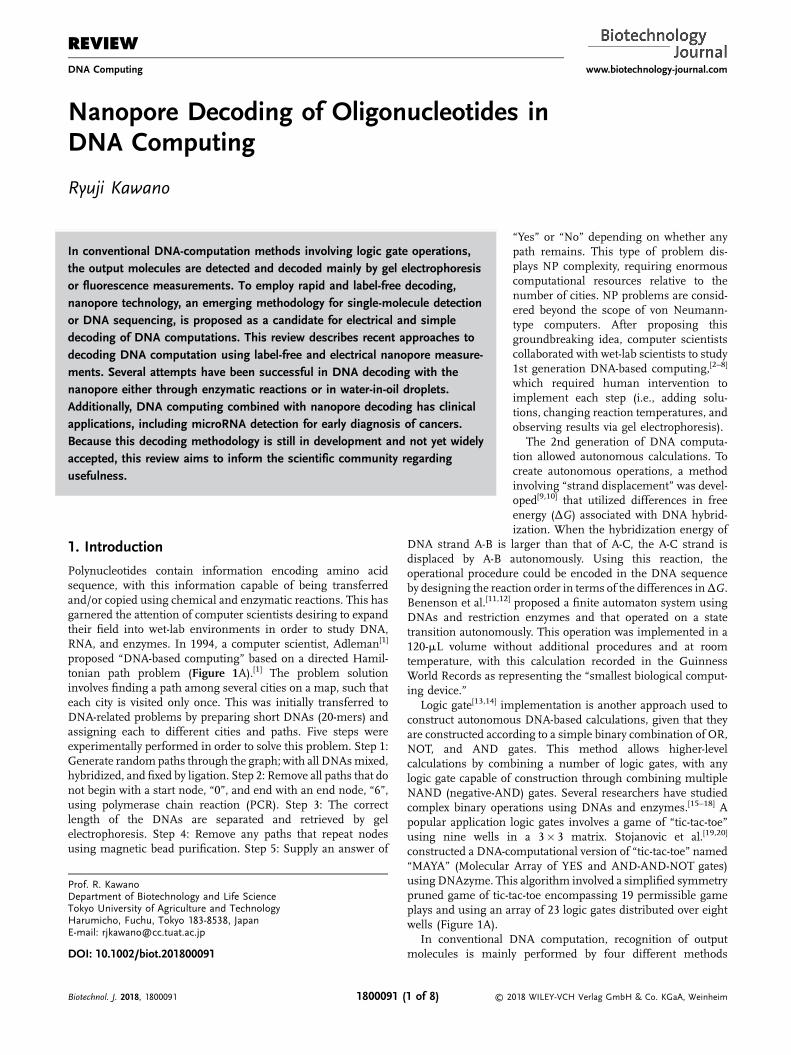
In conventional DNA-computation methods involving logic gate operations,the output molecules are detected and decoded mainly by gel electrophoresisor fluorescence measurements. To employ rapid and label-free decoding,nanopore technology, an emerging methodology for single-molecule detectionor DNA sequencing, is proposed as a candidate for electrical and simpledecoding of DNA computations. This review describes recent approaches todecoding DNA computation using label-free and electrical nanopore measure-ments. Several attempts have been successful in DNA decoding with thenanopore either through enzymatic reactions or in water-in-oil droplets.Additionally, DNA computing combined with nanopore decoding has clinicalapplications, including microRNA detection for early diagnosis of cancers.Because this decoding methodology is still in development and not yet widelyaccepted, this review aims to inform the scientific community regardingusefulness.
1. Introduction
Polynucleotides contain information encoding amino acidsequence, with this information capable of being transferredand/or copied using chemical and enzymatic reactions. This hasgarnered the attention of computer scientists desiring to expandtheir field into wet-lab environments in order to study DNA,RNA, and enzymes. In 1994, a computer scientist, Adleman[1]
proposed “DNA-based computing” based on a directed Hamil-tonian path problem (Figure 1A).[1] The problem solutioninvolves finding a path among several cities on a map, such thateach city is visited only once. This was initially transferred toDNA-related problems by preparing short DNAs (20-mers) andassigning each to different cities and paths. Five steps wereexperimentally performed in order to solve this problem. Step 1:Generate randompaths through the graph; with all DNAsmixed,hybridized, and fixed by ligation. Step 2: Remove all paths that donot begin with a start node, “0”, and end with an end node, “6”,using polymerase chain reaction (PCR). Step 3: The correctlength of the DNAs are separated and retrieved by gelelectrophoresis. Step 4: Remove any paths that repeat nodesusing magnetic bead purification. Step 5: Supply an answer of
Prof. R. KawanoDepartment of Biotechnology and Life ScienceTokyo University of Agriculture and TechnologyHarumicho, Fuchu, Tokyo 183-8538, JapanE-mail: [email protected]
DOI: 10.1002/biot.201800091
Biotechnol. J. 2018, 1800091 © 21800091 (1 of 8)
“Yes” or “No” depending on whether anypath remains. This type of problem dis-plays NP complexity, requiring enormouscomputational resources relative to thenumber of cities. NP problems are consid-ered beyond the scope of von Neumann-type computers. After proposing thisgroundbreaking idea, computer scientistscollaborated with wet-lab scientists to study1st generation DNA-based computing,[2–8]
which required human intervention toimplement each step (i.e., adding solu-tions, changing reaction temperatures, andobserving results via gel electrophoresis).
The 2nd generation of DNA computa-tion allowed autonomous calculations. Tocreate autonomous operations, a methodinvolving “strand displacement” was devel-oped[9,10] that utilized differences in freeenergy (ΔG) associated with DNA hybrid-ization. When the hybridization energy of
DNA strand A-B is larger than that of A-C, the A-C strand isdisplaced by A-B autonomously. Using this reaction, theoperational procedure could be encoded in the DNA sequenceby designing the reaction order in terms of the differences inΔG.Benenson et al.[11,12] proposed a finite automaton system usingDNAs and restriction enzymes and that operated on a statetransition autonomously. This operation was implemented in a120-mL volume without additional procedures and at roomtemperature, with this calculation recorded in the GuinnessWorld Records as representing the “smallest biological comput-ing device.”
Logic gate[13,14] implementation is another approach used toconstruct autonomous DNA-based calculations, given that theyare constructed according to a simple binary combination of OR,NOT, and AND gates. This method allows higher-levelcalculations by combining a number of logic gates, with anylogic gate capable of construction through combining multipleNAND (negative-AND) gates. Several researchers have studiedcomplex binary operations using DNAs and enzymes.[15–18] Apopular application logic gates involves a game of “tic-tac-toe”using nine wells in a 3� 3 matrix. Stojanovic et al.[19,20]
constructed a DNA-computational version of “tic-tac-toe” named“MAYA” (Molecular Array of YES and AND-AND-NOT gates)using DNAzyme. This algorithm involved a simplified symmetrypruned game of tic-tac-toe encompassing 19 permissible gameplays and using an array of 23 logic gates distributed over eightwells (Figure 1A).
In conventional DNA computation, recognition of outputmolecules is mainly performed by four different methods
018 WILEY-VCH Verlag GmbH & Co. KGaA, Weinheim
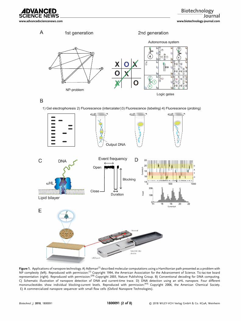
Figure 1. Applications of nanopore technology. A) Adleman[1] describedmolecular computations using a Hamiltonian path presented as a problemwithNP complexity (left). Reproduced with permission.[1] Copyright 1994, the American Association for the Advancement of Science. Tic-tac-toe boardrepresentation (right). Reproduced with permission.[20] Copyright 2003, Nature Publishing Group. B) Conventional decoding for DNA computing.C) Schematic illustration of nanopore detection of DNA and current-time trace. D) DNA detection using an αHL nanopore. Four differentmononucleotides show individual blocking-current levels. Reproduced with permission.[43] Copyright 2006, the American Chemical Society.E) A commercialized nanopore sequencer with small flow cells (Oxford Nanopore Technologies).
www.advancedsciencenews.com www.biotechnology-journal.com
Biotechnol. J. 2018, 1800091 © 2018 WILEY-VCH Verlag GmbH & Co. KGaA, Weinheim1800091 (2 of 8)
www.advancedsciencenews.com www.biotechnology-journal.com
(Figure 1B and Table 1): 1) gel-electrophoretic detectionfollowing PCR amplification; 2) fluorescence detection withisothermal amplification; 3) fluorescence detection by directlabeling without amplification; and 4) fluorescence probingwithout amplification. The pioneering works of Adleman[1] andBenenson[11] in DNA computation used these methods. Despitethe recent development of microscale rapid gel electrophore-sis,[21,22] traditional gel electrophoresis is time consuming. As asubstitute, several fluorescence techniques have been developedinvolving specific amplification of output DNA by isothermalreactions and observation by fluorescence labeling.[20,23] Meth-ods 1 and 2 require an amplification step involving enzymes,which requires long reaction times and temperature control,even under constant conditions at 37 �C. Therefore, non-amplification methods (i.e., methods 3 and 4) can be used,where the output DNA is labeled and detected[24] and a specificfluorescence probe is used.[25] Although neither of thesemethods require an amplification step (considering theirimplementation at relatively high concentrations), the use ofdirect labeling or specific probe molecules is required.
Nanopore technology allows the rapid and electricaldetection of oligonucleotides in the absence of labeling.Several studies reported methods related to nanoporedecoding[26–28] and their applications in diagnosis or clinicalsettings based on DNA computing. Therefore, nanoporemethods represent potential candidate methods for decodingDNA computations.
2. Application of Nanopore Technology forRapid and Label-Free Decoding
Nanopore technology involves electrical measurement of ioncurrent through a nanopore.[29–34] Biological (proteins) orsolid-state nanopores ranging in size from 1nm to �10 nmshow open-pore current conductance (Figure 1C). When amolecule passes through or blocks a nanopore, the open-porecurrent reduces, thereby demonstrating current-signal blockage.The blocking amplitude, duration time, and event frequencyprovide information regarding the size, mobility, and concen-tration of target molecules at the single-molecule level.α-Hemolysin (αHL), a channel toxin from Staphylococcusaureus,[35] is conventionally used as a biological nanopore fordetecting oligonucleotides based on its having a pore sizecomparable to that of single-stranded DNA (ssDNA) or ssRNA.Extensive studies reported using this nanopore as a label-free,rapid, and electrical method for single-oligonucleotide
Table 1. Comparison of conventional fluorescence and nanopore-decoding
Gel electrophoresis[1,11] Fluorescence (intercalator)[23]
Detection method Optical Optical
Measurement time Long Short
Sensitivity Low Low
Throughput Low Low
Pre-treatment Multi-step Few steps
Generality of method High High
Biotechnol. J. 2018, 1800091 1800091 (
determination, with one application targeting nanopore se-quencing (Figure 1D).[36–40] As the first report in 1996 byKasianowicz et al.,[36] enormous efforts have been undertaken toapply this method.[37,39,41–54] In 2015, a company named OxfordNanopore Technologies was launched and provided the firstcommercially available nanopore sequencer for general use(Figure 1E). Currently, nanopore technology can be utilized notonly for single DNA/RNA detection but also for large-scale DNAsequencing.[55]
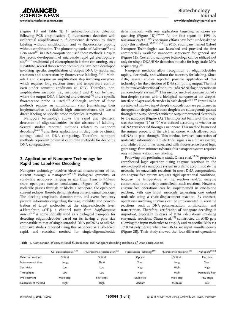
Nanopore methods allow recognition of oligonucleotidesrapidly, electrically, and without the necessity for labeling. Since2016, several studies reported possible application of thistechnology for the detection of DNA-computing output. The firststudy involveddetectionof theoutputofaNANDlogicoperation inamicro-droplet system.[26] Thismethod involved construction of afour-droplet system with a biological nanopore at the droplet-interface bilayer and electrodes in each droplet.[56–59] Input DNAsare injected into two input droplets, calculations are performed inthe operation droplet, and these droplets are subsequently passedthrough the output droplet, with the outputmonitored electricallyby the nanopore (Figure 2A). The important feature of this workwas that output “1” or “0” was defined according to whether anssDNA translocated through a nanopore. This method harnessedthe unique property of the αHL nanopore, which allowed onlyssDNAs to pass through. This method involves conversion ofmolecular information into electrical signals in a binary system,and while output times associated with fluorescence-based logicgates range fromminutes to hours, this nanopore system requiresonly �10min without any labeling.
Following this preliminary study, Ohara et al.[27,60] proposed acomplicated logic operation using enzyme reactions in themicro-droplet of a nanopore system in order to accommodate thenecessity for enzymatic reactions in most DNA computations.An enzyme-free system requires rigid operational conditions,because the temperature of the reaction and/or enzymeconcentrations are strictly controlled in such reactions. However,enzyme-free operations can be implemented in one-to-onereaction, with one input molecule generating one outputmolecule using a chain-displacement reaction. By contrast,operations involving enzymes can be implemented in versatilereactions, such as DNA polymerization, amplification, andtranscription. Therefore, verification of nanopore decoding isimportant, especially in cases of DNA calculations involvingenzymatic reactions. Ohara et al.[27] constructed an AND gateallowing the input molecules to amplify and transcribe DNA viaT7 RNA polymerase when two DNAs are input simultaneously(Figure 2B). Their study showed that four different operations
methods of DNA computation.
Fluorescence (labeling)[24] Fluorescence (probing)[25] Nanopore[26,27]
Optical Optical Electrical
Short Long Short
High High High
High High Potentially high
Multi-step Multi-step Few steps
Medium Medium Low
© 2018 WILEY-VCH Verlag GmbH & Co. KGaA, Weinheim3 of 8)
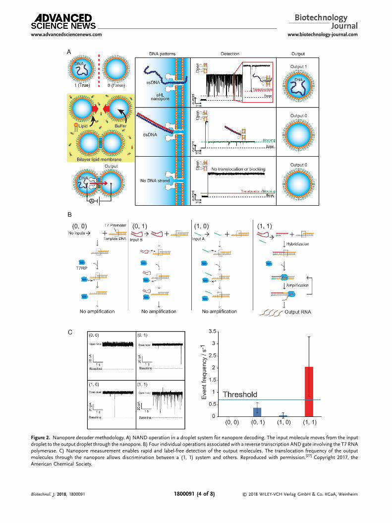
Figure 2. Nanopore decoder methodology. A) NAND operation in a droplet system for nanopore decoding. The input molecule moves from the inputdroplet to the output droplet through the nanopore. B) Four individual operations associated with a reverse transcription AND gate involving the T7 RNApolymerase. C) Nanopore measurement enables rapid and label-free detection of the output molecules. The translocation frequency of the outputmolecules through the nanopore allows discrimination between a (1, 1) system and others. Reproduced with permission.[27] Copyright 2017, theAmerican Chemical Society.
www.advancedsciencenews.com www.biotechnology-journal.com
Biotechnol. J. 2018, 1800091 © 2018 WILEY-VCH Verlag GmbH & Co. KGaA, Weinheim1800091 (4 of 8)
www.advancedsciencenews.com www.biotechnology-journal.com
represented by (0 0), (1 0), (0 1), and (1 1) were implemented inmultiple micro-droplet devices, and that the output could beobtained after 90min, which included a 60-min enzymaticreaction (Figure 2C),[28] suggesting the efficacy of nanoporedecoding for operations involving enzymes in micro-dropletsystems (summarized in Table 1). However, the reactionefficiency differed between that performed in conventionalplastic tubes and droplets with a surrounding lipid bilayer, withreduced efficiency observed in the droplet system.[27] Therefore,these operations need to be improved appropriately withrespect to enzymatic reactions performed in a lipid-dropletenvironment.
3. Application of Nanopore Decoding inMedical Diagnosis
The field of DNA computing was developed largely as a curiositydriven exercise focused on solvingmathematics-related problems,including cryptograms and constructing various types of logicgates (AND, OR, NOT, XOR, and NAND). However, this fieldrecently increased in importancedue to itspotential applications inmedical diagnosis.[61–63] Benenson et al.[61] reported autonomousdiagnosis and drug-release systems using DNA computing usingthe following “if-then” logic: “if” certain diagnostic conditions aretrue, such as low expression levels of certain mRNAs relative tothose of others, “then” the antisense drug is released. After thispioneering study, several studies were undertaken focused onapplication of this technology to diagnosis and therapy.[62] Basedon the favorable compatibility of nanopore technology witholigonucleotide detection, strategies utilizing this method fordiagnosis using nanopores and DNA have been proposed.
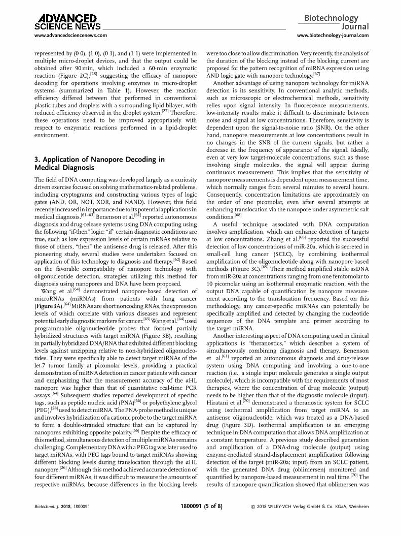
Wang et al.[64] demonstrated nanopore-based detection ofmicroRNAs (miRNAs) from patients with lung cancer(Figure3A).[64]MiRNAsareshortnoncodingRNAs, theexpressionlevels of which correlate with various diseases and representpotential earlydiagnosticmarkers forcancer.[65]Wangetal.[64]usedprogrammable oligonucleotide probes that formed partiallyhybridized structures with target miRNA (Figure 3B), resultinginpartially hybridizedDNA/RNA that exhibiteddifferent blockinglevels against unzipping relative to non-hybridized oligonucleo-tides. They were specifically able to detect target miRNAs of thelet-7 tumor family at picomolar levels, providing a practicaldemonstration ofmiRNAdetection in cancer patients with cancerand emphasizing that the measurement accuracy of the αHLnanopore was higher than that of quantitative real-time PCRassays.[64] Subsequent studies reported development of specifictags, such as peptide nucleic acid (PNA)[66] or polyethylene glycol(PEG),[28] used todetectmiRNA.ThePNA-probemethod isuniqueand involves hybridization of a cationic probe to the targetmiRNAto form a double-stranded structure that can be captured bynanopores exhibiting opposite polarity.[66] Despite the efficacy ofthismethod, simultaneousdetectionofmultiplemiRNAsremainschallenging.ComplementaryDNAwithaPEGtagwas laterusedtotarget miRNAs, with PEG tags bound to target miRNAs showingdifferent blocking levels during translocation through the αHLnanopore.[26] Although thismethod achieved accurate detection offour differentmiRNAs, it was difficult tomeasure the amounts ofrespective miRNAs, because differences in the blocking levels
Biotechnol. J. 2018, 1800091 1800091 (
were too close toallowdiscrimination.Very recently, theanalysisofthe duration of the blocking instead of the blocking current areproposed for the pattern recognition of miRNA expression usingAND logic gate with nanopore technology.[67]
Another advantage of using nanopore technology for miRNAdetection is its sensitivity. In conventional analytic methods,such as microscopic or electrochemical methods, sensitivityrelies upon signal intensity. In fluorescence measurements,low-intensity results make it difficult to discriminate betweennoise and signal at low concentrations. Therefore, sensitivity isdependent upon the signal-to-noise ratio (SNR). On the otherhand, nanopore measurements at low concentrations result inno changes in the SNR of the current signals, but rather adecrease in the frequency of appearance of the signal. Ideally,even at very low target-molecule concentrations, such as thoseinvolving single molecules, the signal will appear duringcontinuous measurement. This implies that the sensitivity ofnanoporemeasurements is dependent uponmeasurement time,which normally ranges from several minutes to several hours.Consequently, concentration limitations are approximately onthe order of one picomolar, even after several attempts atenhancing translocation via the nanopore under asymmetric saltconditions.[68]
A useful technique associated with DNA computationinvolves amplification, which can enhance detection of targetsat low concentrations. Zhang et al.[68] reported the successfuldetection of low concentrations of miR-20a, which is secreted insmall-cell lung cancer (SCLC), by combining isothermalamplification of the oligonucleotide along with nanopore-basedmethods (Figure 3C).[69] Their method amplified stable ssDNAfrommiR-20a at concentrations ranging from one femtomolar to10 picomolar using an isothermal enzymatic reaction, with theoutput DNA capable of quantification by nanopore measure-ment according to the translocation frequency. Based on thismethodology, any cancer-specific miRNAs can potentially bespecifically amplified and detected by changing the nucleotidesequences of the DNA template and primer according tothe target miRNA.
Another interesting aspect of DNA computing used in clinicalapplications is “theranostics,” which describes a system ofsimultaneously combining diagnosis and therapy. Benensonet al.[61] reported an autonomous diagnosis and drug-releasesystem using DNA computing and involving a one-to-onereaction (i.e., a single input molecule generates a single outputmolecule), which is incompatible with the requirements of mosttherapies, where the concentration of drug molecule (output)needs to be higher than that of the diagnostic molecule (input).Hiratani et al.[70] demonstrated a theranostic system for SCLCusing isothermal amplification from target miRNA to anantisense oligonucleotide, which was treated as a DNA-baseddrug (Figure 3D). Isothermal amplification is an emergingtechnique in DNA computation that allows DNA amplification ata constant temperature. A previous study described generationand amplification of a DNA-drug molecule (output) usingenzyme-mediated strand-displacement amplification followingdetection of the target (miR-20a; input) from an SCLC patient,with the generated DNA drug (oblimersen) monitored andquantified by nanopore-based measurement in real time.[70] Theresults of nanopore quantification showed that oblimersen was
© 2018 WILEY-VCH Verlag GmbH & Co. KGaA, Weinheim5 of 8)
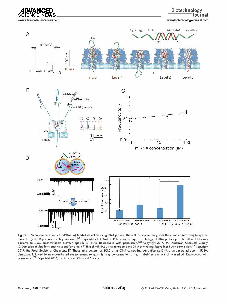
Figure 3. Nanopore detection of miRNAs. A) MiRNA detection using DNA probes. The αHL nanopore recognizes the complex according to specificcurrent signals. Reproduced with permission.[64] Copyright 2011, Nature Publishing Group. B) PEG-tagged DNA probes provide different blockingcurrents to allow discrimination between specific miRNAs. Reproduced with permission.[28] Copyright 2014, the American Chemical Society.C) Detection of ultra-low concentrations (on order of 1 fM) ofmiRNAs using nanopores andDNA computing. Reproduced with permission.[69] Copyright2017, the Royal Society of Chemistry. D) Theranostic system for SCLC using DNA computing. An antisense DNA drug generated upon miR-20adetection; followed by nanopore-based measurement to quantify drug concentration using a label-free and real time method. Reproduced withpermission.[70] Copyright 2017, the American Chemical Society.
www.advancedsciencenews.com www.biotechnology-journal.com
Biotechnol. J. 2018, 1800091 © 2018 WILEY-VCH Verlag GmbH & Co. KGaA, Weinheim1800091 (6 of 8)
www.advancedsciencenews.com www.biotechnology-journal.com
amplified by >20-fold from miR-20a, thereby meeting thedosage requirement for SCLC therapy and suggesting thisautonomous amplification strategy as a potential candidate forbroad-range theranostics using antisense oligonucleotides .
Nanopore decoding might also contribute to molecularrobotics.[71,72] Molecular robots represent next-generationbiochemical machines comprised of biomaterials, such asDNA, proteins, and lipids, with the prerequisites of sensors,intelligence, and actuators proposed as requirements for theconstruction of such robots. To develop sensors necessary toapply a level of “intelligence” to these robots, output decoding,using nanopores will be a valuable tool used to constructing thenecessary parts.
4. Conclusions
In this review, we described recent developments in nanoporedecodingmethods for DNAcomputation and their applications inclinicalfields.Nanopore technology does not require labeling, andthe decoding time is relatively rapid compared with conventionalfluorescence methods. However, for laboratory scale measure-ments, reconstitution of biological nanopores in lipid bilayersrequires training that might be time consuming. Although thedroplet-contact method enables rapid, reproducible, and stablenanopore measurements, it requires experience and training.Powerful strategies based onmicro-fabrication have been recentlyintroduced allowing preparation ofmassive numbers of nanoporechambers in a small device in order to acquire the required dataexclusively from the appropriate chambers. This strategyaddresses current nanopore-specific issues and can be potentiallyapplied to other nanopore technologies, including nanoporedecoding of DNA computation on an industrialized scale.Nanopore technology represents a valuable methodology forenhancing the decoding of DNA computations.
AbbreviationsαHL, α-hemolysin; DNA; microRNA, miRNA; PCR, polymerase chainreaction; PEG, polyethylene glycol; PNA, peptide nucleic acid; SCLC,small-cell lung cancer; SNR, signal-to-noise ratio; ssDNA, single-strandedDNA.
AcknowledgmentsThis research was supported in part by KAKENHI, “Molecular Robotics”(Grant No. 15H00803) and Grant No. 16H06043 from Ministry ofEducation, Culture, Sports, Science and Technology (MEXT), Japan.
Conflict of InterestThe authors declare no commercial or financial conflict of interest.
KeywordsDNA logic gates, DNA sequencing, lipid bilayers, nanopores, singlemolecule detection
Biotechnol. J. 2018, 1800091 1800091 (
Received: April 25, 2018Revised: July 24, 2018
Published online:
[1] L. M. Adleman, Science 1994, 266, 1021.[2] Z. Ezziane, Nanotechnology 2006, 17, R27.[3] R. S. Braich, N. Chelyapov, C. Johnson, P. W. K. Rothemund,
L. Adleman, Science 2002, 296, 499.[4] Q. Ouyang, P. D. Kaplan, S. M. Liu, A. Libchaber, Science 1997, 278,
446.[5] T. Head, G. Rozenberg, R. S. Bladergroen, C. K. Breek,
PH Lommerse, HP Spaink, Biosystems 2000, 57, 87.[6] D. Faulhammer, A. R. Cukras, R. J. Lipton, L. F. Landweber, Proc.
Natl. Acad. Sci. U. S. A. 2000, 97, 1385.[7] A. R. Cukras, D. Faulhammer, R. J. Lipton, L. F. Landweber,
Biosystems 1999, 52, 35.[8] M. Arita, S. Kobayashi, New Generation Computing 2002, 20, 263.[9] D. Y. Zhang, G. Seelig, Nat. Chem. 2011, 3, 103.
[10] G. Seelig, D. Soloveichik, D. Y. Zhang, E. Winfree, Science 2006, 314,1585.
[11] Y. Benenson, R. Adar, T. Paz-Elizur, Z. Livneh, E. Shapiro, Proc. Natl.Acad. Sci. U. S. A. 2003, 100, 2191.
[12] Y. Benenson, T. Paz-Elizur, R. Adar, E. Keinan, Z Livneh, E. Shapiro,Nature 2001, 414, 430.
[13] V. Balzani, A. Credi, M. Venturi, ChemPhysChem 2003, 4, 49.[14] A. Okamoto, K. Tanaka, I. Saito, J. Am. Chem. Soc. 2004, 126, 9458.[15] G. Chatterjee, N. Dalchau, R. A. Muscat, A. Phillips, G. Seelig, Nat.
Nanotechnol. 2017, 12, 920.[16] L. Qian, E. Winfree, J. Bruck, Nature 2011, 475, 368.[17] L. Qian, E. Winfree, Science 2011, 332, 1196.[18] D. Y. Zhang, E. Winfree, J. Am. Chem. Soc. 2009, 131, 17303.[19] M. N. Stojanovic, S. Semova, D. Kolpashchikov, J. Macdonald,
C Morgan, D. Stefanovic, J. Am. Chem. Soc. 2005, 127, 6914.[20] M. N. Stojanovic, D. Stefanovic, Nat. Biotechnol. 2003, 21, 1069.[21] C. T. Lo, D. J. Throckmorton, A. K. Singh, A. E. Herr, Lab Chip 2008, 8,
1273.[22] A. E. Herr, A. V. Hatch, D. J. Throckmorton, H.M. Tran, J. S. Brennan,
W. V. Giannobile, A. K. Singh, Proc. Natl. Acad. Sci. U. S. A. 2007, 104,5268.
[23] X. M. Li, T. R. Ding, L. Sun, C. M. Mao, Biosens. Bioelectron. 2011, 30,241.
[24] A. Saghatelian, N. H. Volcker, K. M. Guckian, V. S. Y. Lin,M. R. Ghadiri, J. Am. Chem. Soc. 2003, 125, 346.
[25] K. S. Park, M. W. Seo, C. Jung, J. Y. Lee, H. G. Park, Small 2012, 8,2203.
[26] H. Yasuga, R. Kawano, M. Takinoue, Y. Tsuji, T Osaki, K Kamiya,N Miki, S. Takeuchi, PLoS ONE 2016, 11, e0149667.
[27] M. Ohara, M. Takinoue, R. Kawano, ACS Synth. Biol. 2017, 6, 1427.[28] X. Y. Zhang, Y. Wang, B. L. Fricke, L. Q. Gu, ACS Nano 2014, 8, 3444.[29] L. Q. Gu, O. Braha, S. Conlan, S. Cheley, H. Bayley,Nature 1999, 398,
686.[30] W. Q. Shi, A. K. Friedman, L. A. Baker, Anal. Chem. 2017, 89, 157.[31] T. Osaki, S. Takeuchi, Anal. Chem. 2017, 89, 216.[32] D. H. Stoloff, M. Wanunu, Curr. Opin. Biotechnol. 2013, 24, 699.[33] J. E. Reiner, A. Balijepalli, J. W. Robertson, J. Campbell, J. Suehle,
J. J. Kasianowicz, Chem. Rev. 2012, 112, 6431.[34] Y. L. Ying, C. Cao, Y. T. Long, Analyst 2014, 139, 3826.[35] L. Z. Song, M. R. Hobaugh, C. Shustak, S. Cheley, H. Bayley,
JE. Gouaux, Science 1996, 274, 1859.[36] J. J. Kasianowicz, E. Brandin, D. Branton, D. W. Deamer, Proc. Natl.
Acad. Sci. U. S. A. 1996, 93, 13770.
© 2018 WILEY-VCH Verlag GmbH & Co. KGaA, Weinheim7 of 8)
www.advancedsciencenews.com www.biotechnology-journal.com
[37] M. Akeson, D. Branton, J. J. Kasianowicz, E. Brandin, D. W. Deamer,Biophys. J. 1999, 77, 3227.
[38] D. W. Deamer, D. Branton, Acc. Chem. Res. 2002, 35, 817.[39] D. Branton, D. W. Deamer, A. Marziali, H. Bayley, S. A. Benner,
T. Butler, M. Di Ventra, S. Garaj, A. Hibbs, X. Huang,S. B. Jovanovich, P. S. Krstic, S. Lindsay, X. S. Ling,C. H. Mastrangelo, A. Meller, J. S. Oliver, Y. V. Pershin,J. M. Ramsey, R. Riehn, G. V. Soni, V. Tabard-Cossa, M. Wanunu,M. Wiggin, J. A. Schloss, Nat. Biotechnol. 2008, 26, 1146.
[40] Y. Astier, O. Braha, H. Bayley, J. Am. Chem. Soc. 2006, 128, 1705.[41] A. Meller, L. Nivon, D. Branton, Phys. Rev. Lett. 2001, 86, 3435.[42] A. Meller, D. Branton, Electrophoresis 2002, 23, 2583.[43] Y. Astier, O. Braha, H. Bayley, J. Am. Chem. Soc. 2006, 128, 1705.[44] S. Benner, R. J. A. Chen, N. A. Wilson, R. Abu-Shumays, N. Hurt,
K. R. Lieberman, D. W. Deamer, W. B. Dunbar, M. Akeson, Nat.Nanotechnol. 2007, 2, 718.
[45] R. J. White, E. N. Ervin, T. Yang, X. Chen, S. Daniel, P. S. Cremer,H. S. White, J. Am. Chem. Soc. 2007, 129, 11766.
[46] T. Z. Butler, M. Pavlenok, I. M. Derrington, M. Niederweis,J. H. Gundlach, Proc. Natl. Acad. Sci. U. S. A. 2008, 105, 20647.
[47] J. Clarke, H. C. Wu, L. Jayasinghe, A. Patel, S. Reid, H. Bayley, Nat.Nanotechnol. 2009, 4, 265.
[48] I. M. Derrington, T. Z. Butler, M. D. Collins, E. Manrao, M. Pavlenok,M. Niederweis, J. H. Gundlach, Proc. Natl. Acad. Sci. U. S. A. 2010,107, 16060.
[49] A. R. Hall, A. Scott, D. Rotem, K. K. Mehta, H. Bayley, C. Dekker,Nat.Nanotechnol. 2010, 5, 874.
[50] F. Olasagasti, K. R. Lieberman, S. Benner, G. M. Cherf, J. M. Dahl,D. W. Deamer, M. Akeson, Nat. Nanotechnol. 2010, 5, 798.
[51] G. M. Cherf, K. R. Lieberman, H. Rashid, C. E. Lam, K. Karplus,M. Akeson, Nat. Biotechnol. 2012, 30, 344.
[52] J. Schreiber, Z. L. Wescoe, R. Abu-Shumays, J. T. Vivian, B. Baatar,K. Karplus, M. Akeson, Proc. Natl. Acad. Sci. U. S. A. 2013, 110,18910.
Biotechnol. J. 2018, 1800091 1800091 (
[53] M. Jain, I. T. Fiddes, K. H. Miga, H. E. Olsen, B. Paten, M. Akeson,Nat. Methods 2015, 12, 351.
[54] R. Kawano, A. Schibel, C. Cauley, H. White, Langmuir 2009, 25,1233.
[55] H. Y. Lu, F. Giordano, Z. M. Ning, Genomics ProteomicsBioinformatics 2016, 14, 265.
[56] K. Funakoshi, H. Suzuki, S. Takeuchi, Anal. Chem. 2006, 78, 8169.[57] A. J. Heron, J. R. Thompson, A. E. Mason, M. I. Wallace, J. Am. Chem.
Soc. 2007, 129, 16042.[58] H. Bayley, B. Cronin, A. Heron, M. A. Holden, W. Hwang, R. Syeda,
J. Thompson, M. Wallace, Mol. BioSyst. 2008, 4, 1191.[59] R. Kawano, Y. Tsuji, K. Sato, T. Osaki, K. Kamiya, M. Hirano, T. Ide,
N. Miki, S. Takeuchi, Sci. Rep. 2013, 3. https://doi.org/10.1038/srep01995
[60] M. Ohara, Y. Sekiya, R. Kawano, Electrochemistry 2016, 84, 338.[61] Y. Benenson, B. Gil, U. Ben-Dor, R. Adar, E. Shapiro, Nature 2004,
429, 423.[62] Y. Benenson, Nat. Rev. Genet. 2012, 13, 455.[63] C. Jung, A. D. Ellington, Acc. Chem. Res. 2014, 47, 1825.[64] Y. Wang, D. L. Zheng, Q. L. Tan, M. X. Wang, L. Q. Gu, Nat.
Nanotechnol. 2011, 6, 668.[65] G. Di Leva, M. Garofalo, C. M. Croce, Annu. Rev. Pathol. Mech. Dis.
2014, 9, 287.[66] K. Tian, Z. J. He, Y. Wang, S. J. Chen, L. Q. Gu, ACS Nano 2013, 7,
3962.[67] M. Hiratani, R. Kawano, Anal. Chem. 2018, 90, 8531.[68] M. Wanunu, W. Morrison, Y. Rabin, A. Y. Grosberg, A. Meller, Nat.
Nanotechnol. 2010, 5, 160.[69] H. L. Zhang, M. Hiratani, K. Nagaoka, R. Kawano,Nanoscale 2017, 9,
16124.[70] M. Hiratani, M. Ohara, R. Kawano, Anal. Chem. 2017, 89, 2312.[71] M. Hagiya, A. Konagaya, S. Kobayashi, H. Saito, S. Murata, Acc.
Chem. Res. 2014, 47, 1681.[72] R. Kawano, ChemPhysChem 2018, 19, 359.
© 2018 WILEY-VCH Verlag GmbH & Co. KGaA, Weinheim8 of 8)