management of supraventricular arrhythmias - the …epsegypt.com/upload/21032013/an approach...
TRANSCRIPT
Management of
Supraventricular
Arrhythmias
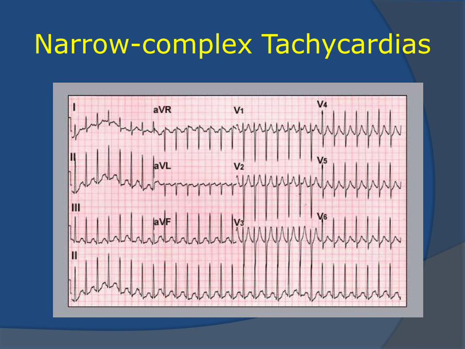
Narrow-complex Tachycardias
Narrow-complex Tachycardias
Rate > 100 beats per minute
QRS duration < 120 msec
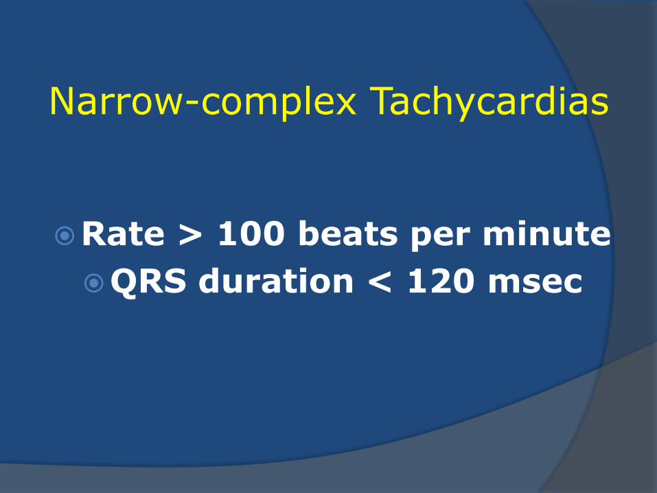
Narrow-complex Tachycardias
Originate in the atria (or adjoining veins)
Depend on the
AV junction
or
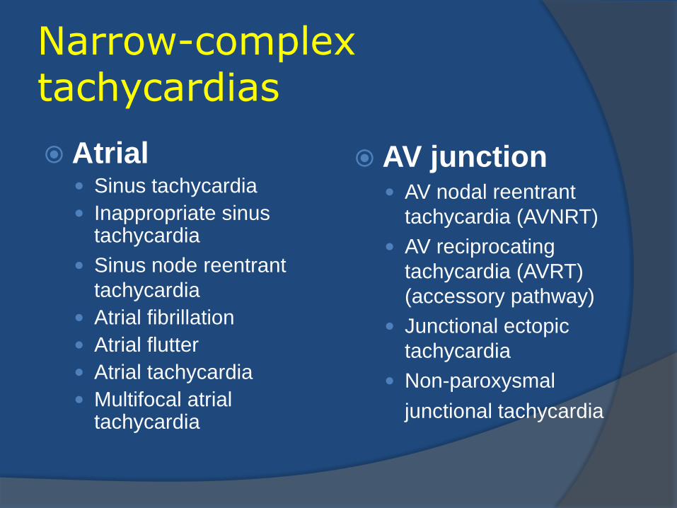
Narrow-complex tachycardias
Atrial Sinus tachycardia
Inappropriate sinus tachycardia
Sinus node reentrant
tachycardia
Atrial fibrillation
Atrial flutter
Atrial tachycardia
Multifocal atrial tachycardia
AV junction AV nodal reentrant
tachycardia (AVNRT)
AV reciprocating
tachycardia (AVRT)
(accessory pathway)
Junctional ectopic
tachycardia
Non-paroxysmal
junctional tachycardia
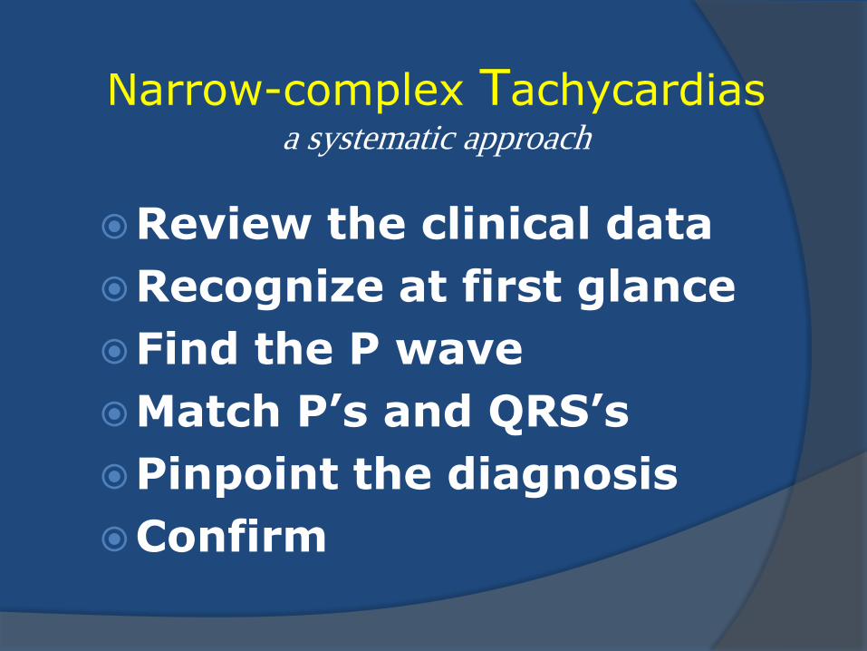
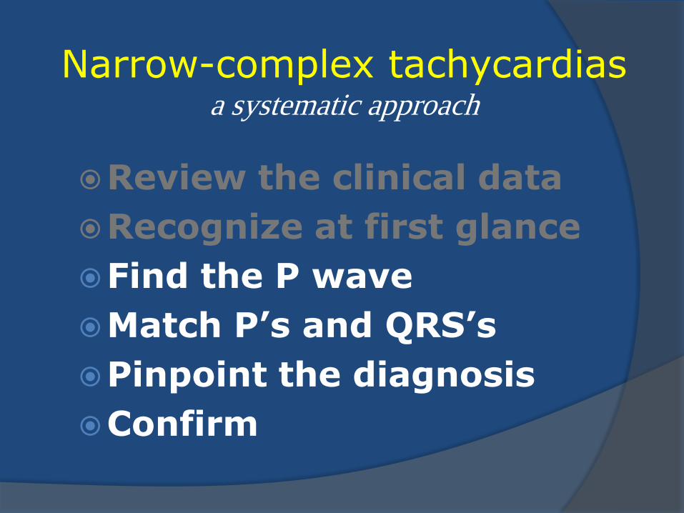
Narrow-complex Tachycardias a systematic approach
Review the clinical data
Recognize at first glance
Find the P wave
Match P’s and QRS’s
Pinpoint the diagnosis
Confirm
Narrow-complex Tachycardias recognize at first glance
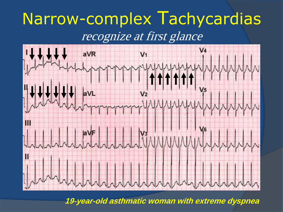
Narrow-complex Tachycardias recognize at first glance
19-year-old asthmatic woman with extreme dyspnea
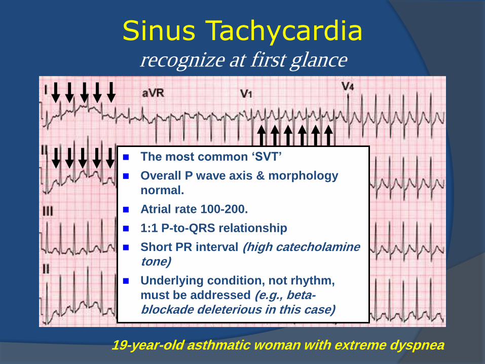
Sinus Tachycardia recognize at first glance
The most common ‘SVT’
Overall P wave axis & morphology
normal.
Atrial rate 100-200.
1:1 P-to-QRS relationship
Short PR interval (high catecholamine tone)
Underlying condition, not rhythm,
must be addressed (e.g., beta-blockade deleterious in this case)
19-year-old asthmatic woman with extreme dyspnea
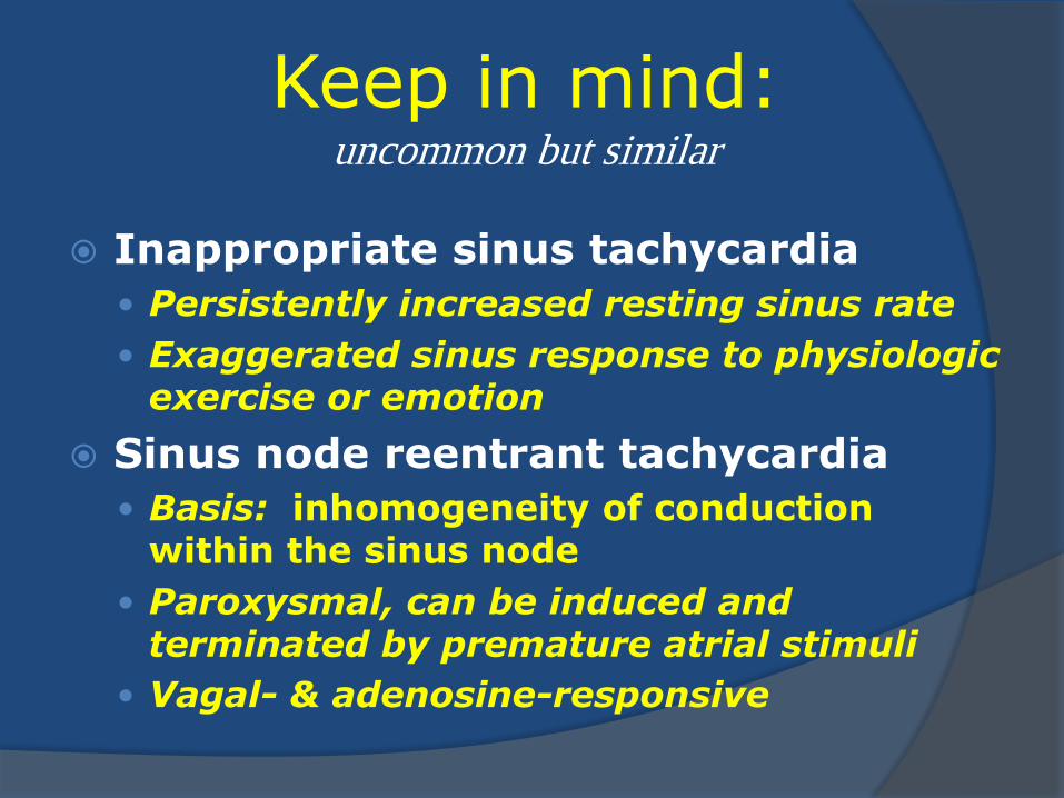
Keep in mind: uncommon but similar
Inappropriate sinus tachycardia
Persistently increased resting sinus rate
Exaggerated sinus response to physiologic exercise or emotion
Sinus node reentrant tachycardia
Basis: inhomogeneity of conduction within the sinus node
Paroxysmal, can be induced and terminated by premature atrial stimuli
Vagal- & adenosine-responsive
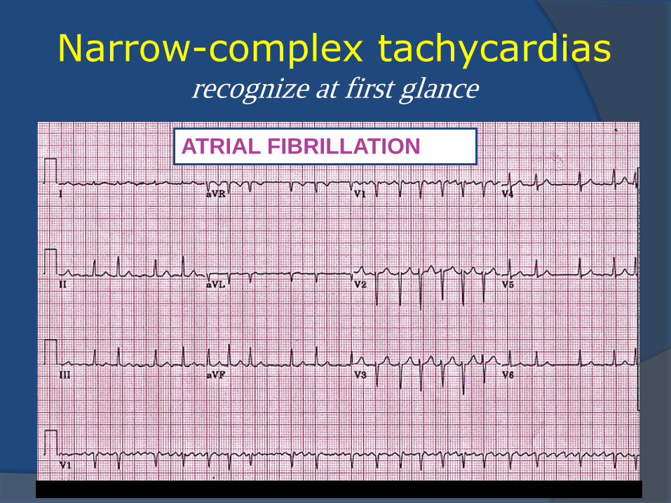
Narrow-complex tachycardias recognize at first glance
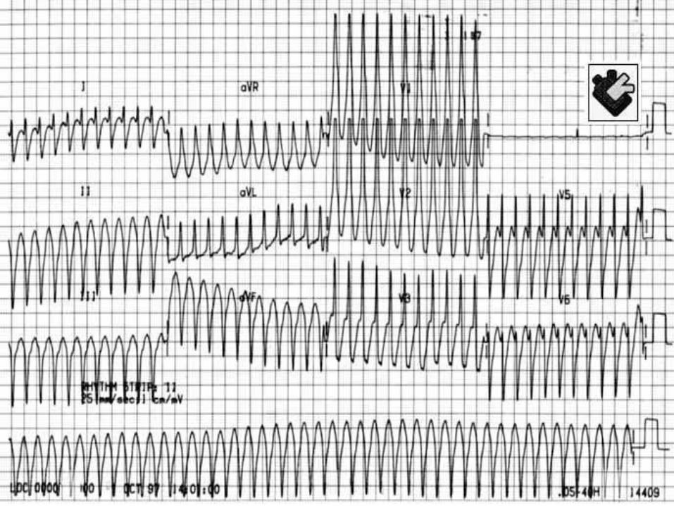
ATRIAL FIBRILLATION
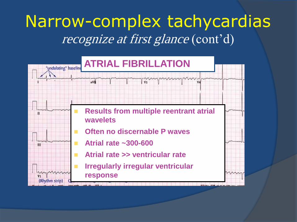
Narrow-complex tachycardias recognize at first glance (cont’d)
ATRIAL FIBRILLATION
Results from multiple reentrant atrial
wavelets
Often no discernable P waves
Atrial rate ~300-600
Atrial rate >> ventricular rate
Irregularly irregular ventricular
response
Narrow-complex tachycardias recognize at first glance (cont’d)
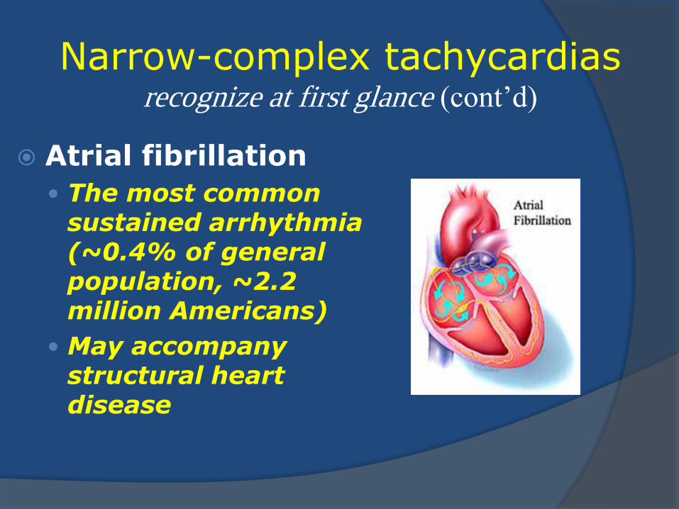
Atrial fibrillation
The most common sustained arrhythmia (~0.4% of general population, ~2.2 million Americans)
May accompany structural heart disease
Narrow-complex tachycardias recognize at first glance (cont’d)
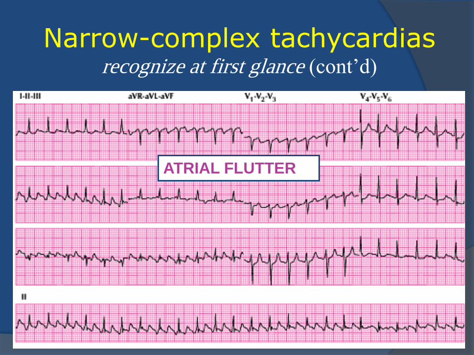
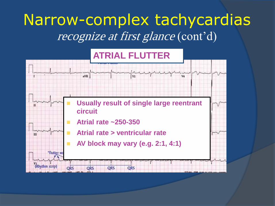
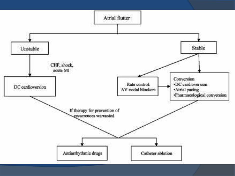
ATRIAL FLUTTER
Narrow-complex tachycardias recognize at first glance (cont’d)
ATRIAL FLUTTER
Usually result of single large reentrant
circuit
Atrial rate ~250-350
Atrial rate > ventricular rate
AV block may vary (e.g. 2:1, 4:1)
Narrow-complex tachycardias recognize at first glance (cont’d)
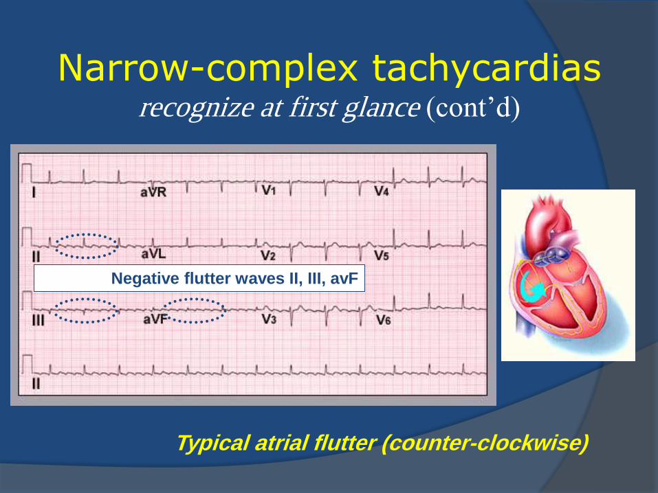
Typical atrial flutter (counter-clockwise)
Negative flutter waves II, III, avF
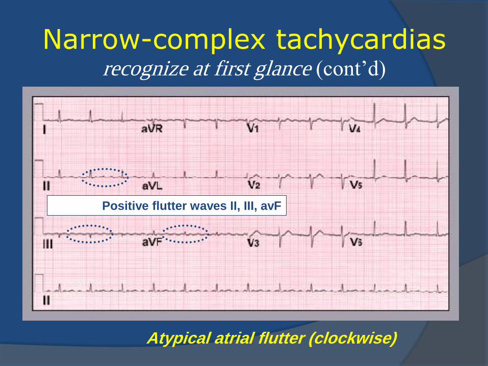
Narrow-complex tachycardias recognize at first glance (cont’d)
Atypical atrial flutter (clockwise)
Positive flutter waves II, III, avF
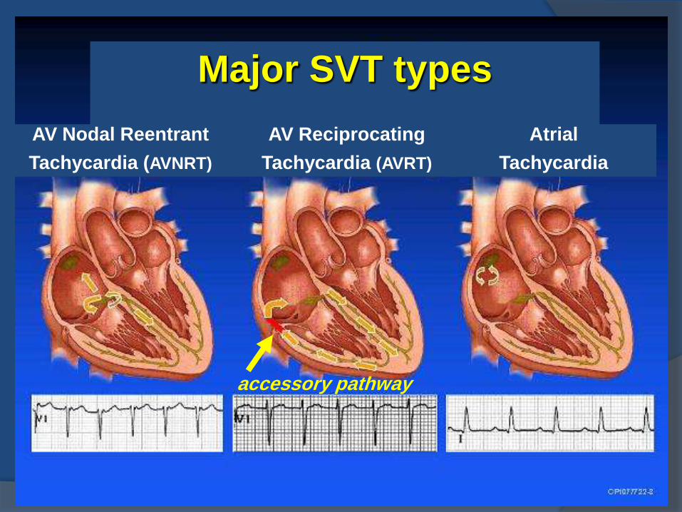
Major SVT types
AV Nodal Reentrant
Tachycardia (AVNRT)
AV Reciprocating
Tachycardia (AVRT)
Atrial
Tachycardia
accessory pathway
Narrow-complex tachycardias a systematic approach
Review the clinical data
Recognize at first glance
Find the P wave
Match P’s and QRS’s
Pinpoint the diagnosis
Confirm
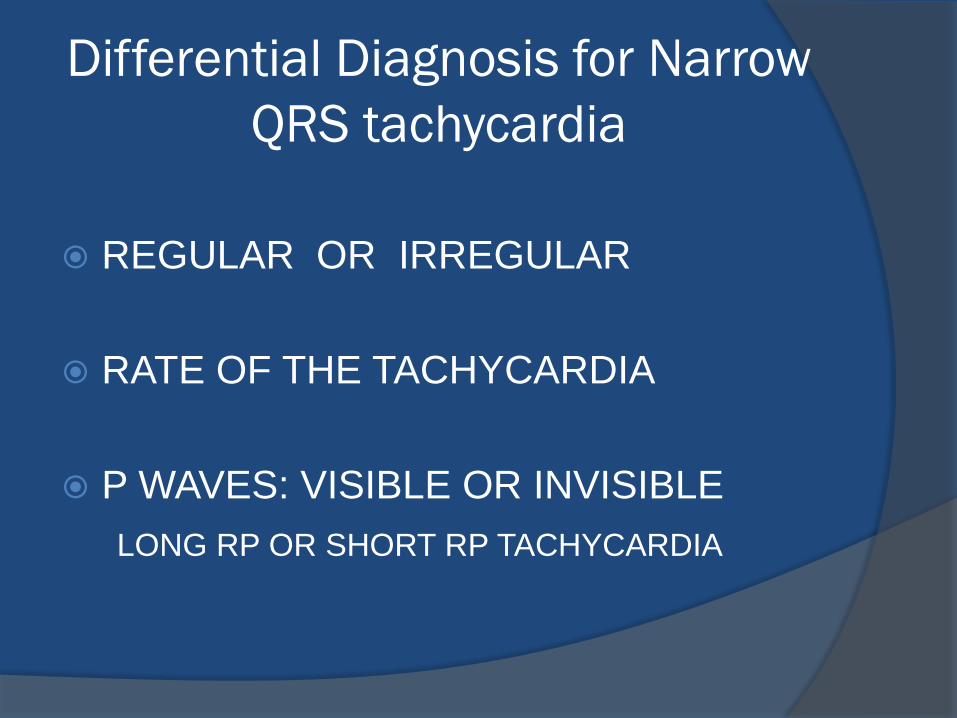
Differential Diagnosis for Narrow
QRS tachycardia
REGULAR OR IRREGULAR
RATE OF THE TACHYCARDIA
P WAVES: VISIBLE OR INVISIBLE
LONG RP OR SHORT RP TACHYCARDIA
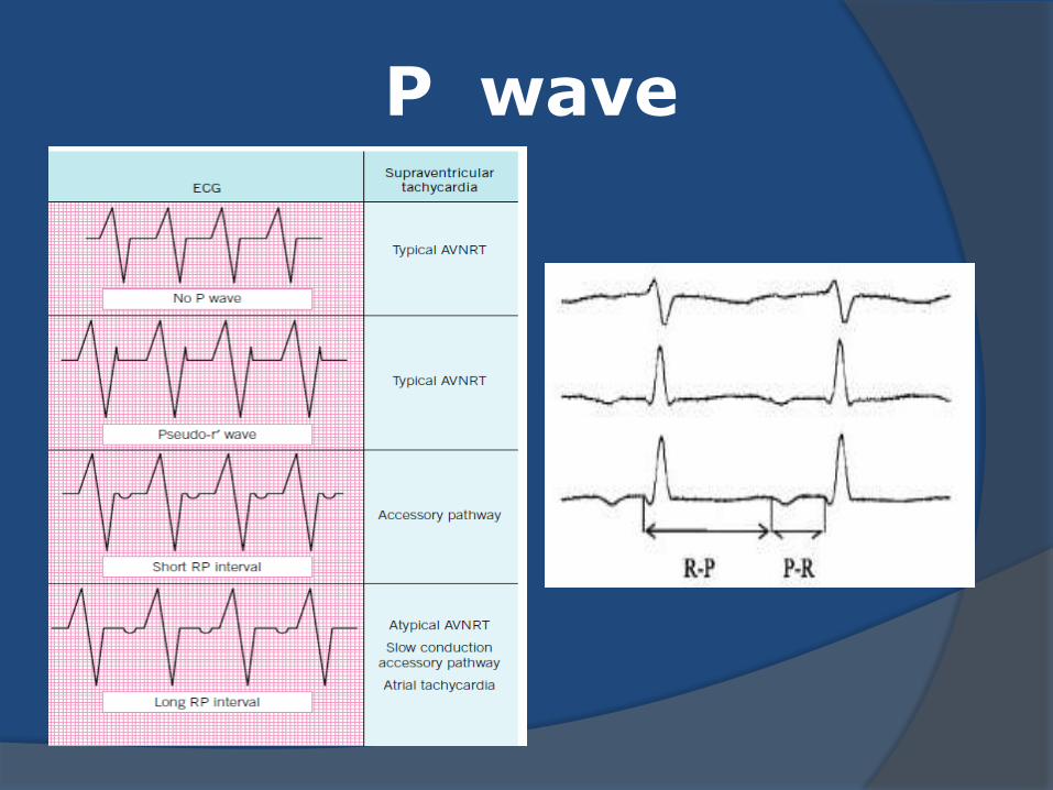
P wave
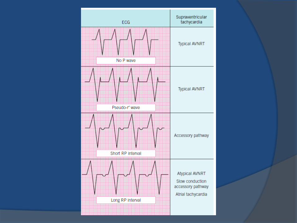
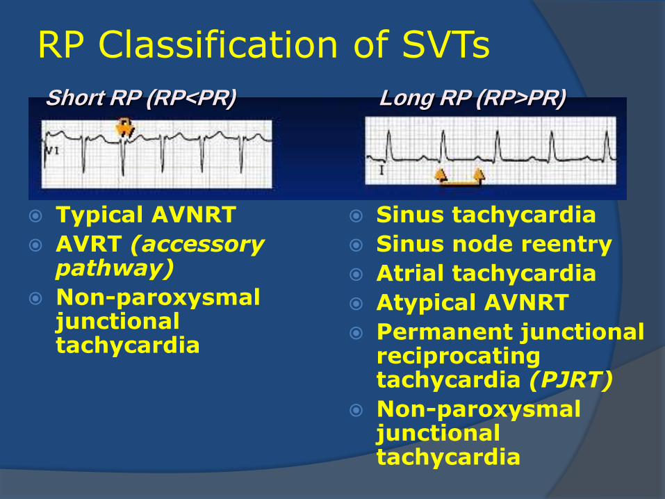
RP Classification of SVTs
Typical AVNRT
AVRT (accessory pathway)
Non-paroxysmal junctional tachycardia
Sinus tachycardia
Sinus node reentry
Atrial tachycardia
Atypical AVNRT
Permanent junctional reciprocating tachycardia (PJRT)
Non-paroxysmal junctional tachycardia
Short RP (RP<PR) Long RP (RP>PR)
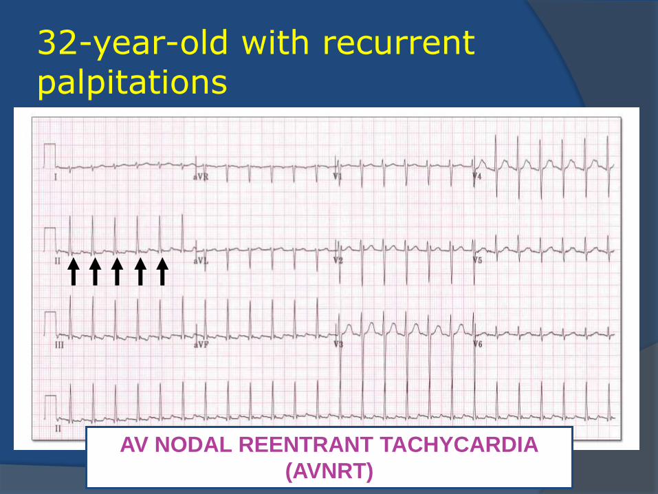
32-year-old with recurrent palpitations
AV NODAL REENTRANT TACHYCARDIA
(AVNRT)
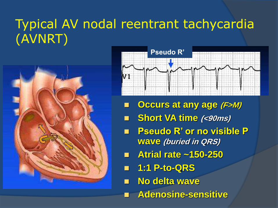
Typical AV nodal reentrant tachycardia (AVNRT)
Pseudo R’
Occurs at any age (F>M)
Short VA time (<90ms)
Pseudo R’ or no visible P wave (buried in QRS)
Atrial rate ~150-250
1:1 P-to-QRS
No delta wave
Adenosine-sensitive
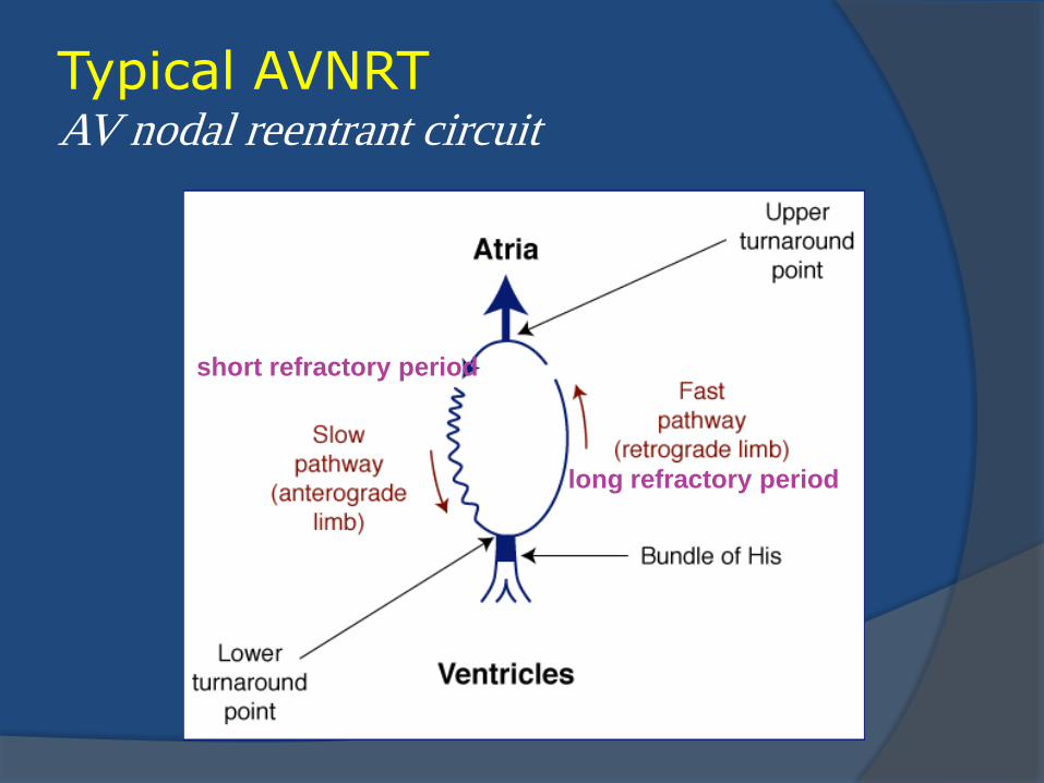
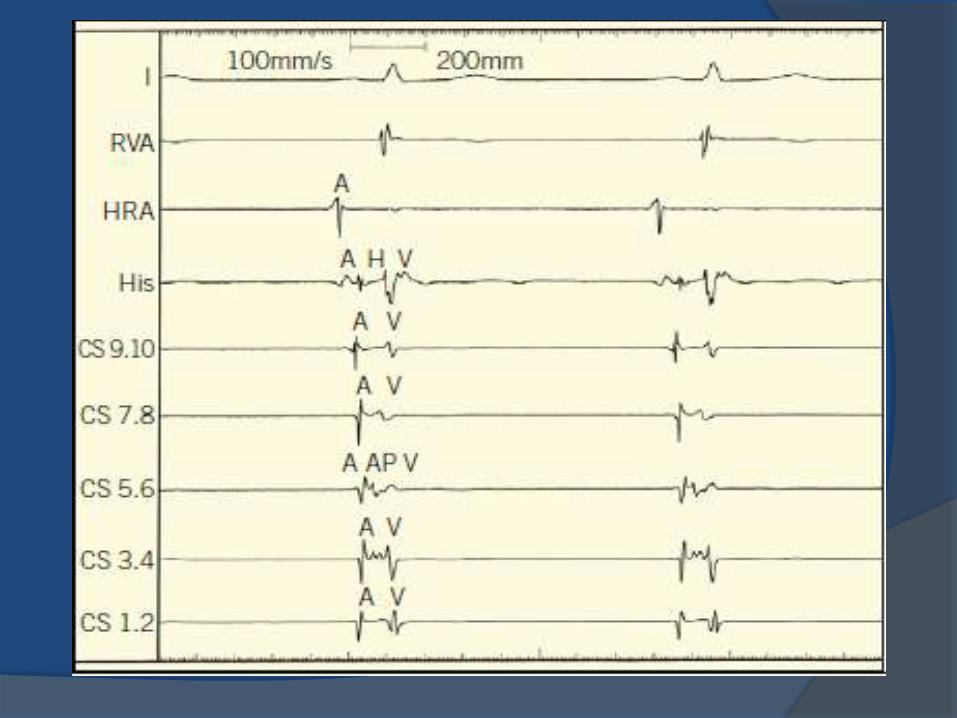
Typical AVNRT AV nodal reentrant circuit
short refractory period
long refractory period
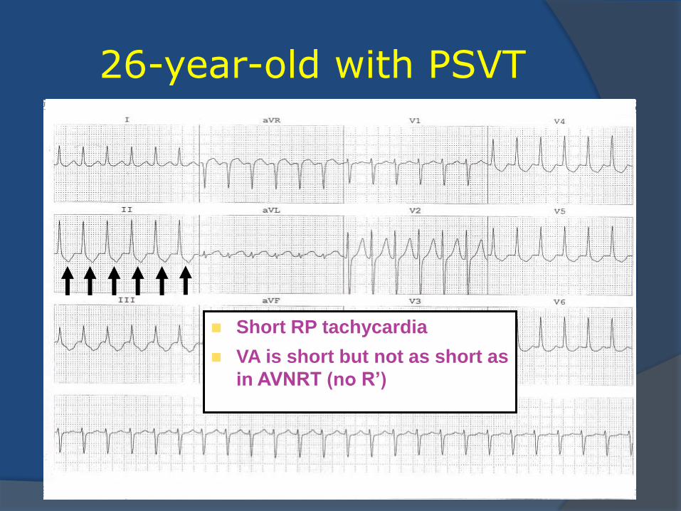
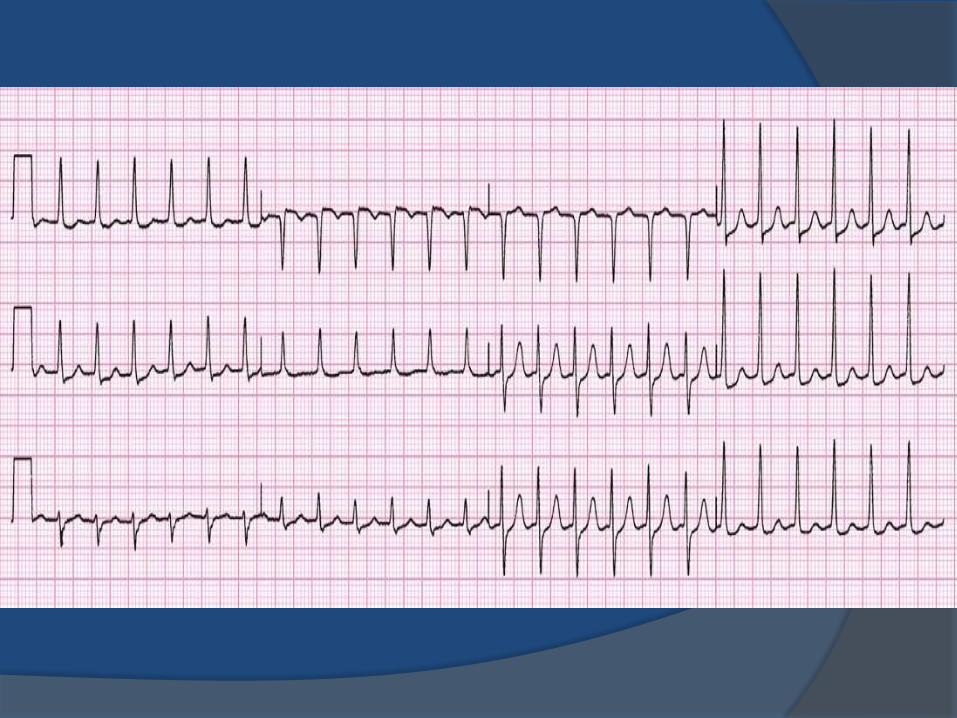
26-year-old with PSVT
Short RP tachycardia
VA is short but not as short as
in AVNRT (no R’)
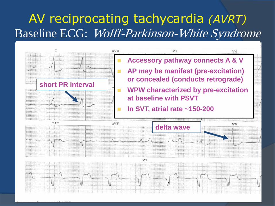
AV reciprocating tachycardia (AVRT)
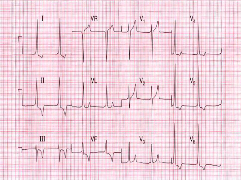
Baseline ECG: Wolff-Parkinson-White Syndrome
short PR interval
delta wave
Accessory pathway connects A & V
AP may be manifest (pre-excitation)
or concealed (conducts retrograde)
WPW characterized by pre-excitation
at baseline with PSVT
In SVT, atrial rate ~150-200
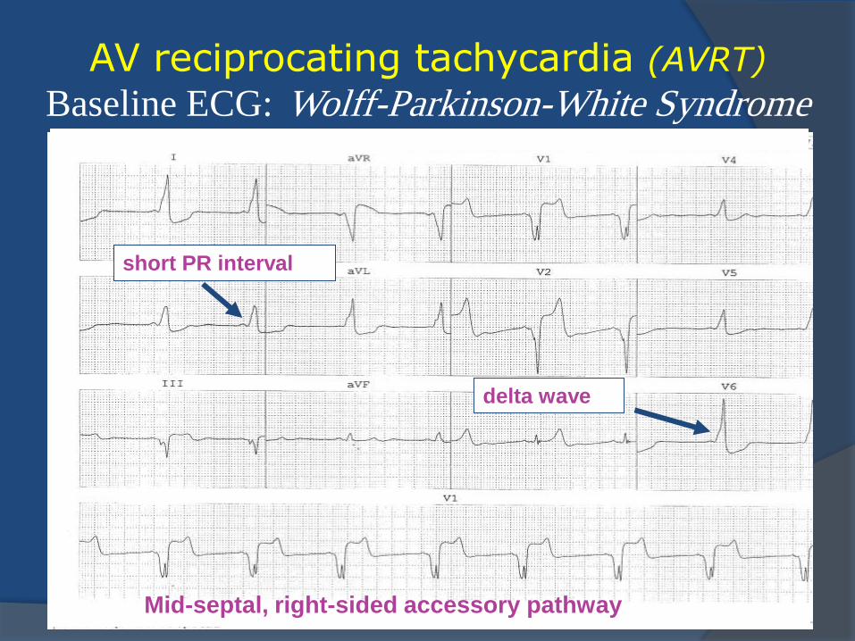
AV reciprocating tachycardia (AVRT)
Baseline ECG: Wolff-Parkinson-White Syndrome
short PR interval
delta wave
Mid-septal, right-sided accessory pathway
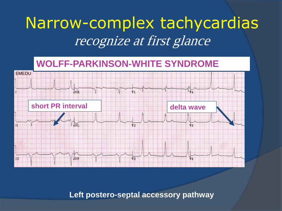
Narrow-complex tachycardias recognize at first glance
short PR interval delta wave
WOLFF-PARKINSON-WHITE SYNDROME
Left postero-septal accessory pathway
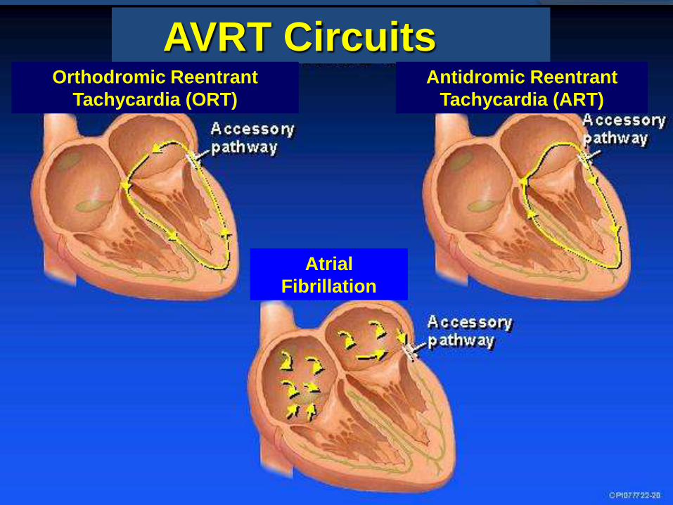
AVRT Circuits Orthodromic Reentrant
Tachycardia (ORT)
Antidromic Reentrant
Tachycardia (ART)
Atrial
Fibrillation
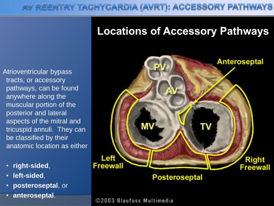
Atrioventricular bypass
tracts, or accessory
pathways, can be found
anywhere along the
muscular portion of the
posterior and lateral
aspects of the mitral and
tricuspid annuli. They can
be classified by their
anatomic location as either
• right-sided,
• left-sided,
• posteroseptal, or
• anteroseptal.
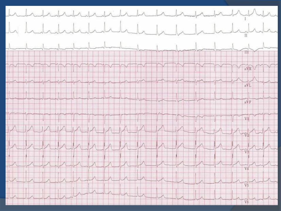
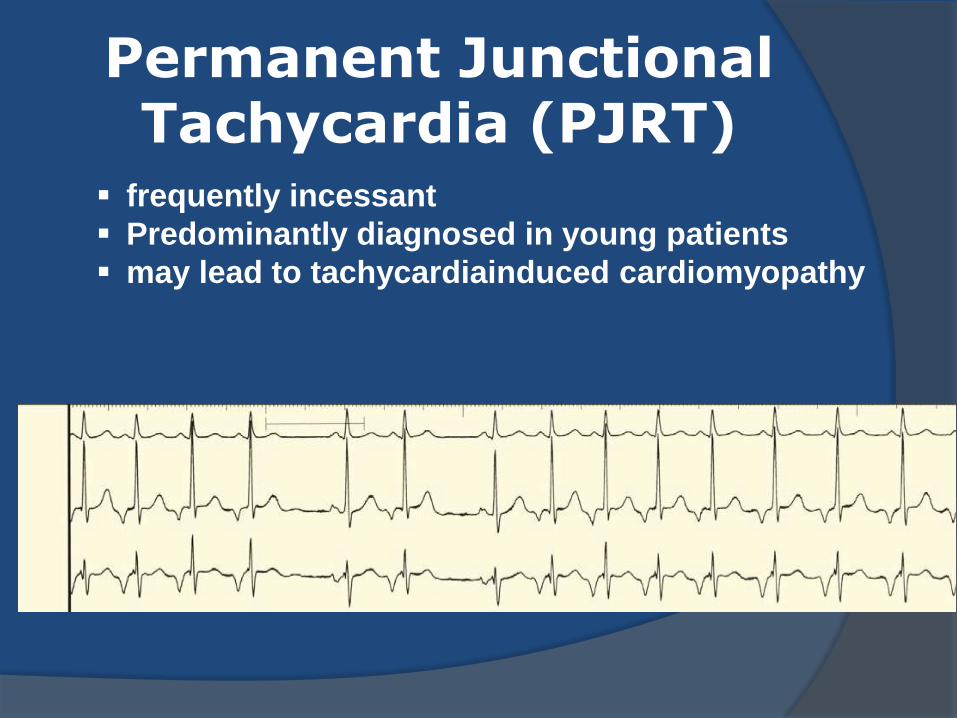
frequently incessant
Predominantly diagnosed in young patients
may lead to tachycardiainduced cardiomyopathy
Permanent Junctional Tachycardia (PJRT)
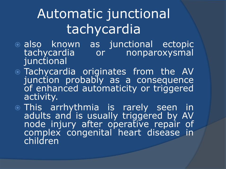
Automatic junctional tachycardia
also known as junctional ectopic tachycardia or nonparoxysmal junctional
Tachycardia originates from the AV junction probably as a consequence of enhanced automaticity or triggered activity.
This arrhythmia is rarely seen in adults and is usually triggered by AV node injury after operative repair of complex congenital heart disease in children
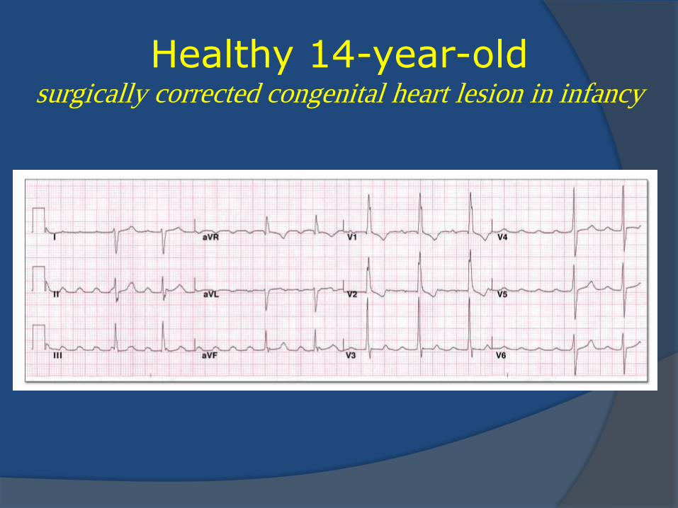
Healthy 14-year-old surgically corrected congenital heart lesion in infancy
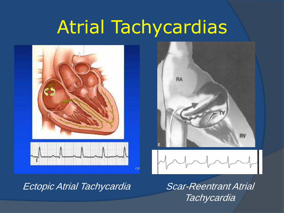
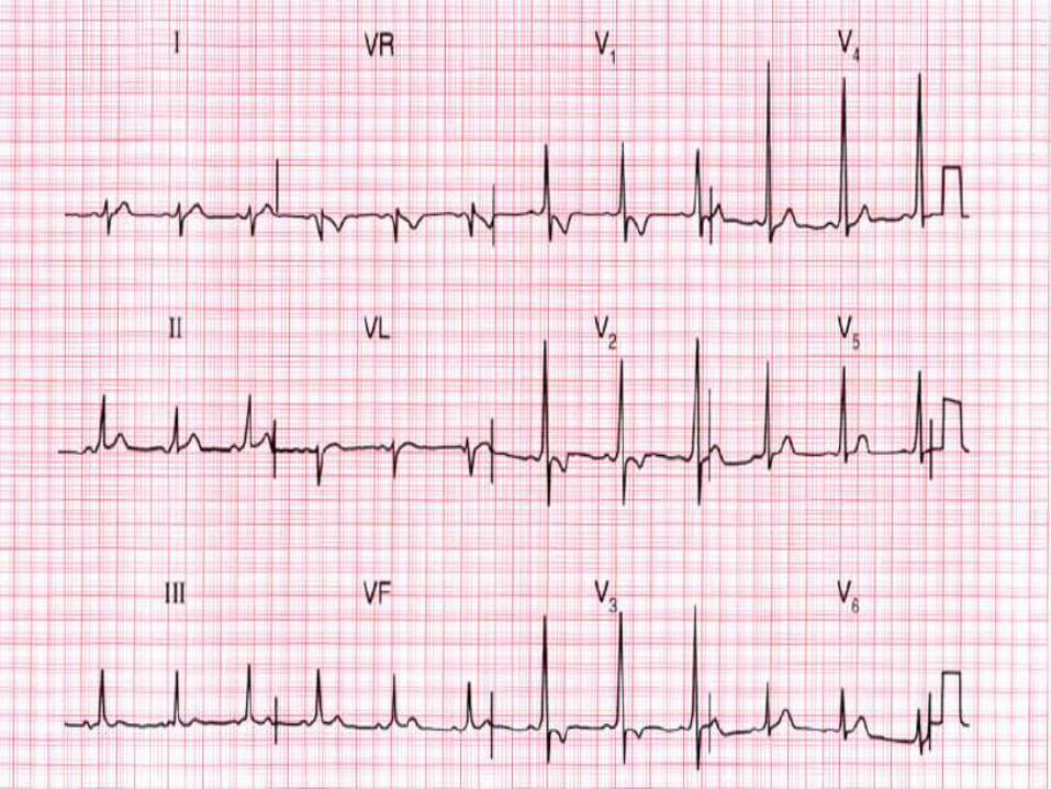
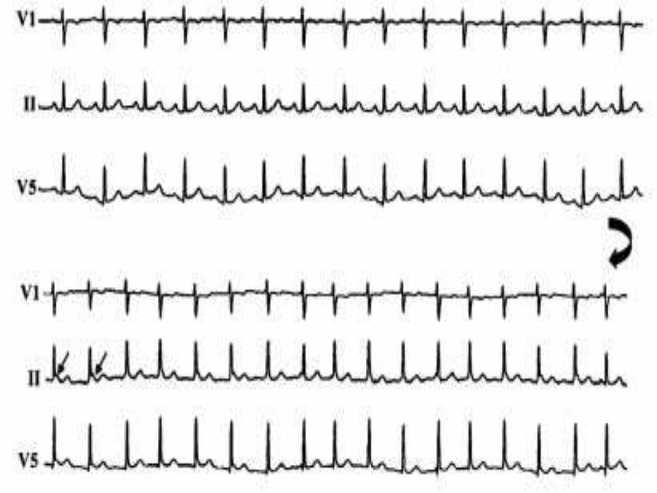
Atrial Tachycardias
Ectopic Atrial Tachycardia Scar-Reentrant Atrial Tachycardia
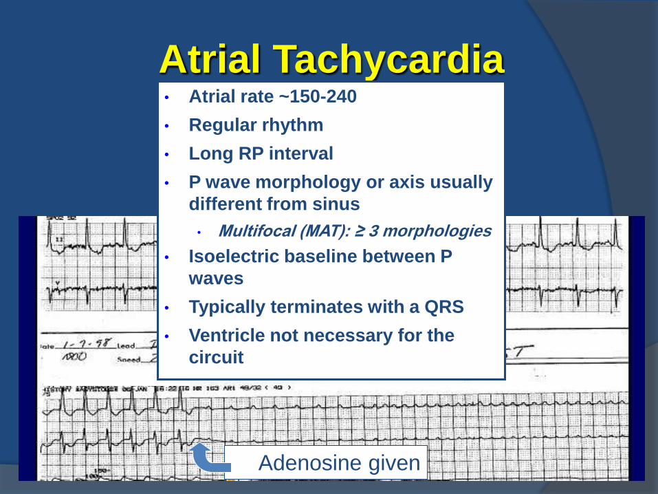
Atrial Tachycardia
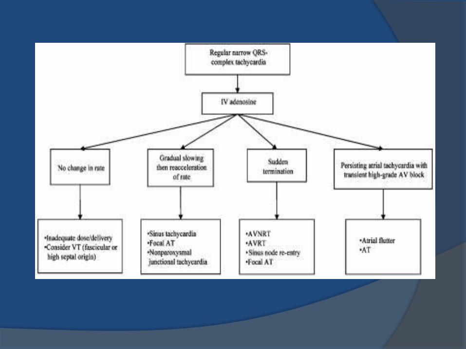
Adenosine given
• Atrial rate ~150-240
• Regular rhythm
• Long RP interval
• P wave morphology or axis usually
different from sinus
• Multifocal (MAT): ≥ 3 morphologies
• Isoelectric baseline between P
waves
• Typically terminates with a QRS
• Ventricle not necessary for the
circuit
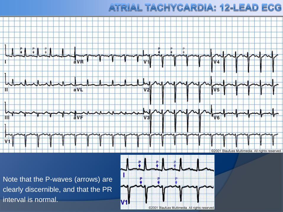
Note that the P-waves (arrows) are
clearly discernible, and that the PR
interval is normal.
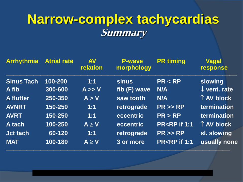
Arrhythmia Atrial rate AV P-wave PR timing Vagal relation morphology response
Sinus Tach 100-200 1:1 sinus PR < RP slowing
A fib 300-600 A >> V fib (F) wave N/A vent. rate
A flutter 250-350 A > V saw tooth N/A AV block
AVNRT 150-250 1:1 retrograde PR >> RP termination
AVRT 150-250 1:1 eccentric PR > RP termination
A tach 100-250 A V eccentric PR<RP if 1:1 AV block
Jct tach 60-120 1:1 retrograde PR >> RP sl. slowing
MAT 100-180 A V 3 or more PR<RP if 1:1 usually none
Narrow-complex tachycardias Summary
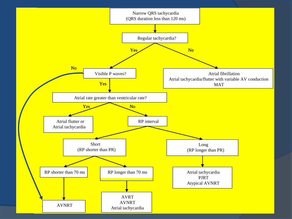
Narrow QRS tachycardia
(QRS duration less than 120 ms)
Visible P waves? Atrial fibrillation
Atrial tachycardia/flutter with variable AV conduction
MAT
Atrial flutter or
Atrial tachycardia
Long
(RP longer than PR)
RP shorter than 70 ms RP longer than 70 ms Atrial tachycardia
PJRT
Atypical AVNRT
AVNRT
AVRT
AVNRT
Atrial tachycardia
Atrial rate greater than ventricular rate?
RP interval
Short
(RP shorter than PR)
Yes
Regular tachycardia?
Yes
Yes
No
No
No

Management Strategies
Acute management
Long-term management
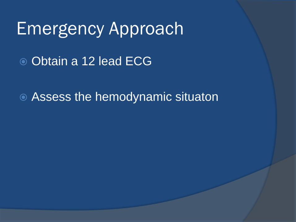
Emergency Approach
Obtain a 12 lead ECG
Assess the hemodynamic situaton
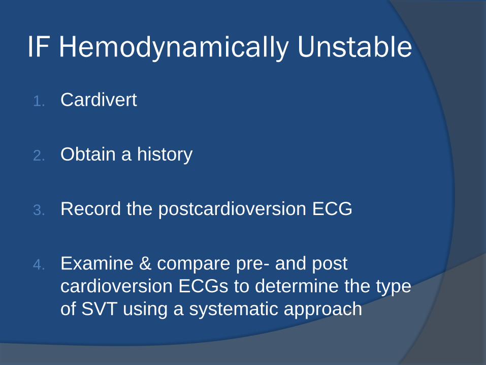
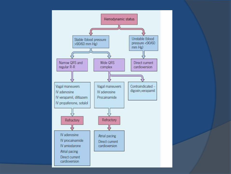
IF Hemodynamically Unstable
1. Cardivert
2. Obtain a history
3. Record the postcardioversion ECG
4. Examine & compare pre- and post
cardioversion ECGs to determine the type
of SVT using a systematic approach
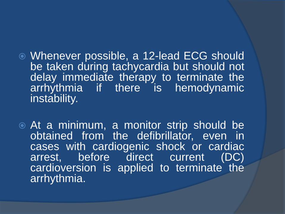
Whenever possible, a 12-lead ECG should be taken during tachycardia but should not delay immediate therapy to terminate the arrhythmia if there is hemodynamic instability.
At a minimum, a monitor strip should be obtained from the defibrillator, even in cases with cardiogenic shock or cardiac arrest, before direct current (DC) cardioversion is applied to terminate the arrhythmia.
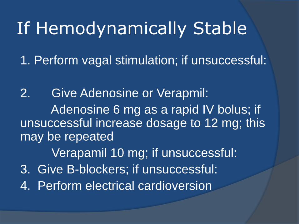
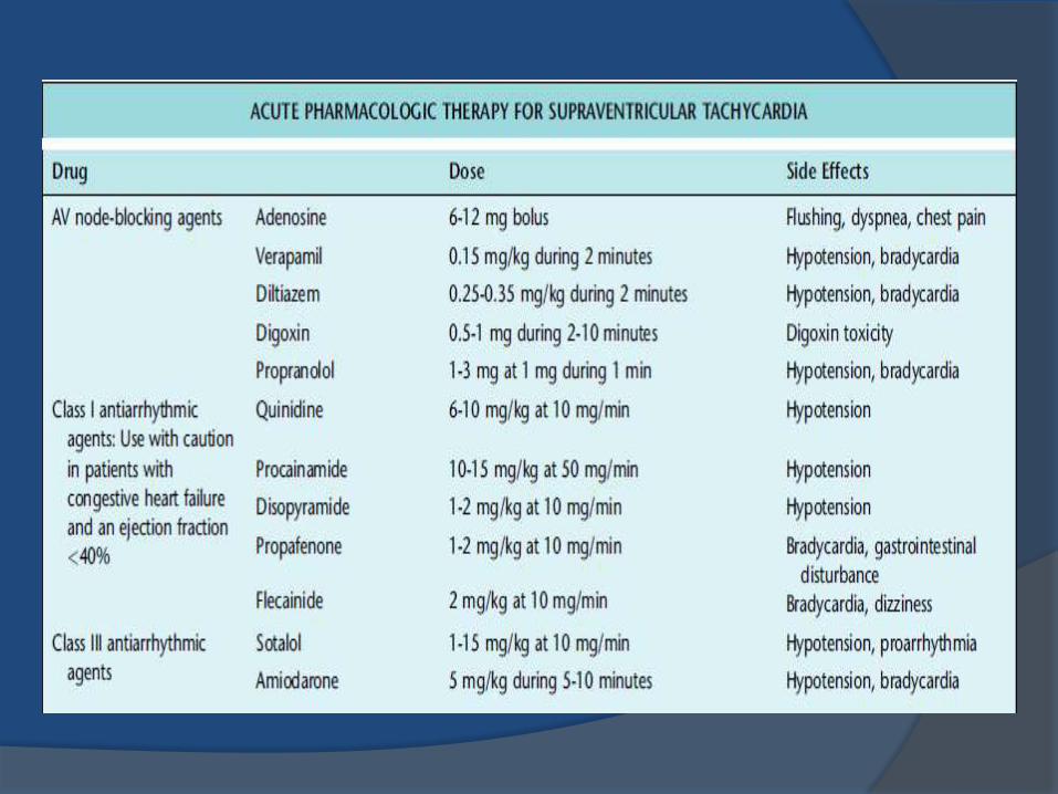
If Hemodynamically Stable
1. Perform vagal stimulation; if unsuccessful:
2. Give Adenosine or Verapmil:
Adenosine 6 mg as a rapid IV bolus; if unsuccessful increase dosage to 12 mg; this may be repeated
Verapamil 10 mg; if unsuccessful:
3. Give B-blockers; if unsuccessful:
4. Perform electrical cardioversion
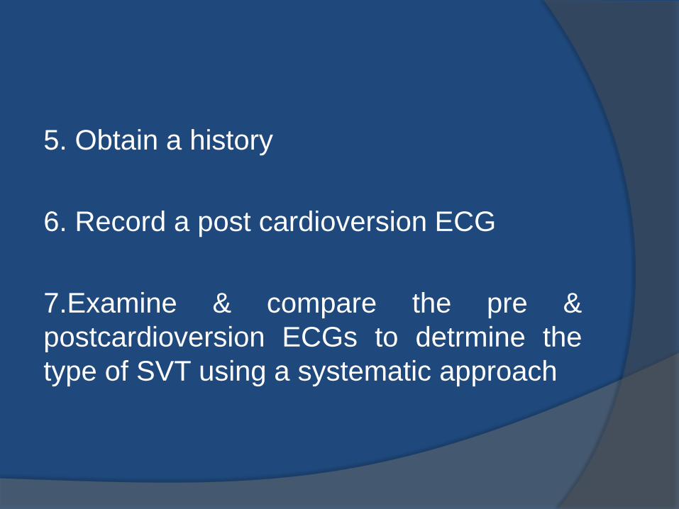
5. Obtain a history
6. Record a post cardioversion ECG
7.Examine & compare the pre &
postcardioversion ECGs to detrmine the
type of SVT using a systematic approach
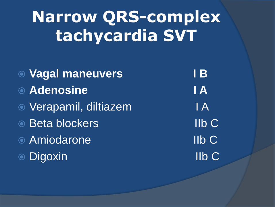
Narrow QRS-complex tachycardia SVT
Vagal maneuvers I B
Adenosine I A
Verapamil, diltiazem I A
Beta blockers IIb C
Amiodarone IIb C
Digoxin IIb C
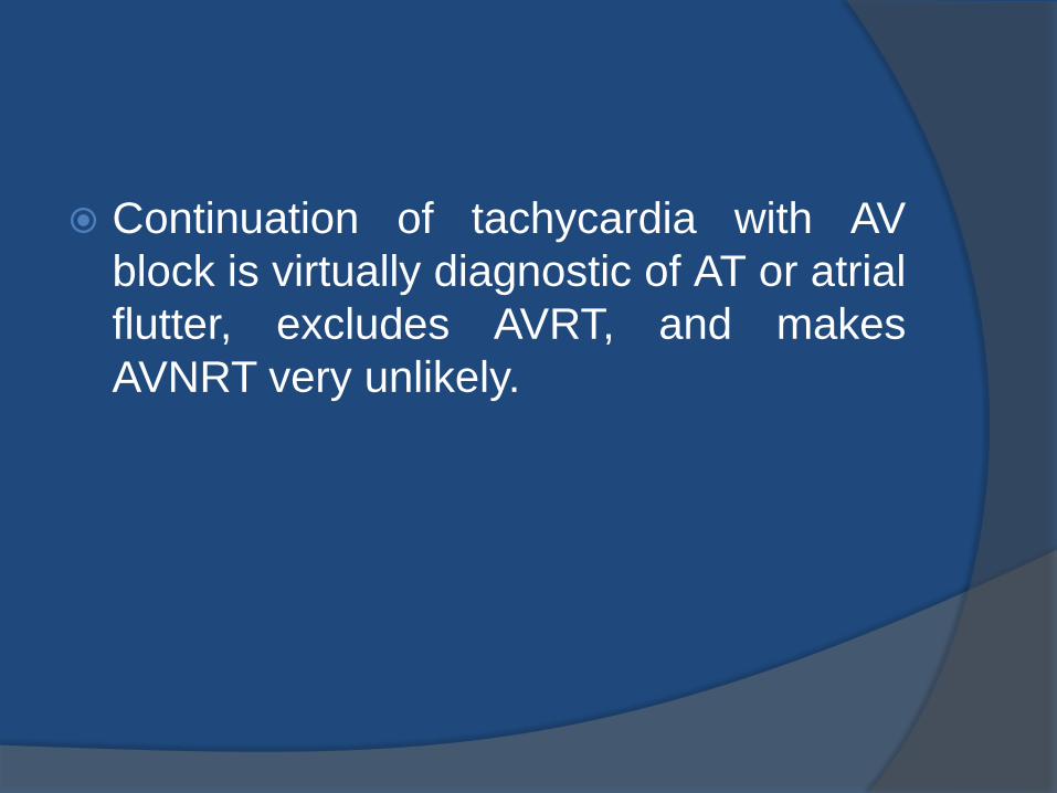
Continuation of tachycardia with AV
block is virtually diagnostic of AT or atrial
flutter, excludes AVRT, and makes
AVNRT very unlikely.
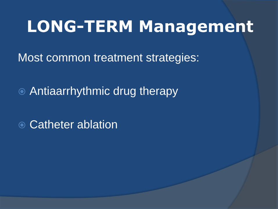
LONG-TERM Management
Most common treatment strategies:
Antiaarrhythmic drug therapy
Catheter ablation
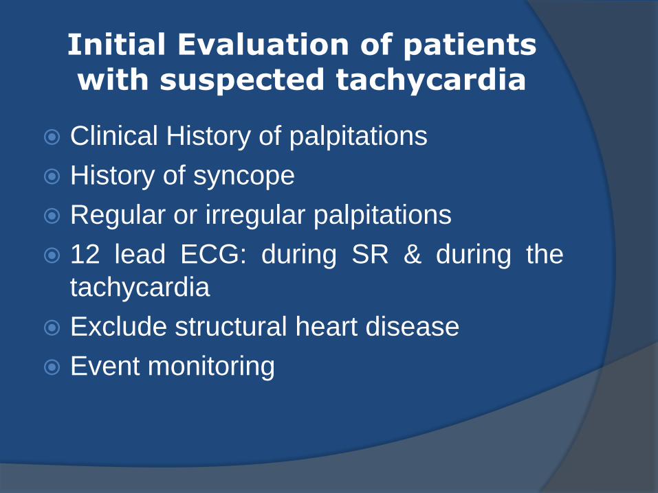
Initial Evaluation of patients with suspected tachycardia
Clinical History of palpitations
History of syncope
Regular or irregular palpitations
12 lead ECG: during SR & during the
tachycardia
Exclude structural heart disease
Event monitoring
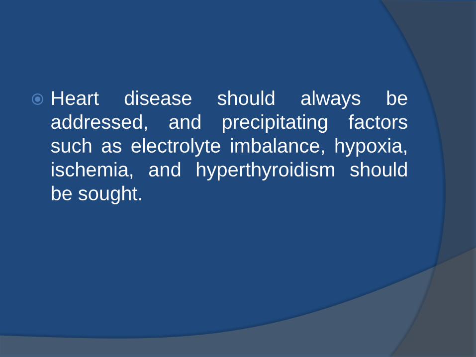
Heart disease should always be
addressed, and precipitating factors
such as electrolyte imbalance, hypoxia,
ischemia, and hyperthyroidism should
be sought.
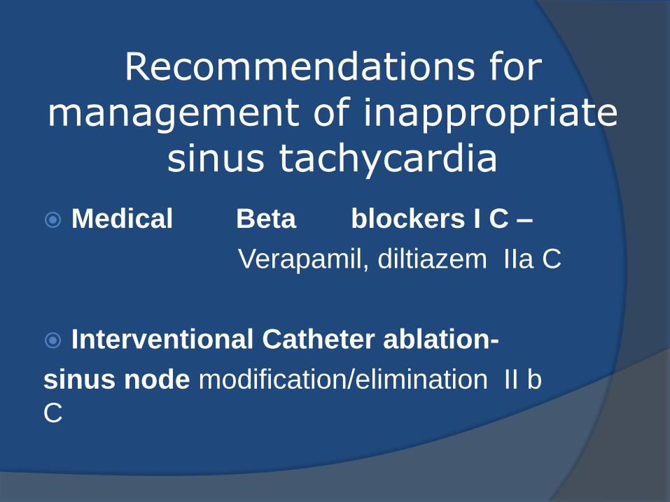
Recommendations for management of inappropriate
sinus tachycardia
Medical Beta blockers I C –
Verapamil, diltiazem IIa C
Interventional Catheter ablation-
sinus node modification/elimination II b
C
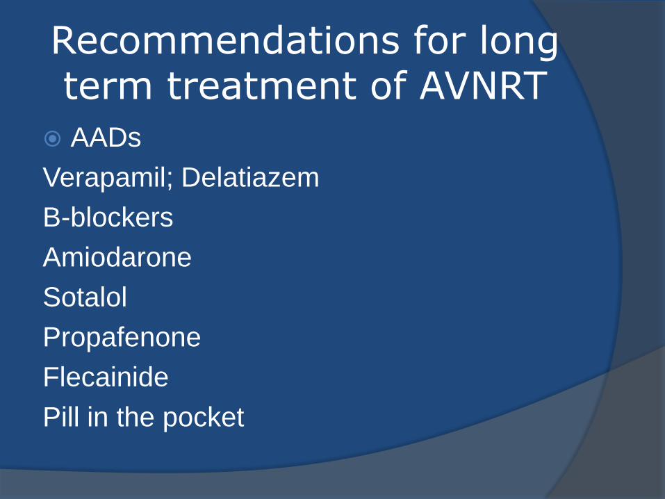
Recommendations for long term treatment of AVNRT
AADs
Verapamil; Delatiazem
B-blockers
Amiodarone
Sotalol
Propafenone
Flecainide
Pill in the pocket
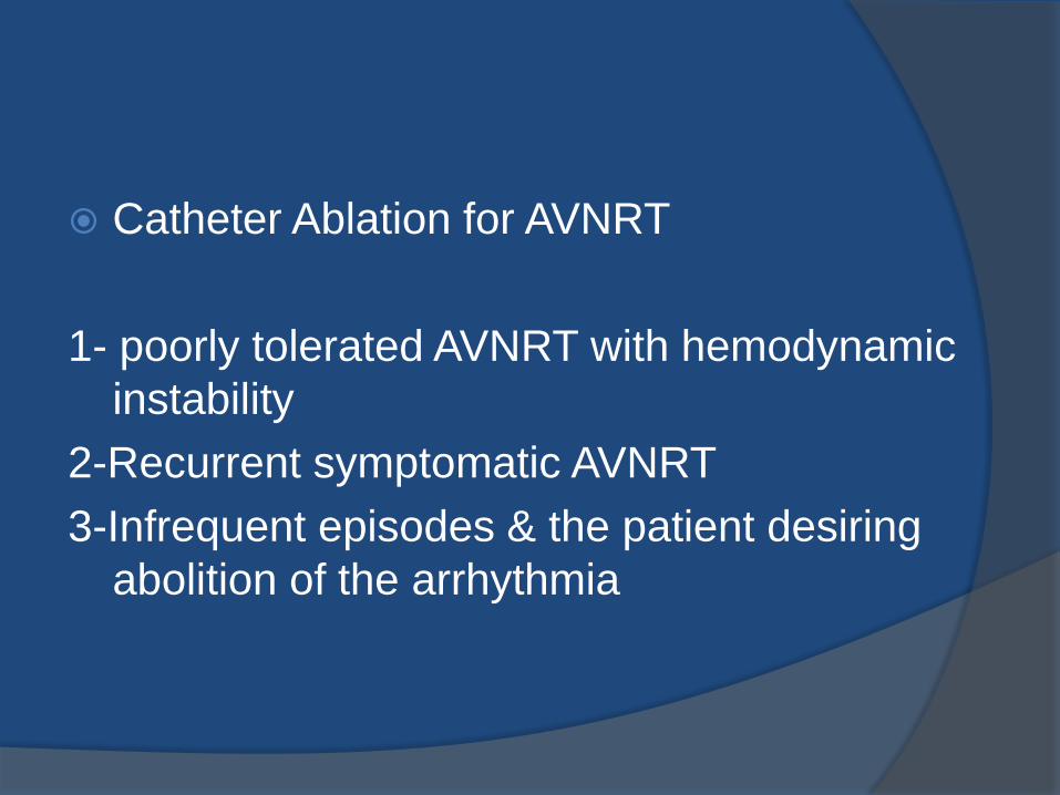
Catheter Ablation for AVNRT
1- poorly tolerated AVNRT with hemodynamic
instability
2-Recurrent symptomatic AVNRT
3-Infrequent episodes & the patient desiring
abolition of the arrhythmia
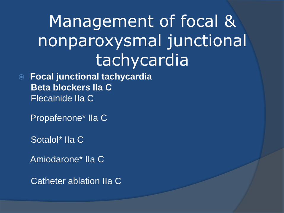
Management of focal & nonparoxysmal junctional
tachycardia
Focal junctional tachycardia
Beta blockers IIa C
Flecainide IIa C
Propafenone* IIa C
Sotalol* IIa C
Amiodarone* IIa C
Catheter ablation IIa C
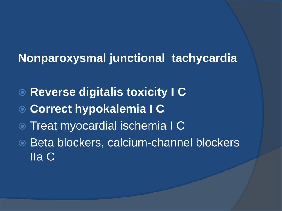
Nonparoxysmal junctional tachycardia
Reverse digitalis toxicity I C
Correct hypokalemia I C
Treat myocardial ischemia I C
Beta blockers, calcium-channel blockers
IIa C
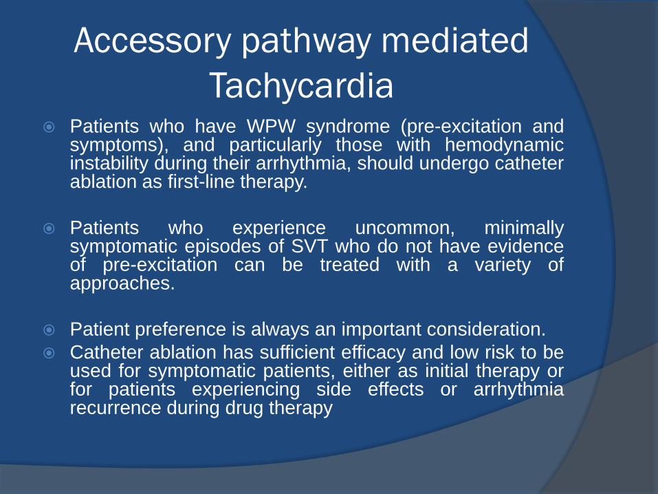
Accessory pathway mediated
Tachycardia Patients who have WPW syndrome (pre-excitation and
symptoms), and particularly those with hemodynamic instability during their arrhythmia, should undergo catheter ablation as first-line therapy.
Patients who experience uncommon, minimally symptomatic episodes of SVT who do not have evidence of pre-excitation can be treated with a variety of approaches.
Patient preference is always an important consideration.
Catheter ablation has sufficient efficacy and low risk to be used for symptomatic patients, either as initial therapy or for patients experiencing side effects or arrhythmia recurrence during drug therapy
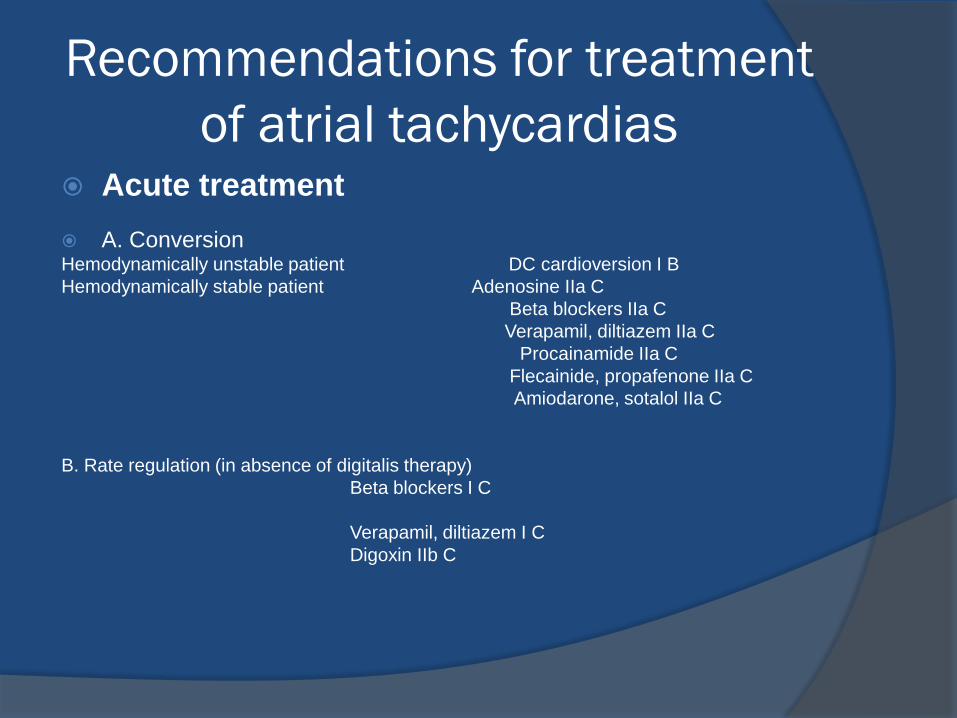
Recommendations for treatment
of atrial tachycardias Acute treatment
A. Conversion Hemodynamically unstable patient DC cardioversion I B
Hemodynamically stable patient Adenosine IIa C
Beta blockers IIa C
Verapamil, diltiazem IIa C
Procainamide IIa C
Flecainide, propafenone IIa C Amiodarone, sotalol IIa C
B. Rate regulation (in absence of digitalis therapy)
Beta blockers I C
Verapamil, diltiazem I C
Digoxin IIb C
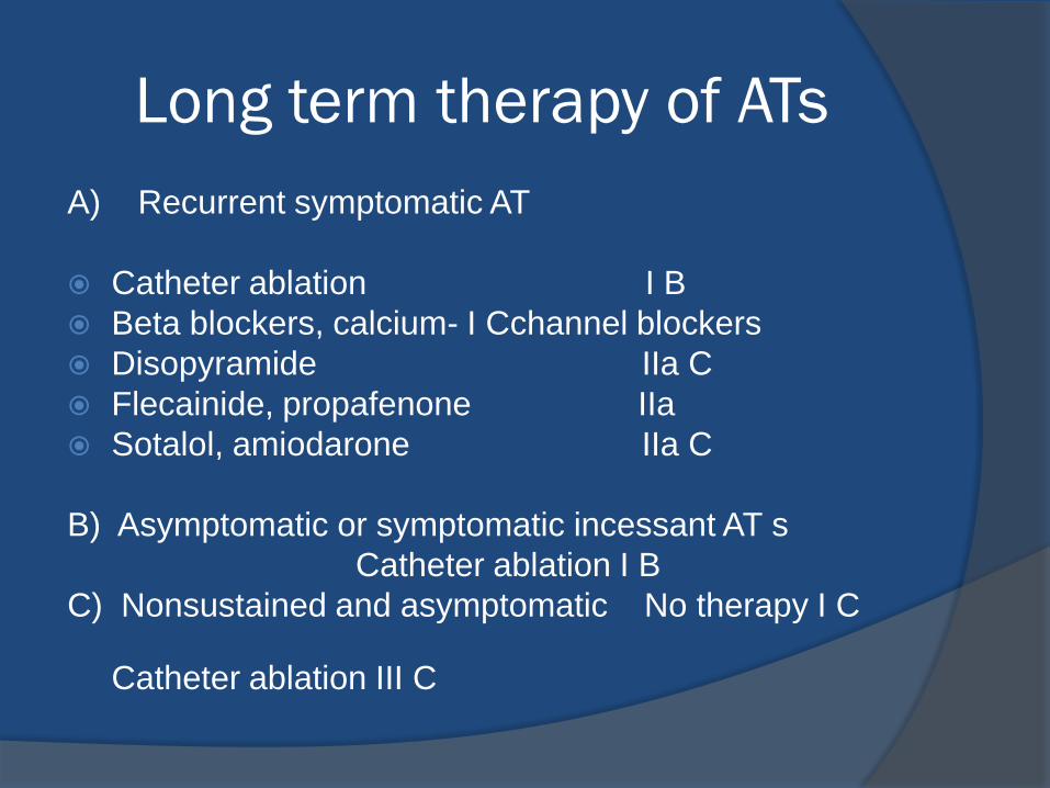
Long term therapy of ATs
A) Recurrent symptomatic AT
Catheter ablation I B
Beta blockers, calcium- I Cchannel blockers
Disopyramide IIa C
Flecainide, propafenone IIa Sotalol, amiodarone IIa C
B) Asymptomatic or symptomatic incessant AT s
Catheter ablation I B
C) Nonsustained and asymptomatic No therapy I C
Catheter ablation III C
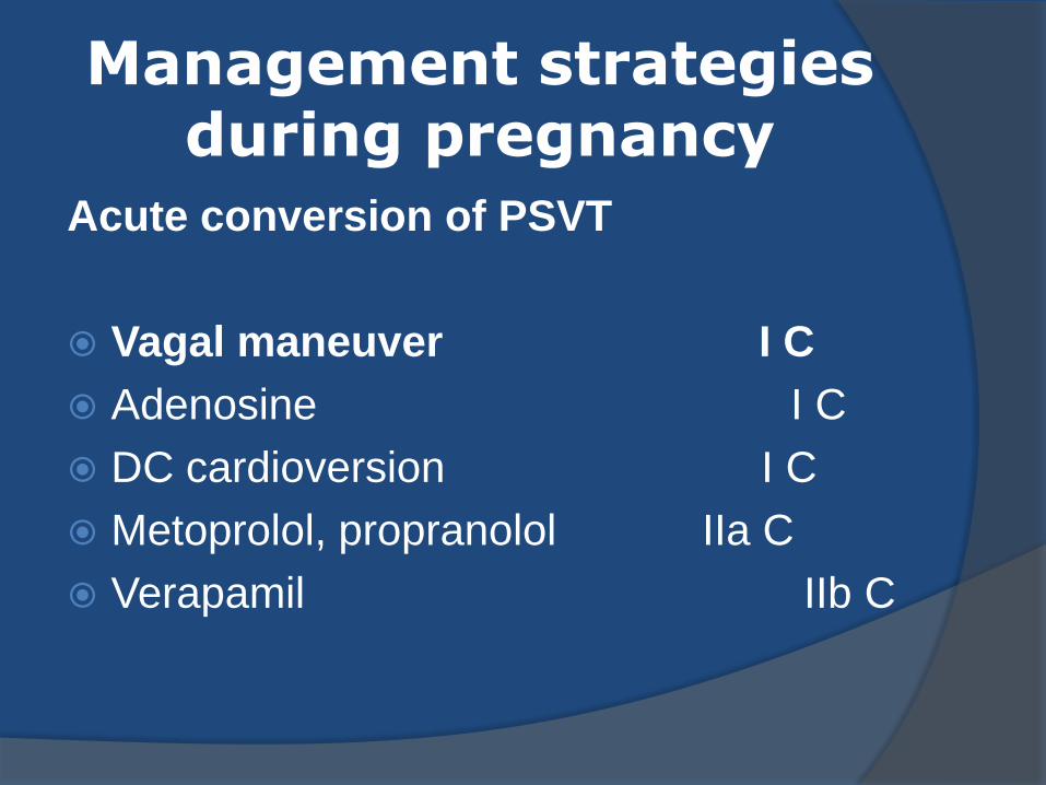
Management strategies during pregnancy
Acute conversion of PSVT
Vagal maneuver I C
Adenosine I C
DC cardioversion I C
Metoprolol, propranolol IIa C
Verapamil IIb C
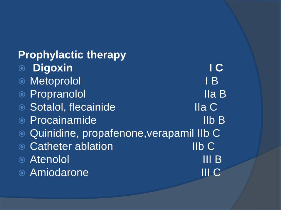
Prophylactic therapy
Digoxin I C
Metoprolol I B
Propranolol IIa B
Sotalol, flecainide IIa C
Procainamide IIb B
Quinidine, propafenone,verapamil IIb C
Catheter ablation IIb C
Atenolol III B
Amiodarone III C
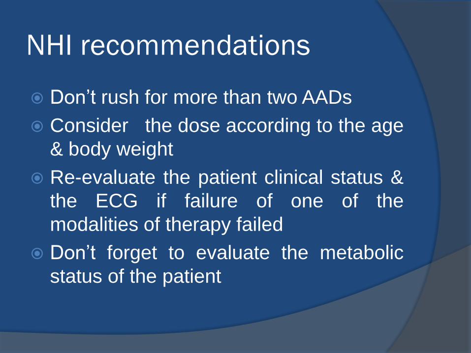
NHI recommendations
Don’t rush for more than two AADs
Consider the dose according to the age
& body weight
Re-evaluate the patient clinical status &
the ECG if failure of one of the
modalities of therapy failed
Don’t forget to evaluate the metabolic
status of the patient

Question
A 76-year-old man with CAD, heart failure, and chronic renal failure
has recurrent SVT despite treatment with beta-blockers and
calcium channel blockers. He declines to undergo an EP study for
further evaluation and treatment of this problem. His arrhythmia
occurs several times during dialysis and causes hypotension.
Which of the following is the most appropriate pharmacotherapy?
A. Procainamide.
B. Amiodarone.
C. Flecainide.
D. Sotalol.
E. Propafenone.
The correct answer is B.
Although amiodarone is not approved for treatment of supraventricular arrhythmias, it is commonly used for this purpose. It is the appropriate choice for selected patients. Low doses of amiodarone are very effective for treatment of SVT, and the risk of adverse effects is acceptable in a patient this age.
Procainamide has a high incidence of GI side effects and drug-induced lupus. It prolongs repolarization and has a 1-3% incidence of torsade de pointes. Although it can be used in patients with renal failure by adjusting the dosage and monitoring levels, it is not as effective as amiodarone and is more difficult to use in patients with renal failure.
Flecainide and propafenone are contraindicated in patients with CAD and heart failure because of their negative inotropic and proarrhythmic effects. Dosage adjustment is required in patients with renal failure because they are excreted by the kidneys.
Sotalol is a negative inotrope and must be used cautiously in patients with heart failure. It is also cleared by the kidneys, which requires careful dosage adjustment and monitoring in patients with renal failure to avoid excessive QT prolongation and induction of torsade de pointes.