lesson assignment lesson 5 lesson assignment lesson … · lesson 5 enterobacteriaceae. lesson...
TRANSCRIPT

MD0856 5-1
LESSON ASSIGNMENT LESSON 5 Enterobacteriaceae. LESSON ASSIGNMENT Paragraph 5-1 through 5-35. LESSON OBJECTIVES Upon completion of this lesson, you should be able to: 5-1. Identify descriptive features of the family Enterobacteriaceae. 5-2. Associate names of genera and species of Enterobacteriaceae with types of diseases they may cause. 5-3. Associate specific types of enteric media with their uses, methods of handling, and interpretation of typical reactions. 5-4. Associate names of Enterobacteriaceae with their typical gram morphology, colony morphology, and biochemical reactions. 5-5. Given two different categories of Enterobacteriaceae, identify tests useful in differentiating them. SUGGESTION: After reading and studying the assignment, complete the exercises at the end of this lesson. These exercises will help you to achieve the lesson objectives.

MD0856 5-2
ENTEROBACTERIACEAE
Section I. INTRODUCTION
5-1. DEFINITION The family Enterobacteriaceae consists of gram-negative, aerobic (facultatively anaerobic), nonsporogenous bacilli that grow well on artificial media. They may be motile or nonmotile, but motile forms must be peritrichous, that is, possess flagella distributed over the entire surface of the bacterial cell. Members of the family reduce nitrates to nitrites, ferment glucose with the production of acid or of acid and gas, do not produce indophenol-oxidase, and do not liquefy alginate. Pectobacterium is the only genus of the family that liquefies pectate. The genera in the family Enterobacteriaceae are Escherichia, Shigella, Edwardsiella, Salmonella Arizona, Citrobacter, Klebsiella, Enterobacter, Serratia, Proteus, Providencia, Erwinia, Pectobacterium, and Yersinia. 5-2. NORMAL FLORA AND GENERAL IDENTIFICATION OF SPECIES Many of these bacteria are normally present in the intestinal tract as part of the normal flora. It becomes the task of the bacteriology laboratory to differentiate between the gram-negative, glucose-fermenting bacilli that are normally present in the intestines, and those that are considered to be pathogens. It must be remembered that the reactions and classifications given in this study guide are broadly accepted, but due to the very nature of the subject itself, different textbooks and authors may vary on certain points and the specific reactions of a particular organism. To completely identify any one of these enteric bacteria, all characteristics of the bacterium must be established to include colony morphology on differential and selective media, numerous biochemical patterns, and often serological characteristics. 5-3. MORPHOLOGY AND CULTURE The enteric gram-negative rods range from 1 to 4 microns in length and from 0.4 to 0.8 microns in breadth. A few longer, filamentous cells may be exhibited by any of the species. The organisms possess no typical cellular arrangement and may be observed singly, in pairs, in clumps, and occasionally in short chains. Microscopic morphology is, therefore, of little diagnostic value. The majority of the enteric gram-negative rods are actively motile. The enteric bacilli grow well on ordinary nutrient media. Most species are facultative anaerobes. Although the majority of these organisms usually yield good growth between 20º C and 40º C, 37º C is optimum for most species, especially the pathogens. A medium of approximately neutral pH is most favorable for growth of all enteric bacilli. A great variety of culture media may be employed for isolation and identification of pathogenic enteric bacilli in fecal specimens. This includes the use of differential, selective and inhibitory plating media as well as selective and enrichment broths.

MD0856 5-3
Section II. PATHOGENICITY OF ENTEROBACTERIACEAE 5-4. PATHOGENICITY OF THE GENUS ESCHERICHIA Escherichia coli is one of the most abundant species of bacteria represented in the normal intestinal tract. In this region, the organism contributes to normal function and nutrition. E. coli and other enteric saprophytes become pathogenic when introduced into tissues outside the intestinal tract, especially the urinary and biliary tracts, peritoneum, or meninges. E. coli more frequently invades the urinary tract and is the most common cause of cystitis. The organism has also been isolated from local infections such as conjunctivitis. E. coli may also be the cause of septicemia. A number of E. coli serotypes have been associated with infant diarrhea, and when E. coli is isolated from pediatric patients, it should always be serotyped. 5-5. PATHOGENICITY OF THE GENUS KLEBSIELLA Klebsiella pneumoniae (Friedlander's bacillus) is isolated with some frequency from the upper respiratory and intestinal tracts of normal individuals and is responsible for approximately two percent of the bacterial pneumonias. Pulmonary infections are characterized by extensive hemorrhagic consolidation of the lobes. The fatality rate is high in untreated cases. Klebsiella species are frequently isolated from various upper respiratory tract infections, although their presence, in many instances, is probably that of secondary invaders. The organisms have definitely been responsible for suppurative abscesses of the other visceral tissue. 5-6. PATHOGENICITY OF THE GENUS ENTEROBACTER Several species of Enterobacter--E. cloacae, E. liquefaciens, E. aerogenes, and E. hafniae--have been recognized and exhibit a pathogenicity similar to Escherichia. Species of Enterobacter are isolated frequently in cases of septicemia and urinary tract infections. 5-7. PATHOGENICITY OF THE GENUS PROTEUS Of the genus Proteus, four species are recognized--Proteus vulagaris, P. mirabilis, M. morganii, and P. rettgeri. Although these organisms are primarily free-living in water, soil, and sewage, they are frequently isolated from fecal specimens of normal individuals. Morganella morganii has been responsible for diarrhea of infants and children. Proteus species often cause human infections and usually do so when introduced into tissues other than the normal intestinal tract. In this connection, Proteus species rank next to E. coli as the etiological agent of cystitis. These organisms are also encountered frequently in eye and ear infections and occasionally in pleurisy, peritonitis, and suppurative abscesses in many areas of the body. Proteus is commonly associated with other bacteria in purulent wounds and may contribute to the severity of such infections.

MD0856 5-4
5-8. PATHOGENICITY OF THE GENUS SALMONELLA It is important to remember that all salmonellae are potential pathogens and may produce enteric fever, septicemia, or gastroenteritis. Such infections often originate from ingestion of contaminated food or drink. a. Enteric Fevers. The enteric fevers consist of typhoid fever and paratyphoid fever. Salmonella typhi is responsible for typhoid fever while S. paratyphi A, S. paratyphi B, and others are most often encountered in paratyphoid fever. Of these salmonellae, S. paratyphi A and S. paratyphi C are only occasionally isolated in the United States. In enteric fevers, the ingested organisms enter the small intestine, spread through the intestinal lymphatics to the thoracic duct and enter the blood stream. The resultant septicemia distributes the infection to many organs including the kidney, intestines, liver, gallbladder, and other tissues. Infections are characterized by an insidious onset, with low-grade fever that ultimately becomes quite elevated during the bacteremic phase. Blood cultures are usually positive only during the first and second week of infection. Stool and urine cultures usually fail to yield the responsible Salmonella species until the third week. The duration of typhoid fever and paratyphoid fever is usually several weeks. Salmonella infections that result in septicemia are often due to Salmonella choleraesuis. The onset of symptoms is abrupt since blood stream invasion occurs within a short period of time following oral ingestion of the organism. This is accompanied by a rapid rise in temperature that spikes during the height of infection. Wide distribution of the organisms results in focal suppuration and abscess formation in various tissues. Meningitis, osteomyelitis, endocarditis, and pneumonia are known complications of such infections. Blood cultures are most often positive when taken during the height of the fever. b. Gastroenteritis. Of the many Salmonella, species that produce acute gastroenteritis in man, S. typhimurium is the most frequent causative agent. Salmonella enteritidis is possibly the second most common cause. S. choleraesuis has also been implicated in gastroenteritis but to a lesser extent than either of the two previously mentioned species. Infections are characterized by fairly sudden onset (15 to 24 hours' incubation), and rather severe gastrointestinal distress with vomiting, diarrhea, and slight elevation of temperature. Recovery is rapid (1 to 3 days) since the intestinal tract is not usually invaded by the organisms. Symptoms result from the irritative action of acids and endotoxin upon the intestinal mucosa. The acids are formed by fermentation of carbohydrates by the responsible organisms. Endotoxins are released following death and cellular lysis of the etiologic agent. Only very rarely do infections develop into septicemia. Outbreaks of gastroenteritis are usually linked with the consumption of certain foods and are often referred to as "food poisoning." Diseases usually originate from unsuspected subclinical cases, convalescent carriers, or healthy permanent carriers who harbor the organisms in their intestine, gallbladder, or the urinary tract. Such individuals may contaminate food or drink either directly or indirectly. The salmonellae produce no exotoxins. Upon death and lysis of the cells, endotoxins are released which largely account for the disease symptoms of man.

MD0856 5-5
5-9. PATHOGENICITY OF THE GENUS SHIGELLA Shigellae are the cause of bacillary dysentery. Infections are usually limited to the gastrointestinal tract. The disease process is essentially an inflammation of the mucous membrane of the large intestine and terminal ileum that leads to necrosis and superficial ulceration. Symptoms occur within 1 to 2 days following ingestion of contaminated food or drink. The illness is characterized by sudden onset of abdominal pains, cramps, diarrhea, and fever. The intense irritation of the bowel is due to the release of somatic endotoxin upon autolysis of the Shigella species. Infections from S. dysenteriae are more severe because, in addition to the endotoxin substance, an exotoxin (neurotoxin) is produced which causes paralytic symptoms. Infections from exotoxin-producing strains of S. dysenteriae are relatively frequent in India, Japan, China, and other parts of Asia. Although some individuals recover quickly from bacillary dysentery and pass infectious bacilli in stools for only a short period, others become chronic carriers (ulcerative colitis) and may suffer frequent relapses of the disease. The latter serve as a reservoir of infection.
Section III. ENTERIC MEDIA 5-10. CATEGORIES OF ENTERIC MEDIA Before you can expect to isolate and tentatively identify members of the enteric bacteria, you must have some knowledge of the different media that are used for this purpose. Generally speaking, enteric media can be divided into three categories. a. Differential media are designed to point out differences in bacteria on the basis of their metabolism. b. Selective media are designed to select certain potential pathogens from among nonpathogenic bacteria. c. Enrichment media are designed to help enrich or promote the growth and recovery of certain types of pathogenic bacteria from clinical specimens, such as feces, which contain large numbers of saprophytic enteric bacteria. 5-11. LACTOSE FERMENTATION The initial division of bacteria comprising the enterics is separated most conveniently using, as a reference point, the ability to ferment the sugar lactose. In this division, three areas of interest emerge: a. The lactose fermenters include those enteric bacteria that are able to ferment lactose with the production of gas within 24 hours. The enteric bacteria exhibiting this characteristic are known as the "coliforms," and they are usually found as saprophytes in the intestinal tract. The coliforms do not usually present a medical problem except in pediatric cases.

MD0856 5-6
b. The late lactose fermenters are able to ferment lactose after prolonged periods of incubation, usually after 48 hours. These enteric bacteria are generally referred to as the "paracolons." They are of interest because they exhibit characteristics of both the coliforms and the pathogens, and must be distinguished from them. The exception is Shigella sonnei that may show delayed (4 to 7 days) lactose fermentation. c. Finally, the lactose nonfermenters represent the third area of interest as regards the ability to ferment lactose. These gram-negative enteric bacilli are usually unable to ferment lactose and include most of the pathogens and some saprophytic bacteria that are usually present in the intestinal tract as normal flora and must be distinguished from pathogens. 5-12. DIFFERENTIAL MEDIA The differential media are designed to distinguish between colonies of lactose fermenters and lactose nonfermenters. Some of the differential (or isolation) media commonly used are eosin-methylene blue (EMB) agar, MacConkey's agar, and deoxycholate agars. These media contain certain carbohydrates, indicators, and chemicals that are inhibitory to a large number of the gram-positive bacteria that so often overgrow the relatively few pathogenic bacteria. a. EMB Agar. Eosin-methylene blue (EMB) agar contains the dyes eosin and methylene as well as the carbohydrates lactose-and sucrose. The dyes act as inhibitors of most gram-positive bacteria and also as indicators of those bacteria capable of fermenting lactose. Colonies of lactose fermenters appear as dark-colored colonies while those of lactose nonfermenters appear as translucent or colorless colonies. EMB agar is particularly valuable in identifying Escherichia coli. On EMB, E.coli produces a very discrete and distinctive colony that has a green metallic sheen. EMB is always sterilized by autoclaving. b. MacConkey's Agar. MacConkey's agar is a differential medium that also distinguishes between lactose fermenters and lactose nonfermenters. Colonies of lactose fermenters appear red or pink, while colonies of lactose nonfermenters are translucent or colorless. MacConkey's agar contains the pH indicator, neutral red, which gives a red color under acid conditions. The growth of gram-positive bacteria is inhibited on MacConkey's agar because of the presence of bile salts and the dye crystal violet. MacConkey's agar is also sterilized by autoclaving. c. Deoxycholate Agar. Deoxycholate agar is very similar to MacConkey's agar and also contains the pH indicator neutral red and distinguishes between lactose fermenters and lactose nonfermenters. The colony appearance is very similar to that produced on MacConkey's agar. Deoxycholate agar is prepared by heating to the boiling point to dissolve the agar; however, it is not autoclaved to sterilize and for this reason it is very suitable for use as a pour plate.

MD0856 5-7
5-13. SELECTIVE MEDIA a. The selective media most commonly used are Salmonella-Shigella agar, deoxycholate citrate agar, brilliant green agar, and bismuth sulfite agar. These media select certain potential pathogenic enteric bacteria from clinical specimens because of the presence of inhibitory substances that inhibit the growth of gram-positive bacteria and retard the growth of coliforms. These inhibitory substances include bile salts and brilliant green dye. b. Salmonella-Shigella (SS) agar also contains the pH indicator neutral red and the carbohydrate lactose. When colonies of "coliforms" (lactose fermenters) appear, they are red or pink in color. Colonies of lactose nonfermenters usually appear colorless or translucent. Although Salmonella-Shigella agar is usually classified as a selective medium, it differentiates between colonies of lactose fermenters and lactose nonfermenters. Incorporated in this selective medium are the salts, ferric citrate and sodium thiosulfate. The presence of these salts provides the medium with an indicator of hydrogen sulfide production. The colonies of bacteria producing hydrogen sulfide may have blackened centers, which is the result of the precipitation of ferric sulfide. The precipitate ferric sulfide results when hydrogen sulfide comes in contact with the ferric ion. SS agar is not prepared by sterilization, but it is heated until in solution, then allowed to cool, and poured into plates. c. Sterilization is not required for SS agar since this highly selective medium contains such strong inhibitors. Brilliant green agar is sterilized by autoclaving. However, deoxycholate citrate agar and bismuth sulfite agar must only be heated sufficiently to dissolve the agar and not autoclaved, as excessive heating will destroy their selectivity. d. All of the selective media mentioned serve the same general purpose, to inhibit many of the coliform bacilli, and especially Escherichia and Proteus strains, while permitting the growth of most Salmonella and some Shigella organisms. Most Salmonella and Shigella species, along with slow or non-lactose-fermenting organisms, generally form colorless colonies. However, some species may appear as black or greenish colonies on certain media as already mentioned, and the lactose-fermenting organisms, which are not inhibited, grow as pink or red colonies. 5-14. ENRICHED MEDIA Enriched media are generally employed for the recovery of enteric pathogens from specimens that might contain few enteric pathogens but many enteric saprophytes or coliform organisms. By the use of these media, enteric saprophytes are inhibited while the growth of enteric is not inhibited. Consequently the recovery of enteric pathogens is enhanced.

MD0856 5-8
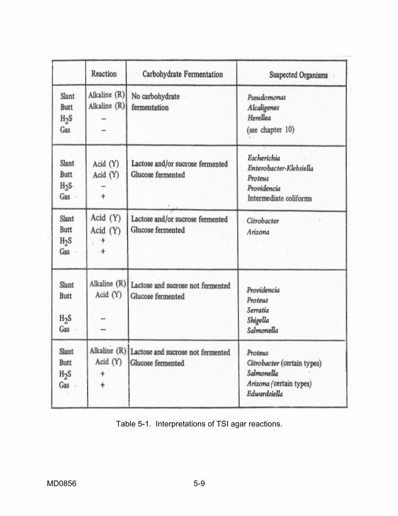
a. Selenite-F Broth. Selenite-F broth is an enriched medium that contains the inhibitor, selenite. In this medium gram-positive bacteria and coliform bacteria are inhibited. When selenite-F broth is used as an enrichment medium, it is essential that subculturing be done no later than 18 hours after inoculation of the medium since coliform bacteria will be inhibited only for a period of about 18 hours following inoculation. Selenite-F broth is prepared by boiling or by use of free- flowing steam. It must not be sterilized by autoclaving. b. Tetrathionate Broth. Tetrathionate broth is an enriched medium that is especially valuable for recovery of Salmonella. This medium contains bile salts, iodine, and brilliant green as inhibitors. Gram-positive bacteria, coliforms, and Shigella species are generally inhibited in this highly inhibitory medium. 5-15. TUBED DIFFERENTIAL MEDIA All of the media mentioned in this chapter so far except the enrichment media are utilized as streak plates or pour plates. Kligler's Iron Agar (KIA) and Triple Sugar Iron Agar (TSI) are tubed differential media having a slant and a butt. The slant represents an aerobic environment; the butt represents an anaerobic environment. a. Medium Contents. (1) KIA contains two sugars and two indicator systems. Glucose is present in 0.1 percent concentration and lactose is present in 1.0 percent concentration. Phenol red is present as the pH indicator, and it gives a yellow color under acid conditions and a red color under alkaline conditions. Hydrogen sulfide production is indicated by the formation of the black precipitate, ferric sulfide. Hydrogen sulfide is formed as a result of action upon the salts, ferric ammonium citrate and sodium thiosulfate. (2) Triple Sugar Iron Agar (TSI) is exactly the same formula as KIA, but with the addition of 1.0 percent sucrose. The glucose concentration is one-tenth the concentration of the lactose and sucrose to enable the detection of glucose fermentation alone. The small amount of acid produced by the fermentation of the glucose is oxidized so rapidly in the slant that the slant either remains or reverts back to alkaline (red), whereas the lower-oxygen tension in the butt retains and maintains an acid (yellow) reaction. Because of this situation that necessitates the free exchange of air with the slant, a tightly stoppered or screw-capped tube creates an acid condition that involves the slant and in this way may give misleading reactions of the medium. (3) Because of the added advantage of three sugars, TSI is usually preferred to KIA. Table 5-1 gives the reactions that are possible using TSI and the interpretation of each reaction, as well as the organisms that would be the most likely suspects.
MD0856 5-9
Table 5-1. Interpretations of TSI agar reactions.

MD0856 5-10
b. Medium Interpretation. (1) Following inoculation and incubation of the organism on TS1 medium, sugar fermentation is evidenced by conversion of all or portions of the medium from orange-red to yellow. The acid end products of such fermentations convert phenol red to its yellow acid state. Those organisms failing to ferment any of the sugars produce no change in the medium or convert it to a deeper red color. The latter reaction is a result of the action of alkaline metabolic end products upon phenol red. (2) TSI agar also contains sodium thiosulfate and ferrous ammonium sulfate to serve as indicators of hydrogen sulfide production. Organisms capable of producing hydrogen sulfide do so through the utilization of sodium thiosulfate in an acid environment. Hydrogen sulfide formed in this manner reacts with ferrous ammonium sulfate to yield ferrous sulfide. The latter substance accumulates as a black precipitate either immediately under the base of the slant or deeper in the butt of the TSI agar. (3) After an 18 to 24 hour incubation period, the TSI medium is examined for the following reactions: (a) Lactose or sucrose fermentation results in an acid (yellow) reaction throughout the medium. (b) Fermentation of only glucose results in an acid (yellow) reaction in the butt of the agar medium with an alkaline (dark red) slant. (c) If no sugars are fermented, an alkaline (dark red) with an orange-red butt is usually observed. (d) Gas production in TSI agar from lactose, sucrose, or glucose fermentation results in bubble formation or splitting of the medium in the butt of the tube. (e) Hydrogen sulfide production results in a blackening in the butt or the area of the medium just under the base of the slant. c. Identification. After interpretation of TSI agar reactions, it is necessary to use numerous biochemical fermentation tests and serotyping to be certain of just which organisms you have.

MD0856 5-11
5-16. ISOLATION OF ENTERIC PATHOGENS FROM FECAL SPECIMENS In attempting to isolate enteric pathogens from fecal specimens, one or two procedures may be used. One method is to plate the specimen directly to any of the differential and selective media. The use of bismuth sulfite agar is highly recommended when Salmonella typhi is suspected, since bismuth sulfite inhibits the growth of most other organisms and promotes the growth of Salmonella typhi, producing a highly characteristic colony. Eosin-methylene-blue agar, MacConkey's agar, and SS agar are probably the most commonly used for the direct plating of fecal specimens. In the indirect procedure for the isolation of the enteric pathogens, either selenite-F or tetrathionate broth are inoculated with a fecal sample for enrichment. If selenite-F broth is used, the culture should be incubated for 8 to 12 hours at 37º C, and then streaked to EMS, MacConkey's, SS, bismuth sulfite, etc. If tetrathionate broth is used, the culture should be incubated for 12 to 24 hours at 37º C, and then streaked to EMS, MacConkey's etc.
Section IV. PRELIMINARY SCREENING OF CULTURES FOR ENTEROBACTERIACEAE
5-17. COLONY CHARACTERISTICS Medical laboratory specialists working in the bacteriology section I become familiar with the colony characteristics of enteric bacilli on blood agar as well as on commonly used plating media for stool examination. Enteric organisms cultivated on blood agar usually reveal large, smooth, shiny, circular, raised colonies which mayor may not be hemolytic or pigmented. Proteus species often exhibit swarming. The colony characteristics of enteric species cultivated on commonly used enteric media are described in table 5-2 as they appear when cultivated for 16 to 24 hours at 37º C. Colorless colonies on differential and selective media indicate the organism is a lactose nonfermenter and, thus, a possible pathogen which must be identified by further studies. (Delayed lactose fermenters will also appear colorless at this stage.) Colored colonies indicate the organism is a lactose fermenter. Organisms producing colored colonies on enteric media are usually nonpathogenic for adults, providing these organisms have been isolated from their normal habitat, the intestinal tract. Certain lactose-fermenting enteric organisms may, however, be etiological agents in cases of infant diarrhea.

MD0856 5-12
Table 5-2. Colony characteristics of enteric bacilli on enteric media (continued).

MD0856 5-13
Note: Certain Proteus and Salmonella species may produce colorless colonies with black centers on SS agar. A bluish water soluble pigment may or may not be evidenced with Pseudomonas aeruginosa. Brilliant green agar is not suitable for growth of Salmonella typhi or Shigella spp.
Table 5-2. Colony characteristics of enteric bacilli on enteric media (concluded)

MD0856 5-14
5-18. USE OF LESS INHIBITORY MEDIA In attempting to isolate enteropathogenic Eschrichia coli, Klebsiella, or Citrobacter from fecal material, the use of tetrathionate and selenite-F broths for enrichment is dispensed with, since both are inhibitory for most strains of these groups. Because of such situations, the less inhibitory media (EMB and MacConkey's agar) are usually used for primary isolation by the direct plating procedure in lieu of the more selective media which are intended to inhibit the majority of these strains. All colonies which exhibit a lactose-nonfermenting appearance on the isolation plates, either from direct streaking or after streaking from an enrichment, should be suspected as possible enteric pathogens and further studies are indicated to establish or rule out the presence of enteric pathogens as opposed to normal flora which are also lactose negative or are late lactose fermenters. Suspected colonies from the isolation plates should be carefully transferred to TSI slants. a. TSI Slants. (1) Special care must be taken to insure that only pure cultures are transferred to a TSI slant, because if more than one type of organism is introduced into the TSI slant, confusing reactions will occur. An inoculating needle should be used for transferring to the TSI slant, and only the center of the desired colony should be touched. The inoculating needle should then be stabbed into the butt of the slant first and then streaked in a zigzag fashion over the slanted surface. Always remember that a TSI slant should be closed with a cotton plug or a loose closure but never with a tightly fitting cap or stopper. (2) Figures 5-1 and 5-2 present in a simplified flow-chart form the typical reactions encountered with TSI slants and a general schematic procedure with which to begin classification of the various types of pathogens and potential pathogens. In routine examinations of fecal materials just for enteric pathogens, all TSI slants with acid butts and alkaline slants should be retained for examination; those which are acid throughout (possible E. coli or other coliform bacilli) may be discarded as nonpathogens. The TS1 slants with alkaline slants and neutral (alkaline) butts may also be discarded when dealing with routine fecal specimens. The latter reactions are indicative of Pseudomonas species and Alcaligenes species, which are usually nonpathogens when present in the intestinal tract of adults. Since some Proteus species may be indistinguishable from salmonella and other lactose-nonfermenting gram-negative bacteria on primary isolation, all TSI slant cultures for positive identification should next be screened with urea.

MD0856 5-15
Figure 5-1. Tentative differentiation of lactose-nonfermenting bacilli.
MD0856 5-16
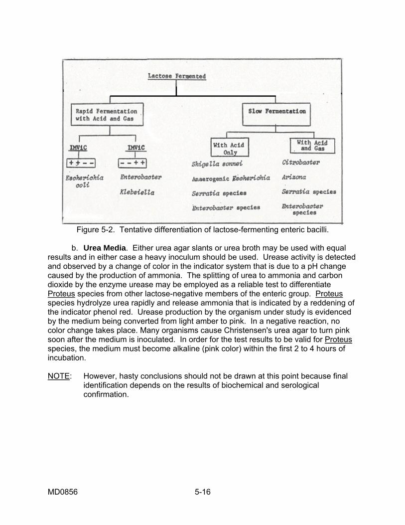
Figure 5-2. Tentative differentiation of lactose-fermenting enteric bacilli.
b. Urea Media. Either urea agar slants or urea broth may be used with equal results and in either case a heavy inoculum should be used. Urease activity is detected and observed by a change of color in the indicator system that is due to a pH change caused by the production of ammonia. The splitting of urea to ammonia and carbon dioxide by the enzyme urease may be employed as a reliable test to differentiate Proteus species from other lactose-negative members of the enteric group. Proteus species hydrolyze urea rapidly and release ammonia that is indicated by a reddening of the indicator phenol red. Urease production by the organism under study is evidenced by the medium being converted from light amber to pink. In a negative reaction, no color change takes place. Many organisms cause Christensen's urea agar to turn pink soon after the medium is inoculated. In order for the test results to be valid for Proteus species, the medium must become alkaline (pink color) within the first 2 to 4 hours of incubation. NOTE: However, hasty conclusions should not be drawn at this point because final identification depends on the results of biochemical and serological confirmation.

MD0856 5-17
5-19. IMVIC TESTS Most of the members comprising the gram-negative bacilli ferment the carbohydrate lactose although some are very slow fermenters and for this reason will resemble most of the enteric pathogens and must be differentiated from them. The exception to the rule is the pathogen Shigella sonnei, which sometimes is a slow lactose fermenter but seldom causes the production of any gas. The lactose fermenters may be tentatively identified according to figure 5-2 using the IMViC reactions, which are used primarily to differentiate between the coliform bacteria. The letters IMViC stand for the tests indole, methyl red, Voges-Proskauer, and citrate. The lower-case "i" is included only to make IMViC easier to pronounce. See Lesson 3, (paras 3-9 - 3-11) for details about these tests. a. Indole Test. The indole test is based on the ability of certain bacteria to split tryptophan to alanine and indole. The liberated indole will combine with paradimethylaminobenzaldehyde in Kovac's reagent to give a deep red color. The presence of the red color at the interphase between the reagent and the broth culture signifies that indole has been liberated. The absence of the red color signifies a negative test. b. Methyl Red Test. The methyl red test is based on the principle that some organisms ferment glucose, and produce small amounts of acids that are converted to neutral end products. This test is designed to differentiate those organisms that, in contrast, produce high acidity. A positive reaction occurs when the culture is sufficiently acid to turn the methyl red reagent to a distinct red color. A yellow color is regarded as a negative test. c. Voges-Proskauer Test. The Voges-Proskauer test is based on the ability of some organisms to produce a neutral end product, acetylmethylcarbinol, from dextrose. A positive test is indicated by the development of a pink or red color. A yellow color is regarded as a negative test. d. Citrate Test. The citrate test is based on the ability of certain bacteria to utilize sodium citrate when it is the sole available source of carbon in a chemically defined medium. A slant of Simmons' citrate agar is lightly inoculated by streaking only. A positive reaction (growth) is accompanied by an alkaline reaction resulting in a change in the green color of the medium to a deep blue color. No change in the indicator or the absence of blue color in the green medium indicates a negative test. A light inoculum must be used to insure that no nutrients are transferred to the chemically defined medium. e. Uses of IMViC Tests. The IMViC reactions are classically used as a group of tests to separate the Escherichia and the Enterobacter-Klebsiella genera. Escherichia organisms are usually IMViC + + - - and Enterobacter-Klebsiella organisms are usually IMViC - - + +. However, any of these tests are of independent value to aid in the biochemical differentiation of other enteric bacilli.

MD0856 5-18
Section V. IDENTIFICATION OF ENTERIC ORGANISMS 5-20. GENERAL Figure 5-3 provides an identification schema for the enteric group and related organisms. Table 5-3 describes the reactions of the different enteric organisms to a series of biochemical tests.
Figure 5-3. Identification schema for the enteric group and related organisms (faculatative gram-positive rods).

MD0856 5-19
Table 5-3. Differentiation of Enterobacteriaceae by biochemical tests. (reprinted by permission of Burgess Publishing Company from Identification of
Enterobacteriaceae, 1972, 3d Ed., by P.R. Edwards and W.H. Ewing)

MD0856 5-20
5-21. LYSINE DECARBOXYLASE TEST (FALKOW METHOD) The lysine decarboxylase test measures the enzymatic ability of organism to decarboxylate the amino acid, lysine, causing its conversion into the amine, cadaverine. This activity is characteristic of the genus Arizona, most members of the genus Klebsiella. The test system employs Falkow lysine broth or lysine iron agar, which include glucose, lysine, peptone and the indicator, bromcresol purple (yellow to purple). The initial reaction of the system is acid (yellow, due to the attack on glucose, lysine production of acid end products. Upon depletion of the glucose, lysine is decarboxylated to form the basic compound, cadaverine, which causes reversion of the reaction to alkalinity and the development of a purple color. 5-22. DECARBOXYLASE TESTS (MOELLER METHOD) a. The basal medium for decarboxylase tests includes a peptone, beef extract, bromcresol purple, cresol red, glucose, and pyridoxal. The basal medium is divided into four portions. One portion, used as a control, is tubed without the addition of an amino acid. To each of the remaining portions, an amino acid is added as follows: (1) Portion 1--1 percent of L-lysine dihydrochloride (2) Portion 2--1 percent of L-arginine monohydrochloride (3) Portion 3--1 percent of L-ornithine dihydrochloride b. The ph of portion 3 must then be adjusted to 6-6.5 prior to sterilization. Each portion may then be tubed in 3 to 4-ml amounts in small screw-capped tubes and sterilized. After light inoculation of tubes including a control, a 10-mm layer of sterile paraffin is added and the tubes are incubated at 37º C for 4 days. A positive reaction, alkalinization, is indicated when the color changes from a yellow to a violet or reddish-violet color. 5-23. PHENYLALANINE DEAMINASE TEST The phenylalanine deaminase test has as its basis the ability of the enzyme deaminase in the presence of oxygen to convert the parent compound L-phenylalanine into phenylpyruvic acid. Organisms of the Proteus and Providencia genera are characterized as having such deaminases. This test system requires a culture on L-phenylalanine medium. Phenylpyruvic acid will react with a solution of acidified Fe2(SO4)3 and FeNH4)2 to produce a green color, and it is the development of this green color that makes the phenylalanine deaminase test positive.
MD0856 5-21
5-24. MALONATE TEST Sterile malonate broth is inoculated from a young agar slant or broth culture; preferably a 3-mm loopful of broth culture. The culture is observed daily during the 48-hour incubation at 37º C. A positive result, utilization of malonate, is indicated by a change of color from green to Prussian blue. 5-25. PECTOLYSIS TEST (MARTIN AND EWING) a. Preparation of Medium. To 100 ml of distilled water is added 0.5 g of yeast extract, 0.9 ml of IN sodium hydroxide, 0.5 ml of 10 percent calcium chloride (CaCl2
. 2H20), 1.25 ml of 0.2 percent bromthymol blue, 1 g of sodium polypectate No.
24 (Sunkist Growers, Inc., Ontario, California), and 2 g of agar. This is stirred thoroughly, heated gently, sterilized, and poured into thin plates. b. Inoculation. Plates are spot inoculated from a young agar slant. Up to 10 cultures can be tested on a single plate. c. Incubation. Incubate at 37º C for 1 to 3 days. d. Interpretation. Liquefaction of pectate, a positive test, is indicated by depression of the medium around a growth. 5-26. MOTILITY TEST A tube of motility test medium is inoculated by stabbing to a depth of about 5 mm. It is incubated at 37º C for 1 to 2 days. If the organisms spread out from the line of inoculation, the test is positive. If organisms grow only along the line of the stab, the test is negative. Negative test should be incubated an additional 5 days at 22º to 25º C. 5-27. THE PROTEUS AND PROVIDENCIA GENERA a. The genus Proteus consists of four species that can be identified by their biochemical patterns. See table 5-3 for typical biochemical reactions of Proteus and Providencia species. Proteus organisms are considered to be normal flora of the intestinal tract, where they usually exist as saprophytes. When grown upon solid media, these bacteria may exhibit the swarming phenomenon and cover the entire plate with a layer of growth. This condition makes recovery of pure cultures of other bacteria present on the plate extremely difficult. Species of Proteus are characterized as being lactose nonfermenters that have the ability to split urea, a characteristic that is extremely helpful in identifying this genus. (1) Proteus vulgaris and P. mirabilis swarm on a blood agar plate and are usually separated on the basis of indole production; P. vulgaris is indole positive and P. mirabilis is indole negative.

MD0856 5-22
(2) Morganella morganii and P. rettgeri usually do not swarm and are usually separated on the basis of mannitol fermentation and the ability to utilize citrate. Morganella morganii is mannitol positive and citrate positive while Proteus rettgeri is mannitol negative and citrate negative. (3) Because Proteus species are lactose negative, they are often selected tentatively from isolation plates as colonies of salmonellae or shigellae; Proteus can usually be ruled out by use of the urease test as previously stated. b. The Providencia species closely resemble Morganella morganii and Proteus rettgeri and for this reason are often included with the genus Proteus. Due to the fact that the Providencia species are H2S-negative and mayor may not produce gas in glucose, they are often mistaken for shigellae on TSI agar. Members of Providencia may be distinguished, however, from the shigellae by their motility and utilization of citrate, as well as by other biochemical reactions. 5-28. THE ENTEROBACTER AND KLEBSIELLA GENERA At present it is not easy to distinguish between Klebsiella pneumoniae and Enterobacter aerogenes on a purely biochemical basis (see table 5-3) since their biochemical characteristics are quite similar. However, Klebsiella is comprised of nonmotile organisms and Enterobacter is comprised of motile organisms. Another procedure that is used for differentiation is the ornithine test. 5-29. THE GENUS SALMONELLA If preliminary biochemical studies are suggestive of a Salmonella species, serological evaluation of the culture is desirable. The serological antigens associated with bacteria are identified by Arabic numerals and alphabetical symbols. The antigens exhibited by the Enterobacteriaceae fall into three main categories--O (somatic), H (flagellar), and K (envelope) antigens. a. Somatic Antigens. Somatic, or body, antigens are assigned the alphabetical symbol O. Somatic antigens are thermostable and determine serological group identification. Using a known specific antiserum that contains antibodies for that antigen identifies each antigen. The reaction of somatic antigens is described as granulation. The time element for this serological reaction is very rapid, occurring within 60 seconds. The granules are very fine and white in color. Any reaction occurring later than 60 seconds should be regarded with suspicion and retested.

MD0856 5-23
b. Flagellar Antigens. The flagellar antigens are represented by the alphabetical symbol H and are associated with motility. These antigens are thermolabile and establish species or serotypes within a particular genus or group. H antigens usually do not interfere with or mask the agglutinability of somatic antigens. The reaction of flagellar antigens is described as a flocculation. This reaction is also rapid and occurs within the prescribed 60-second time element. The flocculation is loose and fluffy, appearing a cloudy gray. Most salmonellae are diphasic; that is, each motile type tends to exhibit two antigenic phases, both phases with the same O antigens but different H antigens. To identify the Salmonella serotype, it is necessary to identify the H antigens in both phases. c. K ("Capsular") Antigens. Bacteria may be surrounded by a capsule, sheath, or envelope that possesses K or envelope antigens. The K antigens are thermolabile and assist in further establishing serotypes within a particular genus and species. K agglutination appears fine in texture and white in color. There are varieties or types of K antigens which are designated by the alphabetical symbols L, A, B, and Vi. The K antigens may interfere with O agglutinability and prevent group identification. This reaction is said to mask the somatic antigens, and heat is required to inactivate the K antigens. This makes it possible to test for the O antigens. d. Polyvalent Antiserum. Polyvalent O antisera are available" commercially. Suspected Salmonella species must first be tested with polyvalent antiserum which contains antibodies against Salmonella somatic O antigens A through E. Complete and immediate agglutination is observed with most salmonellae. A few of the paracolons and some shigellae may exhibit partial to complete agglutination due to close similarities of the antigenic structure and the nature of polyvalent antiserum. Those cultures agglutinated by polyvalent antiserum should then be tested with the individual grouping antisera A through E. The particular serum wherein clumping occurs most rapidly is the serological group to which the organism belongs. Although cross reactions between various O groups of the salmonellae do occur, such reactions are usually not pronounced. Any culture that exhibits prompt, definite clumping in polyvalent serum and in one or more of the O grouping sera is reported as a probable Salmonella species. Biochemical confirmation is necessary, for many paracolon bacteria contain antigens related to the O antigens of Salmonella species. e. Vi Antiserum. If a culture fails to react in polyvalent antiserum, it should be tested with Vi ("virulence") antiserum. This antiserum will detect the presence of Vi antigen, which is present on the cell surface of most freshly isolated strains of Salmonella typhi and occasional Salmonella paratyphi C organisms. If agglutination occurs with Vi antiserum, the saline suspension of the organism should be heated in a boiling water bath for about 15 minutes. Upon cooling, the organism is then retested with polyvalent O. Clumping will occur if the unknown is a Salmonella species. Heating removes the Vi (masking) antigen from the cell surface, permitting the O antibodies to react with somatic antigens to produce agglutination. Cultures failing to react with polyvalent or Vi antiserum may be Shigella, Arizona, or Citrobacter species.

MD0856 5-24
(1) Once and agglutination boiled cells should the presence of Vi antigen has been established observed in polyvalent antiserum, the previously be tested with anti-D or anti-C group serum. The particular antisera used are determined by the preliminary biochemical reactions previously observed. Possible Salmonella typhi strains should agglutinate in group D antiserum, and Salmonella paratyphi C cultures should agglutinate in group C antiserum. One should suspect that cultures that react with Vi antiserum and after boiling agglutinate with group D or group C antiserum are, respectively, strains of Salmonella typhi or Salmonella paratyphi C. (2) All cultures which fail to react quickly and strongly with polyvalent or Vi antiserum should be subjected to further biochemical tests. The same holds true for cultures that react with polyvalent antiserum, but fail to react typically with O grouping antisera. Potassium cyanide medium is excellent in identifying Citrobacter species that can resemble the salmonellae. Citrobacter species grow in KCN broth, while all salmonellae fail to do so. f. Species Definition. Once the group of a Salmonella culture has been determined, species definition is accomplished by subjecting the organism to H (flagellar) antisera, and subsequent biochemical testing. The unheated suspension of the organism is tested on a slide with H antisera (which are diluted 1:50 or 1:100) using the same technique as for O agglutination. Another acceptable technique is to dilute a broth culture of the test organism equally with normal saline containing 0.6 percent formalin and react this with appropriate antisera at 50º C. The antisera for this procedure are diluted 1:100 and the results are read following a one-hour incubation in a water bath. The technique employed will be determined by the specific directions accompanying various lots of antisera procured. It may be necessary on occasion to employ a U-tube or phase-tube to type for both phases of the H antigens. One phase will mask the other preventing complete identification. It is necessary to inoculate the phase tube (which contains a semisolid medium) just below the surface of one end of the tube. Place a small quantity of antisera at the site of inoculation that is specific for the phase you have already identified. The antisera will react with the antigen for which you have established identity and permit the unidentified phase to migrate by means of motility to the opposite end of the tube. Remove the motile bacteria that have reached this point and inoculate to an agar slant and subsequently complete typing for the flagellar antigens of the organism in question. Most bacteria of the Salmonella genus possess both phase 1 and phase 2 antigens. If an organism has two phases of H antigens, you must establish the complete flagellar typing for both phases.

MD0856 5-25
g. Reactions of Salmonella typhi. The typhoid bacillus is one of the most frequently encountered Salmonella species. It is important that the typhoid bacillus be recognized as a highly specialized member of the genus Salmonella with certain characteristics that set it apart from most of the other salmonellae. Salmonella typhi produces acid in the typical media used for biochemical studies, but it does not produce gas as do most members of this genus. Salmonella typhi produces an acid reaction without the production of gas in the fermentation of glucose, mannitol, trehalose, maltose, and dextrin. It does not ferment lactose or sucrose; no gas is formed in a TSI slant and the production of H2S on TSI is variable. Indole is not produced and gelatin is not liquefied. 5-30. THE GENUS SHIGELLA a. Antiserum Reactions. Stool cultures suspected of being Shigella should be tested with the individual polyvalent Shigella grouping antisera. Four antisera are usually recommended for Shigella species, each containing antibodies specific for members of the four Shigella subgroups--subgroup A (S. dysenteriae), B (S. flexneri), C (S. boydii), and D (S. sonnei). Specific agglutination usually takes place within one minute with most commercially prepared antisera. The saline and normal serum controls should reveal no clumping. Delayed or incomplete cross-reactions may be observed between certain members of the Shigella subgroups, but only the prompt or complete reactions are significant. Such cross-reactions are due to antigenic similarities between various Shigella subgroups. It is important to remember that other Enterobacteriaceae are occasionally encountered which possess somatic O antigens related to those of the genus Shigella. For this reason, cultural and biochemical studies, as well as serological procedures, should be employed for identification of suspected cultures. b. Extended Testing. If an organism appears to be a Shigella species yet fails to agglutinate in any of the antisera, a saline suspension should be heated in a boiling water bath for 30 to 60 minutes and then retested. Many Shigella cultures possess envelope surface antigens (K antigens) that prevent the somatic antisera from coming in contact with somatic antigens. Therefore, specific agglutination is impossible unless the surface antigen is first inactivated by heat. When a suspected Shigella culture still fails to react with Shigella grouping sera even after heating, the unheated organism should be tested with Salmonella polyvalent and Vi antisera. This is necessary, because certain nonmotile Salmonella typhi strains which fail to produce hydrogen sulfide (18 to 24 hours) resemble the shigellae on triple sugar iron agar slants. Other atypical salmonellae are occasionally encountered which resemble Shigella species upon tentative biochemical and cultural analyses.

MD0856 5-26
c. Characteristics of Shigella. All species of Shigella are nonmotile. Other characteristics of the genus Shigella are as follows: All species ferment glucose with no formation of gas (the exception is S. flexneri, which may be aerogenic); no species produce gas on any of the other carbohydrate media; some ferment mannitol and others do not; there is no fermentation of salicin or adonitol; they do not grow on Simmons' citrate agar; and they do not hydrolyze urea, liquefy gelatin, or form acetylmethylcarbinol. All strains of Shigella are lactose nonfermenters with the exception of S. sonnei that is a very delayed lactose fermenter (4 to 7 days). 5-31. THE GENUS ESCHERICHIA The genus Escherichia is often not thought of as including those organisms formerly referred to as the Alkalescens-Dispar group. The Alkalescens-Dispar strains closely resemble other Escherichia organisms in biochemical reactions and serologic complexity; however, they neither ferment lactose nor produce gas from other carbohydrates. The typical Escherichia colonies are easily recognized and are characterized by their rapid fermentation of lactose with gas formation, and by the classic IMViC reactions. The Alkalescens-Dispar strains are now considered as separate serotypes of E. coli. a. Antigenic Tests. The antigenic structure of the escherichiae is almost as complex as that of the salmonellae. Three types of antigens possessed by E. coli strains are important in their serological identification. These are O (somatic, cellular) antigens that resist heat inactivation at 100º C for one hour, the H (flagellar) antigens that are inactivated by exposure to heat at 100º C for one hour, and the K (enveloping, capsular) antigens which surround the O antigens and prevent their agglutination in specific O antiserum. The K antigens are inactivated by exposing the organisms to heat at 100º C for one hour. b. Serotype Identification. Isolation of E. coli from infant diarrhea requires that the fecal material be inoculated to differential media (EMB, MacConkey's or deoxycholate agar) rather than the more highly selective media used for isolation of Salmonella and Shigella species; otherwise, growth will be inhibited. Blood agar should also be inoculated since occasional E. coli strains fail to develop on differential media. After 18 to 24 hours' incubation the plates are examined for typical colonies of E. coli. (1) Since the colonies of enteropathogenic forms cannot be grossly distinguished with certainty from nonpathogenic forms, several colonies should be tested serologically. This may be quickly and effectively accomplished by drawing an inoculating loop across a number of colonies and preparing an emulsion of the growth in a drop of saline on a glass slide. A loopful of pooled (polyvalent) antiserum is mixed with the suspension. The antiserum pool contains specific antibodies against each of the OB antigens of the eleven E. coli serotypes most often associated with infant diarrhea. Pronounced agglutination observed in the organism-antiserum mixture is presumptive evidence for the presence of an enteropathogenic E.coli serotype.

MD0856 5-27
(2) Following such results individual colonies are confirmed in the pools and subsequently tested with individual sera to identify the specific type. To accomplish this, depending upon the amount present on the primary plates, it may be necessary to subculture growth on a nutrient agar slant before preparing suspensions for serotyping. Agglutination of a given serotype occurs rapidly in its specific antiserum. 5-32. MORPHOLOGY Yersinia are reasonably large (0.5 to 1.0 by 1 to 2 microns) gram-negative coccobacilli that are ovoid or rod-shaped. Yersinia (Pasteurella) pestis is encapsulated, nonmotile, nonsporogenous, and characteristically bipolar staining. Other species are Yersinia enterocolitica and Yersinia pseudotuberculosis that are not discussed further in this study guide. 5-33. CULTURAL CHARACTERISTICS Y. pestis is not fastidious with respect to nutritive requirements, although trypticase soy agar and broth are recommended for its primary cultivation and blood agar produces the best growth. Once isolated, the organism may be subcultured on ordinary media. After 48 hours' incubation on trypticase soy blood agar, Y. pestis will appear as small (0.1 to 0.2 mm), round, glistening, transparent, colorless colonies that are droplet-like and nonhemolytic. Unlike most bacteria that are pathogenic for man, Y. pestis grows best at 25º to 30º C; however, 37º C is acceptable for incubation. Broth cultures appear turbid or contain flocculent growth. Older colonies become larger, more opaque, and develop grayish-yellow centers with gray-white edges. 5-34. LABORATORY IDENTIFICATION a. Culture. When plague is suspected, materials aspirated from buboes and sputum should be examined. Blood specimens should also be cultured early and late in the course of infection when fever is high. At autopsy, materials from lesions and inflammatory areas of internal organs, especially the spleen, will yield the organisms. Y. pestis may be isolated from blood using routine methods. For specimens other than blood, Trypticase soy (blood agar and broth) media are employed for Y. pestis. When plague is suspected, it is imperative that special media be inoculated in addition to the routine media (blood agar and thioglycollate broth). This is necessary since other bacterial agents may cause systemic disease similar to plague. CAUTION: The plague organisms are extremely dangerous pathogens, and strict aseptic technique must be maintained at all times.

MD0856 5-28
b. Observable Details. Solid media are examined for typical colonies suggestive of Y. pestis. Smears should be prepared of these as well as any growth present in broth media. The tendencies for bipolar staining is demonstrated well with simple methylene blue, crystal violet, or dilute carbolfuchsin stain. Y. pestis reduces nitrites but fails to produce indole, and it is catalase positive and urease negative. It produces scant growth of reddish colonies when subcultured on a plate of deoxycholate citrate agar. c. Definitive Identification. Definitive identification involves the use of bacteriophages (viruses), an agglutination test, a fluoresecent-antibody test, and animal inoculation. Reagents for the first three of these are not commercially available. Animal inoculation should be undertaken only where special facilities are available. 5-35. PATHOGENICITY Yersinia pestis is the etiological agent of plague, a disease primarily of rodents which is secondarily transmitted to man. a. One of the more commonly affected rodents is the wild rat, but guinea pigs, squirrels, prairie dogs, and mice are also susceptible. Plague is spread among rodents through the bites of fleas, previously infected via a blood meal from an infected animal. Human plague results when an infected flea feeds on man. b. Following entry of the plague bacilli into the body, the organisms spread by way of the lymph channels to the regional lymph nodes. The lymphatic channels and nodes become inflamed, hemorrhagic, and greatly enlarged, forming buboes that are usually located in the groin or axilla. Such infections are referred to as bubonic plague, and milder forms of the disease are more or less restricted to the lymphatic system. In many cases, the organisms spread to the blood stream and are distributed to all organs, particularly the spleen, liver, and lungs. The parenchymatous tissues become inflamed and hemorrhagic, ultimately leading to local necrosis. Death may result from a meningitis or overwhelming septicemia. c. The septicemia phase is sometimes accompanied by subcutaneous hemorrhages that cause the formation of dark spots on the skin. Because of this, the plague is sometimes called the "black death". d. Since bubonic infections may progress to involve the lungs, individuals so affected disseminate plague to the respiratory tracts of other persons by coughing or sneezing, producing a highly infectious aerosol. Primary pulmonary infections (pneumonic plague) are always fatal when untreated.
Continue with Exercises

MD0856 5-29
EXERCISES, LESSON 5 INSTRUCTIONS: Answer the following exercises by marking the lettered responses that best answers the exercise, by completing the incomplete statement, or by writing the answer in the space provided. After you have completed all of the exercises, turn to "Solutions to Exercises" at the end of the lesson and check your answers. For each exercise answered incorrectly, reread the material referenced with the solution. 1. Motile organisms belonging to the family Enterobacteriaceae are: a. Atrichous. b. Monotrichous. c. Amphitrichous. d. Lophotrichous. e. Peritrichous. 2. Escherichia coli is a gram-: a. Positive coccus. b. Negative coccus. c. Positive bacillus. d. Negative bacillus. 3. Escherichia coli is: a. ALL OF THE BELOW. b. Part of the normal flora of the intestinal tract. c. An organism sometimes associated with infant diarrhea. d. A frequent cause of urinary tract infections.

MD0856 5-30
4. On TSI agar, organisms responsible for typhoid fever and bacillary dysentery usually produce the following results: a. Red slant, red butt. b. Yellow slant, red butt. c. Red slant, yellow butt. d. Yellow slant, yellow butt. 5. The genus Shigella includes the: a. Paratyphoid bacilli. b. Aberrant coliforms. c. Typhoid bacilli. d. Dysentery bacilli. 6. Most of the enteric pathogens fall into the category of being: a. Lactose fermenters. b. Lactose nonfermenters. c. Late lactose fermenters. d. Able to ferment lactose with the formation of gas. 7. The "coliforms" are generally categorized as being: a. Late lactose fermenters. b. Lactose nonfermenters. c. Unable to grow on a medium containing lactose. d. Able to ferment lactose with the formation of gas in 24 hours.

MD0856 5-31
8. Colonies of Escherichia coli have a green metallic sheen on which agar? a. SS. b. MacConkey's. c. EMB. d. Deoxycholate. 9. MacConkey's agar is a differential medium because: a. Only Salmonella and Shigella will grow on it. b. Gram-positive organisms are inhibited from growing on it. c. Lactose fermenters may be distinguished from nonfermenters when they grow on it. d. No gram-negative rods are able to grow on it. 10. If an organism ferments lactose, it will produce pink or red colonies on Salmonella Shigella agar. a. True, b. False. 11. Which of the following media should NOT be autoclaved? a. EMB agar. b. MacConkey's agar. c. Bismuth sulfite agar. d. Brilliant green agar.

MD0856 5-32
12. Tetrathionate broth is especially valuable for promoting the recovery of: a. Shigella. b. Salmonella. c. Coliforms. d. Gram-positive bacteria. 13. Triple sugar iron agar includes which of the following sugars? a. Lactose, glucose, and sucrose. b. Maltose, lactose, and sucrose. c. Lactose and sucrose only. d. Lactose and glucose only. 14. Reactions on TSI agar cannot be properly interpreted if the slants are incubated more than 48 hours. These reactions should ideally be observed after an incubation of how long? a. 10 to 16 hours. b. 18 to 24 hours. c. 26 to 32 hours. d. 34 to 40 hours. 15. In the butt of a TSI tube, gas production is evidenced by: a. Bubbles. b. A pink color. c. A black color. d. A yellow color.

MD0856 5-33
16. In the isolation of enteric pathogens from fecal specimens, what type of medium is particularly recommended if Salmonella typhi is suspected? a. EMB agar. b. Deoxycholate agar. c. MacConkey's agar. d. Bismuth sulfite agar. 17. Salmonella species produce a pink-white colony surrounded by a brilliant red zone on which of the following types of agar media? a. Bismuth sulfite agar. b. SS agar. c. Brilliant green agar. d. EMB agar. 18. A TSI slant culture after the usual period of incubation at 37O C shows an alkaline slant, an alkaline butt and growth with a slightly greenish pigmentation will most likely prove to be which of the following? a. Salmonella. b. Shigella. c. Pseudomonas. d. Proteus.

MD0856 5-34
19. Which of the following may be employed as a reliable test to differentiate Proteus,species from other lactose negative enterics? a. Urea broth. b. The "swarming" factor of Proteus. c. TSI agar slant. d. Selenite F broth. 20. A TSI slant culture develops an acid butt and an alkaline slant after the usual period of incubation at 37º C. When grown on a urea medium, the same bacterial strain produces a positive reaction (production of urease) within 2 to 4 hours. The bacterial strain belongs to what genus? a. Klebsiella. b. Salmonella. c. Providencia. d. Proteus. 21. The typical IMVIC reaction for the Enterobacter-Klebsiella genera is: a. - - + +. b. + + - -. c. + - - +. d. - + + -. 22. The IMVIC reactions of Escherichia coli are: a. - - + +. b. + - + -. c. + + - -. d. + - - +.

MD0856 5-35
23. Proteus vulgaris and Proteus mirabilis both swarm on blood agar and are separated on the basis of: a. Methyl red. b. Indole production. c. Citrate. d. Motility. 24. To distinguish between Proteus mirabilis and Salmonella species, which of the following would be most useful? a. Color of the butt of the triple sugar iron agar. b. Color of the colonies on MacConkey's agar. c. Reaction in the urease test medium. d. Growth in semisolid motility medium. 25. What isolate should have the following characteristics? Aerobic gram-negative bacilli. T51 agar--no change in the slant, yellow butt with gas, no blackening of the medium. Motile urease test medium--no change, growth in KCN medium. Growth on phenylalanine agar turns green when ferric chloride is added. a. Escherichia. b. Providencia. c. Pseudomonas. d. Klebsiella.

MD0856 5-36
26. What isolate should have the following characteristics? Aerobic gram-negative bacilli. On MacConkey's agar--large, slimy, spreading, pink-white colonies. on TSI agar--yellow throughout with gas in the butt, no blackening of the medium. Nonmotile. Urease test medium-positive reaction. Citrate test medium-positive reaction a. Providencia. b. Neisseria. c. Bacillus. d. Klebsiella. 27. A facultative gram-negative rod gives the following biochemical reactions: VP positive, phenylalanine negative, motility negative, and oxidase negative. This organism could be: a. Vibrio cholerae. b. Yersinia pestis. c. Klebsiella pneumoniae. d. Shigella dysenteriae. e. Proteus rettgeri. 28. Which of the following types of antigens is heat stable? a. Somatic. b. Flagellar c. Envelope. d. Virulence.

MD0856 5-37
29. In order to identify a Salmonella serotype, it is necessary to identify the __________ antigens in both phases. a. Somatic. b. Flagellar. c. Envelope. d. Capsular. 30. If a suspected Shigella culture fails to agglutinate with polyvalent "0" antiserum, the culture should be heated to inactivate a possible __________ antigen that may be masking the somatic antigens. a. H. b. C. c. K. d. Vi. 31. Shigella can be differentiated from Salmonella because Shigella organisms are normally: a. Citrate positive and motile. b. Citrate negative and nonmotile. c. Glucose positive with acid and gas production. d. Lactose negative.

MD0856 5-38
32. Pathogenic strains of E. coli may be differentiated from nonpathogenic strains on the basis of: a. Biochemical reactions. b. Serological typing. c. Morphology. d. Gram reaction. 33. Yersinia pestis, when stained, characteristically shows: a. Gram-positive bacilli. b. Bipolar staining. c. Metachromatic granules. d. Gram-negative cocci. 34. Which staining phenomenon characterizes the Yersinia organisms? a. ALL OF THE BELOW. b. Bipolar staining. c. Acid-fast staining. d. Metachromatic granules.

MD0856 5-39
35. Plague, known as the "black death" because of the areas of skin darkened by subcutaneous hemorrhages, is caused by: a. Shigella dysenteriae. b. Salmonella typhi. c. Klebsiella pneumoniae. d. Yersinia pestis.
Check Your Answers on Next Page
MD0856 5-40
SOLUTIONS TO EXERCISES, LESSON 5 1. e (para 5-1) 2. d (para 5-1) 3. a (par 5-4) 4. c (paras 5-8a; 5-9; table 5-1) 5. d (para 5-9) 6. b (para 5-11c) 7. d (para 5-11a) 8. c (para 5-12a; table 5-2) 9. c (para 5-12b) 10. a (para 5-13b) 11. c (para 5-13c) 12. b (para 5-14b) 13. a (para 5-15a(2)) 14. b (para 5-15b(3)) 15. a (para 5-15b(3)(d)) 16. d (para 5-16) 17. c (table 5-2) 18. c (para 5-18a(2); table 5-1; fig 5-1) 19. a (para 5-18b) 20. d (para 5-18b; fig 5-1) 21. a (para 5-19e; fig 5-2) 22. c (para 5-19e; fig 5-2; table 5-3)

MD0856 5-41
23. b (para 5-27a(1); table 5-3) 24. c (para 5-27a(3)) 25. b (paras 5-27a, 5-23; table 5-3; fig. 5-1; 5-3) 26. d (para 5-28; tables 5-1, 5-2, 5-3; fig 5-2, 5-3) 27. c (para 5-28; table 5-3; fig 5-3) 28. a (para 5-29a) 29. b (para 5-29b) 30. c (para 5-30b) 31. b (para 5-30c; table 5-3) 32. b (para 5-31b(1)) 33. b (para 5-32) 34. b (para 5-32) 35. d (para 5-35)
End of Lesson 5