enrichment of phosphorylated proteins from cell...
TRANSCRIPT
- 1 -
Enrichment of Phosphorylated Proteins from Cell Lysate
- Phosphate Affinity Chromatography using Phos-tagTM Agarose -
Ver. 6 (2010/5) 1. Introduction
Phosphorylation is a fundamental covalent post-translational modification that regulates the function, localization, and binding specificity of target proteins. Methods for determining the phosphorylation status of proteins (i.e., phosphoproteomics) are thus very important with respect to the evaluation of diverse biological and pathological processes. In 2002, Prof. Koike's group (Hiroshima University) reported that a dinuclear metal complex (i.e., 1,3-bis[bis(pyridin-2-ylmethyl)amino]propan-2-olato dizinc(II) complex) acts as a selective phosphate-binding tag molecule, Phos-tagTM in an aqueous solution at a neutral pH (e.g., Kd = 25 nM for phenyl phosphate dianion, Ph-OPO3
2-). Since then, various methods for phosphoproteome research have been developed using Phos-tagTM derivatives. Here, we introduce a simple and efficient protocol to enrich phosphoproteins and phosphorylated peptides at physiological pH. This method is based on immobilized metal affinity chromatography (IMAC) using a phosphate-binding tag molecule (a dinuclear zinc(II) complex) attached on a highly cross-linked agarose (Phos-tagTM Agarose).
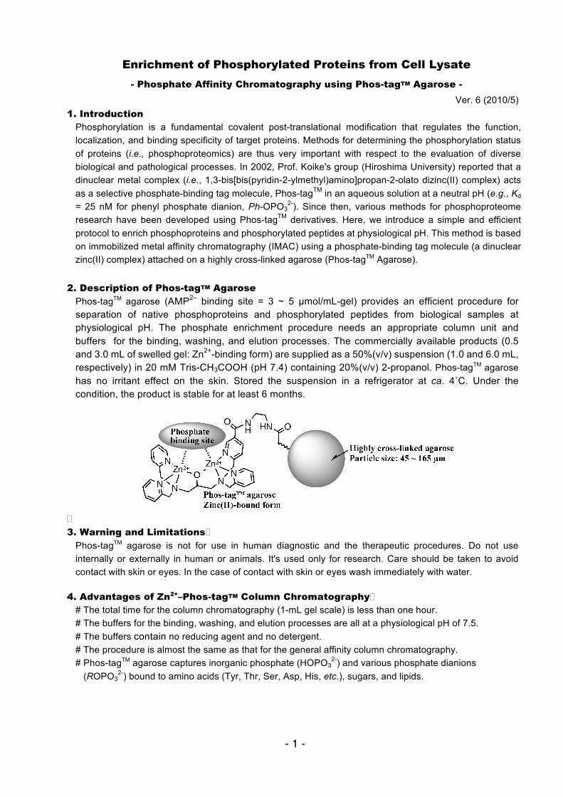
2. Description of Phos-tagTM Agarose
Phos-tagTM agarose (AMP2– binding site = 3 ~ 5 µmol/mL-gel) provides an efficient procedure for separation of native phosphoproteins and phosphorylated peptides from biological samples at physiological pH. The phosphate enrichment procedure needs an appropriate column unit and buffers for the binding, washing, and elution processes. The commercially available products (0.5 and 3.0 mL of swelled gel: Zn2+-binding form) are supplied as a 50%(v/v) suspension (1.0 and 6.0 mL, respectively) in 20 mM Tris-CH3COOH (pH 7.4) containing 20%(v/v) 2-propanol. Phos-tagTM agarose has no irritant effect on the skin. Stored the suspension in a refrigerator at ca. 4˚C. Under the condition, the product is stable for at least 6 months.
3. Warning and Limitations
Phos-tagTM agarose is not for use in human diagnostic and the therapeutic procedures. Do not use internally or externally in human or animals. It's used only for research. Care should be taken to avoid contact with skin or eyes. In the case of contact with skin or eyes wash immediately with water.
4. Advantages of Zn2+–Phos-tagTM Column Chromatography
# The total time for the column chromatography (1-mL gel scale) is less than one hour. # The buffers for the binding, washing, and elution processes are all at a physiological pH of 7.5. # The buffers contain no reducing agent and no detergent. # The procedure is almost the same as that for the general affinity column chromatography. # Phos-tagTM agarose captures inorganic phosphate (HOPO3
2-) and various phosphate dianions (ROPO3
2-) bound to amino acids (Tyr, Thr, Ser, Asp, His, etc.), sugars, and lipids.
- 2 -
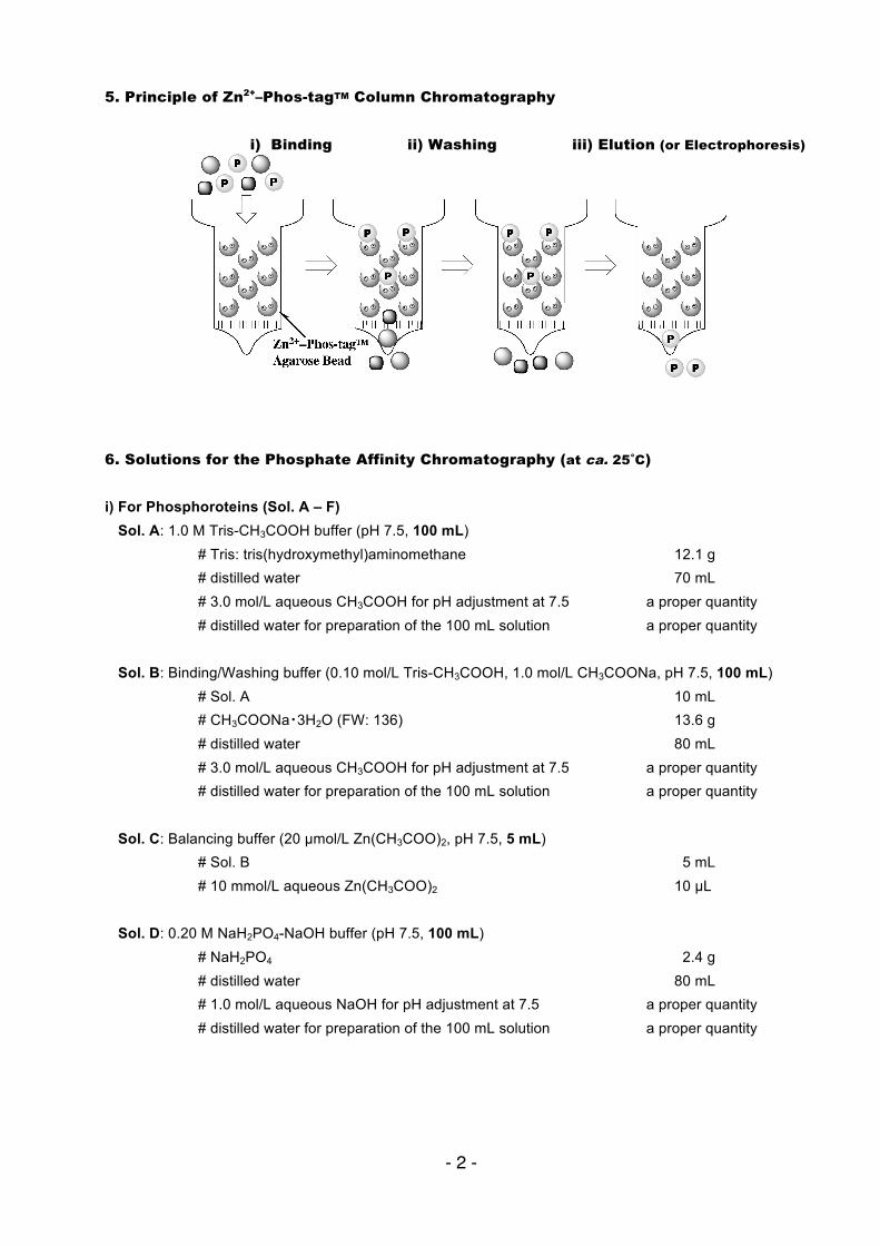
5. Principle of Zn2+–Phos-tagTM Column Chromatography
i) Binding ii) Washing iii) Elution (or Electrophoresis)
6. Solutions for the Phosphate Affinity Chromatography (at ca. 25˚C)
i) For Phosphoroteins (Sol. A – F)
Sol. A: 1.0 M Tris-CH3COOH buffer (pH 7.5, 100 mL) # Tris: tris(hydroxymethyl)aminomethane 12.1 g # distilled water 70 mL # 3.0 mol/L aqueous CH3COOH for pH adjustment at 7.5 a proper quantity # distilled water for preparation of the 100 mL solution a proper quantity Sol. B: Binding/Washing buffer (0.10 mol/L Tris-CH3COOH, 1.0 mol/L CH3COONa, pH 7.5, 100 mL) # Sol. A 10 mL # CH3COONa・3H2O (FW: 136) 13.6 g # distilled water 80 mL # 3.0 mol/L aqueous CH3COOH for pH adjustment at 7.5 a proper quantity # distilled water for preparation of the 100 mL solution a proper quantity Sol. C: Balancing buffer (20 µmol/L Zn(CH3COO)2, pH 7.5, 5 mL) # Sol. B 5 mL # 10 mmol/L aqueous Zn(CH3COO)2 10 µL Sol. D: 0.20 M NaH2PO4-NaOH buffer (pH 7.5, 100 mL) # NaH2PO4 2.4 g # distilled water 80 mL # 1.0 mol/L aqueous NaOH for pH adjustment at 7.5 a proper quantity # distilled water for preparation of the 100 mL solution a proper quantity
- 3 -
Sol. E: Elution buffer (0.10 mol/L Tris-CH3COOH, 1.0 mol/L NaCl, 10 mmol/L NaH2PO4-NaOH, pH 7.5, 50 mL)
# Sol. A 5.0 mL # NaCl 2.9 g # distilled water 38 mL # Sol. D 2.5 mL # 3 mol/L aqueous CH3COOH for pH adjustment at 7.5 a proper quantity # distilled water for preparation of the 50 mL solution a proper quantity Sol. F: Appropriate lysis buffer e.g., RIPA buffer (a radio-immunoprecipitation assay lysis buffer) 50 mmol/L Tris-HCl (pH 7.4) containing 0.15 mol/L NaCl, 0.25%(w/v) sodium deoxycholate, 1.0%(v/v) Nonidet P-40, 1.0 mmol/L EDTA, 1.0 mmol/L phenylmethanesulfonyl fluoride, 1 µg/mL Aprotinin, 1 µg/mL Leupeptin, 1 µg/mL Pepstatin, 1.0 mmol/L Na3VO4, and 1.0 mmol/L NaF
Do not use excess amount of a zinc(II)-chelating agent (e.g., 5 mM EDTA) and/or phosphate derivative (e.g., inorganic phosphate and nucleotides) in the sample.
ii) For Phosphorylated Peptides (Sol. G – J)
Sol. G: 10 mmol/L MES-NaOH, 0.10 mol/L NaCl, 5.0 mmol/L Na2C2O4 (pH 6.0, 100 mL) # MES: 2-(N-morpholino)ethanesulfonic acid (FW: 195) 2.0 g # NaCl (FW: 58.4) 0.58 g # Na2C2O4 (sodium oxalate, FW: 134) 67 mg # distilled water 100 mL # 0.10 mol/L aqueous NaOH for pH adjustment at 6.0 a proper quantity # distilled water for preparation of the 100 mL solution a proper quantity
Sol. H: Binding/Washing buffer (10 mL) # Sol. G 10 ~ 5 mL # 0 ~ 50%(v/v) CH3CN 0 ~ 5 mL
The optimum ratio of CH3CN should be determined for each phosphorylated peptide.
Sol. I: Balancing buffer (5 mL) # Sol. H 5 mL # 10 mmol/l aqueous Zn(CH3COO)2 10 µL
Sol. J: Elution buffers (5 mL)): Please select an appropriate buffer from the follows.
No.1:0.20 mol/L Na2HPO4-NaOH (pH 7) No.2:5%(w/v) aqueous NH3 (1.0 mL of 28%(w/w) aqueous NH3 + 4.0 mL of distilled water) No.3:1%(v/v) H3PO4 (50 µL of H3PO4 + 4.95 mL of distilled water)
Addition of 10 ~ 50%(v/v) CH3CN would increase the recovery of phosphorylated peptides. No.2 is well suited for the MALDI TOF mass analysis.
- 4 -
7. Preparation of Sample (Lysed proteins from A431 human epidermoid carcinoma cells)
1) Remove the culture medium for the cells (7 × 10⁶) on a 100-mm culture plate. 2) Rinse the cells with 20 mmol/L Tris-HCl (pH 7.6) buffer containing 138 mmol/L NaCl.
Do not use a phosphate-containing buffer. 3) The cells are exposed to 0.30 mL of a cold RIPA buffer (Sol. F). 4) The plate is gently rocked for 15 min on ice. 5) Remove the cells from the plate using a cell scraper. 6) Transfer the resulting suspension in a microcentrifuge tube. 7) Wash the plate with 0.20 mL of a cold RIPA buffer. 8) Mix the washing solution with the first suspension in the tube. 9) Incubate the obtained solution for 60 min on ice.
10) Centrifuge the tube at 10000×g for 10 min at 4˚C. 11) The supernatant fluid would contain approximately 1 mg of solubilized proteins. 10) The quantification of the lysed protein is performed according to the Bradford method
using a protein assay kit sold commercially (e.g., Bio-Rad). 11) Adjust the concentration of the solubilized proteins to ca. 2 mg/mL with an appropriate amount of
an RIPA buffer. 12) Dilute the resulting solution (0.20 mL) with 0.80 mL of Sol. B to obtain the sample for the following
column chromatography using 1 mL Zn2+–Phos-tagTM agarose (swelled gel). After the phosphate-affinity chromatography using cell lysate, the Phos-tagTM agarose must be discarded. The column efficiency of the used gel is much smaller than that of unused one due to residual compounds in the gel.
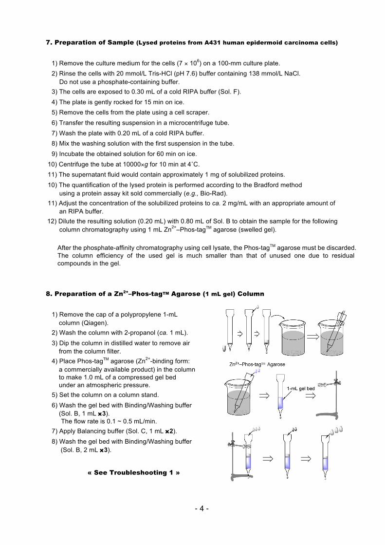
8. Preparation of a Zn2+–Phos-tagTM Agarose (1 mL gel) Column
1) Remove the cap of a polypropylene 1-mL column (Qiagen). 2) Wash the column with 2-propanol (ca. 1 mL). 3) Dip the column in distilled water to remove air from the column filter. 4) Place Phos-tagTM agarose (Zn2+-binding form: a commercially available product) in the column to make 1.0 mL of a compressed gel bed under an atmospheric pressure. 5) Set the column on a column stand. 6) Wash the gel bed with Binding/Washing buffer (Sol. B, 1 mL ×3).
The flow rate is 0.1 ~ 0.5 mL/min. 7) Apply Balancing buffer (Sol. C, 1 mL ×2). 8) Wash the gel bed with Binding/Washing buffer (Sol. B, 2 mL ×3).
« See Troubleshooting 1 »
- 5 -
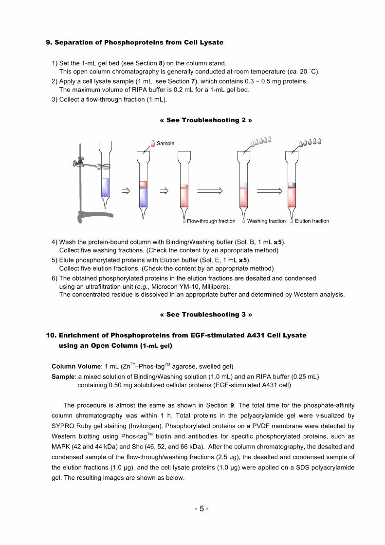
9. Separation of Phosphoproteins from Cell Lysate 1) Set the 1-mL gel bed (see Section 8) on the column stand.
This open column chromatography is generally conducted at room temperature (ca. 20 ˚C). 2) Apply a cell lysate sample (1 mL, see Section 7), which contains 0.3 ~ 0.5 mg proteins.
The maximum volume of RIPA buffer is 0.2 mL for a 1-mL gel bed. 3) Collect a flow-through fraction (1 mL).
« See Troubleshooting 2 »
4) Wash the protein-bound column with Binding/Washing buffer (Sol. B, 1 mL ×5). Collect five washing fractions. (Check the content by an appropriate method)
5) Elute phosphorylated proteins with Elution buffer (Sol. E, 1 mL ×5). Collect five elution fractions. (Check the content by an appropriate method)
6) The obtained phosphorylated proteins in the elution fractions are desalted and condensed using an ultrafiltration unit (e.g., Microcon YM-10, Millipore). The concentrated residue is dissolved in an appropriate buffer and determined by Western analysis.
« See Troubleshooting 3 »
10. Enrichment of Phosphoproteins from EGF-stimulated A431 Cell Lysate
using an Open Column (1-mL gel)
Column Volume: 1 mL (Zn2+–Phos-tagTM agarose, swelled gel) Sample: a mixed solution of Binding/Washing solution (1.0 mL) and an RIPA buffer (0.25 mL)
containing 0.50 mg solubilized cellular proteins (EGF-stimulated A431 cell)
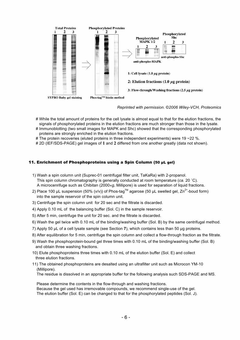
The procedure is almost the same as shown in Section 9. The total time for the phosphate-affinity column chromatography was within 1 h. Total proteins in the polyacrylamide gel were visualized by SYPRO Ruby gel staining (Invitorgen). Phsophorylated proteins on a PVDF membrane were detected by Western blotting using Phos-tagTM biotin and antibodies for specific phosphorylated proteins, such as MAPK (42 and 44 kDa) and Shc (46, 52, and 66 kDa). After the column chromatography, the desalted and condensed sample of the flow-through/washing fractions (2.5 µg), the desalted and condensed sample of the elution fractions (1.0 µg), and the cell lysate proteins (1.0 µg) were applied on a SDS polyacrylamide gel. The resulting images are shown as below.
Flow-through fraction Washing fraction Elution fraction
Sample
- 6 -
Reprinted with permission. ©2006 Wiley-VCH, Proteomics
# While the total amount of proteins for the cell lysate is almost equal to that for the elution fractions, the signals of phosphorylated proteins in the elution fractions are much stronger than those in the lysate. # Immunoblotting (two small images for MAPK and Shc) showed that the corresponding phosphorylated proteins are strongly enriched in the elution fractions. # The protein recoveries (eluted proteins in three independent experiments) were 19 ~22 %. # 2D (IEF/SDS-PAGE) gel images of 1 and 2 differed from one another greatly (data not shown).
11. Enrichment of Phosphoproteins using a Spin Column (50 µL gel)
1) Wash a spin column unit (Suprec-01 centrifugal filter unit, TaKaRa) with 2-propanol.
This spin column chromatography is generally conducted at room temperature (ca. 20 ˚C). A microcenrifuge such as Chibitan (2000×g, Millipore) is used for separation of liquid fractions.
2) Place 100 µL suspension (50% (v/v)) of Phos-tagTM agarose (50 µL swelled gel, Zn2+-boud form) into the sample reservoir of the spin column unit. 3) Centrifuge the spin column unit for 20 sec and the filtrate is discarded. 4) Apply 0.10 mL of the balancing buffer (Sol. C) in the sample reservoir. 5) After 5 min, centrifuge the unit for 20 sec. and the filtrate is discarded. 6) Wash the gel twice with 0.10 mL of the binding/washing buffer (Sol. B) by the same centrifugal method. 7) Apply 50 µL of a cell lysate sample (see Section 7), which contains less than 50 µg proteins. 8) After equilibration for 5 min, centrifuge the spin column and collect a flow-through fraction as the filtrate. 9) Wash the phosphoprotein-bound gel three times with 0.10 mL of the binding/washing buffer (Sol. B) and obtain three washing fractions. 10) Elute phosphoproteins three times with 0.10 mL of the elution buffer (Sol. E) and collect three elution fractions. 11) The obtained phosphoproteins are desalted using an ultrafilter unit such as Microcon YM-10 (Millipore). The residue is dissolved in an appropriate buffer for the following analysis such SDS-PAGE and MS. Please determine the contents in the flow-through and washing fractions. Because the gel used has irremovable compounds, we recommend single-use of the gel. The elution buffer (Sol. E) can be changed to that for the phosphorylated peptides (Sol. J).
- 7 -
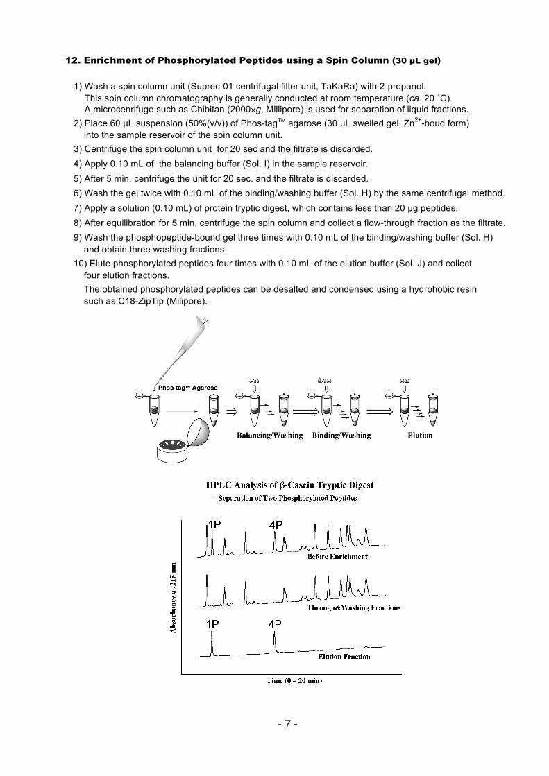
12. Enrichment of Phosphorylated Peptides using a Spin Column (30 µL gel) 1) Wash a spin column unit (Suprec-01 centrifugal filter unit, TaKaRa) with 2-propanol.
This spin column chromatography is generally conducted at room temperature (ca. 20 ˚C). A microcenrifuge such as Chibitan (2000×g, Millipore) is used for separation of liquid fractions.
2) Place 60 µL suspension (50%(v/v)) of Phos-tagTM agarose (30 µL swelled gel, Zn2+-boud form) into the sample reservoir of the spin column unit. 3) Centrifuge the spin column unit for 20 sec and the filtrate is discarded. 4) Apply 0.10 mL of the balancing buffer (Sol. I) in the sample reservoir. 5) After 5 min, centrifuge the unit for 20 sec. and the filtrate is discarded. 6) Wash the gel twice with 0.10 mL of the binding/washing buffer (Sol. H) by the same centrifugal method. 7) Apply a solution (0.10 mL) of protein tryptic digest, which contains less than 20 µg peptides. 8) After equilibration for 5 min, centrifuge the spin column and collect a flow-through fraction as the filtrate. 9) Wash the phosphopeptide-bound gel three times with 0.10 mL of the binding/washing buffer (Sol. H) and obtain three washing fractions. 10) Elute phosphorylated peptides four times with 0.10 mL of the elution buffer (Sol. J) and collect four elution fractions. The obtained phosphorylated peptides can be desalted and condensed using a hydrohobic resin such as C18-ZipTip (Milipore).
- 8 -
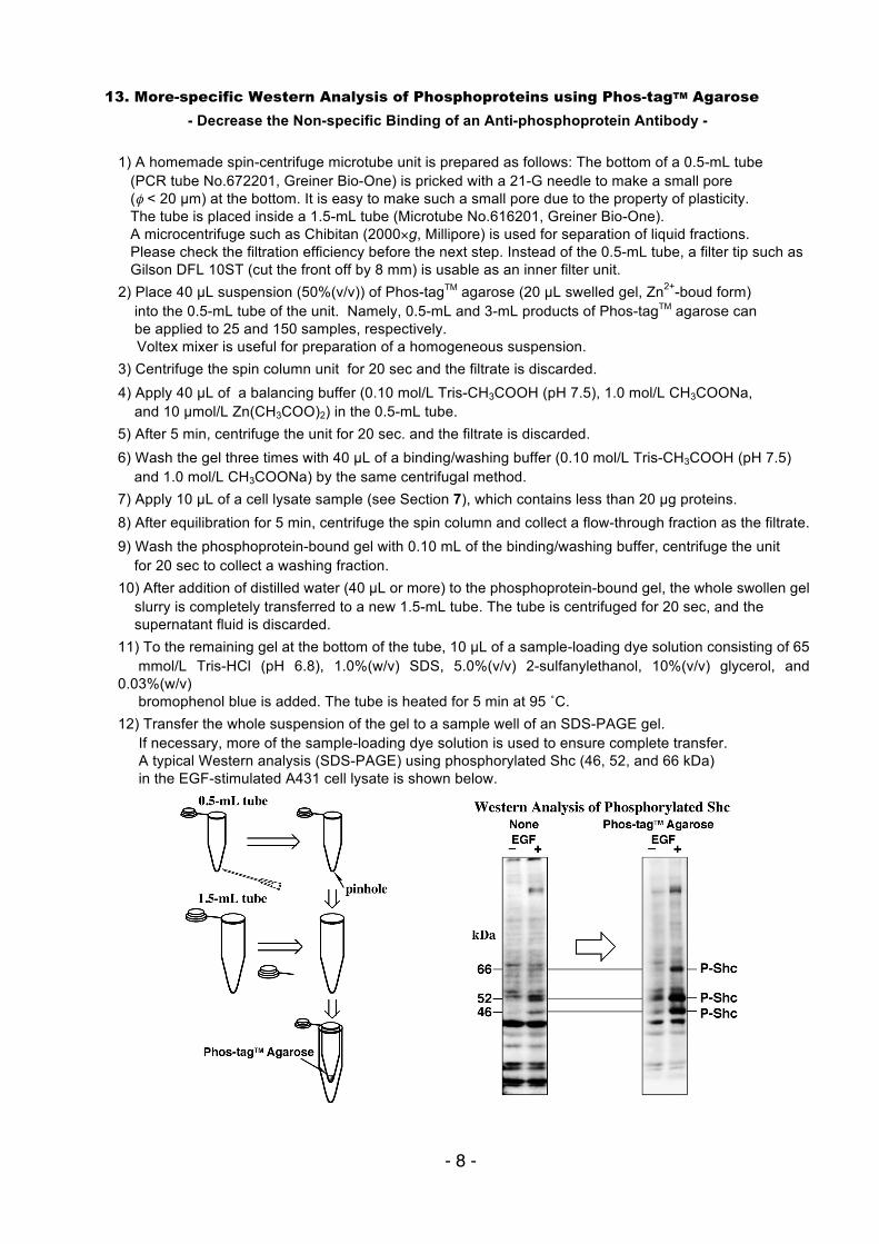
13. More-specific Western Analysis of Phosphoproteins using Phos-tagTM Agarose
- Decrease the Non-specific Binding of an Anti-phosphoprotein Antibody - 1) A homemade spin-centrifuge microtube unit is prepared as follows: The bottom of a 0.5-mL tube (PCR tube No.672201, Greiner Bio-One) is pricked with a 21-G needle to make a small pore (φ < 20 µm) at the bottom. It is easy to make such a small pore due to the property of plasticity. The tube is placed inside a 1.5-mL tube (Microtube No.616201, Greiner Bio-One). A microcentrifuge such as Chibitan (2000×g, Millipore) is used for separation of liquid fractions. Please check the filtration efficiency before the next step. Instead of the 0.5-mL tube, a filter tip such as Gilson DFL 10ST (cut the front off by 8 mm) is usable as an inner filter unit. 2) Place 40 µL suspension (50%(v/v)) of Phos-tagTM agarose (20 µL swelled gel, Zn2+-boud form) into the 0.5-mL tube of the unit. Namely, 0.5-mL and 3-mL products of Phos-tagTM agarose can be applied to 25 and 150 samples, respectively. Voltex mixer is useful for preparation of a homogeneous suspension. 3) Centrifuge the spin column unit for 20 sec and the filtrate is discarded. 4) Apply 40 µL of a balancing buffer (0.10 mol/L Tris-CH3COOH (pH 7.5), 1.0 mol/L CH3COONa, and 10 µmol/L Zn(CH3COO)2) in the 0.5-mL tube. 5) After 5 min, centrifuge the unit for 20 sec. and the filtrate is discarded. 6) Wash the gel three times with 40 µL of a binding/washing buffer (0.10 mol/L Tris-CH3COOH (pH 7.5) and 1.0 mol/L CH3COONa) by the same centrifugal method. 7) Apply 10 µL of a cell lysate sample (see Section 7), which contains less than 20 µg proteins. 8) After equilibration for 5 min, centrifuge the spin column and collect a flow-through fraction as the filtrate. 9) Wash the phosphoprotein-bound gel with 0.10 mL of the binding/washing buffer, centrifuge the unit for 20 sec to collect a washing fraction. 10) After addition of distilled water (40 µL or more) to the phosphoprotein-bound gel, the whole swollen gel slurry is completely transferred to a new 1.5-mL tube. The tube is centrifuged for 20 sec, and the supernatant fluid is discarded. 11) To the remaining gel at the bottom of the tube, 10 µL of a sample-loading dye solution consisting of 65 mmol/L Tris-HCl (pH 6.8), 1.0%(w/v) SDS, 5.0%(v/v) 2-sulfanylethanol, 10%(v/v) glycerol, and 0.03%(w/v) bromophenol blue is added. The tube is heated for 5 min at 95 ˚C. 12) Transfer the whole suspension of the gel to a sample well of an SDS-PAGE gel. If necessary, more of the sample-loading dye solution is used to ensure complete transfer. A typical Western analysis (SDS-PAGE) using phosphorylated Shc (46, 52, and 66 kDa) in the EGF-stimulated A431 cell lysate is shown below.
- 9 -
« Troubleshooting 1 » While the volume of the Phos-tagTM agarose gel bed is fixed at 1 mL in this protocol, it can be easily optimized in proportion to the amount of the sample. Before the pH measurement for the buffer solutions, the pH-electrode system should be calibrated using the two pH buffer solutions (e.g., pH 4 and 7). « Troubleshooting 2 » When a larger amount of the lysate proteins (e.g., 0.60 mg of A431 cell's proteins in 0.25 mL RIPA buffer) were applied on the 1-mL gel bed, some of the phosphoproteins were eluted in the flow-through and washing fractions. A similar leak into the fractions resulted from the use of twice the volume of an RIPA buffer (i.e., 0.50 mL, 0.50 mg lysate proteins) and the 1-mL gel bed, which may be attributed to the competitive binding of HOVO3
2– or zinc(II)-elimination by EDTA. « Troubleshooting 3 » Some proteins adsorb on the centrifugal filter unit. Please estimate the recovery of the eluted proteins by a protein quantification method (e.g., Bradford method).
References on Phos-tagTM Chemistry
・Matrix-assisted laser desorption/ionization time-of-flight mass spectrometry of phosphorylated compounds using a novel phosphate capture molecule, Rapid Communications of Mass Spectrometry, 17, 2075-2081 (2003), H. Takeda, A. Kawasaki, M. Takahashi, A. Yamada, and T. Koike ・Recognition of phosphate monoester dianion by an alkoxide-bridged dinuclear zinc(II) complex, Dalton Transactions, 1189-1193 (2004), E. Kinoshita, M. Takahashi, H. Takeda, M. Shiro, and T. Koike ・Quantitative analysis of lysophosphatidic acid by time-of-flight mass spectrometry using a phosphate capture
molecule, Journal of Lipid Research, 45, 2145-2150 (2004), T. Tanaka, H. Tsutsui, K. Hirano, T. Koike, A. Tokumura, and K. Satouchi ・Production of 1,2-Didocosahexaenoyl Phosphatidylcholine by Bonito Muscle
Lysophosphatidylcholine/Transacylase, Journal of Biochemistry, 136, 477-483 (2004), K. Hirano, H. Matsui, T. Tanaka, F. Matsuura, K. Satouchi, and T. Koike
・Novel immobilized zinc(II) affinity chromatography for phosphopeptides and phosphorylated proteins, Journal of Separation Science, 28, 155-162 (2005), E. Kinoshita, A. Yamada, H. Takeda, E. Kinoshita-Kikuta, and T. Koike ・ Detection and Quantification of On-Chip Phosphorylated Peptides by Surface Plasmon Resonance Imaging Techniques Using a Phosphate Capture Molecule, Analytical Chemistry, 77, 3979-3985 (2005), K. Inamori, M. Kyo, Y. Nishiya, Y. Inoue, T. Sonoda, E. Kinoshita, T. Koike, and Y. Katayama ・Phosphate-binding tag: A new tool to visualize phosphorylated proteins, Molecular & Cellular Proteomics, 5, 749-757 (2006), E. Kinoshita, E. Kinoshita-Kikuta, K. Takiyama, and T. Koike ・Enrichment of phosphorylated proteins from cell lysate using phosphate-affinity chromatography at physiological pH, Proteomics, 6, 5088-5095 (2006), E. Kinoshita-Kikuta, E. Kinoshita, A. Yamada, M. Endo, and T. Koike ・Separation of a phosphorylated histidine protein using phosphate affinity polyacrylamide gel electrophoresis, Analytical Biochemistry, 360, 160-162 (2007), S. Yamada, H. Nakamura, E. Kinoshita, E. Kinoshita-Kikuta, T.
Koike, and Y. Shiro ・Label-free kinase profiling using phosphate-affinity polyacrylamide gel electrophresis, Molecular & Cellular Proteomics, 6, 356-366 (2007), E. Kinoshita-Kikuta, Y. Aoki, E. Kinoshita, and T. Koike ・A SNP genotyping method using phosphate-affinity polyacrylamide gel electrophoresis, Analytical Biochemistry, 361, 294-298 (2007), E. Kinoshita, E. Kinoshita-Kikuta, and T. Koike (The phosphate group at DNA-terminal is efficiently captured by Zn2+–Phos-tag.)
- 10 -
・Identification on Membrane and Characterization of Phosphoproteins Using an Alkoxide-Bridged Dinuclear Metal
Complex as a Phosphate-Binding Tag Molecule Journal of Biomolecular Techniques, 18, 278-286 (2007), T. Nakanishi, E. Ando, M. Furuta, E. Kinoshita, E. Kikuta-Kinoshita, T. Koike, S. Tsunasawa, and O. Nishimura ・A mobility shift detection method for DNA methylation analysis using phosphate affinity polyacrylamide gel
electrophoresis, Analytical Biochemistry, 378, 102-104 (2008), E. Kinoshita-Kikuta, E. Kinoshita, and T. Koike ・Separation of phosphoprotein isotypes having the same number of phosphate groups using phosphate-affinity
SDS-PAGE, Proteomics, 8, 2994-3003 (2008), E. Kinoshita, E. Kinoshita-Kikuta, M. Matsubara, S. Yamada, H. Nakamura, Y. Shiro, Y. Aoki, K. Okita, and T. Koike ・FANCI phosphorylation functions as a molecular switch to turn on the Fanconi anemia pathway Nature Structural & Molecular Biology, 15, 1138-1146 (2008) M. Ishiai, H. Kitao, A. Smogorzewska, J. Tomida, A. Kinomura, E. Uchida, A. Saberi, E. Kinoshita, E. Kinoshita-Kikuta, T. Koike, S. Tashiro, S. J. Elledge, and M. Takata ・Two-dimensional phosphate affinity gel electrophoresis for the analysis of phosphoprotein isotypes Electrophoresis, 30, 550-559 (2009), E. Kinoshita, E. Kinoshita-Kikuta, M. Matsubara, Y. Aoki, S. Ohie, Y. Mouri, and T. Koike ・Formation of lysophosphatidic acid, a wound-healing lipid, during digestion of cabbage leaves Bioscience, Biotechnology, and Biochemistry, in press T. Tanaka, G. Horiuchi, M. Matsuoka, K. Hirano, A. Tokumura, T. Koike, and K. Satouchi ・A Phos-tag-based fluorescence resonance energy transfer system for the analysis of the dephosphorylation of phosphopeptides, Analytical Biochemistry, 388, 235-241, (2009) K. Takiyama, E. Kinoshita, E. Kinoshita-Kikuta, Y. Fujioka, Y. Kubo, and T. Koike ・Phos-tag beads as an immunoblotting enhancer for selective detection of phosphoproteins in cell lysates Analytical Biochemistry, 389, 83-85, (2009) E. Kinoshita-Kikuta, E. Kinoshita, and T. Koike ・Mobility shift detection of phosphorylation on large proteins using a Phos-tag SDS-PAGE gel strengthened with agarose, Proteomics, 9, 4098- 4101 (2009) E. Kinoshita, E. Kinoshita-Kikuta, H. Ujihara, and T. Koike ・Separation and detection of large phosphoproteins using Phos-tag SDS-PAGE, Nature Protocols, 4, 1513-1521 (2009), E. Kinoshita, E. Kinoshita-Kikuta, and T. Koike ・A clean-up technology for the simultaneous determination of lysophosphatidic acid and sphingosine-1-phosphate by matrix-assisted laser desorption/ionization time-of-flight mass spectrometry using a phosphate-capture molecule, Phos-tag, Rapid Communications in Mass Spectrometry, 24, 1075-1084 (2010) J. Morishige, M. Urikura, H. Takagi, K. Hirano, T. Koike, T. Tanaka, and K. Satouchi ・Genotyping and mapping assay of single-nucleotide polymorphisms in CYP3A5 using DNA-binding zinc(II) complexes, Clinical Biochemistry, 43, 302-306 (2010), E. Kinoshita, E. Kinoshita-Kikuta, H. Nakashima, and T. Koike ・The DNA-binding activity of mouse DNA methyltransferase 1 is ragulated phosphorylation with casein kinase 1σ/ε, Biochemical Journal, 427, 489-497 (2010) Y. Sugiyama, N. Hatano, N. Sueyoshi, I. Suetake, S. Tajima, E. Kinoshita, E. Kinoshita-Kikuta, T. Koike, and I. Kameshita
Edited by Phos-tag Consortium