diagnostics for quarantine plant pathogenic … · diagnostics for quarantine plant pathogenic...
TRANSCRIPT
Faculty of Sciences
Department of Biochemistry and Microbiology
Laboratory of Microbiology
Diagnostics for quarantine plant pathogenic Clavibacter and relatives:
molecular characterization, identification and epidemiological studies
Joanna Załuga
Promoter: Prof. Dr. Paul De Vos
Co-promoter: Prof. Dr. Martine Maes
Dissertation submitted in fulfillment of the requirements for the degree of
Doctor (Ph.D.) in Sciences, Biotechnology
ii
Joanna Załuga – Diagnostics for quarantine plant pathogenic Clavibacter and relatives: molecular
characterization, identification and epidemiological studies
Copyright ©2013 Joanna Załuga
ISBN-number:
No part of this thesis protected by its copyright notice may be reproduced or utilized in any form, or
by any means, electronic or mechanical, including photocopying, recording or by any information
storage or retrieval system without written permission of the author and promoters.
Printed by University Press | www.universitypress.be
Ph.D. thesis, Faculty of Sciences, Ghent University, Ghent, Belgium.
This Ph.D. work was financially supported by a QBOL (Quarantine Barcoding Of Life) project KBBE-
2008-1-4-01 nr 226482 funded under the Seventh Framework Program (FP 7) of the European
Union.
Publicly defended in Ghent, Belgium, October 11th, 2013
iii
Examination Committee
Prof. Dr. Savvas Savvides (Chairman)
L-Probe: Laboratory for protein Biochemistry and Biomolecular Engineering
Faculty of Sciences, Ghent University, Belgium
Prof. Dr. Paul De Vos (Promoter)
LM-UGent: Laboratory of Microbiology, Faculty of Science, Ghent University, Belgium
BCCM-LMG Bacteria Collection, Ghent, Belgium
Prof. Dr. Martine Maes (Co-promoter)
Plant Sciences Unit – Crop Protection
Institute for Agricultural and Fisheries Research (ILVO), Merelbeke, Belgium
Dr. Peter Bonants
Plant Research International (PRI)
Business Unit Biointeractions & Plant Health, Wageningen, The Netherlands
Ir. Johan Van Vaerenbergh Plant Sciences Unit – Crop Protection
Institute for Agricultural and Fisheries Research (ILVO), Merelbeke, Belgium
Dr. Pieter Stragier
Laboratory for Microbiology
Faculty of Sciences, Ghent University, Belgium
Prof. Dr. Anne Willems Laboratory for Microbiology
Faculty of Sciences, Ghent University, Belgium
Prof. Dr. Monica Höfte Laboratory of Phytopathology, Department of Crop Protection
Faculty of Bioscience Engineering, Ghent University, Belgium
Prof. Dr. Peter Dawyndt
Department of Applied Mathematics, Computer Science and Statistics
PC-Lab of the Faculty of Science, Ghent University, Belgium

v
Acknowledgements-Podziękowania
To start I wish to thank prof. Dr. Paul De Vos who gave me the opportunity to carry out the PhD in
the Laboratory of Microbiology. Your guidance, thoughtful insights and support were very important
at every stage of my work. I am thankful that I could count on you, especially during the last few,
very intensive months of my PhD. Your encouragement, advices and suggestions were invaluable for
completing this work.
I am grateful to the examination committee for their helpful suggestions, constructive remarks and
fruitful discussions that helped me to improve my thesis. Prof. Dr. Martine Maes, thank you for your
advice and comments on my publications and thesis. Dr. Peter Bonants, Prof. dr. Monica Höfte, Ir.
Johan Van Vaerenbergh and dr. Pieter Stragier, thank you very much for taking your time to read my
PhD and for your positive feedback.
Kim, although you supervised me only at the beginning it was the most important time for me and I
am thankful that I could work with you. Your wide knowledge and scientific input greatly influenced
my first steps in the exciting scientific world. Writing the first article is never easy but with your
guidance and support it was one of the most important and unforgettable experiences for me. I am
grateful not only for your great scientific supervision and what you taught me during these four
years but also for being a great colleague!
Dear Johan, I would like to thank you very, very much for these four years. It was a great experience
to work with you. I am grateful for showing me the real diagnostic lab, for all our discussions and for
a wonderful time in ILVO.
My dearest colleagues, Sven, Ines, Jonas and Bram, I spent with you the best time in the lab. You
were the best company I could imagine to have during my PhD. Our coffee breaks discussions,
Friday lunches, common conferences, meetings and practical exercises, I could not think of anyone
better to share these great moments with. Sven, our philosopher and discussion man, thanks for all
our chats and laughs and for being a super friend. Jonas, our potato/dj man, the most optimistic
person I know, it was great to have someone so positive in the office. Ines, my best desk mate,
thanks for your help at the beginning, for all the translations from Dutch, your support and the great
moments we had together. Bram, the youngest in our team, hopping around, never tired and always
full of ideas. Thanks for all the fun we had in the PDV group.
Margo, you are an amazing person who has so much positive energy and who is always ready to
help others. I will never forget your support and help, especially at the beginning of my adventure in
LM-UGent and during my Erasmus exchange. Thank you for everything!
Bart, Liesbeth, Joke, Evie, thanks for always answering my questions and for assistance in
performing experiments in the lab. I would like to thank all colleagues from the Laboratory of
Microbiology and from ILVO for creating a friendly atmosphere and for your enthusiastic support. It
was a pleasure to work in such a nice environment.

vi
I cannot forget my best friend Kamila who shared with me good and bad moments, who always was
there when I needed her. Without you I would never be where I am now. I still remember our first
days in Ghent when we started together. New places, new people, new challenges and even though
it was tough we did it! Dzięki za wszystko moja najlepsza koleżanko!
My friends, Kasie, Agi, Asie, Anie, Radek, Agata, Mati, Linda, Giacomo, Edyta, Marta, Lidka and many
others I have always a wonderful time with you. Our parties, Friday drinks and chats are
unforgettable moments. I am happy I met such great people!
Moi Rodzice, Aga i Paweł, dziękuję Wam za dodawanie mi otuchy i wiarę we mnie. Dziękuję za to, że
zawsze ciepło mnie witaliście kiedy wracałam do rodzinnego domu. Mamo, tato, wiem, że przyjazd
do Belgii to dla Was duże przeżycie, tym bardziej dziękuję za to, że sie zdecydowaliście.
Ik wil graag de familie van Stan bedanken, vooral Annick en Filip, ik heb veel steun van jullie
gekregen. Jullie hebben me heel warm ontvangen. Ik dank jullie hartelijk voor jullie gastvrijheid,
interesse en vriendschap.
Stan, moje Kochanie, I want to thank you for your support, understanding and for being there for
me. I am really happy that I met you that evening at Brahim’s. You always believed in me and tried
to encourage me during hard times. Dziękuję for all the wonderful days we had, all the great
moments we have now and I am looking forward to the future with you!
Merci!
Bedankt!
Dziękuję!
Thank you!
vii
List of abbreviations
ACN Acetonitrile ATCC American Type Culture Collection BCCM Belgian Coordinated Collections of Micro-organisms BOX-PCR BOX-A1R-based repetitive extragenic palindromic-PCR BLAST Basic Local Alignment Search Tool CDS Coding DNA Sequence CFBP Collection Française de Bactéries associées aux Plantes CHCA α-cyano-4-hydroxycinnamic acid Cmi Clavibacter michiganensis subsp. insidiosus Cmm Clavibacter michiganensis subsp. michiganensis Cmn Clavibacter michiganensis subsp. nebraskensis Cms Clavibacter michiganensis subsp. sepedonicus Cmt Clavibacter michiganensis subsp. tessellarius CMR Comprehensive Microbial Resource CRISPR Clustered Regularly Interspaced Short Palindromic Repeats CWDE Cell Wall Degrading Enzyme dpi days post inoculation DSMZ Deutsche Sammlung von Mikroorganismen und Zellkulturen EDGAR Efficient Database framework for comparative Genome Analyses using
BLAST score Ratios EDTA Ethylene Diamine Tetraacetic Acid ELISA Enzyme-Linked Immunosorbent Assay EPPO European and Mediterranean Plant Protection Organization EPS Extracellular Polysaccharide ERIC-PCR Enterobacterial Repetitive Intergenic Consensus-PCR EU European Union FAME Fatty Acid Methyl Esters FAO Food and Agriculture Organization of the United Nations HGDI Hunter-Gaston Diversity Index HGT Horizontal Gene Transfer HR Hypersensitive Response ILVO Instituut voor Landbouw- en VisserijOnderzoek IS Insertion Sequence IS-LM-PCR Insertion Sequence Ligation-Mediated PCR ISF International Seed Federation
ISSR Inter-Simple Sequence Repeat ITS Internal Transcribed Spacer region LMG Laboratory for Microbiology Ghent MALDI-TOF MS Matrix-Assisted Laser Desorption Ionization-Time Of Flight Mass
Spectrometry MCL Maximum Composite Likelihood model MEGA Molecular Evolutionary Genetics Analysis software ML Maximum Likelihood MLST Multi Locus Sequence Typing MLVA Multilocus Variable-Number Tandem Repeats Analysis
viii
MP Mate-Paired MP Maximum Parsimony MST Minimum Spanning Tree MTNA Mannitol, Trimethoprim, Nalidixic acid, Amphotericin medium NCBI National Centre for Biotechnology Information NCPPB National Collection of Plant Pathogenic Bacteria NJ Neighbor-Joining OEPP Organisation Européenne et Méditerranéenne pour la Protection des
Plantes PAI Pathogenicity Island PAMPs Pathogen-Associated Molecular Patterns PCR Polymerase Chain Reaction NVWA Bacterial collection (Wageningen, The Netherlands) PDDCC Plant Diseases Division Culture Collection PE Paired-Ended PFGE Pulsed-Field Gel Electrophoresis QBOL Quarantine Barcoding Of Life RAPD Random Amplification of Polymorphic DNA RAST Rapid Annotations using Subsystems Technology RH Relative Humidity rep-PCR Repetitive Extragenic Palindromic PCR RMIDb Rapid Microorganism Identification Database ROS Reactive Oxygen Species SNPs Single Nucleotide Polymorphisms TR Tandem Repeat UPGMA Unweighted Pair Group Method with Arithmetic mean VBNC Viable But Non-Culturable V-DICE VNTR diversity and confidence extractor software VNTR Variable Number Tandem Repeat

vii
Table of contents
Examination Committee ................................................................................................................... iii
Acknowledgements-Podziękowania ..................................................................................................v
List of abbreviations ......................................................................................................................... vii
Part I .................................................................................................................................................. 1
Outline .............................................................................................................................................. 2
Background and goals ....................................................................................................................... 3
Summary of work .............................................................................................................................. 6
Samenvatting .................................................................................................................................... 9
Part II ............................................................................................................................................... 13
Literature overview ......................................................................................................................... 13
Chapter 1......................................................................................................................................... 15
1. Genus Clavibacter ....................................................................................................................... 15
1.1 Clavibacter michiganensis subsp. michiganensis-quarantine pathogen ............................ 17
1.2 Bacterial wilt and canker of tomato-quarantine disease ................................................... 18
1.2.1 Symptoms ........................................................................................................... 19
1.2.2 Source of infection, transmission and survival................................................... 21
1.2.3 Disease management and control...................................................................... 21
1.2.4 Disease importance and economic impact on tomato production ................... 23
1.3 Epidemiology of bacterial wilt and canker ......................................................................... 24
1.3.1 Methods used in molecular epidemiology of Cmm strains ................................ 25
1.4 Tomato seed production and health .................................................................................. 26
1.4.1 Cmm detection in seeds ..................................................................................... 28
1.5 Methods for Cmm identification ........................................................................................ 29
1.6 Pathogenicity and virulence in Cmm .................................................................................. 31
1.6.1 Plasmid-encoded virulence factors .................................................................... 32
1.6.2 Pathogenicity Island of Cmm .............................................................................. 33
1.7 Atypical Clavibacter strains and their influence on diagnostics ......................................... 35
1.8 Molecular interaction of Cmm with tomato ...................................................................... 36
1.9 References .......................................................................................................................... 39
viii
Part III .............................................................................................................................................. 45
Experimental work .......................................................................................................................... 45
Chapter 2 ......................................................................................................................................... 47
GyrB sequence analysis and MALDI-TOF MS as identification tools for plant pathogenic
Clavibacter ....................................................................................................................................... 47
Summary.......................................................................................................................................... 48
2.1 Introduction ........................................................................................................................ 49
2.2 Material and methods ........................................................................................................ 52
2.3 Results ................................................................................................................................. 57
2.4 Discussion............................................................................................................................ 64
2.5 General reflections .............................................................................................................. 67
2.6 References .......................................................................................................................... 80
Chapter 3 ......................................................................................................................................... 83
Genetic diversity of non-pathogenic Clavibacter strains isolated from tomato seeds ................... 83
Summary.......................................................................................................................................... 84
3.1 Introduction ........................................................................................................................ 85
3.2 Material and methods ........................................................................................................ 88
3.3 Results and discussion ........................................................................................................ 93
3.4 Conclusions ....................................................................................................................... 105
3.5 References ........................................................................................................................ 107
Chapter 4 ....................................................................................................................................... 111
Comparative genome analysis of pathogenic and non-pathogenic Clavibacter strains reveals
adaptations to their life styles ....................................................................................................... 111
Summary........................................................................................................................................ 112
4.1 Introduction ...................................................................................................................... 113
4.2 Material and methods ...................................................................................................... 117
4.3 Results and discussion ...................................................................................................... 120
4.4 Conclusions ....................................................................................................................... 143
4.5 General reflections ............................................................................................................ 146
4.6 References ........................................................................................................................ 155
ix
Chapter 5....................................................................................................................................... 183
Multilocus Variable-Number-Tandem-Repeats Analysis (MLVA) distinguishes a clonal complex of
Clavibacter michiganensis subsp. michiganensis strains isolated from recent outbreaks of
bacterial wilt and canker in Belgium ............................................................................................. 183
Summary ....................................................................................................................................... 184
5.1 Introduction ...................................................................................................................... 185
5.2 Materials and methods .................................................................................................... 189
5.4 Discussion ......................................................................................................................... 207
5.5 General reflections ........................................................................................................... 215
5.6 References ........................................................................................................................ 218
Part IV............................................................................................................................................ 221
Concluding remarks ...................................................................................................................... 222

x

1
Part I
Background, goals and summary of work
Part I - Background, goals and summary of work
2
Outline
This research was performed within the frame of the QBOL (Quarantine Barcoding of Life)
project funded under the Seventh Framework Program (FP 7) of the European Union and
aimed at the development of a new diagnostic tool using DNA barcoding to identify
quarantine organisms in support of plant health. During three years, the partners of the
project collected a number of important plant pathogenic quarantine organisms which were
DNA barcoded and which will serve in the future as reference materials. New identification
schemes for a range of important quarantine plant pathogenic bacteria, fungi,
phytoplasmas, arthropods and nematodes were developed. The outcome of the project,
including the barcode sequences together with taxonomic features and accompanied with
additional pathogen-specific characteristics, is publically available in an internet-based
database system: Q-Bank (http://www.q-bank.eu).
The thesis starts with a general introduction of the performed research, followed by the
background and goals (Part I). The literature overview (Part II, Chapter 1) presents the
current state of knowledge concerning the genus Clavibacter, including its taxonomic
position, pathogenicity factors and disease characterization, as well as the summary of
currently used molecular identification methods for plant quarantine Clavibacter
michiganensis subsp. michiganensis. The experimental set-up and results (Part III) are
described in four chapters (Chapter 2-5), concerning important aspects of the study of the
plant pathogen Clavibacter. Sections with ‘General reflections’ contain a global discussion of
the performed work with updated information and future perspectives. The last part of the
dissertation contains general conclusions (Chapter 6). The research was performed
between September 2009 and June 2013 at the Laboratory of Microbiology (LM-UGent),
Faculty of Science, Ghent University. Plant tests were carried out in the Crop Protection Unit
in ILVO (Institute for Agricultural and Fisheries Research), Merelbeke, Belgium.
Part I - Background, goals and summary of work
3
Background and goals
Plant pathogens are responsible for many important diseases. Every year plant infections
lead to substantial economic and agricultural losses worldwide. Pest and disease
management helped to double the food production in the last decades. However, each year
more than 10% of the global harvest is still lost due to plant diseases. Quarantine organisms
are especially dangerous because of the potential to induce epidemic outbreaks and to be
widespread in pathogen-free areas. As disease is a natural part of every crop production
system the question is not whether it occurs but when it occurs and how severe it will be.
Therefore, combined efforts have to be undertaken to better understand the mechanisms
and factors triggering the induction of plant diseases. This will help to minimize the scale of
diseases and their agricultural impact.
The bacterial genus Clavibacter contains various plant pathogens of agriculturally important
crops, such as tomatoes, potatoes and maize. Because of the ability to invade seeds and to
induce latent infections, as well as the severity of the induced diseases, some of them are
classified as quarantine organisms. Despite many efforts to prevent their spread and
transmission and intensified control measures new outbreaks occur every year. Limited
information concerning the epidemiology and the population structure of Clavibacter
hampers the development of more reliable and adequate identification/detection
techniques. Therefore, knowledge about the biology of the pathogens, their genetic
diversity and mechanisms of virulence, and how disease cycle relates to disease severity will
help to predict the occurrence of disease and to develop more precise and accurate
detection and identification methods.
Our research aimed at:
i) developing new and more reliable approaches for a correct identification of plant
pathogenic Clavibacter subspecies,
ii) re-evaluation of the taxonomic/phylogenetic position of the genus Clavibacter,
Part I - Background, goals and summary of work
4
iii) investigation of a new group of non-pathogenic Clavibacter strains isolated from tomato
seeds,
iv) studying the epidemiology of the bacterial wilt and canker on tomato caused by
Clavibacter michiganensis subsp. michiganensis in Belgium.
Chapter 1 includes the literature overview concerning the genus Clavibacter.
Chapter 2 presents the gyrB sequence analysis and MALDI-TOF MS as new identification
methods for members of the genus Clavibacter. A fragment of the gyrB housekeeping gene
was shown to be a good DNA barcode for the correct identification of Clavibacter
subspecies. The MALDI-TOF MS generated distinct and reproducible profiles, with unique
peaks that could be used as biomarkers for the accurate Clavibacter subspecies
identification.
Chapter 3 describes the molecular and phenotypic characterization of a new Clavibacter
group containing non-pathogenic strains isolated from tomato seeds. Pathogenicity assays
on tomato confirmed their non-pathogenic nature and in planta tests showed that they are
poorly colonizing the tomato stem. Taxonomic analysis using gyrB and dnaA genes revealed
that they formed a heterogeneous group, distinct from Cmm and other Clavibacter
subspecies. Their high genetic diversity was confirmed by the analysis of BOX-PCR
fingerprinting profiles.
Chapter 4 presents the draft genome of the non-pathogenic seed-borne Clavibacter strain
(LMG 26808). The genome analysis of LMG 26808 aimed at investigation of the presence of
virulence-related factors and other characteristics that could provide new insights into the
genetic basis that may explain its apparent lack of pathogenicity on tomato and its
adaptation to an environmental niche.
In Chapter 5 a newly developed MLVA scheme was applied to study the epidemiology of
Cmm isolated from recent outbreaks of bacterial wilt and canker on tomato in Belgium.
Results demonstrated that all strains from Belgian outbreaks, isolated between 2010 and
2012, together with two French strains from 2010 formed one monomorphic group

Part I - Background, goals and summary of work
5
suggesting that a clonal population, originating probably from contaminated seeds or
seedlings, was responsible for the disease outbreaks.
Chapter 6 is the final chapter in the thesis and describes general conclusions that emphasize
the most important findings of the performed research and summarizes future
perspectives.
Part I - Background, goals and summary of work
6
Summary of work
Bacterial pathogens are responsible for many important plant diseases. Every year,
susceptible plants infected by pathogens lead to substantial economic and agricultural
losses worldwide. Members of the species Clavibacter michiganensis are pathogenic to
several important crops, such as potatoes, tomatoes and maize. Because of the quarantine
status of these organisms, these crops are subjected to the strict and statutory control of
plant protection organizations. Despite many attempts undertaken to provide pathogen-
free certified planting material and intensified eradication processes new outbreaks of
diseases caused by these quarantine organisms still occur.
This study focused on the molecular characterization of plant pathogenic Clavibacter
subspecies. During the course of this research an important group of tomato seed-borne
Clavibacter strains was investigated. These strains showed no pathogenic effect on tomato
and could not be assigned to any of the five known subspecies.
A representative subset of all Clavibacter subspecies and closely related strains was
collected. The majority of the strains were obtained from the public BCCM/LMG bacteria
collection (LM-UGent, Belgium) and from working and research collections as GBBC (ILVO,
Merelbeke) and NVWA (The Netherlands). Collecting an extended number of Clavibacter
strains was hindered by the quarantine status of these strains that implies additional rules
and restrictions regarding the transport and exchange of these cultures. As the reliable
identification is often hampered by a complex taxonomic situation we re-evaluated the
taxonomic/phylogenetic position of members of the genus Clavibacter and developed a two
step identification scheme including barcodes of 16S rRNA and gyrB genes. Additionally, a
protein-based approach, realized by comparative analysis of MALDI-TOF MS patterns was
evaluated for Clavibacter identification. At this stage of the research, a new group of non-
pathogenic seed-borne Clavibacter strains, which were not following the classification of
any of the five known subspecies, was included.
In the next part of the research a more in-depth analysis of the new population of non-
pathogenic tomato, seed-borne Clavibacter strains revealed that they are interfering with
Part I - Background, goals and summary of work
7
the currently applied diagnostic tests used for identification of quarantine Clavibacter
michiganensis subsp. michiganensis (Cmm) on tomato seeds. Non-pathogenic Clavibacter
strains, as demonstrated by BOX-PCR and by gyrB and dnaA gene sequence analysis, were
more heterogonous than the uniform group of Cmm tomato pathogens.
The analysis of the genome sequence of a non-pathogenic Clavibacter strain (LMG 26808)
showed that the majority of known virulence factors were absent. Moreover, there was no
indication of the presence of plasmid-encoded virulence genes, nor could the pathogenicity
island harboring genes influencing colonization efficiency be detected. Probably due to
these reasons and due to the reduced number of plant cell wall degrading enzymes this
seed-borne Clavibacter strain is not pathogenic on tomato. Furthermore, the genome
analysis revealed some adaptations that suggest that LMG 26808 is probably a free-living
environmental strain that at a certain moment of its life cycle is associated with tomato
seeds.
In the last part of the research an epidemiological study of Cmm strains isolated from recent
outbreaks of bacterial wilt and canker on tomato in Belgium was performed. The MLVA
(Multilocus Variable-Number Tandem Repeats Analysis) scheme developed in the course of
this study proved to be useful for the investigation and surveillance of a local outbreak of
bacterial wilt and canker.
In conclusion, the comprehensive molecular investigation combined with taxonomical
studies of the genus Clavibacter resulted in a better understanding of the genetic diversity
and allowed the development of an improved identification scheme for these important
pathogens.
Examination of many strains from different origins and host plants isolated at different
periods of time, proved to be essential for the detection of a new Clavibacter group that
encompasses strains with so far no plant pathogenic characteristics. The presence of similar
but non-pathogenic Clavibacter strains in tomato seeds next to real Cmm members
consequently results in a real danger of false-positive identification and complicates a clear
judgment on the seed health status. Therefore, the knowledge about this group obtained

Part I - Background, goals and summary of work
8
from the molecular investigation and from the genome sequencing will definitely provide
more insights in the diversity and evolution of the Clavibacter group as a whole and will
facilitate the design of more specific and robust diagnostic methods. The newly developed
MLVA scheme for Cmm will improve epidemiological investigations of bacterial wilt and
canker and will enable tracking of the possible sources of infection and transmission routes
of Cmm.
Part I - Background, goals and summary of work
9
Samenvatting
Bacteriële pathogenen zijn verantwoordelijk voor vele, belangrijke plantenziekten. Jaarlijks
veroorzaken plantpathogenen wereldwijd bij gevoelige planten aanzienlijke economische
verliezen in de landbouw. De subspecies van Clavibacter michiganensis zijn pathogeen voor
een aantal belangrijke gewassen zoals aardappelen, tomaten en maïs. Vanwege de
quarantaine-status van deze organismen zijn deze gewassen onderworpen aan strenge en
verplichte controles door instellingen die toezien op de plantveiligheid. Deze strenge en
gereglementeerde controles ter hoogte van de import en verspreiding van plantmateriaal
respectievelijk naar en binnen de EU landen kunnen tot nog toe niet geheel uitsluiten dat
uitbraken van economisch belangrijke bacteriële plantenziekten toch nog voorkomen.
Deze studie richtte zich vooral op moleculaire aspecten van de plantpathogene Clavibacter
subspecies. Tijdens dit onderzoek werden verscheidene Clavibacter-stammen die
geassocieerd waren met tomatenzaad en die niet pathogeen bleken voor tomaten,
gekarakteriseerd.
Er werd een representatieve collectie van alle Clavibacter subspecies en nauw verwante
stammen verzameld. De meerderheid van de stammen werden verkregen van de publieke
BCCM/LMG Bacteriën Collectie (LM-UGent) en uit werk- en onderzoekcollecties waaronder
GBBC (ILVO, Merelbeke) en NVWA (Nederland). Het verzamelen van een uitgebreide
collectie Clavibacter-stammen werd gehinderd door hun quarantaine-status die
aanvullende regels en beperkingen met betrekking tot het vervoer en de uitwisseling van
deze culturen inhoudt. Omdat een betrouwbare identificatie vaak wordt belemmerd door
een onduidelijke taxonomische situatie hebben we de taxonomische/fylogenetische positie
van de (sub)species van het genus Clavibacter opnieuw geëvalueerd en hebben we een
tweestapsidentificatie ontwikkeld op basis van barcodes (sequenties) van 16S rRNA en gyrB
genen. Daarenboven werd een eiwitgebaseerde methode die steunt op de vergelijkende
analyse van MALDI-TOF MS patronen geëvalueerd voor de identificatie van Clavibacter-
isolaten. In dit stadium van het onderzoek werd een nieuwe groep van niet-pathogene
Part I - Background, goals and summary of work
10
Clavibacter-stammen ingesloten, die niet kon worden ingedeeld in de vijf reeds beschreven
subspecies.
Deze niet-pathogene Clavibacter-vertegenwoordigers, gevonden in tomatenzaad,
interfereren met de momenteel beschikbare diagnostische tests voor de identificatie van
quarantaine Clavibacter michiganensis subsp. michiganensis (Cmm) afkomstig van
tomatenzaad. Niet-pathogene Clavibacter-stammen bleken een grotere variabiliteit te
vertonen dan de pathogene stammen van Cmm geïsoleerd van tomaat, zoals aangetoond
door BOX-PCR en gensequentie-analyse.
Uit de analyse van de volledige genoomsequentie van een geselecteerde niet-pathogene
Clavibacter-stam LMG 26808 bleek dat de meerderheid van de bekende virulentiefactoren
afwezig was. Bovendien was er geen aanwijzing voor de aanwezigheid van plasmiden en
kon geen chromosomale pathogeniciteitsregio, die belangrijke pathogene genen herbergt,
gedetecteerd worden. Waarschijnlijk als gevolg hiervan en vanwege het verminderde aantal
plantencelwandafbrekende enzymen is deze Clavibacter-stam niet pathogeen voor tomaat.
Bovendien onthulde de genoomanalyse enkele aanpassingen die suggereren dat LMG
26808 waarschijnlijk een vrij levende omgevingsbacterie is die op een bepaald moment van
zijn levenscyclus geassocieerd is met tomatenzaden.
In het laatste deel van het onderzoek hebben we een epidemiologische studie uitgevoerd
op Cmm-stammen geïsoleerd uit de recente uitbraken van bacteriële verwelkingsziekte en
kanker op de tomaat in België. De MLVA-methode, die in de loop van deze studie
ontwikkeld werd, bleek nuttig voor het onderzoek en de controle van lokale uitbraken in
België. Het moleculaire onderzoek, gecombineerd met de taxonomische studie van het
genus Clavibacter resulteerde dus in een beter inzicht in de genetische diversiteit van deze
groep bacteriën. Tegelijkertijd liet het de ontwikkeling toe van een verbeterd
identificatiesysteem voor deze belangrijke pathogenen. Onderzoek van een groot aantal
stammen van verschillende locaties en planten, geïsoleerd op verschillende tijdstippen
bleek van essentieel belang voor de detectie van een nieuwe Clavibacter-groep. De
aanwezigheid in of op tomatenzaden van niet-pathogene Clavibacter stammen die niet
Part I - Background, goals and summary of work
11
konden onderscheiden worden met de aanbevolen diagnostische testen van de echte Cmm
vormt een reële bedreiging voor vals-positieve identificatie en bemoeilijkt een duidelijke
uitspraak over de gezondheidstoestand van eventueel gecontamineerde zaden. De kennis
over deze groep, verkregen uit het moleculaire onderzoek en uit de analyse van de volledige
genoomsequentie van een vertegenwoordiger uit deze groep, zal zeker zorgen voor een
beter inzicht in de diversiteit en de evolutie van de Clavibacte-groep in het algemeen. Het
zal ook het ontwikkelen van meer robuuste specifieke diagnostische methoden
ondersteunen. De voor Clavibacter nieuw ontwikkelde MLVA-methode zal het
epidemiologische onderzoek van bacteriële verwelkingsziekte en kanker verbeteren en zal
het opsporen van mogelijke bronnen van besmetting en transmissieroutes van Cmm
toelaten.

13
Part II
Literature overview

15
Chapter 1
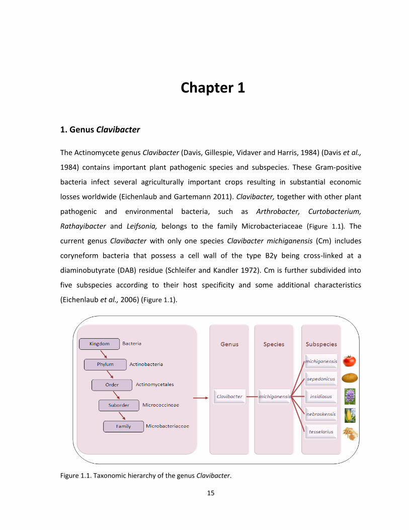
1. Genus Clavibacter
The Actinomycete genus Clavibacter (Davis, Gillespie, Vidaver and Harris, 1984) (Davis et al.,
1984) contains important plant pathogenic species and subspecies. These Gram-positive
bacteria infect several agriculturally important crops resulting in substantial economic
losses worldwide (Eichenlaub and Gartemann 2011). Clavibacter, together with other plant
pathogenic and environmental bacteria, such as Arthrobacter, Curtobacterium,
Rathayibacter and Leifsonia, belongs to the family Microbacteriaceae (Figure 1.1). The
current genus Clavibacter with only one species Clavibacter michiganensis (Cm) includes
coryneform bacteria that possess a cell wall of the type B2γ being cross-linked at a
diaminobutyrate (DAB) residue (Schleifer and Kandler 1972). Cm is further subdivided into
five subspecies according to their host specificity and some additional characteristics
(Eichenlaub et al., 2006) (Figure 1.1).
Figure 1.1. Taxonomic hierarchy of the genus Clavibacter.
Chapter 1- Literature overview
16
Taxonomic history of the genus Clavibacter is rather complicated. As many other plant
pathogenic coryneform bacteria, members of the current genus Clavibacter were assigned
to the genus Corynebacterium mainly based on morphological features and Gram staining
results (Keddie and Jones 1981). However, with time many of these bacteria were shown to
not belong to Corynebacterium group and were reclassified based on additional
chemotaxonomic characteristics to the genus Rhodococcus (Goodfellow and Alderson
1977), Arthrobacter (Collins et al., 1981), Curtobacterium (Collins and Jones 1983),
Rathayibacter (Zgurskaya et al., 1993) and Leifsonia (Evtushenko et al., 2000). The current
genetic classification is based on menaquinone and cell wall composition and the 16S rRNA
phylogeny (Eichenlaub et al., 2006). Despite a generally accepted classification of the genus
Clavibacter there is an ongoing discussion whether the current subspecies warrant the
species status and should be lifted to a species level (Evtushenko and Taekeuchi 2006).
Clavibacter michiganensis subsp. michiganensis (Cmm) causes bacterial wilt and canker of
tomato (Solanum lycopersicum) (Smith 1910), which is considered to be one of the most
important bacterial diseases of tomato (De León et al., 2011). Clavibacter michiganensis
subsp. sepedonicus (Cms) is responsible for ring rot of potato (Solanum tuberosum) (Manzer
and Genereux 1981). Clavibacter michiganensis subsp. insidiosus (Cmi) causes wilting and
stunting in alfalfa (Medicago sativa) (McCulloch 1925). Clavibacter michiganensis subsp.
nebraskensis (Cmn) induces wilt and blight of maize (Zea mays) (Schuster et al., 1975) and
Clavibacter michiganensis subsp. tessellarius (Cmt) causes leaf freckles and leaf spots in
wheat (Triticum aestivum) (Carlson and Vidaver 1982).
Because of the lack of resistant plants, severity of the diseases and problems in effective
controlling the spread of the pathogen, Cmm, Cms and Cmi are classified as quarantine
organisms under the European Union Plant Health Legislation in the European Union (EU)
and many other countries (Anonymous 2000) (http://www.eppo.int/QUARANTINE/
listA2.htm).
Clavibacter michiganensis subspecies, except Cms, produce yellow to orange, smooth, shiny
and flat colonies. Due to the production of exopolysaccharides they often show mucoid
Part II-Introduction
17
colony morphology (Evtushenko and Taekeuchi 2006). Genus Clavibacter is characterized by
high GC content of DNA which is about 73% (Eichenlaub et al., 2006).
Clavibacter subspecies are mostly vascular pathogens causing systemic infections after
entering through wounds or hydathodes. In nature, they show high host specificity.
However, some plants related to host plants (e.g. species of the Solanaceae for Cmm) can
be artificially infected (Anonymous 2005, Eichenlaub et al., 2006). Some solanaceous and
non-solanaceous plants, including Datura stramonium and Amaranthus retroflexus, have
serve as reservoirs for epiphytical survival and spread of Cmm (Chang et al., 1992).
Pathogens belonging to the genus Clavibacter are especially dangerous because of their
ability to induce latent infections with no visible symptoms, which enables spreading of the
bacteria along with infected plant material (Bentley et al., 2008, Gitaitis et al., 1991).
Moreover, these bacteria invade seeds allowing for their long-distance spread (Franken et
al., 1993, McBeath and Adelman 1986, Samac et al., 1998, Tsiantos 2008).
1.1 Clavibacter michiganensis subsp. michiganensis-quarantine pathogen
Clavibacter michiganensis subsp. michiganensis (Cmm) is a seed-borne pathogen and causal
agent of bacterial wilt and canker on tomato. For this reason and because of its unpredicted
and sporadic occurrence in the EU and in many other countries, Cmm is classified as a
quarantine pest present on the A2 EPPO (European and Mediterranean Plant Protection
Organization) list. As a seed pathogen Cmm can be easily transferred over long distances
and distributed throughout all of the tomato-growing regions of the world (Anonymous
2000, OEPP/EPPO 2007).
Primary and solely economically important host of Cmm is tomato (Solanum lycopersicum).
However, Cmm can also infect several solanaceous hosts: Solanum mammosum (spiny
Porto Rican weed), S. douglasii (perennial nightshade, S. nigrum (black nightshade) and S.
triflorum (Lewis Ivey and Miller 2000, Thyr et al., 1975). Other plants, including pepper
(Capsicum annuum) and eggplant (Solanum melongena), are susceptible too (Yim et al.,
2012).
Chapter 1- Literature overview
18
Cmm invades tomato through natural openings: stomata, hydathodes, trichomes and
through wounds (Carlton et al., 1998, Jahr et al., 1999). Widespread dissemination of Cmm
occurs through tomato seeds and transplants (Stephens and Fulbright 1986, Tsiantos 2008).
Frequently, infected transplants are symptomless at the moment they are transported to
other tomato producing areas. It has been proven that, under favourable conditions, even a
few infected seeds or plants can be a source of a large outbreak in the field (Chang et al.,
1991, Fatmi and Schaad 1988). Secondary spread of Cmm can happen via splashing water,
contaminated equipment and agricultural practices (Milijašević et al., 2007). The spread and
increase of number of bacteria is higher in wet, humid conditions and the close proximity of
the tomato transplants. In greenhouses, disease symptoms are more severe during hot and
long summers. Bacterial wilt and canker will be more likely found in the humid and wetter
areas of the greenhouse than in the dry parts. Multiplication and spread of Cmm is less
frequent in open fields because of the lowered humidity, increased air movement and
wider spaces between plants. From the economical point of view it is more efficient to
spray transplants when in the greenhouse than to spray plants once they are placed in the
field. A critical point in spreading of Cmm is the grafting process, which is currently
commonly practiced in the majority of the production greenhouses. In this process tomato
scions are grafted onto rootstock which ensures greater vigor, longevity and sometimes
provide a disease resistance (Kubota et al., 2008) but at the same time it creates a quick
way for bacteria to spread from plant to plant and therefore enabling easier bacterial
transfer and spread.
1.2 Bacterial wilt and canker of tomato-quarantine disease
Bacterial wilt and canker of tomato (Solanum lycopersicum), induced by Cmm is an
important disease occurring in both tomato fields and greenhouses. The disease,
considered as one of the most important and potentially devastating diseases on tomato,
was first described by E. F. Smith in 1905 in Michigan, USA, (Smith 1910). Nowadays, it is
present worldwide. Despite its erratic occurrence (with a typical start/stop pattern)
outbreaks of bacterial wilt and canker are spreading rapidly leading to substantial damage
Part II-Introduction
19
in the tomato producing areas (Anonymous 2005, Stephens and Fulbright 1986, Strider
1969). The erratic nature of this disease is one of the important reasons why there is a slow
progress in improving the efficacy of management practices.
1.2.1 Symptoms
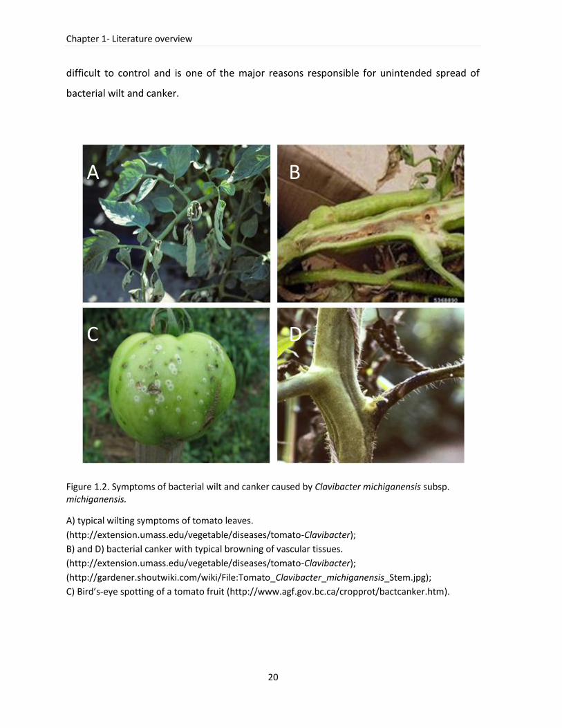
Cmm induces systemic infection and invades the vascular tissues causing wilting (Figure 1.2).
At early stage, the wilt is usually observed on one side of the leaves and canker appears
sometimes on the stem (Figure 1.2). After cutting the stem of infected plants a slight
browning or discoloration of the internal tissue is observed. Systemic infection can be
confirmed by suspending a fragment from a cut tomato stem into a glass of water. If Cmm
causes a systemic tomato infection, after a few minutes, bacteria will form a milky
suspension. Symptoms resulting from a local infection appear as leaf spots or bright or
marginal necrosis; this is commonly called the ‘fring stage’ of disease. In fruit, Cmm might
be transmitted to seeds through vascular tissues. Fruit produced from infected plants may
be stunted or malformed. Often, yellowing or browning of the vascular tissues can be seen.
Fruits develop symptoms of ‘bird’s eye’ spots and lesions observed during surface infection.
During epidemic outbreaks in the fields, the most frequently observed symptoms are
marginal necrosis of leafs. Diagnosis of bacterial wilt and canker based solely on one of the
described symptoms (with the exception of bird’s-eye lesions) can be difficult. Moreover,
symptoms of bacterial wilt and canker on tomato can be confused with systemic infection
induced by Ralstonia solanacearum or Fusarium spp. However, if more typical symptoms
are observed in tomato, they are likely to be a result of Cmm infection.
Symptoms of bacterial wilt and canker vary, depending on the age of the plant at the
moment of infection, environmental conditions, plant susceptibility, systemic or localized
infection and on the virulence level of the infecting strains (De León et al., 2011, Gleason et
al., 1993). Symptoms observed on young plants are usually more severe than those on older
plants. Moreover, Cmm can latently infect and colonize tomato without causing any
symptoms and can remain undetected over several generations. Latent infection is very
Chapter 1- Literature overview
20
difficult to control and is one of the major reasons responsible for unintended spread of
bacterial wilt and canker.
Figure 1.2. Symptoms of bacterial wilt and canker caused by Clavibacter michiganensis subsp. michiganensis.
A) typical wilting symptoms of tomato leaves.
(http://extension.umass.edu/vegetable/diseases/tomato-Clavibacter);
B) and D) bacterial canker with typical browning of vascular tissues.
(http://extension.umass.edu/vegetable/diseases/tomato-Clavibacter);
(http://gardener.shoutwiki.com/wiki/File:Tomato_Clavibacter_michiganensis_Stem.jpg);
C) Bird’s-eye spotting of a tomato fruit (http://www.agf.gov.bc.ca/cropprot/bactcanker.htm).
A B
C D
Part II-Introduction
21
1.2.2 Source of infection, transmission and survival
Cmm can survive in various environments including soil (only for short periods), plant
remnants, secondary host plants and contaminated equipment (Fatmi and Schaad 2002,
Strider 1969). Cmm can persist in soil for more than one year but only in association with
plant debris (Chang et al., 1991, Fatmi and Schaad 2002). Cmm was found to survive for
longer periods in seeds allowing long-distance disseminations and its spread into new
regions (Kaneshiro and Alvarez 2003). Some weeds, such as nightshade and several
Lycopersicon species, can act as a reservoir of Cmm. Plant materials (tomato, weeds, debris)
and Cmm contaminated soil may be transferred via wind and insects into the greenhouses
and fields. Moreover, recirculated water systems, such as irrigation flows, may contain the
pathogen. Excessive watering during tomato production in greenhouses and rainfalls in the
fields facilitate the spread of Cmm, especially when plants were earlier grafted or pruned.
Once Cmm is present in the production fields or greenhouses, transmission can easily occur
on adjacent plants which can be infected via small injuries, recent pruning or natural
openings on the leaves. The pathogen spreads via the machinery and facilities used during
cultivation. Under greenhouse conditions bacteria are transferred during the pruning or
grafting proces by improperly disinfected equipment. Sudden and severe symptoms may be
exhibited when bacteria are transferred directly to the vascular system during grafting.
Cmm can survive at least one month on surfaces, such as plastic and cement and up to a
year in plant material and rock wool. The knowledge about the survival mechanisms and the
common infection sources will facilitate not only the disease management but also will help
to apply more suitable control measures to better follow the disease transmission.
1.2.3 Disease management and control
Management of bacterial wilt and canker is especially problematic because not only there is
no successful treatment but also because Cmm can be difficult to eradicate once it was
present in a greenhouse or field. Once the bacterium is introduced in a production area only
changes in culture practices can limit the risk of spread and of new outbreaks (Gleason et
Chapter 1- Literature overview
22
al., 1993). Furthermore, bacterial wilt and canker with its erratic appearance and
unpredicted frequency is difficult to control. Sanitary measures applied for disease control
regulated in Council Directives 2000/29/EC (Anonymous 2000) aim at prevention of spread
and unintended dissemination of Cmm by following any outbreaks of bacterial wilt and
canker on tomato. As tomato seeds serve as a main source of contamination the most
important recommendation is to start the production with certified, Cmm-free seeds. Only
purchasing tested seeds can minimize the risk of infection. At present, however, there are
no tomato cultivars fully resistant to Cmm (Gleason et al., 1993). Furthermore, the use of
tolerant tomato varieties is not advised since they can disseminate Cmm in symptomless
plants. The most effective actions for controlling the disease, besides the use of healthy
planting material, are regular and strict sanitary procedures during the production of
seedlings, grafting, pruning and harvesting of tomato. A good practice limiting the possible
infection in fields is the rotation of tomato plants with non-host plants of Cmm every two to
three years. In greenhouses, the growing media, such as rockwool, should be replaced to
limit the possibility of Cmm transmission. Furthermore, tomato or pepper cull piles and
solanaceous weeds should not be present in the close neighborhood of the production
regions.
Some trials were undertaken to apply fixed copper combined with either maneb or
mancozeb in order to reduce the epiphytic population of Cmm. Unfortunately, these actions
had a limited impact on the disease control (Hausbeck et al., 2000). Experiments with soil
treatment by formaldehyde and solarization, although used in phytosanitary actions, were
only partially successful due to recontamination from surrounding areas (Antoniou et al.,
1995).
A very interesting and promising approach of disease control employs the use of Clavibacter
specific bacteriophages. Preliminary experiments with endolysins of CMP1 and CN77
bacteriophages that were cloned into Escherichia coli showed a specific activity only against
Clavibacter strains suggesting that these endolysins recognize a unique compound present
in the cell wall of Clavibacter (Wittmann et al., 2010).
Part II-Introduction
23
Effective disease management can be obtained only by applying an integrated pest
management approach which involves a combination of several strategies developed based
on the knowledge of biology and growth of the pathogen.
1.2.4 Disease importance and economic impact on tomato production
Since the first introduction of tomato in Europe somewhere in the 16th century, this crop
became widely grown and cultivated for its fruits. Today, tomato is the second, next to
potato, most important crop worldwide (Kimura and Sinha 2008). The global production of
tomato has significantly increased during the last few decades and in 2011 was estimated
for almost 160 million tons of fruit produced on 4.7 million hectares (FAOSTAT database,
2011). Since 1980, tomato production in Belgium expanded rapidly and this crop became
the most important vegetable (Taragola and Van Lierde 1998) with the production of more
than 215 thousand tons in 2011. Belgium and the Netherlands are the two countries with
the highest yield per hectare (460833 kg/Ha and 478848 kg/Ha) (FAOSTAT database, 2011).
The main focus is on the fresh-market tomatoes grown in the greenhouses and, as in the
Netherlands, the majority of the Belgian production is destined for export.
Bacterial pathogens are estimated to cause between 10-80% yield losses depending on the
conditions, plant variety and its susceptibility (Agrios 1997). With its growing production
area, tomato is continuously exposed to new pathogens, which can easily invade the highly
susceptible tomato plant. Tomato diseases are considered a major limiting factor
influencing its production and leading to substantial yield decrease per hectare every year.
Losses resulting from bacterial wilt and canker are associated with epidemic outbreaks of
the disease. Systemic infections of tomato result in heavily infected plants leading to
substantial plant damage or to its complete collapse. When the foliage is affected, infected
plant becomes less productive and vigorous. Additionally, fruit spots appearing as a result of
secondary infections lower the fraction of tomatoes released onto the market (Davis et al.,
1984). Economic losses can vary substantially among fields, greenhouses and production
years. Direct field losses are an important part of the disease impact. However, the indirect
costs associated with sanitary measures, annual controls and disinfection actions as well as
Chapter 1- Literature overview
24
with tests involved in the certification of planting material are a significant fraction of
expenses spend every year to better control bacterial wilt and canker.
1.3 Epidemiology of bacterial wilt and canker
Knowledge about genetic diversity of Cmm and the epidemiology of the bacterial wilt and
canker on tomato in regions where the disease was detected is limited. Due to the
scattered reports and differences in the methods used it is difficult to investigate the
disease spread and transmission on a global scale. Therefore, there is a constant and urgent
need to develop a common and uniform method which will allow accurate differentiation of
Cmm strains and facilitate comparative analyses of strains from various outbreaks. First
attempts aiming at the molecular investigation of bacterial wilt and canker outbreaks were
undertaken. Recent studies reporting on genetic diversity of Cmm populations from various
outbreaks in Europe and Asia will definitely contribute to a better understanding of the
disease spread and to detection of the probable infection sources and the transmission
routes (Lamichhane et al., 2011) (De León et al., 2009) (Milijašević-Marčić et al., 2012)
(Kawaguchi et al., 2010). These epidemiological reports demonstrated that in many cases
highly homogenous Cmm populations were responsible for devastating outbreaks
suggesting a single introduction of the pathogen as a source of infection (Bella et al., 2012,
De León et al., 2009). In addition, introductions of contaminated tomato seeds and/or
seedlings were still the major source of epidemic outbreaks. Based on the information
obtained from investigations of recent outbreaks there was no link between tomato variety,
year or place of isolation and Cmm type (Kawaguchi et al., 2010) (Bella et al., 2012) (De
León et al., 2009). Interestingly, in Belgium, Israel and in Serbia strains belonging to one
Cmm population were repeatedly isolated from the same locations during subsequent years
(Kleitman et al., 2008, Milijašević-Marčić et al., 2012). The fact that the highly similar or
identical Cmm strains were frequently re-isolated in the same fields or greenhouses implies
that current eradication and sanitary actions are not sufficient and should be improved.
Furthermore, the results derived from the performed studies confirmed that contaminated
Part II-Introduction
25
seeds are still the main cause of infections. Therefore, intensive actions should be carried
out to achieve higher sensitivity and accuracy of Cmm detection in seeds.
Epidemiological studies on bacterial wilt and canker caused by Cmm contribute to a better
understanding of the disease epidemiology but are mostly limited to local investigations. To
overcome this limitation an agreement has to be made to use a common method, which
allows for the data exchange and comparison.
1.3.1 Methods used in molecular epidemiology of Cmm strains
The development of a method that accurately discriminates Cmm strains is of importance to
determine the emergence and evolution of the pathogen. Furthermore, acquiring
knowledge about the genetic diversity of Cmm populations within a country will facilitate
the estimation of quarantine risks posed by newly introduced strains. Moreover, it will
deliver valuable information to design efficient strategies for the control of bacterial wilt
and canker.
The most common and widely used technique in epidemiological studies of Cmm is rep-PCR
(repetitive-sequence-based-PCR). This genomic fingerprinting technique can discriminate
Clavibacter strains at the subspecies level (De León et al., 2009, Nazari et al., 2007, Smith et
al., 2001), however, it is of moderate use for distinguishing highly similar strains within
subspecies (Kawaguchi et al., 2010). Even though, rep-PCR with BOX primers (BOX-PCR) is
commonly applied for genotyping of Cmm (De León et al., 2009) (Kleitman et al., 2008).
However, because of its limited portability and necessity to perform the test in strictly
controlled conditions, BOX-PCR is replaced by other techniques, such as PFGE (pulsed-field
gel electrophoresis), ISSR (Inter Simple Sequence Repeat) and MLST (Multi Locus Sequence
Typing). Although PFGE is labor-intensive and time consuming, it was applied to study the
diversity of Cmm in Israel and in Serbia (Kleitman et al., 2008, Milijašević-Marčić et al.,
2012). Results from the first study divided the tested strains into four groups that were also
discriminated in BOX-PCR analysis (Kleitman et al., 2008) suggesting the usefulness of both
approaches. In the second study the analysis of PFGE was supported by MLST analysis (of
five housekeeping genes: kdpA, sdhA, ligA, gyrB and bipA) confirming the results and
supporting obtained conclusions. ISSR, a method widely used in typing of higher organisms,

Chapter 1- Literature overview
26
mainly plants (Godwin et al., 1997), was applied to study dissemination of bacterial wilt and
canker in Southern Turkey (Baysal et al., 2010). Selected ISSR markers discriminated Cmm
populations into several groups suggesting its high genetic diversity. A comprehensive study
using three widely accepted fingerprinting techniques, rep-PCR, PCR-RFLP and AFLP,
evaluated their applicability in studying diversity of Cmm populations isolated from
outbreaks in Canary Islands (De León et al., 2009). Although these methods differed in their
resolution level, simplicity and the cost of analysis, all of them showed to be useful for
studying genetic diversity of Cmm in Canary Islands.
Each of the above described techniques has strengths and weaknesses that influence their
applicability and usefulness for typing of Cmm strains. Methods developed in a pre-genomic
era, such as PFGE or RAPD, are still valid and work well. However, more advanced
techniques, including MLST or MLVA, developed in the last decades and taking advantage of
the increasing availability of complete genome sequences, are gradually replacing more
time consuming and labor-intensive tests.
With the wide range of techniques used in molecular epidemiological studies of Cmm, the
choice of the most suitable and accurate method will depend on the laboratory facilities in
combination with overall scientific aims. However, to enable drawing general conclusions
and to have a global picture of bacterial wilt and canker spread and transmission of Cmm a
common approach, inclusive the typing methodology is recommended.
1.4 Tomato seed production and health
In order to produce healthy tomato seeds the growing areas should be pathogen-free.
However, Cmm is broadly disseminated and pathogen-free areas where the tomato seed
might be produced are scarce. The dry climate in the United States was believed to limit
disease development. Unfortunately, seeds collected from this region showed to induce
latent infections (Gitaitis and Walcott 2007). Many seed producing regions are located in
developing countries where the labor costs are lower. Because of this situation there is a
fear of Cmm-contaminated seeds originating from these areas that can be at the origin of
new disease introductions. Seed lots originating from Asia (China, Thailand and Taiwan) and
from Bolivia and Brazil are considered to be a concern (De León et al., 2011). However, seed
Part II-Introduction
27
producing companies are reluctant in sharing detail information regarding the origin of the
seeds, consequently hampering the control of the seed transmission between different
countries (EPPO, (http://www.eppo.int)). (De León et al., 2011).
Development and implementation of accurate and reliable seed health testing methods are
of critical importance to prevent dissemination of dangerous diseases. Effective control
through phytosanitary certifications and international quarantine regulations of seed-borne
pathogens is essential to limit the disease occurrences (Gitaitis and Walcott 2007, Munkvold
2009). To date, many various procedures, such as chemical or thermal seed treatments or
fermentation processes used to extract tomato seeds were implemented (Dhanvantari
1989, Fatmi et al., 1991). Although chemical extraction including HCl treatment reduces
greatly the number of Cmm cells, none of the currently available procedures produce Cmm-
free seeds (Pradhanang and Collier 2007).
Morphological differences among Cmm strains and the variable level of seed infection
hampered development of consistent and reliable detection methods with sufficient
sensitivity and reproducibility. Currently, efforts are undertaken to follow uniform protocols
for tomato seed testing which will facilitate a clear judgment of seed health and limit the
possibility of releasing seeds with a questionable quality onto the market (Munkvold 2009).
Contaminated tomato seeds are undistinguishable from the healthy material. Therefore,
the necessary controls have to be applied. Currently available seed tests include destructive
methods and are performed on a representative sample from a given seed lot. A problem
for accurate controlling may appear when a seed lot is large and originates from different
tomato production fields. In such case, the necessary number of representative samples is
difficult to determine. In consequence, there might be a risk of releasing a contaminated
seed lot on the market. Thus, a 100% guarantee of pathogen-free material cannot be
reached. Another very important aspect concerns the sensitivity of the method used to
evaluate the level of inoculum that causes disease induction in the field under given
environmental conditions (Maddox 1998). From the experimental evidences it is known
that, under favorable conditions, one infected seed in a sample of 10.000 can lead to an
outbreak (Chang et al., 1991, Gitaitis et al., 1991). Detection procedures developed to date
Chapter 1- Literature overview
28
include the step of plating the seed extracts on semi-selective media and follow the
recommendation of the sample size containing 10.000 seeds (ISHI rules). This approach
ensures 95% probability of detecting a 0.03% level of contamination in the tested seed lot
(EPPO 2013). To ensure an efficient seed extraction seeds should be crushed and not
soaked since the latter may limit the efficiency of extracting an internal bacterial fraction
(Hadas et al., 2005).
1.4.1 Cmm detection in seeds
In the standard seed assay seed extracts are plated on semi-selective media (e.g. SCM-semi-
selective medium for Clavibacter; KBT-semi-selective medium with K2TeO3 or CNS-
Corynebacterium nebraskense selective medium) (Dhanvantari 1989, Fatmi and Schaad
1988, Gross and Vidaver 1979). Isolation of Cmm is hampered because of the fast growth of
epiphytic bacteria present in seeds. Besides, Cmm is often inhibited by other
microorganisms (De León et al., 2011). To facilitate the procedure and to obtain a more
specific isolation of Cmm currently used semi-selective media are evaluated and improved
(Ftayeh et al., 2011, Koenraadt et al., 2009). During the dilution plating not only a positive
control is included but also spiked controls, which ensure that the presence of other
microorganisms extracted from seeds will not influence the growth of Cmm and the final
test result. For that, a seed extract is spiked with a rifampicin-resistant marked Cmm strain
and plated on a semi-selective medium. To recover colonies of a spiked Cmm, strains are
plated on YPGA (yeast, peptone, glucose agar) medium containing rifampicin. If the labeled
strain is traced back, the seed extract is considered as not inhibiting the growth of Cmm
population. A routine assay to detect Cmm in a seed extract is the immunofluorescence (IF)
test. However, it has some disadvantages including the inability to distinguish viable and
nonviable cells and limited specificity and sensitivity (Fatmi and Schaad 1988). Therefore,
positive results from this test have to be confirmed by another technique e.g. PCR assay
using ITS primers PSA-R and PSA-8 (modified from (Pastrik and Rainey 1999)). Recently, Bio-
PCR, with an extra step including biological amplification on plates, was developed to
increase the sensitivity. Hadas and coworkers demonstrated that PCR with this modification
Part II-Introduction
29
was able to detect one contaminated seed in 10.000 (Hadas et al., 2005). Seed extracts
showing positive results for both tests, IF and PCR, are further investigated in a bioassay
(enrichment) in tomato plantlets. A final confirmation of the presence of Cmm in a seed
sample is achieved when tomato plantlets exhibit disease symptoms. The accepted protocol
for Cmm detection in tomato seed is available on the website of International Seed Health
Initiative for Vegetable Crops (ISHI-Veg) (ISHI 2011) and on EPPO website
(http://www.eppo.int). Although the recommended methods are developed to specifically
detect the presence of Cmm strains recent studies reported the presence of a taxonomically
new group of non-pathogenic Clavibacter strains, which are frequently isolated from
tomato seeds and which interfere with the current detection procedure for Cmm (Jacques
et al., 2012). Because there is very limited information concerning the presence of these
isolates in seeds and the impact on the seed health status, they may complicate the final
judgment on the seed quality. In depth investigation of this new group of isolates can affect
the current seed certification procedures in the future.
1.5 Methods for Cmm identification
An accurate identification of a causal agent is crucial for correct bacterial disease diagnosis,
tracing outbreaks and applying effective treatments. This issue becomes even more
important when it concerns quarantine organisms, such as Cmm, which might induce
epidemic outbreaks resulting often in serious economic and agricultural losses.
To date, several methods for Cmm identification were developed. In the recent version of
EPPO Diagnostics Bulletin for Cmm (EPPO 2013), besides the standard identification
methods, including biochemical tests, colony morphology or BIOLOG system, a range of
molecular tests, including a number of conventional PCRs, as well as real-time PCR assays, is
included. Various applications of PCR assays provided more reliable and accurate diagnostic
tools, which are easily accessible to regular and to more advanced diagnostic laboratories.
Currently, several pairs of primers designed for identification of Cmm exist (CMM5/CMM6
from pat-1 (Dreier et al., 1995), PSA-4/PSA-R from 16S-23S ITS (Pastrik and Rainey 1999)).
Unfortunately, recent studies demonstrated their limited specificity towards Cmm with
Chapter 1- Literature overview
30
false positive identification of non-pathogenic seed-borne Clavibacter strains (Jacques et al.,
2012). At present, identification of Cmm using Ptssk 10/11 (from a gene coding for a protein
two-component system sensor kinase) primers is the most reliable (Jacques et al., 2012).
However, the situation might change when more strains of non-pathogenic Clavibacter
strains will be included. Another technique used for Cmm identification is BOX-PCR. It is a
well known method used not only for Cmm identification but also for typing; its limitations
of application have been mentioned previously. Serological approaches, including
immunofluorescence test and immunostrip, provide a quick identification; however, they
are known to cross-react with some other bacteria present in seeds or soil (De León et al.,
2007). SA (serum/stained antigen agglutination) test enables quick screening of Cmm by
using specific antibodies absorbed on cells of Staphylococcus aureus, which are used as
agglutination reagent. Its advantages are the speed (results within minutes) and its
simplicity. Immunofluorescence assays with mono- and polyclonal antibodies were used for
Cmm detection but demonstrated cross-reactions with other Clavibacter subspecies and
plant associated bacteria (Alvarez et al., 1993, De León et al., 2007, Franken et al., 1993,
Kaneshiro et al., 2006). Detection of Cmm using serological methods is hampered by the
lack of specificity of the available antisera. It is attributed to the fact that the antigen
determinants, namely teichoic acids peptidoglycans and capsular polysaccharides present in
Gram-positive bacteria are relative ubiquitous (Schleifer and Seidl 1977, Wicken and Knox
1975). According to EPPO diagnostics for Cmm also fatty acid profiling using Midi (Microbial
Identification System) can be an alternative for Cmm identification (EPPO 2013).
As for many other plant pathogens, such as Xanthomonas axonopodis pv. citri (EPPO 2005)
and Cms (EPPO 2006), also for presumable Cmm isolates additional pathogenicity tests have
to be performed to confirm a final diagnosis. To confirm the identity of Cmm isolated from
seeds, a bioassay on tomato is recommended. It is performed at the last stage of the seeds
examination. Bioassay on tomato is sensitive and specific, but it requires a three weeks
incubation time from the moment of inoculation to the manifestation of symptoms. In case
of a positive reaction and exhibition of symptoms, the bacterium should be reisolated and
confirmed by dilution plating on semi-selective medium. To identify occasional strains which
Part II-Introduction
31
do not produce wilting symptoms an additional test on cotyledons can be applied. In case of
a pathogenic strain, after inoculation and a 48 hour incubation period, symptoms including
white blisters or craters on the cotyledon surface, confirm the pathogenicity of a strain.
As some of the methods used for Cmm identification are prone to false-negative and false-
positive results (Jacques et al., 2012, Kaneshiro and Alvarez 2001) and required
pathogenicity tests need even several days before a final confirmation can be achieved
there is a need for faster and more reliable methods, which replace unspecific molecular
assays and the costly and time-consuming pathogenicity tests. With the growing tendency
to use sequencing and because of its gradually decreasing prices gene sequencing will be
soon universally acceptable approach for strains identification. However, before such a
technique becomes widely used it must be compared with standard methods and its
specificity should be tested on a vast number of genetically diverse strains including
taxonomically and phenotypically similar isolates. Moreover, annual validation by
independent institutions and tests with new strains and hosts should be performed to
ensure the high specificity and accuracy of a given identification method.
1.6 Pathogenicity and virulence in Cmm
The molecular interactions of Cmm with its host are not well understood. However, recent
discoveries with regard to pathogenesis of Cmm are increasing our understanding of
molecular mechanisms of infection. Initial observation of infected plants led to the
hypothesis that typical wilting symptoms are caused either by water stress, resulted from
the presence of high titers of the bacterium in the vascular system, by a presence of
phytotoxic EPS (extracellular polysaccharides) or by enzymatic degradation of the plant
tissues (Denny 1995, Vidaver 1982). Cmm produces many cell wall-degrading enzymes (e.g.,
xylanase, pectate lyases, cellulases and polygalacturonase), which might be involved in
canker production and tissue maceration (Gartemann et al., 2008). The molecular
investigation of Cmm was facilitated by the construction of cloning vectors (Meletzus and
Eichenlaub 1991). Initial experiments were performed using Cmm NCPPB 382 that harbors
two circular plasmids, pCM1 (27 kb) and pCM2 (70 kb) (Meletzus and Eichenlaub 1991). It
Chapter 1- Literature overview
32
was observed that the cured, plasmid-free strain CMM100 was able to colonize a plant in a
high titer but did not induce any disease symptoms in tomato. In the plant, CMM100
behaved as an endophyte demonstrating that genes involved in colonization, host
recognition, invasion and suppression of host defenses have to be chromosomally-encoded.
Moreover, CMM100 produced the same amounts of EPS as the wild type Cmm strain
(Meletzus et al., 1993), which rejected the hypotheses that the high titer colonization of the
xylem or EPS was responsible for wilting symptoms (Bermpohl et al., 1996).
Further analysis revealed that mutants containing only one of two plasmids are still virulent
but the development of disease symptoms was delayed. Experiments with Cmm plasmids
helped to identify two genes, celA and pat-1, which are essential, plasmid-encoded
pathogenicity determinants.
1.6.1 Plasmid-encoded virulence factors
CelA, a cellulase gene and pat-1, a putative serine protease gene encoded on pCM1 and
pCM2, respectively, have been demonstrated to be important in the virulence of Cmm
(Dreier et al., 1997, Jahr et al., 2000). Both proteins, CelA and Pat-1, are secreted by Cmm
and necessary for disease induction (Dreier et al., 1997, Jahr et al., 2000). However,
multiple genes encoded on plasmids must be expressed in Cmm to exhibit full virulence
(Jahr et al., 1999).
CelA, the first important virulence gene, encodes the endo-β-1,4-glucanase, a protein of 78
kDa (746 amino acids) (Jahr et al., 2000). CelA has a three domain structure: (i) catalytic (ii)
cellulose-binding and (iii) a domain at the C-terminus with a low similarity to plant α-
expansins. The celA gene was shown to be an important virulence factor also in Cms in
which the gene is carried by the 50 kb-plasmid pCS1 (Laine et al., 2000). The α-expansin-like
domain of CelA is believed to be required for the degradation of native cell walls in plants,
especially crystalline cellulose regions (Cosgrove 1998).
Pat-1, the second essential virulence factor of Cmm encodes a protein of 280 amino acids
with a molecular mass of 29.7 kDa. Pat-1 gene shows homology to serine proteases of the
chymotrypsine type. It contains a repetitive region called pat-1 rep which was found in the
Part II-Introduction
33
majority of Cmm strains examined. The repetitive sequence is thought to protect pat-1
mRNA from degradation and its deletion resulted in a reduced virulence and decreased
level of pat-1 mRNA in cells (Dreier et al., 1997).
1.6.2 Pathogenicity Island of Cmm
The putative pathogenicity island (PI) of 129 kb is a region with significantly lower GC
content that was identified in Cmm NCPPB 382 during the analysis of the complete genome
sequence (Gartemann et al., 2008) (Figure 1.3). PI, also called chp/tomA region, contains
genes important for colonization and modulation of plant defense systems. Serine
proteases encoded in this region showed homology to plasmid-located pat-1 gene (Figure
1.3). Most of them are functional genes with the exception of chpA, chpB and chpD which
are pseudogenes (Stork et al., 2008). Some genes from chp/tomA fragment are involved in
sugar metabolism and are suggested to play an important function in utilization of plant
derived nutrients (Flügel et al., 2012). The loss of the chp/tomA region leads to a significant
reduction in colonization abilities. In Cmm mutants lacking this region wilting symptoms did
not appear, even when virulence factors such as celA were present (Chalupowicz et al.,
2010, Gartemann et al., 2008).
Homologues of genes from PI in Cmm exist also in the genome of Cms (Bentley et al., 2008,
Burger et al., 2005). The chp7 of Cms is the chromosomal homologue of pat-1 in Cmm and it
was demonstrated to be involved in the hypersensitive response (HR) in tobacco and
virulence of Cms (Nissinen et al., 2001). Other members of the Chp and Php gene families
were recognized in the genome of Cms, however, neither their functions, nor their potential
participation in pathogenesis of Cms have been investigated.
Another important gene located on PI is tomA encoding a putative tomatinase. In Fusarium
oxysporum f. sp. lycopersici this enzyme is known to detoxify α-tomatine (alkaloid produced
by tomato) (Pareja-Jaime et al., 2008) and it plays an essential function in overcoming plant
defense (Roldán-Arjona et al., 1999). Although experiments with tomA mutants
demonstrated their higher sensitivity to α-tomatine no changes in virulence were observed
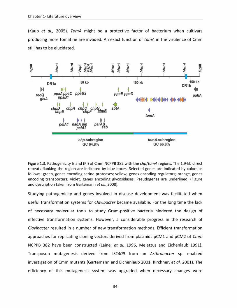
Chapter 1- Literature overview
34
(Kaup et al., 2005). TomA might be a protective factor of bacterium when cultivars
producing more tomatine are invaded. An exact function of tomA in the virulence of Cmm
still has to be elucidated.
Figure 1.3. Pathogenicity Island (PI) of Cmm NCPPB 382 with the chp/tomA regions. The 1.9-kb direct repeats flanking the region are indicated by blue boxes. Selected genes are indicated by colors as follows: green, genes encoding serine proteases; yellow, genes encoding regulators; orange, genes encoding transporters; violet, genes encoding glycosidases. Pseudogenes are underlined. (Figure and description taken from Gartemann et al., 2008).
Studying pathogenicity and genes involved in disease development was facilitated when
useful transformation systems for Clavibacter became available. For the long time the lack
of necessary molecular tools to study Gram-positive bacteria hindered the design of
effective transformation systems. However, a considerable progress in the research of
Clavibacter resulted in a number of new transformation methods. Efficient transformation
approaches for replicating cloning vectors derived from plasmids pCM1 and pCM2 of Cmm
NCPPB 382 have been constructed (Laine, et al. 1996, Meletzus and Eichenlaub 1991).
Transposon mutagenesis derived from IS1409 from an Arthrobacter sp. enabled
investigation of Cmm mutants (Gartemann and Eichenlaub 2001, Kirchner, et al. 2001). The
efficiency of this mutagenesis system was upgraded when necessary changes were
Part II-Introduction
35
introduced in the electroporation technique (Kirchner, et al. 2001). The availability of
complete genome sequences of Cmm and Cms enabled the application of directed
mutagenesis that proved useful when studied tomA gene in Cmm NCPPB 382 (Kaup, et al.
2005). Although developed for Cmm these directed mutagenesis approach can also be used
in other Clavibacter subspecies (Laine, et al. 1996, Nissinen, et al. 2009).
1.7 Atypical Clavibacter strains and their influence on diagnostics
Although the genus Clavibacter includes mostly pathogenic bacteria, non-virulent or
avirulent strains arise sometimes in nature (Kleitman et al., 2008, Schuster et al., 1975).
Various atypical Cmm strains showing genotypic and phenotypic differences include strains
with dry, sticky more orange/red or white colony morphology (Davis and Vidaver 2001,
Kaneshiro et al., 2006). Strains varying in virulence levels were isolated from plants and
seeds (Kaneshiro and Alvarez 2001, Kaneshiro et al., 2006, Louws et al., 1998). Investigation
of these strains is impeded because isolates are collected after an outbreak of the disease
resulting in possible changes in gene content of isolated strains (populations containing
different virulence factors: celA, pat-1 and PI). In addition, the examination of avirulent or
weakly virulent strains is hampered because these strains are often not identified during the
diagnostics procedures. Especially, when the methods used for screening are based on
virulence factors, which might be lacking in this population. There are at least a few possible
mechanisms responsible for the presence of Cmm strains with different levels of virulence.
The lack of one or two plasmids, containing virulence factors, leads to a reduced virulence
or to a disability of a strain to induce disease, respectively. The virulence plasmids can be
lost under stress condition (e.g. at temperatures higher than 30˚C). The experiments in
planta performed by the group of Gartemann and Eichenlaub demonstrated that plasmids
pCM1 and pCM2 are conjugative (Gartemann et al., 2003). These findings suggest that the
partial loss of the pathogenicity might be compensated by conjugational acquisition of the
plasmids from a donor strain and an initially avirulent or weakly virulent strain might
become an effective pathogen. The modulation of virulence might also result from a loss of
the pathogenicity island (PI) containing some possible virulence factors. With these
Chapter 1- Literature overview
36
alternations bacteria can still colonize the plant, but because of the lack of essential
virulence determinants (pat-1 and celA) it turns into a non-virulent endophyte (Eichenlaub
and Gartemann 2011). A very interesting example of atypical Cmm strains isolated from
pepper was recently reported in Korea (Yim et al., 2012). These isolates showed only limited
disease symptoms on tomato (slight stem canker and leaf blight) but produced stem and
leaf blight on pepper. Moreover, they were negative in PCR-tests for plasmid-encoded as
well as PI-encoded genes involved in virulence. As most of them exhibited high
morphological similarity to Cmm strains and were identified as Cmm by ELISA, 16S rRNA and
fatty acid analysis, they were assumed to belong to the Cmm group. However, these
methods are not specific enough to confirm the Cmm identity. Therefore, it would be
advisable to use more accurate techniques, such as PCR assay with Ptssk 10/11 primers,
DNA barcoding or MLSA to reassure their correct identification as Cmm. These strains might
belong to the Clavibacter group that is actually distinct from Cmm strains.
1.8 Molecular interaction of Cmm with tomato
Plants must employ various strategies to resist infections caused by different pathogens.
These strategies constitute a part of the plant’s innate immune system that can be divided
into two main groups. The first is an early warning defense, which recognizes pathogen-
associated molecular patterns (PAMPs). After PAMPs are recognized a basal plant defense is
induced and PAMP triggered immunity (PTI) is elicited (Schneider and Collmer 2010). A
second plant defense system is highly specific and involves an effector molecule of a
pathogen and an associated resistance protein (R) of a host plant (Jones and Dangl 2006). A
branch of plant immune system that is activated by the presence of pathogen effectors is
called effector triggered immunity (ETI).
In a majority of Gram-negative bacteria, the T3SS (Type III Secretion System) which transfers
various effector proteins into the plant cell, plays a crucial role in ETI. In susceptible non-
host plants, this enables growth of the pathogen that in consequence leads to disease. In
resistant host plants, effectors called avirulence factors are recognized by the products of
Part II-Introduction
37
corresponding resistance genes of the plant, which induce an HR (hypersensitive reaction)
defense reaction and prevent the spreading of the pathogen in the plant.
Pathogens, such as Cmm, must suppress plant defenses and overcome PTI to be able to
successfully attack the plant. Although data analysis of complete genomes of Cmm, Cms and
Cmn revealed that T3SS and genes encoding effector proteins similar to those found in
Gram-negative bacteria are not present (Bentley et al., 2008, Gartemann et al., 2008),
Clavibacter can induce a hypersensitive response in non-hosts. Clavibacter PAMPs are
recognized in non-hosts and induce an effective defense mechanism, which has to be
overcome in host plants.
The transcriptome analysis of tomato plants infected by Cmm showed that many tomato
genes involved in defense were activated. Therefore, it was suggested that Cmm contains
unknown bacterial PAMPs recognized by the tomato and able to trigger tomato defense
responses (Balaji et al., 2008).
The study of the Cmm-tomato interactome demonstrated that during infection, Cmm
recognizes the plant environment, transfers signals, secretes a variety of hydrolytic enzymes
including serine proteases of the Pat-1, Ppa and Sbt familes, the CelA, XysA and NagA
glycosyl hydrolases as well as other cell wall-degrading enzymes (Savidor et al,. 2012) (Figure
1.4).
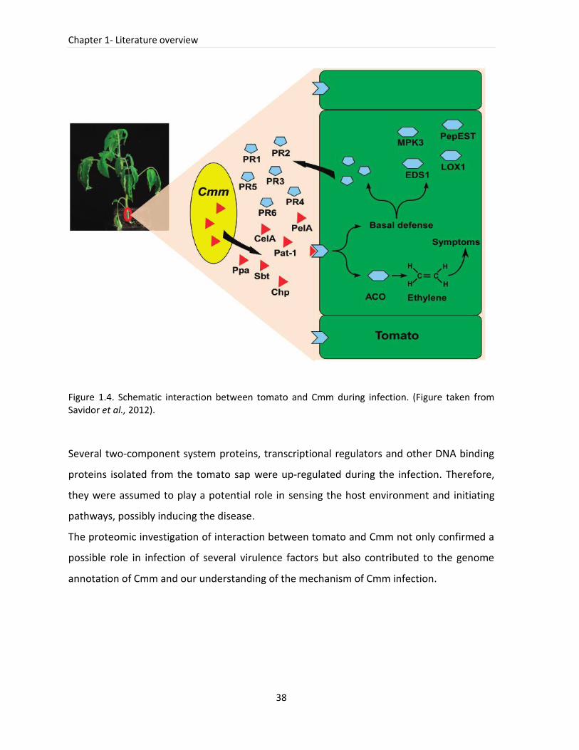
Chapter 1- Literature overview
38
Figure 1.4. Schematic interaction between tomato and Cmm during infection. (Figure taken from Savidor et al., 2012).
Several two-component system proteins, transcriptional regulators and other DNA binding
proteins isolated from the tomato sap were up-regulated during the infection. Therefore,
they were assumed to play a potential role in sensing the host environment and initiating
pathways, possibly inducing the disease.
The proteomic investigation of interaction between tomato and Cmm not only confirmed a
possible role in infection of several virulence factors but also contributed to the genome
annotation of Cmm and our understanding of the mechanism of Cmm infection.
Part II-Introduction
39
1.9 References
1. Agrios GN. 1997. Plant Pathology. Academic press, London, UK 2. Alvarez A, Derie M, Benedict A, Gabrielson R. 1993. Characteristics of a monoclonal antibody to
Clavibacter michiganensis subsp. michiganensis. Phytopathology 83 3. Anonymous. 2000. Council Directive 2000/29/EC of 8 May 2000 on protective measures against
the introduction into the Community of organisms harmful to plants or plant products and against their spread within the Community. Office for Official Publications of the European Communities. Consolidated Text. 1-139
4. Anonymous. 2005. Clavibacter michiganensis subsp. michiganensis OEPP/EPPO Bull.: 275-83 5. Anonymous. 2011. Combating plant diseases is key for sustainable crops. ScienceDaily
http://www.sciencedaily.com/releases/2011/04/110411194819.htm. 6. Antoniou P, Tjamos E, Panagopoulos C. 1995. Use of soil solarization for controlling bacterial
canker of tomato in plastic houses in Greece. Plant Pathology 44: 438-47 7. Balaji V, Mayrose M, Sherf O, Jacob-Hirsch J, Eichenlaub R, et al. 2008. Tomato transcriptional
changes in response to Clavibacter michiganensis subsp. michiganensis reveal a role for ethylene in disease development. Plant Physiology 146: 1797-809
8. Baysal Ö, Mercati F, İkten H, YIldIz R, Carimi F, et al. 2010. Clavibacter michiganensis subsp. michiganensis: tracking strains using their genetic differentiations by ISSR markers in Southern Turkey. Physiological and Molecular Plant Pathology 75: 113-9
9. Bella P, Ialacci G, Licciardello G, La Rosa R, Catara V. 2012. Characterization of atypical Clavibacter michiganensis subsp. michiganensis populations in greenhouse tomatoes in Italy. Journal of Plant Pathology 94: 635-42
10. Bentley SD, Corton C, Brown SE, Barron A, Clark L, et al. 2008. Genome of the actinomycete plant pathogen Clavibacter michiganensis subsp. sepedonicus suggests recent niche adaptation. Journal of Bacteriology 190: 2150-60
11. Bermpohl A, Dreier J, Bahro R, Eichenlaub R. 1996. Exopolysaccharides in the pathogenic interaction of Clavibacter michiganensis subsp. michiganensis with tomato plants. Microbiological Research 151: 391-9
12. Burger A, Grafen I, Engemann J, Niermann E, Pieper M, et al. 2005. Identification of homologues to the pathogenicity factor pat-1, a putative serine protease of Clavibacter michiganensis subsp. michiganensis. Microbiological Research 160: 417-27
13. Carlson RR, Vidaver AK. 1982. Taxonomy of Corynebacterium plant-pathogens, including a new pathogen of wheat, based on polyacrylamide-gel electrophoresis of cellular proteins International Journal of Systematic Bacteriology 32: 315-26
14. Carlton W, Braun E, Gleason M. 1998. Ingress of Clavibacter michiganensis subsp. michiganensis into tomato leaves through hydathodes. Phytopathology 88: 525-9
15. Chalupowicz L, Cohen-Kandli M, Dror O, Eichenlaub R, Gartemann KH, et al. 2010. Sequential expression of bacterial virulence and plant defense genes during infection of tomato with Clavibacter michiganensis subsp. michiganensis. Phytopathology 100: 252-61
16. Chang R, Ries S, Pataky J. 1991. Dissemination of Clavibacter michiganensis subsp. michiganensis by practices used to produce tomato transplants. Phytopathology 81: 1276-81
17. Collins M, Jones D. 1983. Reclassification of Corynebacterium flaccumfaciens, Corynebacterium betae, Corynebacterium oortii and Corynebacterium poinsettiae in the genus Curtobacterium, as Curtobacterium flaccumfaciens comb. nov. Microbiology 129: 3545
18. Collins MD, Jones D, Kroppenstedt RM. 1981. Reclassification of Corynebacterium ilicis (Mandel, Guba and Litsky) in the genus Arthrobacter, as Arthrobacter ilicis comb. nov. Zentralbl. Bakteriol. Parasitenkd. Infektionskr. Hyg. Abt. 1 Orip,. Reihe 2: 318-23
19. Cosgrove DJ. 1998. Cell wall loosening by expansins. Plant Physiology 118: 333-9 20. Davis M, Vidaver A. 2001. Coryneform plant pathogens. Plant pathogenic bacteria. Schaad NW,
Jones JB, Chun W, Chapter: Diseases caused by bacteria: 218-35
Chapter 1- Literature overview
40
21. Davis MJ, Gillaspie AG, Vidaver AK, Harris RW. 1984. Clavibacter: a new genus containing some phytopathogenic coryneform bacteria, including Clavibacter xyli subsp. xyli sp. nov., subsp. nov. and Clavibacter xyli subsp. cynodontis subsp. nov., pathogens that cause ratoon stunting disease of sugarcane and bermudagrass stunting disease. International Journal of Systematic Bacteriology 34: 107-17
22. De León L, Rodriguez A, Lopez M, Siverio F. 2007. Evaluation of the efficacy of immunomagnetic separation for the detection of Clavibacter michiganensis subsp. michiganensis in tomato seeds. Journal of Applied Microbiology 104: 776-86
23. De León L, Rodriguez A, Llop P, Lopez MM, Siverio F. 2009. Comparative study of genetic diversity of Clavibacter michiganensis subsp. michiganensis isolates from the Canary Islands by RAPD-PCR, BOX-PCR and AFLP. Plant Pathology 58: 862-71
24. De León L, Siverio F, Lopez MM, Rodriguez A. 2011. Clavibacter michiganensis subsp. michiganensis, a seedborne tomato pathogen: healthy seeds are still the goal. Plant Disease 95: 1328-38
25. Denny T. 1995. Involvement of bacterial polysaccharides in plant pathogenesis. Annual Review of Phytopathology 33: 173-97
26. Dhanvantari B. 1989. Effect of seed extraction methods and seed treatments on control of tomato bacterial canker. Canadian Journal of Plant Pathology 11: 400-8
27. Dreier J, Bermpohl A, Eichenlaub R. 1995. Southern hybridization and PCR for specific detection of phytopathogenic Clavibacter michiganensis subsp. michiganensis. Phytopathology 85: 462-8
28. Dreier J, Meletzus D, Eichenlaub R. 1997. Characterization of the plasmid encoded virulence region pat-1 of phytopathogenic Clavibacter michiganensis subsp. michiganensis. Molecular Plant-Microbe Interactions 10: 195-206
29. Eichenlaub R, Gartemann K-H. 2011. The Clavibacter michiganensis subspecies: molecular investigation of Gram-positive bacterial plant pathogens. Annual Review of Phytopathology 49: 445-64
30. Eichenlaub R, Gartemann K, Burger A. 2006. Clavibacter michiganensis, a group of Gram-positive phytopathogenic bacteria. Plant-associated bacteria. Springer, Dordrecht, The Netherlands: 385-422
31. EPPO. 2005. Xanthomonas axonopodis pv. citri Diagnostics, PM7/44. Bulletin OEPP/EPPO 35: 271-3
32. EPPO. 2006. Clavibacter michiganensis subsp. sepedonicus Diagnostics, PM7/59. Bulletin OEPP/EPPO 36: 99-109
33. EPPO. 2013. Clavibacter michiganensis subsp. michiganensis Diagnostics, PM7/42 (2). Bulletin OEPP/EPPO 43: 46-67
34. Evtushenko L, Taekeuchi M. 2006. The family Microbacteriaceae. Prokaryotes 3: 1020-98 35. Evtushenko LI, Dorofeeva LV, Subbotin SA, Cole JR, Tiedje JM. 2000. Leifsonia poae gen. nov., sp
nov., isolated from nematode galls on Poa annua and reclassification of 'Corynebacterium aquaticum' Leifson 1962 as Leifsonia aquatica (ex Leifson 1962) gen. nov., nom. rev., comb. nov and Clavibacter xyli Davis et al., 1984 with two subspecies as Leifsonia xyli (Davis et al., 1984) gen. nov., comb. nov. International Journal of Systematic and Evolutionary Microbiology 50: 371-80
36. Fatmi M, Schaad N. 1988. Semiselective agar medium for isolation of Clavibacter michiganense subsp. michiganense from tomato seed. Phytopathology 78: 121-6
37. Fatmi M, Schaad N, Bolkan H. 1991. Seed treatments for eradicating Clavibacter michiganensis subsp. michiganensis from naturally infected tomato seeds. Plant Disease 75: 383-5
38. Fatmi M, Schaad NW. 2002. Survival of Clavibacter michiganensis ssp. michiganensis in infected tomato stems under natural field conditions in California, Ohio and Morocco. Plant Pathology 51: 149-54
39. Flügel M, Becker A, Gartemann K, Eichenlaub R. 2012. Analysis of the interaction of Clavibacter michiganensis subsp. michiganensis with its host plant tomato by genome-wide expression profiling. Journal of Biotechnology 160: 42-54
Part II-Introduction
41
40. Franken A, Kamminga GC, Snijders W, Vanderzouwen PS, Birnbaum YE. 1993. Detection of Clavibacter michiganensis ssp. michiganensis in tomato seeds by immunofluorescence microscopy and dilution plating. Netherlands Journal of Plant Pathology 99: 125-37
41. Ftayeh RM, von Tiedemann A, Rudolph KWE. 2011. A New Selective Medium for Isolation of Clavibacter michiganensis subsp. michiganensis from Tomato Plants and Seed. Phytopathology 101: 1355-64
42. Gartemann KH, Abt B, Bekel T, Burger A, Engemann J, et al. 2008. The genome sequence of the tomato-pathogenic actinomycete Clavibacter michiganensis subsp. michiganensis NCPPB382 reveals a large island involved in pathogenicity. Journal of Bacteriology 190: 2138-49
43. Gartemann KH, Kirchner O, Engemann J, Grafen I, Eichenlaub R, Burger A. 2003. Clavibacter michiganensis subsp. michiganensis: first steps in the understanding of virulence of a Gram-positive phytopathogenic bacterium. Journal of Biotechnology 106: 179-91
44. Gartemann K-H, Eichenlaub R. 2001. Isolation and Characterization of IS1409, an Insertion Element of 4-Chlorobenzoate-Degrading Arthrobacter sp. Strain TM1, and Development of a System for Transposon Mutagenesis. Journal of Bacteriology 183: 3729-36
45. Gitaitis R, Beaver R, Voloudakis A. 1991. Detection of Clavibacter michiganensis subsp. michiganensis in symptomless tomato transplants. Plant Disease 75: 834-8
46. Gitaitis R, Walcott R. 2007. The epidemiology and management of seedborne bacterial diseases. Annual Review of Phytopathology 45: 371-97
47. Gleason ML, Gitaitis RD, Ricker MD. 1993. Recent progress in understanding and controlling bacterial canker of tomato in eastern North America. Plant Disease 77: 1069-76
48. Godwin ID, Aitken EA, Smith LW. 1997. Application of inter simple sequence repeat (ISSR) markers to plant genetics. Electrophoresis 18: 1524-8
49. Goodfellow M, Alderson G. 1977. The actinomycete-genus Rhodococcus: a home for the 'rhodochrous' complex. Journal of general microbiology 100: 99-122
50. Gross D, Vidaver A. 1979. A selective medium for isolation of Corynebacterium nebraskense from soil and plant parts Phytopathology 69
51. Hadas R, Kritzman G, Klietman F, Gefen T, Manulis S. 2005. Comparison of extraction procedures and determination of the detection threshold for Clavibacter michiganensis ssp. michiganensis in tomato seeds. Plant Pathology 54: 643-9
52. Hausbeck MK, Bell J, Medina-Mora C, Podolsky R, Fulbright DW. 2000. Effect of bactericides on population sizes and spread of Clavibacter michiganensis subsp. michiganensis on tomatoes in the greenhouse and on disease development and crop yield in the field. Phytopathology 90: 38-44
53. Hogenhout SA, Van der Hoorn RA, Terauchi R, Kamoun S. 2009. Emerging concepts in effector biology of plant-associated organisms. Molecular Plant-Microbe Interactions 22: 115-22
54. ISHI. 2011. Method for the Detection of Clavibacter michiganensis subsp. michiganensis on Tomato seed (Version 4). International Seed Health Initiative: http://www.worldseed.org/isf/ishi_vegetable.html.
55. Jacques MA, Durand K, Orgeur G, Balidas S, Fricot C, et al. 2012. Phylogenetic analysis and polyphasic characterization of Clavibacter michiganensis strains isolated from tomato seeds reveal that non-pathogenic strains are distinct from C. michiganensis subsp. michiganensis. Applied and Environmental Microbiology 78: 8388-402
56. Jahr H, Bahro R, Burger A, Ahlemeyer J, Eichenlaub R. 1999. Interactions between Clavibacter michiganensis and its host plants. Environmental Microbiology 1: 113-8
57. Jahr H, Dreier J, Meletzus D, Bahro R, Eichenlaub R. 2000. The endo-beta-1,4-glucanase CelA of Clavibacter michiganensis subsp. michiganensis is a pathogenicity determinant required for induction of bacterial wilt of tomato. Molecular Plant-Microbe Interactions 13: 703-14
58. Jones JD, Dangl JL. 2006. The plant immune system. Nature 444: 323-9 59. Kaneshiro WS, Alvarez AM. 2001. Specificity of PCR and ELISA assays for hypovirulent and
avirulent Clavibacter michiganensis subsp. michiganensis. Phytopathology 91 60. Kaneshiro WS, Alvarez AM. 2003. Variability and survival of Clavibacter michiganensis subsp.
michiganensis (Cmm) in seed. Phytopathology (Abstr.) 93
Chapter 1- Literature overview
42
61. Kaneshiro WS, Mizumoto CY, Alvarez AM. 2006. Differentiation of Clavibacter michiganensis subsp. michiganensis from seed-borne saprophytes using ELISA, Biolog and 16S rDNA sequencing. European Journal of Plant Pathology 116: 45-56
62. Kaup O, Grafen I, Zellermann EM, Eichenlaub R, Gartemann KH. 2005. Identification of a tomatinase in the tomato-pathogenic actinomycete Clavibacter michiganensis subsp. michiganensis NCPPB382. Molecular Plant-Microbe Interactions 18: 1090-8
63. Kawaguchi A, Tanina K, Inoue K. 2010. Molecular typing and spread of Clavibacter michiganensis subsp. michiganensis in greenhouses in Japan. Plant Pathology 59: 76-83
64. Keddie R, Jones D. 1981. Saprophytic, aerobic coryneform bacteria. The Prokaryotes: A handbook on habitats, isolation and identification of bacteria. Berlin: Springer-Verlag: 1838-78
65. Kimura S, Sinha N. 2008. Tomato (Solanum lycopersicum): A model fruit-bearing crop. Cold Spring Harbor Protocols 2008: pdb. emo105
66. Kirchner O, Gartemann K-H, Zellermann E-M, Eichenlaub R, Burger A. 2001. A highly efficient transposon mutagenesis system for the tomato pathogen Clavibacter michiganensis subsp. michiganensis. Molecular Plant-Microbe Interactions 14: 1312-8
67. Kleitman F, Barash I, Burger A, Iraki N, Falah Y, et al. 2008. Characterization of a Clavibacter michiganensis subsp. michiganensis population in Israel. European Journal of Plant Pathology 121: 463-75
68. Koenraadt H, van Vliet A, Neijndorff N, Woudt B. 2009. Improvement of semi-selective media for the detection of Clavibacter michiganensis subsp. michiganensis in seeds of tomato. Presented at Annual Meeting of the American-Phytopathology-Society, Portland, OR
69. Kubota C, McClure MA, Kokalis-Burelle N, Bausher MG, Rosskopf EN. 2008. Vegetable grafting: History, use and current technology status in North America. Hortscience 43: 1664-9
70. Laine MJ, Nakhei H, Dreier J, Lehtila K, Meletzus D, et al. 1996. Stable transformation of the gram-positive phytopathogenic bacterium Clavibacter michiganensis subsp. sepedonicus with several cloning vectors. Applied and Environmental Microbiology 62: 1500-6
71. Laine MJ, Haapalainen M, Wahlroos T, Kankare K, Nissinen R, et al. 2000. The cellulase encoded by the native plasmid of Clavibacter michiganensis ssp. sepedonicus plays a role in virulence and contains an expansin-like domain. Physiological and Molecular Plant Pathology 57: 221-33
72. Lamichhane JR, Balestra GM, Varvaro L. 2011. Severe Outbreak of Bacterial Canker Caused by Clavibacter michiganensis subsp. michiganensis on Tomato in Central Italy. Plant Disease 95: 221
73. Lewis Ivey M, Miller S. 2000. First report of bacterial canker of pepper in Ohio. Plant Disease 84: 810
74. Louws F, Bell J, Medina-Mora C, Smart C, Opgenorth D, et al. 1998. Rep-PCR-mediated genomic fingerprinting: a rapid and effective method to identify Clavibacter michiganensis. Phytopathology 88: 862-8
75. Maddox DA. 1998. Implications of new technologies for seed health testing and the worldwide movement of seed. Seed Science Research 8: 277-87
76. Manzer F, Genereux H. 1981. Ring rot. Compendium of potato diseases. American Phytopathological Society, St. Paul, Minn: 31-2
77. McBeath J, Adelman M. 1986. Detection of Corynebacterium michiganense subsp. tessellarius in seeds and wheat plants. Phytopathology 76: 1099
78. McCulloch L. 1925. Aplanobacter insidiosum n. sp., the cause of an alfalfa disease. Phytopathology 15
79. Meletzus D, Bermphol A, Dreier J, Eichenlaub R. 1993. Evidence for plasmid-encoded virulence factors in the phytopathogenic bacterium Clavibacter michiganensis subsp. michiganensis NCPPB382. Journal of bacteriology 175: 2131
80. Meletzus D, Eichenlaub R. 1991. Transformation of the phytopathogenic bacterium Clavibacter michiganense subsp. michiganense by electroporation and development of a cloning vector. Journal of bacteriology 173: 184
81. Milijašević-Marčić S, Gartemann KH, Frohwitter J, Eichenlaub R, Todorović B, et al. 2012. Characterization of Clavibacter michiganensis subsp. michiganensis strains from recent outbreaks of bacterial wilt and canker in Serbia. European Journal of Plant Pathology 134: 697-711
Part II-Introduction
43
82. Milijašević S, Todorović B, Rekanović E, Potočnik I, Balaž J. 2007. Clavibacter michiganensis subsp. michiganensis, bacterial canker of tomato, 2: Comparison of the effectiveness of extraction procedures and sensitivity of methods for detection in tomato seeds. Pesticidi i fitomedicina 22: 121-30
83. Munkvold GP. 2009. Seed pathology progress in academia and industry. Annual Review of Phytopathology 47: 285-311
84. Nazari F, Niknam GR, Ghasemi A, Taghavi SM, Momeni H, Torabi S. 2007. An investigation on strains of Clavibacter michiganensis subsp. michiganensis in north and north west of Iran. Journal of Phytopathology 155: 563-9
85. Nissinen R, Kassuwi S, Peltola R, Metzler MC. 2001. In planta - complementation of Clavibacter michiganensis subsp. sepedonicus strains deficient in cellulase production or HR induction restores virulence. European Journal of Plant Pathology 107: 175-82
86. Nissinen R, Xia YJ, Mattinen L, Ishimaru CA, Knudson DL, et al. 2009. The Putative Secreted Serine Protease Chp-7 Is Required for Full Virulence and Induction of a Nonhost Hypersensitive Response by Clavibacter michiganensis subsp. sepedonicus. Molecular Plant-Microbe Interactions 22: 809-19
87. OEPP/EPPO. 2007. EPPO Plant Quarantine Data Retrieval System, version 4.6 July 2007. http://www.eppo.org/DATABASES/pqr/pqr.htm.
88. Pareja-Jaime Y, Roncero MIG, Ruiz-Roldan MC. 2008. Tomatinase from Fusarium oxysporum f. sp. lycopersici is required for full virulence on tomato plants. Molecular Plant-Microbe Interactions 21: 728-36
89. Pastrik K, Rainey F. 1999. Identification and differentiation of Clavibacter michiganensis subspecies by polymerase chain reaction-based techniques. Journal of Phytopathology 147: 687-93
90. Pradhanang P, Collier G. 2007. How effective is hydrochloric acid treatment to control Clavibacter michiganensis subsp. michiganensis contamination in tomato seed? Presented at II International Symposium on Tomato Diseases 808
91. Roldán-Arjona T, Pérez-Espinosa A, Ruiz-Rubio M. 1999. Tomatinase from Fusarium oxysporum f. sp. lycopersici defines a new class of saponinases. Molecular Plant-Microbe Interactions 12: 852-61
92. Samac DA, Nix RJ, Oleson AE. 1998. Transmission frequency of Clavibacter michiganensis subsp. insidiosus to alfalfa seed and identification of the bacterium by PCR. Plant Disease 82: 1362-7
93. Savidor A, Teper D, Gartemann KH, Eichenlaub R, Chalupowicz L, et al. 2012. The Clavibacter michiganensis subsp. michiganensis-Tomato Interactome Reveals the Perception of Pathogen by the Host and Suggests Mechanisms of Infection. Journal of Proteome Research 11: 736-50
94. Schleifer K, Seidl H. 1977. Structure and immunological aspects of peptidoglycans. Microbiology 1977: 339-43
95. Schleifer KH, Kandler O. 1972. Peptidoglycan types of bacterial cell walls and their taxonomic implications. Bacteriological reviews 36: 407
96. Schneider DJ, Collmer A. 2010. Studying plant-pathogen interactions in the genomics era: beyond molecular Koch's postulates to systems biology. Annual Review of Phytopathology 48: 457-79
97. Schuster M, Hoff B, Compton W. 1975. Variation, maintenance, loss and failure to recover virulence in Corynebacterium nebraskense [Leaf freckles, wilt, maize, bacterial diseases]. Plant Disease Reporter 59
98. Smith E. 1910. A new tomato disease of economic importance. Science 31: 794-6 99. Smith N, Hennessy J, Stead D. 2001. Repetitive sequence-derived PCR profiling using the BOX-
A1R primer for rapid identification of the plant pathogen Clavibacter michiganensis subspecies sepedonicus. European Journal of Plant Pathology 107: 739-48
100. Stephens CT, Fulbright DW. 1986. Bacterial Canker of Tomato: Cooperative Extension Service, Michigan State University
101. Stork I, Gartemann KH, Burger A, Eichenlaub R. 2008. A family of serine proteases of Clavibacter michiganensis subsp. michiganensis: chpC plays a role in colonization of the host plant tomato. Molecular Plant Pathology 9: 599-608
Chapter 1- Literature overview
44
102. Strider DL. 1969. Bacterial canker of tomato caused by Corynebacterium michiganense: a literature review and bibliography: Technical Bulletin North Carolina Agricultural Experiment Station, No. 193
103. Taragola N, Van Lierde D. 1998. Competitive strategies in the sector of greenhouse tomato production in Belgium. Presented at XXV International Horticultural Congress, Part 14: Horticultural Economics at Micro and Macro Level, International Trade and 524
104. Thyr BD, Samuel MJ, Brown PG. 1975. New solanaceous host records for Corynebacterium michiganense. Plant Disease Reporter 59: 595-8
105. Tsiantos J. 2008. Transmission of bacterium Corynebacterium michiganense pv. michiganense by seeds. Journal of Phytopathology 119: 142-6
106. Vidaver AK. 1982. The plant pathogenic corynebacteria. Annual Reviews in Microbiology 36: 495-517
107. Wicken A, Knox KW. 1975. Lipoteichoic acids: a new class of bacterial antigen. Science 187: 1161-7
108. Wittmann J, Eichenlaub R, Dreiseikelmann B. 2010. The endolysins of bacteriophages CMP1 and CN77 are specific for the lysis of Clavibacter michiganensis strains. Microbiology-Sgm 156: 2366-73
109. Yim KO, Lee HI, Kim JH, Lee SD, Cho JH, Cha JS. 2012. Characterization of phenotypic variants of Clavibacter michiganensis subsp. michiganensis isolated from Capsicum annuum. European Journal of Plant Pathology 133: 559-75
110. Zgurskaya H, Evtushenko L, Akimov V, Kalakoutskii L. 1993. Rathayibacter gen. nov., including the species Rathayibacter rathayi comb. nov., Rathayibacter tritici comb. nov., Rathayibacter iranicus comb. nov. and six strains from annual grasses. International Journal of Systematic and Evolutionary Microbiology 43: 143

45
Part III
Experimental work

47
Chapter 2
GyrB sequence analysis and MALDI-TOF MS as identification
tools for plant pathogenic Clavibacter
Redrafted from:
Zaluga J, Heylen K, Van Hoorde K, Hoste B, Van Vaerenbergh J, Maes M, De Vos P (2011)
GyrB sequence analysis and MALDI-TOF MS as identification tools for plant pathogenic
Clavibacter. Systematic and Applied Microbiology 34(6), 400-407.
Authors’ contributions:
PDV, KH, MM, JVV and JZ designed the experiment set-up. JZ has done the practical work,
performed the sequence and MALDI-TOF MS analysis and drafted the manuscript. PDV and
JVV provided the necessary bacterial cultures. KVH and BH assisted with MALDI-TOF MS and
helped with the spectral data analysis. KH supervised the work and assisted in writing. PDV,
KH, MM, JVV and KVH proofread the manuscript.
Chapter 2 – GyrB sequence analysis and MALDI-TOF MS as identification tools for plant pathogenic Clavibacter
48
Summary
The bacterial genus Clavibacter has only one species, Clavibacter michiganensis, containing
five subspecies. All five are known as plant pathogens, among which three are recognized as
quarantine pests (mentioned on the EPPO A2 list). Prevention of their introduction and
epidemic outbreaks requires a reliable and accurate identification. Currently, identification
of these bacteria is time consuming and often problematic, mainly because of cross-
reactions with other plant-associated bacteria in immunological tests and false-positive
results in PCR detection methods. This study aimed at evaluating the use of matrix-assisted
laser desorption ionization-time of flight mass spectrometry (MALDI-TOF MS) and the
sequence of a gyrB gene fragment for reliable and fast identification of Clavibacter
subspecies. Amplification and sequencing of the gyrB fragment using a single primer set had
sufficient resolution and specificity to identify each subspecies based on both sequence
similarities in cluster analyses and specific signatures within the sequences. All five
subspecies also generated distinct and reproducible MALDI-TOF MS profiles, with unique
and specific ion peaks for each subspecies, which could be used as biomarkers for
identification. Results from both methods were in agreement and were able to distinguish
the five Clavibacter subspecies from each other and from representatives of closely related
Rathayibacter, Leifsonia or Curtobacterium species. Our study suggests that the proteomic
profiles produced by MALDI-TOF MS and the gyrB sequences are powerful diagnostic tools
for the accurate identification of the known plant pathogenic Clavibacter.
Keywords: Clavibacter, bacterial identification, mass spectrometry, gyrB, MALDI-TOF MS
Part III – Experimental work
49
2.1 Introduction
Clavibacter michiganensis consists of aerobic, non-sporulating rods which belong to the GC
rich subgroup of Gram-positive bacteria. It constitutes the only species within the genus
Clavibacter and currently harbours five subspecies, all of which are plant pathogens: C. m.
subsp. insidiosus (Cmi), C. m. subsp. michiganensis (Cmm), C. m. subsp. nebraskensis (Cmn),
C. m. subsp. sepedonicus (Cms) and C. m. subsp. tesselarius (Cmt) (Evtushenko and
Taekeuchi 2006). They are harmful on important agricultural crops such as alfalfa, tomato,
corn, potato and wheat, respectively. The host range of each subspecies is limited, mostly
restricted to one plant species and most of them are pathogens of vascular plants, causing
wilt as main symptom. Based on cell wall composition, menaquinones and additional
phenotypic markers, former Clavibacter species were reclassified into the genus
Curtobacterium, Leifsonia and Rathayibacter (Collins and Jones 1983; Evtushenko et al.,
2000; Zgurskaya et al,. 1993). Cmm, Cms and Cmi have quarantine status in plant health
legislation in Europe (Anonymous 2000). Every year they cause serious economic damage
due to actual yield reduction (Easton 1979) but also due to statutory measures taken to
eliminate the pathogen from seed stocks and production systems (Van der Wolf et al.,
2005). Transport of certain plants and plant products within the EU is regulated, as well as
introduction of certain commodities from third countries into the EU. Plant health and seed
certifications are mandatory to prevent the introduction, spread and establishment of
specific plant pests and, if detected, to implement control strategies with the aim of
eradication (Anonymous 2000).
Diagnostic procedures for phytopathogenic Clavibacter michiganensis subspecies are
available from either the European Union or the European Plant Protection Organization
(EPPO) (EPPO 2005a; 2006; 2010). Fundamental in a diagnostic process is the rapid, reliable
and accurate identification of the phytopathogen, which for Clavibacter michiganensis
subspecies can be done by phenotypic properties, serological tests, fatty acid or protein
profiles and molecular assays. Final confirmation is obtained in a pathogenicity test. Usually,
a serological test or fatty acid profile is combined with a molecular test, such as PCR, real-
time PCR or fingerprint PCR, e.g. BOX-PCR. PCR identification of the Clavibacter
Chapter 2 – GyrB sequence analysis and MALDI-TOF MS as identification tools for plant pathogenic Clavibacter
50
michiganensis subspecies targets the 16S-23S rRNA spacer region (Pastrik and Rainey 1999),
other chromosomal regions (Mills et al., 1997) or plasmid encoded genes, such as pat-1 and
cel-A (Kleitman et al., 2008; Ozdemir 2005). This diversity of tests and targets, each with its
intrinsic specificity, can trigger inconsistent or conflicting results in identification of
Clavibacter michiganensis subspecies. Sequence analysis based on a uniform target region
present in all Clavibacter michiganensis subspecies will allow differentiation within the
whole species. Sequencing of the 16S rRNA gene is the most common identification tool in
bacterial taxonomy (Janda and Abbott 2007), but its resolution is not sufficient for accurate
delineation at subspecies or even at species level. Other targets, such as the housekeeping
genes atpA, rpoB, gyrB and others, have already proven to be useful for reliable
identification in genera such as Ensifer (Martens et al., 2008), Microbacterium (Richert et
al., 2007) and others. Sequence analysis of the gyrase B gene (gyrB), deserves attention as
potential identification tool for the genus Clavibacter as it was already proposed as a
possible useful phylogenetic marker for members of the family Microbacteriaceae, to which
Clavibacter also belongs (Richert et al., 2005) (Waleron et al., 2011).
Recently, a number of studies demonstrated the usefulness of MALDI-TOF MS (matrix
assisted laser desorption/ionization time of flight mass spectrometry) for rapid
identification of clinical pathogens (Barbuddhe et al., 2008; Ferreira et al., 2010), lactic acid
bacteria (Tanigawa et al., 2010), non-fermenting bacteria (Mellmann et al., 2009),
environmental bacteria (Ruelle et al., 2004), etc. In this type of mass spectrometry, samples
are prepared by embedding analyte molecules in a crystal matrix of small acidic molecules.
A brief laser pulse irradiates the sample and the matrix absorbs the laser energy resulting in
ablation of a small volume of matrix and desorption of the embedded analyte molecules
which are ionized. Subsequently, predominantly single charged analyte ions can be
detected and analyzed (Lay 2001). Characterization and/or identification using MALDI-TOF
MS is based on differences in mass to charge ratio (m/z) fingerprints of whole cell proteins,
mainly representing ribosomal proteins which are most abundantly expressed under all
growth conditions (Ryzhov and Fenselau 2001).
Part III – Experimental work
51
The objective of this study was to develop a robust identification method applicable to and
discriminative for all Clavibacter michiganensis subspecies. GyrB and MALDI-TOF MS
profiling were evaluated as identification tools on a large set of strains of all Clavibacter
michiganensis subspecies retrieved from outbreaks worldwide, complemented with a
collection of reference strains and representatives of phylogenetically closely related taxa.
Applicability of both techniques for correct subspecies assignment was evaluated in two
ways: by cluster analysis and use of specific signature positions in case of sequence data and
by cluster analysis together with biomarkers for spectral data.
Chapter 2 – GyrB sequence analysis and MALDI-TOF MS as identification tools for plant pathogenic Clavibacter
52
2.2 Material and methods
Bacterial strains and growth conditions. A total of 173 strains were received from the
BCCM/LMG Bacteria Collection (Ghent, Belgium), NVWA collection (Wageningen, The
Netherlands), GBBC collection (ILVO, Merelbeke, Belgium) and Instituto Canario de
Investigaciones Agrarias (Tenerife, Spain). This Clavibacter strain subset consisted of 67
Cmm, 65 Cms, 13 Cmi, 9 Cmn, 4 Cmt and 7 unidentified Clavibacter sp. These Clavibacter
strains were isolated from tomato seeds, positively identified as belonging to the subspecies
Cmm using the current diagnostic protocols (EPPO 2005a), though failed in the
pathogenicity test (personal communication NVWA collection). The following outgroup
strains were included, Rathayibacter rathayi LMG 3717, Rathayibacter tritici LMG 3726,
Rathayibacter iranicus LMG 3677T, Leifsonia aquatica LMG 18699 and four Curtobacterium
strains. All strains are listed in (Supplementary Table 2.1). The bacteria were grown
aerobically at 25°C for 24-48h. Stock cultures were stored at -20˚C in MicrobankTM beads
(Pro-Lab Diagnostics, Canada) until analysis. For MALDI-TOF MS analysis, bacteria were
grown on MTNA (Mannitol, trimethoprim, nalidixic acid, amphotericin) medium without
antibiotics added (Jansing and Rudolph 1998). To assess the influence of different growth
media on species recognition using MALDI-TOF MS, two additional media recommended by
the BCCM/LMG bacteria collection (http://bccm.belspo.be/index.php), M6 (glucose 10 g,
yeast extract 5 g, peptone 5 g, agar 15 g, distilled water 1 L, pH 7.0) and M39 (medium M6
supplemented with 0.1 g casein hydrolysate per liter), were used. These two media were
also used to obtain cultures for DNA extraction.
DNA extraction and sequencing. Total genomic DNA was extracted according to the
guanidium-thiocyanate-EDTA-sarkosyl method described by (Pitcher et al., 1989) which was
adapted for Gram-positive bacteria with a pre-treatment with lysozyme (5 mg/µl lysosyme
powder in TE buffer) and incubation for 40 minutes at 37°C. 16S rRNA gene amplification
was performed as described previously with primers pA-forward and pH-reverse (Heyrman
and Swings 2001). Subsequently, the PCR products were purified and partially sequenced as
described below using the primers BKL1-reverse (5’- ACTGCTGCCTCCCGTAGGAG-3’, position
Part III – Experimental work
53
536–516 in the Escherichia coli numbering system) and gamma-reverse (5’
ACTGCTGCCTCCCGTAGGAG 3’, position 358–339). (Heyrman and Swings 2001). A fragment
of the gyrB was amplified by PCR with the previously described primers gyrB-2F and gyrB-4R
(Richert et al., 2005). The PCR mixture had a total volume of 25 µl, containing 1 x PCR buffer
(100 mM Tris-HCl, 15 mM MgCl2, 500 mM KCl [pH 8.3]), dNTP’s 0.2 mM each, 0.6 µM of
each primer, 0.5 U AmpliTaq DNA polymerase and 50-60 ng template DNA. The PCR
conditions were as described previously (Richert et al., 2005). The expected amplicon size
was about 550bp. Resulting amplicons were purified using the Nucleofast®96 PCR clean up
membrane system (Macherey-Nagel, Germany). Sequencing reactions were performed in a
total volume of 10 µl with 3 µl of purified amplicon, 0.286 µl of BigDyeTM mixture
(Terminator Cycle Sequencing Kit version 3.1, Applied Biosystems), 1x sequencing buffer and
1.2 µM of each of the amplification primers (gyrB 2F and gyrB 4R). The thermal program
consisted of 30 cycles (96°C for 15 s, 35°C for 1 s, 60°C for 4 min). Subsequently, the
sequencing products were purified using the BigDye XTerminator Kit (Applied Biosystems)
and analyzed on a 3130xl Genetic Analyzer (Applied Biosystems).
Sequence analysis. The 16S rRNA and gyrB gene sequences were assembled with
BioNumerics version 5.1 (Applied Maths, Belgium) and aligned using ClustalW (Thompson et
al., 1994). The identity assigned to each strain by the three culture collections was verified
with a BLASTn of the 16S rRNA gene sequence of each strain and a neighbor-joining cluster
analysis with all type strains of all species of all genera mentioned in the twenty highest
BLAST hits. The strains were assigned to a genus based on the obtained pairwise 16S rRNA
gene sequence similarity (> 98% gene sequence similarity).
GyrB sequences were checked by amino acid translation with Transseq
(http://www.ebi.ac.uk/Tools/emboss/transeq) and presence of the gyrB protein domain
was confirmed with BLASTp (Altschul et al., 1990). Molecular Evolutionary Genetics Analysis
software (Mega 4.1) (Tamura et al., 2007) was used to calculate evolutionary distances
based on a 500 bp (the same length was used for all strains) fragment of the gyrB amplicon
Chapter 2 – GyrB sequence analysis and MALDI-TOF MS as identification tools for plant pathogenic Clavibacter
54
and to infer Maximum Parsimony (MP), Maximum Likelihood (ML) and Neighbor-Joining
(NJ) trees using the maximum composite likelihood (MCL) model.
TaxonGap software, version 2.4.1 (Slabbinck et al., 2008), was used to define the
discriminatory power of both sequence markers, 16S rDNA and gyrB and to identify
sequence characters by using strains of Clavibacter subspecies with established name and
confirmed pathogenicity. To establish the positions within the gyrB and 16S rDNA gene, the
whole Cmm genome sequence (AM711867) was used as a reference.
MALDI-TOF MS. Preparation of Cell extracts. The formic acid-acetonitrile extraction as
developed by Bruker Daltonics (Maier et al., 2008) was optimized to retain mostly ribosomal
proteins. For this, a small amount of bacterial cells (yellow loop) was suspended in 300 µl of
distilled water after which 900 µl of absolute ethanol was added. This mixture was
centrifuged at 18,000 x g for 3 min and the supernatant was discarded. Subsequently, the
pellet was resuspended in 50 µl of formic acid (70% [vol/vol]) after which 50 µl of
acetonitrile (ACN) was added. Following centrifugation at 18,000 x g for 3 min, one
microliter of the supernatant was spotted on a steel target plate (AB Sciex, Belgium) and air
dried at room temperature. Finally, the sample spot was overlaid with 1 µl of a 0.5% (wt/v)
α-cyano-4-hydroxy cinnamic acid (α-CHCA) solution in ACN-MilliQ water-trifluoroacetic
(50:48:2 v/v/v) and air dried.
MALDI-TOF MS analysis. Measurements were performed on a 4800 Plus MALDI TOF/TOF™
Analyzer (AB Sciex, Belgium) in linear, positive-ion mode with a 200-Hz frequency tripled UV
Nd:YAG laser operating at a wavelength of 355 nm. Generated ions were accelerated at 20
kV through a grid at 19.2 kV and separated according to their m/z ratio in a 1.5 m long
linear, field-free drift region. Each generated spectrum resulted from 40 laser shots at 50
random positions within the measuring spot (2,000 spectra/spot). MALDI-TOF mass spectra
were generated in the range 2-20 kDa. Calibration was performed by using an external
protein calibration mix composed of the Protein Calibration Standard I developed by Bruker
Daltonics (Bruker) [composition: insulin ([M+H]+, m/z 5734.6), ubiquitin I ([M+H]+, m/z
Part III – Experimental work
55
8565.9), cytochrome C ([M+H]+, m/z 12361.5), myoglobin ([M+H]+, m/z 16952.3)] and an
ACTH clip 18-39 ([M+H]+, m/z 2466.7) (Sigma-Aldrich), resulting in five peaks spread over
the whole fingerprint spectrum. The error allowed to match the peak of the proteins was
set at 200 ppm. Bruker Bacterial Test Standard (Bruker) of Escherichia coli DH5α was
included with every set of measurements as a positive control. Each sample was spotted at
least in duplicate to check the reproducibility.
Analysis of the spectral data. Mass spectra were generated in t2d format and converted to
txt files using ABI Data Explorer 4.0 software (AB Sciex), after which they were imported
into BioNumerics 5.1 software (Applied Maths, Belgium). To obtain a reliable data analysis,
the spectra with extensive noise and/or insufficient signal intensity were excluded from
further analysis. High levels of noise signals, possibly due to interference of remaining
polysaccharides or salts in the cell extracts at the beginning of the profile were left out of
the analysis as excluding the first 5% of the profile (shifting the m/z range from 2 kDa to 3
kDa) was enough to obtain more reliable profiles.
The similarity between the spectra was expressed using Pearson's product moment
correlation coefficient, a curve based analysis and the spectra were clustered using the
UPGMA (unweighted pair group method with arithmetic mean) clustering algorithm. Next
to cluster analyses, spectral data were investigated for the presence of biomarkers
characteristic for each Clavibacter subspecies. After visual inspection and comparison, the
most intensive and predominantly present protein peaks were selected and screened in
representatives of each subspecies. Because the in-house cell extraction protocol was
designed to retain mainly ribosomal proteins, selected biomarkers could be assigned to
specific ribosomal proteins by comparing protein masses to the Rapid Microorganism
Identification Database (RMIDb, http://www.rmidb.org/cgi-bin). This database was created
to identify bacteria and viruses using existing mass spectrometry protocols. RMIDb contains
all bacterial, virus, plasmid and environmental protein sequences from the Ventor
Institute's Comprehensive Microbial Resource (CMR), UniProt's Swiss-Prot and TrEMBL,
Genbank's Protein and RefSeq's Protein and Genome (Pineda et al., 2003).

Chapter 2 – GyrB sequence analysis and MALDI-TOF MS as identification tools for plant pathogenic Clavibacter
56
Nucleotide accession numbers. The Clavibacter sequences have been deposited in the
EMBL Database with accession numbers FR728257 to FR728381 and FR727975 to FR728147
for the 16S rRNA and gyrB gene sequences, respectively.
Part III – Experimental work
57
2.3 Results
Strain set. A total of 165 Clavibacter strains and eight outgroup strains were included in this
study, covering a wide geographical and temporal spread (Supplementary Table 2.1).
Information received from the culture collections on subspecies allocation of strains was
initially regarded as correct. However, partial 16S rRNA gene sequence analysis
demonstrated that not all strains belonged to Clavibacter (data not shown). Neighbor
joining analysis of 16S rRNA gene sequences distinguished two separate groups within our
Clavibacter strains, one containing a majority of clavibacters and the second containing four
strains, three of which were originally assigned to Cmm and one to Cms subspecies.
However, BLAST analysis of members of this second group showed 100% 16S rRNA gene
sequence similarity to members of the genus Curtobacterium (Curtobacterium
flaccumfaciens, accession number GU586309.1, Curtobacterium sp., accession number
GU120656.1 and Curtobacterium flaccumfaciens pv. flaccumfaciens, accession number
AM410688.1). These results indicated that these strains had been incorrectly identified as
Clavibacter, which was confirmed by pairwise sequence similarity analysis with type strains
of genus Curtobacterium. The separate position of these four Curtobacterium strains was
later also observed in the gyrB sequence and MALDI-TOF MS analyses. Because of the
relevance to diagnostic purposes, non-pathogenic Clavibacter strains from the NVWA
collection were also included in the strain set. These bacteria were isolated from tomato
seeds and had been positively identified as Cmm although they could not be confirmed as
pathogenic with the pathogenicity test. Both MALDI-TOF MS and gyrB sequence analysis
grouped these non-pathogenic Clavibacter strains in separate clusters. Moreover, both gyrB
sequence analysis and MALDI-TOF MS were able to assign three unidentified Clavibacter
spp., with strain numbers PD 5715, PD 5716 and PD 5714, to subsp. nebraskensis, subsp.
michiganensis and subsp. tesselarius, respectively. In addition, the outgroup strains
Rathayibacter; Curtobacterium and Leifsonia were included to evaluate the sensitivity and
robustness of MALDI-TOF MS and gyrB sequence analysis.
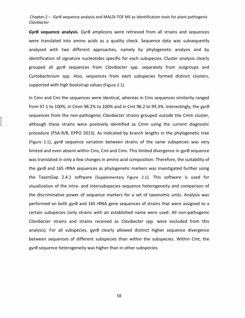
Chapter 2 – GyrB sequence analysis and MALDI-TOF MS as identification tools for plant pathogenic Clavibacter
58
GyrB sequence analysis. GyrB amplicons were retrieved from all strains and sequences
were translated into amino acids as a quality check. Sequence data was subsequently
analyzed with two different approaches, namely by phylogenetic analysis and by
identification of signature nucleotides specific for each subspecies. Cluster analysis clearly
grouped all gyrB sequences from Clavibacter spp. separately from outgroups and
Curtobacterium spp. Also, sequences from each subspecies formed distinct clusters,
supported with high bootstrap values (Figure 2.1).
In Cmn and Cmi the sequences were identical, whereas in Cms sequences similarity ranged
from 97.1 to 100%, in Cmm 98.2% to 100% and in Cmt 96.2 to 99.3%. Interestingly, the gyrB
sequences from the non-pathogenic Clavibacter strains grouped outside the Cmm cluster,
although these strains were positively identified as Cmm using the current diagnostic
procedure (PSA-R/8, EPPO 2013). As indicated by branch lengths in the phylogenetic tree
(Figure 2.1), gyrB sequence variation between strains of the same subspecies was very
limited and even absent within Cms, Cmi and Cmn. This limited divergence in gyrB sequence
was translated in only a few changes in amino acid composition. Therefore, the suitability of
the gyrB and 16S rRNA sequences as phylogenetic markers was investigated further using
the TaxonGap 2.4.1 software (Supplementary Figure 2.1). This software is used for
visualization of the intra- and intersubspecies sequence heterogeneity and comparison of
the discriminative power of sequence markers for a set of taxonomic units. Analysis was
performed on both gyrB and 16S rRNA gene sequences of strains that were assigned to a
certain subspecies (only strains with an established name were used. All non-pathogenic
Clavibacter strains and strains received as Clavibacter spp. were excluded from this
analysis). For all subspecies, gyrB clearly allowed distinct higher sequence divergence
between sequences of different subspecies than within the subspecies. Within Cmt, the
gyrB sequence heterogeneity was higher than in other subspecies.
Part III – Experimental work
59
Figure 2.1. Phylogenetic analysis of gyrB sequences. The Rooted neighbor-joining tree based on partial gyrB sequences (position 646-1143 of Cmm NCPPB 382 accession number AM711867) of 173 Clavibacter strains. Bootstrap values were generated from 1000 replicates. Rathayibacter sp. and Leifsonia aquatica were included as outgroups. NJ tree was in accordance with maximum parsimony and maximum likelihood analysis. GyrB sequences had minimum lengths of 500 bp.
In addition to the standard cluster analysis approach, aligned gyrB sequences were
manually searched for the presence of signature positions specific for each subspecies. In
total, 39 positions differed among subspecies throughout the gyrB fragment of 500 bp.
Based on these positions; unique signatures could be selected for each subspecies
(Supplementary Figure 2.3). Numbers of unique nucleotides ranged from two for Cmm to nine
Number of base substitutions per site
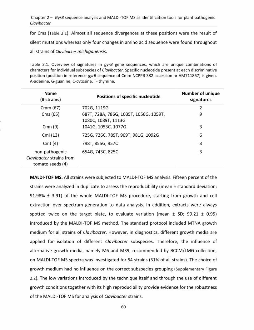
Chapter 2 – GyrB sequence analysis and MALDI-TOF MS as identification tools for plant pathogenic Clavibacter
60
for Cms (Table 2.1). Almost all sequence divergences at these positions were the result of
silent mutations whereas only four changes in amino acid sequence were found throughout
all strains of Clavibacter michiganensis.
Table 2.1. Overview of signatures in gyrB gene sequences, which are unique combinations of characters for individual subspecies of Clavibacter. Specific nucleotide present at each discriminative position (position in reference gyrB sequence of Cmm NCPPB 382 accession nr AM711867) is given. A-adenine, G-guanine, C-cytosine, T- thymine.
Name (# strains)
Positions of specific nucleotide Number of unique
signatures
Cmm (67) 702G, 1119G 2
Cms (65) 687T, 728A, 786G, 1035T, 1056G, 1059T, 1080C, 1089T, 1113G
9
Cmn (9) 1041G, 1053C, 1077G 3
Cmi (13) 725G, 726C, 789T, 969T, 981G, 1092G 6
Cmt (4) 798T, 855G, 957C 3
non-pathogenic Clavibacter strains from
tomato seeds (4)
654G, 743C, 825C 3
MALDI-TOF MS. All strains were subjected to MALDI-TOF MS analysis. Fifteen percent of the
strains were analyzed in duplicate to assess the reproducibility (mean ± standard deviation;
91.98% ± 3.91) of the whole MALDI-TOF MS procedure, starting from growth and cell
extraction over spectrum generation to data analysis. In addition, extracts were always
spotted twice on the target plate, to evaluate variation (mean ± SD; 99.21 ± 0.95)
introduced by the MALDI-TOF MS method. The standard protocol included MTNA growth
medium for all strains of Clavibacter. However, in diagnostics, different growth media are
applied for isolation of different Clavibacter subspecies. Therefore, the influence of
alternative growth media, namely M6 and M39, recommended by BCCM/LMG collection,
on MALDI-TOF MS spectra was investigated for 54 strains (31% of all strains). The choice of
growth medium had no influence on the correct subspecies grouping (Supplementary Figure
2.2). The low variations introduced by the technique itself and through the use of different
growth conditions together with its high reproducibility provide evidence for the robustness
of the MALDI-TOF MS for analysis of Clavibacter strains.
Part III – Experimental work
61
The MALDI-TOF MS data analyzed in BioNumerics 5.1 and DataExplorer 4.0 software were in
agreement with the gyrB sequence analysis. Indeed, cluster analysis of the whole spectral
profile showed distinct clusters for each subspecies, non-pathogenic Clavibacter strains and
outgroups (data not shown). However, Cmm strains formed two separate groups, one with
only a few strains that closely clustered to Cmn strains and the other with a majority of the
Cmm strains. Next to cluster analyses, the obtained spectral profiles were also screened for
the presence of recurring peaks or biomarker ions specific for the whole species or a
subspecies. A selection of 20 profiles of representatives of all Clavibacter subspecies,
including the type strains, was visually screened for the most informative and intensive
peaks.
A typical MALDI-TOF MS spectrum of Clavibacter michiganensis contains about 50 ion peaks
between 2,000 and 20,000 Da, with the highest intensity peaks between 4,000 and 10,000
Da. Table 2.2 summarizes 32 selected m/z values. Six m/z values were detected in all
subspecies, making them characteristic for the genus Clavibacter. Unique peaks for each
subspecies ranged from one in Cmt to five in Cms (Figure 2.2, Table 2.2). In order to validate
these peaks as biomarkers, analysis of another 25 randomly selected Clavibacter profiles
was performed and the assignment to the expected subspecies was obtained.
The applied cell extraction protocol favors isolation of ribosomal subunit proteins.
Therefore, we tried to assign the selected biomarkers by comparing the experimentally
obtained protein masses with those of the ribosomal subunit proteins deduced from two
fully sequenced Clavibacter genomes (Cmm NCPPB 382 and Cms ATCC 33113) using the
RMIDb database. In this way, eleven biomarkers could be identified as specific ribosomal
proteins (Figure 2.2). However, our assignment procedure left several masses unidentified,
probably because of slight differences in protein mass between different subspecies (only
genomes of representatives of two subspecies were available), inclusion of nonribosomal
proteins in the analyzed cell extracts and possible occurrence of 2+ ions.
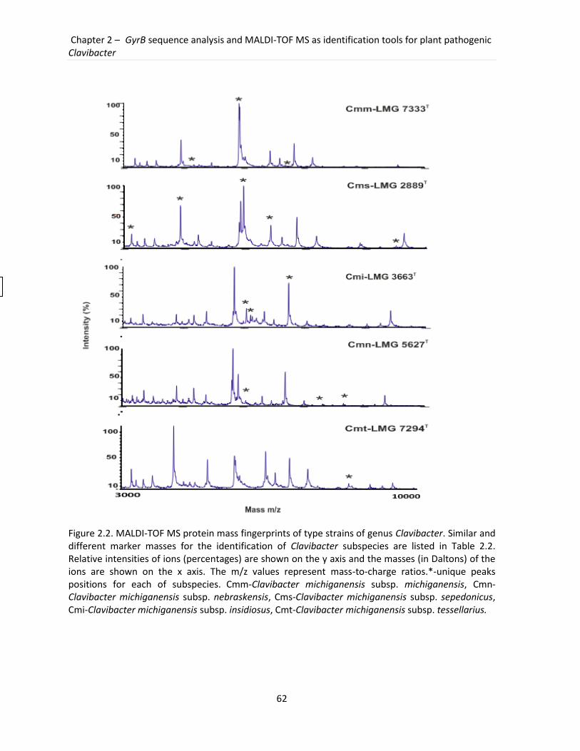
Chapter 2 – GyrB sequence analysis and MALDI-TOF MS as identification tools for plant pathogenic Clavibacter
62
Figure 2.2. MALDI-TOF MS protein mass fingerprints of type strains of genus Clavibacter. Similar and different marker masses for the identification of Clavibacter subspecies are listed in Table 2.2. Relative intensities of ions (percentages) are shown on the y axis and the masses (in Daltons) of the ions are shown on the x axis. The m/z values represent mass-to-charge ratios.*-unique peaks positions for each of subspecies. Cmm-Clavibacter michiganensis subsp. michiganensis, Cmn-Clavibacter michiganensis subsp. nebraskensis, Cms-Clavibacter michiganensis subsp. sepedonicus, Cmi-Clavibacter michiganensis subsp. insidiosus, Cmt-Clavibacter michiganensis subsp. tessellarius.
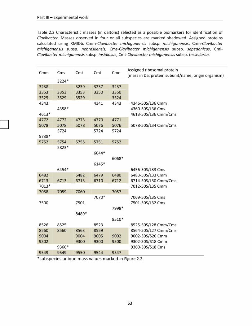
Part III – Experimental work
63
Table 2.2 Characteristic masses (in daltons) selected as a possible biomarkers for identification of Clavibacter. Masses observed in four or all subspecies are marked shadowed. Assigned proteins calculated using RMIDb. Cmm-Clavibacter michiganensis subsp. michiganensis, Cmn-Clavibacter michiganensis subsp. nebraskensis, Cms-Clavibacter michiganensis subsp. sepedonicus, Cmi-Clavibacter michiganensis subsp. insidiosus, Cmt-Clavibacter michiganensis subsp. tessellarius.
Cmm Cms Cmt Cmi Cmn Assigned ribosomal protein (mass in Da, protein subunit/name, origin organism)
3224*
3238
3239 3237 3237 3353 3353 3353 3350 3350 3525 3529 3529
3524
4343
4341 4343 4346-50S/L36 Cmm
4358*
4360-50S/L36 Cms
4613*
4613-50S/L36 Cmm/Cms 4772 4772 4773 4770 4771
5078 5078 5078 5076 5076 5078-50S/L34 Cmm/Cms
5724
5724 5724
5738* 5752 5754 5755 5751 5752
5823*
6044*
6068*
6145*
6454*
6456-50S/L33 Cms 6482
6482 6479 6480 6483-50S/L33 Cmm
6713 6713 6713 6710 6712 6714-50S/L30 Cmm/Cms 7013*
7012-50S/L35 Cmm
7058 7059 7060
7057
7070*
7069-50S/L35 Cms
7500
7501
7501-50S/L32 Cms
7998*
8489*
8510*
8526 8525
8523
8525-50S/L28 Cmm/Cms 8560 8560 8563 8559
8564-50S/L27 Cmm/Cms
9004
9004 9005 9002 9002-30S/S20 Cmm 9302
9300 9300 9300 9302-30S/S18 Cmm
9360*
9360-30S/S18 Cms
9549 9549 9550 9544 9547 *subspecies unique mass values marked in Figure 2.2.
Chapter 2 – GyrB sequence analysis and MALDI-TOF MS as identification tools for plant pathogenic Clavibacter
64
2.4 Discussion
This study demonstrated the suitability of gyrB sequences and MALDI-TOF MS protein
profiles for identification of plant pathogenic bacteria of the genus Clavibacter. The
identification can be based on the generated data as a whole, or on signatures within these
data which have enough discriminatory power to distinguish between the different
subspecies of Clavibacter michiganensis. Moreover, both the sequence and the protein
method suggested that diversity within this Clavibacter michiganensis species is higher than
currently accepted. This study reports for the first time on the use of gyrB as a phylogenetic
marker in the genus Clavibacter. Molecular markers for identification purposes must exhibit
the smallest amount of heterogeneity within a taxonomic unit and should maximally
discriminate between different species/subspecies (Martens et al., 2008), which was
demonstrated in this study for gyrB sequences. In addition to cluster analysis, a selection of
positions in the gyrB contained nucleotide signatures characteristic for each subspecies and
were shown to be decisive for subspecies assignment. This character-based identification
approach was previously proposed as an alternative for tree building methods (Rach et al.,
2008). However, in bacterial taxonomy, this approach is not as popular as in human
medicine where SNPs (single nucleotide polymorphisms) are commonly used in
identification of pathogens or loci associated with complex diseases (Degefu 2006; Lai
2001). These SNPs have also been used to design highly specific molecular tools for
identification (Umeyama et al., 2006). Furthermore, the fact that the gyrB fragment can be
amplified and sequenced with the same protocol in all Clavibacter and closely related
strains tested, creates a great potential for future applications as a new diagnostic tool.
Based on the signature nucleotide positions, new methods for specific PCR and qPCR
detection of quarantine Cmm, Cmi and Cms in plant material may be developed.
Because of its simplicity and applicability, MALDI-TOF MS is already widely used for
identification and characterization of diverse microorganisms (Giebel et al., 2010, Qian et
al., 2008, Rezzonico et al., 2010). Currently, spectral data are commonly analyzed using two
different approaches. Specific biomarker ions are identified based on additional information
on theoretical protein masses (from e.g. genome projects). New profiles of unknown strains

Part III – Experimental work
65
are then screened for presence/absence of these unique biomarker ions to subsequently
assign to a certain taxonomic group (Pineda et al., 2003). This approach is more suitable for
discrimination between closely related species as well as for accurate identification
(Dieckmann et al., 2010). Another workflow uses a database with reference fingerprinting
profiles obtained as a consensus profile from a set of type strains. New spectra are then
compared with database entries and the closest spectrum is found (Jarman et al., 2000).
Widely used commercial systems, such as SARAMIS (Spectral ARchiving and Microbial
Identification System, AnagnosTec) and Maldi Biotyper (Bruker Daltonics), contain average
spectra together with peak lists of the most discriminating biomarker ions which are scored
according to their occurrence and specificity. Because we wanted to use both the whole
spectral profile and biomarker ions, we used BioNumerics and DataExplorer 4.0 (AB Sciex)
respectively, as an alternative to commercially available database systems. Both software
applications allowed us to look at the spectra in detail, perform our own quality check and
analyze the data in different ways. Moreover, BioNumerics software allowed the coupling of
additional data from more than one experiment type (e.g. sequencing information) making
the analysis easier, more comprehensive and straightforward. However, the use of non-
specialized software also had the disadvantages that (i) no specific experiment type was
available for spectral profiles and (ii) biomarker ions detection had to be performed
manually because appropriate tools were lacking. There is still debate on the discriminatory
level of MALDI-TOF MS, which is said to be accurate at genus, species (Rezzonico et al.,
2010) or even below species level (Barbuddhe et al., 2008). Nevertheless, a very good
discrimination between Clavibacter subspecies was consistently obtained. As MALDI-TOF
MS is gaining more popularity as a quick and robust identification tool, required software
tools will probably be available in the near future.
To evaluate the robustness and discriminatory power of gyrB sequencing and MALDI-TOF
MS analysis, the closely related Clavibacter strains were analyzed together with non-
pathogenic Clavibacter strains. These strains are an especially interesting group of
organisms that possibly form a new group of Clavibacter since up to now it is little known
about non-pathogenic bacteria within this genus (Zinniel et al., 2002). Both techniques
Chapter 2 – GyrB sequence analysis and MALDI-TOF MS as identification tools for plant pathogenic Clavibacter
66
applied in this study were discriminative enough to differentiate strains of the closely
related genus Curtobacterium. Misidentification of Curtobacterium as Clavibacter could be
attributed to their high morphological similarities or contamination during the handling.
Most of the members of the genus Curtobacterium have been isolated from plants but only
one species, Curtobacterium flaccumfaciens pv. flaccumfaciens, is regarded as a plant
pathogen. Because of its importance, this species is also classified as a quarantine organism
on the A2 EPPO list and in the Council Directive 2000/29/EC (Anonymous 2000; EPPO
2005b). Problems in the discrimination of Curtobacterium from Clavibacter were
encountered previously in a numerical classification study of coryneforms (Kämpfer et al.,
1993), where members of both groups were allocated in the same cluster.
The results obtained with the genetic- and protein-based approach used in our study were
in perfect agreement and showed their value as stand-alone diagnostic tools for the genus
Clavibacter. Whereas the sequence-based approach is more time consuming, it is surely a
valuable tool when MALDI-TOF MS is not available. But, increasing applicability of MALDI-
TOF MS will make it more accessible for the end users. Besides, low costs of sample
preparation and rapid measurement procedure are of great value for unbiased and fast
diagnostics.
Acknowledgements
We thank the NVWA, GBBC and BCCM/LMG collections and Ana Rodríguez Pérez (Spain) for
providing necessary strains. We also thank An Coorevits for assistance with TaxonGap
software. This work was performed in the Seventh Framework Programme of project KBBE-
2008-1-4-01 nr 226482 (QBOL) funded by the European Commission. The Research Fund
University College Ghent is acknowledged for the postdoctoral fellowship of Koenraad Van
Hoorde and the Belgian NPPO (FAVV) for partially financing ILVO-research.
Part III – Experimental work
67
2.5 General reflections
In this chapter, gyrB barcode and MALDI-TOF MS were applied as new identification
approaches for plant pathogenic clavibacters. A high number of members of Clavibacter and
closely related taxa, such as Rathayibacter, Curtobacterium and Leifsonia, as well as strains
that occupy the same niche and morphologically resemble Clavibacter, was included to
cover as much as possible the natural diversity. In a two-step barcoding approach including
16S rRNA and gyrB sequencing the strains were first assigned at the genus and
subsequently at the subspecies level, respectively. In addition to the sequence based
approach, a protein-based method realized by MALDI-TOF MS proved to be useful for
identification of Clavibacter subspecies.
As identification might be hampered by the taxonomic/phylogenetic complexity of the
group under investigation we have first re-evaluated the taxonomic situation of Clavibacter.
Therefore, we needed to collect a sufficient number of strains covering the natural diversity.
The main obstacle we experienced at the beginning of the project was the limited number
of available specimens and general difficulties to obtain bacteria classified as quarantine
organisms. Because of additional restrictions concerning the transport and exchange of
organisms with a quarantine status collecting the necessary number of strains was very
problematic. The common efforts of many collaborators and significant contributions of
three main bacterial collections, namely BCCM-LMG (Ghent, Belgium), GBBC (Merelbeke,
Belgium) and NVWA (The Netherlands) helped to obtain the required strains. The subset of
173 strains of Clavibacter and close relatives that were subjected to phylogenetic analysis
by 16S rRNA and gyrB gene sequences confirmed the current taxonomic classification of five
Clavibacter subspecies. In addition, the presence of a new group of non-pathogenic seed-
borne Clavibacter strains was detected. These findings suggested that the intragenetic
diversity of Clavibacter was higher than originally expected.
The development of a new identification method for Clavibacter originated from the
necessity to provide a more reliable, robust and universal technique because the currently
applied identification methods are prone to false positive and/or negative results (Jacques
et al., 2012). Barcoding had been successfully applied to identify various organisms,
Chapter 2 – GyrB sequence analysis and MALDI-TOF MS as identification tools for plant pathogenic Clavibacter
68
especially a 648 bp fragment of COX1 gene proved to be very useful in the identification of a
wide range of animal species (Hajibabaei et al., 2007). However, the assumption of a
development of only one universal barcode for identification of different bacteria, fungi or
phytoplasmas, appeared to be an unachievable goal and the preliminary results of the QBOL
project showed that the strategy had to be directed towards the development of one or
more group-specific barcodes. In the case of Clavibacter, first, a partial 16S rRNA gene
sequence was used to assign strains at the genus level and a second barcode, gyrB, was
applied for identification at the subspecies level. The identification scheme developed for
the genus Clavibacter was working well in the collection of various strains that were
included in the tests. In the genus Xanthomonas, other bacterial pathogens also included in
the QBOL project, the situation appeared to be much more complicated, which resulted in
an identification employing a multi-step approach with more than two barcodes (data not
published).
A very important aspect of this project concerned the construction of an internet-based
freely available database, in which not only the sequences generated in the course of the
project were deposited, but also additional information regarding taxonomy, physiology
and biology of the quarantine organisms is included. The database Q-Bank (www.q-bank.eu)
is now a comprehensive source of information about quarantine organisms. The database is
not restricted to bacteria but contains information about quarantine phytoplasmas,
nematodes, viruses, arthropods and fungi.
A unique asset of this project was also the creation of a DNA bank where the reference
voucher specimens are available and can be requested by the end users for positive and
negative controls during the barcoding identification procedure.
Despite obvious advantages of barcoding, such as rapid and relatively inexpensive
identification of a specimen and the possibility to create an exchangeable database to
identify an unknown organism, the use of barcoding is still not fully accepted for the
discovery of a new species and for taxonomic studies. The main issue of DNA barcoding
concerns the way how barcode data should be read and interpreted. Some approaches
utilize distance measures to be able to delineate a species (Hebert et al., 2003). The
Part III – Experimental work
69
simplest example of this approach is the use of BLAST (Altschul et al., 1990) in which
similarities between sequences determine the best matches.
A limitation of distance based approaches for identification concerns the fact that similarity
values indicating the nearest neighbour do not necessary direct to the closest relative
(which might not yet be included in the database). As a consequence, the results might be
misleading. The distance methods need to use a threshold value that enables delineation of
a group. So far, however, threshold values differ depending on the group of organism under
investigation and no uniformly accepted value has been approved to for example assign a
species. Having in mind the possibility to use various models in distance methods it can be
supposed that the end result may be different depending on the chosen parameters. To
overcome this obstacle an alternative manner of barcode interpretation should be included,
namely, the identification based on the specific character position. In that way the presence
of certain characters (nucleotide bases) in the particular sequence positions will provide an
evidence for identification. In our study, both approaches, namely distance- and character-
based, were successfully applied to identify unknown Clavibacter strains. In the character-
based approach the number of subspecies specific characters was rather small (only two
specific characters-signatures for Cmm strains, Table 2.1). Despite there are no rules
concerning the sufficient number of group-specific characters, a higher number of such
markers provide an extra assurance for correct identification. The reason for the limited
number of subspecies-specific positions in Clavibacter strains was a direct consequence of
the relatively high homogeneity among e.g. Cmm strains (Figure 3.3). To overcome this
limitation instead of the gyrB barcode of 500 bp length a longer fragment could have been
used. After the decision about the gyrB barcode was made and a significant number of
sequences were generated in our study, the group of Schneider and coworkers proposed
the sequence of dnaA that showed promising results in classification of plant pathogenic
bacteria including Clavibacter (Schneider et al., 2011). The proposed sequence of dnaA was
indeed longer than the gyrB barcode and like the gyrB allowed successful identification of
the Clavibacter subspecies. But it was only tested on a limited number of strains and the
primers used for amplification of this gene appeared to be specific for Clavibacter. At the
Chapter 2 – GyrB sequence analysis and MALDI-TOF MS as identification tools for plant pathogenic Clavibacter
70
initial stage of the barcoding project we intended to find a single barcode for clavibacters
and relatives. Therefore, designed primers for a selected gene had to be universal enough
to obtain an amplicon also from close relatives to strengthen the identification. Only later,
after it was decided that two-step identification had to be applied this issue became less
important and dnaA primers specific for Clavibacter would be equally useful. Although not
included in the barcoding scheme for Clavibacter, the applicability of dnaA sequences to
study the Clavibacter population was assessed at a later stage of our research where dnaA
and gyrB gene sequences were used for characterization and diversity studies of non-
pathogenic Clavibacter strains as well as for a phylogenetic analysis of Cmm strains from
Belgian outbreaks of bacterial wilt and canker on tomato.
The length and the specificity of the chosen gyrB barcode at the beginning of the project
were sufficient for a correct identification of the five known Clavibacter subspecies and four
non-pathogenic Clavibacter strains that could not be classified to any of the known
subspecies. After more representatives of seed-borne non-pathogenic Clavibacters were
included it appeared that their diversity is much higher than originally anticipated. As a
consequence of the dynamic changes of the genetic diversity in the population of non-
pathogenic Clavibacter strains, the number of representatives included in the QBOL project
might not completely reflect the actual diversity of these non-pathogenic strains.
With the changing standards in the diagnostic laboratories and the growing needs to
analyze many strains at the same time it is required to use new, more sophisticated
techniques that allow rapid and high throughput identification. To meet these expectations
we subjected a protein-based approach realized by MALDI-TOF MS for identification of
Clavibacter strains. Over the last two decades MALDI-TOF MS gained a lot of attention and
was applied for identification of various organisms including many bacteria (Barbuddhe et
al., 2008, Hsieh et al., 2008, Ruelle et al., 2004). Its success is attributed to the ease of use,
the possibility of automation and to its high throughput capacity. When we started testing
MALDI-TOF MS for Clavibacter identification the system was only recently introduced in the
laboratory and the way of material preparation and data analysis was not yet fully
developed. Therefore, the initial steps were directed to explore the effect of growth
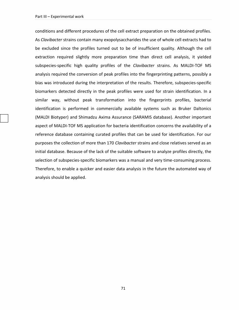
Part III – Experimental work
71
conditions and different procedures of the cell extract preparation on the obtained profiles.
As Clavibacter strains contain many exopolysaccharides the use of whole cell extracts had to
be excluded since the profiles turned out to be of insufficient quality. Although the cell
extraction required slightly more preparation time than direct cell analysis, it yielded
subspecies-specific high quality profiles of the Clavibacter strains. As MALDI-TOF MS
analysis required the conversion of peak profiles into the fingerprinting patterns, possibly a
bias was introduced during the interpretation of the results. Therefore, subspecies-specific
biomarkers detected directly in the peak profiles were used for strain identification. In a
similar way, without peak transformation into the fingerprints profiles, bacterial
identification is performed in commercially available systems such as Bruker Daltonics
(MALDI Biotyper) and Shimadzu Axima Assurance (SARAMIS database). Another important
aspect of MALDI-TOF MS application for bacteria identification concerns the availability of a
reference database containing curated profiles that can be used for identification. For our
purposes the collection of more than 170 Clavibacter strains and close relatives served as an
initial database. Because of the lack of the suitable software to analyze profiles directly, the
selection of subspecies-specific biomarkers was a manual and very time-consuming process.
Therefore, to enable a quicker and easier data analysis in the future the automated way of
analysis should be applied.
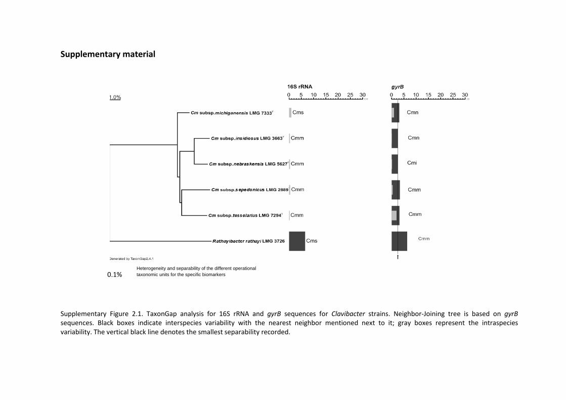
0.1%
Supplementary material
Supplementary Figure 2.1. TaxonGap analysis for 16S rRNA and gyrB sequences for Clavibacter strains. Neighbor-Joining tree is based on gyrB sequences. Black boxes indicate interspecies variability with the nearest neighbor mentioned next to it; gray boxes represent the intraspecies variability. The vertical black line denotes the smallest separability recorded.
Heterogeneity and separability of the different operational
taxonomic units for the specific biomarkers
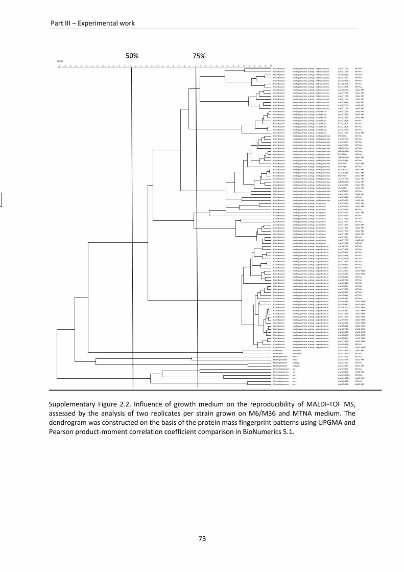
Part III – Experimental work
73
Supplementary Figure 2.2. Influence of growth medium on the reproducibility of MALDI-TOF MS, assessed by the analysis of two replicates per strain grown on M6/M36 and MTNA medium. The dendrogram was constructed on the basis of the protein mass fingerprint patterns using UPGMA and Pearson product-moment correlation coefficient comparison in BioNumerics 5.1.
Pearson correlation [5.0%-100.0%]
MALDI
100
989694929088868482807876747270686664626058565452504846444240383634323028262422
.
.
.
.
.
.
.
.
.
.
.
.
.
.
.
.
.
.
.
.
.
.
.
.
.
.
.
.
.
.
.
.
.
.
.
.
.
.
.
.
.
.
.
.
.
.
.
.
.
.
.
.
.
.
.
.
.
.
.
.
.
.
.
.
.
.
.
.
.
.
.
.
.
.
.
.
.
.
.
.
.
.
.
.
.
.
.
.
.
.
.
.
.
.
.
.
.
.
.
.
.
.
.
.
Clavibacter
Clavibacter
Clavibacter
Clavibacter
Clavibacter
Clavibacter
Clavibacter
Clavibacter
Clavibacter
Clavibacter
Clavibacter
Clavibacter
Clavibacter
Clavibacter
Clavibacter
Clavibacter
Clavibacter
Clavibacter
Clavibacter
Clavibacter
Clavibacter
Clavibacter
Clavibacter
Clavibacter
Clavibacter
Clavibacter
Clavibacter
Clavibacter
Clavibacter
Clavibacter
Clavibacter
Clavibacter
Clavibacter
Clavibacter
Clavibacter
Clavibacter
Clavibacter
Clavibacter
Clavibacter
Clavibacter
Clavibacter
Clavibacter
Clavibacter
Clavibacter
Clavibacter
Clavibacter
Clavibacter
Clavibacter
Clavibacter
Clavibacter
Clavibacter
Clavibacter
Clavibacter
Clavibacter
Clavibacter
Clavibacter
Clavibacter
Clavibacter
Clavibacter
Clavibacter
Clavibacter
Clavibacter
Clavibacter
Clavibacter
Clavibacter
Clavibacter
Clavibacter
Clavibacter
Clavibacter
Clavibacter
Clavibacter
Clavibacter
Clavibacter
Clavibacter
Clavibacter
Clavibacter
Clavibacter
Clavibacter
Clavibacter
Clavibacter
Clavibacter
Clavibacter
Clavibacter
Clavibacter
Clavibacter
Clavibacter
Clavibacter
Clavibacter
Clavibacter
Clavibacter
Clavibacter
Clavibacter
Leifsonia
Leifsonia
Rathayibacter
Rathayibacter
Rathayibacter
Rathayibacter
Curtobacterium
Curtobacterium
Curtobacterium
Curtobacterium
Curtobacterium
Curtobacterium
michiganensis subsp. nebraskensis
michiganensis subsp. nebraskensis
michiganensis subsp. nebraskensis
michiganensis subsp. nebraskensis
michiganensis subsp. nebraskensis
michiganensis subsp. nebraskensis
michiganensis subsp. nebraskensis
michiganensis subsp. nebraskensis
michiganensis subsp. nebraskensis
michiganensis subsp. nebraskensis
michiganensis subsp. nebraskensis
michiganensis subsp. nebraskensis
michiganensis subsp. nebraskensis
michiganensis subsp. nebraskensis
michiganensis subsp. tesselarius
michiganensis subsp. tesselarius
michiganensis subsp. tesselarius
michiganensis subsp. tesselarius
michiganensis subsp. tesselarius
michiganensis subsp. tesselarius
michiganensis subsp. tesselarius
michiganensis subsp. tesselarius
michiganensis subsp. michiganensis
michiganensis subsp. michiganensis
michiganensis subsp. michiganensis
michiganensis subsp. michiganensis
michiganensis subsp. michiganensis
michiganensis subsp. michiganensis
michiganensis subsp. michiganensis
michiganensis subsp. michiganensis
michiganensis subsp. michiganensis
michiganensis subsp. michiganensis
michiganensis subsp. michiganensis
michiganensis subsp. michiganensis
michiganensis subsp. michiganensis
michiganensis subsp. michiganensis
michiganensis subsp. michiganensis
michiganensis subsp. michiganensis
michiganensis subsp. michiganensis
michiganensis subsp. michiganensis
michiganensis subsp. michiganensis
michiganensis subsp. michiganensis
michiganensis subsp. michiganensis
michiganensis subsp. michiganensis
michiganensis subsp. insidiosus
michiganensis subsp. insidiosus
michiganensis subsp. insidiosus
michiganensis subsp. insidiosus
michiganensis subsp. insidiosus
michiganensis subsp. insidiosus
michiganensis subsp. insidiosus
michiganensis subsp. insidiosus
michiganensis subsp. insidiosus
michiganensis subsp. insidiosus
michiganensis subsp. insidiosus
michiganensis subsp. insidiosus
michiganensis subsp. insidiosus
michiganensis subsp. insidiosus
michiganensis subsp. sepedonicus
michiganensis subsp. sepedonicus
michiganensis subsp. sepedonicus
michiganensis subsp. sepedonicus
michiganensis subsp. sepedonicus
michiganensis subsp. sepedonicus
michiganensis subsp. sepedonicus
michiganensis subsp. sepedonicus
michiganensis subsp. sepedonicus
michiganensis subsp. sepedonicus
michiganensis subsp. sepedonicus
michiganensis subsp. sepedonicus
michiganensis subsp. sepedonicus
michiganensis subsp. sepedonicus
michiganensis subsp. sepedonicus
michiganensis subsp. sepedonicus
michiganensis subsp. sepedonicus
michiganensis subsp. sepedonicus
michiganensis subsp. sepedonicus
michiganensis subsp. sepedonicus
michiganensis subsp. sepedonicus
michiganensis subsp. sepedonicus
michiganensis subsp. sepedonicus
michiganensis subsp. sepedonicus
michiganensis subsp. sepedonicus
michiganensis subsp. sepedonicus
michiganensis subsp. sepedonicus
michiganensis subsp. sepedonicus
michiganensis subsp. sepedonicus
michiganensis subsp. sepedonicus
michiganensis subsp. sepedonicus
michiganensis subsp. sepedonicus
michiganensis subsp. sepedonicus
michiganensis subsp. sepedonicus
aquatica
aquatica
tritici
tritici
rathayi
rathayi
sp.
sp.
sp.
sp.
sp.
sp.
.
.
.
.
.
.
.
.
.
.
.
.
.
.
.
.
.
.
.
.
.
.
.
.
.
.
.
.
.
.
.
.
.
.
.
.
.
.
.
.
.
.
.
.
.
.
.
.
.
.
.
.
.
.
.
.
.
.
.
.
.
.
.
.
.
.
.
.
.
.
.
.
.
.
.
.
.
.
.
.
.
.
.
.
.
.
.
.
.
.
.
.
.
.
.
.
.
.
.
.
.
.
.
.
LMG7174
LMG7176
LMG3699
LMG7177
LMG3700
LMG5625
LMG7285
LMG5625
LMG7285
LMG7176
LMG7174
LMG3699
LMG3700
LMG7177
LMG7296
LMG7299
LMG7300
LMG7299
LMG7294
LMG7296
LMG7300
LMG7294
PD5753
LMG5726
LMG3683
LMG3681
GBBC209
GBBC296
PD5736
GBBC296
LMG5644
PD5726
PD5719
LMG5602
LMG5644
PD5753
LMG5726
GBBC209
LMG3681
PD5719
LMG5602
LMG3683
LMG3695
LMG3595
LMG3660
LMG3662
LMG3660
LMG3663
LMG3663
LMG7326
LMG7267
LMG7324
LMG7332
LMG7272
LMG7326
LMG7324
LMG7267
LMG7272*
LMG6720
LMG7368
LMG5860
LMG2889
LMG2901*
LMG7359
LMG5960
LMG2894
LMG2894
LMG2899
LMG5872
LMG6712
LMG2899
LMG5874
LMG7362
LMG6526
LMG5842
LMG6317
LMG5876
LMG5842
LMG6720
LMG6317
LMG7359
LMG7362
LMG5960
LMG2889
LMG5872
LMG6712
LMG6526
LMG5860
LMG5874
LMG7368
LMG5876
LMG2901
LMG18702
LMG18702
LMG3726
LMG3726
LMG3717
LMG3717
LMG3680
LMG3680
LMG18661
LMG18661
LMG2896
LMG2896
.
.
.
.
.
.
.
.
.
.
.
.
.
.
.
.
.
.
.
.
.
.
.
.
.
.
.
.
.
.
.
.
.
.
.
.
.
.
.
.
.
.
.
.
.
.
.
.
.
.
.
.
.
.
.
.
.
.
.
.
.
.
.
.
.
.
.
.
.
.
.
.
.
.
.
.
.
.
.
.
.
.
.
.
.
.
.
.
.
.
.
.
.
.
.
.
.
.
.
.
.
.
.
.
.
.
.
.
.
.
.
.
.
.
.
.
.
.
.
.
.
.
.
.
.
.
.
.
.
.
.
.
.
.
.
.
.
.
.
.
.
.
.
.
.
.
.
.
.
.
.
.
.
.
.
.
.
.
.
.
.
.
.
.
.
.
.
.
.
.
.
.
.
.
.
.
.
.
.
.
.
.
.
.
.
.
.
.
.
.
.
.
.
.
.
.
.
.
.
.
.
.
.
.
.
.
.
.
.
.
.
.
.
.
.
.
.
.
.
.
.
.
.
.
.
.
.
.
.
.
.
.
.
.
.
.
.
.
.
.
.
.
.
.
.
.
.
.
.
.
.
.
.
.
.
.
.
.
.
.
.
.
.
.
.
.
.
.
.
.
.
.
.
.
.
.
.
.
.
.
.
.
.
.
.
.
.
.
.
.
.
.
.
.
.
.
.
.
.
.
.
.
.
.
.
.
.
.
.
.
.
.
.
.
.
.
.
.
.
.
.
.
.
.
.
.
.
.
.
.
.
.
.
.
.
.
.
.
.
.
.
.
.
.
.
.
.
.
.
.
.
.
.
.
.
.
.
.
.
.
.
.
.
.
.
.
.
.
.
.
MTNA
MTNA
MTNA
MTNA
MTNA
MTNA
MTNA
LMG M6
LMG M6
LMG M6
LMG M6
LMG M6
LMG M6
LMG M6
LMG M6
LMG M6
LMG M6
MTNA
MTNA
MTNA
MTNA
LMG M6
MTNA
MTNA
MTNA
MTNA
MTNA
MTNA
MTNA
LMG M6
MTNA
LMG M6
MTNA
LMG M6
LMG M6
LMG M6
LMG M6
LMG M6
LMG M6
LMG M6
MTNA
LMG M6
MTNA
LMG M6
LMG M6
LMG M6
MTNA
LMG M6
MTNA
MTNA
MTNA
LMG M6
LMG M6
LMG M6
LMG M6
MTNA
LMG M6
MTNA
MTNA
MTNA
MTNA
MTNA
MTNA
MTNA
MTNA
MTNA
LMG M39
LMG M39
MTNA
MTNA
MTNA
MTNA
MTNA
MTNA
MTNA
MTNA
LMG M39
LMG M39
LMG M39
LMG M39
LMG M39
LMG M39
LMG M39
LMG M39
LMG M39
LMG M39
LMG M39
LMG M39
LMG M39
LMG M39
MTNA
LMG M39
LMG M6
MTNA
MTNA
LMG M6
MTNA
LMG M6
MTNA
LMG M6
MTNA
LMG M6
MTNA
LMG M6
75% 50%
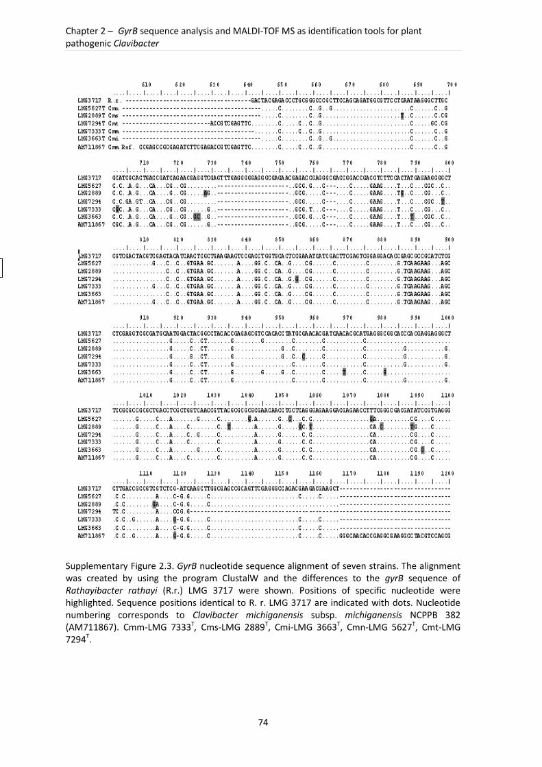
Chapter 2 – GyrB sequence analysis and MALDI-TOF MS as identification tools for plant pathogenic Clavibacter
74
Supplementary Figure 2.3. GyrB nucleotide sequence alignment of seven strains. The alignment was created by using the program ClustalW and the differences to the gyrB sequence of Rathayibacter rathayi (R.r.) LMG 3717 were shown. Positions of specific nucleotide were highlighted. Sequence positions identical to R. r. LMG 3717 are indicated with dots. Nucleotide numbering corresponds to Clavibacter michiganensis subsp. michiganensis NCPPB 382 (AM711867). Cmm-LMG 7333T, Cms-LMG 2889T, Cmi-LMG 3663T, Cmn-LMG 5627T, Cmt-LMG 7294T.
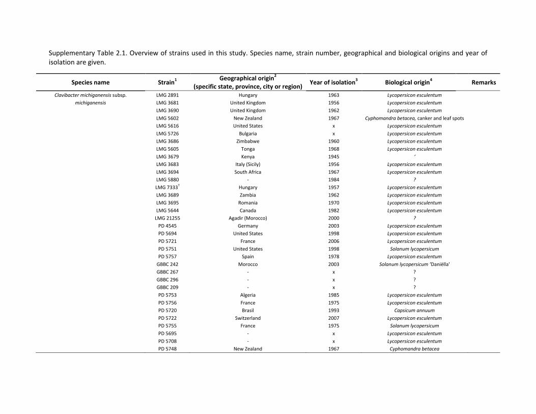
Supplementary Table 2.1. Overview of strains used in this study. Species name, strain number, geographical and biological origins and year of isolation are given.
Species name Strain1
Geographical origin2
(specific state, province, city or region) Year of isolation
3 Biological origin
4
Remarks
Clavibacter michiganensis subsp. LMG 2891 Hungary 1963 Lycopersicon esculentum
michiganensis LMG 3681 United Kingdom 1956 Lycopersicon esculentum
LMG 3690 United Kingdom 1962 Lycopersicon esculentum
LMG 5602 New Zealand 1967 Cyphomandra betacea, canker and leaf spots
LMG 5616 United States x Lycopersicon esculentum
LMG 5726 Bulgaria x Lycopersicon esculentum
LMG 3686 Zimbabwe 1960 Lycopersicon esculentum
LMG 5605 Tonga 1968 Lycopersicon esculentum
LMG 3679 Kenya 1945 ‘
LMG 3683 Italy (Sicily) 1956 Lycopersicon esculentum
LMG 3694 South Africa 1967 Lycopersicon esculentum
LMG 5880 - 1984 ?
LMG 7333T Hungary 1957 Lycopersicon esculentum
LMG 3689 Zambia 1962 Lycopersicon esculentum
LMG 3695 Romania 1970 Lycopersicon esculentum
LMG 5644 Canada 1982 Lycopersicon esculentum
LMG 21255 Agadir (Morocco) 2000 ?
PD 4545 Germany 2003 Lycopersicon esculentum
PD 5694 United States 1998 Lycopersicon esculentum
PD 5721 France 2006 Lycopersicon esculentum
PD 5751 United States 1998 Solanum lycopersicum
PD 5757 Spain 1978 Lycopersicon esculentum
GBBC 242 Morocco 2003 Solanum lycopersicum ’Daniëlla'
GBBC 267 - x ?
GBBC 296 - x ?
GBBC 209 - x ?
PD 5753 Algeria 1985 Lycopersicon esculentum
PD 5756 France 1975 Lycopersicon esculentum
PD 5720 Brasil 1993 Capsicum annuum
PD 5722 Switzerland 2007 Lycopersicon esculentum
PD 5755 France 1975 Solanum lycopersicum
PD 5695 - x Lycopersicon esculentum
PD 5708 - x Lycopersicon esculentum
PD 5748 New Zealand 1967 Cyphomandra betacea
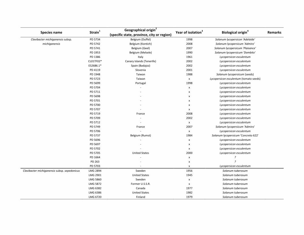
Species name Strain1
Geographical origin2
(specific state, province, city or region) Year of isolation
3 Biological origin
4 Remarks
Clavibacter michiganensis subsp. PD 5734 Belgium (Duffel) 1998 Solanum lycopersicon 'Adelaïde'
michiganensis PD 5742 Belgium (Kontich) 2008 Solanum lycopersicum 'Admiro'
PD 5741 Belgium (Geel) 2007 Solanum lycopersicum 'Plaisance'
PD 1953 Belgium (Melsele) 1990 Solanum lycopersicum 'Dombito'
PD 1386 Italy 1961 Lycopersicon esculentum
CL01TF02* Canary Islands (Tenerife) 2002 Lycopersicon esculentum
ES2686.1* Spain (Badajoz) 2002 Lycopersicon esculentum
PD 4119 Slovenia 2001 Lycopersicon esculentum
PD 1948 Taiwan 1988 Solanum lycopersicum (seeds)
PD 5723 Taiwan x Lycopersicon esculentum (tomato seeds)
PD 5699 Portugal 1998 Lycopersicon esculentum
PD 5704 - x Lycopersicon esculentum
PD 5711 - x Lycopersicon esculentum
PD 5698 - x Lycopersicon esculentum
PD 5701 - x Lycopersicon esculentum
PD 5700 - x Lycopersicon esculentum
PD 5707 - x Lycopersicon esculentum
PD 5719 France 2008 Lycopersicon esculentum
PD 5709 - 2002 Lycopersicon esculentum
PD 5712 - x Lycopersicon esculentum
PD 5749 France 2007 Solanum lycopersicum 'Admiro'
PD 5706 - x Lycopersicon esculentum
PD 5737 Belgium (Rumst) 1984 Solanum lycopersicum 'Concreto 622'
PD 5696 - x Lycopersicon esculentum
PD 5697 - x Lycopersicon esculentum
PD 5702 - x Lycopersicon esculentum
PD 5705 United States 2000 Lycopersicon esculentum
PD 1664 - x ?
PD 263 - x ?
PD 5703 - x Lycopersicon esculentum
Clavibacter michiganensis subsp. sepedonicus LMG 2894 Sweden 1956 Solanum tuberosum
LMG 2901 United States 1945 Solanum tuberosum
LMG 5860 Sweden x Solanum tuberosum
LMG 5872 Former U.S.S.R. x Solanum tuberosum
LMG 6382 Canada 1977 Solanum tuberosum
LMG 6386 United States 1982 Solanum tuberosum
LMG 6720 Finland 1979 Solanum tuberosum
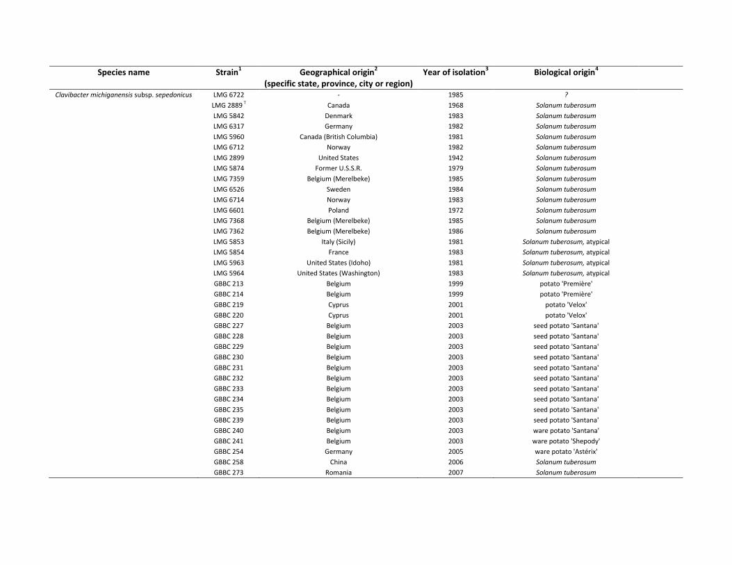
Species name Strain1 Geographical origin
2 Year of isolation
3 Biological origin
4
(specific state, province, city or region)
Clavibacter michiganensis subsp. sepedonicus LMG 6722 - 1985 ?
LMG 2889 T Canada 1968 Solanum tuberosum
LMG 5842 Denmark 1983 Solanum tuberosum
LMG 6317 Germany 1982 Solanum tuberosum
LMG 5960 Canada (British Columbia) 1981 Solanum tuberosum
LMG 6712 Norway 1982 Solanum tuberosum
LMG 2899 United States 1942 Solanum tuberosum
LMG 5874 Former U.S.S.R. 1979 Solanum tuberosum
LMG 7359 Belgium (Merelbeke) 1985 Solanum tuberosum
LMG 6526 Sweden 1984 Solanum tuberosum
LMG 6714 Norway 1983 Solanum tuberosum
LMG 6601 Poland 1972 Solanum tuberosum
LMG 7368 Belgium (Merelbeke) 1985 Solanum tuberosum
LMG 7362 Belgium (Merelbeke) 1986 Solanum tuberosum
LMG 5853 Italy (Sicily) 1981 Solanum tuberosum, atypical
LMG 5854 France 1983 Solanum tuberosum, atypical
LMG 5963 United States (Idoho) 1981 Solanum tuberosum, atypical
LMG 5964 United States (Washington) 1983 Solanum tuberosum, atypical
GBBC 213 Belgium 1999 potato 'Première'
GBBC 214 Belgium 1999 potato 'Première'
GBBC 219 Cyprus 2001 potato 'Velox'
GBBC 220 Cyprus 2001 potato 'Velox'
GBBC 227 Belgium 2003 seed potato 'Santana'
GBBC 228 Belgium 2003 seed potato 'Santana'
GBBC 229 Belgium 2003 seed potato 'Santana'
GBBC 230 Belgium 2003 seed potato 'Santana'
GBBC 231 Belgium 2003 seed potato 'Santana'
GBBC 232 Belgium 2003 seed potato 'Santana'
GBBC 233 Belgium 2003 seed potato 'Santana'
GBBC 234 Belgium 2003 seed potato 'Santana'
GBBC 235 Belgium 2003 seed potato 'Santana'
GBBC 239 Belgium 2003 seed potato 'Santana'
GBBC 240 Belgium 2003 ware potato 'Santana'
GBBC 241 Belgium 2003 ware potato 'Shepody'
GBBC 254 Germany 2005 ware potato 'Astérix'
GBBC 258 China 2006 Solanum tuberosum
GBBC 273 Romania 2007 Solanum tuberosum
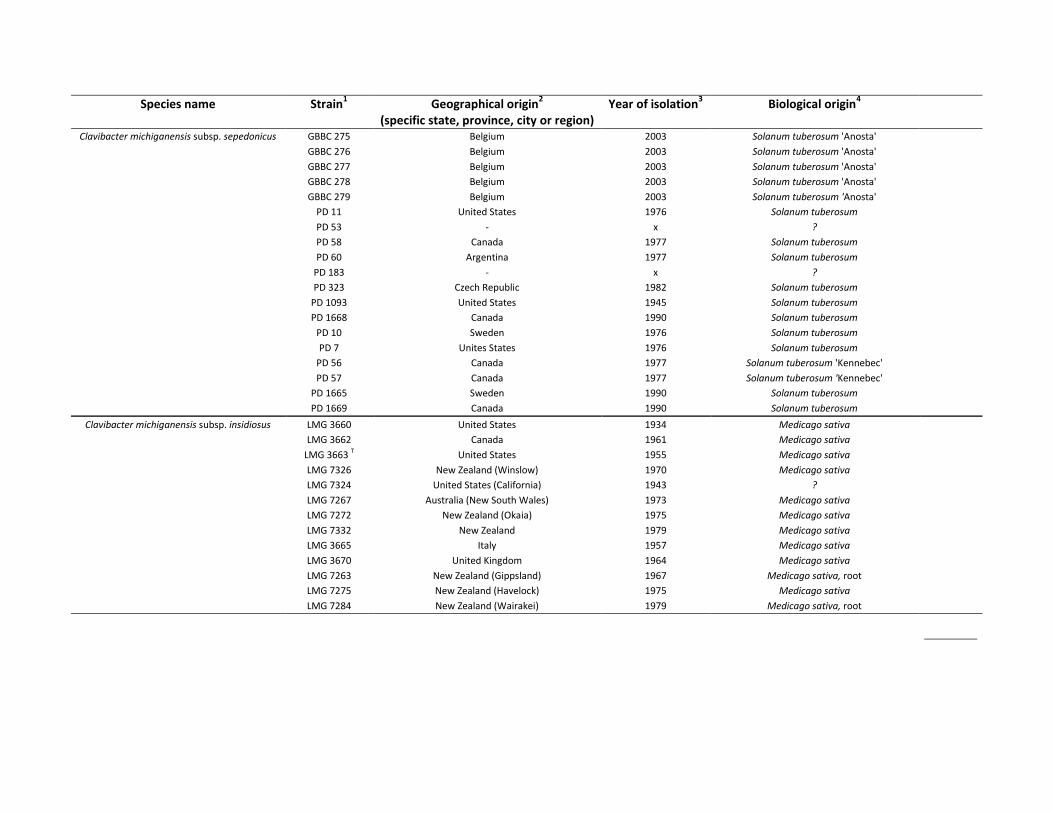
Species name Strain1 Geographical origin
2 Year of isolation
3 Biological origin
4
(specific state, province, city or region)
Clavibacter michiganensis subsp. sepedonicus GBBC 275 Belgium 2003 Solanum tuberosum 'Anosta'
GBBC 276 Belgium 2003 Solanum tuberosum 'Anosta'
GBBC 277 Belgium 2003 Solanum tuberosum 'Anosta'
GBBC 278 Belgium 2003 Solanum tuberosum 'Anosta'
GBBC 279 Belgium 2003 Solanum tuberosum 'Anosta'
PD 11 United States 1976 Solanum tuberosum
PD 53 - x ?
PD 58 Canada 1977 Solanum tuberosum
PD 60 Argentina 1977 Solanum tuberosum
PD 183 - x ?
PD 323 Czech Republic 1982 Solanum tuberosum
PD 1093 United States 1945 Solanum tuberosum
PD 1668 Canada 1990 Solanum tuberosum
PD 10 Sweden 1976 Solanum tuberosum
PD 7 Unites States 1976 Solanum tuberosum
PD 56 Canada 1977 Solanum tuberosum 'Kennebec'
PD 57 Canada 1977 Solanum tuberosum 'Kennebec'
PD 1665 Sweden 1990 Solanum tuberosum
PD 1669 Canada 1990 Solanum tuberosum
Clavibacter michiganensis subsp. insidiosus LMG 3660 United States 1934 Medicago sativa
LMG 3662 Canada 1961 Medicago sativa
LMG 3663 T United States 1955 Medicago sativa
LMG 7326 New Zealand (Winslow) 1970 Medicago sativa
LMG 7324 United States (California) 1943 ?
LMG 7267 Australia (New South Wales) 1973 Medicago sativa
LMG 7272 New Zealand (Okaia) 1975 Medicago sativa
LMG 7332 New Zealand 1979 Medicago sativa
LMG 3665 Italy 1957 Medicago sativa
LMG 3670 United Kingdom 1964 Medicago sativa
LMG 7263 New Zealand (Gippsland) 1967 Medicago sativa, root
LMG 7275 New Zealand (Havelock) 1975 Medicago sativa
LMG 7284 New Zealand (Wairakei) 1979 Medicago sativa, root

Species name Strain
1 Geographical origin
2 Year of isolation
3 Biological origin
4
(specific state, province, city or region)
Clavibacter michiganensis subsp. nebraskensis
LMG 3699 United States 1971 Zea mays
LMG 3700 T United States 1971 Zea mays
LMG 5630 United States 1972 Zea mays
LMG 7176 United States 1971 Zea mays
LMG 7285 United States 1927 Zea mays
LMG 5627 T United States 1971 Zea mays
LMG 5625 - 1972 Zea mays
LMG 7174 United States (Nebraska) 1979 Zea mays
LMG 7177 United States (Nebraska) 1972 Zea mays
Clavibacter michiganensis subsp. tesselarius LMG 7294 T - 1978 Triticum aestivum (Aestivum Group)
LMG 7299 - 1977 Triticum aestivum (Aestivum Group)
LMG 7300 - 1977 Triticum aestivum (Aestivum Group)
LMG 7296 - 1978 Triticum aestivum (Aestivum Group)
Curtobacterium sp. LMG 2896 - 1963 Solanum melongena Deposited
as Clavibacter
species
LMG 18661 Philippines 1996 Oryza sativa
LMG 18697 Philippines (Iloilo) 1996 Oryza sativa
LMG 3680 Australia 1933 ?
Clavibacter sp. PD 5714 - 1997 Lycopersicon esculentum
PD 5715 - x ?
PD 5687 - x Lycopersicon esculentum (tomato seeds) Non-
pathogenic Clavibacter
PD 5688 - x Lycopersicon esculentum (tomato seeds)
PD 5689 - x Lycopersicon esculentum (tomato seeds)
PD 5693 - x Lycopersicon esculentum (tomato seeds)
Rathayibacter rathayi LMG 3717 United Kingdom 1934 Dactylis glomerata
Rathayibacter tritici LMG 3726 Egypt 1941 Triticum aestivum (Aestivum Group)
Rathayibacter iranicus LMG 3677 T Iran 1966 Triticum aestivum (Aestivum Group)
Leifsonia aquatica LMG 18699 Philippines (Iloilo) 1996 Oryza sativa
1 Bacterial collection abbreviations: BCCM/LMG-Bacteria Collection (Ghent, Belgium), PD collection (Wageningen, The Netherlands) and GBBC collection (ILVO, Merelbeke, Belgium),
2 -, geographical origin unknown,
3 x, year of isolation unknown,
4 ?, biological origin unknown,
* Instituto Canario de Investigaciones Agrarias (Tenerife, Spain).
Chapter 2 – GyrB sequence analysis and MALDI-TOF MS as identification tools for plant pathogenic Clavibacter
80
2.6 References
1. Altschul SF, Gish W, Miller W, Myers EW, Lipman DJ. 1990. Basic local alignment search tool. J. Mol. Biol. 215:403-10
2. Anonymous. 2000. Council Directive 2000/29/EC of 8 May 2000 on protective measures against the introduction into the Community of organisms harmful to plants or plant products and against their spread within the Community. Office for Official Publications of the European Communities. Consolidated Text. 1-139
3. Barbuddhe SB, Maier T, Schwarz G, Kostrzewa M, Hof H et al., 2008. Rapid identification and typing of Listeria species by matrix-assisted laser desorption ionization-time of flight mass spectrometry. Appl. Environ. Microbiol. 74:5402-7
4. Collins M, Jones D. 1983. Reclassification of Corynebacterium flaccumfaciens, Corynebacterium betae, Corynebacterium oortii and Corynebacterium poinsettiae in the genus Curtobacterium, as Curtobacterium flaccumfaciens comb. nov. Microbiology 129:3545
5. Degefu Y. 2006. Molecular diagnostic technologies and end user applications: potentials and challenges. Published at www.smts.fi
6. Dieckmann R, Strauch E, Alter T. 2010. Rapid identification and characterization of Vibrio species using whole-cell MALDI-TOF mass spectrometry. J. Appl. Microbiol. 109:199-211
7. Easton GD. 1979. The biology and epidemiology of potato ring rot Am. Potato J. 56:459-60 8. EPPO. 2005a. Clavibacter michiganensis subsp. michiganensis Diagnostics, PM7/42. Bulletin OEPP/EPPO
35:273-83 9. EPPO. 2005b. Curtobacterium flaccumfaciens pv. flaccumfaciens OEPP/EPPO Data Sheets on Quarantine
Pests 10. EPPO. 2006. Clavibacter michiganensis subsp. sepedonicus Diagnostics, PM7/59. Bulletin OEPP/EPPO
36:99-109 11. EPPO. 2010. Clavibacter michiganensis subsp. insidiosus Diagnostics, PM7/99 Bulletin OEPP/EPPO
40:353-64 12. Evtushenko L, Taekeuchi M. 2006. The family Microbacteriaceae. Prokaryotes 3:1020-98 13. Evtushenko LI, Dorofeeva LV, Subbotin SA, Cole JR, Tiedje JM. 2000. Leifsonia poae gen. nov., sp nov.,
isolated from nematode galls on Poa annua and reclassification of 'Corynebacterium aquaticum' Leifson 1962 as Leifsonia aquatica (ex Leifson 1962) gen. nov., nom. rev., comb. nov and Clavibacter xyli Davis et al., 1984 with two subspecies as Leifsonia xyli (Davis et al., 1984) gen. nov., comb. nov. Int. J. Syst. Evol. Microbiol. 50:371-80
14. Ferreira L, Sanchez-Juanes F, Gonzalez-Avila M, Cembrero-Fucinos D, Herrero-Hernandez A et al. 2010. Direct Identification of Urinary Tract Pathogens from Urine Samples by Matrix-Assisted Laser Desorption Ionization-Time of Flight Mass Spectrometry. Journal of Clinical Microbiology 48:2110
15. Giebel R, Worden C, Rust S, Kleinheinz G, Robbins M, Sandrin T. 2010. Microbial Fingerprinting using Matrix-Assisted Laser Desorption Ionization Time-Of-Flight Mass Spectrometry (MALDI-TOF MS): Applications and Challenges. Adv. Appl. Microbiol.:149-84
16. Hajibabaei M, Singer G, Hebert P, Hickey D. 2007. DNA barcoding: how it complements taxonomy, molecular phylogenetics and population genetics. Trends in Genetics 23: 167-72
17. Hebert P, Cywinska A, Ball S, Dewaard J. 2003. Biological identifications through DNA barcodes. Proceedings of the Royal Society of London. Series B: Biological Sciences 270: 313
18. Heyrman J, Swings J. 2001. 16S rDNA sequence analysis of bacterial isolates from biodeteriorated mural paintings in the Servilia Tomb (Necropolis of Carmona, Seville, Spain). Syst. Appl. Microbiol. 24:417-22
19. Hsieh S, Tseng C, Lee Y, Kuo A, Sun C, et al. 2008. Highly efficient classification and identification of human pathogenic bacteria by MALDI-TOF MS. Molecular & Cellular Proteomics 7: 448
20. Jacques MA, Durand K, Orgeur G, Balidas S, Fricot C, et al. 2012. Phylogenetic analysis and polyphasic characterization of Clavibacter michiganensis strains isolated from tomato seeds reveal that non-pathogenic strains are distinct from C. michiganensis subsp. michiganensis. Applied and Environmental Microbiology 78: 8388-402
Part III – Experimental work
81
21. Janda J, Abbott S. 2007. 16S rRNA gene sequencing for bacterial identification in the diagnostic laboratory: pluses, perils and pitfalls. Journal of Clinical Microbiology 45:2761
22. Jansing H, Rudolph K. 1998. Physiological capabilities of Clavibacter michiganensis subsp. sepedonicus and development of a semi-selective medium. Zeitschrift Fur Pflanzenkrankheiten Und Pflanzenschutz-Journal of Plant Diseases and Protection 105:590-601
23. Jarman K, Cebula S, Saenz A, Petersen C, Valentine N et al., 2000. An algorithm for automated bacterial identification using matrix-assisted laser desorption/ionization mass spectrometry. Anal. Chem 72:1217-23
24. Kämpfer P, Seiler H, Dott W. 1993. Numerical classification of coryneform bacteria and related taxa. The Journal of General and Applied Microbiology 39:135-214
25. Kleitman F, Barash I, Burger A, Iraki N, Falah Y et al., 2008. Characterization of a Clavibacter michiganensis subsp. michiganensis population in Israel. Eur. J. Plant Pathol. 121:463-75
26. Lai E. 2001. Application of SNP technologies in medicine: lessons learned and future challenges. Genome Res. 11:927
27. Lay J. 2001. MALDI-TOF mass spectrometry of bacteria. Mass Spectrometry Reviews 20:172-94 28. Maier T, Schwarz G, Kostrzewa M. 2008. Microorganism Identification and Classification Based on
MALDI-TOF MS Fingerprinting with MALDI Biotyper In Application Note # MT-80 29. Martens M, Dawyndt P, Coopman R, Gillis M, De Vos P, Willems A. 2008. Advantages of multilocus
sequence analysis for taxonomic studies: a case study using 10 housekeeping genes in the genus Ensifer (including former Sinorhizobium). Int. J. Syst. Evol. Microbiol. 58:200
30. Mellmann A, Bimet F, Bizet C, Borovskaya A, Drake R et al., 2009. High interlaboratory reproducibility of matrix-assisted laser desorption ionization-time of flight mass spectrometry-based species identification of nonfermenting bacteria. Journal of Clinical Microbiology 47:3732
31. Mills D, Russell B, Hanus J. 1997. Specific detection of Clavibacter michiganensis subsp. sepedonicus by amplification of three unique DNA sequences isolated by subtraction hybridization. Phytopathology 87:853-61
32. Ozdemir Z. 2005. Development of a Multiplex PCR Assay for Concurrent Detection of Clavibacter michiganensis ssp. michiganensis and Xanthomonas axonopodis pv. vesicatoria. Plant Pathology Journal 4:133-7
33. Pastrik K, Rainey F. 1999. Identification and differentiation of Clavibacter michiganensis subspecies by polymerase chain reaction-based techniques. J. Phytopathol. 147:687-93
34. Pineda FJ, Antoine MD, Demirev PA, Feldman AB, Jackman J et al., 2003. Microorganism identification by matrix-assisted laser/desorption ionization mass spectrometry and model-derived ribosomal protein biomarkers. Anal. Chem. 75:3817-22
35. Pitcher D, Saunders N, Owen R. 1989. Rapid extraction of bacterial genomic DNA with guanidium thiocyanate. Lett. Appl. Microbiol. 8:151-6
36. Qian J, Cutler J, Cole R, Cai Y. 2008. MALDI-TOF mass signatures for differentiation of yeast species, strain grouping and monitoring of morphogenesis markers. Anal. Bioanal. Chem. 392:439-49
37. Rach J, DeSalle R, Sarkar I, Schierwater B, Hadrys H. 2008. Character-based DNA barcoding allows discrimination of genera, species and populations in Odonata. Proceedings of the Royal Society B: Biological Sciences 275:237
38. Rezzonico F, Vogel G, Duffy B, Tonolla M. 2010. Whole Cell MALDI-TOF Mass Spectrometry Application for Rapid Identification and Clustering Analysis of Pantoea species. Appl. Environ. Microbiol.
39. Richert K, Brambilla E, Stackebrandt E. 2005. Development of PCR primers specific for the amplification and direct sequencing of gyrB genes from microbacteria, order Actinomycetales. J. Microbiol. Methods 60:115-23
40. Richert K, Brambilla E, Stackebrandt E. 2007. The phylogenetic significance of peptidoglycan types: Molecular analysis of the genera Microbacterium and Aureobacterium based upon sequence comparison of gyrB, rpoB, recA and ppk and 16SrRNA genes. Syst. Appl. Microbiol. 30:102-8
41. Ruelle V, El Moualij B, Zorzi W, Ledent P, De Pauw E. 2004. Rapid identification of environmental bacterial strains by matrix-assisted laser desorption/ionization time-of-flight mass spectrometry. Rapid Commun. Mass Spectrom. 18:2013-9
Chapter 2 – GyrB sequence analysis and MALDI-TOF MS as identification tools for plant pathogenic Clavibacter
82
42. Ryzhov V, Fenselau C. 2001. Characterization of the protein subset desorbed by MALDI from whole bacterial cells. Anal. Chem. 73:746-50
43. Schneider KL, Marrero G, Alvarez AM, Presting GG. 2011. Classification of plant associated bacteria using RIF, a computationally derived DNA marker. PLoS ONE 6: e18496
44. Slabbinck B, Dawyndt P, Martens M, De Vos P, De Baets B. 2008. TaxonGap: a visualization tool for intra-and inter-species variation among individual biomarkers. Bioinformatics 24:866
45. Tamura K, Dudley J, Nei M, Kumar S. 2007. MEGA4: Molecular evolutionary genetics analysis (MEGA) software version 4.0. Mol. Biol. Evol. 24:1596-9
46. Tanigawa K, Kawabata H, Watanabe K. 2010. Identification and Typing of Lactococcus lactis by Matrix-Assisted Laser Desorption Ionization-Time of Flight Mass Spectrometry. Appl. Environ. Microbiol. 76:4055-62
47. Thompson J, Higgins D, Gibson T. 1994. CLUSTAL W: improving the sensitivity of progressive multiple sequence alignment through sequence weighting, position-specific gap penalties and weight matrix choice. Nucleic Acids Res. 22:4673
48. Umeyama T, Sano A, Kamei K, Niimi M, Nishimura K, Uehara Y. 2006. Novel approach to designing primers for identification and distinction of the human pathogenic fungi Coccidioides immitis and Coccidioides posadasii by PCR amplification. Journal of Clinical Microbiology 44:1859
49. Van der Wolf J, Elphinstone J, Stead D, Metzler M, Müller P et al., 2005. Epidemiology of Clavibacter michiganensis subsp. sepedonicus in relation to control of bacterial ring rot. Plant Research International, Report 95
50. Waleron M, Waleron K, Kamasa J, Przewodowski W, Lojkowska E. 2011. Polymorphism analysis of housekeeping genes for identification and differentiation of Clavibacter michiganensis subspecies. European Journal of Plant Pathology 131: 341-54
51. Zaluga J, Van Vaerenbergh J, Stragier P, Maes M, De Vos P. 2013. Genetic diversity of non-pathogenic Clavibacter strains isolated from tomato seeds. Systematic and Applied Microbiology
52. Zgurskaya H, Evtushenko L, Akimov V, Kalakoutskii L. 1993. Rathayibacter gen. nov., including the species Rathayibacter rathayi comb. nov., Rathayibacter tritici comb. nov., Rathayibacter iranicus comb. nov. and six strains from annual grasses. Int. J. Syst. Evol. Microbiol. 43:143
53. Zinniel DK, Lambrecht P, Harris NB, Feng Z, Kuczmarski D et al., 2002. Isolation and characterization of endophytic colonizing bacteria from agronomic crops and prairie plants. Appl. Environ. Microbiol. 68:2198-208
83
Chapter 3
Genetic diversity of non-pathogenic Clavibacter strains
isolated from tomato seeds
Redrafted from:
Zaluga J, Van Vaerenbergh J, Stragier P, Maes M, De Vos P. 2013. Genetic diversity of non-
pathogenic Clavibacter strains isolated from tomato seeds. Systematic and Applied
Microbiology 36, pp. 426-435
Authors’ contributions:
PDV, PS, MM, JVV and JZ designed the experiment set-up. JZ has done the practical work,
performed the sequence, BOX-PCR, DDH analyses and drafted the manuscript. PDV and JVV
provided the necessary bacterial cultures. JVV and JZ performed pathogenicity tests. JVV
performed the colonization tests. PDV, MM, JVV and PS proofread the manuscript.
Chapter 3 – Genetic diversity of non-pathogenic Clavibacter strains isolated from tomato seeds
84
Summary
Clavibacter michiganensis subsp. michiganensis (Cmm) is a seed-transmitted, quarantine
pathogen that causes bacterial wilt and canker of tomato. Despite efforts to prevent seed
contamination new introductions associated with new regions of tomato seed production
are regularly detected.
Non-pathogenic Clavibacter isolates from tomato seed are phenotypically similar to Cmm in
the common diagnostic semi-selective media and are also identified as Cmm in the
customary diagnostic tests but they are negative in pathogenicity assays on tomato. In our
first study four representatives of this non-pathogenic group formed a separate cluster
based on gyrB sequence and MALDI-TOF MS analysis. Their presence in tomato seeds
complicates a clear judgment on the health status of tested seed lots. As their nature and
genetic background are unknown we aimed to investigate and compare them to Cmm.
Twenty representatives of Clavibacter strains isolated from tomato seeds and not
pathogenic to tomato were investigated. In planta tests showed no leaf spots, wilting or
canker symptoms after local or systemic inoculation. Tomato stems were not colonized nor
was there evidence of survival in tomato stems. Total DNA-DNA hybridization and sequence
analysis of gyrB and dnaA proved that they belong to the Clavibacter michiganensis species
but can be unambiguously separated from Cmm and from other Clavibacter subspecies.
Some of the genes encoding virulence determinants in Cmm strains were also detected in
some of the non-pathogenic isolates. Cmm strains formed a coherent group, while the non-
pathogenic Clavibacter strains were heterogenic. The latter was confirmed by BOX-PCR. We
speculate that tomato seeds likely represent a larger reservoir of unexplored Clavibacter
diversity.
Keywords: Clavibacter, bacterial identification, tomato seeds
Part III – Experimental work
85
3.1 Introduction
As a seed pathogen Clavibacter michiganensis subsp. michiganensis (Cmm) can be
disseminated over long distances and introduced into Europe and elsewhere from countries
where the seed production is currently taking place (De León et al., 2009). Cmm strains
isolated from tomato plants and seeds differ in virulence level and weakly virulent (inducing
only a slight canker at the inoculation site, no wilt symptoms) and avirulent (no visible
symptoms on plants) strains were identified (Alvarez et al., 2004, Kaneshiro and Alvarez
2001, Louws et al., 1998). Atypical Cmm strains lacking pat-1 gene and inducing weaker
symptoms on tomato were found on symptomatic tomato plants in greenhouses in Italy
(Bella et al., 2012). Non-pathogenic to tomato Clavibacter strains isolated from tomato
seeds that cross-reacted with identification methods for Cmm were recently included in the
study of Jacques and coworkers (Jacques et al., 2012). These strains showed positive
reaction in commonly used serological identification methods (immunofluorescence) and in
different PCR assays (Cmm F/R, PSA 4/R, PSA 8/R). Some of non-pathogenic clavibacters
showed a positive amplification with primers specific to celA (PCF 3/5).
Avirulent strains exist for bacteria pathogenic to human, animals and plants (Cheng et al.,
2010, Karki et al., 2012, Tenaillon et al., 2010). In Cmm, avirulent strains were linked to the
lack of plasmids carrying virulence genes, celA and pat-1, which are required for symptom
expression. When these determinants were lost strains could still colonize a plant but were
not inducing disease (Eichenlaub and Gartemann 2011). The other explanation for the
existence of non-virulent Cmm strains is a deletion of the chromosomal pathogenicity island
of Cmm. This region encodes several serine proteases, tomatinase and homologues of the
plasmid-borne virulence factor pat-1. Some of these genes were important for efficient
colonization of tomato plant tissues (Stork et al., 2008). When PI (pathogenicity island) was
partially or completely lost, the Cmm strains poorly colonized vascular tissues and although
the plasmid-borne pathogenicity genes were present, it was assumed that their population
sizes inside the plant were too low to induce disease (Kleitman et al., 2008). Saprophytic
and environmental Clavibacter strains were sometimes found on other plants and seeds,
such as those reported on rice seed (Cottyn et al., 2009, Cottyn et al., 2001) and on healthy
Chapter 3 – Genetic diversity of non-pathogenic Clavibacter strains isolated from tomato seeds
86
aerial tissues of prairie plants (Zinniel et al., 2002). In the latter example Clavibacter strains
constituted the fraction of endophytic colonizing bacteria inhabiting various prairie plants.
Little is known about these environmental strains which are mostly identified only at the
genus level.
The challenge to seed health organizations lays in accurately differentiating Cmm from
other seed contaminants and to determine the significance of seed-borne Cmm with
various levels of virulence. Cmm coexist with numerous other bacteria on tomato seed,
some of which share similar colony morphologies on the selective media used in the seed
assay. Some non-pathogenic Clavibacter strains resembling Cmm have been isolated from
tomato seed lots imported into Europe and from seed lots for which the country of origin is
mostly unknown. Those strains cross-react with commercially available antisera for Cmm
and are positively identified as Cmm by the commonly used PCR tests but fail in the
pathogenicity test (Jacques et al., 2012). A set of those non-pathogenic Clavibacter strains,
isolated from tomato seeds, was recently investigated by gyrB sequence and MALDI-TOF
MS (matrix-assisted laser desorption/ionization mass spectrometry) analysis (Zaluga et al.,
2011). They clustered in a separate Clavibacter clade outside Cmm and were most related
to Clavibacter michiganensis subsp. tessellarius (Cmt).
The host specialization of Cmm is known to be very strict with natural infections occurring
on tomato, although other solanaceous and non-solanaceous plant species were reported
as potential hosts after artificial inoculation (EPPO 2005, Thyr et al., 1975). Just recently,
Cmm, identified based on 16S rRNA, ELISA and fatty acid analysis were isolated from pepper
plants (Capsicum annum) in Korea (Yim et al., 2012). These isolates displayed distinct
orange-coloured and less mucoid colonies which were noticeably different from the
common Cmm phenotype isolated from tomato. The Cmm strains from pepper were
virulent on its host of origin but only weakly virulent in tomato plants (no wilting, only slight
stem canker). Furthermore, Cmm strains from pepper produced no PCR amplicons for the
plasmid-borne virulence factors, nor for the virulence related genes located on the
chromosomal pathogenicity island. Authors suggest that these strains represent a separate
population of Cmm strains that evolved within the limits of the host (Yim et al., 2012).
Part III – Experimental work
87
So it is evident that various ‘atypical’ strains of Cmm exist with clear differences in
phenotype, genotype and virulence. This paper reports on the investigation of a novel
group of Clavibacter isolates from tomato seeds that were shown not to colonize or infect
tomato plants and therefore regarded as non-pathogenic. In our initial work we observed a
stable separate grouping of these strains within the Clavibacter clade using gyrB amplicon
sequences and MALDI-TOF MS (Zaluga et al., 2011). Their apparent lack of pathogenicity on
tomato and the risk of erroneous identification as Cmm make them an interesting study
object. In the present work the genetic diversity of these strains was investigated by gyrB
and dnaA gene sequence analysis and by BOX-PCR. These new isolates from tomato seeds
were then compared with Cmm and their separate taxonomic position within the genus
Clavibacter was confirmed. We tested their interaction with tomato plants in inoculation
experiments. In addition, the presence of known virulence factors of Cmm was investigated.
Chapter 3 – Genetic diversity of non-pathogenic Clavibacter strains isolated from tomato seeds
88
3.2 Material and methods
Bacterial strains and growth conditions: The strains used in this study were obtained from
the BCCM/LMG Bacteria Collection (Ghent, Belgium), the NVWA collection (Wageningen,
The Netherlands) and the GBBC (ILVO-Diagnostic Centre for Plants, Merelbeke, Belgium).
This Clavibacter strain subset consisted of nineteen Cmm strains, twenty non-pathogenic
Clavibacter strains, five strains belonging to the Family Microbacteriaceae and five type
strains of Clavibacter: Clavibacter michiganensis subsp. michiganensis (Cmm) LMG 7333T,
Clavibacter michiganensis subsp. nebraskensis (Cmn) LMG 5627T, Clavibacter michiganensis
subsp. sepedonicus (Cms) LMG 2889T, Clavibacter michiganensis subsp. insidiosus (Cmi)
LMG 3663T, Clavibacter michiganensis subsp. tessellarius (Cmt) LMG 7294T (Table 3.1).
Strains isolated from tomato seed were received from Naktuinbouw (The Netherlands).
They were isolated according to the current method for detection of Cmm in tomato seed
recommended by International Seed Federation (ISF) (ISHI 2011). Seed extracts were
obtained by soaking overnight at 4°C in phosphate buffer to which Tween 20 was added,
followed by a Stomacher treatment to crush the seeds. The Clavibacter strains originated
from bacterial collections were assumed to be correctly identified by their depositors. Their
identity was further confirmed by 16S rRNA, gyrB and dnaA gene sequencing. The strains
were cultured aerobically on M6 medium (glucose 10 g, yeast extract 5 g, peptone 5 g, agar
15 g, distilled water 1 L, pH 7.0) at 25°C for 24h-48h. Stock cultures were stored at -80˚C in
MicrobankTM beads (Pro-Lab Diagnostics, Canada).
In planta tests (pathogenicity and virulence) Tomato plants (cv. Moneymaker) were
produced from certified seed in a greenhouse. They were inoculated in the 3rd – 4th true leaf
stage. Strains in this study were revived from cryopreservation and cultured at 28˚C on M6
medium for 24-48 hours. A cell suspension was prepared by suspending a film of bacterial
culture in a 10 µl calibrated loop in 1 ml sterile 10 mM phosphate buffer (pH 7.2). For
development of systemic infection, inoculation was performed by pipetting 5 µl of the 1:10
diluted bacterial suspension, containing approximately 106 cells as determined by dilution
plating, on the stem at the cotyledons. The stem was punctured through the droplet with a
needle allowing the droplet to be absorbed in the vascular tissues. Plants were not watered
Part III – Experimental work
89
48 hours before inoculation to reduce turgor pressure which facilitates the access to the
vascular tissues. Plants were scored for wilting and canker formation. For development of
local infection individual leaves were pressure inoculated with the 1:10 diluted cell
suspension until run-off. Plants were scored for development of leaf spots. Control plants
were inoculated with sterile 10 mM phosphate buffer. Test plants for systemic infection
were maintained on a greenhouse bench at a night/day regime of 18°C/25°C and
uncontrolled humidity conditions. Test plants for local infection were maintained in
transparent boxes closed with the lid for symptom development for at least 21 days. Strains
were considered virulent if inoculated plants exhibited some degree of leaf wilting. Strains
were considered hypovirulent if only a canker had developed at the inoculation point. Non-
virulent (avirulent) strains caused no symptoms in inoculated plants. All strains were
inoculated into at least five plants. For avirulent strains colonization of stem tissues was
investigated by aseptically removing from each plant a 1 cm stem tissue section at the first
true leaf. The tissues were individually analyzed. They were comminuted in 5 ml of sterile
10 mM phosphate buffer pH 7.2 and dilution plated onto Difco Pseudomonas Agar F.
DNA–DNA hybridization experiments were performed with photobiotin-labelled probes in
microplate wells as described by (Ezaki et al., 1989), with some modifications (Cleenwerck
et al., 2002) using an HTS7000 Bio Assay Reader for the fluorescence measurements. DNA-
DNA hybridizations were performed at 47˚C with DNA prepared as described below. A
representative of the non-pathogenic Clavibacter strains, LMG 26814, was hybridized
against the type strain of Cmm LMG 7333T. Reciprocal reactions (A x B and B x A) were
performed for each DNA pair from included strains and their variation was within the limits
for this method (Goris et al., 1998).
The GC% estimation. DNA was enzymatically degraded into nucleosides as described by
Mesbah and coworkers (Mesbah et al., 1989). The nucleoside mixture obtained was then
separated using a Waters Breeze HPLC system and XBridge Shield RP18 column
thermostabilized at 37°C. The solvent was 0.02M NH4H2PO4 (pH 4.0) with 1.5% (v/v)
Chapter 3 – Genetic diversity of non-pathogenic Clavibacter strains isolated from tomato seeds
90
acetonitrile. Non-methylated lambda phage (Sigma) and Escherichia coli DNA were used as
calibration reference and control, respectively.
DNA extraction, amplification and sequencing. Total genomic DNA was extracted according
to the guanidium-thiocyanate-EDTA-sarkosyl method described by (Pitcher et al., 1989)
which was adapted for Gram-positive bacteria with a pre-treatment with lysozyme (5 mg/µl
lysosyme powder in TE buffer) and incubation for 40 minutes at 37°C. 16S rRNA gene
amplification was performed with primers pA-forward and pH-reverse as described
previously (Heyrman and Swings 2001). Subsequently, PCR products were purified and
partially sequenced using the BKL1-reverse primer (5’- ACTGCTGCCTCCCGTAGGAG-3’,
position 536–516 in the Escherichia coli numbering system) and gamma-reverse primer (5’
ACTGCTGCCTCCCGTAGGAG 3’, position 358–339) (Heyrman and Swings 2001). Amplification
and sequencing primers used in this study are listed in Supplementary Table 3.1. The expected
amplicons were obtained using 50-60 ng template, AmpliTaq polymerase, GeneAmp®
dNTP’s and GeneAmp® 10x PCR buffer according to the manufacturer specifications
(Applied Biosystems, Belgium). All primers were from Sigma Aldrich, Belgium. Resulting
amplicons were purified using the Nucleofast®96 PCR clean up membrane system
(Macherey-Nagel, Germany). Sequencing PCR was performed in a total volume of 10 µl with
3 µl of purified amplicon, 0.286 µl of BigDyeTM mixture (Terminator Cycle Sequencing Kit
version 3.1, Applied Biosystems), 1x sequencing buffer and 1.2 µM of each of the
amplification primers mentioned above. The thermal program consisted of 30 cycles (96°C
for 15 s, 35°C for 1 s, 60°C for 4 min). Subsequently, the sequencing products were purified
using the BigDye XTerminator Kit (Applied Biosystems) and analyzed on a 3130xl Genetic
Analyzer (Applied Biosystems).
PCR-based assays for Cmm identification. The PCR primers for identification of Cmm were
Cmm F/R, PSA 4/R, PSA 8/R and Ptssk 10/11. The list of primers, their target regions and
expected product sizes are listed in Supplementary Table 3.1. PCR reactions were performed
in a 20 µl volume containing 200 µM deoxynucleotide triphosphate (dNTP), 1.5 mM MgCl2,
0.6 µM each primer, 1 U/reaction of AmpliTaq DNA polymerase (Applied Biosystems) and
Part III – Experimental work
91
50-80 ng of DNA solution. PCR conditions were: 5 min at 94 ˚C followed by 35 cycles of 30
sec at 94 ˚C, 1 min at Ta and 1 min at 72 ˚C with 7 min final extention at 72 ˚C. Amplification
products were separated on a 1% agarose gel in 1 X Tris-acetate-EDTA (TAE).
Sequence analysis. The 16S rRNA gene, celA, pat-1, tomA, chpC, ppaA, gyrB and dnaA PCR
amplicon sequences were assembled with BioNumerics version 5.1 (Applied Maths,
Belgium) and aligned using ClustalW (Thompson et al., 1994). The identity assigned to each
strain by the three culture collections was verified with the BLASTn (Altschul et al., 1990) of
the obtained 16S rRNA gene sequence and a neighbor-joining cluster analysis with all type
strains of all species from all genera mentioned in the twenty highest BLAST hits. The strains
were assigned to a genus based on the obtained pairwise 16S rRNA gene sequence
similarity (>98% gene sequence similarity). DnaA sequences were checked by amino acid
translation with Transseq (http://www.ebi.ac.uk/Tools/emboss/transeq/) and presence of
the DnaA protein domain was confirmed with BLASTp (Altschul et al., 1990). Molecular
Evolutionary Genetics Analysis software (Mega 5.1) (Tamura et al., 2011) was used to
calculate evolutionary distances based on a 666 bp (this equal length was used for all
strains) fragment of the dnaA amplicon and to infer Maximum Parsimony (MP), Maximum
Likelihood (ML) and Neighbor-Joining (NJ) trees using the maximum composite likelihood
(MCL) model. To establish the positions within the dnaA, pat-1, celA and 16S rRNA gene the
whole Cmm genome sequence (AM711867) was used as a reference. The newly generated
sequences were deposited in the NCBI database under the accession numbers JX259275-
JX259316 and KC594297-KC594349.
Endocellulase activity. Endocellulase activity was detected on M9CMC agar plates
consisting of M9 medium (Maniatis 1982) lacking glucose but supplemented with 0.1%
(wt/vol) yeast extract and 0.5% (wt/vol) carboxymethylcellulose. Strains to be tested were
incubated for 4 to 5 days at 24 to 26°C. Plates were stained with 0.1% (wt/vol) Congo red
for 20 min and finally bleached with 1 M NaCl (Meletzus et al., 1993). Endocellulase activity
is detected as a clear, yellowish halo around producing colonies.
Chapter 3 – Genetic diversity of non-pathogenic Clavibacter strains isolated from tomato seeds
92
Rep-fingerprinting. Rep-PCR was performed with the BOX-primers because in-house tests
with (GTG)5 and ERIC primers showed less informative profiles. The PCR-mixture was
prepared as described previously (Rademaker and De Bruijn 1997). Amplification was
performed in a GeneAmp PCR System 9600 (Applied Biosystems) with the following
temperature-time profile: 7 min 95 °C, 30 cycles of 94 °C for 1 min, 52 °C for 1 min and 65 °C
for 8 min and a final step of 16 min at 65 °C. Electrophoresis was performed in 1 × TAE
buffer (40 mM Tris, 20 mM acetic acid and 1 mM EDTA) on a 1.5% agarose gel (w/v) under
standardized conditions (55 V, 400 mA, 960 min, 4 °C). Sixteen samples and four reference
marker lines, each composed of 1.10 μl Molecular Ruler 500 bp (Bio-Rad), 1.40 μl Molecular
Ruler 100 bp (Bio-Rad), 2 μl TE buffer (1 mM EDTA, 10 mM Tris–HCl (pH 8.0)) and 1.50 μl
loading dye, were loaded per gel. Profiles were visualized under ultraviolet light after
staining with ethidium bromide. Digitized images of gels were normalized and analyzed with
the BioNumerics 5.1 software (Applied Maths, Belgium). Similarity matrices of
densitometric curves of the gel tracks were calculated with the Pearson's product-moment
correlation coefficient. Cluster analyses of similarity matrices were performed by
unweighted pair group method with arithmetic averages (UPGMA).
Part III – Experimental work
93
3.3 Results and discussion
Pathogenicity, colonization tests and bacterial colony morphology
Pathogenicity tests on tomato plants with 20 seed-borne Clavibacter strains and members
of the Family Microbacteriaceae showed no disease symptoms after 21 days of incubation
whereas Cmm strains produced wilting or/and canker (Figure 3.1, Table 3.1). The Cmm group
contained very aggressive strains (e.g. PD 1664) which led to fast disease development and
complete devastation of the plant (Figure 3.1). Among Cmm strains there were three strains
that did not induce disease symptoms despite presence of one or more virulence factors.
Non-pathogenic Clavibacter strains isolated from tomato seeds were not able to colonize
the plant. In addition, they could not be reisolated, already after several days after
inoculation which suggests that they do not survive in the plant tissues. Their disability to
invade in planta suggests that their genetic structure is lacking some important genes
responsible for effective plant colonization. Since these non-pathogenic Clavibacter strains
are not able to successfully colonize plant tissues they will not be able to reach seeds via the
vascular system and contaminate them that way. Therefore, their retrieval from tomato
seeds is probably to be explained by a post factum external contamination, e.g. at the stage
of fruit collecting and processing.
Some of the non-pathogenic Clavibacter strains showed differences in colony morphology.
Most of the Cmm strains studied previously displayed typically yellow, fluidal colonies
whereas some of non-pathogenic Clavibacter strains (LMG 26808 and LMG 26809) showed
more orange and fluidal colonies. Morphological variants within Cmm strains were reported
previously but the link with their pathogenic properties was not recognized (Kaneshiro et
al., 2006). However, using only these indications we cannot ascertain the differentiation
between Cmm and non-pathogenic Clavibacter strains; it can be a first indication that a
strain can indeed be not pathogenic. Recently, Yim and coworkers characterized Cmm
strains isolated from Capsicum annum which were significantly less aggressive to tomato
plants and exhibited different colony morphology (Yim et al., 2012). In our opinion,
identification of these strains based on 16S rRNA, ELISA and fatty acid analysis is not
Chapter 3 – Genetic diversity of non-pathogenic Clavibacter strains isolated from tomato seeds
94
sufficient to assign them as Cmm and more tests should be performed to ascertain their
taxonomic position. Because of the significantly lower virulence to tomato, the separate
position in the 16S rRNA tree and the lack of amplification of genes coding for known
virulence factors these strains may actually represent a new group within the genus
Clavibacter and it would be very interesting to compare these isolates with strains
characterized in our study.
To better understand the ecology of the Cmm and non-pathogenic Clavibacter strains,
experimental set ups should aim at unraveling the natural reservoirs of non-pathogenic
Clavibacter strains as well as the routes to seeds contamination by different Clavibacter
strains.
Taxonomic position of non-pathogenic Clavibacter strains
In our previous study we selected strains of all Clavibacter subspecies and performed gyrB
gene sequencing together with MALDI-TOF MS (Zaluga et al., 2011). GyrB gene sequence
barcode was selected in the frame of the EU-FP7 Quarantine Barcoding of Life (QBOL)
project as a suitable gene for identification of Clavibacter strains at the subspecies level. A
Clavibacter strain is identified as belonging to one of the five plant pathogenic Clavibacter
michiganensis subspecies when its gyrB sequence is 99% similar to a reference sequence
present in the Q-Bank database (http://www.q-bank.eu/). Non-pathogenic Clavibacter
strains differ in the gyrB gene sequence and can be separated from the other Clavibacter
subspecies on the phylogenetic gyrB tree (Maximum Likelihood). BLAST search of the 16S
rRNA gene sequences identified the 20 non-pathogenic strains as belonging to the genus
Clavibacter (Table 3.2). Because i) they are able to grow on the semi-selective media used in
tomato seed testing, ii) their colony morphology is similar to this of Cmm, iii) they are
identified as Cmm in serological tests and by PCR with ITS primers PSA-R and PSA-4 (Pastrik
and Rainey 1999), iv) they are not pathogenic to tomato and v) they are identified as
belonging to the genus Clavibacter in the 16S rRNA gene sequence analysis, they were
temporarily assigned to a group of non-pathogenic Clavibacter strains. Their distinct
taxonomic positions were confirmed by the sequence analysis of dnaA (chromosomal
Part III – Experimental work
95
replication initiator) and gyrB (Figure 3.2). For dnaA, all sequences were generated in the
course of this study, gyrB sequences were obtained for additional non-pathogenic
Clavibacter strains which were not included in our first study (Zaluga et al., 2011).
Since the non-pathogenic Clavibacter strains could not be classified within the known
subspecies, in the next part of our work the relatedness between them and Cmm was
checked by DNA-DNA hybridization. DNA–DNA binding values of 71% (reciprocal values,
71.2-71.8) relatedness were observed between non-pathogenic Clavibacter strain LMG
26814 and Cmm LMG 7333T. Additionally, GC% of four tested strains (LMG 26807, LMG
26808, LMG 26810, and LMG 26814) was measured. The results demonstrated that the
average GC content of representatives of non-pathogenic Clavibacter strains was 73.36%
which is very close to 72.66%, a value deduced from the complete genome sequence of
Cmm NCPPB 382 (Gartemann et al., 2008).
These data indicate unambiguously that the non-pathogenic Clavibacter strains indeed
belong to the Clavibacter michiganensis species. For practical reasons it would be better to
allocate them to a different taxon, preferably a new subspecies, taking into account the
nomenclature definitions and rules of the Bacterial Code (Lapage et al., 1992). According to
a generally agreed polyphasic approach for the delineation of bacterial taxa, phenotypic,
chemotaxonomic and genotypic data as well as phylogenetic information are used.
However, there are no clear guidelines for the establishment of subspecies (Brenner et al.,
2001, Schleifer 2009). Subspecies designation can be given to genetically determined
clusters of strains or to groups of strains exhibiting minor but consistent phenotypic
variation within a species (Schloter et al., 2006) and are usually designated according to
specific needs (Brenner et al., 2001). Five already known Clavibacter michiganensis
subspecies were subdivided mainly based on host specificity and their stable grouping was
confirmed by gyrB sequence analysis (Zaluga et al., 2011), as well as other techniques such
as rep-PCR (Louws et al., 1998). The diversity of non-pathogenic Clavibacter strains is much
higher than of Cmm (Figure 3.2, Figure 3.3) or of highly homogenic Cms strains (Smith et al.,
2001) which may implicate the necessity to delineate more than one subspecies. Because
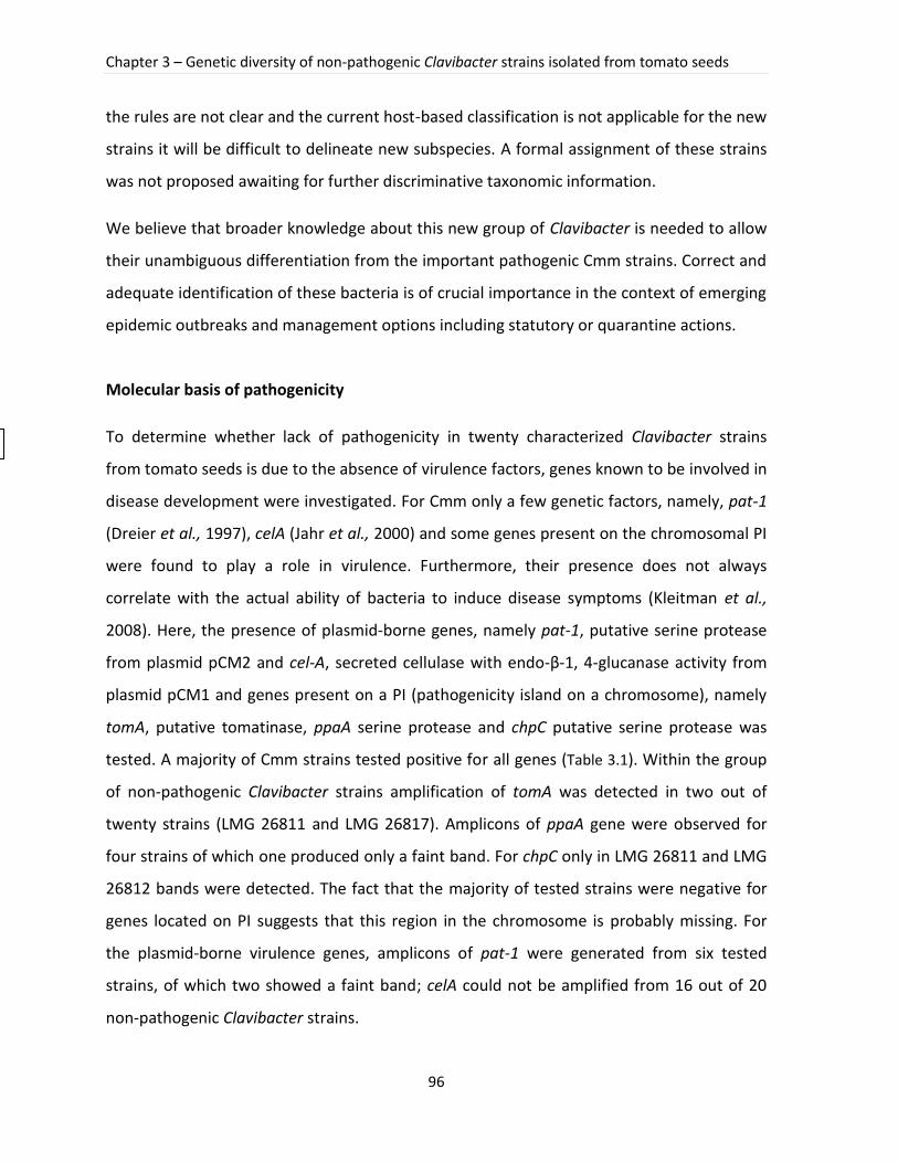
Chapter 3 – Genetic diversity of non-pathogenic Clavibacter strains isolated from tomato seeds
96
the rules are not clear and the current host-based classification is not applicable for the new
strains it will be difficult to delineate new subspecies. A formal assignment of these strains
was not proposed awaiting for further discriminative taxonomic information.
We believe that broader knowledge about this new group of Clavibacter is needed to allow
their unambiguous differentiation from the important pathogenic Cmm strains. Correct and
adequate identification of these bacteria is of crucial importance in the context of emerging
epidemic outbreaks and management options including statutory or quarantine actions.
Molecular basis of pathogenicity
To determine whether lack of pathogenicity in twenty characterized Clavibacter strains
from tomato seeds is due to the absence of virulence factors, genes known to be involved in
disease development were investigated. For Cmm only a few genetic factors, namely, pat-1
(Dreier et al., 1997), celA (Jahr et al., 2000) and some genes present on the chromosomal PI
were found to play a role in virulence. Furthermore, their presence does not always
correlate with the actual ability of bacteria to induce disease symptoms (Kleitman et al.,
2008). Here, the presence of plasmid-borne genes, namely pat-1, putative serine protease
from plasmid pCM2 and cel-A, secreted cellulase with endo-β-1, 4-glucanase activity from
plasmid pCM1 and genes present on a PI (pathogenicity island on a chromosome), namely
tomA, putative tomatinase, ppaA serine protease and chpC putative serine protease was
tested. A majority of Cmm strains tested positive for all genes (Table 3.1). Within the group
of non-pathogenic Clavibacter strains amplification of tomA was detected in two out of
twenty strains (LMG 26811 and LMG 26817). Amplicons of ppaA gene were observed for
four strains of which one produced only a faint band. For chpC only in LMG 26811 and LMG
26812 bands were detected. The fact that the majority of tested strains were negative for
genes located on PI suggests that this region in the chromosome is probably missing. For
the plasmid-borne virulence genes, amplicons of pat-1 were generated from six tested
strains, of which two showed a faint band; celA could not be amplified from 16 out of 20
non-pathogenic Clavibacter strains.

Figure 3.1. Symptoms of infection with Clavibacter michiganensis subsp. michiganensis and non-pathogenic Clavibacter strains. A) non-pathogenic Clavibacter strain LMG 26813; B) Cmm LMG 5720, stem canker at the inoculation site; C) Cmm PD 5720, leaves wilting; D) Cmm PD 1664, an example of a very aggressive strain, complete plant collapse.
A B C D
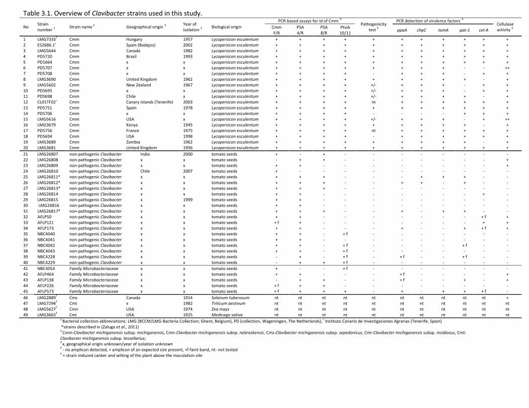
1 Bacterial collection abbreviations: LMG (BCCM/LMG-Bacteria Collection; Ghent, Belgium), PD (collection, Wageningen, The Netherlands),˚ Instituto Canario de Investigaciones Agrarias (Tenerife, Spain) *strains described in (Zaluga et al., 2011) 2 Cmm-Clavibacter michiganensis subsp. michiganensis, Cmn-Clavibacter michiganensis subsp. nebraskensis, Cms-Clavibacter michiganensis subsp. sepedonicus, Cmi-Clavibacter michiganensis subsp. insidiosus, Cmt-Clavibacter michiganensis subsp. tessellarius;
3 x, geographical origin unknown/year of isolation unknown
4 - no amplicon detected, + amplicon of an expected size present, +f faint band, nt- not tested 5 + strain induced canker and wilting of the plant above the inoculation site
Table 3.1. Overview of Clavibacter strains used in this study.
No Strain number
1
Strain name 2
Geographical origin 3
Year of isolation
3
Biological origin
PCR-based assays for id of Cmm 4
Pathogenicity test
5
PCR detection of virulence factors 4
Cellulase activity
4
Cmm F/R
PSA 4/R
PSA 8/R
Ptssk 10/11
ppaA chpC tomA pat-1 cel-A
1 LMG7333T Cmm Hungary 1957 Lycopersicon esculentum + + + + + + + + + + + 2 ES2686.1˚ Cmm Spain (Badajoz) 2002 Lycopersicon esculentum + + + + + + + + + + + 3 LMG5644 Cmm Canada 1982 Lycopersicon esculentum + + + + + + + + + + + 4 PD5720 Cmm Brasil 1993 Lycopersicon esculentum + + + + + + + + + + + 5 PD1664 Cmm x x Lycopersicon esculentum + + + + + + + + + + + 6 PD5707 Cmm x x Lycopersicon esculentum + + + + + + + + - - ++ 7 PD5708 Cmm x x Lycopersicon esculentum + + + + - + + + - + + 8 LMG3690 Cmm United Kingdom 1962 Lycopersicon esculentum + + + + + + + + + - + 9 LMG5602 Cmm New Zealand 1967 Lycopersicon esculentum + + + + +/- + + + - + + 10 PD5695 Cmm x x Lycopersicon esculentum + + + + +/- + + + - + + 11 PD5698 Cmm Chile x Lycopersicon esculentum + + + + +/- + + + + - + 12 CL01TF02˚ Cmm Canary Islands (Tenerife) 2003 Lycopersicon esculentum + + + + nt + + + + + + 13 PD5751 Cmm Spain 1978 Lycopersicon esculentum + + + + + + + + + + + 14 PD5706 Cmm x x Lycopersicon esculentum + + + + - - - - + + + 15 LMG5616 Cmm USA x Lycopersicon esculentum + + + + +/- + + + - + ++ 16 LMG3679 Cmm Kenya 1945 Lycopersicon esculentum + + + + + + + + + - + 17 PD5756 Cmm France 1975 Lycopersicon esculentum + + + + nt + + + + + + 18 PD5694 Cmm USA 1998 Lycopersicon esculentum + + + + - + + + + + + 19 LMG3689 Cmm Zambia 1962 Lycopersicon esculentum + + + + + + + + + - + 20 LMG3681 Cmm United Kingdom 1956 Lycopersicon esculentum + + + + + + + + + + +
21 LMG26807 non-pathogenic Clavibacter India 2000 tomato seeds + - + - - - - - - - - 22 LMG26808 non-pathogenic Clavibacter x x tomato seeds - + + - - - - - - - + 23 LMG26809 non-pathogenic Clavibacter x x tomato seeds + + - - - - - - - - + 24 LMG26810 non-pathogenic Clavibacter Chile 2007 tomato seeds + - - - - - - - - - - 25 LMG26811* non-pathogenic Clavibacter x x tomato seeds + + + - - - + + + - - 26 LMG26812* non-pathogenic Clavibacter x x tomato seeds + + + - - + + - + - - 27 LMG26813* non-pathogenic Clavibacter x x tomato seeds + + + - - - - - - - - 28 LMG26814 non-pathogenic Clavibacter x x tomato seeds + + - - - - - - - + - 29 LMG26815 non-pathogenic Clavibacter x 1999 tomato seeds + + - - - - - - - - - 30 LMG26816 non-pathogenic Clavibacter x x tomato seeds + + - - - - - - - - - 31 LMG26817* non-pathogenic Clavibacter x x tomato seeds + + + - - + - + + - - 32 AFLP50 non-pathogenic Clavibacter x x tomato seeds + + - - - - - - - + f + 33 AFLP121 non-pathogenic Clavibacter x x tomato seeds + f + - - - - - - - + + 34 AFLP173 non-pathogenic Clavibacter x x tomato seeds + + - - - + - - + + f + 35 NBC4040 non-pathogenic Clavibacter x x tomato seeds + + - + f - - - - - - - 36 NBC4041 non-pathogenic Clavibacter x x tomato seeds + + - - - - - - - - - 37 NBC4042 non-pathogenic Clavibacter x x tomato seeds + + - + f - - - - + f - - 38 NBC4043 non-pathogenic Clavibacter x x tomato seeds + + - + f - - - - - - - 39 NBC4228 non-pathogenic Clavibacter x x tomato seeds - + - + f - + f - - + f - - 40 NBC4229 non-pathogenic Clavibacter x x tomato seeds - + + + f - - - - - - -
41 NBC4054 Family Microbacteriaceae x x tomato seeds + - - + f - - - - - - - 42 AFLP464 Family Microbacteriaceae x x tomato seeds + + - - - + f - - - - + 43 AFLP138 Family Microbacteriaceae x x tomato seeds - + + - - + f - - + - + 44 AFLP226 Family Microbacteriaceae x x tomato seeds + f + + - - - - - - - - 45 AFLP573 Family Microbacteriaceae x x tomato seeds + f + + + - + - + + + f -
46 LMG2889T Cms Canada 1914 Solanum tuberosum nt nt nt nt nt nt nt nt nt nt + 47 LMG7294T Cmt x 1982 Triticum aestivum nt nt nt nt nt nt nt nt nt nt nt 48 LMG5627T Cmn USA 1974 Zea mays nt nt nt nt nt nt nt nt nt nt nt 49 LMG3663T Cmi USA 1925 Medicago sativa nt nt nt nt nt nt nt nt nt nt nt
Part III – Experimental work
99
Table 3.2. BLAST result of a non-pathogenic Clavibacter strain based on its 16S rDNA sequence.
Strain number
Identification Accession number
Maximum identity (%)
Query coverage (%)
Total score
LMG26807 Clavibacter michiganensis subsp. michiganensis strain PF008 JN603288.1 100 100 815
LMG26808 Clavibacter michiganensis subsp. michiganensis strain PF008 JN603288.1 100 100 815
LMG26809 Clavibacter michiganensis subsp. sepedonicus strain ATCC33113 NR_074600.1 99 100 822
LMG26810 Clavibacter michiganensis subsp. michiganensis strain PF008 JN603288.1 100 100 811
LMG26811 Clavibacter michiganensis subsp. michiganensis strain PS006 JN603286.1 100 100 815
LMG26812 Clavibacter michiganensis subsp. michiganensis strain PF008 JN603288.1 100 100 809
LMG26813 Clavibacter michiganensis subsp. michiganensis strain PF008 JN603288.1 99 100 802
LMG26814 Clavibacter michiganensis subsp. michiganensis strain PF008 JN603288.1 100 100 815
LMG26815 Clavibacter michiganensis subsp. michiganensis strain PS006 JN603286.1 100 100 819
LMG26816 Clavibacter michiganensis subsp. michiganensis strain PS006 JN603286.1 100 100 828
LMG26817 Clavibacter michiganensis subsp. michiganensis strain PS005 JN603285.1 100 100 815
AFLP50 Clavibacter michiganensis subsp. michiganensis strain LMG3683 FR728298.1 100 100 725
AFLP121 Clavibacter michiganensis subsp. nebraskensis LMG5625 FR728278.1 100 100 835
AFLP173 Clavibacter michiganensis subsp. sepedonicus strain ATCC33113 NR_074600.1 100 100 809
NBC4228 Clavibacter sp. BM-M2 JX164054.1 100 100 811
NBC4229 Clavibacter michiganensis subsp. sepedonicus strain ATCC33113 NR_074600.1 100 100 815
NBC4040 Clavibacter michiganensis strains LPPA982 HE608962.1 100 100 802
NBC4041 Clavibacter michiganensis subsp. michiganensis strain PF008 JN603288.1 100 100 811
NBC4042 Clavibacter michiganensis subsp. michiganensis strain PF008 JN603288.1 100 100 811
NBC4043 Clavibacter michiganensis subsp. michiganensis strain PF008 JN603288.1 100 100 815
NBC4054 Actinobacterium iRIII11 AY358003.1 99 100 826
AFLP464 Curtobacterium sp. 1043 JX566549.1 99 100 809
AFLP138 Microbacterium testaceum strain 38A KC329834.1 100 100 459
AFLP226 Curtobacterium sp. SAP758.3 JX067681.1 100 100 808
AFLP573 Microbacterium testaceum strain CE648 AF474330.1 100 100 806
To confirm the presence of a correct amplicon, sequencing was performed. In all cases the
correct gene sequence was obtained. However, in the case of two pat-1 sequences we were
unable to obtain a long amplicon. Additional sequencing did not result in an increase of the
length. Despite of only a short fragment BLAST results indicated the correct gene. In
addition, the sequence analysis of tested virulence genes showed low divergence between
sequences obtained from non-pathogenic Clavibacter strains and from Cmm strains
suggesting their high conservation.
Pat-1 gene present on the plasmid pCM2 was found to be unstable and frequently lost
during the laboratory handling (Gartemann et al., 2003). Gartemann and coworkers showed
that both plasmids, pCM1 and pCM2, carrying the virulence factors can be successfully
transferred to recipients lacking one or both of them. These results from in planta
experiments suggest that both plasmids are transmissible and contain the genes required
Chapter 3 – Genetic diversity of non-pathogenic Clavibacter strains isolated from tomato seeds
100
for transport (tra) or mobilization (mob) functions (Eichenlaub and Gartemann 2011,
Gartemann et al., 2003).
PCR-based assays for Cmm identification
Forty five strains were included in the analysis of four PCR-based assays recommended for
Cmm identification. Cmm strains tested positively for all PCR sets. None of the tested assays
was specific only for Cmm. Primer sets Cmm F/R and PSA 4/R appeared to be the least
specific with more than half of the strains testing positively (Table 3.1). The highest
specificity was observed for Ptssk 10/11 for which some of the non-pathogenic Clavibacter
strains showed faint bands. Jacques and coworkers also found Ptssk PCR assay the most
specific, however, they tested only six of non-pathogenic Clavibacter strains (Jacques et al.,
2012).
Cellulase activity
The endoglucanase gene celA of Cmm is known to be an important virulence factor required
for wilt-induction in tomato (Jahr et al., 2000). To determine whether cellulase activity is
absent in non-pathogenic Clavibacter strains, an assay was carried out on M9CMC agar
plates. Fifteen non-pathogenic Clavibacter strains showed no cellulase activity whereas the
tested Cmm strains appeared to have high cellulase activity with two strains, LMG 5616 and
PD 5707, which produced noticeably larger ‘halo’ around their colonies (Table 3.1). These
results suggest that the majority of non-pathogenic Clavibacter strains lack an important
enzymatic activity which can be another evidence for their non-pathogenic nature.
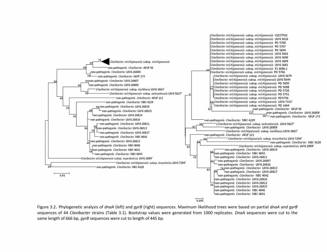
Figure 3.2. Phylogenetic analysis of dnaA (left) and gyrB (right) sequences. Maximum likelihood trees were based on partial dnaA and gyrB
sequences of 44 Clavibacter strains (Table 3.1). Bootstrap values were generated from 1000 replicates. DnaA sequences were cut to the same length of 666 bp, gyrB sequences were cut to length of 445 bp.
Chapter 3 – Genetic diversity of non-pathogenic Clavibacter strains isolated from tomato seeds
102
Genetic heterogeneity
Genetic heterogeneity of both Cmm and non-pathogenic Clavibacter strains was assessed
by BOX-PCR fingerprinting. First, the results showed that Cmm can be easily separated from
non-pathogenic Clavibacter strains investigated so far. Second, as expected, the diversity
within the non-pathogenic Clavibacter strains group appeared to be higher. While the group
of Cmm strain formed a tight cluster, non-pathogenic Clavibacter strains fell into several
groups (Figure 3.3). Cmm strains showed several common bands in a region above 1000 bp
(Figure 3.3). Other primers targeting interspersed repetitive sequences did not generate
valuable patterns to distinguish both groups: (GTG)5 only showed bands in a narrow range
of length (500-2000 bp), whereas ERIC primers revealed only two to three bands per strain
in the 1500 bp to 2000 bp range making these rep patterns useless for comparative
purposes (data not shown). Although it was known that rep-PCR in general was not suitable
to differentiate highly homogenous Cms strains (Fousek and Mraz 2003), we could
successfully distinguish the non-pathogenic Clavibacter strains by BOX- PCR fingerprinting.
As profiles of non-pathogenic Clavibacter strains are more diverse compared to the highly
similar profiles of Cmm (Figure 3.3) one can speculate that this is due to a more versatile life
style of the non-pathogenic Clavibacter strains occupying different ecological niches on
plants and perhaps even soil.
Importance of investigation of atypical strains
Trade between countries, especially trade of plant materials, may bring unintentional
imports of accompanying microorganisms. In most cases such transmissions form no threat,
but the danger exists that these microorganisms adapt to a new environment and cause
serious damage in agriculture. Therefore, the international plant protection organizations
established regulations in order to prevent spreading of agricultural pests as effectively as
possible. It is becoming a crucial issue for quarantine organisms, especially for Cmm for
which the number of new and reoccurring outbreaks increased over the last few years with
some severe occurrences in Spain and Italy (De León et al., 2009, Lamichhane et al., 2011).
Since contaminated seeds are considered the main source of infection there is a big
Part III – Experimental work
103
challenge for the seed industry to produce a pathogen-free, high quality material. As non-
pathogenic Clavibacter strains isolated from tomato seeds interfere with currently applied
diagnostic procedures for Cmm, their presence complicates a clear judgment on tomato
seed health. A better understanding of the genetic basis of the lack of pathogenicity of
these strains and their molecular investigation will help to design more reliable and specific
identification approaches. In the future these strains should be included in the development
and validation processes when designing new diagnostic methods for Cmm in seeds.
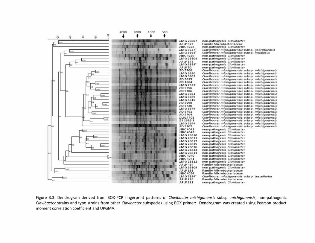
Figure 3.3. Dendrogram derived from BOX-PCR fingerprint patterns of Clavibacter michiganensis subsp. michiganensis, non-pathogenic Clavibacter strains and type strains from other Clavibacter subspecies using BOX primers. Dendrogram was created using Pearson product moment correlation coefficient and UPGMA.
Part III – Experimental work
105
3.4 Conclusions
From an economical perspective, it is most imperative to confirm that seeds contaminated
only with non-pathogenic strains are safe and thus can be released onto the market.
However, our results clearly demonstrated that, although the investigated Clavibacter
strains isolated from tomato seeds were not pathogenic, they seem to contain some
virulence genes, located both on plasmids and on the chromosome. Moreover, not all
virulence factors were investigated and we cannot exclude an eventual latent state of these
strains in a tomato plant. Favourable conditions can induce disease symptoms or the
tomato can be only a carrier of these bacteria onto other susceptible crops. As the impact of
non-pathogenic Clavibacter strains for the expression of pathogenicity is not
straightforward and as these strains were found to react positively with the identification
methods for Cmm, we suggest that in all cases when the results from at least two methods
based on different biological principles (serological, PCR-assay) are positive, a gyrB
sequence is determined and verified with the sequences deposited in the reference
database (Q-bank) or with sequences available on EMBL/NCBI. In case of a positive match
the identification is completed, in case of a negative match, the pathogenicity test should
be performed to confirm the final conclusion.
Acknowledgements
We thank the NVWA, GBBC and BCCM/LMG collections for providing necessary strains. The
authors wish to acknowledge Harrie Koenraad (Naktuinbouw) and Bert Wondt (Syngenta)
for helpful discussions and for providing a collection of non-pathogenic strains. We thank
Dr. Kim Heylen for fruitful discussions and proofreading the manuscript. This work was
performed in the Seventh Framework Programme of project KBBE-2008-1-4-01 (QBOL) nr
226482 funded by the European Commission. The Belgian NPPO (FAVV) is acknowledged for
partially financing ILVO-research and help with in planta test.
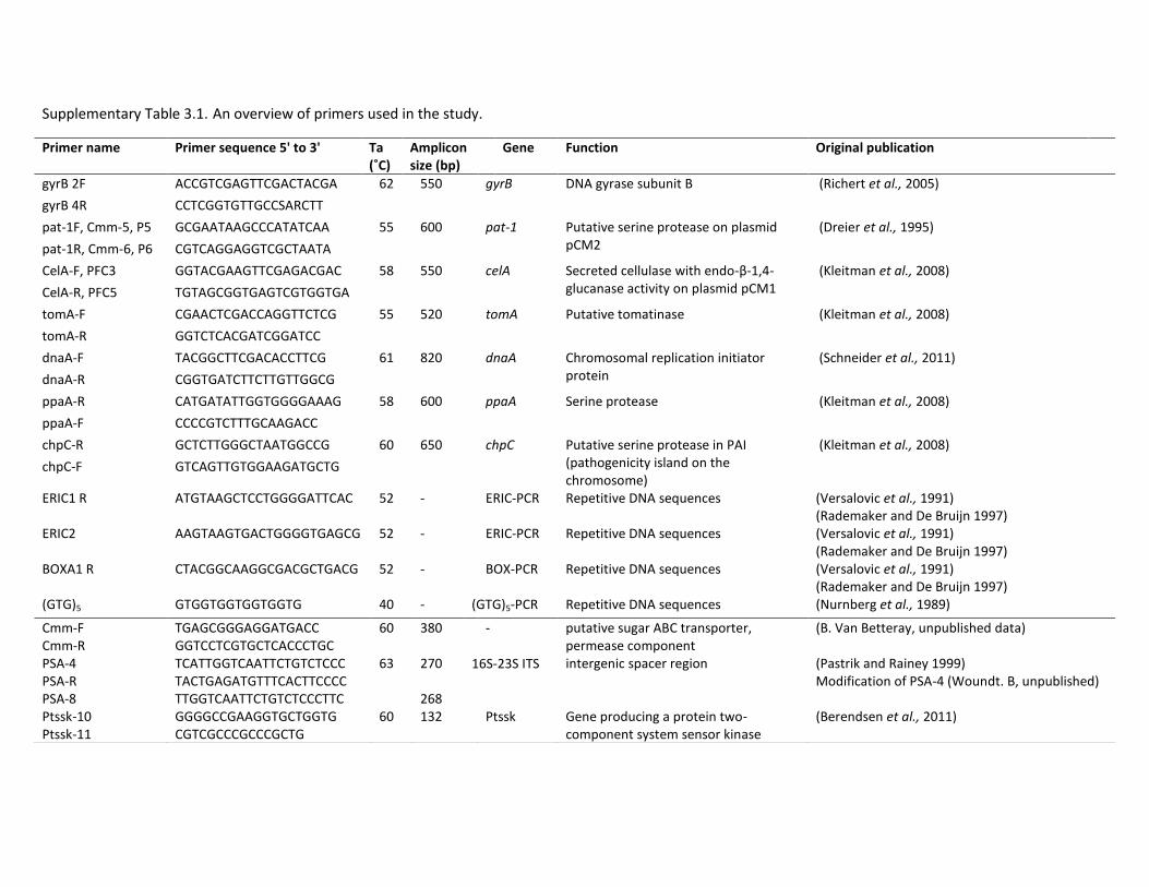
Supplementary Table 3.1. An overview of primers used in the study.
Primer name Primer sequence 5' to 3' Ta (˚C)
Amplicon size (bp)
Gene Function Original publication
gyrB 2F ACCGTCGAGTTCGACTACGA 62 550 gyrB DNA gyrase subunit B (Richert et al., 2005)
gyrB 4R CCTCGGTGTTGCCSARCTT
pat-1F, Cmm-5, P5 GCGAATAAGCCCATATCAA 55 600 pat-1 Putative serine protease on plasmid pCM2
(Dreier et al., 1995)
pat-1R, Cmm-6, P6 CGTCAGGAGGTCGCTAATA
CelA-F, PFC3 GGTACGAAGTTCGAGACGAC 58 550 celA Secreted cellulase with endo-β-1,4-glucanase activity on plasmid pCM1
(Kleitman et al., 2008)
CelA-R, PFC5 TGTAGCGGTGAGTCGTGGTGA
tomA-F CGAACTCGACCAGGTTCTCG 55 520 tomA Putative tomatinase (Kleitman et al., 2008)
tomA-R GGTCTCACGATCGGATCC
dnaA-F TACGGCTTCGACACCTTCG 61 820 dnaA Chromosomal replication initiator protein
(Schneider et al., 2011)
dnaA-R CGGTGATCTTCTTGTTGGCG
ppaA-R CATGATATTGGTGGGGAAAG 58 600 ppaA Serine protease (Kleitman et al., 2008)
ppaA-F CCCCGTCTTTGCAAGACC
chpC-R GCTCTTGGGCTAATGGCCG 60 650 chpC Putative serine protease in PAI (pathogenicity island on the chromosome)
(Kleitman et al., 2008) chpC-F GTCAGTTGTGGAAGATGCTG
ERIC1 R ATGTAAGCTCCTGGGGATTCAC 52 - ERIC-PCR Repetitive DNA sequences (Versalovic et al., 1991) (Rademaker and De Bruijn 1997)
ERIC2 AAGTAAGTGACTGGGGTGAGCG 52 - ERIC-PCR Repetitive DNA sequences (Versalovic et al., 1991) (Rademaker and De Bruijn 1997)
BOXA1 R CTACGGCAAGGCGACGCTGACG 52 - BOX-PCR Repetitive DNA sequences (Versalovic et al., 1991) (Rademaker and De Bruijn 1997)
(GTG)5 GTGGTGGTGGTGGTG 40 - (GTG)5-PCR Repetitive DNA sequences (Nurnberg et al., 1989)
Cmm-F Cmm-R
TGAGCGGGAGGATGACC GGTCCTCGTGCTCACCCTGC
60 380 - putative sugar ABC transporter, permease component
(B. Van Betteray, unpublished data)
PSA-4 PSA-R PSA-8
TCATTGGTCAATTCTGTCTCCC TACTGAGATGTTTCACTTCCCC TTGGTCAATTCTGTCTCCCTTC
63 270 268
16S-23S ITS intergenic spacer region (Pastrik and Rainey 1999) Modification of PSA-4 (Woundt. B, unpublished)
Ptssk-10 Ptssk-11
GGGGCCGAAGGTGCTGGTG CGTCGCCCGCCCGCTG
60 132 Ptssk Gene producing a protein two-component system sensor kinase
(Berendsen et al., 2011)
Part III – Experimental work
107
3.5 References
1. Altschul SF, Gish W, Miller W, Myers EW, Lipman DJ. 1990. Basic local alignment search tool. Journal of Molecular Biology 215: 403-10
2. Alvarez AM, Kaneshiro WS, Vine BG. 2004. Diversity of Clavibacter michiganensis subsp. michiganensis populations in tomato seed: what is the significance? Presented at 1st International Symposium on Tomato Diseases, Orlando, FL
3. Baysal Ö, Mercati F, İkten H, YIldIz R, Carimi F, et al. 2010. Clavibacter michiganensis subsp. michiganensis: tracking strains using their genetic differentiations by ISSR markers in Southern Turkey. Physiological and Molecular Plant Pathology 75: 113-9
4. Bella P, Ialacci G, Licciardello G, La Rosa R, Catara V. 2012. Characterization of atypical Clavibacter michiganensis subsp. michiganensis populations in greenhouse tomatoes in Italy. Journal of Plant Pathology 94: 635-42
5. Berendsen SMH, Koenraadt H, Woudt B, Oosterhof J. 2011. The development of a specific Real-Time TaqMan for the detection of Clavibacter michiganensis subsp. michiganensis. APS-IPPC Meeting, Honolulu (Hawaii), 6-10 August 2011
6. Brenner DJ, Staley JT, Krieg NR. 2001. Classification of prokaryotic organisms and the concept of bacterial speciation. In D. R. Boone, R. W. Castenholz and G. M. Garrity (ed.), Bergey's manual of systematic bacteriology, 2nd ed., vol. 1. Springer-Verlag, New York, N.Y.: 27-31
7. Cheng S, Hu Y-h, Zhang M, Sun L. 2010. Analysis of the vaccine potential of a natural avirulent Edwardsiella tarda isolate. Vaccine 28: 2716-21
8. Cleenwerck I, Vandemeulebroecke K, Janssens D, Swings J. 2002. Re-examination of the genus Acetobacter, with descriptions of Acetobacter cerevisiae sp. nov. and Acetobacter malorum sp. nov. International Journal of Systematic and Evolutionary Microbiology 52: 1551-8
9. Cottyn B, Debode J, Regalado E, Mew TW, Swings J. 2009. Phenotypic and genetic diversity of rice seed-associated bacteria and their role in pathogenicity and biological control. Journal of Applied Microbiology 107: 885-97
10. Cottyn B, Regalado E, Lanoot B, De Cleene M, Mew TW, Swings J. 2001. Bacterial populations associated with rice seed in the tropical environment. Phytopathology 91: 282-92
11. De León L, Rodriguez A, Llop P, Lopez MM, Siverio F. 2009. Comparative study of genetic diversity of Clavibacter michiganensis subsp. michiganensis isolates from the Canary Islands by RAPD-PCR, BOX-PCR and AFLP. Plant Pathology 58: 862-71
12. Dreier J, Bermpohl A, Eichenlaub R. 1995. Southern hybridization and PCR for specific detection of phytopathogenic Clavibacter michiganensis subsp. michiganensis. Phytopathology 85: 462-8
13. Dreier J, Meletzus D, Eichenlaub R. 1997. Characterization of the plasmid encoded virulence region pat-1 of phytopathogenic Clavibacter michiganensis subsp. michiganensis. Molecular Plant-Microbe Interactions 10: 195-206
14. Eichenlaub R, Gartemann K-H. 2011. The Clavibacter michiganensis subspecies: molecular investigation of Gram-positive bacterial plant pathogens. Annual Review of Phytopathology 49: 445-64
15. EPPO. 2005. Clavibacter michiganensis subsp. michiganensis Diagnostics, PM7/42. Bulletin OEPP/EPPO 35: 273-83
16. Ezaki T, Hashimoto Y, Yabuuchi E. 1989. Fluorometric deoxyribonucleic acid-deoxyribonucleic acid hybridization in microdilution wells as an alternative to membrane filter hybridization in which radioisotopes are used to determine genetic relatedness among bacterial strains. Int. J. Syst. Evol. Microbiol. 39: 224-9
17. Fousek J, Mraz I. 2003. Determination of genetic differences between fluid and nonfluid variants of Clavibacter michiganensis subsp. sepedonicus using rep-PCR technique. Folia Microbiologica 48: 682-6
18. Gartemann KH, Abt B, Bekel T, Burger A, Engemann J, et al. 2008. The genome sequence of the tomato-pathogenic actinomycete Clavibacter michiganensis subsp. michiganensis NCPPB382 reveals a large island involved in pathogenicity. Journal of Bacteriology 190: 2138-49
Chapter 3 – Genetic diversity of non-pathogenic Clavibacter strains isolated from tomato seeds
108
19. Gartemann KH, Kirchner O, Engemann J, Grafen I, Eichenlaub R, Burger A. 2003. Clavibacter michiganensis subsp. michiganensis: first steps in the understanding of virulence of a Gram-positive phytopathogenic bacterium. Journal of Biotechnology 106: 179-91
20. Goris J, Suzuki K, De Vos P, Nakase T, Kersters K. 1998. Evaluation of a microplate DNA-DNA hybridization method compared with the initial renaturation method. Canadian Journal of Microbiology 44: 1148-53
21. Heyrman J, Swings J. 2001. 16S rDNA sequence analysis of bacterial isolates from biodeteriorated mural paintings in the Servilia Tomb (Necropolis of Carmona, Seville, Spain). Systematic and applied microbiology 24: 417-22
22. ISHI. 2011. Method for the Detection of Clavibacter michiganensis subsp. michiganensis on Tomato seed (Version 4). International Seed Health Initiative: http://www.worldseed.org/isf/ishi_vegetable.html.
23. Jacques MA, Durand K, Orgeur G, Balidas S, Fricot C, et al. 2012. Phylogenetic analysis and polyphasic characterization of Clavibacter michiganensis strains isolated from tomato seeds reveal that non-pathogenic strains are distinct from C. michiganensis subsp. michiganensis. Applied and Environmental Microbiology 78: 8388-402
24. Jahr H, Dreier J, Meletzus D, Bahro R, Eichenlaub R. 2000. The endo-beta-1,4-glucanase CelA of Clavibacter michiganensis subsp. michiganensis is a pathogenicity determinant required for induction of bacterial wilt of tomato. Molecular Plant-Microbe Interactions 13: 703-14
25. Kaneshiro WS, Alvarez AM. 2001. Specificity of PCR and ELISA assays for hypovirulent and avirulent Clavibacter michiganensis subsp. michiganensis. Phytopathology 91
26. Kaneshiro WS, Mizumoto CY, Alvarez AM. 2006. Differentiation of Clavibacter michiganensis subsp. michiganensis from seed-borne saprophytes using ELISA, Biolog and 16S rDNA sequencing. European Journal of Plant Pathology 116: 45-56
27. Karki HS, Shrestha BK, Han JW, Groth DE, Barphagha IK, et al. 2012. Diversities in virulence, antifungal activity, pigmentation and DNA fingerprint among strains of Burkholderia glumae. PLoS ONE 7: e45376
28. Kleitman F, Barash I, Burger A, Iraki N, Falah Y, et al. 2008. Characterization of a Clavibacter michiganensis subsp. michiganensis population in Israel. European Journal of Plant Pathology 121: 463-75
29. Lamichhane JR, Balestra GM, Varvaro L. 2011. Severe Outbreak of Bacterial Canker Caused by Clavibacter michiganensis subsp. michiganensis on Tomato in Central Italy. Plant Disease 95: 221-
30. Lapage SP, Sneath PHA, Lessel EF. 1992. International Code of Nomenclature of Bacteria: Bacteriological Code, 1990 Revision. Washington (DC): ASM Press; 1992. Statutes of the International Committee on Systematic Bacteriology. Available from: http://www.ncbi.nlm.nih.gov/books/NBK8815/. Washington (DC): ASM Press
31. Louws F, Bell J, Medina-Mora C, Smart C, Opgenorth D, et al. 1998. Rep-PCR-mediated genomic fingerprinting: a rapid and effective method to identify Clavibacter michiganensis. Phytopathology 88: 862-8
32. Maniatis T, E. F. Fritsch and J. Sambrook., ed. 1982. Molecular cloning: a laboratory manual. 33. Meletzus D, Bermphol A, Dreier J, Eichenlaub R. 1993. Evidence for plasmid-encoded virulence factors
in the phytopathogenic bacterium Clavibacter michiganensis subsp. michiganensis NCPPB382. Journal of bacteriology 175: 2131
34. Mesbah M, Premachandran U, Whitman WB. 1989. Precise measurement of the G+ C content of deoxyribonucleic acid by high-performance liquid chromatography. Int. J. Syst. Bacteriol. 39: 159
35. Milijašević-Marčić S, Gartemann KH, Frohwitter J, Eichenlaub R, Todorović B, et al. 2012. Characterization of Clavibacter michiganensis subsp. michiganensis strains from recent outbreaks of bacterial wilt and canker in Serbia. European Journal of Plant Pathology 134: 697-711
36. Nurnberg P, Roewer L, Neitzel H, Sperling K, Popperl A, et al. 1989. DNA fingerprinting with the oligonucleotide probe (CAC) 5/(GTG) 5: somatic stability and germline mutations. Human Genetics 84: 75-8
37. Pastrik K, Rainey F. 1999. Identification and differentiation of Clavibacter michiganensis subspecies by polymerase chain reaction-based techniques. Journal of Phytopathology 147: 687-93
Part III – Experimental work
109
38. Pitcher D, Saunders N, Owen R. 1989. Rapid extraction of bacterial genomic DNA with guanidium thiocyanate. Letters in Applied Microbiology 8: 151-6
39. Rademaker J, De Bruijn F. 1997. Characterization and classification of microbes by rep-PCR genomic fingerprinting and computer assisted pattern analysis. DNA markers: protocols, applications and overviews. J. eds Caetano-Anollés G., Gresshoff P. M. (Wiley and Sons, New York, N.Y): 151-71
40. Richert K, Brambilla E, Stackebrandt E. 2005. Development of PCR primers specific for the amplification and direct sequencing of gyrB genes from microbacteria, order Actinomycetales. Journal of Microbiological Methods 60: 115-23
41. Savidor A, Teper D, Gartemann KH, Eichenlaub R, Chalupowicz L, et al. 2012. The Clavibacter michiganensis subsp. michiganensis-Tomato Interactome Reveals the Perception of Pathogen by the Host and Suggests Mechanisms of Infection. Journal of Proteome Research 11: 736-50
42. Schleifer KH. 2009. Classification of Bacteria and Archaea: Past, present and future. Systematic and Applied Microbiology 32: 533-42
43. Schloter M, Lebuhn M, Heulin T, Hartmann A. 2006. Ecology and evolution of bacterial microdiversity. FEMS Microbiology Reviews 24: 647-60
44. Schneider KL, Marrero G, Alvarez AM, Presting GG. 2011. Classification of plant associated bacteria using RIF, a computationally derived DNA marker. PLoS ONE 6: e18496
45. Smith N, Hennessy J, Stead D. 2001. Repetitive sequence-derived PCR profiling using the BOX-A1R primer for rapid identification of the plant pathogen Clavibacter michiganensis subspecies sepedonicus. European Journal of Plant Pathology 107: 739-48
46. Stork I, Gartemann KH, Burger A, Eichenlaub R. 2008. A family of serine proteases of Clavibacter michiganensis subsp. michiganensis: chpC plays a role in colonization of the host plant tomato. Molecular Plant Pathology 9: 599-608
47. Tamura K, Peterson D, Peterson N, Stecher G, Nei M, Kumar S. 2011. MEGA5: molecular evolutionary genetics analysis using maximum likelihood, evolutionary distance and maximum parsimony methods. Molecular Biology and Evolution 28: 2731-9
48. Tenaillon O, Skurnik D, Picard B, Denamur E. 2010. The population genetics of commensal Escherichia coli. Nature Reviews Microbiology 8: 207-17
49. Thompson J, Higgins D, Gibson T. 1994. CLUSTAL W: improving the sensitivity of progressive multiple sequence alignment through sequence weighting, position-specific gap penalties and weight matrix choice. Nucleic Acids Research 22: 4673
50. Thyr BD, Samuel MJ, Brown PG. 1975. New solanaceous host records for Corynebacterium michiganense. Plant Disease Reporter 59: 595-8
51. Versalovic J, Koeuth T, Lupski JR. 1991. Distribution of repetitive DNA-sequences in Eubacteria and application to fingerprinting of bacterial genomes. Nucleic Acids Research 19: 6823-31
52. Yim KO, Lee HI, Kim JH, Lee SD, Cho JH, Cha JS. 2012. Characterization of phenotypic variants of Clavibacter michiganensis subsp. michiganensis isolated from Capsicum annuum. European Journal of Plant Pathology 133: 559-75
53. Zaluga J, Heylen K, Van Hoorde K, Hoste B, Van Vaerenbergh J, et al. 2011. GyrB sequence analysis and MALDI-TOF MS as identification tools for plant pathogenic Clavibacter. Systematic and Applied Microbiology 34: 400-7
54. Zinniel DK, Lambrecht P, Harris NB, Feng Z, Kuczmarski D, et al. 2002. Isolation and characterization of endophytic colonizing bacteria from agronomic crops and prairie plants. Applied and Environmental Microbiology 68: 2198-208

111
Chapter 4
Comparative genome analysis of pathogenic and non-
pathogenic Clavibacter strains reveals adaptations to their
life styles
Zaluga J, Van Vaerenbergh J, Stragier P, Maes M, De Vos P.
Authors’ contributions:
MM, JVV and PDV conceived the study and participated in its design. JZ participated in the
study design, carried out the molecular work, performed the data analysis and drafted the
manuscript. PS participated in the study design, conducted the genome assembly and
calculated ANI values. All authors read and approved the final manuscript.
Chapter 4 - Comparative genome analysis of pathogenic and non-pathogenic Clavibacter strain reveals adaptations to their life styles
112
Summary
It is well known that the genus Clavibacter harbors economically important plant pathogens
infecting agricultural crops such as potato and tomato. Although the vast majority of
Clavibacter strains are pathogenic, there is an increasing number of non-pathogenic isolates
reported. Especially problematic are non-pathogenic Clavibacter strains isolated from
tomato seeds because of their interference with the current detection/identification
methods for quarantine Clavibacter michiganensis subsp. michiganensis (Cmm). Their
misidentification with pathogenic Cmm strains hampers the clear judgment on the seed
quality and health. In order to get more insight in the genetic content linked to lifestyles of
these bacteria a whole-genome sequence of the seed-borne non-pathogenic Clavibacter
LMG 26808 was determined. To gain a better understanding of the molecular determinants
of pathogenicity, the genome sequence of LMG 26808 was compared to that of the
pathogenic Cmm strains. The comparative analysis revealed that LMG 26808 does not
contain plasmids pCM1 and pCM2 and also lacks the majority of important virulence factors
described so far for pathogenic Cmm. This explains its apparent non-pathogenic character in
tomato plants. Moreover, it appeared that at a certain moment of evolution LMG 26808
acquired sequences from a plasmid of a Klebsiella pneumoniae member. Genes received
that way and coding for antibiotic resistance may provide a competitive advantage for
survival of LMG 26808 in its ecological niche. Genetically, LMG 26808 was highly similar to
the pathogenic Cmm NCPPB 382 but contained more mobile genetic elements suggesting
that there is a higher gene dissemination among these bacteria than observed for
pathogenic Cmm NCPPB 382. High numbers of regulatory genes and transporters compared
to those of soil organisms imply the non-pathogenic Clavibacter strain is most probably a
free-living organism.

Part III – Experimental work
113
4.1 Introduction
Clavibacter is generally considered a genus of plant pathogens, but some ecological surveys
suggested that environmental, non-pathogenic members occur more commonly than
previously thought (Zinniel et al., 2002, Nazina et al., 2002). Non-pathogenic isolates are
often overlooked since diagnostic procedures are focused only on strains responsible for
disease development. Thus, studies were undertaken to initiate the characterization of
these non-pathogenic isolates (Jacques et al., 2012) (Zaluga et al., 2013). The main focus of
this study was dedicated to investigate non-pathogenic Clavibacter strains isolated from
tomato seeds because of their high genetic and physiological similarity to pathogenic Cmm
strains resulting in their misidentification as Cmm. In consequence, a clear judgment on the
seed quality and health is hampered.
Initial in planta experiments demonstrated that this group of isolates is not pathogenic to
the tomato plant and they seem to have lower colonization efficiency than Cmm strains
which are able to move within the vascular system of tomato (Zaluga et al., 2013). Non-
pathogenic clavibacters neither induce an HR (hypersensitive reaction) after infiltration of
Nicotiana tabaccum and Nicotiana benthamiana leaves (Jacques et al., 2012), nor when
inoculated to Mirabilis jalapa (J. Van Vaerenbergh, personal communication). Furthermore,
a majority of these strains is lacking one or both Cmm plasmids carrying important virulence
factors. The majority of non-pathogenic Clavibacter strains isolated from tomato seeds
exhibit similar cell and colony morphology to the genuine Cmm (Zaluga et al., 2011).
Because of the common biological origin (tomato seed), high sequence similarities and
similar physiological characteristics, the non-pathogenic Clavibacter strains are suggested to
be the most related to Cmm.
So far there is very little information available on non-pathogenic Clavibacter strains
isolated from tomato seeds. Reports concerning the ecological niche, survival abilities or
nutritional requirements are lacking. Knowledge about the biology of these strains is
limited, not only because they were only recently traced as constituting a separate
Clavibacter group but also because their importance in the Cmm identification procedure
Chapter 4 - Comparative genome analysis of pathogenic and non-pathogenic Clavibacter strain reveals adaptations to their life styles
114
has not been evaluated previously. To date, non-pathogenic Clavibacter strains have been
found in association with tomato seeds and their ecological niche remains unknown. Also,
routes of transmission and possible sources of these strains have not yet been recognized. It
may be hypothesized that the seed infection occurs at the stage of tomato fruit harvesting
and processing since the movement of non-pathogenic Clavibacter strains in the vascular
tissues of tomato was very limited. This suggests that they could not enter the seeds this
way (Zaluga et al., 2013).
High genetic and phenotypic similarities of non-pathogenic Clavibacter and pathogenic
Cmm strains were the main reasons for their misidentifications as Cmm in the currently
recommended detection/identification methods for Cmm in tomato seeds (ISHI 2011).
Cross-reactions with the antisera and/or positive signals for some of the identification
primers observed in non-pathogenic seed-borne Clavibacter strains illustrate their proximity
in the genetic information and surface antigens to the pathogenic Cmm (Jacques et al.,
2012). Recent studies demonstrated that neither PCR assays based on commonly used 16S
rRNA genes or ITS region, nor those designed for the detection of known virulence factors
are specific to only pathogenic Cmm (Jacques et al., 2012, Kleitman et al., 2008).
Furthermore, some non-pathogenic Clavibacter strains showed fainter PCR amplicons on
gel, impeding the correct interpretation of the results (Zaluga et al., 2013). Taxonomically,
these non-pathogenic clavibacters from tomato seeds are distinct from all Clavibacter
subspecies. Therefore, it was suggested to assign them into a new subspecies (Jacques et
al., 2012). Because of the lack of clear recommendations for subspecies delineation and
because this heterogeneous group of non-pathogenic Clavibacter strains should probably
be assigned to more than one subspecies, based on sequence differences, a formal
allocation was not proposed awaiting for further discriminative taxonomic information.
Pathogenic Cmm strains varying in virulence are sometimes isolated from tomato plants
and seeds but the molecular basis of their reduced virulence is not fully explored (Alvarez et
al., 2004, Kaneshiro and Alvarez 2001, Kleitman et al., 2008). Weakly virulent populations of
Cmm are mostly lacking some virulence factors limiting their ability to fully develop disease
Part III – Experimental work
115
symptoms (Kleitman et al., 2008) but these strain are still able to produce an HR when
tested on Mirabilis jalapa (Milijašević-Marčić et al., 2012). Avirulent Cmm strains do not
produce disease symptoms in tomato plants and are not able to induce an HR in nonhost
plants. However, reports concerning these strains should be carefully checked before
conclusions are made because in some cases strains were assigned to Cmm based on
unsuitable identification methods. Therefore, it is possible that some of the strains regarded
as ‘avirulent Cmm’ are probably not true Cmm since methods used for their identification,
such as ELISA or BIOLOG are not sufficient for a reliable identification at the subspecies
level. An example of this confusing classification concerns strain A4748 (CFBP 7500) that
was first identified as an avirulent Cmm (Alvarez et al., 2004), when later it was shown that
this strain does not belong to Cmm but should be included in a group of saprophytic
Clavibacter strains (Jacques et al., 2012). In order to avoid confusion, more careful and
accurate identification methods should be applied. It is possible that some of the
Clavibacter strains, originally identified as Cmm and producing no symptoms on tomato
plants (classified as avirulent), might represent a novel subspecies within Clavibacter.
In a recent proteomic study in which they investigated tomato-Cmm interaction, many
bacterial proteins with possible functions in the disease development were identified
(Savidor et al., 2012). Numerous hydrolytic enzymes including serine proteases, cellulase,
glycosyl hydrolases and cell wall-degrading enzymes were secreted during tomato infection
of Cmm. Some of them are believed to play a crucial role in colonization and pathogenicity
as their substrates may be plant proteins that participate in defense responses against Cmm
(Savidor et al., 2012). In addition, a number of ABC transporters and two-component
proteins expressed during the infection were assumed to participate in signal transmission
and in sensing the plant environment.
Recent developments in the field of molecular biology and sequencing allowed generating
complete genome sequences and subsequently determining metabolic traits for many
organisms. Genome sequencing enables detailed reconstructions of the physiology and a
functional analysis of the genes present. Complete and draft genomes of plant-associated
Chapter 4 - Comparative genome analysis of pathogenic and non-pathogenic Clavibacter strain reveals adaptations to their life styles
116
and plant-pathogenic bacteria revealed many essential aspects, showed differences in
genome structure, organization and composition and helped to understand the mechanisms
of pathogenicity and the host specificity (Nelson et al., 2000).
Complete genome sequences of Cmm, Cms and Cmn provided genetic information that
allowed for comparative studies and helped to better understand their pathogenicity
characteristics and host adaptation. However, no information is available about the genome
content of non-pathogenic Clavibacter strains, which could deliver some informative
insights into the differences in virulence determinants, genetic content and adaptation to a
life style in their natural ecological niche(s). Genome comparison between pathogenic and
non-pathogenic strains belonging to the same species is an important and valuable
approach to identify genes that may contribute to virulence and general fitness of the
organism.
In this report we present the genome analysis of the non-pathogenic Clavibacter LMG
26808 isolated from tomato seed. The specific purpose of this study were a) to generate a
draft genome sequence of this strain, b) to analyze it for virulence-related gene content by
comparing it to the available genome of the pathogenic Clavibacter michiganensis subsp.
michiganensis NCPPB 382, c) to perform a comparative analysis with the genomes of strains
of Cmm (NCPPB 382), Cmn (NCPPB 2581) and Cms (ATCC 33113), pathogenic to tomato,
maize and potato, respectively, d) to search for adaptations to a non-pathogenic life style.
Part III – Experimental work
117
4.2 Material and methods
Strains and DNA extraction. Non-pathogenic Clavibacter LMG 26808 was obtained from
Naktuinbouw, The Netherlands. Non-pathogenic Clavibacter isolates from tomato seeds are
phenotypically similar to Cmm in the common diagnostic semi-selective media and are
identified as Cmm in customary applied tests but are negative in pathogenicity tests on
tomato (Jacques et al., 2012, Zaluga et al., 2011). They were isolated according to the
current method for detection of Cmm in tomato seed recommended by the International
Seed Federation (ISF) (ISHI 2011). LMG 26808 was aerobically grown on MTNA (mannitol,
trimethoprim, nalidixic acid, amphotericin) medium without antibiotics at 25°C for 24h-48h
(Jansing and Rudolph 1998). Stock cultures were stored at -80˚C in MicrobankTM beads (Pro-
Lab Diagnostics, Canada). Total genomic DNA was extracted according to the guanidium-
thiocyanate-EDTA-sarkosyl method described by Pitcher (Pitcher et al., 1989), which was
adapted for Gram-positive bacteria by a pre-treatment with lysozyme (5 mg/µl lysozyme in
TE buffer) and incubation for 40 minutes at 37°C.
Genome sequencing. Library preparation and genome sequencing was performed by
BaseClear (Leiden, The Netherlands). First, a paired-end (PE) DNA library with a mean insert
size between 150-350 bp was sequenced with average reads of 101 bp on an Illumina
HiSeq2500. Next, a mate-paired (MP) DNA library with a mean insert size between 2000-
3800 bp was sequenced with average reads of 51 bp on Illumina HiSeq2500 (Illumina Inc.).
Automatic trimming (based on a threshold of Q=20) and assembly was performed using CLC
Genomics Workbench v5.0. An initial de novo assembly was performed in CLC Genomics
Workbench v5.0 using the quality trimmed and paired reads from the PE and MP reads. All
contigs shorter than 200 bp were discarded. Finally, the draft genome sequence was
manually edited with the Editseq tool of DNAStar Lasergene core suite v.10.0.1. Remaining
N-nucleotides in the scaffolds, introduced during scaffolding, were removed from the final
sequence by breaking up the scaffolds back into contigs where they were encountered. The
quality of the final draft genome sequence was compared to the initial PE-based de novo
assembly through comparative read-mapping in CLC Genomics Workbench v5.0 using the
Chapter 4 - Comparative genome analysis of pathogenic and non-pathogenic Clavibacter strain reveals adaptations to their life styles
118
trimmed read sets. Contigs were ordered automatically with MAUVE (Darling et al., 2010)
and manually with Artemis (Rutherford et al., 2000) by comparing with Cmm.
Genome annotation. Functional annotation and metabolic reconstruction were performed
with (1) the Rapid Annotation Subsystem Technology (RAST) server (Aziz et al., 2008), using
Glimmer (Delcher et al., 1999) for gene calling and allowing frameshift correction,
backfilling of gaps and automatic fixing of errors, with (2) the Integrated Microbial Genomes
Expert Review (IMG-ER) annotation pipeline using GenePRIMP for genome gene prediction
(https://img.jgi.doe.gov/cgi-bin/er/main.cgi) (Mavromatis et al., 2009) and with (3) the
NCBI's Prokaryotic Genome Automatic Annotation Pipeline (PGAAP)
(http://www.ncbi.nlm.nih.gov/genomes/static/Pipeline.html) which uses GeneMark and
GeneMark.HMM for gene calling (Borodovsky and McIninch 1993) (Lukashin and
Borodovsky 1998). Assigned functions were checked with BLASTp (Altschul et al., 1997).
Alignment and phylogenetic analysis were performed with MEGA 5.0 (Tamura et al., 2011).
Comparative genomic analysis. Artemis software was used for data management and
DNAPlotter was used for genome visualization (Carver et al., 2009). The MAUVE alignment
tool was used for multiple genomes sequence alignment and visualization. IslandViewer
was used to analyze possible genomic islands on the draft genome (Langille and Brinkman
2009). IslandViewer integrates two sequence composition GI prediction methods, namely
IslandPath-DIMOB (Hsiao et al., 2005) and SIGI-HMM (Waack et al., 2006) and one single
comparative GI prediction method, namely IslandPick (Langille et al., 2008) for genomic
island prediction.
ISsaga application from ISfinder server (Varani et al., 2011) was used to identify insertion
sequences (IS) and transposons in the draft genome of LMG 26808. Sequences exhibiting
homology to IS and transposon sequences were verified with the Mobilomics software
(Holmes 2011). The core genome was estimated using the Phylogenetic profiler tool that is
part of the IMG system (http://img.jgi.doe.gov/cgi-bin/m/main.cgi) at a similarity cutoff of
50%.
Part III – Experimental work
119
The presence of possible virulence-related genes and genes expressed during tomato
infection in the draft genome of the non-pathogenic Clavibacter was analyzed by comparing
it with tomato pathogen Cmm NCPPB 382. The comparative screening of the gene content
was performed in RAST, IMG-ER and EDGAR (Blom et al., 2009). Absence or presence of
coding sequences in each genome, as reported by RAST, IMG-ER and EDGAR were
independently confirmed by performing protein and nucleotide BLAST queries in the target
genomes. Proteins with amino acid sequence similarities higher than 50% and with a
coverage higher than 70% were considered homologs. Based on the RAST, IMG-ER and
EDGAR annotation results, the presence of known and putative virulence factors,
pathogenicity related genes and genes uniquely present in the non-pathogenic Clavibacter
LMG 26808 were investigated.
Depositions. The raw sequence data received from Baseclear N.V. will be deposited at the
Short Read Archive (SRA) of Genbank. The current draft genome sequence will be deposited
at Genbank after automatic annotation by the Prokaryotic Genomes Automatic Annotation
Pipeline (PGAAP; http://www.ncbi.nlm.nih.gov/genomes/static/Pipeline.html).
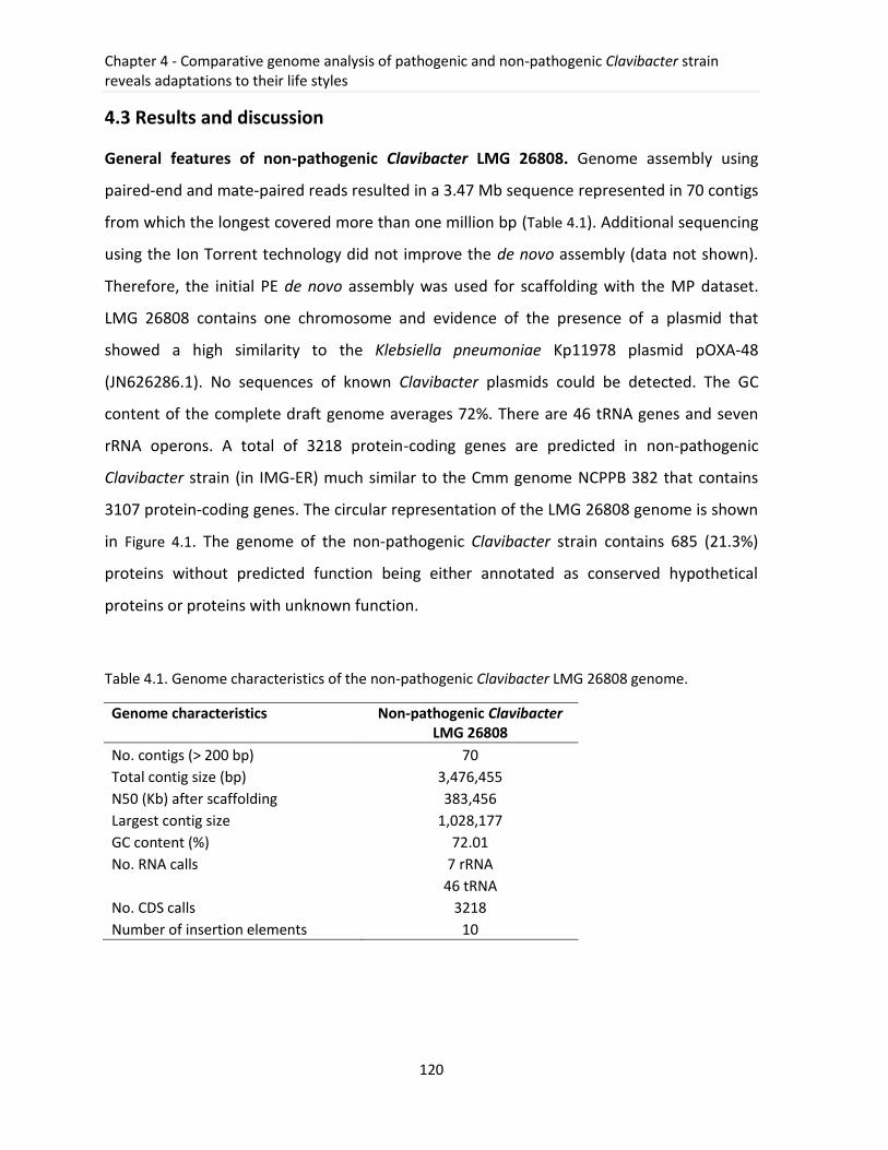
Chapter 4 - Comparative genome analysis of pathogenic and non-pathogenic Clavibacter strain reveals adaptations to their life styles
120
4.3 Results and discussion
General features of non-pathogenic Clavibacter LMG 26808. Genome assembly using
paired-end and mate-paired reads resulted in a 3.47 Mb sequence represented in 70 contigs
from which the longest covered more than one million bp (Table 4.1). Additional sequencing
using the Ion Torrent technology did not improve the de novo assembly (data not shown).
Therefore, the initial PE de novo assembly was used for scaffolding with the MP dataset.
LMG 26808 contains one chromosome and evidence of the presence of a plasmid that
showed a high similarity to the Klebsiella pneumoniae Kp11978 plasmid pOXA-48
(JN626286.1). No sequences of known Clavibacter plasmids could be detected. The GC
content of the complete draft genome averages 72%. There are 46 tRNA genes and seven
rRNA operons. A total of 3218 protein-coding genes are predicted in non-pathogenic
Clavibacter strain (in IMG-ER) much similar to the Cmm genome NCPPB 382 that contains
3107 protein-coding genes. The circular representation of the LMG 26808 genome is shown
in Figure 4.1. The genome of the non-pathogenic Clavibacter strain contains 685 (21.3%)
proteins without predicted function being either annotated as conserved hypothetical
proteins or proteins with unknown function.
Table 4.1. Genome characteristics of the non-pathogenic Clavibacter LMG 26808 genome.
Genome characteristics Non-pathogenic Clavibacter LMG 26808
No. contigs (> 200 bp) 70
Total contig size (bp) 3,476,455
N50 (Kb) after scaffolding 383,456
Largest contig size 1,028,177
GC content (%) 72.01
No. RNA calls 7 rRNA
46 tRNA
No. CDS calls 3218
Number of insertion elements 10
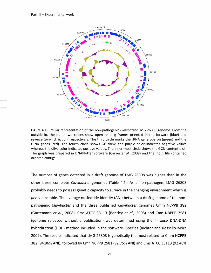
Part III – Experimental work
121
Figure 4.1.Circular representation of the non-pathogenic Clavibacter LMG 26808 genome. From the outside in, the outer two circles show open reading frames oriented in the forward (blue) and reverse (pink) direction, respectively. The third circle marks the rRNA gene operon (green) and the tRNA genes (red). The fourth circle shows GC skew, the purple color indicates negative values whereas the olive color indicates positive values. The inner-most circle shows the GC% content plot. The graph was prepared in DNAPlotter software (Carver et al., 2009) and the input file contained ordered contigs.
The number of genes detected in a draft genome of LMG 26808 was higher than in the
other three complete Clavibacter genomes (Table 4.2). As a non-pathogen, LMG 26808
probably needs to possess genetic capacity to survive in the changing environment which is
per se unstable. The average nucleotide identity (ANI) between a draft genome of the non-
pathogenic Clavibacter and the three published Clavibacter genomes Cmm NCPPB 382
(Gartemann et al., 2008), Cms ATCC 33113 (Bentley et al., 2008) and Cmn NBPPB 2581
(genome released without a publication) was determined using the in silico DNA-DNA
hybridization (DDH) method included in the software JSpecies (Richter and Rosselló-Móra
2009). The results indicated that LMG 26808 is genetically the most related to Cmm NCPPB
382 (94.96% ANI), followed by Cmn NCPPB 2581 (92.75% ANI) and Cms ATCC 33113 (92.48%
Chapter 4 - Comparative genome analysis of pathogenic and non-pathogenic Clavibacter strain reveals adaptations to their life styles
122
ANI). Although based on the ANI values the LMG 26808 genome is the most similar to that
of pathogenic Cmm NCPPB 382, the synteny plots of LMG 26808 and Cmn NCPPB 2581 and
the percentage of homologous genes shared by LMG 26808 and Cmn NCPPB 2581 are also
considerably high (Table 4.2, Figure 4.2, Figure 4.3).
Comparison on a genomic scale revealed a high conservation in the gene sequence among
genomes of LMG 26808, NCPPB 382 and NCPPB 2581 (Figure 4.2). There are 299 genes
(~10%) present in the LMG 26808 draft genome that were not detected in the Cmm NCPPB
382 genome. Forty eight of them could be shown to be present in Cmn and/or Cms
genomes (Supplementary Table 4.1). Almost half of the genes specific for LMG 26808
belonged to hypothetical or unknown proteins (142). Remaining sequences were coding for
additional ABC transporters, antibiotic resistance genes, acetyltransferases and several
enzymes that in majority could not be assigned to any KEGG (Kyoto Encyclopedia of Genes
and Genomes) pathway (Supplementary Table 4.1). When compared to other Clavibacter
genomes, LMG 26808 appeared to not have experienced gene loss and despite it is
considered only a draft, a majority of important genes involved in basic metabolism and
gene regulation could be detected. The core genome consists of 2316 homologs found in all
four Clavibacter genomes. LMG 26808 contains 12 genomic regions exhibiting a lower GC
content (Supplementary Table 4.2). Several genes coding for proteins within these regions
were found to contribute to the fitness of the bacterium (Cl_02679 coding for ABC-type
Fe3+-siderophore transport system; Cl_03044 coding for permease component,
chloramphenicol acetyltransferase (EC 2.3.1.28); Cl_03094 coding for multidrug-efflux
transporter). The genome heterogeneity and genetic diversity among Clavibacter strains
most likely contribute to the differences in the bacterial life style.
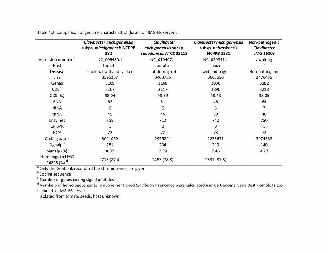
Table 4.2. Comparison of genome characteristics (based on IMG-ER server).
Clavibacter michiganensis subps. michiganensis NCPPB
382
Clavibacter michiganensis subsp.
sepedonicus ATCC 33113
Clavibacter michiganensis subsp. nebraskensis
NCPPB 2581
Non-pathogenic Clavibacter LMG 26808
Accession number a NC_009480.1 NC_010407.1 NC_020891.1 awaiting
Host tomato potato maize *
Disease bacterial wilt and canker potato ring rot wilt and blight Non-pathogenic
Size 3395237 3403786 3063596 3476455
Genes 3169 3168 2936 3282
CDS b 3107 3117 2890 3218
CDS (%) 98.04 98.39 98.43 98.05
RNA 62 51 46 64
rRNA 6 6 6 7
tRNA 45 45 30 46
Enzymes 759 712 740 750
CRISPR 1 0 0 1
GC% 72 72 73 72
Coding bases 3041059 2955244 2823671 3074588
Signalp c 281 234 219 140
Signalp (%) 8.87 7.39 7.46 4.27 Homologs to LMG
26808 (%) d 2716 (87.4) 2457 (78.8) 2531 (87.5) -
a Only the Genbank records of the chromosomes are given b Coding sequence
C Number of genes coding signal peptides d Numbers of homologous genes in abovementioned Clavibacter genomes were calculated using a Genome Gene Best Homologs tool included in IMG-ER server. * Isolated from tomato seeds, host unknown.

Figure 4.2. Local collinear blocks (LCBs) of the chromosomal sequences of non-pathogenic Clavibacter LMG 26808, Clavibacter michiganensis subsp. michiganensis NCPPB 382 and Clavibacter michiganensis subsp. nebraskensis NCPPB 2581 strains. Representation of chromosomal similarity of the three strains was generated by the MAUVE alignment software (Darling et al., 2010). Eighteen local collinear blocks (LCBs) were identified with connecting lines joining the regions on the draft genome that are homologous in the three genomes. LCBs drawn below the black horizontal lines represent homology found in the reverse strand of the genome. Sequence similarity plots are displayed in the LCBs and the height of the sequence identity plot reflects the degree of sequence similarity for the region of the respective alignment. The uncolored regions within the LCBs or in-between LCBs indicate the presence of strain-specific sequences.
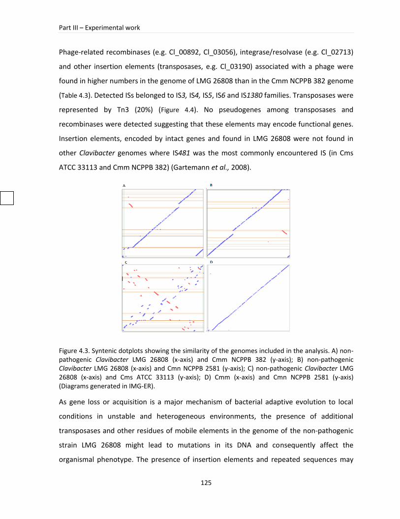
Part III – Experimental work
125
Phage-related recombinases (e.g. Cl_00892, Cl_03056), integrase/resolvase (e.g. Cl_02713)
and other insertion elements (transposases, e.g. Cl_03190) associated with a phage were
found in higher numbers in the genome of LMG 26808 than in the Cmm NCPPB 382 genome
(Table 4.3). Detected ISs belonged to IS3, IS4, IS5, IS6 and IS1380 families. Transposases were
represented by Tn3 (20%) (Figure 4.4). No pseudogenes among transposases and
recombinases were detected suggesting that these elements may encode functional genes.
Insertion elements, encoded by intact genes and found in LMG 26808 were not found in
other Clavibacter genomes where IS481 was the most commonly encountered IS (in Cms
ATCC 33113 and Cmm NCPPB 382) (Gartemann et al., 2008).
Figure 4.3. Syntenic dotplots showing the similarity of the genomes included in the analysis. A) non-pathogenic Clavibacter LMG 26808 (x-axis) and Cmm NCPPB 382 (y-axis); B) non-pathogenic Clavibacter LMG 26808 (x-axis) and Cmn NCPPB 2581 (y-axis); C) non-pathogenic Clavibacter LMG 26808 (x-axis) and Cms ATCC 33113 (y-axis); D) Cmm (x-axis) and Cmn NCPPB 2581 (y-axis) (Diagrams generated in IMG-ER).
As gene loss or acquisition is a major mechanism of bacterial adaptive evolution to local
conditions in unstable and heterogeneous environments, the presence of additional
transposases and other residues of mobile elements in the genome of the non-pathogenic
strain LMG 26808 might lead to mutations in its DNA and consequently affect the
organismal phenotype. The presence of insertion elements and repeated sequences may
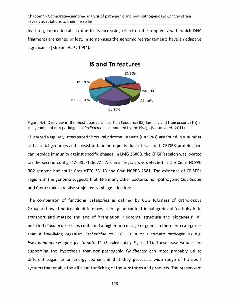
Chapter 4 - Comparative genome analysis of pathogenic and non-pathogenic Clavibacter strain reveals adaptations to their life styles
126
lead to genomic instability due to its increasing effect on the frequency with which DNA
fragments are gained or lost. In some cases the genomic rearrangements have an adaptive
significance (Moxon et al., 1994).
Figure 4.4. Overview of the most abundant Insertion Sequence (IS) families and transposons (Tn) in the genome of non-pathogenic Clavibacter, as annotated by the ISsaga (Varani et al., 2011).
Clustered Regularly Interspaced Short Palindrome Repeats (CRISPRs) are found in a number
of bacterial genomes and consist of tandem repeats that interact with CRISPR-proteins and
can provide immunity against specific phages. In LMG 26808, the CRISPR region was located
on the second contig (126395-126672). A similar region was detected in the Cmm NCPPB
382 genome but not in Cms ATCC 33113 and Cmn NCPPB 2581. The existence of CRISPRs
regions in the genome suggests that, like many other bacteria, non-pathogenic Clavibacter
and Cmm strains are also subjected to phage infections.
The comparison of functional categories as defined by COG (Clusters of Orthologous
Groups) showed noticeable differences in the gene content in categories of ‘carbohydrate
transport and metabolism’ and of ‘translation, ribosomal structure and biogenesis’. All
included Clavibacter strains contained a higher percentage of genes in these two categories
than a free-living organism Escherichia coli 081 ED1a or a tomato pathogen as e.g.
Pseudomonas syringae pv. tomato T1 (Supplementary Figure 4.1). These observations are
supporting the hypothesis that non-pathogenic Clavibacter can most probably utilize
different sugars as an energy source and that they possess a wide range of transport
systems that enable the efficient trafficking of the substrates and products. The presence of
IS3: 30%
IS4:10%
IS5: 10%
IS6:20%
IS1380: 10%
Tn3:20%
IS and Tn features
Part III – Experimental work
127
a high number of genes involved in translation, ribosomal structure and biogenesis implies
that these bacteria respond more effectively and rapidly to nutritional resources, which can
be an important advantage in a changing environment. As demonstrated by Klappenbach
and coworkers, soil bacteria that formed colonies rapidly after exposure to nutrient-rich
medium possesed a higher number of rRNA operons (average 5.5 copy) in comparison to
bacteria that responded slower (average 1.4 copies) (Klappenbach et al., 2000).
Genomic islands. The analysis of the LMG 26808 genome showed that at least 12 regions
with lower GC contents distributed among different contigs could be distinguished
(Supplementary Table 4.2). Genomic islands with lower GC% are thought to be integrative
elements that exhibit different codon usage relative to the rest of the genome, encode for
transposases, integrases and are typically found at tRNA loci. Their acquisition is mostly a
result of actions of phages, transposons or horizontal gene transfer (Juhas et al., 2009).
Some of the genes present in these regions in LMG 26808 were detected previously in the
genome of Cmm NCPPB 382 but the majority represents regions that were not found in
Clavibacter subspecies. The total size of these regions accounts for 265 kb (~7% of the
genome size). However, the pathogenicity island in Cmm NCPPB 382, containing important
genes responsible for effective plant colonization, was not detected in the genome of the
non-pathogenic Clavibacter strain. Genomic regions with lower GC content can contain
diverse genes exhibiting functions in many metabolic processes. The longest region found in
LMG 26808 (more than 100 kb) included genes coding for the antibiotic resistance (beta-
lactamase class A, Cl_03208, Cl_03230), transposases (Cl_03209, Cl_03212) and many
hypothetical proteins (Cl_03223, Cl_03183) some of which showed the highest similarity on
the protein level to genes found on Klebsiella pneumoniae plasmids. Genomic region 3
contains some genes that showed similarities to the genes found in pCM2 plasmid of Cmm
NCPPB 382. The majority of them are hypothetical proteins and two of them code for
acetyltransferases (Supplementary Table 4.2). Previous studies indicated that some
pathogenic Clavibacter strains lacking pCM1 and pCM2 showed a positive signal in
hybridization experiments with specific plasmids regions of Cmm NCPPB 382 implying that
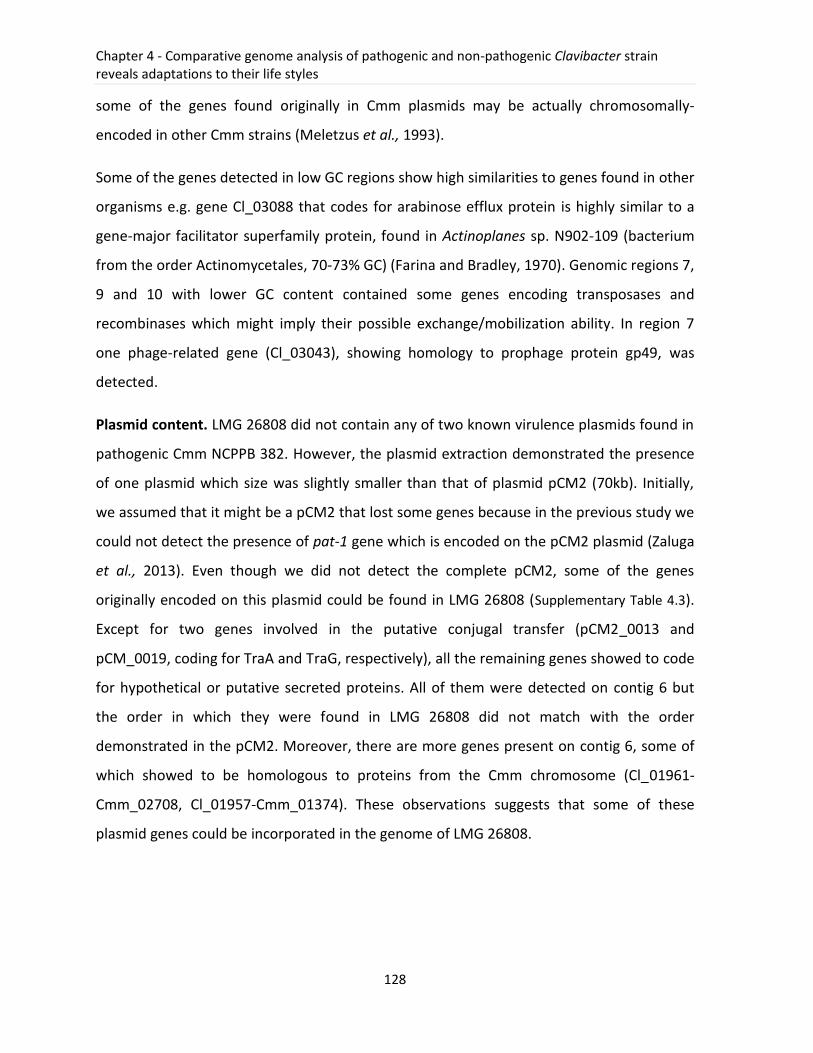
Chapter 4 - Comparative genome analysis of pathogenic and non-pathogenic Clavibacter strain reveals adaptations to their life styles
128
some of the genes found originally in Cmm plasmids may be actually chromosomally-
encoded in other Cmm strains (Meletzus et al., 1993).
Some of the genes detected in low GC regions show high similarities to genes found in other
organisms e.g. gene Cl_03088 that codes for arabinose efflux protein is highly similar to a
gene-major facilitator superfamily protein, found in Actinoplanes sp. N902-109 (bacterium
from the order Actinomycetales, 70-73% GC) (Farina and Bradley, 1970). Genomic regions 7,
9 and 10 with lower GC content contained some genes encoding transposases and
recombinases which might imply their possible exchange/mobilization ability. In region 7
one phage-related gene (Cl_03043), showing homology to prophage protein gp49, was
detected.
Plasmid content. LMG 26808 did not contain any of two known virulence plasmids found in
pathogenic Cmm NCPPB 382. However, the plasmid extraction demonstrated the presence
of one plasmid which size was slightly smaller than that of plasmid pCM2 (70kb). Initially,
we assumed that it might be a pCM2 that lost some genes because in the previous study we
could not detect the presence of pat-1 gene which is encoded on the pCM2 plasmid (Zaluga
et al., 2013). Even though we did not detect the complete pCM2, some of the genes
originally encoded on this plasmid could be found in LMG 26808 (Supplementary Table 4.3).
Except for two genes involved in the putative conjugal transfer (pCM2_0013 and
pCM_0019, coding for TraA and TraG, respectively), all the remaining genes showed to code
for hypothetical or putative secreted proteins. All of them were detected on contig 6 but
the order in which they were found in LMG 26808 did not match with the order
demonstrated in the pCM2. Moreover, there are more genes present on contig 6, some of
which showed to be homologous to proteins from the Cmm chromosome (Cl_01961-
Cmm_02708, Cl_01957-Cmm_01374). These observations suggests that some of these
plasmid genes could be incorporated in the genome of LMG 26808.
Table 4.3. Mobile genetic elements found in the genome of LMG 26808 (Based on the annotation results from IMG-ER, RAST and EDGAR).
CDS identifiers COG COG annotation Pfam Position Length
Cl_00892 COG4974 Site-specific recombinase XerD, phage_integrase pfam00589 Contig 3 (50199 to 51350) 1152
Cl_00935 COG4974 Site-specific recombinase XerD, phage_integrase pfam00589 Contig 5 (8554 to 9540) 987
Cl_01562 COG4974 Site-specific recombinase XerD, phage_integrase pfam00589 Contig 5 (682570 to 683556) 987
Cl_01811 COG1842 Phage shock protein A (IM30), suppresses sigma54-dependent transcription pfam04012 Contig 5 (938633 to 939376) 744
Cl_01968 COG3600 Uncharacterized phage-associated protein pfam13274 contig 7 (7492 to 7956) 753
Cl_03043 COG4679 Phage-related protein pfam05973 contig 15 (974 to 1303) 330
Cl_03056 COG4974 Site-specific recombinase XerD, phage_integrase pfam00589 contig 15 (22760 to 23485) 726
Cl_03252 COG4974 Site-specific recombinase XerD, phage_integrase pfam00589 contig 46 (189 to 866) 678
Cl_02713 COG2452 Predicted site-specific integrase-resolvase pfam12728 contig 11 (350076 to 350522) 447
Cl_03047 COG2801 Transposase and inactivated derivatives, Tnp1, IS3_IS150 pfam01527 contig 15 (9545 to 11077) 1533
Cl_03189 - Transposase DDE domain, Tnp1, IS1380 pfam01609 contig 28 (1417 to 2532) 1116
Cl_03190 COG4644 Transposase and inactivated derivatives, TnpA family, Tn3 pfam01526 contig 28 (4206 to 5753) 1530
Cl_03209 COG4644 Transposase and inactivated derivatives, TnpA family, Tn3 pfam01526 contig 33 (10984 to 12801) 1818
Cl_03210 COG3316 Transposase and inactivated derivatives, IS6 pfam13610 contig 33 (12848 to 13552) 705
Cl_03211 COG2801 Transposase and inactivated derivatives, IS3_IS150 pfam13276 contig 33 (14010 to 13498) 513
Cl_03212 COG2963 Transposase and inactivated derivatives, Tnp1, IS3 pfam01527 contig 33 (14489 to 14343) 147
Cl_03214 COG3316 Transposase and inactivated derivatives, IS6 pfam13610 contig 34 (391 to 116) 276
Cl_03235 - Transposase, Tnp1, IS5_IS903 pfam13737 contig 39 (1373 to 2212) 840
Cl_03261 - Transposase DDE domain, Tnp1, IS4 pfam01609 contig 51 (1 to 1188) 1188
Cl_03204 - Mobile element protein - contig 33 (530 to 366) 165
peg.1244 - Mobile element protein - contig 28 (149 to 742) 594
Cl_03063 - Mobile element protein - contig 15 (32765 to 34252) 1488
peg.807 - Resolvase-like - contig 15 (6452 to 6847) 396
peg.1245 - Tn1 transposase - contig 28 (741 to 1088) 348
Cl_03045 - Gifsy-2 prophage protein - contig 15 (7251 to 7544) 294
Cl_03251 - putative bacteriophage protein - contig 45 (3200 to 4171) 972
Cl_01918 - elements of external origin; phage-related functions and prophages - contig 6 (14793 to15281) 489
Chapter 4 - Comparative genome analysis of pathogenic and non-pathogenic Clavibacter strain reveals adaptations to their life styles
130
The integration into the chromosome will assure that these genes are passed on to
daughter cells. The observation that some genes from pCM2 that were expressed during
tomato infection by Cmm (Savidor et al., 2012) had homologous genes found in LMG 26808,
might suggest that although their function is unknown, they may be essential for non-
pathogenic Clavibacter as well as pathogenic Cmm strains (Supplementary Table 4.3). Further
investigation is needed to elucidate the exact functions of these genes. The smaller plasmid
pCM1 was not detected during the plasmid extraction, nor were its sequences found in the
genome sequence of LMG 26808. Despite that two DNA fragments of LMG 26808 showed
significant similarity to two plasmid-encoded genes, namely pCM1_0018 and pCM1_0020,
the reciprocal BLAST search revealed that these sequences from the non-pathogenic
Clavibacter genome are more similar to the chromosomally encoded CMM_1065 and
CMM_2443, respectively. Interestingly, the latter gene encodes CelB, which is a putative
secreted cellulase that contains a cellulose-binding domain (endo-1,4-beta-glucanase).
Chromosomally encoded celB misses one of three protein coding domains present in the
celA gene. The missing endoglucanase C-terminal domain is similar to α-expansin protein
family from plant and essential for development of wilting and for degradation of crystalline
cellulose (Gartemann et al., 2008, Jahr et al., 2000). The lack or disruption of any of these
domains of celA inevitably led to disability to induce disease symptoms in a tomato plant
(Jahr et al., 2000).
Unexpectedly, the genome analysis showed the presence of sequences found in a Klebsiella
pneumoniae plasmid pOXA-48 (61881 bp). The presence of sequences from a plasmid of
Gram-negative bacteria in a Gram-positive Clavibacter strain is rather unusual and has not
been reported previously. Although the genome sequence of LMG 26808 is only a draft and
therefore incomplete, we could not detect any sequences that could be attributed to a
Klebsiella pneumoniae Kp11978 chromosome. In order to assure that the original biological
material of LMG 26808 and therefore also DNA extracted from it, was not contaminated by
Klebsiella pneumoniae cells, additional tests were performed. Growth tests on VRBG (Violet
Red Bile agar with Glucose) a specific medium for the growth of Enterobacteriaceae were
negative for growth of LMG 26808, whereas R-31932 a positive control of Klebsiella
Part III – Experimental work
131
pneumoniae, grew fast and produced characteristic colonies. Growth tests on EMB (Eosin-
Methylene Blue) (Levine 1918), commonly used for isolation and differentiation of Gram-
negative enteric bacilli isolated from various medical and environmental samples, showed a
delayed growth of the Gram-positive non-pathogenic Clavibacter strain versus the growth
of the control strain. Although it was reported that some Gram-positive bacteria, such as
enterococci, staphylococci and yeast will grow on this medium forming usually pinpoint
colonies, we decided to perform a gram staining and sequencing to confirm the identity of
the observed colonies. As expected, cells isolated from EMB medium were Gram-positive
and subsequent 16S rRNA sequencing showed that this strain belonged to the genus
Clavibacter. Additionally, primers designed for some of the plasmid sequences found in the
genome of LMG 26808 were tested in order to check if the Klebsiella pneumoniae plasmid is
still detectable in the in house plasmid extracts of LMG 26808. The results were positive for
three different plasmid extracts confirming the presence of the sequences from a Klebsiella
pneumoniae plasmid. A PCR detection (primers Pf and Pr1) based on 16S–23S rDNA internal
transcribed spacer (ITS) of Klebsiella pneumoniae was used to check the presence of a
Klebsiella pneumoniae chromosome (Liu et al., 2008). This test showed only amplification in
the positive control and no amplicons were detected in the DNA and plasmid extractions of
LMG 26808. These results firmly support the lack of contamination of LMG 26808 by
Klebsiella pneumoniae cell material.
The exchange of genetic material between various prokaryotes is well known and has been
extensively studied over the last few decades (Gevers et al., 2003, Mazodier and Davies
1991, Musovic et al., 2006, Schäfer et al., 1990, Trieu-Cuot et al., 1987). Lateral gene
transfer (LTG, also known as horizontal gene transfer HTG) realized via the following
mechanisms: i) conjugation (mediated by plasmids), ii) transformation (an uptake of naked
DNA from the environment), iii) transduction (mediated by phages), iv) gene transfer agents
(transposons, phage like-vehicles) and v) microtubes, is affecting almost all known genes
and is contributing substantially to the bacterial evolution (Popa et al., 2011b). Although it
was demonstrated for bacteria that the gene exchange is observed more frequently
between closely related genera with a similar GC content and exhibiting high sequence
Chapter 4 - Comparative genome analysis of pathogenic and non-pathogenic Clavibacter strain reveals adaptations to their life styles
132
similarities there are examples of recent gene transfers between distantly related bacteria
(e.g. Actinobacteria and gammaproteobacteria) (Popa et al., 2011a). Barriers responsible for
the limited transfer between dissimilar genomes are not yet well understood but are likely
related to a particular transfer mechanism, to physical distances between donor and
recipient and to functional differences, such as absence of required promoters that lower
the chances for maintenance of newly acquired genetic material (Popa et al., 2011b).
Interspecies barriers between various genera can be lower in complex bacterial
communities as demonstrated for marine environments in which distantly related bacterial
genera showed a high conjugation level (Dahlberg et al., 1998).
Conjugational transfer is considered the most efficient way of LTG (Dahlberg et al., 1998,
Grohmann et al., 2003) and contributes the most to the spread of antibiotic resistance
among different bacteria (Bennett 2008). This type of LTG is widely encountered among
various bacterial species and even between bacteria and Archaea (Dodsworth et al., 2010)
on the one hand and between bacteria and higher organisms such as Saccharomyces
cerevisiae (Heinemann and Sprague Jr 1989), or plants (Zambryski et al., 1989) on the other
hand. Conjugational plasmid exchange was also observed within the genus Clavibacter in
which the endophytic CMM100 strain (cured of pCM1 and pCM2 plasmids) was able to
acquire these plasmids from other pathogenic Cmm strains and restore pathogenicity
(Eichenlaub et al., 2006). Furthermore, transformation experiments carried out with
Clavibacter xyli subsp. cynodontis (currently reclassified to the genus Leifsonia)
demonstrated the possibility to acquire an IncP plasmid from Enterobacteriaceae by this
Gram-positive Actinobacteria which provided another evidence of conjugational transfer
between diverse taxa.
Klebsiella pneumoniae strains were found in many important crops such as potato, maize,
soybean, cotton and tomato (Quadt-Hallmann et al., 1997, Rosenblueth and Martinez-
Romero 2006). Many of these strains carry plasmids that contain antibiotic resistance genes
and possess the conjugation transfer systems which enable the gene mobilization and
exchange among and outside Enterobacteriaceae and other bacterial genera (Zhao et al.,
2010). Some genes encoded on the Klebsiella pneumoniae plasmids exhibit high similarities
Part III – Experimental work
133
to regions found previously in Escherichia coli and Yersinia genomes, implying that there is
an active genetic exchange among strains of these genera (Bistué et al., 2008).
Plasmid pOXA-48, of which sequences showed high similarities to genes detected in the
genome of LMG 26808, was self-transmissible and highly similar in various
Enterobacteriaceae species, such as Klebsiella pneumoniae, Citrobacter freundii and
Escherichia coli (Carrër et al., 2010). But with its rapid spread throughout Europe, Africa and
Middle East, its similarity to broad-host range plasmid IncL/M (Poirel et al., 2011) and its
effective mobilization system the transfer to other bacteria can also occur, but at lower
frequencies. Additionally, because a reservoir of blaOXA-48 (carrying the carbapenemase
gene) has been recently identified in the waterborne Gram-negative Shewanella oneidensis
species (Poirel et al., 2004), it is possible that the dissemination of this gene is subject of a
widespread interspecies exchange with a consequent presence of plasmids encoding blaOXA-
48 gene in a wide variety of bacteria. Characteristic features of this plasmid, including a
transposon system and many tra (transfer) and mob (mobilization) genes were detected in
the non-pathogenic Clavibacter strain (Supplementary Table 4.4).
Although an acquisition by LMG 26808 of a relatively large plasmid originating most
probably from a Klebsiella pneumoniae was unexpected and unprecedented, a similar
occurrence was previously reported by Baltrus and coworkers. They detected a recent
acquisition by two cucumber isolates of Pseudomonas lachrymans of a megaplasmid which
has not been detected in Pseudomonas members so far (Baltrus et al., 2011). It was
suggested that this acquisition resulted from an important ecological shift across closely
related Pseudomonas members and that the plasmid-encoded genes may be advantageous
for the recipient bacteria.
As Klebsiella pneumoniae and Clavibacter strains thrive in the same environmental niche
(associated with tomato) and because of examples of possible genetic material exchange
between distantly related bacteria we can hypothesize that the acquisition of plasmid
sequences encoding antibiotic resistance genes may provide a competitive advantage for
the non-pathogenic Clavibacter strain LMG 26808.
Chapter 4 - Comparative genome analysis of pathogenic and non-pathogenic Clavibacter strain reveals adaptations to their life styles
134
Non-pathogenic life style. Non-pathogenic Clavibacter strains from tomato seeds tested in
the previous study (Zaluga et al., 2013) as well as other strains tested by Jacques and
coworkers (Jacques et al., 2012) did not introduce any disease symptoms when tested on
tomato plants. Possible explanations for the non-pathogenic nature of these strains are i)
the lack of two plasmids present in pathogenic Cmm and carrying virulence factors, ii) the
absence of the pathogenicity island and iii) a significantly lower number of genes coding for
extracellular hydrolytic enzymes including several important serine proteases, glycosyl
hydrolases and the plant cell wall-hydrolyzing enzymes.
In pathogenic Cmm, main virulence factors cel-A and pat-1, encoded on pCM1 and pCM2,
respectively, are required to induce disease symptoms (wilting and canker) in tomato plants
(Dreier et al., 1997, Jahr et al., 2000). Moreover, genes coding for the production of
extracellular enzymes, such as endoglucanase (Meletzus et al., 1993), polygalacturonase,
xylanase (Beimen et al., 1992), serine proteases and other secreted proteins have been
implicated as possible virulence factors in recent reports (Dreier et al., 1997, Savidor et al.,
2012). The genome of LMG 26808 was checked for the presence of several groups of
different genes exhibiting a possible role in plant recognition, colonization and disease
development, as reported in a proteomic study of Savidor and coworkers (Savidor et al.,
2012) (Supplementary Table 4.5).
The most prominent virulence factors pat-1 and celA could not be detected in the genome
of LMG 26808. Their absence may be directly linked with the absence of plasmids pCM1 and
pCM2 in the non-pathogenic Clavibacter. However, southern hybridization experiments
with plasmid fragments containing virulence factors showed that in some plasmid-free
pathogenic Cmm strains these virulence determinants were present on the chromosome
(Meletzus et al., 1993).
A proteomic study of tomato-Cmm interaction identified several bacterial proteins with a
putative role in signal perception, transduction and response to impulses. They belong to
two-component system proteins, transcriptional regulators and other DNA binding proteins.
They are believed to play a role in sensing the tomato plant environment and initiating
pathways, possibly leading to disease development (Savidor et al., 2012). All putative genes
Part III – Experimental work
135
encoding proteins that are probably involved in signal exchange between tomato and
bacterium could be identified in the genome sequence of LMG 26808 (Supplementary Table
4.5). The absence of frameshifts or truncations within the sequences suggest that the genes
might be functional.
As a non-pathogenic bacterium, LMG 26808 was hypothesized to contain less genomic
information for hydrolytic enzymes that are known to be expressed during tomato infection
with Cmm (Savidor et al., 2012). As expected, the most important group containing genes
coding for secreted proteases from Pat-1 family was largely absent in LMG 26808
(Supplementary Table 4.5). Additional pat-1 homologues encoded on the pCM2 plasmid
(plasmid homologs of pat-1, phpA and phpB) were also absent. From seven genes encoding
putative serine proteases chpA-chpG (chromosomal homologs of pat-1) (Gartemann et al.,
2003) only sequences similar to chpF and chpG could be detected. Both of the sequences,
however, matched to the same region and a reciprocal best BLAST hit confirmed the
presence of only chpF. Interestingly, the lack of chpG may be a possible explanation for the
disability of LMG 26808 to produce a hypersensitive response (HR) in nonhost plants since
the chpG mutant in Cmm was unable to cause an HR in Mirabilis jalapa (Gartemann et al.,
2008). Moreover, the low colonization efficiency of LMG 26808 could be attributed to the
lack of chpC gene. A chpC mutation in the pathogenic Cmm NCPPB 382 resulted in a drastic
reduction in colonization abilities in tomato plants (Gartemann et al., 2008, Stork et al.,
2008). Pseudogenes chpA, chpB and chpD were not found in LMG 26808. The family of chp
genes is important for plant-pathogen interaction in Cmm, but probably also in Cms where
11 homologs were found.
The majority of members of secreted serine proteases of Ppa family (PpaA-PpaJ) that are
encoded in several different loci on the chromosome and on pCM1 could not be found in
LMG 26808. Since ppaA and ppaC genes are important for plant colonization (Gartemann et
al., 2008) and they were absent in LMG 26808, it can be another evidence why non-
pathogenic Clavibacter strains are poorly colonizing tomato plants. Indeed, secreted serine
proteases studied in pathogenic Cmm are thought to presumably facilitate the interaction
Chapter 4 - Comparative genome analysis of pathogenic and non-pathogenic Clavibacter strain reveals adaptations to their life styles
136
between Cmm and its host plant and are believed to play a function in pathogenicity by a
possible utilization of plant proteins (Savidor et al., 2012). Their lack might imply that
interaction between LMG 26808 and tomato is actually very limited. Two out of three genes
coding for subtilases sbtA, sbtB and sbtC which are known to be secreted during the plant
infection (Savidor et al., 2012) were also present in LMG 26808. Sbt proteins of Cmm are
highly similar to different tomato subtilases, some of which have been associated with
wound formation and pathogen responses (Jorda et al., 1999) and may play a role in the
disease development. Because they are present in the non-pathogenic Clavibacter strain
their function probably cannot be solely associated with the disease development.
Cellulases and pectinases are the most important enzymes degrading plant cell walls. In
many bacteria, genes encoding these enzymes were found to be virulence determinants
(Salmond 1994). In the genome of LMG 26808 genes for pectate lyases, pelA1, and pelA2
and cellulase celA were not found. However, another cellulase celB, the polygalacturonase
pgaA (whose substrate is pectin), xysA (whose substrate is β-1,4-xylan) and an arylesterase
(which hydrolyzes ester bonds between hemicelluloses and lignin) (Borneman et al., 1990)
were present in LMG 26808 (Supplementary Table 4.5). These findings support the thesis that
non-pathogenic Clavibacter strain is probably less efficient in digesting pectins and cellulose
into simpler by-products than the pathogenic Cmm that is equipped with many various
plant cell degrading enzymes.
Enzymes from a large group of glycosyl hydrolases (GH), which hydrolyze the glycosidic
bond between two carbohydrates or between a carbohydrate and a noncarbohydrate
molecule (Henrissat et al., 1995) were also expressed during plant infection of Cmm.
Therefore, many of them are assumed to be potential virulence factors that can hydrolyze
substrates of a plant origin (Savidor et al., 2012). Our results demonstrated that genes for
the majority of these enzymes are present in LMG 26808 suggesting that their function
might not be restricted to disease development alone.
Very important functions involved in transport and metabolism are linked to the presence
of ABC and other transporters that ensure the uptake and intake of amino acids, metals,
Part III – Experimental work
137
sugars, oligopeptides, etc. Some of these transporters that were expressed during tomato
infection by Cmm may utilize plant molecules to support metabolism of Cmm. The genes
found in the LMG 26808 genome that code for transporters that are known to be expressed
during plant infection by Cmm are listed in Supplementary Table 4.6. Interestingly, only four
out of fifty seven transporters could not be found in LMG 26808. Furthermore, the genome
of non-pathogenic Clavibacter contained additional transporters that were not present in
the pathogenic Cmm genome (Supplementary Table 4.1). Some of them are supposed to play
a role in the active drug transport and cell protection from toxic metabolites (C_03094 and
Cl_03219). Another very important example of additional ABC transporters in the genome
of LMG 26808 are transporters involved in iron complex transport (ABC-type Fe3+
siderophore transport system Cl_02679 and ABC-type transporter, ATPase component
Cl_02678). Iron is an essential ion for bacteria because it is needed to properly build the
active site of various key enzymes among which cytochrome c, a component of the electron
transport chain in a respiratory system. It also influences cell composition, secondary
metabolism and enzymes activity (Church et al., 2000). Hence, a lack or limitation of iron
can restrict the bacterial growth efficiency. An alternative iron uptake system found in LMG
26808 might be advantageous in an iron deficient environment. The number of various
transporters detected in the genome of non-pathogenic Clavibacter (ABC transporters,
Pfam00005-77) was as high as in Cmm (ABC transporters, Pfam00005-75) and significantly
higher than in the intracellular bacterium Mycobacterium tuberculosis (strain H37 Rv),
which contained only 38 ABC transporters. This data suggests that LMG 26808 is probably
an environmental bacterium able to utilize a broad variety of compounds to maximize its
survival changes. Many environmental bacteria were shown to contain a high number of
transporter genes in support of their life style (Harland et al., 2005).
Observations described above correlate well with the initial assumptions that suggested
that non-pathogenic Clavibacter strains must have lost or never contained prominent
virulence determinants responsible for disease induction in tomato plants. These
hypotheses were partially underpinned by similar findings in another draft genome of non-
pathogenic Clavibacter LMG 26811, which lacks the majority of virulence factors including
Chapter 4 - Comparative genome analysis of pathogenic and non-pathogenic Clavibacter strain reveals adaptations to their life styles
138
two main determinants. It also contained less plant cell degrading enzymes than pathogenic
Cmm NCPPB 382 (data not shown). Furthermore, the comparative genome analysis of LMG
26808 and Cmm NCPPB 382 revealed that some putative virulence factors, determined
based on expression levels obtained from the proteomic study of tomato-Cmm interaction
(Savidor et al., 2012), were also present in LMG 26808, which may imply their redundant
functions and suggest that they are not critical for Cmm virulence.
Survival in the environmental niche. Non-pathogenic Clavibacter strains were isolated from
the tomato seeds yet knowledge on their environmental niche is largely lacking. Despite the
association of non-pathogenic clavibacters with tomato plants their ecological niche(s) and
transmission routes are not yet found. Preliminary results with colonization experiments
showed their poor colonization abilities of vascular tomato tissues and a seemingly lower
survival potential than Cmm in plant material (Zaluga et al., 2013). The hypersensitive
reaction (HR) performed in the Mirabilis jalapa (J. Van Vaerenbergh, data not published)
was negative, indicating that non-pathogenic Clavibacter strains do not contain genes
producing proteins that would be recognized by the plant and trigger the active plant
defense response.
LMG 26808 and NCPPB 382 share many homologous genes, which indicates that their basic
metabolism is probably quite similar. As described for the pathogenic Cmm strain also LMG
26808 does not contain genes coding for nitrate or nitrite reduction. Therefore, it needs
other nitrogen sources for growth. Ammonia is a main intermediate in the nitrogen
metabolism that can be produced from inorganic sources (nitrate, nitrite, molecular
nitrogen). In bacteria that are not able to assimilate oxidized nitrogen sources, ammonium
can potentially be generated catabolically from purines and urea (Doroshchuk et al., 2006)
or acquired from the environment. In LMG 26808 urea can be metabolized by urea
carboxylase (Cl_02362) to ammonium. An ammonium transporter Cl_01263 found in LMG
26808 is probably responsible for ammonium acquisition from the environment. Bacteria
can also obtain nitrogen by uptake of amino acids, such as glutamine, glutamate, aspartate
and asparagine from the environment. Several transporters involved in this acquisition were
indeed detected in LMG 26808 (Cl_02150, Cl_02359, Cl_02768-Cl_2772). Amino acids can
Part III – Experimental work
139
then be enzymatically changed to produce ammonia in a reaction involving asparaginases
(Cl_00001, Cl_01800) and aspartate ammonia-lyase (Cl_00099). Remarkably, the genome of
the pathogenic Cmm NCPPB 382 does not have the tRNA synthetase for asparagine that has
to be produced using the gatABC system whereas LMG 26808 is an L-asparagine prototroph.
Antibiotic resistance. In the genome of LMG 26808 several additional genes responsible for
antibiotic resistance were detected (Supplementary Table 4.1). They coded for beta-
lactamases (Cl_03263, peg.1233, peg.1766, peg.1776), chloramphenicol acetyltransferase
(Cl_03044) and tetracycline efflux protein TetA (peg.1764). They showed the highest
similarity to genes found in Klebsiella pneumonia, Escherichia coli and Salmonella enterica.
In addition to the above genes, the genome of LMG 26808 contains two drug efflux
transporters (Cl_03219, Cl_03094) not found in pathogenic Cmm. Interestingly, LMG 26808
contained glyoxalase/bleomycin resistance protein (Cl_03100), which probably constitutes
the resistance to bleomycin-antibiotic produced by some Streptomyces strains (Semon et
al., 1987).
The presence of additional acetyltransferases might suggest that LMG 26808 exhibits broad
resistance to certain antibiotics as some of the acetyltransferases (GNAT superfamily)
catalyze the selective acetylation of one of the four amino groups found on a diverse set of
aminoglycosides with antibiotic properties (Vetting et al., 2005). Acetylation reduces the
affinity of these compounds for the acceptor tRNA site on the 30S ribosome. As a
consequence, bacteria expressing these genes are resistant to some aminoglycosides with
antibiotic properties. A BLAST search of a gene encoding 6'-N-acetyltransferase (peg.1234,
found in LMG 26808) against the nucleotide database showed 100% similarity to the
aminoglycoside N(6')-acetyltransferase from Escherichia coli and to Aac6'-lb-cr
(aminoglycoside N(6')-acetyltransferase) from Klebsiella pneumoniae. Aminoglycoside N(6')-
acetyltransferases found in strains of Escherichia coli were responsible for the resistance to
antibiotics such as amikacin, kanamycin or netilmicin (Chavideh et al., 1999). The ability to
cope with antibiotics produced by organisms with which non-pathogenic Clavibacter strains
share the environmental niche is a significant adaptive advantage. The growth of
Chapter 4 - Comparative genome analysis of pathogenic and non-pathogenic Clavibacter strain reveals adaptations to their life styles
140
Clavibacter strains in culture is often inhibited by other faster growing organisms.
Therefore, the presence of genes coding for antibiotic resistance might be the reason why
non-pathogenic Clavibacter strains are more frequently encountered and isolated from the
semi-selective medium during the tomato seed certification.
Toxins. The presence of the toxin-antitoxin genes (YefM Cl_00198, peg.1235 and YoeB
Cl_00197) in the genome of non-pathogenic Clavibacter is intriguing and raises questions
concerning their origin and potential functions in relation to the physiology of the
bacterium (Supplementary Table 4.1). The YefM and YoeB toxin-antitoxin (T-A) genes were
found in many bacterial genomes and sometimes more than one copy per genome (Pandey
and Gerdes 2005). It was demonstrated that T-A systems are present in many
environmental, free-living and pathogenic organisms (Pandey and Gerdes 2005, Yamaguchi
et al., 2011). The BLAST analysis of YefM and YoeB genes from LMG 26808 revealed the high
similarities to genes encoding proteins from Rhodococcus pyridinivorans AK37 and
Microbacterium testaceum StLB037, respectively. The T-A system found in LMG 26808 was
not present in the pathogenic Cmm, but YefM (peg.1235) was present in other non-
pathogenic Clavibacter strain LMG 26811 (data not shown). Interestingly, Cmn contained
another putative toxin-antitoxin system. T-A systems are not essential for normal cell
growth, nevertheless they are present in many bacteria and Archaea (Pandey and Gerdes
2005). Based on the frequency of T-A systems, it was suggested that they play subtle roles
that are advantageous for cell survival in their natural habitats. Toxins may facilitate cellular
adaptation of an organism to changing environments by slowing down its cell growth,
inhibiting its cell growth, or causing some of its cells to die (Yamaguchi et al., 2011). It is
possible that the presence of a T-A system in the LMG 26808 genome increases the fitness
of this bacterium in the occupied environmental niche. Differences in the detected toxin-
antitoxin systems in particular Clavibacter subspecies might be attributed to different
ecological niches and inhabited hosts.
In addition to the identified T-A system, LMG 26808 contained a gene (peg.942) coding for a
putative nisin resistance. Nisin is a bacteriocin produced by certain Lactococcus species that
Part III – Experimental work
141
inhibits the growth of a range of Gram-positive bacteria (Shimizu et al., 1999). The presence
of a nisin resistance gene may help the non-pathogenic Clavibacter strain to overcome the
inhibiting effect of this bacteriocin. Moreover, LMG 26808 contained genes involved in the
production of other types of bacteriocins such as vicibactin (Cl_00225) and microcins
(peg.820 and peg.817), which can limit the growth of various bacteria. The ability to cope
with bacteriocins and production of some others is probably beneficial for non-pathogenic
clavibacters that are expected to coexist with many different bacteria in the same ecological
niche.
Error prone UmuDC operon. SOS mutagenesis response in bacteria includes error-prone
and error-free DNA damage repair responses that are activated after exposure to different
antibiotics, chemical compounds or radiation (Hare et al., 2012). In Escherichia coli UmuDC
proteins are involved in error-prone bypass of UV lesions and UmuC protein posses DNA
polymerase activity. In the SOS process many genes get induced and their products are
involved in DNA repair, replication and cell cycle control in order to repair the DNA damage
(Hare et al., 2006). The genes coding for this operon were found in LMG 26808 and also in
another non-pathogenic Clavibacter, LMG 26811 (data not shown), implying that their cells
might have higher abilities to recover after exposures to UV and/or other types of
chemicals. It is tempting to hypothesize that because of a more efficient system of DNA
repair and damage control present in these non-pathogenic strains they may be more
resistant to the heat or acid treatments applied in the seed extraction processes. In a
consequence, they might be easier retrieved during the seed certification procedures.
The extracellular polysaccharide (EPS). The genome of the non-pathogenic Clavibacter
strain and pathogenic Cmm and Cms, contained four gene clusters involved in
exopolysaccharides production (Supplementary Table 4.7). The EPS production in LMG 26808
is expected to effectively occur since all genes involved in that process are functional (no
frameshifts, no pseudogenes). There is, however, one notable difference between
pathogenic Cmm NCPPB 382 and LMG 26808. In the EPS2 of LMG 26808 the order of the
genes is disrupted because they are located at different contigs. The functionality of this
Chapter 4 - Comparative genome analysis of pathogenic and non-pathogenic Clavibacter strain reveals adaptations to their life styles
142
gene cluster is therefore unknown. Even if functional, it will probably be dependent on
different regulation factors which may eventually influence the EPS production. EPS gene
clusters in the pathogenic Cms underwent quite some drastic changes with disruptions by
insertion elements and most likely the EPS production in Cms ATCC 33113 is limited. In
many bacteria the ability to produce EPS and their presence in the cell wall surface has been
shown to participate in the interaction between bacteria and the environment. EPS is
believed to prevent bacterial attachment to host cells which in consequence prevents the
recognition of the bacteria by the host and the induction of an HR reaction (Van den Bulk et
al., 1991). In many plant pathogens EPS production prevents bacterial immobilization by
host lectins and in that way allows bacteria to spread in the xylem vessels (Bermpohl et al.,
1996). The presence of at least three functional EPS gene clusters in LMG 26808 might
facilitate the EPS production in different environmental conditions and can be an advantage
for non-pathogenic Clavibacter, which probably inhabits not only tomato seeds but also
other environmental niches.
Part III – Experimental work
143
4.4 Conclusions
The analysis of the genome sequence of the non-pathogenic Clavibacter LMG 26808
revealed that this strain is adapted to a non-pathogenic life style. This is reflected by the
lack of prominent virulence factors present in pathogenic Cmm and by the presence of a
significantly lower number of genes encoding enzymes involved in digesting plant material
and extracellular proteins that are potential virulence determinants. Also, LMG 26808
contained many transport proteins and transcriptional regulators implying its capacity to
utilize various compounds and to respond rapidly to a changing environment. The numbers
of ABC transporters and genes involved in the cell signaling were as high as in some free-
living bacteria, which can be an evidence that the non-pathogenic Clavibacter strain can live
freely in the environment.
The draft genome of the non-pathogenic Clavibacter strain and the comparative analysis
with other completely sequenced Clavibacter genomes provided valuable insights into the
genetic bases of pathogenicity and mechanisms involved in the adaptation to host plants
and to environmental niches. Our results demonstrated that some of the putative virulence
factors were also present in LMG 26808, which suggests that these genes rather contribute
to the general fitness (iron uptake systems, proteases) of the bacterium by increasing
competitiveness and adaptive abilities in the same environment than playing a role in
virulence. Whether the non-pathogenic Clavibacter strain can turn into a pathogen will
depend not only on the presence of additional fitness genes that allow for efficient host
colonization and adaptation, but mainly on the presence of functional virulence genes. LMG
26808 does not contain celA and pat-1, the two most important virulence factors and lacks
some other important determinants contributing to the effective plant colonization and
involved in cell maceration and degradation. Furthermore, it exhibits some phenotypic
features, such as an additional iron uptake mechanism, antibiotic resistance, UmuDC
operon and a toxin-antitoxin system. This specific combination of various features likely
represents the basis of its nature as a free-living bacterium and might exhibit the possible
evolutionary process that involves horizontal gene transfer and gene loss, which shaped this
bacterium into a non-pathogen. Because the diversity of non-pathogenic Clavibacter strains
Chapter 4 - Comparative genome analysis of pathogenic and non-pathogenic Clavibacter strain reveals adaptations to their life styles
144
investigated so far is much higher than the one observed for Cmm it will be very interesting
to investigate more of these strains in order to reveal the common genetic features and to
determine factors responsible for their non-pathogenic nature. So far, some of the genomic
adaptations, such as presence of additional antibiotic resistance genes and toxin-antitoxin
system could be confirmed in a draft genome sequence of another non-pathogenic strain
(data unpublished). In the future genes uniquely found in genomes of non-pathogenic
Clavibacter strains can serve as targets for designing group-specific primers for their
unambiguous identification. Comparative analysis with pathogenic strains may help to
determine genes involved in plant-pathogen interaction and in consequence better
understand the mechanism of pathogenic invasion of Cmm.
The availability of genome sequences of Clavibacter strains is a crucial point in the
understanding of the processes involved in the evolution of these subspecies and in gaining
more insight into the genetic basis of their pathogenic and non-pathogenic nature. Our
findings confirmed the thesis that the non-pathogenic Clavibacter strain contains specific
fitness factors but lacks crucial virulence determinants, which likely contribute to its poor
colonization abilities and survival in the tomato plant. The comparison of Cmm and a non-
pathogenic Clavibacter strain demonstrated that it is difficult to define real virulence factors
since some of the genes previously assigned as putative virulence factors for Cmm are also
present in the non-pathogenic strain. The role of many putative virulence factors is not
clear, which partially can be attributed to the functional redundancy of these genes and to
the complex and not well understood processes of their regulation. In consequence, it is
dependent on the environmental niche and growth conditions (pathogen inside the host
versus non-pathogenic strain in the environment) if the presence of additional factors
increasing the general strain fitness will contribute to virulence.
Acknowledgements
The authors would like to thank Dr. Joachim Vandroemme (Plant Sciences, ILVO, Belgium)
and Steve Baeyen (Plant Sciences, ILVO, Belgium) for their useful comments and suggestions
on a draft version of this manuscript. We wish to thank Dr. Annelies Haegeman (Plant
Part III – Experimental work
145
Sciences, ILVO, Belgium) for her help in performing global BLAST analyses in the genome
comparisons. This work was performed in the Seventh Framework Programme of project
KBBE-2008-1-4-01 (QBOL) nr 226482 funded by the European Commission. Het Fonds
Wetenschappelijk Onderzoek-Vlaanderen (FWO) is acknowledged for the postdoctoral
fellowship of Pieter Stragier and the Belgian NPPO (FAVV) for partially financing ILVO-
research.
Chapter 4 - Comparative genome analysis of pathogenic and non-pathogenic Clavibacter strain reveals adaptations to their life styles
146
4.5 General reflections
As mentioned in the second chapter the application of the identification based on the
barcode approach with 16S rRNA and gyrB gene sequencing together with MALDI-TOF MS
analysis, detected the presence of a new group of non-pathogenic Clavibacter strains
isolated from tomato seeds. These strains could not be assigned to any of the five currently
accepted Clavibacter subspecies and occupied separate positions in the genus Clavibacter.
At that moment no other reports concerning their potential role, genetic diversity and
taxonomic position were available. They were, therefore, subjected to a more in-depth
molecular characterization. The presence of these strains was only recently noticed and
reported by the tomato seed companies that frequently isolated them from the semi-
selective medium used during the identification procedure of Cmm strains. Their presence
on the tomato seeds can probably be linked to the fact that the seed producing areas are
expanding new regions, mostly in South America, which could introduce a new diversity of
Clavibacter strains. There is, however, no detailed information about the exact time when
these strains were isolated and from which seed lots. To be able to trace their transmission
routes and the sources of origin, information concerning their geographical origin should be
more detailed. At the beginning of this research path we received only a few non-
pathogenic isolates from Naktuinbouw, The Netherlands. Nonetheless, in order to perform
a more thorough characterization we needed to include more isolates, originating
preferably from different seed lots and isolations. We requested and received additional
strains and at the last stage of this PhD we could investigate twenty isolates that
demonstrated a high genetic diversity (Figure 3.2). Unfortunately, information concerning
their origins remained unknown which limited the possibility to draw conclusions regarding
their spread and transfer between countries.
Part III – Experimental work
147
Importance
The recent interest concerning non-pathogenic Clavibacter strains is motivated by the fact
that these isolates are often retrieved from the tomato seed samples and interfere with the
current diagnostic procedures for identification of pathogenic Cmm (ISHI 2011). As a
consequence, the judgment on the seeds health status is not straightforward. Unlike for the
pathogenic Cmm, the knowledge about non-pathogenic clavibacters is very limited. Their
presence was reported so far only on tomato seeds but other possible sources are
unknown. The objective for this study was not only to characterize these strains but to
determine their significance regarding the seed health. The rationale for the investigation of
their genetic content, more specifically the assessment of the presence of virulence factors,
is their misidentification as pathogenic Cmm.
Currently used detection and identification methods for quarantine Cmm include several
tests based on different biological principles (e.g. combination of serological, biochemical
and sequence-based approaches) (EPPO 2013). Preliminary investigations of morphological
and molecular features of non-pathogenic seed-borne strains revealed their high similarity
to Cmm. Therefore, it was not surprising that serological tests and a majority of PCR-based
identification assays specific to pathogenic Cmm gave positive identification for non-
pathogenic Clavibacter strains (Jacques et al., 2012) (Table 3.1). It has to be noted that most
of the techniques included in the current seed-testing procedure were developed before
the presence of non-pathogenic Clavibacters was reported. For that reason, these strains
should in the future be included in a validation process of newly developed
detection/identification approaches for pathogenic Cmm. The genome sequence
determined in the frame of this project is expected to be of great value in selecting possible
targets for new and reliable PCR primers for unbiased identification of Cmm on seeds.
Chapter 4 - Comparative genome analysis of pathogenic and non-pathogenic Clavibacter strain reveals adaptations to their life styles
148
Taxonomic position Taxonomic position of the members of the genus Clavibacter, although generally accepted,
is still a subject of an ongoing discussion (Evtushenko and Taekeuchi 2006). Some of the
researchers are convinced that the current subspecies warrant a species level and should be
as such assigned. The basis of this argumentation is attributed to the fact that members of
the particular subspecies differ not only in their colony morphology, pigmentation and
menaquinone pattern but can be also distinguished based on the sequence analysis. The
attempts to clarify the taxonomic status of the members of the Clavibacter genus were
undertaken and DNA-DNA hybridization (DDH) experiments were performed. Due to
technical problems, though, the results were not unequivocal. The general trend showed
that the hybridization similarities escalated on the border of a species delineation. The
results obtained from the DDH of a representative of non-pathogenic Clavibacter LMG
26814 and Cmm type strain LMG 7333T revealed that they belong to the same species but
the hybridization values only slightly exceeded the threshold border. An additional evidence
suggesting lifting Clavibacter subspecies to the species level was delivered from the ANI
analysis in which the strains of Cmm NCPPB 382, Cms ATCC 33113, Cmn NCPPB 2581 and
non-pathogenic Clavibacter LMG 26808 exhibited ANI values lower than 96% implying that
they indeed may form separate species. This problematic situation cannot be easily
resolved as results from techniques applied to delineate species (for example DDH) are not
clear for some microbial groups (e.g. escalate at the level of 70% of sequence relatedness
for DDH) and their interpretations remain subjective. Despite the confusing results obtained
from the first hybridization experiments another trial is going to be undertaken aiming at
resolving the current subspecies/species status of the genus Clavibacter including more
than one representative of non-pathogenic Clavibacter strains.
Part III – Experimental work
149
Nomenclature
Initially, non-pathogenic Clavibacter strains isolated from tomato seeds were called ‘Cmm
look-alike’ because these strains resembled morphologically the true pathogenic Cmm
strains on the semi-selective medium and were isolated from the common isolation source:
tomato seeds. After the taxonomic analysis with 16S rRNA, gyrB and dnaA sequences it
became clear that these strains are actually distinct from other Clavibacter subspecies with
some strains more closely related to Cmm and others grouping closer to Cmn and Cmi
(Figure 3.2). Thus, the preliminary name ‘Cmm look-alike’ was no longer accurate. At that
moment the publication concerning the phylogenetic analysis and polyphasic
characterization of Clavibacter strains also included some representatives of non-
pathogenic strains and were called ‘Clavibacter-like’ (Jacques et al., 2012). This was, in fact,
also not correct since we already knew that these strains taxonomically belonged to the
genus Clavibacter and the most appropriate name at this stage should have been
Clavibacter sp. To avoid more confusion the terminology used in the publication of Jacques
and coworkers (Jacques et al., 2012) was followed in our article describing the genetic
diversity of these strains. To clarify the situation with the non-pathogenic isolates Jacques
and coworkers proposed to create for them a new subspecies within the genus Clavibacter.
Their suggestion was made based on the results of only a few non-pathogenic strains, which
coincidently formed a monophyletic group. The situation changed drastically when more
non-pathogenic seed-borne isolates were included. Both, the sequence-based and BOX-PCR
analyses demonstrated their high genetic diversity, which would imply the creation of more
than one subspecies only based on the sequence differences and would not be supported
by any additional characteristics. Strains in other Clavibacter subspecies were assigned
based on their host specificity and some additional characteristics and since non-pathogenic
Clavibacter strains do not have a host (as far as we know) they would not follow the current
rules of Clavibacter subspecies classification. In this dissertation all strains called at the
certain stage of the research ‘Cmm look-alike’ or ‘Clavibacter-like’ are referred to as ‘non-
pathogenic Clavibacter strains’ in order to avoid any confusion and to facilitate the reading.
Chapter 4 - Comparative genome analysis of pathogenic and non-pathogenic Clavibacter strain reveals adaptations to their life styles
150
Unexplored diversity
Although the Clavibacter genus comprises mainly plant pathogens saprophytic and
environmental clavibacters are sometimes found and reported during isolations such as the
strains found in oil fields in China (Nazina et al., 2002) or strains isolated from rice seeds in
tropical environment (Cottyn et al., 2009). Except the identification on the genus level there
is very limited information available concerning the non-pathogenic fraction of clavibacters.
Some of the strains presumably identified as belonging to Clavibacter based on for example
fatty acid analysis after subjecting to 16S rRNA sequencing turned out to belong to the
closely related genus Curtobacterium (Supplementary Table 2.1). Therefore, we want to
advocate the use of 16S rRNA sequencing as a first step in the identification of Clavibacter in
order to avoid confusing results derived from various methods. The diversity within the
genus Clavibacter is not only attributed to the presence of various subspecies or pathogenic
and saprophytic strains but results also from the occurrence of strains exhibiting different
levels of virulence (avirulent, weakly and highly virulent) (Alvarez et al., 2004, Brown et al.,
2002). Strains with lower level of virulence represent populations of Clavibacter strains that
lost the ability to induce or fully develop a disease because of the absence or the presence
of a very limited number of virulence factors. The true saprophytic clavibacters, however,
most likely do not contain virulence determinants and constitute a separate population that
was never thoroughly studied, because of its irrelevance in the regard to the disease
induction. Non-pathogenic seed-borne Clavibacter strains that probably represent an
environmental fraction of Clavibacter population would perhaps never be investigated.
Nevertheless due to their interference with the current diagnostic methods applied to
identify pathogenic Cmm, their importance was recognized and they were subjected to a
more in-depth characterization. As atypical Clavibacter strains isolated from pepper and not
pathogenic to tomato were recently described by the Korean group (Yim et al., 2012), it
would be very interesting to check their relatedness to the group of non-pathogenic strains
investigated in this study. This comparison would be especially interesting because the
pepper isolates (assigned by the authors to the Cmm group) like some of non-pathogenic
seed-borne Clavibacter exhibit some genetic and phenotypic differences which might allow
Part III – Experimental work
151
classifying them as a new Clavibacter subspecies. It is possible that these strains are a part
of an unexplored population of non-pathogenic Clavibacter strains.
Non-pathogenic nature
Pathogenicity tests performed on tomato plants confirmed the non-pathogenic nature of
seed-borne Clavibacter strains. In the follow-up experiments, hypersensitive reactions (HR)
on four o’clock plants (Mirabilis jalapa) showed negative results providing an additional
evidence that these strains are not harmful. As non-pathogenic Clavibacter strains did not
induce an HR they most likely do not possess genes triggering the active plant response.
Pathogenic Cmm and Cms produced an HR reaction on nonhost plants. Unlikely to many
proteobacterial pathogens Clavibacter strains probably trigger only a first level of plant
defence that is associated with PAMP (pathogen-associated microbial pattern). The second
level of plant defence system associated with effectors, as described for Gram-negative
bacterial pathogens, was not found in any of the so far studied Clavibacter genomes
(Eichenlaub and Gartemann 2011). Although some studies reported a new class of proteins
with HR-inducing properties in Cmm (Alarcón et al., 1998) and Cms (Nissinen et al., 1997)
there is little known about these possible HR-elicitors. The comparative analysis between
pathogenic and non-pathogenic Clavibacter genomes that differ in the hypersensitive
reaction might reveal the presence of genes uniquely present in pathogenic strains that are
involved in triggering the plant defence system. Their analysis could deliver very interesting
information related to the plant-Clavibacter interaction. Results obtained so far from the
comparative genome analysis provided explanations why non-pathogenic Clavibacter
strains are not able to efficiently colonize and attack a tomato plant and why these strains
are most probably free-living environmental organisms. Initial hypotheses concerning the
possible explanations of their non-pathogenic nature, such as the lack of main virulence
factors and a significantly lower number of plant cell degrading enzymes were confirmed by
the genome analysis of LMG 26808. These observations were also crucial in understanding
their poor survival in vascular tissues that resulted from the limited abilities to digest plant
cells and to obtain in this way necessary nutrients and energy. In addition to these findings
Chapter 4 - Comparative genome analysis of pathogenic and non-pathogenic Clavibacter strain reveals adaptations to their life styles
152
the non-pathogenic strain showed some interesting adaptation mechanisms, such as genes
coding for production of bacteriocins, antibiotic resistance, SOS system (UmuDC operon)
and genes involved in iron scavenging. All these mechanisms together with the presence of
many ABC transporters and genes involved in utilization of various energy sources provide
strong evidence that the non-pathogenic Clavibacter strain might be a free-living
environmental bacteria.
Endophyte or epiphyte?
To shed some light on the behavior of these strains inside a tomato plant colonization
experiments were initiated. The results indicated that these strains are not able to
successfully colonize vascular tissues and do not survive long inside a plant. This was the
first evidence that these bacteria, even though taxonomically related to endophytic vascular
pathogens of the Clavibacter genus, are probably epiphytes. This major difference in life
style was reflected in the adaptation mechanisms (the ability to utilize various carbon
sources, the presence of various ABC transporters and transcriptional regulators) and the
metabolic differences (limited ability to degrade plant cell wall components) revealed by
the subsequent whole genome sequencing of the non-pathogenic Clavibacter strain.
Knowing that non-pathogenic Clavibacter strains are probably epiphytes helped to deduce
possible seed contamination routes. If non-pathogenic Clavibacter strains are not moving
inside the vessels of tomato plants they will not be able to contaminate the seed that way.
Hence, it was proposed that these strains originate from the environment and enter tomato
seeds from outside at the stage of tomato fruit harvesting and processing. To confirm these
assumptions it would be very interesting to perform isolations from different sources
including soil, surface of equipment used during tomato production and tomato fruit in
order to find a possible source of these non-pathogenic strains.
Part III – Experimental work
153
Genetic diversity and genome size of non-pathogenic Clavibacter strains
The genetic diversity of non-pathogenic Clavibacter strains, assessed by gene sequence
analysis and by BOX-PCR fingerprinting, was higher than that of plant pathogenic Cmm
which constituted a highly homogenous group. These results were in agreement with the
fact that environmental bacteria which are not restricted to a certain niche are often more
diverse than rather homogenous populations of host-specific and pathogenic organisms
(Mann and Chen 2010). Environmental bacteria are also known to possess larger genomes
than pathogens or parasites and to contain more genes, allowing a better adaptation and
survival in rapidly changing environments outside a host (Mira et al., 2001). This trend was
not followed by LMG 26808. This non-pathogenic Clavibacter strain which is probably an
environmental bacterium, has a genome size comparable to the pathogenic Cmm NCPPB
382 and a number of genes only slightly higher than that of Cmm (Table 4.2). A possible
explanation might be the fact that the draft version of the genome lacks a part which would
introduce some additional genes. To answer this question attempts should be undertaken
to try to close the draft genome of LMG 26808 or to determine a complete genome
sequence of another non-pathogenic Clavibacter strain. This would enable an estimation of
the average genome size and gene number of a non-pathogenic Clavibacter strain. Despite
an increasing number of generated genomes and decreasing prices for WGS it is not yet
feasible to routinely sequence a genome of interest. For that reason, the choice of the right
strain for sequencing remains a crucial issue. In case of diverse environmental strains, such
as non-pathogenic Clavibacter strains, the decision concerning the right choice of a strain
that will be the best representation of a population remains very difficult. In this respect
sequencing more strains representing the known diversity and subsequent high-throughput
comparative data analysis can contribute to a better understanding of genetic differences
governing diversity in environmental strains. The investigation of more non-pathogenic
strains will enable the comparative analyses with pathogenic Clavibacter strains and provide
important insights into the genetic and metabolic differences between these organisms.
Additional whole genome sequences will provide a confirmation of the findings derived
from the first sequenced genome and possibly reveal alternative mechanisms and

Chapter 4 - Comparative genome analysis of pathogenic and non-pathogenic Clavibacter strain reveals adaptations to their life styles
154
adaptations to a non-pathogenic life style. The initial results will allow for additional
experiments in the future aiming at deduction of the evolution and at determining ecology
of these bacteria. The draft genome of LMG 26808 is the starting point that will serve as a
framework in the further exploration and investigation of the non-pathogenic fraction of
clavibacters.
Part III – Experimental work
155
4.6 References
1. Alarcón C, Castro J, Munoz F, Arce-Johnson P, Delgado J. 1998. Protein (s) from the gram-positive bacterium Clavibacter michiganensis subsp. michiganensis induces a hypersensitive response in plants. Phytopathology 88: 306-10
2. Altschul S, Madden T, Schaffer A, Zhang JH, Zhang Z, et al. 1997. Gapped BLAST and PSI-BLAST: A new generation of protein database search programs. Faseb Journal 12: 102
3. Alvarez AM, Kaneshiro WS, Vine BG. 2004. Diversity of Clavibacter michiganensis subsp. michiganensis populations in tomato seed: what is the significance? Presented at 1st International Symposium on Tomato Diseases, Orlando, FL
4. Aziz R, Bartels D, Best A, DeJongh M, Disz T, et al. 2008. The RAST Server: rapid annotations using subsystems technology. BMC Genomics 9: 75
5. Baltrus DA, Nishimura MT, Romanchuk A, Chang JH, Mukhtar MS, et al. 2011. Dynamic evolution of pathogenicity revealed by sequencing and comparative genomics of 19 Pseudomonas syringae isolates. PLoS pathogens 7: e1002132
6. Beimen A, Bermpohl A, Meletzus D, Eichenlaub R, Barz W. 1992. Accumulation of phenolic compounds in leaves of tomato plants after infection with Clavibacter michiganense subsp. michiganense strains differing in virulence. Zeitschrift fuer Naturforschung. Section C 47: 898-909
7. Bennett P. 2008. Plasmid encoded antibiotic resistance: acquisition and transfer of antibiotic resistance genes in bacteria. British Journal of Pharmacology 153: S347-S57
8. Bentley SD, Corton C, Brown SE, Barron A, Clark L, et al. 2008. Genome of the actinomycete plant pathogen Clavibacter michiganensis subsp. sepedonicus suggests recent niche adaptation. Journal of Bacteriology 190: 2150-60
9. Bermpohl A, Dreier J, Bahro R, Eichenlaub R. 1996. Exopolysaccharides in the pathogenic interaction of Clavibacter michiganensis subsp. michiganensis with tomato plants. Microbiological Research 151: 391-9
10. Bistué AJS, Birshan D, Tomaras AP, Dandekar M, Tran T, et al. 2008. Klebsiella pneumoniae multiresistance plasmid pMET1: similarity with the Yersinia pestis plasmid pCRY and integrative conjugative elements. PLoS ONE 3: e1800
11. Blom J, Albaum SP, Doppmeier D, Pühler A, Vorhölter F-J, et al. 2009. EDGAR: a software framework for the comparative analysis of prokaryotic genomes. BMC Bioinformatics 10: 154
12. Borneman W, Akin D, Ljungdahl L, Wilson J, Harris P. 1990. Lignocellulose degradation by rumen fungi and bacteria: ultrastructure and cell wall degrading enzymes. Microbial and plant opportunities to improve lignocellulose utilization by ruminants. Elsevier, Nueva York: 325-39
13. Borodovsky M, McIninch J. 1993. GeneMark: Parallel Gene Recognition for Both DNA Strands. Computers & Chemistry 17: 123-33
14. Brown SE, Reilley AA, Knudson DL, Ishimaru CA. 2002. Genomic fingerprinting of virulent and avirulent strains of Clavibacter michiganensis subspecies sepedonicus. Current Microbiology 44: 112-9
15. Carrër A, Poirel L, Yilmaz M, Akan Ö, Arikan, Feriha C, et al. 2010. Spread of OXA-48-encoding plasmid in Turkey and beyond. Antimicrobial Agents and Chemotherapy 54: 1369-73
16. Carver T, Thomson N, Bleasby A, Berriman M, Parkhill J. 2009. DNAPlotter: circular and linear interactive genome visualization. Bioinformatics 25: 119-20
17. Chavideh R, Sholly S, Panaite D, Tolmasky ME. 1999. Effects of F171 mutations in the 6′-N-Acetyltransferase Type Ib [AAC(6′)-Ib] enzyme on susceptibility to aminoglycosides. Antimicrobial Agents and Chemotherapy 43: 2811-2
18. Church MJ, Hutchins DA, Ducklow HW. 2000. Limitation of bacterial growth by dissolved organic matter and iron in the Southern Ocean. Applied and Environmental Microbiology 66: 455-66
19. Cottyn B, Debode J, Regalado E, Mew TW, Swings J. 2009. Phenotypic and genetic diversity of rice seed-associated bacteria and their role in pathogenicity and biological control. Journal of Applied Microbiology 107: 885-97
Chapter 4 - Comparative genome analysis of pathogenic and non-pathogenic Clavibacter strain reveals adaptations to their life styles
156
20. Dahlberg C, Bergström M andreasen M, Christensen BB, Molin S, Hermansson M. 1998. Interspecies bacterial conjugation by plasmids from marine environments visualized by gfp expression. Molecular Biology and Evolution 15: 385-90
21. Darling AE, Mau B, Perna NT. 2010. progressiveMauve: multiple genome alignment with gene gain, loss and rearrangement. PLoS ONE 5: e11147
22. Davis MJ, Gillaspie AG, Vidaver AK, Harris RW. 1984. Clavibacter: a new genus containing some phytopathogenic coryneform bacteria, including Clavibacter xyli subsp. xyli sp. nov., subsp. nov. and Clavibacter xyli subsp. cynodontis subsp. nov., pathogens that cause ratoon stunting disease of sugarcane and bermudagrass stunting disease. International Journal of Systematic Bacteriology 34: 107-17
23. Delcher AL, Harmon D, Kasif S, White O, Salzberg SL. 1999. Improved microbial gene identification with GLIMMER. Nucleic Acids Research 27: 4636-41
24. Dodsworth JA, Li L, Wei S, Hedlund BP, Leigh JA, de Figueiredo P. 2010. Interdomain conjugal transfer of DNA from bacteria to archaea. Applied and Environmental Microbiology 76: 5644-7
25. Doroshchuk N, Gelfand M, Rodionov D. 2006. Regulation of nitrogen metabolism in Gram-positive bacteria. Molecular Biology 40: 829-36
26. Dreier J, Meletzus D, Eichenlaub R. 1997. Characterization of the plasmid encoded virulence region pat-1 of phytopathogenic Clavibacter michiganensis subsp. michiganensis. Molecular Plant-Microbe Interactions 10: 195-206
27. Eichenlaub R, Gartemann K-H. 2011. The Clavibacter michiganensis subspecies: molecular investigation of Gram-positive bacterial plant pathogens. Annual Review of Phytopathology 49: 445-64
28. Eichenlaub R, Gartemann K, Burger A. 2006. Clavibacter michiganensis, a group of Gram-positive phytopathogenic bacteria. Plant-associated bacteria. Springer, Dordrecht, The Netherlands: 385-422
29. EPPO. 2013. Clavibacter michiganensis subsp. michiganensis Diagnostics, PM7/42 (2). Bulletin OEPP/EPPO 43: 46-67
30. Farina G, Bradley S. 1970. Reassociation of deoxyribonucleic acids from Actinoplanes and other actinomycetes. Journal of Bacteriology 102: 30-5
31. Gartemann KH, Abt B, Bekel T, Burger A, Engemann J, et al. 2008. The genome sequence of the tomato-pathogenic actinomycete Clavibacter michiganensis subsp. michiganensis NCPPB382 reveals a large island involved in pathogenicity. Journal of Bacteriology 190: 2138-49
32. Gartemann KH, Kirchner O, Engemann J, Grafen I, Eichenlaub R, Burger A. 2003. Clavibacter michiganensis subsp. michiganensis: first steps in the understanding of virulence of a Gram-positive phytopathogenic bacterium. Journal of Biotechnology 106: 179-91
33. Gevers D, Huys G, Swings J. 2003. In vitro conjugal transfer of tetracycline resistance from Lactobacillus isolates to other Gram-positive bacteria. FEMS Microbiology Letters 225: 125-30
34. Grohmann E, Muth G, Espinosa M. 2003. Conjugative plasmid transfer in Gram-positive bacteria. Microbiology and Molecular Biology Reviews 67: 277-301
35. Hare JM, Bradley JA, Lin C-l, Elam TJ. 2012. Diverse responses to UV light exposure in Acinetobacter include the capacity for DNA damage-induced mutagenesis in the opportunistic pathogens Acinetobacter baumannii and Acinetobacter ursingii. Microbiology 158: 601-11
36. Hare JM, Perkins SN, Gregg-Jolly LA. 2006. A constitutively expressed, truncated umuDC operon regulates the recA-dependent DNA damage induction of a gene in Acinetobacter baylyi strain ADP1. Applied and Environmental Microbiology 72: 4036-43
37. Harland DN, Garmory HS, Brown KA, Titball RW. 2005. An association between ATP binding cassette systems, genome sizes and lifestyles of bacteria. Research in Microbiology 156: 434-42
38. Heinemann JA, Sprague Jr GF. 1989. Bacterial conjugative plasmids mobilize DNA transfer between bacteria and yeast. Nature 340: 205-9
39. Henrissat B, Callebaut I, Fabrega S, Lehn P, Mornon J-P, Davies G. 1995. Conserved catalytic machinery and the prediction of a common fold for several families of glycosyl hydrolases. Proceedings of the National Academy of Sciences 92: 7090-4
40. Holmes DS. 2011. TnpPred: A Web Service for the Robust Prediction of Prokaryotic Transposases. International Journal of Genomics 2012
Part III – Experimental work
157
41. Hsiao WW, Ung K, Aeschliman D, Bryan J, Finlay BB, Brinkman FS. 2005. Evidence of a large novel gene pool associated with prokaryotic genomic islands. PLoS Genetics 1: e62
42. ISHI. 2011. Method for the Detection of Clavibacter michiganensis subsp. michiganensis on Tomato seed (Version 4). International Seed Health Initiative: http://www.worldseed.org/isf/ishi_vegetable.html.
43. Jacques MA, Durand K, Orgeur G, Balidas S, Fricot C, et al. 2012. Phylogenetic analysis and polyphasic characterization of Clavibacter michiganensis strains isolated from tomato seeds reveal that non-pathogenic strains are distinct from C. michiganensis subsp. michiganensis. Applied and Environmental Microbiology 78: 8388-402
44. Jahr H, Dreier J, Meletzus D, Bahro R, Eichenlaub R. 2000. The endo-beta-1,4-glucanase CelA of Clavibacter michiganensis subsp. michiganensis is a pathogenicity determinant required for induction of bacterial wilt of tomato. Molecular Plant-Microbe Interactions 13: 703-14
45. Jansing H, Rudolph K. 1998. Physiological capabilities of Clavibacter michiganensis subsp. sepedonicus and development of a semi-selective medium. Zeitschrift Fur Pflanzenkrankheiten Und Pflanzenschutz-Journal of Plant Diseases and Protection 105: 590-601
46. Jorda L, Coego A, Conejero V, Vera P. 1999. A genomic cluster containing four differentially regulated subtilisin-like processing protease genes is in tomato plants. Journal of Biological Chemistry 274: 2360-5
47. Juhas M, Der Meer V, Roelof J, Gaillard M, Harding RM, et al. 2009. Genomic islands: tools of bacterial horizontal gene transfer and evolution. FEMS Microbiology Reviews 33: 376-93
48. Kaneshiro WS, Alvarez AM. 2001. Specificity of PCR and ELISA assays for hypovirulent and avirulent Clavibacter michiganensis subsp. michiganensis. Phytopathology 91
49. Klappenbach JA, Dunbar JM, Schmidt TM. 2000. rRNA operon copy number reflects ecological strategies of bacteria. Applied and Environmental Microbiology 66: 1328-33
50. Kleitman F, Barash I, Burger A, Iraki N, Falah Y, et al. 2008. Characterization of a Clavibacter michiganensis subsp. michiganensis population in Israel. European Journal of Plant Pathology 121: 463-75
51. Langille MG, Brinkman FS. 2009. IslandViewer: an integrated interface for computational identification and visualization of genomic islands. Bioinformatics 25: 664-5
52. Langille MG, Hsiao WW, Brinkman FS. 2008. Evaluation of genomic island predictors using a comparative genomics approach. BMC Bioinformatics 9: 329
53. Levine M. 1918. Differentiation of B. coli and B. aerogenes on a simplified eosin-methylene blue agar. The Journal of Infectious Diseases 23: 43-7
54. Liu Y, Liu C, Zheng W, Zhang X, Yu J, et al. 2008. PCR detection of Klebsiella pneumoniae in infant formula based on 16S-23S internal transcribed spacer. International Journal of Food Microbiology 125: 230-5
55. Lukashin AV, Borodovsky M. 1998. GeneMark.hmm: new solutions for gene finding. Nucleic Acids Research 26: 1107-15
56. Mann S, Chen Y-PP. 2010. Bacterial genomic G+C composition-eliciting environmental adaptation. Genomics 95: 7-15
57. Mavromatis K, Ivanova NN, Chen I-MA, Szeto E, Markowitz VM, Kyrpides NC. 2009. The DOE-JGI standard operating procedure for the annotations of microbial genomes. Standards in Genomic Sciences 1: 63
58. Mazodier P, Davies J. 1991. Gene transfer between distantly related bacteria. Annual Review of Genetics 25: 147-71
59. Meletzus D, Bermphol A, Dreier J, Eichenlaub R. 1993. Evidence for plasmid-encoded virulence factors in the phytopathogenic bacterium Clavibacter michiganensis subsp. michiganensis NCPPB382. Journal of bacteriology 175: 2131
60. Milijašević-Marčić S, Gartemann KH, Frohwitter J, Eichenlaub R, Todorović B et al., 2012. Characterization of Clavibacter michiganensis subsp. michiganensis strains from recent outbreaks of bacterial wilt and canker in Serbia. European Journal of Plant Pathology 134: 697-711
61. Mira A, Ochman H, Moran NA. 2001. Deletional bias and the evolution of bacterial genomes. Trends in Genetics 17: 589-96
Chapter 4 - Comparative genome analysis of pathogenic and non-pathogenic Clavibacter strain reveals adaptations to their life styles
158
62. Moxon ER, Rainey PB, Nowak MA, Lenski RE. 1994. Adaptive evolution of highly mutable loci in pathogenic bacteria. Current Biology 4: 24-33
63. Musovic S, Oregaard G, Kroer N, Sørensen SrJ. 2006. Cultivation-independent examination of horizontal transfer and host range of an IncP-1 plasmid among Gram-positive and Gram-negative bacteria indigenous to the barley rhizosphere. Applied and Environmental Microbiology 72: 6687-92
64. Nazina T, Grigor'yan A, Xue Y, Sokolova D, Novikova E, et al. 2002. Phylogenetic diversity of aerobic saprotrophic bacteria isolated from the Daqing oil field. Microbiology 71: 91-7
65. Nelson KE, Paulsen IT, Heidelberg JF, Fraser CM. 2000. Status of genome projects for nonpathogenic bacteria and archaea. Nature Biotechnology 18: 1049-54
66. Nissinen R, Lai FM, Laine MJ, Bauer PJ, Reilley AA, et al. 1997. Clavibacter michiganensis subsp. sepedonicus elicits a hypersensitive response in tobacco and secretes hypersensitive response-inducing protein(s). Phytopathology 87: 678-84
67. Pandey DP, Gerdes K. 2005. Toxin-antitoxin loci are highly abundant in free-living but lost from host-associated prokaryotes. Nucleic Acids Research 33: 966-76
68. Pitcher D, Saunders N, Owen R. 1989. Rapid extraction of bacterial genomic DNA with guanidium thiocyanate. Letters in Applied Microbiology 8: 151-6
69. Poirel L, Bonnin RA, Nordmann P. 2011. Genetic features of the widespread plasmid coding for the carbapenemase OXA-48. Antimicrobial Agents and Chemotherapy 56: 559-62
70. Poirel L, Heritier C, Nordmann P. 2004. Chromosome-encoded Ambler class D ß-lactamase of Shewanella oneidensis as a progenitor of carbapenem-hydrolyzing oxacillinase. Antimicrob. Agents Chemother 48: 348-51
71. Popa O, Hazkani-Covo E, Landan G, Martin W, Dagan T. 2011. Directed networks reveal genomic barriers and DNA repair bypasses to lateral gene transfer among prokaryotes. Genome Research 21: 599-609
72. Popa O, Dagan T. 2011. Trends and barriers to lateral gene transfer in prokaryotes. Current Opinion in Microbiology 14: 615-23
73. Quadt-Hallmann A, Hallmann J, Kloepper J. 1997. Bacterial endophytes in cotton: location and interaction with other plant-associated bacteria. Canadian Journal of Microbiology 43: 254-9
74. Richter M, Rosselló-Móra R. 2009. Shifting the genomic gold standard for the prokaryotic species definition. Proceedings of the National Academy of Sciences 106: 19126-31
75. Rosenblueth M, Martinez-Romero E. 2006. Bacterial endophytes and their interactions with hosts. Molecular Plant-Microbe Interactions 19: 827-37
76. Rutherford K, Parkhill J, Crook J, Horsnell T, Rice P, et al. 2000. Artemis: sequence visualization and annotation. Bioinformatics 16: 944-5
77. Salmond GP. 1994. Secretion of extracellular virulence factors by plant pathogenic bacteria. Annual Review of Phytopathology 32: 181-200
78. Savidor A, Teper D, Gartemann KH, Eichenlaub R, Chalupowicz L, et al. 2012. The Clavibacter michiganensis subsp. michiganensis-Tomato Interactome Reveals the Perception of Pathogen by the Host and Suggests Mechanisms of Infection. Journal of Proteome Research 11: 736-50
79. Schäfer A, Kalinowski J, Simon R, Seep-Feldhaus A, Pühler A. 1990. High-frequency conjugal plasmid transfer from Gram-negative Escherichia coli to various Gram-positive coryneform bacteria. Journal of Bacteriology 172: 1663-6
80. Semon D, Movva NR, Smith T, El Alama M, Davies J. 1987. Plasmid-determined bleomycin resistance in Staphylococcus aureus. Plasmid 17: 46-53
81. Shimizu H, Mizuguchi T, Tanaka E, Shioya S. 1999. Nisin Production by a Mixed-Culture System Consisting of Lactococcus lactis and Kluyveromyces marxianus. Applied and Environmental Microbiology 65: 3134-41
82. Stork I, Gartemann KH, Burger A, Eichenlaub R. 2008. A family of serine proteases of Clavibacter michiganensis subsp. michiganensis: chpC plays a role in colonization of the host plant tomato. Molecular Plant Pathology 9: 599-608
83. Tamura K, Peterson D, Peterson N, Stecher G, Nei M, Kumar S. 2011. MEGA5: molecular evolutionary genetics analysis using maximum likelihood, evolutionary distance and maximum parsimony methods. Molecular Biology and Evolution 28: 2731-9

Part III – Experimental work
159
84. Trieu-Cuot P, Carlier C, Martin P, Courvalin P. 1987. Plasmid transfer by conjugation from Escherichia coli to Gram-positive bacteria. FEMS Microbiology Letters 48: 289-94
85. Van den Bulk R, Zevenhuizen L, Cordewener J, Dons J. 1991. Characterization of the extracellular polysaccharide produced by Clavibacter michiganensis subsp. michiganensis. Phytopathology 81: 619-23
86. Varani AM, Siguier P, Gourbeyre E, Charneau V, Chandler M. 2011. ISsaga is an ensemble of web-based methods for high throughput identification and semi-automatic annotation of insertion sequences in prokaryotic genomes. Genome Biol 12: R30
87. Vetting MW, S de Carvalho LP, Yu M, Hegde SS, Magnet S, et al. 2005. Structure and functions of the GNAT superfamily of acetyltransferases. Archives of Biochemistry and Biophysics 433: 212-26
88. Waack S, Keller O, Asper R, Brodag T, Damm C, et al. 2006. Score-based prediction of genomic islands in prokaryotic genomes using hidden Markov models. BMC Bioinformatics 7: 142
89. Yamaguchi Y, Park J-H, Inouye M. 2011. Toxin-antitoxin systems in bacteria and archaea. Annual Review of Genetics 45: 61-79
90. Yim KO, Lee HI, Kim JH, Lee SD, Cho JH, Cha JS. 2012. Characterization of phenotypic variants of Clavibacter michiganensis subsp. michiganensis isolated from Capsicum annuum. European Journal of Plant Pathology 133: 559-75
91. Zaluga J, Heylen K, Van Hoorde K, Hoste B, Van Vaerenbergh J, et al. 2011. GyrB sequence analysis and MALDI-TOF MS as identification tools for plant pathogenic Clavibacter. Systematic and Applied Microbiology 34: 400-7
92. Zaluga J, Van Vaerenbergh J, Stragier P, Maes M, De Vos P. 2013. Genetic diversity of non-pathogenic Clavibacter strains isolated from tomato seeds. Systematic and Applied Microbiology
93. Zambryski P, Tempe J, Schell J. 1989. Transfer and function of T-DNA genes from Agrobacterium Ti and Ri plasmids in plants. Cell 56: 193-201
94. Zhao F, Bai J, Wu J, Liu J, Zhou M, et al. 2010. Sequencing and genetic variation of multidrug resistance plasmids in Klebsiella pneumoniae. PLoS ONE 5: e10141
95. Zinniel DK, Lambrecht P, Harris NB, Feng Z, Kuczmarski D, et al. 2002. Isolation and characterization of endophytic colonizing bacteria from agronomic crops and prairie plants. Applied and Environmental Microbiology 68: 2198-208
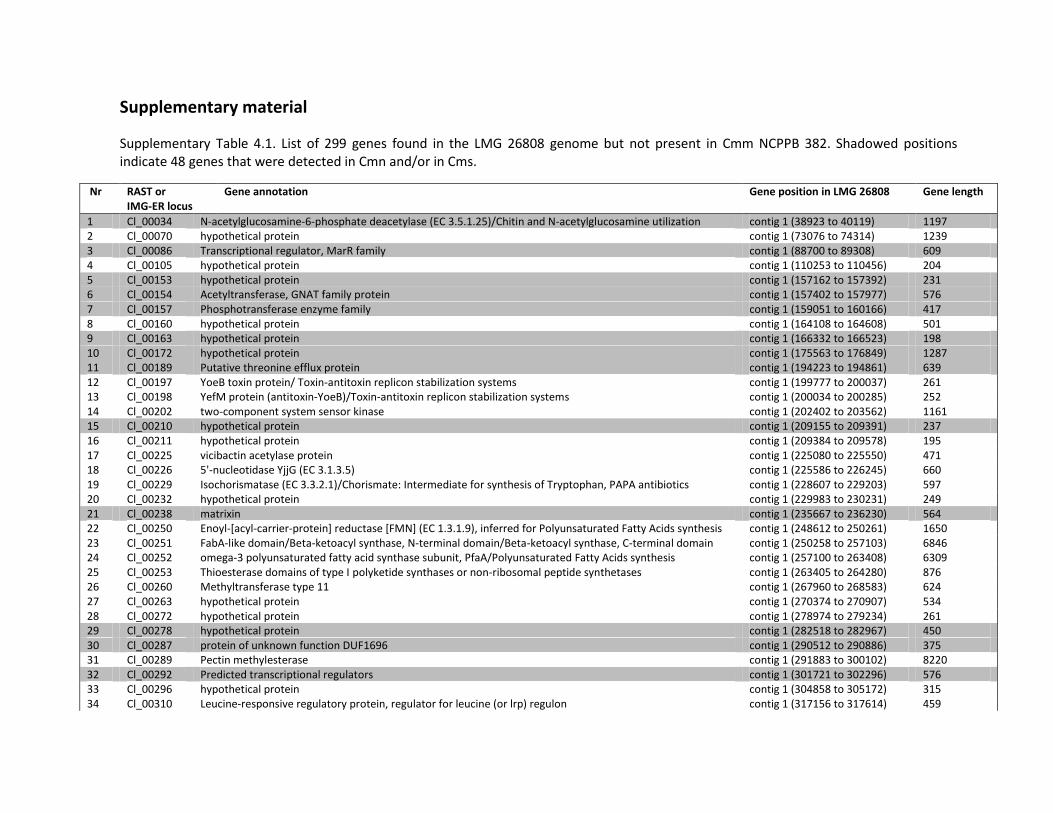
Supplementary material
Supplementary Table 4.1. List of 299 genes found in the LMG 26808 genome but not present in Cmm NCPPB 382. Shadowed positions indicate 48 genes that were detected in Cmn and/or in Cms.
Nr RAST or IMG-ER locus
Gene annotation Gene position in LMG 26808 Gene length
1 Cl_00034 N-acetylglucosamine-6-phosphate deacetylase (EC 3.5.1.25)/Chitin and N-acetylglucosamine utilization contig 1 (38923 to 40119) 1197 2 Cl_00070 hypothetical protein contig 1 (73076 to 74314) 1239 3 Cl_00086 Transcriptional regulator, MarR family contig 1 (88700 to 89308) 609 4 Cl_00105 hypothetical protein contig 1 (110253 to 110456) 204 5 Cl_00153 hypothetical protein contig 1 (157162 to 157392) 231 6 Cl_00154 Acetyltransferase, GNAT family protein contig 1 (157402 to 157977) 576 7 Cl_00157 Phosphotransferase enzyme family contig 1 (159051 to 160166) 417 8 Cl_00160 hypothetical protein contig 1 (164108 to 164608) 501 9 Cl_00163 hypothetical protein contig 1 (166332 to 166523) 198 10 Cl_00172 hypothetical protein contig 1 (175563 to 176849) 1287 11 Cl_00189 Putative threonine efflux protein contig 1 (194223 to 194861) 639 12 Cl_00197 YoeB toxin protein/ Toxin-antitoxin replicon stabilization systems contig 1 (199777 to 200037) 261 13 Cl_00198 YefM protein (antitoxin-YoeB)/Toxin-antitoxin replicon stabilization systems contig 1 (200034 to 200285) 252 14 Cl_00202 two-component system sensor kinase contig 1 (202402 to 203562) 1161 15 Cl_00210 hypothetical protein contig 1 (209155 to 209391) 237 16 Cl_00211 hypothetical protein contig 1 (209384 to 209578) 195 17 Cl_00225 vicibactin acetylase protein contig 1 (225080 to 225550) 471 18 Cl_00226 5'-nucleotidase YjjG (EC 3.1.3.5) contig 1 (225586 to 226245) 660 19 Cl_00229 Isochorismatase (EC 3.3.2.1)/Chorismate: Intermediate for synthesis of Tryptophan, PAPA antibiotics contig 1 (228607 to 229203) 597 20 Cl_00232 hypothetical protein contig 1 (229983 to 230231) 249 21 Cl_00238 matrixin contig 1 (235667 to 236230) 564 22 Cl_00250 Enoyl-[acyl-carrier-protein] reductase [FMN] (EC 1.3.1.9), inferred for Polyunsaturated Fatty Acids synthesis contig 1 (248612 to 250261) 1650 23 Cl_00251 FabA-like domain/Beta-ketoacyl synthase, N-terminal domain/Beta-ketoacyl synthase, C-terminal domain contig 1 (250258 to 257103) 6846 24 Cl_00252 omega-3 polyunsaturated fatty acid synthase subunit, PfaA/Polyunsaturated Fatty Acids synthesis contig 1 (257100 to 263408) 6309 25 Cl_00253 Thioesterase domains of type I polyketide synthases or non-ribosomal peptide synthetases contig 1 (263405 to 264280) 876 26 Cl_00260 Methyltransferase type 11 contig 1 (267960 to 268583) 624 27 Cl_00263 hypothetical protein contig 1 (270374 to 270907) 534 28 Cl_00272 hypothetical protein contig 1 (278974 to 279234) 261 29 Cl_00278 hypothetical protein contig 1 (282518 to 282967) 450 30 Cl_00287 protein of unknown function DUF1696 contig 1 (290512 to 290886) 375 31 Cl_00289 Pectin methylesterase contig 1 (291883 to 300102) 8220 32 Cl_00292 Predicted transcriptional regulators contig 1 (301721 to 302296) 576 33 Cl_00296 hypothetical protein contig 1 (304858 to 305172) 315 34 Cl_00310 Leucine-responsive regulatory protein, regulator for leucine (or lrp) regulon contig 1 (317156 to 317614) 459
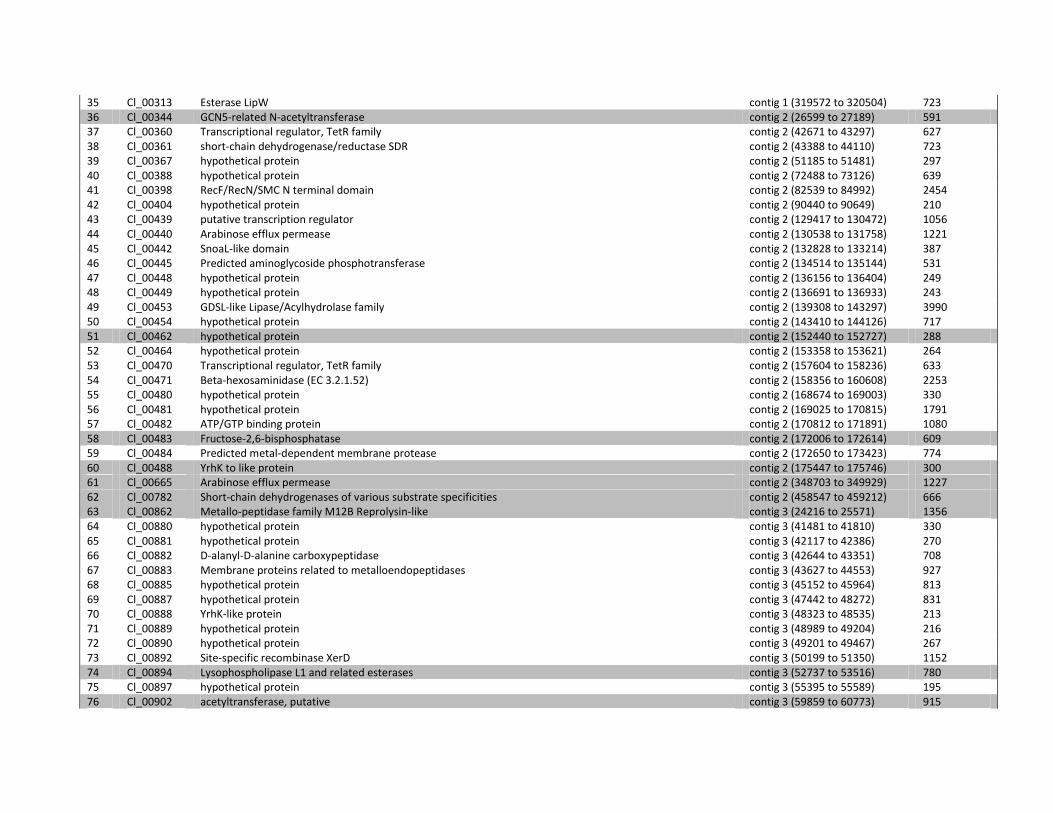
35 Cl_00313 Esterase LipW contig 1 (319572 to 320504) 723 36 Cl_00344 GCN5-related N-acetyltransferase contig 2 (26599 to 27189) 591 37 Cl_00360 Transcriptional regulator, TetR family contig 2 (42671 to 43297) 627 38 Cl_00361 short-chain dehydrogenase/reductase SDR contig 2 (43388 to 44110) 723 39 Cl_00367 hypothetical protein contig 2 (51185 to 51481) 297 40 Cl_00388 hypothetical protein contig 2 (72488 to 73126) 639 41 Cl_00398 RecF/RecN/SMC N terminal domain contig 2 (82539 to 84992) 2454 42 Cl_00404 hypothetical protein contig 2 (90440 to 90649) 210 43 Cl_00439 putative transcription regulator contig 2 (129417 to 130472) 1056 44 Cl_00440 Arabinose efflux permease contig 2 (130538 to 131758) 1221 45 Cl_00442 SnoaL-like domain contig 2 (132828 to 133214) 387 46 Cl_00445 Predicted aminoglycoside phosphotransferase contig 2 (134514 to 135144) 531 47 Cl_00448 hypothetical protein contig 2 (136156 to 136404) 249 48 Cl_00449 hypothetical protein contig 2 (136691 to 136933) 243 49 Cl_00453 GDSL-like Lipase/Acylhydrolase family contig 2 (139308 to 143297) 3990 50 Cl_00454 hypothetical protein contig 2 (143410 to 144126) 717 51 Cl_00462 hypothetical protein contig 2 (152440 to 152727) 288 52 Cl_00464 hypothetical protein contig 2 (153358 to 153621) 264 53 Cl_00470 Transcriptional regulator, TetR family contig 2 (157604 to 158236) 633 54 Cl_00471 Beta-hexosaminidase (EC 3.2.1.52) contig 2 (158356 to 160608) 2253 55 Cl_00480 hypothetical protein contig 2 (168674 to 169003) 330 56 Cl_00481 hypothetical protein contig 2 (169025 to 170815) 1791 57 Cl_00482 ATP/GTP binding protein contig 2 (170812 to 171891) 1080 58 Cl_00483 Fructose-2,6-bisphosphatase contig 2 (172006 to 172614) 609 59 Cl_00484 Predicted metal-dependent membrane protease contig 2 (172650 to 173423) 774 60 Cl_00488 YrhK to like protein contig 2 (175447 to 175746) 300 61 Cl_00665 Arabinose efflux permease contig 2 (348703 to 349929) 1227 62 Cl_00782 Short-chain dehydrogenases of various substrate specificities contig 2 (458547 to 459212) 666 63 Cl_00862 Metallo-peptidase family M12B Reprolysin-like contig 3 (24216 to 25571) 1356 64 Cl_00880 hypothetical protein contig 3 (41481 to 41810) 330 65 Cl_00881 hypothetical protein contig 3 (42117 to 42386) 270 66 Cl_00882 D-alanyl-D-alanine carboxypeptidase contig 3 (42644 to 43351) 708 67 Cl_00883 Membrane proteins related to metalloendopeptidases contig 3 (43627 to 44553) 927 68 Cl_00885 hypothetical protein contig 3 (45152 to 45964) 813 69 Cl_00887 hypothetical protein contig 3 (47442 to 48272) 831 70 Cl_00888 YrhK-like protein contig 3 (48323 to 48535) 213 71 Cl_00889 hypothetical protein contig 3 (48989 to 49204) 216 72 Cl_00890 hypothetical protein contig 3 (49201 to 49467) 267 73 Cl_00892 Site-specific recombinase XerD contig 3 (50199 to 51350) 1152 74 Cl_00894 Lysophospholipase L1 and related esterases contig 3 (52737 to 53516) 780 75 Cl_00897 hypothetical protein contig 3 (55395 to 55589) 195 76 Cl_00902 acetyltransferase, putative contig 3 (59859 to 60773) 915
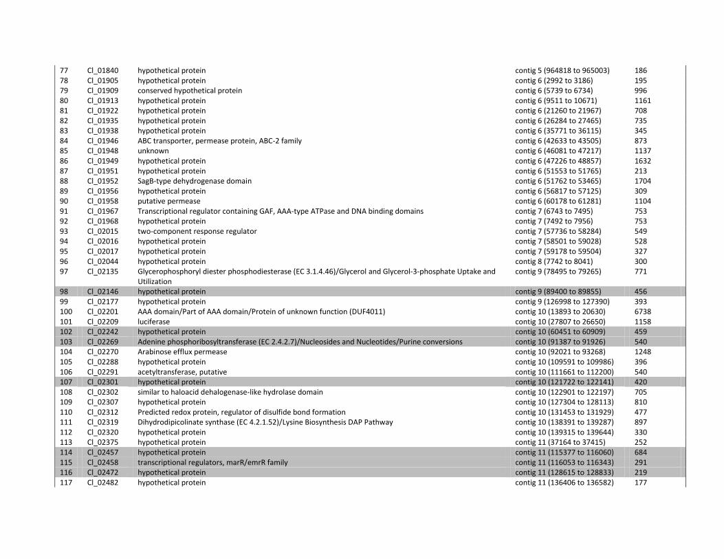
77 Cl_01840 hypothetical protein contig 5 (964818 to 965003) 186 78 Cl_01905 hypothetical protein contig 6 (2992 to 3186) 195 79 Cl_01909 conserved hypothetical protein contig 6 (5739 to 6734) 996 80 Cl_01913 hypothetical protein contig 6 (9511 to 10671) 1161 81 Cl_01922 hypothetical protein contig 6 (21260 to 21967) 708 82 Cl_01935 hypothetical protein contig 6 (26284 to 27465) 735 83 Cl_01938 hypothetical protein contig 6 (35771 to 36115) 345 84 Cl_01946 ABC transporter, permease protein, ABC-2 family contig 6 (42633 to 43505) 873 85 Cl_01948 unknown contig 6 (46081 to 47217) 1137 86 Cl_01949 hypothetical protein contig 6 (47226 to 48857) 1632 87 Cl_01951 hypothetical protein contig 6 (51553 to 51765) 213 88 Cl_01952 SagB-type dehydrogenase domain contig 6 (51762 to 53465) 1704 89 Cl_01956 hypothetical protein contig 6 (56817 to 57125) 309 90 Cl_01958 putative permease contig 6 (60178 to 61281) 1104 91 Cl_01967 Transcriptional regulator containing GAF, AAA-type ATPase and DNA binding domains contig 7 (6743 to 7495) 753 92 Cl_01968 hypothetical protein contig 7 (7492 to 7956) 753 93 Cl_02015 two-component response regulator contig 7 (57736 to 58284) 549 94 Cl_02016 hypothetical protein contig 7 (58501 to 59028) 528 95 Cl_02017 hypothetical protein contig 7 (59178 to 59504) 327 96 Cl_02044 hypothetical protein contig 8 (7742 to 8041) 300 97 Cl_02135 Glycerophosphoryl diester phosphodiesterase (EC 3.1.4.46)/Glycerol and Glycerol-3-phosphate Uptake and
Utilization contig 9 (78495 to 79265) 771
98 Cl_02146 hypothetical protein contig 9 (89400 to 89855) 456 99 Cl_02177 hypothetical protein contig 9 (126998 to 127390) 393 100 Cl_02201 AAA domain/Part of AAA domain/Protein of unknown function (DUF4011) contig 10 (13893 to 20630) 6738 101 Cl_02209 luciferase contig 10 (27807 to 26650) 1158 102 Cl_02242 hypothetical protein contig 10 (60451 to 60909) 459 103 Cl_02269 Adenine phosphoribosyltransferase (EC 2.4.2.7)/Nucleosides and Nucleotides/Purine conversions contig 10 (91387 to 91926) 540 104 Cl_02270 Arabinose efflux permease contig 10 (92021 to 93268) 1248 105 Cl_02288 hypothetical protein contig 10 (109591 to 109986) 396 106 Cl_02291 acetyltransferase, putative contig 10 (111661 to 112200) 540 107 Cl_02301 hypothetical protein contig 10 (121722 to 122141) 420 108 Cl_02302 similar to haloacid dehalogenase-like hydrolase domain contig 10 (122901 to 122197) 705 109 Cl_02307 hypothetical protein contig 10 (127304 to 128113) 810 110 Cl_02312 Predicted redox protein, regulator of disulfide bond formation contig 10 (131453 to 131929) 477 111 Cl_02319 Dihydrodipicolinate synthase (EC 4.2.1.52)/Lysine Biosynthesis DAP Pathway contig 10 (138391 to 139287) 897 112 Cl_02320 hypothetical protein contig 10 (139315 to 139644) 330 113 Cl_02375 hypothetical protein contig 11 (37164 to 37415) 252 114 Cl_02457 hypothetical protein contig 11 (115377 to 116060) 684 115 Cl_02458 transcriptional regulators, marR/emrR family contig 11 (116053 to 116343) 291 116 Cl_02472 hypothetical protein contig 11 (128615 to 128833) 219 117 Cl_02482 hypothetical protein contig 11 (136406 to 136582) 177
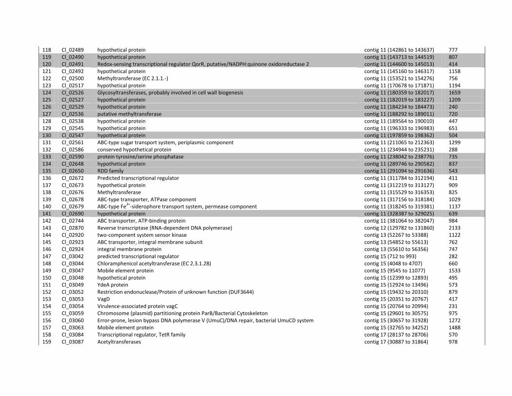
118 Cl_02489 hypothetical protein contig 11 (142861 to 143637) 777 119 Cl_02490 hypothetical protein contig 11 (143713 to 144519) 807 120 Cl_02491 Redox-sensing transcriptional regulator QorR, putative/NADPH:quinone oxidoreductase 2 contig 11 (144600 to 145013) 414 121 Cl_02492 hypothetical protein contig 11 (145160 to 146317) 1158 122 Cl_02500 Methyltransferase (EC 2.1.1.-) contig 11 (153521 to 154276) 756 123 Cl_02517 hypothetical protein contig 11 (170678 to 171871) 1194 124 Cl_02526 Glycosyltransferases, probably involved in cell wall biogenesis contig 11 (180359 to 182017) 1659 125 Cl_02527 hypothetical protein contig 11 (182019 to 183227) 1209 126 Cl_02529 hypothetical protein contig 11 (184234 to 184473) 240 127 Cl_02536 putative methyltransferase contig 11 (188292 to 189011) 720 128 Cl_02538 hypothetical protein contig 11 (189564 to 190010) 447 129 Cl_02545 hypothetical protein contig 11 (196333 to 196983) 651 130 Cl_02547 hypothetical protein contig 11 (197859 to 198362) 504 131 Cl_02561 ABC-type sugar transport system, periplasmic component contig 11 (211065 to 212363) 1299 132 Cl_02586 conserved hypothetical protein contig 11 (234944 to 235231) 288 133 Cl_02590 protein tyrosine/serine phosphatase contig 11 (238042 to 238776) 735 134 Cl_02648 hypothetical protein contig 11 (289746 to 290582) 837 135 Cl_02650 RDD family contig 11 (291094 to 291636) 543 136 Cl_02672 Predicted transcriptional regulator contig 11 (311784 to 312194) 411 137 Cl_02673 hypothetical protein contig 11 (312219 to 313127) 909 138 Cl_02676 Methyltransferase contig 11 (315529 to 316353) 825 139 Cl_02678 ABC-type transporter, ATPase component contig 11 (317156 to 318184) 1029 140 Cl_02679 ABC-type Fe
3+-siderophore transport system, permease component contig 11 (318245 to 319381) 1137
141 Cl_02690 hypothetical protein contig 11 (328387 to 329025) 639 142 Cl_02744 ABC transporter, ATP-binding protein contig 11 (381064 to 382047) 984 143 Cl_02870 Reverse transcriptase (RNA-dependent DNA polymerase) contig 12 (129782 to 131860) 2133 144 Cl_02920 two-component system sensor kinase contig 13 (52267 to 53388) 1122 145 Cl_02923 ABC transporter, integral membrane subunit contig 13 (54852 to 55613) 762 146 Cl_02924 integral membrane protein contig 13 (55610 to 56356) 747 147 Cl_03042 predicted transcriptional regulator contig 15 (712 to 993) 282 148 Cl_03044 Chloramphenicol acetyltransferase (EC 2.3.1.28) contig 15 (4048 to 4707) 660 149 Cl_03047 Mobile element protein contig 15 (9545 to 11077) 1533 150 Cl_03048 hypothetical protein contig 15 (12399 to 12893) 495 151 Cl_03049 YdeA protein contig 15 (12924 to 13496) 573 152 Cl_03052 Restriction endonuclease/Protein of unknown function (DUF3644) contig 15 (19432 to 20310) 879 153 Cl_03053 VagD contig 15 (20351 to 20767) 417 154 Cl_03054 Virulence-associated protein vagC contig 15 (20764 to 20994) 231 155 Cl_03059 Chromosome (plasmid) partitioning protein ParB/Bacterial Cytoskeleton contig 15 (29601 to 30575) 975 156 Cl_03060 Error-prone, lesion bypass DNA polymerase V (UmuC)/DNA repair, bacterial UmuCD system contig 15 (30657 to 31928) 1272 157 Cl_03063 Mobile element protein contig 15 (32765 to 34252) 1488 158 Cl_03084 Transcriptional regulator, TetR family contig 17 (28137 to 28706) 570 159 Cl_03087 Acetyltransferases contig 17 (30887 to 31864) 978
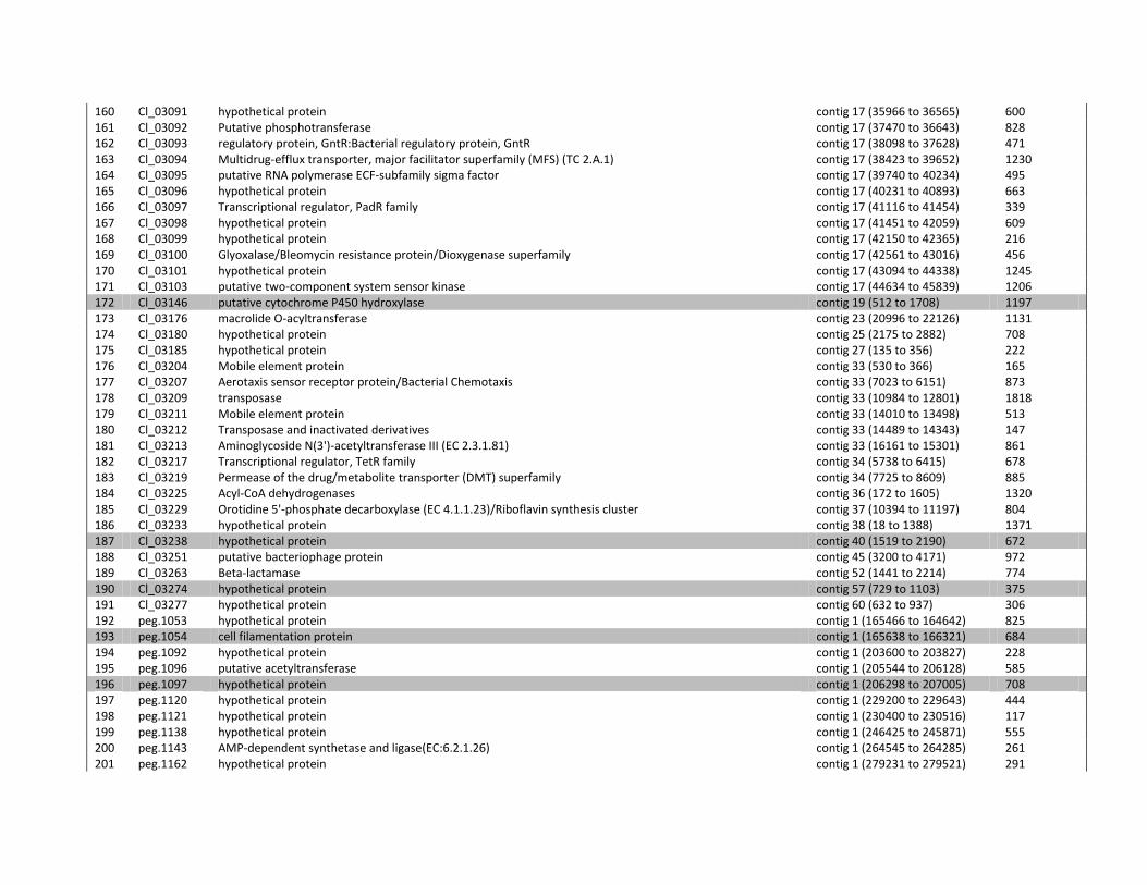
160 Cl_03091 hypothetical protein contig 17 (35966 to 36565) 600 161 Cl_03092 Putative phosphotransferase contig 17 (37470 to 36643) 828 162 Cl_03093 regulatory protein, GntR:Bacterial regulatory protein, GntR contig 17 (38098 to 37628) 471 163 Cl_03094 Multidrug-efflux transporter, major facilitator superfamily (MFS) (TC 2.A.1) contig 17 (38423 to 39652) 1230 164 Cl_03095 putative RNA polymerase ECF-subfamily sigma factor contig 17 (39740 to 40234) 495 165 Cl_03096 hypothetical protein contig 17 (40231 to 40893) 663 166 Cl_03097 Transcriptional regulator, PadR family contig 17 (41116 to 41454) 339 167 Cl_03098 hypothetical protein contig 17 (41451 to 42059) 609 168 Cl_03099 hypothetical protein contig 17 (42150 to 42365) 216 169 Cl_03100 Glyoxalase/Bleomycin resistance protein/Dioxygenase superfamily contig 17 (42561 to 43016) 456 170 Cl_03101 hypothetical protein contig 17 (43094 to 44338) 1245 171 Cl_03103 putative two-component system sensor kinase contig 17 (44634 to 45839) 1206 172 Cl_03146 putative cytochrome P450 hydroxylase contig 19 (512 to 1708) 1197 173 Cl_03176 macrolide O-acyltransferase contig 23 (20996 to 22126) 1131 174 Cl_03180 hypothetical protein contig 25 (2175 to 2882) 708 175 Cl_03185 hypothetical protein contig 27 (135 to 356) 222 176 Cl_03204 Mobile element protein contig 33 (530 to 366) 165 177 Cl_03207 Aerotaxis sensor receptor protein/Bacterial Chemotaxis contig 33 (7023 to 6151) 873 178 Cl_03209 transposase contig 33 (10984 to 12801) 1818 179 Cl_03211 Mobile element protein contig 33 (14010 to 13498) 513 180 Cl_03212 Transposase and inactivated derivatives contig 33 (14489 to 14343) 147 181 Cl_03213 Aminoglycoside N(3')-acetyltransferase III (EC 2.3.1.81) contig 33 (16161 to 15301) 861 182 Cl_03217 Transcriptional regulator, TetR family contig 34 (5738 to 6415) 678 183 Cl_03219 Permease of the drug/metabolite transporter (DMT) superfamily contig 34 (7725 to 8609) 885 184 Cl_03225 Acyl-CoA dehydrogenases contig 36 (172 to 1605) 1320 185 Cl_03229 Orotidine 5'-phosphate decarboxylase (EC 4.1.1.23)/Riboflavin synthesis cluster contig 37 (10394 to 11197) 804 186 Cl_03233 hypothetical protein contig 38 (18 to 1388) 1371 187 Cl_03238 hypothetical protein contig 40 (1519 to 2190) 672 188 Cl_03251 putative bacteriophage protein contig 45 (3200 to 4171) 972 189 Cl_03263 Beta-lactamase contig 52 (1441 to 2214) 774 190 Cl_03274 hypothetical protein contig 57 (729 to 1103) 375 191 Cl_03277 hypothetical protein contig 60 (632 to 937) 306 192 peg.1053 hypothetical protein contig 1 (165466 to 164642) 825 193 peg.1054 cell filamentation protein contig 1 (165638 to 166321) 684 194 peg.1092 hypothetical protein contig 1 (203600 to 203827) 228 195 peg.1096 putative acetyltransferase contig 1 (205544 to 206128) 585 196 peg.1097 hypothetical protein contig 1 (206298 to 207005) 708 197 peg.1120 hypothetical protein contig 1 (229200 to 229643) 444 198 peg.1121 hypothetical protein contig 1 (230400 to 230516) 117 199 peg.1138 hypothetical protein contig 1 (246425 to 245871) 555 200 peg.1143 AMP-dependent synthetase and ligase(EC:6.2.1.26) contig 1 (264545 to 264285) 261 201 peg.1162 hypothetical protein contig 1 (279231 to 279521) 291
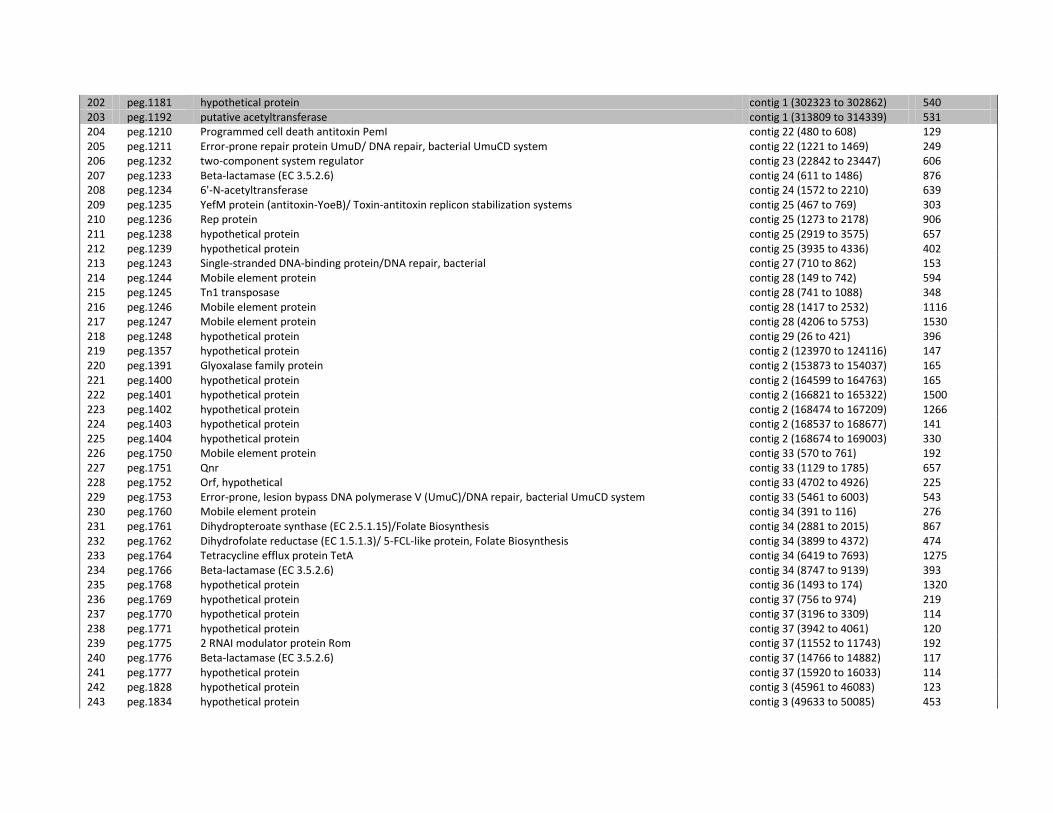
202 peg.1181 hypothetical protein contig 1 (302323 to 302862) 540 203 peg.1192 putative acetyltransferase contig 1 (313809 to 314339) 531 204 peg.1210 Programmed cell death antitoxin PemI contig 22 (480 to 608) 129 205 peg.1211 Error-prone repair protein UmuD/ DNA repair, bacterial UmuCD system contig 22 (1221 to 1469) 249 206 peg.1232 two-component system regulator contig 23 (22842 to 23447) 606 207 peg.1233 Beta-lactamase (EC 3.5.2.6) contig 24 (611 to 1486) 876 208 peg.1234 6'-N-acetyltransferase contig 24 (1572 to 2210) 639 209 peg.1235 YefM protein (antitoxin-YoeB)/ Toxin-antitoxin replicon stabilization systems contig 25 (467 to 769) 303 210 peg.1236 Rep protein contig 25 (1273 to 2178) 906 211 peg.1238 hypothetical protein contig 25 (2919 to 3575) 657 212 peg.1239 hypothetical protein contig 25 (3935 to 4336) 402 213 peg.1243 Single-stranded DNA-binding protein/DNA repair, bacterial contig 27 (710 to 862) 153 214 peg.1244 Mobile element protein contig 28 (149 to 742) 594 215 peg.1245 Tn1 transposase contig 28 (741 to 1088) 348 216 peg.1246 Mobile element protein contig 28 (1417 to 2532) 1116 217 peg.1247 Mobile element protein contig 28 (4206 to 5753) 1530 218 peg.1248 hypothetical protein contig 29 (26 to 421) 396 219 peg.1357 hypothetical protein contig 2 (123970 to 124116) 147 220 peg.1391 Glyoxalase family protein contig 2 (153873 to 154037) 165 221 peg.1400 hypothetical protein contig 2 (164599 to 164763) 165 222 peg.1401 hypothetical protein contig 2 (166821 to 165322) 1500 223 peg.1402 hypothetical protein contig 2 (168474 to 167209) 1266 224 peg.1403 hypothetical protein contig 2 (168537 to 168677) 141 225 peg.1404 hypothetical protein contig 2 (168674 to 169003) 330 226 peg.1750 Mobile element protein contig 33 (570 to 761) 192 227 peg.1751 Qnr contig 33 (1129 to 1785) 657 228 peg.1752 Orf, hypothetical contig 33 (4702 to 4926) 225 229 peg.1753 Error-prone, lesion bypass DNA polymerase V (UmuC)/DNA repair, bacterial UmuCD system contig 33 (5461 to 6003) 543 230 peg.1760 Mobile element protein contig 34 (391 to 116) 276 231 peg.1761 Dihydropteroate synthase (EC 2.5.1.15)/Folate Biosynthesis contig 34 (2881 to 2015) 867 232 peg.1762 Dihydrofolate reductase (EC 1.5.1.3)/ 5-FCL-like protein, Folate Biosynthesis contig 34 (3899 to 4372) 474 233 peg.1764 Tetracycline efflux protein TetA contig 34 (6419 to 7693) 1275 234 peg.1766 Beta-lactamase (EC 3.5.2.6) contig 34 (8747 to 9139) 393 235 peg.1768 hypothetical protein contig 36 (1493 to 174) 1320 236 peg.1769 hypothetical protein contig 37 (756 to 974) 219 237 peg.1770 hypothetical protein contig 37 (3196 to 3309) 114 238 peg.1771 hypothetical protein contig 37 (3942 to 4061) 120 239 peg.1775 2 RNAI modulator protein Rom contig 37 (11552 to 11743) 192 240 peg.1776 Beta-lactamase (EC 3.5.2.6) contig 37 (14766 to 14882) 117 241 peg.1777 hypothetical protein contig 37 (15920 to 16033) 114 242 peg.1828 hypothetical protein contig 3 (45961 to 46083) 123 243 peg.1834 hypothetical protein contig 3 (49633 to 50085) 453
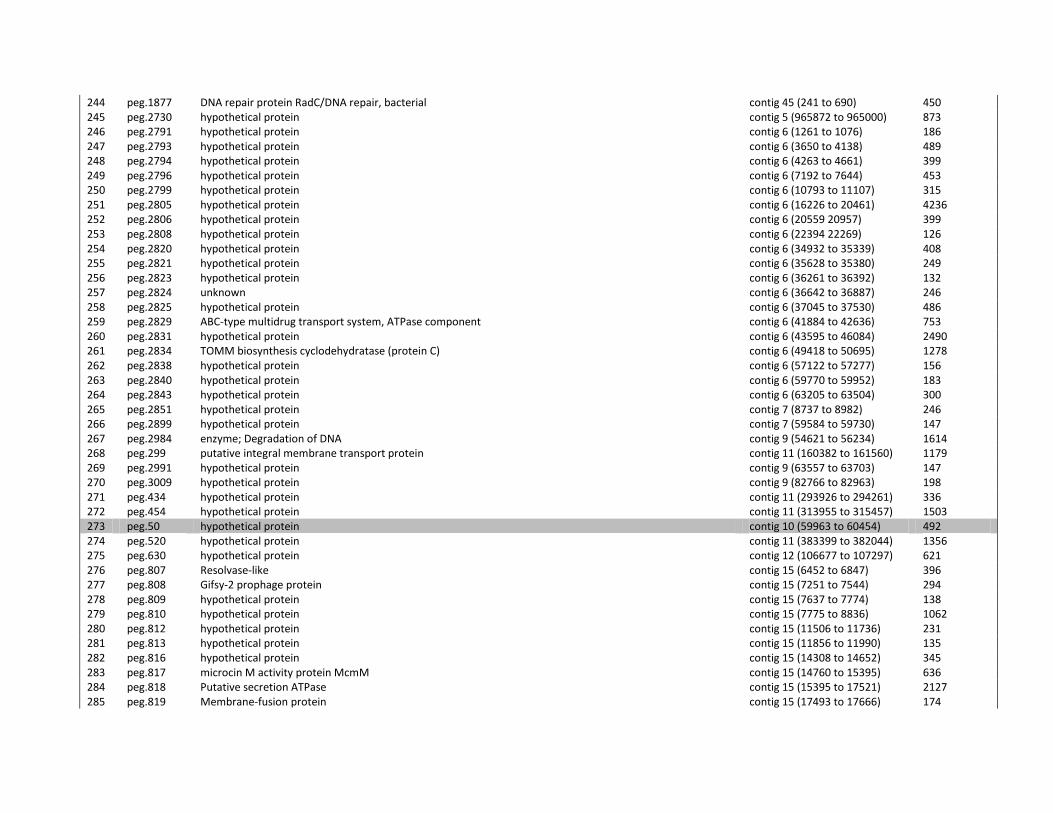
244 peg.1877 DNA repair protein RadC/DNA repair, bacterial contig 45 (241 to 690) 450 245 peg.2730 hypothetical protein contig 5 (965872 to 965000) 873 246 peg.2791 hypothetical protein contig 6 (1261 to 1076) 186 247 peg.2793 hypothetical protein contig 6 (3650 to 4138) 489 248 peg.2794 hypothetical protein contig 6 (4263 to 4661) 399 249 peg.2796 hypothetical protein contig 6 (7192 to 7644) 453 250 peg.2799 hypothetical protein contig 6 (10793 to 11107) 315 251 peg.2805 hypothetical protein contig 6 (16226 to 20461) 4236 252 peg.2806 hypothetical protein contig 6 (20559 20957) 399 253 peg.2808 hypothetical protein contig 6 (22394 22269) 126 254 peg.2820 hypothetical protein contig 6 (34932 to 35339) 408 255 peg.2821 hypothetical protein contig 6 (35628 to 35380) 249 256 peg.2823 hypothetical protein contig 6 (36261 to 36392) 132 257 peg.2824 unknown contig 6 (36642 to 36887) 246 258 peg.2825 hypothetical protein contig 6 (37045 to 37530) 486 259 peg.2829 ABC-type multidrug transport system, ATPase component contig 6 (41884 to 42636) 753 260 peg.2831 hypothetical protein contig 6 (43595 to 46084) 2490 261 peg.2834 TOMM biosynthesis cyclodehydratase (protein C) contig 6 (49418 to 50695) 1278 262 peg.2838 hypothetical protein contig 6 (57122 to 57277) 156 263 peg.2840 hypothetical protein contig 6 (59770 to 59952) 183 264 peg.2843 hypothetical protein contig 6 (63205 to 63504) 300 265 peg.2851 hypothetical protein contig 7 (8737 to 8982) 246 266 peg.2899 hypothetical protein contig 7 (59584 to 59730) 147 267 peg.2984 enzyme; Degradation of DNA contig 9 (54621 to 56234) 1614 268 peg.299 putative integral membrane transport protein contig 11 (160382 to 161560) 1179 269 peg.2991 hypothetical protein contig 9 (63557 to 63703) 147 270 peg.3009 hypothetical protein contig 9 (82766 to 82963) 198 271 peg.434 hypothetical protein contig 11 (293926 to 294261) 336 272 peg.454 hypothetical protein contig 11 (313955 to 315457) 1503 273 peg.50 hypothetical protein contig 10 (59963 to 60454) 492 274 peg.520 hypothetical protein contig 11 (383399 to 382044) 1356 275 peg.630 hypothetical protein contig 12 (106677 to 107297) 621 276 peg.807 Resolvase-like contig 15 (6452 to 6847) 396 277 peg.808 Gifsy-2 prophage protein contig 15 (7251 to 7544) 294 278 peg.809 hypothetical protein contig 15 (7637 to 7774) 138 279 peg.810 hypothetical protein contig 15 (7775 to 8836) 1062 280 peg.812 hypothetical protein contig 15 (11506 to 11736) 231 281 peg.813 hypothetical protein contig 15 (11856 to 11990) 135 282 peg.816 hypothetical protein contig 15 (14308 to 14652) 345 283 peg.817 microcin M activity protein McmM contig 15 (14760 to 15395) 636 284 peg.818 Putative secretion ATPase contig 15 (15395 to 17521) 2127 285 peg.819 Membrane-fusion protein contig 15 (17493 to 17666) 174
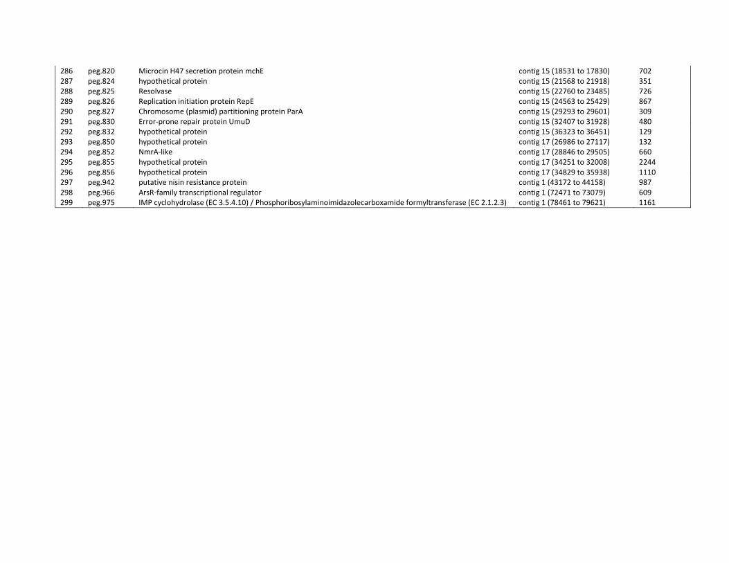
286 peg.820 Microcin H47 secretion protein mchE contig 15 (18531 to 17830) 702 287 peg.824 hypothetical protein contig 15 (21568 to 21918) 351 288 peg.825 Resolvase contig 15 (22760 to 23485) 726 289 peg.826 Replication initiation protein RepE contig 15 (24563 to 25429) 867 290 peg.827 Chromosome (plasmid) partitioning protein ParA contig 15 (29293 to 29601) 309 291 peg.830 Error-prone repair protein UmuD contig 15 (32407 to 31928) 480 292 peg.832 hypothetical protein contig 15 (36323 to 36451) 129 293 peg.850 hypothetical protein contig 17 (26986 to 27117) 132 294 peg.852 NmrA-like contig 17 (28846 to 29505) 660 295 peg.855 hypothetical protein contig 17 (34251 to 32008) 2244 296 peg.856 hypothetical protein contig 17 (34829 to 35938) 1110 297 peg.942 putative nisin resistance protein contig 1 (43172 to 44158) 987 298 peg.966 ArsR-family transcriptional regulator contig 1 (72471 to 73079) 609 299 peg.975 IMP cyclohydrolase (EC 3.5.4.10) / Phosphoribosylaminoimidazolecarboxamide formyltransferase (EC 2.1.2.3) contig 1 (78461 to 79621) 1161
Supplementary Table 4.2. Low GC regions of LMG 26808 detected by IslandViewer.
Nr Size GI Prediction Program Position of CDS in LMG 26808 Avr GC%
ORF(s) Gene Name
1 4266 Predicted by SIGI-HMM method
Contig 2 (481757 - 487234) 70 Cl_00805 hypothetical protein
Cl_00806 Alpha-glucosidases, family 31 of glycosyl hydrolases
Cl_00807 carbohydrate ABC transporter membrane protein 2, CUT1 family (TC 3.A.1.1.-)
Cl_00808 carbohydrate ABC transporter membrane protein 1, CUT1 family (TC 3.A.1.1.-)
Cl_00809 Maltose-binding periplasmic proteins/domains
2 7166 Predicted by SIGI-HMM method
Contig 5 (19001 - 25028) 56 Cl_00945 hypothetical protein
Cl_00946 hypothetical protein
Cl_00947 hypothetical protein
Cl_00948 hypothetical protein
Cl_00949 hypothetical protein
Cl_00950 Predicted phosphohydrolases
Cl_00951 DNA adenine methylase (dam)
3 13711 Predicted by SIGI-HMM method
Contig 6 (1970 - 2779) 65 Cl_01904 hypothetical protein
Cl_01905 hypothetical protein
Cl_01906 Acetyltransferases, including N-acetylases of ribosomal proteins
Cl_01907 hypothetical protein
Cl_01908 hypothetical protein
Cl_01909 hypothetical protein
Cl_01910 hypothetical protein
Cl_01911 hypothetical protein
Cl_01912 hypothetical protein
Cl_01913 hypothetical protein
Cl_01914 hypothetical protein
Cl_01915 Protein involved in cell division
Cl_01916 Nucleotidyl transferase of unknown function (DUF1814)
Cl_01917 Domain of unknown function (DUF4095)
Cl_01918 hypothetical protein
Cl_01919 hypothetical protein
4 19917 Predicted by SIGI-HMM method
Contig 6 (36516 - 56433) 66 Cl_01940 hypothetical protein
Cl_01941 hypothetical protein
Cl_01942 plasmid segregation oscillating ATPase
Cl_01943 hypothetical protein
Cl_01944 thiazolylpeptide-type bacteriocin precursor
Cl_01945 ABC-type multidrug transport system, ATPase component
Cl_01946 ABC-type multidrug transport system, permease component
Cl_01947 Lantibiotic dehydratase, C terminus
Cl_01948 hypothetical protein
Cl_01949 hypothetical protein

Cl_01950 bacteriocin biosynthesis cyclodehydratase
Cl_01951 hypothetical protein
Cl_01952 SagB-type dehydrogenase domain
Cl_01953 Transcriptional regulator
Cl_01954 hypothetical protein
Cl_01955 hypothetical protein
5 7323 Predicted by SIGI-HMM method
Contig 6 (57486 - 64809) 61 Cl_01957 signal peptidase I (EC:3.4.21.89). Serine peptidase. MEROPS family S26A
Cl_01958 ABC-type antimicrobial peptide transport system, permease component
Cl_01959 Membrane-fusion protein
Cl_01960 Lsr2
Cl_01961 Helix-turn-helix domain
6 8592 Predicted by SIGI-HMM Contig 12 (126631 - 135226) 64 Cl_02867 Trehalose utilization protein
method
Cl_02868 Sugar phosphate isomerases/epimerases
Cl_02869 Reverse transcriptase (RNA-dependent DNA polymerase)
Cl_02870 Reverse transcriptase (RNA-dependent DNA polymerase)
Cl_02871 ABC-type multidrug transport system, ATPase and permease components
Cl_02872 hypothetical protein
7 43152 Predicted by SIGI-HMM and IslandPath-DIMOB methods
Contig 14,15 52 Cl_03040 hypothetical protein
Contig 16 (2785 - 3504)
Cl_03041 DNA segregation ATPase FtsK/SpoIIIE and related proteins
Contig 17 (3 - 1409)
Cl_03042 Transcriptional regulator, contains sigma factor-related N-terminal domain
Cl_03043 Phage-related protein
Cl_03044 Chloramphenicol O-acetyltransferase
Cl_03045 hypothetical protein
Cl_03046 hypothetical protein
Cl_03047 Transposase and inactivated derivatives
Cl_03048 hypothetical protein
Cl_03049 hypothetical protein
Cl_03050 colicin V processing peptidase. Cysteine peptidase. MEROPS family C39
Cl_03051 Multidrug resistance efflux pump
Cl_03052 Restriction endonuclease/Protein of unknown function (DUF3644)
Cl_03053 Predicted nucleic acid-binding protein, contains PIN domain
Cl_03054 Virulence-associated protein and related proteins
Cl_03055 hypothetical protein
Cl_03056 Site-specific recombinase XerD
Cl_03057 hypothetical protein
Cl_03058 CobQ/CobB/MinD/ParA nucleotide binding domain
Cl_03059 ParB-like partition proteins
Cl_03060 Nucleotidyltransferase/DNA polymerase involved in DNA repair
Cl_03061 SOS-response transcriptional repressors (RecA-mediated autopeptidases
Cl_03063 Retron-type reverse transcriptase
Cl_03065 MbeD/MobD like
Cl_03066 MbeB-like, N-term conserved region
Cl_03067 NAD-dependent aldehyde dehydrogenases
8 6638 Predicted by SIGI-HMM method
Contig 17 (27649 - 34287) 67 Cl_03083 hypothetical protein
Cl_03084 transcriptional regulator, TetR family
Cl_03085 NADH(P)-binding
Cl_03086 Response regulator containing a CheY-like receiver
Cl_03087 Acetyltransferases
Cl_03088 Arabinose efflux permease
Cl_03089 transcriptional regulator, TetR family
9 107832 Predicted by IslandPath- Contig 23 (22159 - 23453) 55 Cl_03177 Predicted acyltransferases
DIMOB method Contig 25-30, 32-39
Cl_03178 two component transcriptional regulator, LuxR family
Contig 40 (1 - 135)
Cl_03179 Histidine kinase/Histidine kinase-, DNA gyrase B- and HSP90-like ATPase
Cl_03180 hypothetical protein
Cl_03181 hypothetical protein
Cl_03182 AAA-like domain
Cl_03183 hypothetical protein
Cl_03184 hypothetical protein
Cl_03185 hypothetical protein
Cl_03186 hypothetical protein
Cl_03187 hypothetical protein
Cl_03188 plasmid transfer ATPase TraJ
Cl_03189 Transposase DDE domain
Cl_03190 Transposase and inactivated derivatives, TnpA family
Cl_03191 Transglycosylase SLT domain
Cl_03192 hypothetical protein
Cl_03193 hypothetical protein
Cl_03194 Protein of unknown function (DUF3625)
Cl_03195 Macrophage killing protein with similarity to conjugation protein
Cl_03199 Acetyltransferases
Cl_03200 ABC-type multidrug transport system, ATPase and permease components
Cl_03201 ABC-type multidrug transport system, ATPase and permease components
Cl_03202 Methyltransferase domain
Cl_03203 Predicted thioesterase involved in non-ribosomal peptide biosynthesis
Cl_03204 hypothetical protein
Cl_03205 hypothetical protein
Cl_03206 hypothetical protein
Cl_03207 Methyl-accepting chemotaxis protein
Cl_03208 Beta-lactamase class A
Cl_03209 Transposase and inactivated derivatives, TnpA family
Cl_03210 Transposase and inactivated derivatives
Cl_03211 Transposase and inactivated derivatives
Cl_03212 Transposase and inactivated derivatives
Cl_03213 Aminoglycoside N3'-acetyltransferase
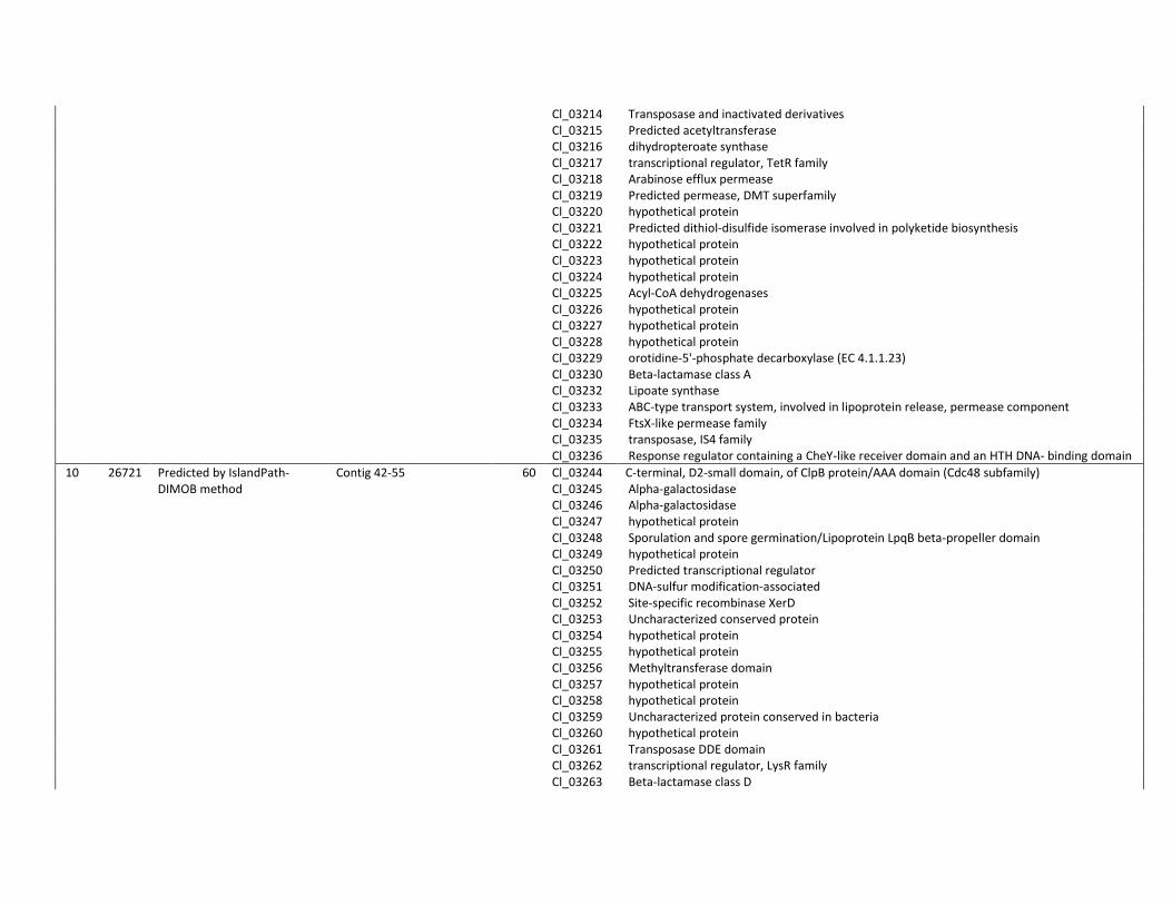
Cl_03214 Transposase and inactivated derivatives
Cl_03215 Predicted acetyltransferase
Cl_03216 dihydropteroate synthase
Cl_03217 transcriptional regulator, TetR family
Cl_03218 Arabinose efflux permease
Cl_03219 Predicted permease, DMT superfamily
Cl_03220 hypothetical protein
Cl_03221 Predicted dithiol-disulfide isomerase involved in polyketide biosynthesis
Cl_03222 hypothetical protein
Cl_03223 hypothetical protein
Cl_03224 hypothetical protein
Cl_03225 Acyl-CoA dehydrogenases
Cl_03226 hypothetical protein
Cl_03227 hypothetical protein
Cl_03228 hypothetical protein
Cl_03229 orotidine-5'-phosphate decarboxylase (EC 4.1.1.23)
Cl_03230 Beta-lactamase class A
Cl_03232 Lipoate synthase
Cl_03233 ABC-type transport system, involved in lipoprotein release, permease component
Cl_03234 FtsX-like permease family
Cl_03235 transposase, IS4 family
Cl_03236 Response regulator containing a CheY-like receiver domain and an HTH DNA- binding domain
10 26721 Predicted by IslandPath-DIMOB method
Contig 42-55 60 Cl_03244 C-terminal, D2-small domain, of ClpB protein/AAA domain (Cdc48 subfamily)
Cl_03245 Alpha-galactosidase
Cl_03246 Alpha-galactosidase
Cl_03247 hypothetical protein
Cl_03248 Sporulation and spore germination/Lipoprotein LpqB beta-propeller domain
Cl_03249 hypothetical protein
Cl_03250 Predicted transcriptional regulator
Cl_03251 DNA-sulfur modification-associated
Cl_03252 Site-specific recombinase XerD
Cl_03253 Uncharacterized conserved protein
Cl_03254 hypothetical protein
Cl_03255 hypothetical protein
Cl_03256 Methyltransferase domain
Cl_03257 hypothetical protein
Cl_03258 hypothetical protein
Cl_03259 Uncharacterized protein conserved in bacteria
Cl_03260 hypothetical protein
Cl_03261 Transposase DDE domain
Cl_03262 transcriptional regulator, LysR family
Cl_03263 Beta-lactamase class D
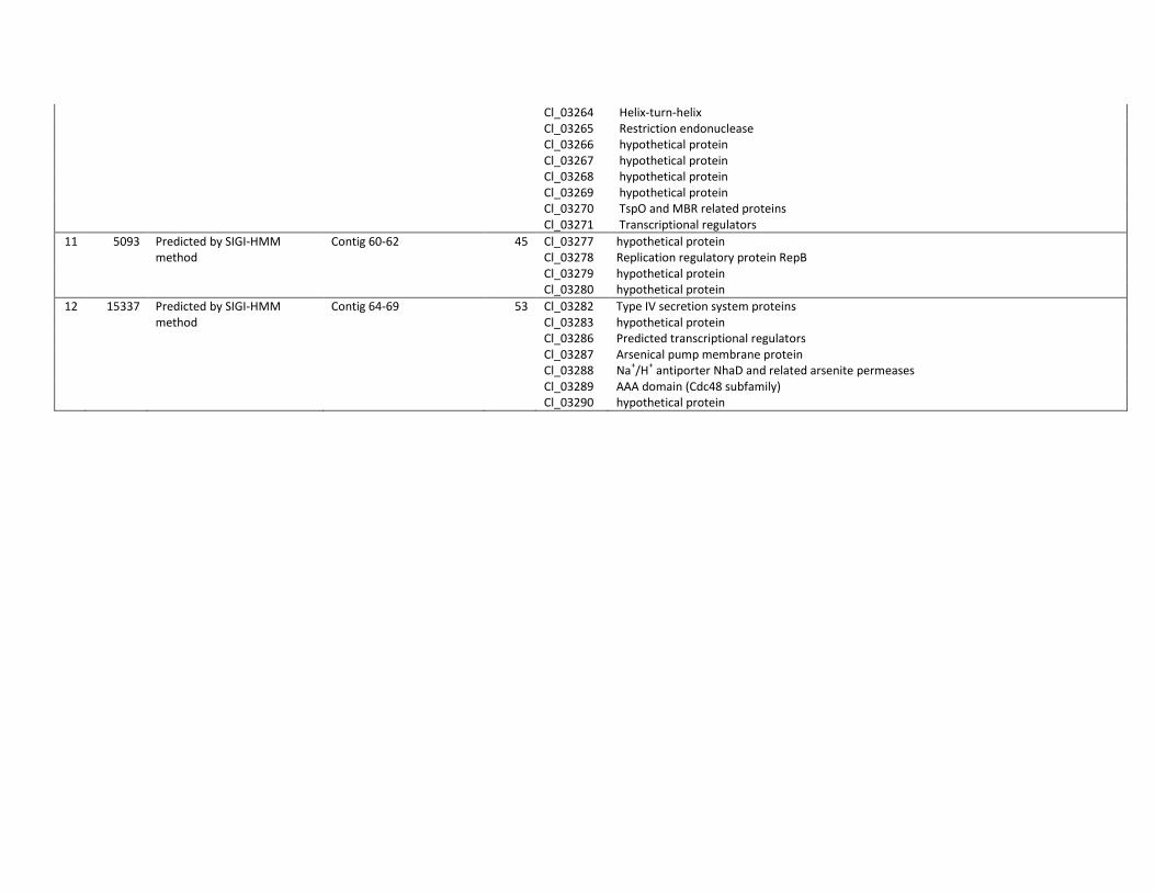
Cl_03264 Helix-turn-helix
Cl_03265 Restriction endonuclease
Cl_03266 hypothetical protein
Cl_03267 hypothetical protein
Cl_03268 hypothetical protein
Cl_03269 hypothetical protein
Cl_03270 TspO and MBR related proteins
Cl_03271 Transcriptional regulators
11 5093 Predicted by SIGI-HMM method
Contig 60-62 45 Cl_03277 hypothetical protein
Cl_03278 Replication regulatory protein RepB
Cl_03279 hypothetical protein
Cl_03280 hypothetical protein
12 15337 Predicted by SIGI-HMM method
Contig 64-69 53 Cl_03282 Type IV secretion system proteins
Cl_03283 hypothetical protein
Cl_03286 Predicted transcriptional regulators
Cl_03287 Arsenical pump membrane protein
Cl_03288 Na+/H
+ antiporter NhaD and related arsenite permeases
Cl_03289 AAA domain (Cdc48 subfamily)
Cl_03290 hypothetical protein
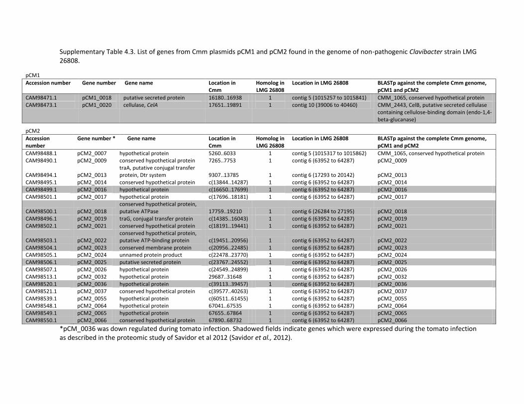
Supplementary Table 4.3. List of genes from Cmm plasmids pCM1 and pCM2 found in the genome of non-pathogenic Clavibacter strain LMG 26808.
pCM1 Accession number Gene number Gene name Location in
Cmm Homolog in LMG 26808
Location in LMG 26808 BLASTp against the complete Cmm genome, pCM1 and pCM2
CAM98471.1 pCM1_0018 putative secreted protein 16180..16938 1 contig 5 (1015257 to 1015841) CMM_1065, conserved hypothetical protein CAM98473.1 pCM1_0020 cellulase, CelA 17651..19891 1 contig 10 (39006 to 40460) CMM_2443, CelB, putative secreted cellulase
containing cellulose-binding domain (endo-1,4-beta-glucanase)
pCM2 Accession
number Gene number * Gene name Location in
Cmm Homolog in LMG 26808
Location in LMG 26808 BLASTp against the complete Cmm genome, pCM1 and pCM2
CAM98488.1 pCM2_0007 hypothetical protein 5260..6033 1 contig 5 (1015317 to 1015862) CMM_1065, conserved hypothetical protein CAM98490.1 pCM2_0009 conserved hypothetical protein 7265..7753 1 contig 6 (63952 to 64287) pCM2_0009
CAM98494.1 pCM2_0013 traA, putative conjugal transfer protein, Dtr system 9307..13785 1 contig 6 (17293 to 20142) pCM2_0013
CAM98495.1 pCM2_0014 conserved hypothetical protein c(13844..14287) 1 contig 6 (63952 to 64287) pCM2_0014 CAM98499.1 pCM2_0016 hypothetical protein c(16650..17699) 1 contig 6 (63952 to 64287) pCM2_0016 CAM98501.1 pCM2_0017 hypothetical protein c(17696..18181) 1 contig 6 (63952 to 64287) pCM2_0017
CAM98500.1 pCM2_0018 conserved hypothetical protein, putative ATPase 17759..19210 1 contig 6 (26284 to 27195) pCM2_0018
CAM98496.1 pCM2_0019 traG, conjugal transfer protein c(14385..16043) 1 contig 6 (63952 to 64287) pCM2_0019 CAM98502.1 pCM2_0021 conserved hypothetical protein c(18191..19441) 1 contig 6 (63952 to 64287) pCM2_0021
CAM98503.1 pCM2_0022 conserved hypothetical protein, putative ATP-binding protein c(19451..20956) 1 contig 6 (63952 to 64287) pCM2_0022
CAM98504.1 pCM2_0023 conserved membrane protein c(20956..22485) 1 contig 6 (63952 to 64287) pCM2_0023 CAM98505.1 pCM2_0024 unnamed protein product c(22478..23770) 1 contig 6 (63952 to 64287) pCM2_0024 CAM98506.1 pCM2_0025 putative secreted protein c(23767..24552) 1 contig 6 (63952 to 64287) pCM2_0025 CAM98507.1 pCM2_0026 hypothetical protein c(24549..24899) 1 contig 6 (63952 to 64287) pCM2_0026 CAM98513.1 pCM2_0032 hypothetical protein 29687..31648 1 contig 6 (63952 to 64287) pCM2_0032 CAM98520.1 pCM2_0036 hypothetical protein c(39113..39457) 1 contig 6 (63952 to 64287) pCM2_0036 CAM98521.1 pCM2_0037 conserved hypothetical protein c(39577..40263) 1 contig 6 (63952 to 64287) pCM2_0037 CAM98539.1 pCM2_0055 hypothetical protein c(60511..61455) 1 contig 6 (63952 to 64287) pCM2_0055 CAM98548.1 pCM2_0064 hypothetical protein 67041..67535 1 contig 6 (63952 to 64287) pCM2_0064 CAM98549.1 pCM2_0065 hypothetical protein 67655..67864 1 contig 6 (63952 to 64287) pCM2_0065 CAM98550.1 pCM2_0066 conserved hypothetical protein 67890..68732 1 contig 6 (63952 to 64287) pCM2_0066
*pCM_0036 was down regulated during tomato infection. Shadowed fields indicate genes which were expressed during the tomato infection as described in the proteomic study of Savidor et al 2012 (Savidor et al., 2012).
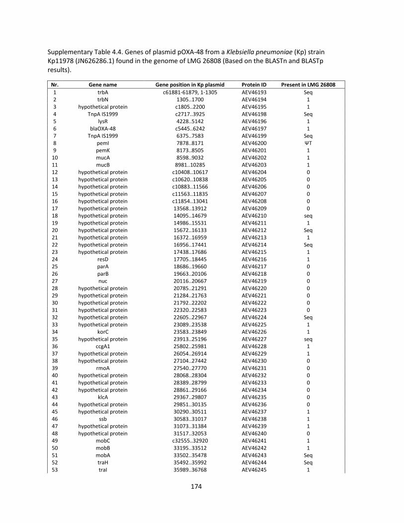
174
Supplementary Table 4.4. Genes of plasmid pOXA-48 from a Klebsiella pneumoniae (Kp) strain Kp11978 (JN626286.1) found in the genome of LMG 26808 (Based on the BLASTn and BLASTp results).
Nr. Gene name Gene position in Kp plasmid Protein ID Present in LMG 26808
1 trbA c61881-61879, 1-1305 AEV46193 Seq 2 trbN 1305..1700 AEV46194 1 3 hypothetical protein c1805..2200 AEV46195 1 4 TnpA IS1999 c2717..3925 AEV46198 Seq 5 lysR 4228..5142 AEV46196 1 6 blaOXA-48 c5445..6242 AEV46197 1 7 TnpA IS1999 6375..7583 AEV46199 Seq 8 pemI 7878..8171 AEV46200 ΨT 9 pemK 8173..8505 AEV46201 1
10 mucA 8598..9032 AEV46202 1 11 mucB 8981..10285 AEV46203 1 12 hypothetical protein c10408..10617 AEV46204 0 13 hypothetical protein c10620..10838 AEV46205 0 14 hypothetical protein c10883..11566 AEV46206 0 15 hypothetical protein c11563..11835 AEV46207 0 16 hypothetical protein c11854..13041 AEV46208 0 17 hypothetical protein 13568..13912 AEV46209 0 18 hypothetical protein 14095..14679 AEV46210 seq 19 hypothetical protein 14986..15531 AEV46211 1 20 hypothetical protein 15672..16133 AEV46212 Seq 21 hypothetical protein 16372..16959 AEV46213 1 22 hypothetical protein 16956..17441 AEV46214 Seq 23 hypothetical protein 17438..17686 AEV46215 1 24 resD 17705..18445 AEV46216 1 25 parA 18686..19660 AEV46217 0 26 parB 19663..20106 AEV46218 0 27 nuc 20116..20667 AEV46219 0 28 hypothetical protein 20785..21291 AEV46220 0 29 hypothetical protein 21284..21763 AEV46221 0 30 hypothetical protein 21792..22202 AEV46222 0 31 hypothetical protein 22320..22583 AEV46223 0 32 hypothetical protein 22605..22967 AEV46224 Seq 33 hypothetical protein 23089..23538 AEV46225 1 34 korC 23583..23849 AEV46226 1 35 hypothetical protein 23913..25196 AEV46227 seq 36 ccgA1 25802..25981 AEV46228 1 37 hypothetical protein 26054..26914 AEV46229 1 38 hypothetical protein 27104..27442 AEV46230 0 39 rmoA 27540..27770 AEV46231 0 40 hypothetical protein 28068..28304 AEV46232 0 41 hypothetical protein 28389..28799 AEV46233 0 42 hypothetical protein 28861..29166 AEV46234 0 43 klcA 29367..29807 AEV46235 0 44 hypothetical protein 29851..30135 AEV46236 0 45 hypothetical protein 30290..30511 AEV46237 1 46 ssb 30583..31017 AEV46238 1 47 hypothetical protein 31073..31384 AEV46239 1 48 hypothetical protein 31517..32053 AEV46240 0 49 mobC c32555..32920 AEV46241 1 50 mobB 33195..33512 AEV46242 1 51 mobA 33502..35478 AEV46243 Seq 52 traH 35492..35992 AEV46244 Seq 53 traI 35989..36768 AEV46245 1
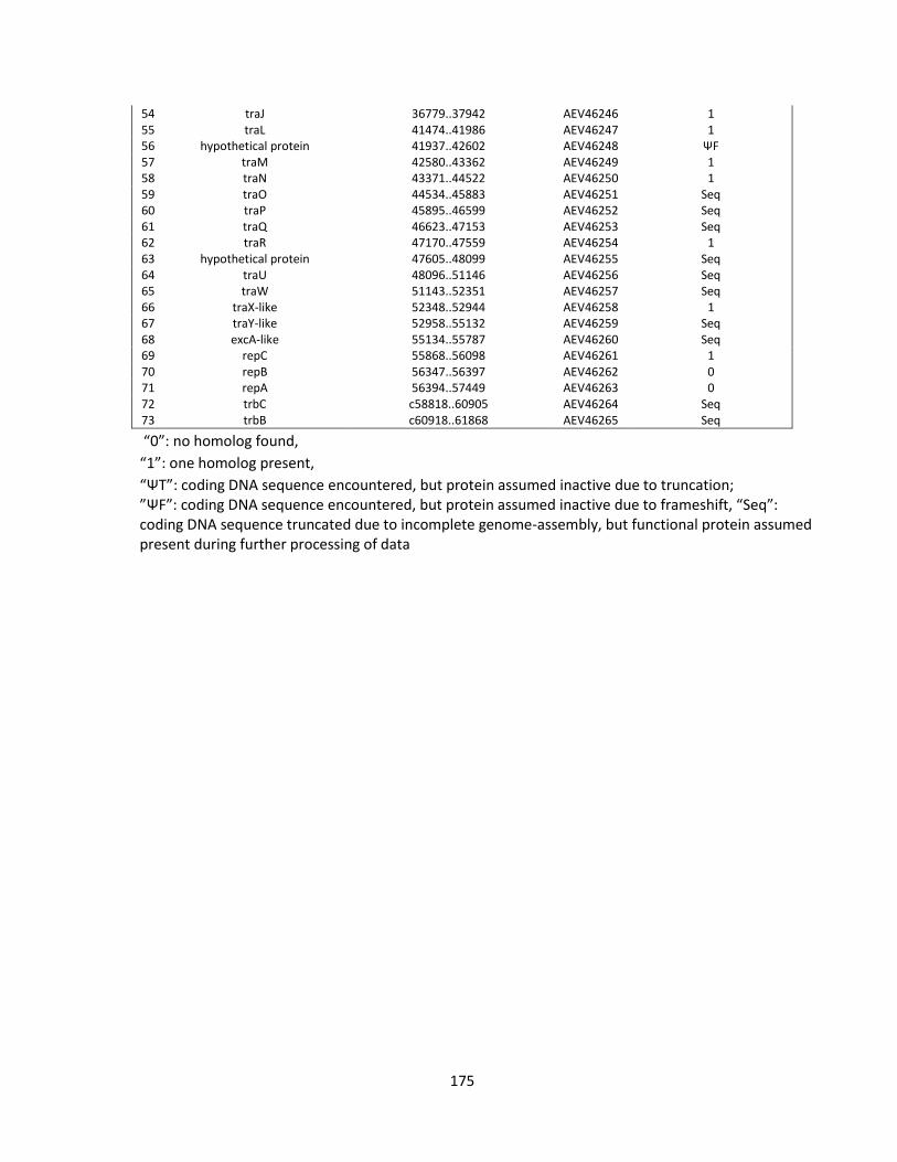
175
54 traJ 36779..37942 AEV46246 1 55 traL 41474..41986 AEV46247 1 56 hypothetical protein 41937..42602 AEV46248 ΨF 57 traM 42580..43362 AEV46249 1 58 traN 43371..44522 AEV46250 1 59 traO 44534..45883 AEV46251 Seq 60 traP 45895..46599 AEV46252 Seq 61 traQ 46623..47153 AEV46253 Seq 62 traR 47170..47559 AEV46254 1 63 hypothetical protein 47605..48099 AEV46255 Seq 64 traU 48096..51146 AEV46256 Seq 65 traW 51143..52351 AEV46257 Seq 66 traX-like 52348..52944 AEV46258 1 67 traY-like 52958..55132 AEV46259 Seq 68 excA-like 55134..55787 AEV46260 Seq 69 repC 55868..56098 AEV46261 1 70 repB 56347..56397 AEV46262 0 71 repA 56394..57449 AEV46263 0 72 trbC c58818..60905 AEV46264 Seq 73 trbB c60918..61868 AEV46265 Seq
“0”: no homolog found,
“1”: one homolog present,
“ΨT”: coding DNA sequence encountered, but protein assumed inactive due to truncation; ”ΨF”: coding DNA sequence encountered, but protein assumed inactive due to frameshift, “Seq”: coding DNA sequence truncated due to incomplete genome-assembly, but functional protein assumed present during further processing of data

Supplementary Table 4.5. A list of genes from Cmm containing known and putative virulence factors and other bacterial genes that are possibly involved in functions such as signal perception and transduction and interaction with tomato plant (as described in the publication of Savidor et al., 2012) and their homologs in LMG 26808.
Locus Annotations/gene name CDS position in Cmm
Accession number
Homolog in LMG 26808
Contig Remarks
CMM_2969 putative transcriptional regulator containing an aminotransferase domain
3289507-3290829 YP_001223714.1 1 contig_11 (78977 to 80299)
CMM_1564 putative transcriptional regulator, HrcA family 1769447-1770469 YP_001222306.1 1 contig_5 (474086 to 475108) CMM_0789 putative transcriptional regulator, GntR family 887879-888640 YP_001221529.1 1 contig_2 (25785 to 26546) CMM_0893 putative transcriptional regulator, LuxR family 1018383-1021241 YP_001221633.1 1 contig_7 (71558 to 74350) CMM_1063 putative transcription factor 1219388-1219882 YP_001221804.1 1 contig_5 (1016576 to 1017070) CMM_1931 putative transcriptional regulator 2180204-2180872 YP_001222673.1 1 contig_5 (59453 to 60121) CMM_1689 putative transcriptional regulator 1913463-1914470 YP_001222432.1 1 contig_5 (326367 to 327374) CMM_2255 putative antisigma factor antagonist 2550877-2551215 YP_001223000.1 1 contig_9 (100292 to 100630) CMM_2645 putative transcriptional regulator, TetR family 2971696-2972319 YP_001223390.1 1 contig_12 (104151 to 104774) CMM_2841 putative transcriptional regulator, LacI family 3167961-3169025 YP_001223586.1 1 contig_11 (206119 to 207120) CMM_2492 putative two-component system response regulator 2807423-2808103 YP_001223586.1 1 contig_3 (19743 to 20423) CMM_1059 putative two-component system sensor kinase 1215851-1217356 YP_001221800.1 1 contig_5 (1019101 to 1020606)
pCM2_0054 pat-1, putative extracellular serine protease, Chp Family 55371-56213 YP_001220724.1 0 pCM2_0053 phpA, putative extracellular serine protease,
homologues of pat-1 encoded on the pCM2, Chp Family 53465-54298 YP_001220723.1 0
pCM2_0052 phpB, putative extracellular serine protease, homologues of pat-1 encoded on the pCM1, Chp Family
52086-52940 YP_001220722.1 0
CMM_PS_05 chpA, pseudogene, not expressed in planta, Chp Family 58928-59730 - 0 CMM_PS_10 chpB, pseudogene, not expressed in planta, Chp Family 88997-89862 - 0 CMM_0052 chpC, serine protease, Chp Family 70785-71645 YP_001220791.1 0 CMM_PS_04 chpD, pseudogene, not expressed in planta, Chp Family 46723-47512 - 0 CMM_0039 chpE, Serine protease, Chp Family 48734-49567 YP_001220778.1 0 CMM_0053 chpF, putative serine protease, Chp Family 74779-75633 YP_001220792.1 1 contig_10 (132109 to 132804) RBH is CMM_0053 CMM_0059 chpG, putative serine protease, Chp Family 82939-83772 YP_001220798.1 1 contig_10 (132106 to 132804) RBH is CMM_0053
CMM_0041 ppaA, putative extracellular serine protease, Ppa Family, important for plant colonization
50376-51356 YP_001220780.1 0
CMM_0042 ppaB1, putative extracellular protease, Ppa Family 51503-52507 YP_001220781.1 0 CMM_0050 ppaB2, putative extracellular protease, Ppa Family 67885-68886 YP_001220789.1 0 CMM_0044 ppaC, putative extracellular protease, Ppa Family,
important for plant colonization 53883-54896 YP_001220783.1 0
CMM_0075 ppaD, serine protease, Ppa Family 113031-114005 YP_001220814.1 0 CMM_0071 ppaE, putative serine protease, Ppa Family 103428-104384 YP_001220810.1 0 CMM_0764 ppaF, putative glutamyl peptidase, Ppa Family 859301-860284 YP_001221504.1 1 contig_2 (54705 to 55265) RBH is CMM_0764
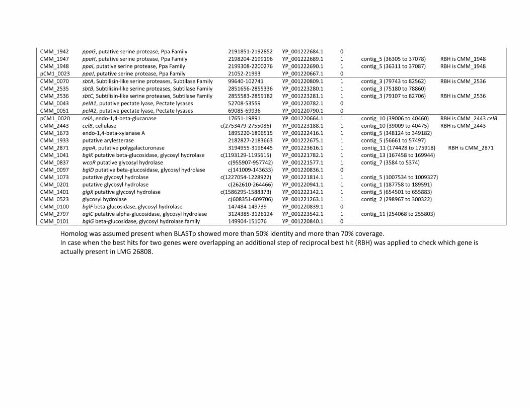
CMM_1942 ppaG, putative serine protease, Ppa Family 2191851-2192852 YP_001222684.1 0 CMM_1947 ppaH, putative serine protease, Ppa Family 2198204-2199196 YP_001222689.1 1 contig_5 (36305 to 37078) RBH is CMM_1948 CMM_1948 ppaI, putative serine protease, Ppa Family 2199308-2200276 YP_001222690.1 1 contig_5 (36311 to 37087) RBH is CMM_1948 pCM1_0023 ppaJ, putative serine protease, Ppa Family 21052-21993 YP_001220667.1 0
CMM_0070 sbtA, Subtilisin-like serine proteases, Subtilase Family 99640-102741 YP_001220809.1 1 contig_3 (79743 to 82562) RBH is CMM_2536 CMM_2535 sbtB, Subtilisin-like serine proteases, Subtilase Family 2851656-2855336 YP_001223280.1 1 contig_3 (75180 to 78860) CMM_2536 sbtC, Subtilisin-like serine proteases, Subtilase Family 2855583-2859182 YP_001223281.1 1 contig_3 (79107 to 82706) RBH is CMM_2536 CMM_0043 pelA1, putative pectate lyase, Pectate lysases 52708-53559 YP_001220782.1 0 CMM_0051 pelA2, putative pectate lyase, Pectate lysases 69085-69936 YP_001220790.1 0
pCM1_0020 celA, endo-1,4-beta-glucanase 17651-19891 YP_001220664.1 1 contig_10 (39006 to 40460) RBH is CMM_2443 celB CMM_2443 celB, cellulase c(2753479-2755086) YP_001223188.1 1 contig_10 (39009 to 40475) RBH is CMM_2443 CMM_1673 endo-1,4-beta-xylanase A 1895220-1896515 YP_001222416.1 1 contig_5 (348124 to 349182) CMM_1933 putative arylesterase 2182827-2183663 YP_001222675.1 1 contig_5 (56661 to 57497) CMM_2871 pgaA, putative polygalacturonase 3194955-3196445 YP_001223616.1 1 contig_11 (174428 to 175918) RBH is CMM_2871 CMM_1041 bglK putative beta-glucosidase, glycosyl hydrolase c(1193129-1195615) YP_001221782.1 1 contig_13 (167458 to 169944) CMM_0837 wcoR putative glycosyl hydrolase c(955907-957742) YP_001221577.1 1 contig_7 (3584 to 5374) CMM_0097 bglD putative beta-glucosidase, glycosyl hydrolase c(141009-143633) YP_001220836.1 0 CMM_1073 putative glycosyl hydrolase c(1227054-1228922) YP_001221814.1 1 contig_5 (1007534 to 1009327) CMM_0201 putative glycosyl hydrolase c(262610-264466) YP_001220941.1 1 contig_1 (187758 to 189591) CMM_1401 glgX putative glycosyl hydrolase c(1586295-1588373) YP_001222142.1 1 contig_5 (654501 to 655883) CMM_0523 glycosyl hydrolase c(608351-609706) YP_001221263.1 1 contig_2 (298967 to 300322) CMM_0100 bglF beta-glucosidase, glycosyl hydrolase 147484-149739 YP_001220839.1 0 CMM_2797 aglC putative alpha-glucosidase, glycosyl hydrolase 3124385-3126124 YP_001223542.1 1 contig_11 (254068 to 255803) CMM_0101 bglG beta-glucosidase, glycosyl hydrolase family 149904-151076 YP_001220840.1 0
Homolog was assumed present when BLASTp showed more than 50% identity and more than 70% coverage. In case when the best hits for two genes were overlapping an additional step of reciprocal best hit (RBH) was applied to check which gene is actually present in LMG 26808.
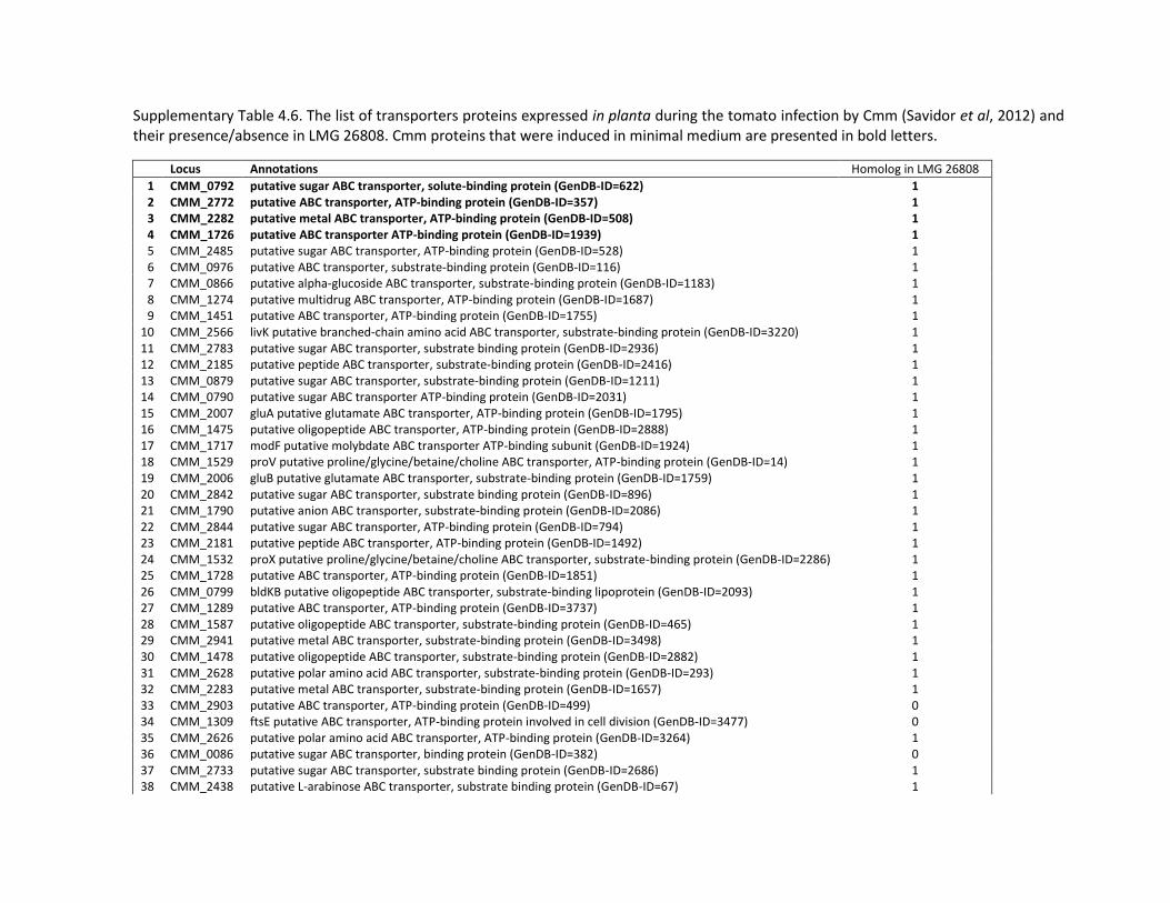
Supplementary Table 4.6. The list of transporters proteins expressed in planta during the tomato infection by Cmm (Savidor et al, 2012) and their presence/absence in LMG 26808. Cmm proteins that were induced in minimal medium are presented in bold letters.
Locus Annotations Homolog in LMG 26808
1 CMM_0792 putative sugar ABC transporter, solute-binding protein (GenDB-ID=622) 1 2 CMM_2772 putative ABC transporter, ATP-binding protein (GenDB-ID=357) 1 3 CMM_2282 putative metal ABC transporter, ATP-binding protein (GenDB-ID=508) 1 4 CMM_1726 putative ABC transporter ATP-binding protein (GenDB-ID=1939) 1 5 CMM_2485 putative sugar ABC transporter, ATP-binding protein (GenDB-ID=528) 1 6 CMM_0976 putative ABC transporter, substrate-binding protein (GenDB-ID=116) 1 7 CMM_0866 putative alpha-glucoside ABC transporter, substrate-binding protein (GenDB-ID=1183) 1 8 CMM_1274 putative multidrug ABC transporter, ATP-binding protein (GenDB-ID=1687) 1 9 CMM_1451 putative ABC transporter, ATP-binding protein (GenDB-ID=1755) 1
10 CMM_2566 livK putative branched-chain amino acid ABC transporter, substrate-binding protein (GenDB-ID=3220) 1 11 CMM_2783 putative sugar ABC transporter, substrate binding protein (GenDB-ID=2936) 1 12 CMM_2185 putative peptide ABC transporter, substrate-binding protein (GenDB-ID=2416) 1 13 CMM_0879 putative sugar ABC transporter, substrate-binding protein (GenDB-ID=1211) 1 14 CMM_0790 putative sugar ABC transporter ATP-binding protein (GenDB-ID=2031) 1 15 CMM_2007 gluA putative glutamate ABC transporter, ATP-binding protein (GenDB-ID=1795) 1 16 CMM_1475 putative oligopeptide ABC transporter, ATP-binding protein (GenDB-ID=2888) 1 17 CMM_1717 modF putative molybdate ABC transporter ATP-binding subunit (GenDB-ID=1924) 1 18 CMM_1529 proV putative proline/glycine/betaine/choline ABC transporter, ATP-binding protein (GenDB-ID=14) 1 19 CMM_2006 gluB putative glutamate ABC transporter, substrate-binding protein (GenDB-ID=1759) 1 20 CMM_2842 putative sugar ABC transporter, substrate binding protein (GenDB-ID=896) 1 21 CMM_1790 putative anion ABC transporter, substrate-binding protein (GenDB-ID=2086) 1 22 CMM_2844 putative sugar ABC transporter, ATP-binding protein (GenDB-ID=794) 1 23 CMM_2181 putative peptide ABC transporter, ATP-binding protein (GenDB-ID=1492) 1 24 CMM_1532 proX putative proline/glycine/betaine/choline ABC transporter, substrate-binding protein (GenDB-ID=2286) 1 25 CMM_1728 putative ABC transporter, ATP-binding protein (GenDB-ID=1851) 1 26 CMM_0799 bldKB putative oligopeptide ABC transporter, substrate-binding lipoprotein (GenDB-ID=2093) 1 27 CMM_1289 putative ABC transporter, ATP-binding protein (GenDB-ID=3737) 1 28 CMM_1587 putative oligopeptide ABC transporter, substrate-binding protein (GenDB-ID=465) 1 29 CMM_2941 putative metal ABC transporter, substrate-binding protein (GenDB-ID=3498) 1 30 CMM_1478 putative oligopeptide ABC transporter, substrate-binding protein (GenDB-ID=2882) 1 31 CMM_2628 putative polar amino acid ABC transporter, substrate-binding protein (GenDB-ID=293) 1 32 CMM_2283 putative metal ABC transporter, substrate-binding protein (GenDB-ID=1657) 1 33 CMM_2903 putative ABC transporter, ATP-binding protein (GenDB-ID=499) 0 34 CMM_1309 ftsE putative ABC transporter, ATP-binding protein involved in cell division (GenDB-ID=3477) 0 35 CMM_2626 putative polar amino acid ABC transporter, ATP-binding protein (GenDB-ID=3264) 1 36 CMM_0086 putative sugar ABC transporter, binding protein (GenDB-ID=382) 0 37 CMM_2733 putative sugar ABC transporter, substrate binding protein (GenDB-ID=2686) 1 38 CMM_2438 putative L-arabinose ABC transporter, substrate binding protein (GenDB-ID=67) 1
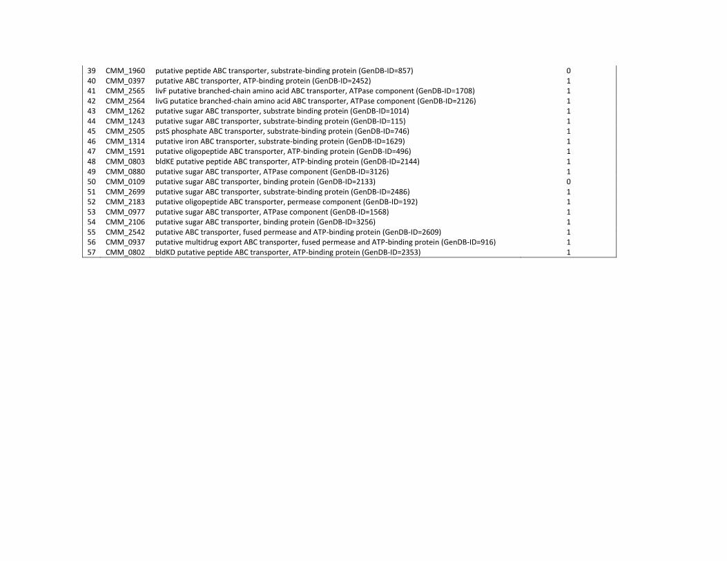
39 CMM_1960 putative peptide ABC transporter, substrate-binding protein (GenDB-ID=857) 0 40 CMM_0397 putative ABC transporter, ATP-binding protein (GenDB-ID=2452) 1 41 CMM_2565 livF putative branched-chain amino acid ABC transporter, ATPase component (GenDB-ID=1708) 1 42 CMM_2564 livG putatice branched-chain amino acid ABC transporter, ATPase component (GenDB-ID=2126) 1 43 CMM_1262 putative sugar ABC transporter, substrate binding protein (GenDB-ID=1014) 1 44 CMM_1243 putative sugar ABC transporter, substrate-binding protein (GenDB-ID=115) 1 45 CMM_2505 pstS phosphate ABC transporter, substrate-binding protein (GenDB-ID=746) 1 46 CMM_1314 putative iron ABC transporter, substrate-binding protein (GenDB-ID=1629) 1 47 CMM_1591 putative oligopeptide ABC transporter, ATP-binding protein (GenDB-ID=496) 1 48 CMM_0803 bldKE putative peptide ABC transporter, ATP-binding protein (GenDB-ID=2144) 1 49 CMM_0880 putative sugar ABC transporter, ATPase component (GenDB-ID=3126) 1 50 CMM_0109 putative sugar ABC transporter, binding protein (GenDB-ID=2133) 0 51 CMM_2699 putative sugar ABC transporter, substrate-binding protein (GenDB-ID=2486) 1 52 CMM_2183 putative oligopeptide ABC transporter, permease component (GenDB-ID=192) 1 53 CMM_0977 putative sugar ABC transporter, ATPase component (GenDB-ID=1568) 1 54 CMM_2106 putative sugar ABC transporter, binding protein (GenDB-ID=3256) 1 55 CMM_2542 putative ABC transporter, fused permease and ATP-binding protein (GenDB-ID=2609) 1 56 CMM_0937 putative multidrug export ABC transporter, fused permease and ATP-binding protein (GenDB-ID=916) 1 57 CMM_0802 bldKD putative peptide ABC transporter, ATP-binding protein (GenDB-ID=2353) 1
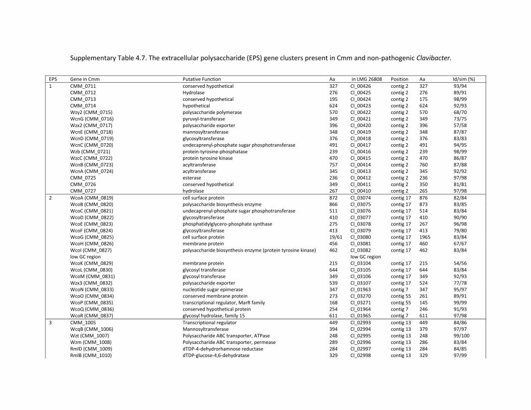
Supplementary Table 4.7. The extracellular polysaccharide (EPS) gene clusters present in Cmm and non-pathogenic Clavibacter.
EPS Gene in Cmm Putative Function Aa in LMG 26808 Position Aa Id/sim (%)
1 CMM_0711 conserved hypothetical 327 Cl_00426 contig 2 327 93/94 CMM_0712 Hydrolase 276 Cl_00425 contig 2 276 89/91 CMM_0713 conserved hypothetical 195 Cl_00424 contig 2 175 98/99 CMM_0714 hypothetical 624 Cl_00423 contig 2 624 92/93 Wzy2 (CMM_0715) polysaccharide polymerase 570 Cl_00422 contig 2 570 68/70 WcnG (CMM_0716) pyruvyl-transferase 349 Cl_00421 contig 2 349 73/75 Wzx2 (CMM_0717) polysaccharide exporter 396 Cl_00420 contig 2 396 57/58 WcnE (CMM_0718) mannosyltransferase 348 Cl_00419 contig 2 348 87/87 WcnD (CMM_0719) glycosyltransferase 376 Cl_00418 contig 2 376 83/83 WcnC (CMM_0720) undecaprenyl-phosphate sugar phosphotransferase 491 Cl_00417 contig 2 491 94/95 Wzb (CMM_0721) protein-tyrosine-phosphatase 239 Cl_00416 contig 2 239 98/99 WzcC (CMM_0722) protein tyrosine kinase 470 Cl_00415 contig 2 470 86/87 WcnB (CMM_0723) acyltransferase 757 Cl_00414 contig 2 760 87/88 WcnA (CMM_0724) acyltransferase 345 Cl_00413 contig 2 345 92/92 CMM_0725 esterase 236 Cl_00412 contig 2 236 97/98 CMM_0726 conserved hypothetical 349 Cl_00411 contig 2 350 81/81 CMM_0727 hydrolase 267 Cl_00410 contig 2 265 97/98
2 WcoA (CMM_0819) cell surface protein 872 Cl_03074 contig 17 876 82/84 WcoB (CMM_0820) polysaccharide biosynthesis enzyme 866 Cl_03075 contig 17 873 83/85 WcoC (CMM_0821) undecaprenyl-phosphate sugar phosphotransferase 511 Cl_03076 contig 17 514 83/84 WcoD (CMM_0822) glycosyltransferase 410 Cl_03077 contig 17 410 90/90 WcoE (CMM_0823) phosphatidylglycero-phosphate synthase 275 Cl_03078 contig 17 267 96/98 WcoF (CMM_0824) glycosyltransferase 413 Cl_03079 contig 17 413 79/80 WcoG (CMM_0825) cell surface protein 19/61 Cl_03080 contig 17 1965 83/84 WcoH (CMM_0826) membrane protein 456 Cl_03081 contig 17 460 67/67 WcoI (CMM_0827) polysaccharide biosynthesis enzyme (protein tyrosine kinase) 462 Cl_03082 contig 17 462 83/84 low GC region low GC region WcoK (CMM_0829) membrane protein 215 Cl_03104 contig 17 215 54/56 WcoL (CMM_0830) glycosyl transferase 644 Cl_03105 contig 17 644 83/84 WcoM (CMM_0831) glycosyl transferase 349 Cl_03106 contig 17 349 92/93 Wzx3 (CMM_0832) polysaccharide exporter 539 Cl_03107 contig 17 524 77/78 WcoN (CMM_0833) nucleotide sugar epimerase 347 Cl_01963 contig 7 347 95/97 WcoO (CMM_0834) conserved membrane protein 273 Cl_03270 contig 55 261 89/91 WcoP (CMM_0835) transcriptional regulator, MarR family 168 Cl_03271 contig 55 145 99/99 WcoQ (CMM_0836) conserved hypothetical protein 254 Cl_01964 contig 7 246 91/93 WcoR (CMM_0837) glycosyl hydrolase, family 15 611 Cl_01965 contig 7 611 97/98
3 CMM_1005 Transcriptional regulator 449 Cl_02993 contig 13 449 84/86 WcqB (CMM_1006) Mannosyltransferase 394 Cl_02994 contig 13 379 97/97 Wzt (CMM_1007) Polysaccharide ABC transporter, ATPase 248 Cl_02995 contig 13 248 99/100 Wzm (CMM_1008) Polysaccharide ABC transporter, permease 289 Cl_02996 contig 13 286 83/84 RmlD (CMM_1009) dTDP-4-dehydrorhamnose reductase 284 Cl_02997 contig 13 284 84/85 RmlB (CMM_1010) dTDP-glucose-4,6-dehydratase 329 Cl_02998 contig 13 329 97/99
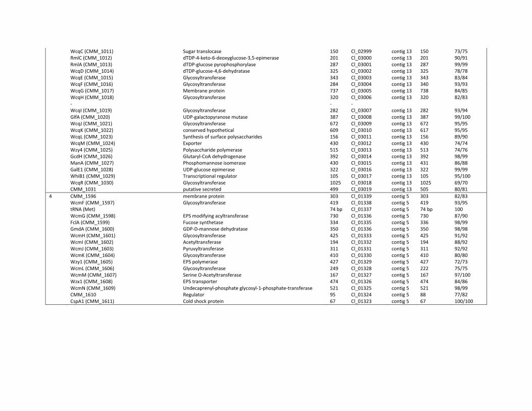
WcqC (CMM_1011) Sugar translocase 150 Cl_02999 contig 13 150 73/75 RmlC (CMM_1012) dTDP-4-keto-6-deoxyglucose-3,5-epimerase 201 Cl_03000 contig 13 201 90/91 RmlA (CMM_1013) dTDP-glucose pyrophosphorylase 287 Cl_03001 contig 13 287 99/99 WcqD (CMM_1014) dTDP-glucose-4,6-dehydratase 325 Cl_03002 contig 13 325 78/78 WcqE (CMM_1015) Glycosyltransferase 343 Cl_03003 contig 13 343 83/84 WcqF (CMM_1016) Glycosyltransferase 284 Cl_03004 contig 13 340 93/93 WcqG (CMM_1017) Membrane protein 737 Cl_03005 contig 13 738 84/85 WcqH (CMM_1018) Glycosyltransferase 320 Cl_03006 contig 13 320 82/83 - - - WcqI (CMM_1019) Glycosyltransferase 282 Cl_03007 contig 13 282 93/94 GlfA (CMM_1020) UDP-galactopyranose mutase 387 Cl_03008 contig 13 387 99/100 WcqJ (CMM_1021) Glycosyltransferase 672 Cl_03009 contig 13 672 95/95 WcqK (CMM_1022) conserved hypothetical 609 Cl_03010 contig 13 617 95/95 WcqL (CMM_1023) Synthesis of surface polysaccharides 156 Cl_03011 contig 13 156 89/90 WcqM (CMM_1024) Exporter 430 Cl_03012 contig 13 430 74/74 Wzy4 (CMM_1025) Polysaccharide polymerase 515 Cl_03013 contig 13 513 74/76 GcdH (CMM_1026) Glutaryl-CoA dehydrogenase 392 Cl_03014 contig 13 392 98/99 ManA (CMM_1027) Phosphomannose isomerase 430 Cl_03015 contig 13 431 86/88 GalE1 (CMM_1028) UDP-glucose epimerase 322 Cl_03016 contig 13 322 99/99 WhiB1 (CMM_1029) Transcriptional regulator 105 Cl_03017 contig 13 105 95/100 WcqR (CMM_1030) Glycosyltransferase 1025 Cl_03018 contig 13 1025 69/70 CMM_1031 putative secreted 499 Cl_03019 contig 13 505 80/81
4 CMM_1596 membrane protein 303 Cl_01339 contig 5 303 82/83 WcmF (CMM_1597) Glycosyltransferase 419 Cl_01338 contig 5 419 93/95 tRNA (Met) 74 bp Cl_01337 contig 5 74 bp 100 WcmG (CMM_1598) EPS modifying acyltransferase 730 Cl_01336 contig 5 730 87/90 FclA (CMM_1599) Fucose synthetase 334 Cl_01335 contig 5 336 98/99 GmdA (CMM_1600) GDP-D-mannose dehydratase 350 Cl_01336 contig 5 350 98/98 WcmH (CMM_1601) Glycosyltransferase 425 Cl_01333 contig 5 425 91/92 WcmI (CMM_1602) Acetyltransferase 194 Cl_01332 contig 5 194 88/92 WcmJ (CMM_1603) Pyruvyltransferase 311 Cl_01331 contig 5 311 92/92 WcmK (CMM_1604) Glycosyltransferase 410 Cl_01330 contig 5 410 80/80 Wzy1 (CMM_1605) EPS polymerase 427 Cl_01329 contig 5 427 72/73 WcmL (CMM_1606) Glycosyltransferase 249 Cl_01328 contig 5 222 75/75 WcmM (CMM_1607) Serine O-Acetyltransferase 167 Cl_01327 contig 5 167 97/100 Wzx1 (CMM_1608) EPS transporter 474 Cl_01326 contig 5 474 84/86 WcmN (CMM_1609) Undecaprenyl-phosphate glycosyl-1-phosphate-transferase 521 Cl_01325 contig 5 521 98/99 CMM_1610 Regulator 95 Cl_01324 contig 5 88 77/82 CspA1 (CMM_1611) Cold shock protein 67 Cl_01323 contig 5 67 100/100
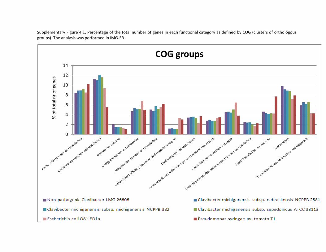
Supplementary Figure 4.1. Percentage of the total number of genes in each functional category as defined by COG (clusters of orthologous groups). The analysis was performed in IMG-ER.
0
2
4
6
8
10
12
14
COG groups
Clavibacter-like LMG 26808 Clavibacter michiganensis subsp. nebraskensis NCPPB 2581
Clavibacter michiganensis subsp. michiganensis NCPPB 382 Clavibacter michiganensis subsp. sepedonicus ATCC 33113
Escherichia coli O81 ED1a Pseudomonas syringae pv. tomato T1
% o
f to
tal n
r o
f ge
nes

183
Chapter 5
Multilocus Variable-Number-Tandem-Repeats Analysis (MLVA)
distinguishes a clonal complex of Clavibacter michiganensis subsp.
michiganensis strains isolated from recent outbreaks of bacterial
wilt and canker in Belgium
Redrafted from:
Zaluga J, Stragier P, Van Vaerenbergh J, Maes M, De Vos P. 2013. Multilocus Variable-
Number-Tandem-Repeats Analysis (MLVA) distinguishes a clonal complex of Clavibacter
michiganensis subsp. michiganensis strains isolated from recent outbreaks of bacterial wilt
and canker in Belgium. BMC Microbiology 13: 126
Authors’ contributions:
PS, MM, JVV and PDV conceived the study and participated in its design. JVV and PDV
provided the bacteria culture collection for the study. JZ participated in the design of the
study, carried out the molecular work, performed the data analysis and drafted the
manuscript. PS coordinated the work and performed the statistical analysis. All authors read
and approved the final manuscript.
Chapter 5 – MLVA analysis of Cmm strains isolated from recent outbreaks of bacterial wilt and canker in Belgium
184
Summary
Background Clavibacter michiganensis subsp. michiganensis (Cmm) causes bacterial wilt
and canker in tomato. Cmm is present nearly in all European countries. During the last three
years several local outbreaks were detected in Belgium. The lack of a convenient high-
resolution strain-typing method has hampered the study of the routes of transmission of
Cmm and epidemiology in tomato cultivation. In this study the genetic relatedness among a
worldwide collection of Cmm strains and their relatives was approached by gyrB and dnaA
gene sequencing. Further, we developed and applied a multilocus variable number of
tandem repeats analysis (MLVA) scheme to discriminate Cmm strains.
Results A phylogenetic analysis of gyrB and dnaA gene sequences of 56 Cmm strains
demonstrated that Belgian Cmm strains from recent outbreaks of 2010-2012 form a
genetically uniform group within the Cmm clade and Cmm is phylogenetically distinct from
other Clavibacter subspecies and from non-pathogenic Clavibacter strains. MLVA conducted
with eight minisatellite loci detected 25 haplotypes within Cmm. All strains from Belgian
outbreaks, isolated between 2010 and 2012, together with two French strains from 2010
seem to form one monomorphic group. Regardless of the isolation year, location or tomato
cultivar, Belgian strains from recent outbreaks belonged to the same haplotype. On the
contrary, strains from diverse geographical locations or isolated over longer periods of time
formed mostly singletons.
Conclusions We hypothesise that the introduction might have originated from one lot of
seeds or contaminated tomato seedlings which was the source of the outbreak in 2010 and
that these Cmm strains persisted and induced infection in 2011 and 2012. Our results
demonstrate that MLVA is a promising typing technique for a local surveillance and
outbreaks investigation in epidemiological studies of Cmm.
Part III – Experimental work
185
5.1 Introduction
Clavibacter michiganensis subsp. michiganensis, a Gram-positive bacterium, is the causative
agent of bacterial wilt and canker, one of the most destructive bacterial diseases in tomato
(De León et al., 2011). Contaminated tomato seeds are considered to be the main source of
infection. The bacterium survives for a long period of time in seeds, soil and plant debris
(EPPO 2005, Strider 1969). Every year, new or reoccurring outbreaks are detected causing
substantial economic losses worldwide (Jahr et al., 1999). Bacterial wilt and canker was
described for the first time in 1905 in Michigan, USA and since that moment it has been
reported in nearly all tomato growing areas of the world (Strider 1969, EPPO 2013).
Difficulties in controlling the spread of the pathogen, the lack of resistant tomato varieties
and severity of disease symptoms led to the classification of Cmm as quarantine organisms.
Cmm is listed as an A2 quarantine pest by the European and Mediterranean Plant
Protection Organization (EPPO) (EPPO 2005) in Europe and in many countries all over the
world (De León et al., 2011).
The epidemiology and the population structure of Cmm in areas where outbreaks of Cmm
are common remains scantily investigated and poorly understood. Recent studies describing
outbreaks of Cmm in Europe and Asia (Lamichhane et al., 2011) (De León et al., 2009)
(Milijašević-Marčić et al., 2012) (Kawaguchi et al., 2010) have shed some light on this issue.
In Italy a clonal population of Cmm was responsible for the outbreak in 2007 (Bella et al.,
2012). A high homogeneity was also observed among strains isolated from 2002 to 2007 in
Canary Islands suggesting a single introduction of the pathogen as a source of infection (De
León et al., 2009). Primary infections in many countries were attributed to the introductions
of contaminated tomato seeds and/or seedlings (Milijašević-Marčić et al., 2012) (Kleitman
et al., 2008). These findings indicate that seeds play an important role in long-distance
spread of the pathogen. A direct link between tomato cultivar, year or place of isolation and
Cmm type mostly could not be recognized (Kawaguchi et al., 2010) (Bella et al., 2012) (De
León et al., 2009) except the outbreak in 2001 in Turkey where bacterial wilt and canker
was detected only on one tomato cultivar ‘Target’ (Sahin et al., 2002). Interestingly, in Israel
and Serbia Cmm strains showing the same haplotypes were repeatedly isolated from the
Chapter 5 – MLVA analysis of Cmm strains isolated from recent outbreaks of bacterial wilt and canker in Belgium
186
same locations during several subsequent years (Kleitman et al., 2008, Milijašević-Marčić et
al., 2012). Reoccurring outbreaks suggest that despite intensified efforts for eradication,
reliable control of this disease remains an unattainable goal. The limited progress in
improving its management is mainly due to the erratic nature of the disease outbreaks and
to limited and scattered epidemiological data. Therefore, access to an accurate, efficient
and cost-effective strain typing technique could be very useful.
Bacterial typing techniques are applied to quickly and reliably differentiate closely related
strains in an epidemiological survey, to determinate the relatedness among the strains and
to track their origin and pathways of spread. Over the past decades a variety of different
typing methods have been developed to generate strain-specific patterns. They are also
applied for comprehensive investigation of bacterial population structure and dynamics. A
range of methods has already been applied to study the diversity of Clavibacter, particularly
to investigate Cmm strains. Rep-PCR (repetitive-element-based PCR), a relatively easy and
fast technique, was shown to be of moderate utility (Kawaguchi et al., 2010), mainly
because of the lack of a database and the rather low discriminatory power needed to study
closely related strains. Moreover, rep-PCR is mostly not portable between different
laboratories (Cangelosi et al., 2004). PFGE (pulsed-field gel electrophoresis of macro-
restricted bacterial DNA), one of the oldest techniques used in epidemiology, is labor
intensive and expensive but is still used as a gold standard in typing of some bacterial
species (Kleitman et al., 2008) (Bosch et al., 2010). PFGE was applied to study the diversity
of Cmm strains from outbreaks in Serbia (Milijašević-Marčić et al., 2012) and in Israel
(Kleitman et al., 2008) where the results of PFGE showed similar resolution of those
obtained by gene sequence analysis and rep-PCR, respectively. Also, AFLP, a high resolution
molecular typing method was applied by De León and coworkers to study genetic diversity
of Cmm strains from Canary Islands (De León et al., 2009). This technique generated more
bands per strain and resulted in more reproducible and robust discriminatory clustering of
the strains (De León et al., 2009). Highly reproducible multilocus sequence typing (MLST)
was used to analyze Cmm population from Serbia. Cmm strains were divided into seven
groups and the results were confirmed by PFGE analysis (Milijašević-Marčić et al., 2012).
Part III – Experimental work
187
MLVA (Multiple-Locus Variable number tandem repeat Analysis) is a PCR-based typing
technique that has been widely applied in medical microbiology (Van Belkum 2006). It takes
advantage of the inherent variability encountered in regions with a number of tandem
repeats. The origin of the repetitive regions can be accounted to slipped strand mispairing
events occurring during DNA duplication, in which repetitive regions are incorrectly copied
resulting in deletion or insertion of one or several copies of the repeat (Van Belkum et al.,
1998). PCR primers designed to board different VNTR (Variable Number of Tandem Repeats)
regions in the genome can be easily combined in a multiplex PCR in an MLVA scheme. The
differences between strains are assessed by the different lengths of the repeats visualized
by gel electrophoresis or automated fragment analysis on a sequencer. From these sizes,
the number of repeat units at each locus can be deduced. The resulting information forms a
strain-specific numerical code which can be easily compared to a reference database. The
MLVA technique was introduced to bacterial typing as a promising alternative or a
complement to already existing typing methods, such as AFLP, MLST, rep-PCR or PFGE. The
discriminatory power of MLVA is generally higher than other standard typing techniques
(Harth-Chu et al., 2009). However, the final result is group dependent and can vary
considerably between different bacterial species. VNTRs have been used to discriminate
among individual strains within many food-borne pathogens with little genetic differences,
including Escherichia coli O157:H7 (Lindstedt et al., 2003) and Vibrio cholerae (Danin-Poleg
et al., 2007) and to study other important human pathogens, such as Neisseria gonorrhoeae
(Heymans et al., 2011), Streptococcus pneumoniae (van Cuyck et al., 2012) and
Mycobacterium tuberculosis (Skuce et al., 2002). MLVA has been extensively used for
tracking transmissions of important human and animal pathogens (Marsh et al., 2006)
(Hidalgo et al., 2010) and for typing monomorphic bacterial pathogens including Bacillus
anthracis (Le Fleche et al., 2001) and Yersinia pestis (Li et al., 2009). To date, several MLVA
schemes have been published on plant pathogens, such as Xanthomonas citri pv. citri (3), X.
oryzae pv. oryzicola (Zhao et al., 2007), Pseudomonas syringae pv. maculicola and tomato
(Gironde and Manceau 2012), Xylella fastidiosa (Coletta-Filho et al., 2001) and on fungi e.g.
Aspergillus flavus (Wang et al., 2012), but not for Clavibacter subspecies. In plant
Chapter 5 – MLVA analysis of Cmm strains isolated from recent outbreaks of bacterial wilt and canker in Belgium
188
pathogens, such as Xanthomonas arbolicola pv. pruni, MLVA was proposed as a
complementary molecular typing method to AFLP, BOX and ERIC-PCR (Bergsma-Vlami et al.,
2012). In the epidemiological study of pathotypes of Xanthomonas citri MLVA was
compared to AFLP and insertion sequence ligation-mediated PCR (IS-LM-PCR) and was
found the best method to describe the variations among strains originating from the same
country or group of neighboring countries (Bui Thi Ngoc et al., 2009).
The objectives of this study were: 1) to characterize a Belgian population of Cmm strains by
a newly developed MLVA scheme; 2) to compare its genetic variability with some strains of
Cmm isolated in other countries; 3) to investigate whether the strains responsible for
bacterial wilt and canker outbreaks in Belgium in 2010-2012 have one or several infection
sources and 4) to assess the genetic relatedness of the Cmm strains from Belgium by gyrB
and dnaA gene sequence analysis.
Part III – Experimental work
189
5.2 Materials and methods
Bacterial strains. The bacterial strains used in this study are listed in Table 5.1. The strains
were obtained from the BCCM/LMG Bacteria Collection (Ghent, Belgium), the GBBC (ILVO
Plant Clinic, Merelbeke, Belgium) and the NVWA collection (Wageningen, The Netherlands).
The Clavibacter strain subset consisted of five type strains Cmm LMG 7333T (species type
strain), Clavibacter michiganensis subsp. nebraskensis (Cmn) LMG 5627T, Clavibacter
michiganensis subsp. sepedonicus (Cms) LMG 2889T, Clavibacter michiganensis subsp.
insidiosus (Cmi) LMG 3663T, Clavibacter michiganensis subsp. tessellarius (Cmt) LMG 7294T,
two non-pathogenic Clavibacter strains and fifty five Cmm originating from Belgian
outbreaks and other geographical locations. Twenty three Cmm strains were sampled from
symptomatic tomato plants in fields and greenhouses in northeast Belgium. They were
isolated from five different tomato cultivars and seven different locations, in the period
February 2010 till February 2012 (Table 5.1). Non-pathogenic Clavibacter isolates from
tomato seed are phenotypically similar to Cmm in the common diagnostic semi-selective
media and are identified as Cmm in the standard tests but are non-pathogenic to tomato
(Jacques et al., 2012, Zaluga et al., 2011). They were isolated according to the current
method for detection of Cmm in tomato seed recommended by International Seed
Federation (ISF) (Naktuinbouw 2011). The strains were cultured aerobically on MTNA
(Mannitol, trimethoprim, nalidixic acid, amphotericin) medium without antibiotics (Jansing
and Rudolph 1998) at 25°C for 24-48h. Stock cultures were stored at -80˚C in MicrobankTM
beads (Pro-Lab Diagnostics, Canada).
DNA extraction, amplification and sequencing. Total genomic DNA was extracted according
to the guanidium-thiocyanate-EDTA-sarkosyl method described by (Pitcher et al., 1989)
which was adapted for Gram-positive bacteria with a pre-treatment with lysozyme (5 mg/µl
lysosyme powder in TE buffer) and incubation for 40 minutes at 37°C. Amplification and
sequencing primers are listed in Table 5.2. The expected amplicons were generated with the
Qiagen Taq DNA polymerase kit (supplemented with a Q-Solution) and GeneAmp® dNTP’s
(Applied Biosystems, Belgium) according to the manufacturer specifications and with
primers from Sigma Aldrich (Belgium). Amplicons were purified using the Nucleofast®96 PCR

Chapter 5 – MLVA analysis of Cmm strains isolated from recent outbreaks of bacterial wilt and canker in Belgium
190
clean up membrane system (Macherey-Nagel, Germany). Sequencing PCR was performed in
a total volume of 10 µl with 3 µl of a purified amplicon, 0.286 µl of BigDyeTM mixture
(Terminator Cycle Sequencing Kit version 3.1, Applied Biosystems), 1x sequencing buffer and
1.2 µM of each of the amplification primers listed in Table 5.2. The PCR program consisted of
30 cycles (96°C for 15 s, 35°C for 1 s, 60°C for 4 min). Subsequently, the sequencing
products were purified using the BigDye XTerminator Kit (Applied Biosystems) and analyzed
on a 3130xl Genetic Analyzer (Applied Biosystems).
Sequence analysis. In the frame of the European project QBOL (Quarantine Barcoding Of
Life) we developed a gyrB barcode that was proven suitable to identify members of the
genus Clavibacter at the subspecies level (http://www.q-bank.eu/) (Zaluga et al., 2011).
Moreover, gyrB gene was used in MLST schemes developed to type Cmm strains
(Milijašević-Marčić et al., 2012) (Waleron et al., 2011) (Jacques et al., 2012). DnaA sequence
was shown a good taxonomic marker to identify and classify plant pathogenic bacteria, such
as Clavibacter, Xanthomonas and Ralstonia (Schneider et al., 2011). The partial sequencing
of dnaA was successfully used to study genetic diversity of non-pathogenic Clavibacter
strains and to identify members of the genus Clavibacter (J. Zaluga, data unpublished). GyrB
and dnaA sequences were assembled with BioNumerics version 5.1 (Applied Maths,
Belgium) and aligned using ClustalW (Thompson et al., 1994). GyrB sequences and dnaA
sequences were checked by amino acid translation with Transseq
(http://www.ebi.ac.uk/Tools/st/emboss_transeq/) and presence of the GyrB and DnaA
protein domain was confirmed with BLASTp (Altschul et al., 1990). DnaA and gyrB
amplicons were 675 bp and 440 bp long (equal length was used for all strains), respectively.
A phylogenetic tree was constructed on dnaA-gyrB concatenated sequence data with
Molecular Evolutionary Genetics Analysis software (Mega 5.1) (Tamura et al., 2007), using
the Maximum Likelihood method with the Tamura-Nei model (Tamura and Nei 1993) and
1000 bootstrap replicates.
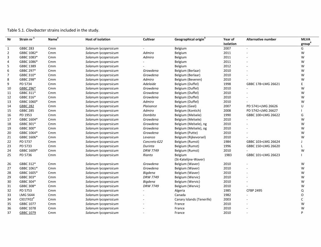
Table 5.1. Clavibacter strains included in the study.
Nr Strain nr 1 Name
2 Host of isolation Cultivar Geographical origin
3 Year of
isolation Alternative number MLVA
group4
1 GBBC 283 Cmm Solanum lycopersicum - Belgium 2007 - G 2 GBBC 1082* Cmm Solanum lycopersicum Admiro Belgium 2011 - W 3 GBBC 1083* Cmm Solanum lycopersicum Admiro Belgium 2011 - W 4 GBBC 1086* Cmm Solanum lycopersicum - Belgium 2011 - W 5 GBBC 1389 Cmm Solanum lycopersicum - Belgium 2012 - W 6 GBBC 297* Cmm Solanum lycopersicum Growdena Belgium (Berlaar) 2010 - W 7 GBBC 310* Cmm Solanum lycopersicum Growdena Belgium (Berlaar) 2010 - W 8 GBBC 298* Cmm Solanum lycopersicum Admiro Belgium (Beveren) 2010 - W 9 PD 5734 Cmm Solanum lycopersicum Adelaide Belgium (Duffel) 1998 GBBC 178=LMG 26621 E 10 GBBC 296* Cmm Solanum lycopersicum Growdena Belgium (Duffel) 2010 - W 11 GBBC 311* Cmm Solanum lycopersicum Growdena Belgium (Duffel) 2010 - W 12 GBBC 316* Cmm Solanum lycopersicum Growdena Belgium (Duffel) 2010 - W 13 GBBC 1060* Cmm Solanum lycopersicum Admiro Belgium (Duffel) 2010 - W 14 GBBC 282 Cmm Solanum lycopersicum Plaisance Belgium (Geel) 2007 PD 5741=LMG 26626 U 15 GBBC 285 Cmm Solanum lycopersicum Admiro Belgium (Kontich) 2008 PD 5742=LMG 26627 I 16 PD 1953 Cmm Solanum lycopersicum Dombito Belgium (Melsele) 1990 GBBC 100=LMG 26622 G 17 GBBC 1604* Cmm Solanum lycopersicum Growdena Belgium (Melsele) 2010 - W 18 GBBC 301* Cmm Solanum lycopersicum Growdena Belgium (Melsele), ng 2010 - W 19 GBBC 300* Cmm Solanum lycopersicum Growdena Belgium (Melsele), og 2010 - W 20 GBBC 1064* Cmm Solanum lycopersicum Growdena Belgium (Putte) 2010 - W 21 GBBC 1606* Cmm Solanum lycopersicum Levanzo Belgium (Rijkevorsel) 2010 - W 22 PD 5737 Cmm Solanum lycopersicum Concreto 622 Belgium (Rumst) 1984 GBBC 103=LMG 26624 J 23 PD 5733 Cmm Solanum lycopersicum Durinta Belgium (Rumst) 1996 GBBC 150=LMG 26620 L 24 GBBC 1609* Cmm Solanum lycopersicum DRW 7749 Belgium (Rumst) 2010 - W 25 PD 5736 Cmm Solanum lycopersicum Rianto Belgium
(St-Katelijne-Waver) 1983 GBBC 101=LMG 26623 I
26 GBBC 312* Cmm Solanum lycopersicum Growdena Belgium (Waver) 2010 - W 27 GBBC 1061* Cmm Solanum lycopersicum Growdena Belgium (Waver) 2010 - W 28 GBBC 1605* Cmm Solanum lycopersicum Bigdena Belgium (Waver) 2010 - W 29 GBBC 303* Cmm Solanum lycopersicum DRW 7749 Belgium (Wervic) 2010 - W 30 GBBC 304* Cmm Solanum lycopersicum Bigdena Belgium (Wervic) 2010 - W 31 GBBC 308* Cmm Solanum lycopersicum DRW 7749 Belgium (Wervic) 2010 - W 32 PD 5753 Cmm Solanum lycopersicum - Algeria 1985 CFBP 2495 Q 33 LMG 5644 Cmm Solanum lycopersicum - Canada 1982 - O 34 Cl01TF02
# Cmm Solanum lycopersicum - Canary Islands (Tenerife) 2003 - C
35 GBBC 1077 Cmm Solanum lycopersicum - France 2010 - W 36 GBBC 1078 Cmm Solanum lycopersicum - France 2010 - W 37 GBBC 1079 Cmm Solanum lycopersicum - France 2010 - P
1 Bacterial collection abbreviations: LMG-BCCM - Belgian Coordinated Collections of Micro-organisms (Ghent, Belgium), PD-PD collection (Wageningen, The Netherlands) and GBBC-GBBC collection (ILVO, Merelbeke, Belgium); * Nursery BPK, Duffel, Belgium;
# Instituto Canario de Investigaciones Agrarias (Tenerife, Spain); eight underlined strains were included in an initial testing of VNTR loci; 2 Cmm-Clavibacter michiganensis subsp. michiganensis, Cmn-Clavibacter michiganensis subsp. nebraskensis, Cms-Clavibacter michiganensis subsp. sepedonicus, Cmi-Clavibacter michiganensis subsp. insidiosus, Cmt-Clavibacter michiganensis subsp. tessellarius;
- Cultivar unknown; geographical origin unknown; year of isolation unknown; no alternative number; 3 ng-new greenhouse, og-old greenhouse; 4 na-not applicable
Nr Strain nr 1 Name
2 Host of isolation Cultivar Geographical origin
3 Year of
isolation Alternative number MLVA
group4
39 PD 5721 Cmm Solanum lycopersicum - France 2006 LMG 26819 S 40 PD 5719 Cmm Solanum lycopersicum - France 2008 - K 41 PD 5749 Cmm Solanum lycopersicum Admiro France 2007 GBBC 261=LMG 26628 A 42 PD 4545 Cmm Solanum lycopersicum - Germany 2003 LMG 26617 D 43 LMG 7333
T Cmm Solanum lycopersicum - Hungary 1957 - R
44 PD 1386 Cmm Solanum lycopersicum - Italy 1961 NCPPB 1064=LMG 3687 K 45 GBBC 242 Cmm Solanum lycopersicum Daniëlla Morocco 2003 PD 5750=LMG 26629 M 46 LMG 5602 Cmm Cyphomandra betacea - New Zealand 1967 - X 47 PD 5699 Cmm Solanum lycopersicum - Portugal 1998 - N 48 LMG 3695 Cmm Solanum lycopersicum - Romania 1970 - F 49 PD 4149 Cmm Solanum lycopersicum - Slovenia 2001 LMG 26619 B 50 ES 2686.1
# Cmm Solanum lycopersicum - Spain (Granada) 2002 - J
51 PD 1664 Cmm Solanum lycopersicum - Sweden - LMG 26805 R 52 PD 5722 Cmm Solanum lycopersicum - Switzerland 2007 - T 53 PD 1948 Cmm Solanum lycopersicum (seeds) - Taiwan 1988 PD 1683=LMG 26625 G 54 GBBC 247 Cmm Solanum lycopersicum - The Netherlands (Velden) 2004 - H 55 LMG 3681 Cmm Solanum lycopersicum - United Kingdom 1956 NCPPB 382 V 56 PD 5751 Cmm Solanum lycopersicum - USA 1998 GBBC 172=LMG 26630 O 57 LMG 26807 Non-pathogenic
Clavibacter Solanum lycopersicum - (seeds)
India 2000 PD 5683 na
58 LMG 26810 Non-pathogenic Clavibacter
Solanum lycopersicum (seeds)
- Chile 2007 PD 5686 na
59 LMG 3663T Cmi Medicago sativa - USA 1955 NCPPB 1109 na
60 LMG 5627T Cmn Zea mays - USA 1971 NCPPB 2581=LMG 3700
T na
61 LMG 7294T Cmt Triticum aestivum (Aestivum Group) - 1978 ATCC 33566 na
62 LMG 2889T Cms Solanum tuberosum - Canada 1968 NCPPB 2137 na
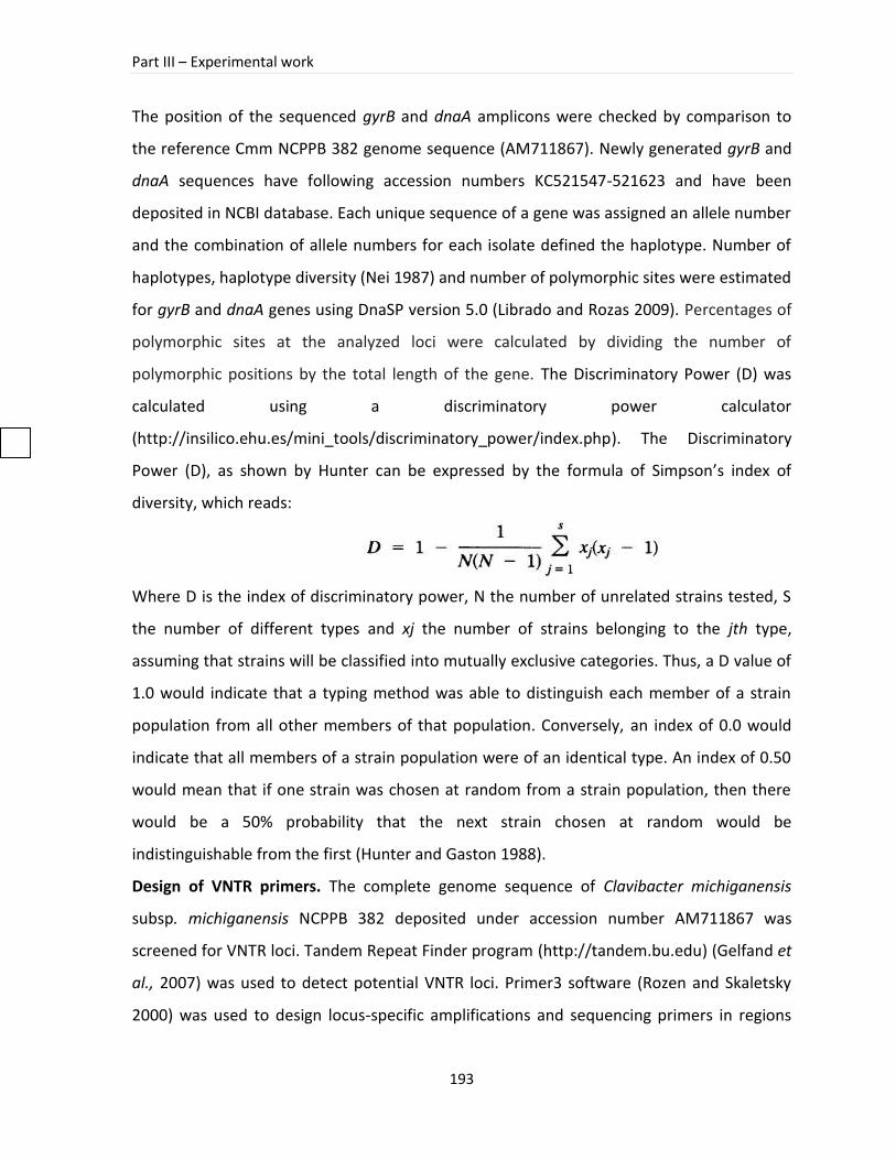
Part III – Experimental work
193
The position of the sequenced gyrB and dnaA amplicons were checked by comparison to
the reference Cmm NCPPB 382 genome sequence (AM711867). Newly generated gyrB and
dnaA sequences have following accession numbers KC521547-521623 and have been
deposited in NCBI database. Each unique sequence of a gene was assigned an allele number
and the combination of allele numbers for each isolate defined the haplotype. Number of
haplotypes, haplotype diversity (Nei 1987) and number of polymorphic sites were estimated
for gyrB and dnaA genes using DnaSP version 5.0 (Librado and Rozas 2009). Percentages of
polymorphic sites at the analyzed loci were calculated by dividing the number of
polymorphic positions by the total length of the gene. The Discriminatory Power (D) was
calculated using a discriminatory power calculator
(http://insilico.ehu.es/mini_tools/discriminatory_power/index.php). The Discriminatory
Power (D), as shown by Hunter can be expressed by the formula of Simpson’s index of
diversity, which reads:
Where D is the index of discriminatory power, N the number of unrelated strains tested, S
the number of different types and xj the number of strains belonging to the jth type,
assuming that strains will be classified into mutually exclusive categories. Thus, a D value of
1.0 would indicate that a typing method was able to distinguish each member of a strain
population from all other members of that population. Conversely, an index of 0.0 would
indicate that all members of a strain population were of an identical type. An index of 0.50
would mean that if one strain was chosen at random from a strain population, then there
would be a 50% probability that the next strain chosen at random would be
indistinguishable from the first (Hunter and Gaston 1988).
Design of VNTR primers. The complete genome sequence of Clavibacter michiganensis
subsp. michiganensis NCPPB 382 deposited under accession number AM711867 was
screened for VNTR loci. Tandem Repeat Finder program (http://tandem.bu.edu) (Gelfand et
al., 2007) was used to detect potential VNTR loci. Primer3 software (Rozen and Skaletsky
2000) was used to design locus-specific amplifications and sequencing primers in regions
Chapter 5 – MLVA analysis of Cmm strains isolated from recent outbreaks of bacterial wilt and canker in Belgium
194
flanking VNTR loci. Eight loci (Table 5.3) of 20 bp to 45 bp long tandem repeat (TR) units
were selected. TRs longer than 20 bp were chosen to enable easier interpretation of results
from an agarose gel. Primer pairs targeting single locus alleles were manually designed in
the conserved regions to obtain amplicons of no more than 450 bp in length.
VNTR PCR amplification and sequencing. The PCR mixture had a total volume of 25 µl,
containing 1 x PCR buffer (100 mM Tris-HCl, 15 mM MgCl2, 500 mM KCl [pH 8.3]) (Qiagen),
dNTP’s 0.2 mM each, 0.6 µM of each primer, 0.5 U DNA Taq polymerase and 50-60 ng
template DNA. The PCR amplifications were performed under following conditions: 3 min
denaturation step at 94˚ C; 35 cycles of 94˚ C for 1 min, annealing at 60˚ C for 1 min and
extention at 72˚ C for 1 min; and a final extension step at 72˚ C for 10 min. Amplified
products were run on a 2.5% Gel Pilot® Small Fragment Agarose (Qiagen) at 110 V for 2.5
hrs at 4˚C using 25 bp size marker (Invitrogen) and visualized by ethidium bromide staining.
PCR amplicons from one representative strain per different locus of a particular VNTR were
sequenced using sequencing primers (Table 5.2) according to the sequencing protocol
described above for gyrB and dnaA genes.
VNTR analysis and statistics. Product sizes were estimated and the exact number of repeats
present was calculated using a derived allele-naming table, based on the number of repeats
which could theoretically be present in a PCR product of a given size, allowing for extra
flanking nucleotides and primer size. Theoretical number of repeats was confirmed
subsequently by sequencing. Loci were named simply on the basis of the order in which
they were found by the initial search. VNTR allele calls were analyzed in BioNumerics as
‘character’ data. Composite datasets were created for the eight Clav-VNTR loci. Distance
trees were derived by clustering with the unweighted pair group method with arithmetic
means (UPGMA), using ‘categorical’ character table values. All markers were given equal
weight, irrespective of the number of repeats. The percentages in the dendrogram reflect
the percentage of homology between the specific markers. Relatedness between the
different haplotypes was investigated based on comparison of allelic profiles using the
minimum spanning tree (MS tree) method from BioNumerics v 5.1. We used the classical
criterium of one allelic mismatch to group haplotypes into clonal complexes. In order to
Part III – Experimental work
195
assess the evolutionary relatedness between haplotypes the MLVA data was analyzed
taking into account the number of repeat differences. The type strain LMG 7333T served as
a reference and a starting point for calculations of the differences in other strains. For each
VNTR locus the Hunter–Gaston and Simpson’s diversity indices were calculated using the
VNTR diversity and confidence extractor software (V-DICE) available at the Health
Protection Agency bioinformatics tools website (http://www.hpa-bioinformatics.org.uk/cgi-
bin/DICI/DICI.pl) (Simpson 1949). Shannon-Wiener index of diversity was calculated using
BioNumerics version 5.1.
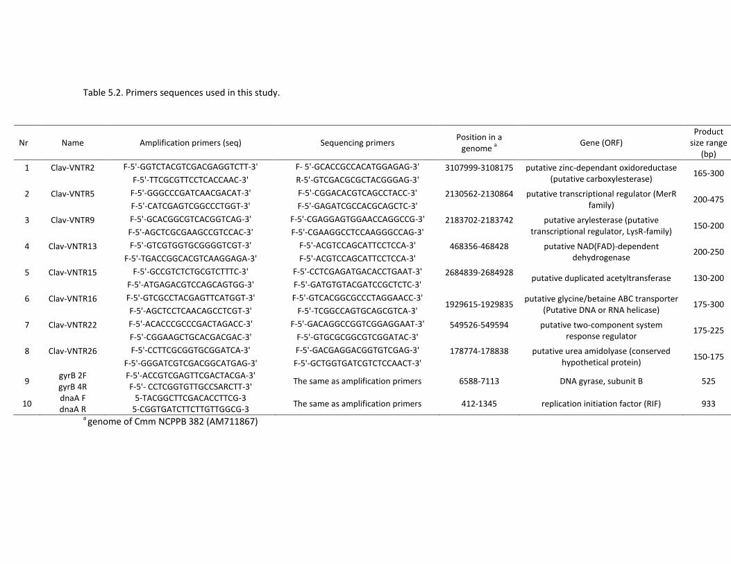
Table 5.2. Primers sequences used in this study.
Nr Name Amplification primers (seq) Sequencing primers Position in a
genome a
Gene (ORF) Product
size range (bp)
1
Clav-VNTR2
F-5'-GGTCTACGTCGACGAGGTCTT-3' F- 5'-GCACCGCCACATGGAGAG-3' 3107999-3108175
putative zinc-dependant oxidoreductase (putative carboxylesterase)
165-300 F-5'-TTCGCGTTCCTCACCAAC-3' R-5'-GTCGACGCGCTACGGGAG-3'
2
Clav-VNTR5
F-5'-GGGCCCGATCAACGACAT-3' F-5'-CGGACACGTCAGCCTACC-3' 2130562-2130864
putative transcriptional regulator (MerR family)
200-475 F-5'-CATCGAGTCGGCCCTGGT-3' F-5'-GAGATCGCCACGCAGCTC-3'
3
Clav-VNTR9
F-5'-GCACGGCGTCACGGTCAG-3' F-5'-CGAGGAGTGGAACCAGGCCG-3' 2183702-2183742
putative arylesterase (putative transcriptional regulator, LysR-family)
150-200 F-5'-AGCTCGCGAAGCCGTCCAC-3' F-5'-CGAAGGCCTCCAAGGGCCAG-3'
4
Clav-VNTR13
F-5'-GTCGTGGTGCGGGGTCGT-3' F-5'-ACGTCCAGCATTCCTCCA-3' 468356-468428
putative NAD(FAD)-dependent dehydrogenase
200-250 F-5'-TGACCGGCACGTCAAGGAGA-3' F-5'-ACGTCCAGCATTCCTCCA-3'
5
Clav-VNTR15
F-5'-GCCGTCTCTGCGTCTTTC-3' F-5'-CCTCGAGATGACACCTGAAT-3' 2684839-2684928
putative duplicated acetyltransferase 130-200 F-5'-ATGAGACGTCCAGCAGTGG-3' F-5'-GATGTGTACGATCCGCTCTC-3'
6
Clav-VNTR16
F-5'-GTCGCCTACGAGTTCATGGT-3' F-5'-GTCACGGCGCCCTAGGAACC-3' 1929615-1929835
putative glycine/betaine ABC transporter (Putative DNA or RNA helicase)
175-300 F-5'-AGCTCCTCAACAGCCTCGT-3' F-5'-TCGGCCAGTGCAGCGTCA-3'
7
Clav-VNTR22
F-5'-ACACCCGCCCGACTAGACC-3' F-5'-GACAGGCCGGTCGGAGGAAT-3' 549526-549594
putative two-component system response regulator
175-225 F-5'-CGGAAGCTGCACGACGAC-3' F-5'-GTGCGCGGCGTCGGATAC-3'
8
Clav-VNTR26
F-5'-CCTTCGCGGTGCGGATCA-3' F-5'-GACGAGGACGGTGTCGAG-3' 178774-178838
putative urea amidolyase (conserved hypothetical protein)
150-175 F-5'-GGGATCGTCGACGGCATGAG-3' F-5'-GCTGGTGATCGTCTCCAACT-3'
9 gyrB 2F gyrB 4R
F-5'-ACCGTCGAGTTCGACTACGA-3' F-5'- CCTCGGTGTTGCCSARCTT-3'
The same as amplification primers 6588-7113 DNA gyrase, subunit B 525
10 dnaA F dnaA R
5-TACGGCTTCGACACCTTCG-3 5-CGGTGATCTTCTTGTTGGCG-3
The same as amplification primers 412-1345 replication initiation factor (RIF) 933
a genome of Cmm NCPPB 382 (AM711867)
Part III – Experimental work
197
5.3 Results
Assessment of genetic diversity among Clavibacter strains
In total, 62 strains representing the Clavibacter subspecies and non-pathogenic Clavibacter strains were included in this study. The identity of included Cmm strains was confirmed by analysis of the gyrB and dnaA gene sequences. The gene sequence analyses were performed on several related Clavibacter strains in order to study the genetic diversity in the genus Clavibacter. Phylogenetic analysis of two tested genes confirmed a clear separation of Clavibacter subspecies and a distinct position of non-pathogenic Clavibacter strains. Phylogenetic relationship between the Clavibacter subspecies and non-pathogenic
Chapter 5 – MLVA analysis of Cmm strains isolated from recent outbreaks of bacterial wilt and canker in Belgium
198
Clavibacter strains was strongly supported by high bootstrap values
(
Figure 5.1). The number of polymorphic sites was 47 (10.7%) and 87 (12.9%), for gyrB and dnaA, respectively. It has to be noted that diversity among Cmm strains, especially among strains from recent Belgian outbreaks, was small which resulted in a limited number of
Part III – Experimental work
199
clusters. Despite a low genetic diversity, a number of groups could be distinguished in a
Cmm cluster (
Figure 5.1). The largest cluster, containing Belgian strains from recent outbreaks and two French strains from 2010 (GBBC 1077 and GBBC 1078), was separated from the Cmm strains
Chapter 5 – MLVA analysis of Cmm strains isolated from recent outbreaks of bacterial wilt and canker in Belgium
200
isolated previously in Belgium
(
Figure 5.1). Furthermore, strains originating from the same location mostly grouped together, such as French strains GBBC 1079, GBBC 1080 and PD 5719. However, based on the concatenated Maximum Likelihood tree of gyrB and dnaA no clear geographical separation among Cmm strains could be demonstrated. In gyrB and dnaA trees (data not
Part III – Experimental work
201
shown) and in a concatenated tree Clavibacter subspecies are separated from each other and from non-pathogenic strains, which suggests that these trees present the same phylogenetic information
(
Figure 5.1).
Chapter 5 – MLVA analysis of Cmm strains isolated from recent outbreaks of bacterial wilt and canker in Belgium
202
Development and implementation of MLVA
In parallel with the sequence analysis Cmm strains were investigated with MLVA. Fifty eight
VNTR loci were identified in the genome of Cmm NCPPB 382. Thirty one of them were
tested on a set of eight genetically diverse Cmm strains originating from geographically
spread locations (Table 5.1). Subsequently, eight loci that were successfully amplified and
showed to be polymorphic in the tested subset of strains were selected for further analysis.
Successful amplification was obtained in all tested Cmm strains. Regarding the non-
pathogenic seed-borne Clavibacter strains the results varied from no amplification for Clav-
VNTR5 or unspecific (more than one band, not expected product size) bands in Clav-VNTR26
(data not shown). Similar findings were observed for Clavibacter michiganensis subspecies
other than Cmm. In the cluster analysis, a total of 24 MLVA types were detected among 56
Cmm strains when the data from eight loci were combined, with allele numbers per locus
ranging from two (Clav-VNTR22, Clav-VNTR26) to six (Clav-VNTR5) (Table 5.3, Figure 5.2).
A large cluster, comprised of Cmm strains from recent Belgian outbreaks together with two
French strains isolated in 2010, exhibited identical MLVA haplotypes. Strains from other
countries formed mostly a separate branch or a cluster with two strains with an identical
MLVA haplotype. No direct connection between strains from recent Belgian outbreaks of
2010-2012 and other Belgian strains included in this study could be observed.
Remarkably, Belgian strains PD 5736 and GBBC 285, isolated in 1983 and 2008, respectively, showed the same MLVA haplotypes. In the concatenated tree of gyrB and dnaA these two
Part III – Experimental work
203
Belgian strains clustered together among strains originating from other countries
(
Figure 5.1). Similar findings were observed for other two Belgian strains PD 1953 and GBBC
283, isolated in 1984 and 2002, respectively.
Chapter 5 – MLVA analysis of Cmm strains isolated from recent outbreaks of bacterial wilt and canker in Belgium
204
The discriminatory abilities of the MLVA technique was determined by calculating the
discriminatory index (D) for 56 typed strains. MLVA differentiated 25 Cmm strains and
showed a level of discrimination, with a D value of 0.8006. The discriminatory power of
each VNTR was estimated by the number of alleles detected and the allele diversity. The
number of different alleles ranged from two for Cmm-V22 and Cmm-V26 to six for Cmm-V5.
Highest allelic diversities measured by Hunter–Gaston, Simpson’s and Shannon-Wiener
diversity indices were 0.664; 0.652; 1.3377, respectively and were observed for the loci
Clav-VNTR5 (Table 5.3). For the set under study, 27 different alleles of eight VNTR loci were
observed. The relationship among the strains based on MLVA results is presented in a
minimum spanning tree (MST).
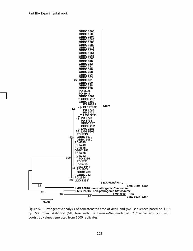
Part III – Experimental work
205
Figure 5.1. Phylogenetic analysis of concatenated tree of dnaA and gyrB sequences based on 1115 bp. Maximum Likelihood (ML) tree with the Tamura-Nei model of 62 Clavibacter strains with bootstrap values generated from 1000 replicates.
GBBC 1605 GBBC 1606 GBBC 1604 GBBC 1086 GBBC 1083 GBBC 1082 GBBC 1078 GBBC 1077 GBBC 1064 GBBC 1061 GBBC 1060 GBBC 316 GBBC 312 GBBC 311 GBBC 310 GBBC 308 GBBC 304 GBBC 303 GBBC 301 GBBC 300 GBBC 298 GBBC 296 PD 5699 PD 1948 GBBC 1609
GBBC 297 GBBC 1389
ES 2686.1 CL01TF02 PD 5737 PD 5734 LMG 3695
PD 5722 PD 5733 GBBC 247 GBBC 282 LMG 3681 LMG 5602
PD 5719 GBBC 1079 GBBC 1080
PD 4149 PD 5749 PD 4545 GBBC 285 PD 5736 PD 5753
PD 1386 PD 5721 PD 5751 LMG 5644 PD 1953 GBBC 283 GBBC 242
PD 1664 LMG 7333
T
Cmm
LMG 2889T Cms
LMG 7294T Cmt
LMG 26810 non-pathogenic Clavibacter LMG 26807 non-pathogenic Clavibacter
LMG 3663T Cmi
LMG 5627T
Cmn 98 78
92
52
83
64
100
40
42
14
26
16
42
0.005
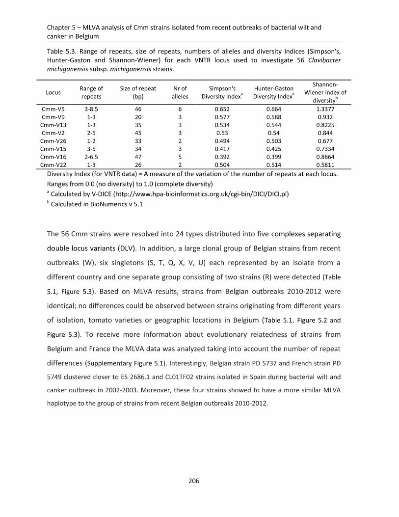
Chapter 5 – MLVA analysis of Cmm strains isolated from recent outbreaks of bacterial wilt and canker in Belgium
206
Table 5.3. Range of repeats, size of repeats, numbers of alleles and diversity indices (Simpson's, Hunter-Gaston and Shannon-Wiener) for each VNTR locus used to investigate 56 Clavibacter michiganensis subsp. michiganensis strains.
Locus Range of repeats
Size of repeat (bp)
Nr of alleles
Simpson's Diversity Index
a
Hunter-Gaston Diversity Index
a
Shannon-Wiener index of
diversityb
Cmm-V5 3-8.5 46 6 0.652 0.664 1.3377 Cmm-V9 1-3 20 3 0.577 0.588 0.932
Cmm-V13 1-3 35 3 0.534 0.544 0.8225 Cmm-V2 2-5 45 3 0.53 0.54 0.844
Cmm-V26 1-2 33 2 0.494 0.503 0.677 Cmm-V15 3-5 34 3 0.417 0.425 0.7334 Cmm-V16 2-6.5 47 5 0.392 0.399 0.8864 Cmm-V22 1-3 26 2 0.504 0.514 0.5811
Diversity Index (for VNTR data) = A measure of the variation of the number of repeats at each locus.
Ranges from 0.0 (no diversity) to 1.0 (complete diversity) a Calculated by V-DICE (http://www.hpa-bioinformatics.org.uk/cgi-bin/DICI/DICI.pl) b Calculated in BioNumerics v 5.1
The 56 Cmm strains were resolved into 24 types distributed into five complexes separating
double locus variants (DLV). In addition, a large clonal group of Belgian strains from recent
outbreaks (W), six singletons (S, T, Q, X, V, U) each represented by an isolate from a
different country and one separate group consisting of two strains (R) were detected (Table
5.1, Figure 5.3). Based on MLVA results, strains from Belgian outbreaks 2010-2012 were
identical; no differences could be observed between strains originating from different years
of isolation, tomato varieties or geographic locations in Belgium (Table 5.1, Figure 5.2 and
Figure 5.3). To receive more information about evolutionary relatedness of strains from
Belgium and France the MLVA data was analyzed taking into account the number of repeat
differences (Supplementary Figure 5.1). Interestingly, Belgian strain PD 5737 and French strain PD
5749 clustered closer to ES 2686.1 and CL01TF02 strains isolated in Spain during bacterial wilt and
canker outbreak in 2002-2003. Moreover, these four strains showed to have a more similar MLVA
haplotype to the group of strains from recent Belgian outbreaks 2010-2012.
Part III – Experimental work
207
5.4 Discussion
Over the last few decades, bacterial wilt and canker has been frequently detected in tomato
production areas, leading to substantial financial and economical losses. Only during the last
three years several local outbreaks of Cmm were reported in Belgium. In some cases,
reoccurring infections were detected in the primarily contaminated farms, suggesting a
persistence of an initial infection source. Despite a quite frequent detection of tomato
canker and wilting in Belgian tomato production areas there is little known about the
genetic diversity of Cmm strains, which hinders the correct conclusions about the probable
sources of epidemics and transmission routes of Cmm.
This study is the first MLVA approach developed for efficient genotyping of Cmm strains. To
date typing of Cmm strains was performed by RAPD-PCR (De León et al., 2009), BOX-PCR
(Kawaguchi et al., 2010) (Nazari et al., 2007), AFLP (De León et al., 2009), PFGE (Kleitman et
al., 2008) and MLST (Milijašević-Marčić et al., 2012). Despite the fact that some of these
methods were found to have a good resolution most of them have limitations, such as a
poor interlaboratory portability or limited exchangeability of results that were generated on
a specific machine or compared to an in-house database. Nowadays, fully sequenced
genomes give a unique opportunity for a development of more robust and accurate typing
methods such as MLVA. Its advantages including high reproducibility, exchangeability of
results and the possibility to add loci greatly facilitate epidemiological studies of
economically important pathogens such as Cmm.
In this work, Clav-VNTR5 showed to be the most polymorphic loci with six different alleles
and the highest HGDI of 0.664. Combined data from MLVA analysis of all eight investigated
loci resulted in 25 different haplotypes and a discriminatory power of 0.8006. Cmm strains
from the recent epidemics in Belgium in 2010-2012 showed identical MLVA haplotypes,
which suggests that a clonal population was responsible for the outbreaks. The presence of
the same MLVA haplotypes of Cmm strains from 2011 and 2012 could mean that bacteria
persisted in the used equipment, devices or soil and induced the outbreaks in the following
years. Population of Belgian strains isolated from 2010-2011 is epidemiologically related to
at least two French strains that exhibited the same MLVA haplotype. Moreover, based on
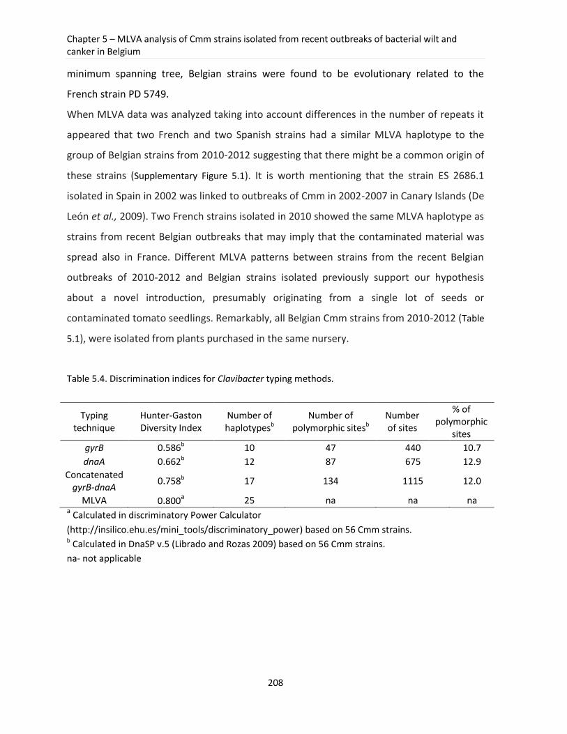
Chapter 5 – MLVA analysis of Cmm strains isolated from recent outbreaks of bacterial wilt and canker in Belgium
208
minimum spanning tree, Belgian strains were found to be evolutionary related to the
French strain PD 5749.
When MLVA data was analyzed taking into account differences in the number of repeats it
appeared that two French and two Spanish strains had a similar MLVA haplotype to the
group of Belgian strains from 2010-2012 suggesting that there might be a common origin of
these strains (Supplementary Figure 5.1). It is worth mentioning that the strain ES 2686.1
isolated in Spain in 2002 was linked to outbreaks of Cmm in 2002-2007 in Canary Islands (De
León et al., 2009). Two French strains isolated in 2010 showed the same MLVA haplotype as
strains from recent Belgian outbreaks that may imply that the contaminated material was
spread also in France. Different MLVA patterns between strains from the recent Belgian
outbreaks of 2010-2012 and Belgian strains isolated previously support our hypothesis
about a novel introduction, presumably originating from a single lot of seeds or
contaminated tomato seedlings. Remarkably, all Belgian Cmm strains from 2010-2012 (Table
5.1), were isolated from plants purchased in the same nursery.
Table 5.4. Discrimination indices for Clavibacter typing methods.
Typing technique
Hunter-Gaston Diversity Index
Number of haplotypesb
Number of polymorphic sitesb
Number of sites
% of polymorphic
sites
gyrB 0.586b 10 47 440 10.7
dnaA 0.662b 12 87 675 12.9
Concatenated gyrB-dnaA
0.758b 17 134 1115 12.0
MLVA 0.800a 25 na na na a Calculated in discriminatory Power Calculator
(http://insilico.ehu.es/mini_tools/discriminatory_power) based on 56 Cmm strains. b Calculated in DnaSP v.5 (Librado and Rozas 2009) based on 56 Cmm strains.
na- not applicable
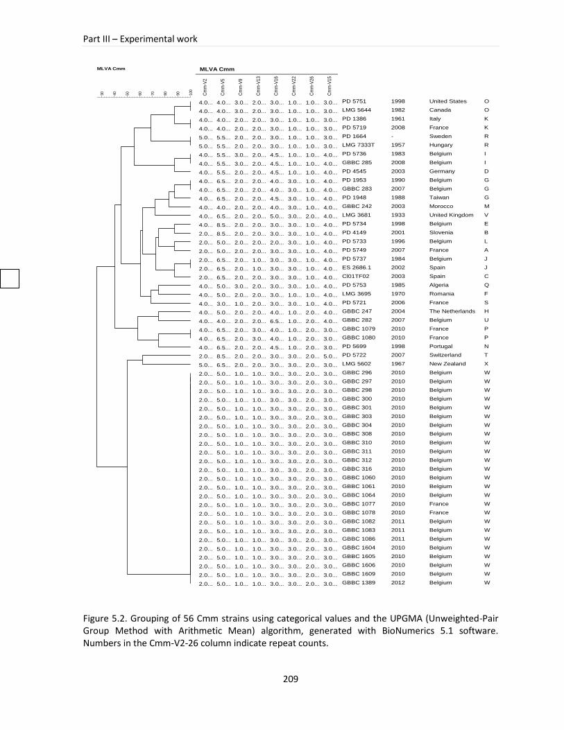
Part III – Experimental work
209
Figure 5.2. Grouping of 56 Cmm strains using categorical values and the UPGMA (Unweighted-Pair Group Method with Arithmetic Mean) algorithm, generated with BioNumerics 5.1 software. Numbers in the Cmm-V2-26 column indicate repeat counts.
Categorical
MLVA Cmm
10
0
90
80
70
60
50
40
30
MLVA Cmm
Cm
m-V
2
Cm
m-V
5
Cm
m-V
9
Cm
m-V
13
Cm
m-V
16
Cm
m-V
22
Cm
m-V
26
Cm
m-V
15
4.0... 4.0... 3.0... 2.0... 3.0... 1.0... 1.0... 3.0...
4.0... 4.0... 3.0... 2.0... 3.0... 1.0... 1.0... 3.0...
4.0... 4.0... 2.0... 2.0... 3.0... 1.0... 1.0... 3.0...
4.0... 4.0... 2.0... 2.0... 3.0... 1.0... 1.0... 3.0...
5.0... 5.5... 2.0... 2.0... 3.0... 1.0... 1.0... 3.0...
5.0... 5.5... 2.0... 2.0... 3.0... 1.0... 1.0... 3.0...
4.0... 5.5... 3.0... 2.0... 4.5... 1.0... 1.0... 4.0...
4.0... 5.5... 3.0... 2.0... 4.5... 1.0... 1.0... 4.0...
4.0... 5.5... 2.0... 2.0... 4.5... 1.0... 1.0... 4.0...
4.0... 6.5... 2.0... 2.0... 4.0... 3.0... 1.0... 4.0...
4.0... 6.5... 2.0... 2.0... 4.0... 3.0... 1.0... 4.0...
4.0... 6.5... 2.0... 2.0... 4.5... 3.0... 1.0... 4.0...
4.0... 4.0... 2.0... 2.0... 4.0... 3.0... 1.0... 4.0...
4.0... 6.5... 2.0... 2.0... 5.0... 3.0... 2.0... 4.0...
4.0... 8.5... 2.0... 2.0... 3.0... 3.0... 1.0... 4.0...
2.0... 8.5... 2.0... 2.0... 3.0... 3.0... 1.0... 4.0...
2.0... 5.0... 2.0... 2.0... 2.0... 3.0... 1.0... 4.0...
2.0... 5.0... 2.0... 2.0... 3.0... 3.0... 1.0... 4.0...
2.0... 6.5... 2.0... 1.0... 3.0... 3.0... 1.0... 4.0...
2.0... 6.5... 2.0... 1.0... 3.0... 3.0... 1.0... 4.0...
2.0... 6.5... 2.0... 2.0... 3.0... 3.0... 1.0... 4.0...
4.0... 5.0... 3.0... 2.0... 3.0... 3.0... 1.0... 4.0...
4.0... 5.0... 2.0... 2.0... 3.0... 1.0... 1.0... 4.0...
4.0... 3.0... 1.0... 2.0... 3.0... 3.0... 1.0... 4.0...
4.0... 5.0... 2.0... 2.0... 4.0... 1.0... 2.0... 4.0...
4.0... 4.0... 2.0... 2.0... 6.5... 1.0... 2.0... 4.0...
4.0... 6.5... 2.0... 3.0... 4.0... 1.0... 2.0... 3.0...
4.0... 6.5... 2.0... 3.0... 4.0... 1.0... 2.0... 3.0...
4.0... 6.5... 2.0... 2.0... 4.5... 1.0... 2.0... 3.0...
2.0... 8.5... 2.0... 2.0... 3.0... 3.0... 2.0... 5.0...
5.0... 6.5... 2.0... 2.0... 3.0... 3.0... 2.0... 3.0...
2.0... 5.0... 1.0... 1.0... 3.0... 3.0... 2.0... 3.0...
2.0... 5.0... 1.0... 1.0... 3.0... 3.0... 2.0... 3.0...
2.0... 5.0... 1.0... 1.0... 3.0... 3.0... 2.0... 3.0...
2.0... 5.0... 1.0... 1.0... 3.0... 3.0... 2.0... 3.0...
2.0... 5.0... 1.0... 1.0... 3.0... 3.0... 2.0... 3.0...
2.0... 5.0... 1.0... 1.0... 3.0... 3.0... 2.0... 3.0...
2.0... 5.0... 1.0... 1.0... 3.0... 3.0... 2.0... 3.0...
2.0... 5.0... 1.0... 1.0... 3.0... 3.0... 2.0... 3.0...
2.0... 5.0... 1.0... 1.0... 3.0... 3.0... 2.0... 3.0...
2.0... 5.0... 1.0... 1.0... 3.0... 3.0... 2.0... 3.0...
2.0... 5.0... 1.0... 1.0... 3.0... 3.0... 2.0... 3.0...
2.0... 5.0... 1.0... 1.0... 3.0... 3.0... 2.0... 3.0...
2.0... 5.0... 1.0... 1.0... 3.0... 3.0... 2.0... 3.0...
2.0... 5.0... 1.0... 1.0... 3.0... 3.0... 2.0... 3.0...
2.0... 5.0... 1.0... 1.0... 3.0... 3.0... 2.0... 3.0...
2.0... 5.0... 1.0... 1.0... 3.0... 3.0... 2.0... 3.0...
2.0... 5.0... 1.0... 1.0... 3.0... 3.0... 2.0... 3.0...
2.0... 5.0... 1.0... 1.0... 3.0... 3.0... 2.0... 3.0...
2.0... 5.0... 1.0... 1.0... 3.0... 3.0... 2.0... 3.0...
2.0... 5.0... 1.0... 1.0... 3.0... 3.0... 2.0... 3.0...
2.0... 5.0... 1.0... 1.0... 3.0... 3.0... 2.0... 3.0...
2.0... 5.0... 1.0... 1.0... 3.0... 3.0... 2.0... 3.0...
2.0... 5.0... 1.0... 1.0... 3.0... 3.0... 2.0... 3.0...
2.0... 5.0... 1.0... 1.0... 3.0... 3.0... 2.0... 3.0...
2.0... 5.0... 1.0... 1.0... 3.0... 3.0... 2.0... 3.0...
PD 5751
LMG 5644
PD 1386
PD 5719
PD 1664
LMG 7333T
PD 5736
GBBC 285
PD 4545
PD 1953
GBBC 283
PD 1948
GBBC 242
LMG 3681
PD 5734
PD 4149
PD 5733
PD 5749
PD 5737
ES 2686.1
Cl01TF02
PD 5753
LMG 3695
PD 5721
GBBC 247
GBBC 282
GBBC 1079
GBBC 1080
PD 5699
PD 5722
LMG 5602
GBBC 296
GBBC 297
GBBC 298
GBBC 300
GBBC 301
GBBC 303
GBBC 304
GBBC 308
GBBC 310
GBBC 311
GBBC 312
GBBC 316
GBBC 1060
GBBC 1061
GBBC 1064
GBBC 1077
GBBC 1078
GBBC 1082
GBBC 1083
GBBC 1086
GBBC 1604
GBBC 1605
GBBC 1606
GBBC 1609
GBBC 1389
1998
1982
1961
2008
-
1957
1983
2008
2003
1990
2007
1988
2003
1933
1998
2001
1996
2007
1984
2002
2003
1985
1970
2006
2004
2007
2010
2010
1998
2007
1967
2010
2010
2010
2010
2010
2010
2010
2010
2010
2010
2010
2010
2010
2010
2010
2010
2010
2011
2011
2011
2010
2010
2010
2010
2012
United States
Canada
Italy
France
Sweden
Hungary
Belgium
Belgium
Germany
Belgium
Belgium
Taiwan
Morocco
United Kingdom
Belgium
Slovenia
Belgium
France
Belgium
Spain
Spain
Algeria
Romania
France
The Netherlands
Belgium
France
France
Portugal
Switzerland
New Zealand
Belgium
Belgium
Belgium
Belgium
Belgium
Belgium
Belgium
Belgium
Belgium
Belgium
Belgium
Belgium
Belgium
Belgium
Belgium
France
France
Belgium
Belgium
Belgium
Belgium
Belgium
Belgium
Belgium
Belgium
O
O
K
K
R
R
I
I
D
G
G
G
M
V
E
B
L
A
J
J
C
Q
F
S
H
U
P
P
N
T
X
W
W
W
W
W
W
W
W
W
W
W
W
W
W
W
W
W
W
W
W
W
W
W
W
W
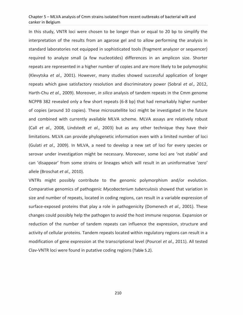
Chapter 5 – MLVA analysis of Cmm strains isolated from recent outbreaks of bacterial wilt and canker in Belgium
210
In this study, VNTR loci were chosen to be longer than or equal to 20 bp to simplify the
interpretation of the results from an agarose gel and to allow performing the analysis in
standard laboratories not equipped in sophisticated tools (fragment analyzer or sequencer)
required to analyze small (a few nucleotides) differences in an amplicon size. Shorter
repeats are represented in a higher number of copies and are more likely to be polymorphic
(Klevytska et al., 2001). However, many studies showed successful application of longer
repeats which gave satisfactory resolution and discriminatory power (Sobral et al., 2012,
Harth-Chu et al., 2009). Moreover, in silico analysis of tandem repeats in the Cmm genome
NCPPB 382 revealed only a few short repeats (6-8 bp) that had remarkably higher number
of copies (around 10 copies). These microsatellite loci might be investigated in the future
and combined with currently available MLVA scheme. MLVA assays are relatively robust
(Call et al., 2008, Lindstedt et al., 2003) but as any other technique they have their
limitations. MLVA can provide phylogenetic information even with a limited number of loci
(Gulati et al., 2009). In MLVA, a need to develop a new set of loci for every species or
serovar under investigation might be necessary. Moreover, some loci are ‘not stable’ and
can ‘disappear’ from some strains or lineages which will result in an uninformative ‘zero’
allele (Broschat et al., 2010).
VNTRs might possibly contribute to the genomic polymorphism and/or evolution.
Comparative genomics of pathogenic Mycobacterium tuberculosis showed that variation in
size and number of repeats, located in coding regions, can result in a variable expression of
surface-exposed proteins that play a role in pathogenicity (Domenech et al., 2001). These
changes could possibly help the pathogen to avoid the host immune response. Expansion or
reduction of the number of tandem repeats can influence the expression, structure and
activity of cellular proteins. Tandem repeats located within regulatory regions can result in a
modification of gene expression at the transcriptional level (Pourcel et al., 2011). All tested
Clav-VNTR loci were found in putative coding regions (Table 5.2).
Part III – Experimental work
211
Figure 5.3. Minimum spanning tree of 56 Cmm strains based on eight VNTR loci. Each circle represents an MLVA type with a size corresponding to the number of strains that share an identical MLVA type. MLVA types connected by a thick solid line differ from one another by one VNTR locus, while MLVA types connected by a thin solid line differ by two VNTR loci. MLVA types that differ from each other by three, four or more VNTR loci are connected by dashed and dotted lines. MLVA types were distinguished to define clonal complexes and to group in zones MLVA types that differ from one another by at most two locus variants. Letters visible on each circle are corresponding to strains described in Table 5.1. CC-Clonal complex.
Chapter 5 – MLVA analysis of Cmm strains isolated from recent outbreaks of bacterial wilt and canker in Belgium
212
At least two of them were found within genes linked to processes taking place in a cell
envelope (Clav-VNTR-13: putative NAD (FAD)-dependent dehydrogenase and Clav-VNTR 16:
putative glycine/betaine ABC transporter). We could speculate that variability observed
within these regions might possibly help bacteria to alternate the proteins of a cell
envelope. However, more research has to be performed on the role of tandem repeat copy,
number variation and virulence in Cmm.
The genetic structure of the studied strains was assessed by the sequence analysis of two
housekeeping genes, gyrB and dnaA, which were previously reported to be good molecular
markers for studying populations of the genus Clavibacter (Schneider et al., 2011, Zaluga et
al., 2011). The phylogenetic position of Cmm strains was supported by high bootstrap
values in a Maximum Likelihood tree. High similarity of Belgian strains from recent
outbreaks was detected both, in a gene sequence analysis and by an MLVA typing method,
supporting the hypothesis about their monomorphic nature. The percentages of
polymorphic sites observed for the concatenated set of gyrB and dnaA genes (Table 5.4) was
higher than the value obtained from five concatenated genes described in a recently
published MLSA scheme of Clavibacter michiganensis subsp. michiganensis, (12 versus 8.8)
(Jacques et al., 2012). Based on these parameters the genes selected in this work can be
applied in MLST studies to investigate highly similar Cmm populations.
In this study, MLVA was successfully applied to investigate a genetic relationship of Cmm
strains from recent Belgian outbreaks. Its discriminatory power, measured by HGDI, was
higher than these of each of the tested genes, gyrB and dnaA (Table 5.4). Our study has
shown that MLVA analysis offers better discrimination of Cmm strains (HGDI=0.8) than the
typing method based on the concatenated tree of gyrB and dnaA (HGDI=0.758) (Table 5.4). A
significant advantage of the MLVA method is the excellent interlaboratory reproducibility
(Kremer et al., 2005), which makes this method well-suited for accurate and reproducible
bacterial typing applicable in epidemiological studies of Clavibacter. MLVA, with its high
discriminatory power to separate closely related strains, might be very useful for tracking
sources of epidemic outbreaks as well as for investigating various haplotypes occurring
during these outbreaks, as illustrated in the differentiation of Cmm strains. The technique is
Part III – Experimental work
213
fast (results within one day), easy to perform, user-friendly, cost-effective and showed an
excellent reproducibility (intra- and interlaboratory) compared to other typing techniques
(e.g. AFLP). Additionally, data storage, comparison and exchange of the results are possible
and easy. Moreover, the use of fluorescence-labeled primers enables multiplex PCR and
subsequent analysis in a fragment analyzer. It is worth mentioning that the MLVA scheme,
derived from in silico analysis of a complete genome sequence of Cmm, was experimentally
confirmed to be accurate. It is consistent with previous findings demonstrated for
Xanthomonas citri pv. citri and is advantageous over other experimentally tested techniques
such as AFLP or IS-LM-PCR, where in vitro vs. in silico accuracy values of 75% and 87%,
respectively, were reported (Bui Thi Ngoc et al., 2009).
The MLVA method, with eight novel VNTR loci identified within the genome of Cmm,
demonstrated its applicability as a new tool for the molecular investigation of bacterial
wilting and canker outbreaks. In the future, additional VNTR loci and Clavibacter isolates
might enable unraveling intrapopulation genetic variation and assessing the robustness of
the method for investigating bacterial wilt and canker outbreaks on a global scale.
Competing interest
The other authors declare that they have no competing interests.
Acknowledgements
We thank the NVWA, GBBC and BCCM/LMG collections and Ana Rodríguez Pérez (Spain) for
providing necessary strains. This work was performed in the Seventh Framework
Programme of project KBBE-2008-1-4-01 (QBOL) nr 226482 funded by the European
Commission. Het Fonds Wetenschappelijk Onderzoek-Vlaanderen (FWO) is acknowledged
for the postdoctoral fellowship of Pieter Stragier and the Belgian NPPO (FAVV) for partially
financing ILVO-research. We thank Dr Kim Heylen for her critical reading and valuable
comments on the manuscript.
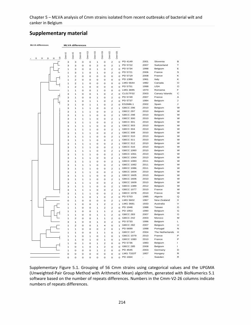
Chapter 5 – MLVA analysis of Cmm strains isolated from recent outbreaks of bacterial wilt and canker in Belgium
214
Supplementary material
Supplementary Figure 5.1. Grouping of 56 Cmm strains using categorical values and the UPGMA (Unweighted-Pair Group Method with Arithmetic Mean) algorithm, generated with BioNumerics 5.1 software based on the number of repeats differences. Numbers in the Cmm-V2-26 columns indicate numbers of repeats differences.
Pearson correlation
MLVA differences
100
806040200
MLVA differences
Cm
m-V
2
Cm
m-V
5
Cm
m-V
9
Cm
m-V
13
Cm
m-V
15
Cm
m-V
16
Cm
m-V
22
Cm
m-V
26
3 3 0 0 1 0 2 0
3 3 0 0 2 0 2 1
1 3 0 0 1 0 2 0
1 3 1 0 1 0 2 0
1 2 0 0 0 0 0 0
1 2 0 0 0 0 0 0
1 2 1 0 0 0 0 0
1 2 1 0 0 0 0 0
1 1 0 0 1 0 0 0
3 1 0 0 1 0 2 0
3 1 0 0 1 0 2 0
3 1 0 1 1 0 2 0
3 1 0 1 1 0 2 0
3 1 1 1 0 0 2 1
3 1 1 1 0 0 2 1
3 1 1 1 0 0 2 1
3 1 1 1 0 0 2 1
3 1 1 1 0 0 2 1
3 1 1 1 0 0 2 1
3 1 1 1 0 0 2 1
3 1 1 1 0 0 2 1
3 1 1 1 0 0 2 1
3 1 1 1 0 0 2 1
3 1 1 1 0 0 2 1
3 1 1 1 0 0 2 1
3 1 1 1 0 0 2 1
3 1 1 1 0 0 2 1
3 1 1 1 0 0 2 1
3 1 1 1 0 0 2 1
3 1 1 1 0 0 2 1
3 1 1 1 0 0 2 1
3 1 1 1 0 0 2 1
3 1 1 1 0 0 2 1
3 1 1 1 0 0 2 1
3 1 1 1 0 0 2 1
3 1 1 1 0 0 2 1
3 1 1 1 0 0 2 1
3 1 1 1 0 0 2 1
1 1 1 0 1 0 2 0
0 1 0 0 0 0 2 1
1 1 0 0 1 2 2 1
1 1 0 0 1 2 2 0
1 1 0 0 1 1 2 0
1 1 0 0 1 1 2 0
1 2 0 0 1 1 2 0
3 1 0 0 1 3 2 0
1 2 0 0 1 4 0 1
1 1 0 0 0 2 0 1
1 1 0 0 1 1 0 1
1 1 0 1 0 1 0 1
1 1 0 1 0 1 0 1
1 0 1 0 1 2 0 0
1 0 1 0 1 2 0 0
1 0 0 0 1 2 0 0
0 0 0 0 0 0 0 0
0 0 0 0 0 0 0 0
PD 4149
PD 5722
PD 5734
PD 5721
PD 5719
PD 1386
LMG 5644
PD 5751
LMG 3695
CL01TF02
PD 5749
PD 5737
ES2686.1
GBCC 296
GBCC 297
GBCC 298
GBCC 300
GBCC 301
GBCC 303
GBCC 304
GBCC 308
GBCC 310
GBCC 311
GBCC 312
GBCC 316
GBCC 1060
GBCC 1061
GBCC 1064
GBCC 1083
GBCC 1082
GBCC 1086
GBCC 1604
GBCC 1605
GBCC 1606
GBCC 1609
GBCC 1389
GBCC 1077
GBCC 1078
PD 5753
LMG 5602
LMG 3681
PD 1948
PD 1953
GBCC 283
GBCC 242
PD 5733
GBCC 282
PD 5699
GBCC 247
GBCC 1079
GBCC 1080
PD 5736
GBCC 285
PD 4545
LMG 7333T
PD 1664
2001
2007
1998
2006
2008
1961
1982
1998
1970
2003
2007
1984
2002
2010
2010
2010
2010
2010
2010
2010
2010
2010
2010
2010
2010
2010
2010
2010
2011
2011
2011
2010
2010
2010
2010
2012
2010
2010
1985
1967
1933
1988
1990
2007
2003
1996
2007
1998
2004
2010
2010
1983
2008
2003
1957
-
Slovenia
Switzerland
Belgium
France
France
Italy
Canada
USA
Romania
Canary Islands
France
Belgium
Spain
Belgium
Belgium
Belgium
Belgium
Belgium
Belgium
Belgium
Belgium
Belgium
Belgium
Belgium
Belgium
Belgium
Belgium
Belgium
Belgium
Belgium
Belgium
Belgium
Belgium
Belgium
Belgium
Belgium
France
France
Algerie
New Zealand
Australia
Taiwan
Belgium
Belgium
Moroco
Belgium
Belgium
Portugal
The Netherlands
France
France
Belgium
Belgium
Germany
Hungary
Sweden
B
T
E
S
K
K
O
O
F
C
A
J
J
W
W
W
W
W
W
W
W
W
W
W
W
W
W
W
W
W
W
W
W
W
W
W
W
W
Q
X
V
G
G
G
M
L
U
N
H
P
P
I
I
D
R
R
Part III – Experimental work
215
5.5 General reflections
In this chapter the development and successful application of a newly designed MLVA
scheme for studying Cmm diversity was presented. Cmm strains isolated from outbreaks of
bacterial wilt and canker reported in 2010, 2011 and 2012 in Belgium were typed to assess
their molecular epidemiology. The necessity to perform molecular typing of Cmm strains
involved in the epidemic outbreaks is crucial to study transmission routes, to localize
sources of infection and to monitor the spread of the disease. Although tomato diseases
caused by Cmm are responsible for substantial agricultural losses little is known about their
epidemiology. In Belgium, bacterial wilt and canker is present for at least a few decades but
there are almost no records describing genetic diversity of Cmm strains involved in disease
occurrences. As Belgium and the Netherlands are occupying a leading position in fresh-
tomato production in greenhouses in Europe (FAOSTAT, 2011), any disease on this crop will
have a great impact on the production and tomato yield. An epidemiological analysis,
aiming at investigating possible spread of Cmm strains and at determining the origin of
infections, could be initiated because a collection of Cmm strains with detailed information
concerning their isolation place, time and tomato variety was available.
Recent studies describing a genetic diversity of Cmm populations in different countries
employed various typing methods: BOX-PCR, PFGE, AFLP, MLST and ISSR (Baysal et al., 2010,
De León et al., 2009, Milijašević-Marčić et al., 2012). These methods differ in many aspects,
such as labour time, complexity in handling, user-friendliness and most important the
resolution. Their applicability and usefulness is dependent on the epidemiological questions
and the choice of the most suitable technique remains a crucial decision in epidemiological
studies. Since the genome of Cmm NCPPB 382 was available and MLVA demonstrated its
usefulness in epidemiological studies of many different bacterial pathogens (Sabat et al.,
2013) we developed a MLVA scheme that allowed for typing of Cmm strains from recent
Belgian outbreaks.
The in silico analysis of repeat sequences from Cmm genomes revealed many regions with
only two or three repeat copies, which are generally considered to be less polymorphic than
regions with higher number of repeats. Furthermore, after testing of more than thirty
Chapter 5 – MLVA analysis of Cmm strains isolated from recent outbreaks of bacterial wilt and canker in Belgium
216
selected VNTR regions with repeats longer than 20 bp only eight showed to be polymorphic
and could be actually included in the further work. Since our objective was to develop a
technique in which a simple agarose gel can be used to obtain the results we needed to
select repeats longer than 20 bp, which also could have an influence on the limited
polymorphism we observed in these loci. To improve the current MLVA scheme it would be
interesting to test some shorter patterns which show more repeat copies. Additional
information gained this way would probably increase the resolution of the current scheme.
Additional VNTR loci may reveal some differences among Cmm strains of the monomorphic
group that might be related for example to an isolation place or crop variety, which in turn
could help in a better understanding of the disease development pattern.
A great advantage of the MLVA technique is the possibility to generate easy to read and
informative codes that can be compared to other entries in the database in order to answer
the question about origin of a strain or relatedness to other strains. However, before such a
database for Cmm can be created more strains for which important epidemiological
information is available should be included and tested in order to assess the robustness of
the developed technique. The creation of a database of MLVA codes for Cmm would
facilitate the global comparison and epidemiological studies of Cmm strains involved in
outbreaks of bacterial wilt and canker. Despite a relatively low number of strains included in
our study we could detect connections between French and Belgian strains isolated from
2010 that indicates a common origin (contaminated seeds or seedlings) (Figure 5.2, Figure
5.3). Moreover, MLVA analysis demonstrated that Belgian strains involved in recent
outbreaks have probably a common origin with the Spanish strain ES 2686.1 that was
responsible for outbreaks in Canary Islands in 2002.
A very interesting aspect of the MLVA application for Cmm typing concerns the possibility of
multiplexing various loci and subsequent semi-automation of the procedure. It offers an
alternative for the gel-based approach in the laboratories where the necessary facilities,
such as a sequencer or a fragment analyzer, are available. Development of a multiplex
protocol will be advantageous in future studies of Cmm once the currently developed MLVA

Part III – Experimental work
217
scheme has proved its robustness. Additional MLVA typing data for Cmm strains will open
possibilities to investigate transmission routes and sources of contamination on a more
global scale.
Chapter 5 – MLVA analysis of Cmm strains isolated from recent outbreaks of bacterial wilt and canker in Belgium
218
5.6 References
1. Altschul SF, Gish W, Miller W, Myers EW, Lipman DJ. 1990. Basic local alignment search tool. Journal of Molecular Biology 215: 403-10
2. Baysal Ö, Mercati F, İkten H, YIldIz R, Carimi F, et al. 2010. Clavibacter michiganensis subsp. michiganensis: tracking strains using their genetic differentiations by ISSR markers in Southern Turkey. Physiological and Molecular Plant Pathology 75: 113-9
3. Bella P, Ialacci G, Licciardello G, La Rosa R, Catara V. 2012. Characterization of atypical Clavibacter michiganensis subsp. michiganensis populations in greenhouse tomatoes in Italy. Journal of Plant Pathology 94: 635-42
4. Bergsma-Vlami M, Martin W, Koenraadt H, Teunissen H, Pothier J, et al. 2012. Molecular typing of Dutch isolates of Xanthomonas arboricola pv. pruni isolated from ornamental cherry laurel. Journal of Plant Pathology 94: S1. 29-S1. 35
5. Bosch T, de Neeling A, Schouls L, Zwaluw K, Kluytmans J, et al. 2010. PFGE diversity within the methicillin-resistant Staphylococcus aureus clonal lineage ST398. BMC Microbiology 10: 40
6. Broschat S, Call D, Davis M, Meng D, Lockwood S, et al. 2010. Improved identification of epidemiologically related strains of Salmonella enterica by use of a fusion algorithm based on pulsed-field gel electrophoresis and multiple-locus variable-number tandem-repeat analysis. Journal of clinical microbiology 48: 4072-82
7. Bui Thi Ngoc L, Vernire C, Jarne P, Brisse S, Guerin F, et al. 2009. From local surveys to global surveillance: three high-throughput genotyping methods for epidemiological monitoring of Xanthomonas citri pv. citri pathotypes. Applied and Environmental Microbiology 75: 1173-84
8. Call DR, Orfe L, Davis MA, Lafrentz S, Kang M-S. 2008. Impact of compounding error on strategies for subtyping pathogenic bacteria. Foodborne pathogens and disease 5: 505-16
9. Cangelosi GA, Freeman RJ, Lewis KN, Livingston-Rosanoff D, Shah KS, et al. 2004. Evaluation of a high-throughput repetitive-sequence-based PCR system for DNA fingerprinting of Mycobacterium tuberculosis and Mycobacterium avium complex strains. Journal of clinical microbiology 42: 2685-93
10. Coletta-Filho HD, Takita MA, De Souza AA, Aguilar-Vildoso CI, Machado MA. 2001. Differentiation of strains of Xylella fastidiosa by a variable number of tandem repeat analysis. Applied and Environmental Microbiology 67: 4091-5
11. Danin-Poleg Y, Cohen LA, Gancz H, Broza YY, Goldshmidt H, et al. 2007. Vibrio cholerae strain typing and phylogeny study based on simple sequence repeats. Journal of clinical microbiology 45: 736-46
12. De León L, Rodriguez A, Llop P, Lopez MM, Siverio F. 2009. Comparative study of genetic diversity of Clavibacter michiganensis subsp. michiganensis isolates from the Canary Islands by RAPD-PCR, BOX-PCR and AFLP. Plant Pathology 58: 862-71
13. De León L, Siverio F, Lopez MM, Rodriguez A. 2011. Clavibacter michiganensis subsp. michiganensis, a seedborne tomato pathogen: healthy seeds are still the goal. Plant Disease 95: 1328-38
14. Domenech P, Barry 3rd C, Cole ST. 2001. Mycobacterium tuberculosis in the post-genomic age. Current Opinion in Microbiology 4: 28
15. EPPO. 2005. Clavibacter michiganensis subsp. michiganensis Diagnostics, PM7/42. Bulletin OEPP/EPPO 35: 273-83
16. Gelfand Y, Rodriguez A, Benson G. 2007. TRDB - the tandem repeats database. Nucleic Acids Research 35: D80-D7
17. Gironde S, Manceau C. 2012. Housekeeping Gene Sequencing and Multilocus Variable-Number Tandem-Repeat Analysis To Identify Subpopulations within Pseudomonas syringae pv. maculicola and Pseudomonas syringae pv. tomato That Correlate with Host Specificity. Applied and Environmental Microbiology 78: 3266-79
18. Gulati P, Varshney R, Virdi J. 2009. Multilocus variable number tandem repeat analysis as a tool to discern genetic relationships among strains of Yersinia enterocolitica biovar 1A. Journal of Applied Microbiology 107: 875-84
Part III – Experimental work
219
19. Harth-Chu E, Espejo RT, Christen R, Guzman CA, Hofle MG. 2009. Multiple-locus variable-number tandem-repeat analysis for clonal identification of Vibrio parahaemolyticus isolates by using capillary electrophoresis. Applied and Environmental Microbiology 75: 4079-88
20. Heymans R, Schouls LM, van der Heide HG, Schim van der Loeff MF, Bruisten SM. 2011. Multiple-locus variable-number tandem repeat analysis of Neisseria gonorrhoeae. Journal of clinical microbiology 49: 354-63
21. Hidalgo Ă, Carvajal A, La T, Naharro G, Rubio P, et al. 2010. Multiple-locus variable-number tandem-repeat analysis of the swine dysentery pathogen, Brachyspira hyodysenteriae. Journal of clinical microbiology 48: 2859-65
22. Hunter PR, Gaston MA. 1988. Numerical index of the discriminatory ability of typing systems: an application of Simpson's index of diversity. Journal of clinical microbiology 26: 2465-6
23. Jacques MA, Durand K, Orgeur G, Balidas S, Fricot C, et al. 2012. Phylogenetic analysis and polyphasic characterization of Clavibacter michiganensis strains isolated from tomato seeds reveal that non-pathogenic strains are distinct from C. michiganensis subsp. michiganensis. Applied and Environmental Microbiology 78: 8388-402
24. Jahr H, Bahro R, Burger A, Ahlemeyer J, Eichenlaub R. 1999. Interactions between Clavibacter michiganensis and its host plants. Environmental Microbiology 1: 113-8
25. Jansing H, Rudolph K. 1998. Physiological capabilities of Clavibacter michiganensis subsp. sepedonicus and development of a semi-selective medium. Zeitschrift Fur Pflanzenkrankheiten Und Pflanzenschutz-Journal of Plant Diseases and Protection 105: 590-601
26. Kawaguchi A, Tanina K, Inoue K. 2010. Molecular typing and spread of Clavibacter michiganensis subsp. michiganensis in greenhouses in Japan. Plant Pathology 59: 76-83
27. Kleitman F, Barash I, Burger A, Iraki N, Falah Y, et al. 2008. Characterization of a Clavibacter michiganensis subsp. michiganensis population in Israel. European Journal of Plant Pathology 121: 463-75
28. Klevytska AM, Price LB, Schupp JM, Worsham PL, Wong J, Keim P. 2001. Identification and characterization of variable-number tandem repeats in the Yersinia pestis genome. Journal of clinical microbiology 39: 3179-85
29. Kremer K, Arnold C, Cataldi A, Gutierrez MC, Haas WH, et al. 2005. Discriminatory power and reproducibility of novel DNA typing methods for Mycobacterium tuberculosis complex strains. Journal of clinical microbiology 43: 5628-38
30. Lamichhane JR, Balestra GM, Varvaro L. 2011. Severe Outbreak of Bacterial Canker Caused by Clavibacter michiganensis subsp. michiganensis on Tomato in Central Italy. Plant Disease 95: 221
31. Le Fleche P, Hauck Y, Onteniente L, Prieur A, Denoeud F, et al. 2001. A tandem repeats database for bacterial genomes: application to the genotyping of Yersinia pestis and Bacillus anthracis. BMC Microbiology 1: 2
32. Li Y, Cui Y, Hauck Y, Platonov ME, Dai E, et al. 2009. Genotyping and phylogenetic analysis of Yersinia pestis by MLVA: insights into the worldwide expansion of Central Asia plague foci. PLoS ONE 4: e6000
33. Librado P, Rozas J. 2009. DnaSP v5: a software for comprehensive analysis of DNA polymorphism data. Bioinformatics 25: 1451-2
34. Lindstedt BA, Heir E, Gjernes E, Vardund T, Kapperud G. 2003. DNA fingerprinting of Shiga-toxin producing Escherichia coli O157 based on multiple-locus variable-number tandem-repeats analysis (MLVA). Annals of clinical microbiology and antimicrobials 2: 12
35. Marsh JW, O'Leary MM, Shutt KA, Pasculle AW, Johnson S, et al. 2006. Multilocus variable-number tandem-repeat analysis for investigation of Clostridium difficile transmission in hospitals. Journal of clinical microbiology 44: 2558-66
36. Milijašević-Marčić S, Gartemann KH, Frohwitter J, Eichenlaub R, Todorović B, et al. 2012. Characterization of Clavibacter michiganensis subsp. michiganensis strains from recent outbreaks of bacterial wilt and canker in Serbia. European Journal of Plant Pathology 134: 697-711
37. Naktuinbouw. 2011. Method for the detection of Clavibacter michiganensis subsp. michiganensis on tomato seeds. Version 4
Chapter 5 – MLVA analysis of Cmm strains isolated from recent outbreaks of bacterial wilt and canker in Belgium
220
38. Nazari F, Niknam GR, Ghasemi A, Taghavi SM, Momeni H, Torabi S. 2007. An investigation on strains of Clavibacter michiganensis subsp. michiganensis in north and north west of Iran. Journal of Phytopathology 155: 563-9
39. Nei M. 1987. Molecular evolutionary genetics: Columbia University Press 40. Pitcher D, Saunders N, Owen R. 1989. Rapid extraction of bacterial genomic DNA with guanidium
thiocyanate. Letters in Applied Microbiology 8: 151-6 41. Pourcel C, Minandri F, Hauck Y, D'Arezzo S, Imperi F, et al. 2011. Identification of variable-number
tandem-repeat (VNTR) sequences in Acinetobacter baumannii and interlaboratory validation of an optimized multiple-locus VNTR analysis typing scheme. Journal of clinical microbiology 49: 539-48
42. Rozen S, Skaletsky H. 2000. Primer3 on the WWW for general users and for biologist programmers. Methods in Molecular Biology 132: 365-86
43. Sabat A, Budimir A, Nashev D, Sa-Leao R, van Dijl J, et al. 2013. Overview of molecular typing methods for outbreak detection and epidemiological surveillance. Euro Surveill 18
44. Sahin F, Uslu H, Kotan R, Donmez M. 2002. Bacterial canker, caused by Clavibacter michiganensis subsp. michiganensis, on tomatoes in eastern Anatolia region of Turkey. Plant Pathology 51: 399-
45. Schneider KL, Marrero G, Alvarez AM, Presting GG. 2011. Classification of plant associated bacteria using RIF, a computationally derived DNA marker. PLoS ONE 6: e18496
46. Simpson EH. 1949. Measurement of diversity. Nature; Nature 47. Skuce RA, McCorry TP, McCarroll JF, Roring SMM, Scott AN, et al. 2002. Discrimination of
Mycobacterium tuberculosis complex bacteria using novel VNTR-PCR targets. Microbiology 148: 519-28
48. Sobral D, Schwarz S, Bergonier D, Brisabois A, Feßler AT, et al. 2012. High Throughput Multiple Locus Variable Number of Tandem Repeat Analysis (MLVA) of Staphylococcus aureus from Human, Animal and Food Sources. PLoS ONE 7: e33967
49. Strider DL. 1969. Bacterial canker of tomato caused by Corynebacterium michiganense: a literature review and bibliography: Technical Bulletin North Carolina Agricultural Experiment Station, No. 193
50. Tamura K, Dudley J, Nei M, Kumar S. 2007. MEGA4: Molecular evolutionary genetics analysis (MEGA) software version 4.0. Molecular Biology and Evolution 24: 1596-9
51. Tamura K, Nei M. 1993. Estimation of the number of nucleotide substitutions in the control region of mitochondrial DNA in humans and chimpanzees. Molecular Biology and Evolution 10: 512-26
52. Thompson J, Higgins D, Gibson T. 1994. CLUSTAL W: improving the sensitivity of progressive multiple sequence alignment through sequence weighting, position-specific gap penalties and weight matrix choice. Nucleic Acids Research 22: 4673
53. Van Belkum A. 2006. Tracing isolates of bacterial species by multilocus variable number of tandem repeat analysis (MLVA). FEMS Immunology and Medical Microbiology 49: 22-7
54. Van Belkum A, Scherer S, Van Alphen L, Verbrugh H. 1998. Short-sequence DNA repeats in prokaryotic genomes. Microbiology and Molecular Biology Reviews 62: 275
55. van Cuyck H, Pichon B, Leroy P, Granger-Farbos A, Underwood A, et al. 2012. Multiple-locus variable-number tandem-repeat analysis of Streptococcus pneumoniae and comparison with multiple loci sequence typing. BMC Microbiology 12: 241
56. Waleron M, Waleron K, Kamasa J, Przewodowski W, Lojkowska E. 2011. Polymorphism analysis of housekeeping genes for identification and differentiation of Clavibacter michiganensis subspecies. European Journal of Plant Pathology 131: 341-54
57. Wang DY, Hadj-Henni L, Thierry S, Arna P, Chermette R, et al. 2012. Simple and Highly Discriminatory VNTR-Based Multiplex PCR for Tracing Sources of Aspergillus flavus Isolates. PLoS ONE 7: e44204
58. Zaluga J, Heylen K, Van Hoorde K, Hoste B, Van Vaerenbergh J, et al. 2011. GyrB sequence analysis and MALDI-TOF MS as identification tools for plant pathogenic Clavibacter. Systematic and Applied Microbiology 34: 400-7
59. Zhao WJ, Chen HY, Zhu SF, Xia MX, Tan TW. 2007. One-step detection of Clavibacter michiganensis subsp. michiganensis in symptomless tomato seeds using a Taqman probe. Journal of Plant Pathology 89: 349-51
221
Part IV
Concluding remarks
Part IV – Concluding remarks
222
Concluding remarks
Development of accurate and specific identification and typing approaches for plant
pathogens is crucial for protection and management of susceptible crops. Nowadays, this
issue is especially important because of the growing transport and exchange of plants and
plant-related material between countries. As a consequence of these increased trades, the
introduction of a contaminated material poses a real threat. Therefore, correct and reliable
detection and identification methods included in the preventive and curative measures are
important parts of strategies for controlling plant disease.
Application of more specific and accurate techniques for identification and typing of plant
pathogens is facilitated when underpinning knowledge about taxonomy, biology and
genetics of the pathogen is available. Great focus is therefore placed on molecular
characterization of plant pathogens and the genetic basis of their host-pathogen
interaction. As important quarantine pathogens of agricultural crops, Clavibacter strains,
especially belonging to Cms and Cmm, were subjects of many studies over the last few
decades. The results obtained so far shed some light on their molecular mechanisms of
pathogenicity and diversity. However, various aspects such as population structure, origin
and spread of strains involved in disease outbreaks, genes determining adaptation to a host
and diversity of non-pathogenic populations of Clavibacter members present in nature,
remain still unknown. Further investigation is required in order to better understand their
infection mode, the diversity forces behind their life style and molecular factors
determining their interaction with host plants.
This research project led to the development of new and more reliable identification
methods for plant pathogenic Clavibacter members including sequence-based approaches
with DNA barcode and a protein-based strategy using MALDI-TOF MS. The techniques were
tested on a set of more than 170 strains and proved to be accurate for identification of
Clavibacter strains at the subspecies level. Both techniques also showed the presence of a
new taxonomically distant group of non-pathogenic Clavibacter strains. Further
investigation of members of this interesting group revealed that these strains can be
Part IV – Concluding remarks
223
misidentified as Cmm with the currently applied identification protocols, which complicates
the clear judgment of Cmm presence on tested tomato seed lots. To explore the molecular
basis that determine differences between pathogenic and non-pathogenic Clavibacter
strains originating from tomato seed, a draft genome of a representative non-pathogenic
strain was determined. The genome analysis revealed that because of i) the lack of a
majority of known and putative virulence factors, ii) a limited number of plant cell wall
degrading enzymes and iii) a lower number of extracellular serine proteases this strain is
probably not able to successfully colonize the vascular tissues of tomato and to degrade
plant cells. This in consequence, significantly lowers its survival inside the nutrient-poor
xylem sap. The draft genome sequence of this Clavibacter strain was a first step towards
providing some important explanations for its non-pathogenic nature. In the future,
Clavibacter genomes will serve as a framework for determining the origin of evolution of
metabolic traits that contribute to bacterial fitness and the adaptations to a certain life
style. A more in-depth comparative analysis with other Clavibacter genomes will allow
generating more knowledge about underlying biology of these bacteria and enabling the
selection of group-specific regions that will serve as targets for development of reliable
identification primers for novel control strategies. Comparative genomics of pathogenic and
non-pathogenic Clavibacter strains opened new opportunities to identify genes important
for plant invasion and symptoms development and to confirm the virulence status of
currently putative determinants.
The avaiablity of fully sequenced and draft genomes of Clavibacter open an exicting
possibilities to study the function of particular genes using the currently available
mutagenesis and transformation systems developed for Clavibacter. For a long time these
approaches were not feasible for Gram-positive bacteria but recent years brought a real
breakthrough that enables detailed studies of gene function. Plasmid transformation
systems in Clavibacter (using naturally occurring plasmids in Clavibacter, pCM1 and pCM2)
were developed and more efficient electroporation protocols are now available and await
further applications. Another, highly efficient transformation system using transposon
mutagenesis with Tn1409 (based on IS1409 from an Arthrobacter sp.) was also introduced
Part IV – Concluding remarks
224
and poved to be very useful for studying the function of some of putative virulence factors.
These approaches will definitely help to confirm or reject the possible role in pathogenesis
of many putative virulence factors described so far in Clavibacter.
In the frame of this project a new MLVA typing method that can be used in epidemiological
studies of disease outbreaks caused by Cmm was delivered. A newly developed scheme
including eight VNTR regions demonstrated its utility to study Cmm strains involved in
recent outbreaks of bacterial wilt and canker on tomato in Belgium. The results provided
strong indications for a probable source of infection linked to contaminated tomato seeds
or seedlings. MLVA identified a monomorphic group of Cmm strains from recent Belgian
outbreaks and two French isolates. This molecular typing method proved to be useful for
molecular investigation of Cmm strains on a local scale. Future epidemiological
investigations of prevalent MLVA types will help to reveal the origins and transmission
routes of Cmm strains responsible for disease outbreaks.
Finally, this work represents a comprehensive study in which populations of Clavibacter
strains including Cmm and a newly identified group of non-pathogenic tomato seed-borne
strains are described in detail. The results from this study provided more insights in the
genetic diversity of Clavibacter and molecular differences between pathogenic and non-
pathogenic clavibacters. Furthermore, newly developed identification approaches based on
DNA barcodes and MALDI-TOF MS profiles will facilitate more reliable and accurate
Clavibacter identification at the species and subspecies level. MLVA schemes will enable
molecular typing that supports epidemiological investigation of outbreaks of bacterial wilt
and canker.
To conclude, I believe that the recent advances in Clavibacter genomics and proteomics will
lead to the discovery of new targets for pathogen control and to a better understanding of
pathogenesis of these bacteria that in the future will help to improve prevention and
treatment strategies.
225
CURRICULUM VITAE
PERSONAL INFORMATION
Name Date of birth Address Phone e-mail
Joanna Zaluga 12.12.1985 Sint-Lievenspoortstraat 30, 9000 Ghent, Belgium +32476047894 [email protected]
EDUCATION
2009-present
Doctoral degree in Science (Biochemistry and Microbiology), Ghent University, Ghent, Belgium Thesis: “Diagnostics for quarantine plant pathogenic Clavibacter and relatives: molecular characterization, identification and epidemiological studies”, Promoters: prof. Dr Paul De Vos, Prof. Dr Martine Maes
2004-2009 Master Degree, Warsaw University of Life Sciences, Warsaw, Poland Interfaculty Studies of Biotechnology, specialization: food biotechnology, Thesis: “Isolation and characterization of tetracycline and erythromycin resistant bacteria from artisan Gouda-type cheeses”
2000-2004 Jozef Chelmonski High School, Lowicz, Poland, Biological Sciences
WORK EXPERIENCE
February- June 2008
Erasmus project at the Laboratory of Microbiology, Ghent University, Ghent, Belgium Participation in the project aiming at identification and characterization of antibiotic resistant fraction of cheese microflora.
August- September 2007
Summer internship in Cardinal Stefan Wyszynski Institute of Cardiology, Warsaw, Poland Participation in the laboratory research concerning the detection of mutations responsible for heart diseases (main focus on patients having phaeochromosytoma and von Hippel-Lindau disease)
TRAININGS
Photoshop course Microbial genomes (including genomes annotation and analysis) Conference Skills Advanced Academic Writing Skills Personal Effectiveness
Syntra, Midden Vlaanderen, 2013 Doctoral School of Ghent University, 2012 Doctoral School of Ghent University, 2011 Doctoral School of Ghent University, 2010 Doctoral School of Ghent University, 2010
LANGUAGES
Polish mother tongue English fluent Dutch fluent
226
SCIENTIFIC OUTPUT
Oral presentations:
Zaluga J., Heylen K., Maes M., De Vos P. “Polyphasic characterization of non-pathogenic Clavibacter strains from tomato seeds”, QBOL-EPPO Conference on DNA Barcoding and diagnostic methods for plants pests, Haarlem, The Netherlands, 21-25 May 2012 Zaluga J., Heylen K., Maes M., De Vos P. “New identification approaches for plant pathogenic Clavibacter”, Sixth QBOL meeting, Ghent, Belgium, 22-23 September 2011 Zaluga J., Heylen K., Maes M., De Vos P. “GyrB barcode for identification of plant pathogenic Clavibacter”, Fourth QBOL meeting, Bologna, Italy, 20-22 October 2010 Zaluga J., Heylen K., Maes M., De Vos P. “Barcoding as a diagnostic tool for plant pathogenic Clavibacter”, European Consortium For the Barcode of Life (ECBOL), 2-nd meeting, Braga, Portugal, 2-6 June 2010 Zaluga J., Heylen K., Maes M., De Vos P. ”Diagnostics for plant quarantine: barcoding Clavibacter”, Third QBOL meeting, York, England, 6-7 May 2010 Poster presentations:
Zaluga J., Heylen K., Maes M., De Vos P. “Polyphasic characterization and diversity studies within a group of plant
pathogenic Clavibacter”, 14th International Symposium on Microbial Ecology (ISME) Copenhagen, Denmark, 19-24 August 2012.
Zaluga J., Heylen K., Maes M., De Vos P. “Diagnostics for plant quarantine Clavibacter” 17th Annual Symposium of the Belgian Society for Microbiology, Brussel, Belgium, 16-17 November 2011
Zaluga J., Heylen K., Maes M., De Vos P. “Taxonomic study and identification of quarantine plant pathogenic Clavibacter “, The 4th Congress of European Microbiologists FEMS, Geneva, Switzerland, 26-30 June 2011
Zaluga J., Heylen K., Maes M., De Vos P. “Phylogenetic study and identification of plant pathogenic Clavibacter”, 64rd International Symposium on Crop Protection, Ghent, Belgium, 24 May 2011 Zaluga J., Heylen K., Maes M., De Vos P. “New approaches in identification of quarantine Clavibacter strains”, Spring Symposium NVvM/NvMM, Arnhem, The Netherlands, 18-20 April 2011
Zaluga J., Heylen K., Maes M., De Vos P. “New identification tools for plant pathogenic Clavibacter”, Annual meeting of Belgium Society for Microbiology, Brussels, Belgium, 19 November 2010
227
Publications:
A1 publications Zaluga J, Stragier P, Van Vaerenbergh J, Maes M, De Vos P. “Comparative genome analysis of pathogenic and non-pathogenic Clavibacter strain reveals adaptations to their life styles”, in preparation for submission to BMC Genomics
Zaluga J, Stragier P, Van Vaerenbergh J, Maes M, De Vos P. 2013. “Multilocus Variable-Number-Tandem-Repeats Analysis (MLVA) distinguishes a clonal complex of Clavibacter michiganensis subsp. michiganensis strains isolated from recent outbreaks of bacterial wilt and canker in Belgium”. BMC Microbiology 13: 126 Zaluga J, Van Vaerenbergh J, Stragier P, Maes M, De Vos P. 2013. ”Genetic diversity of non-pathogenic Clavibacter strains isolated from tomato seeds”. Systematic and Applied Microbiology 36, 426-435 Zaluga J, Heylen K, Van Hoorde K, Hoste B, Van Vaerenbergh J et al., 2011. ”GyrB sequence analysis and MALDI-TOF MS as identification tools for plant pathogenic Clavibacter”. Systematic and Applied Microbiology 34, 400-407 Application note
Steve Baeyen, Joanna Zaluga, Paul De Vos, Martine Maes, 2013. ”ISSR-PCR fingerprinting of plant pathogen strains using the QIAxcel® system and BioNumerics®”, Qiagen SCIENTIFIC ACTIVITIES
Supervision of Erasmus students projects: Margit Rid (September 2012- January 2013) Supervision of Master projects: Annelies Tourny (2011-2012), Merel Vansevenant (2011-2012), Iebe Rossey (2010-2011), Karen Dendoncker (2010-2011), Dorien Van de Wouwer (2009-2010). Assistance in practical course of microbiology (2010-2012)