catalytic conversion of cellulose into...
TRANSCRIPT
CATALYTIC CONVERSION OF CELLULOSE INTO
NANOCELLULOSE IN IONIC LIQUID
TAN XIAO YUN
INSTITUTE OF GRADUATE STUDIES
UNIVERSITY OF MALAYA
KUALA LUMPUR
2016
CATALYTIC CONVERSION OF CELLULOSE INTO
NANOCELLULOSE IN IONIC LIQUID
TAN XIAO YUN
DISSERTATION SUBMITTED IN FULFILMENT OF
THE REQUIREMENTS FOR THE DEGREE OF MASTER
OF PHILOSOPHY
INSTITUTE OF GRADUATE STUDIES
UNIVERSITY OF MALAYA
KUALA LUMPUR
2016
ii
UNIVERSITY OF MALAYA
ORIGINAL LITERARY WORK DECLARATION
Name of Candidate: TAN XIAO YUN (I.C/Passport No: 900608065720)
Matric No: HGA 130017
Name of Degree: Master of Philosophy
Title of Project Paper/Research Report/Dissertation/Thesis (“this Work”):
Catalytic Conversion of Cellulose into Nanocellulose in Ionic Liquid
Field of Study: Chemistry (Catalysis)
I do solemnly and sincerely declare that:
(1) I am the sole author/writer of this Work;
(2) This Work is original;
(3) Any use of any work in which copyright exists was done by way of fair dealing
and for permitted purposes and any excerpt or extract from, or reference to or
reproduction of any copyright work has been disclosed expressly and
sufficiently and the title of the Work and its authorship have been
acknowledged in this Work;
(4) I do not have any actual knowledge nor do I ought reasonably to know that the
making of this work constitutes an infringement of any copyright work;
(5) I hereby assign all and every rights in the copyright to this Work to the
University of Malaya (“UM”), who henceforth shall be owner of the copyright
in this Work and that any reproduction or use in any form or by any means
whatsoever is prohibited without the written consent of UM having been first
had and obtained;
(6) I am fully aware that if in the course of making this Work I have infringed any
copyright whether intentionally or otherwise, I may be subject to legal action
or any other action as may be determined by UM.
Candidate’s Signature Date:
Subscribed and solemnly declared before,
Witness’s Signature Date:
Name:
Designation:
iii
ABSTRACT
Nanocellulose have emerged as a promising material and have attracted considerable
attention owing to their promising properties such as high surface area, high strength and
stiffness as well as biodegradability. In the present study, 1-butyl-3-methylimidazolium
hydrogen sulfate (BmimHSO4) and 1-butyl-3-methylimidazolium acetate (BmimOAc)
ionic liquids were utilized to function as catalyst and solvent to convert cellulose into
nanocellulose. Despite having the same cations, BmimHSO4 with higher acidity is
suggested to induce hydrolytic cleavage of glycosidic bonds. On contrary, higher
electronegativity of BmimOAc much prone to cause solvolysis of cellulose.
Comprehensive investigations on different synthesis parameters including reaction
temperature, time, concentration (mass loading of MCC) and sonication treatments were
conducted in order to control the specific architecture and properties of nanocellulose.
Improved crystallinity of nanocellulose with the preservation of crystalline cellulose I
structure has been successfully acquired with acidic BmimHSO4 through hydrolysis.
Hydrolytic reaction of BmimHSO4 performed at a temperature of 90 ºC for 1.5 hours has
contributed to the formation of highly crystalline nanocellulose (CrI 92.2 %) with smaller
diameter (~15 nm). Increasing the temperature and time of the hydrolytic reaction
predominantly increased the crystallinity of nanocellulose and produced smaller size of
nanocellulose. However, the crystallinity decreased gradually with further increased in
the temperature and time beyond the optimum conditions. Besides that, the cellulose
concentration (in wt%) also has significant impact on the crystallinity as well as size of
nanocellulose. Interestingly, the size of nanocellulose increased proportionally with
increasing cellulose concentration.
On the other hand, transformation of crystal structure from cellulose I into cellulose II
took place in nanocellulose obtained from solvolysis reaction with BmimOAc.
iv
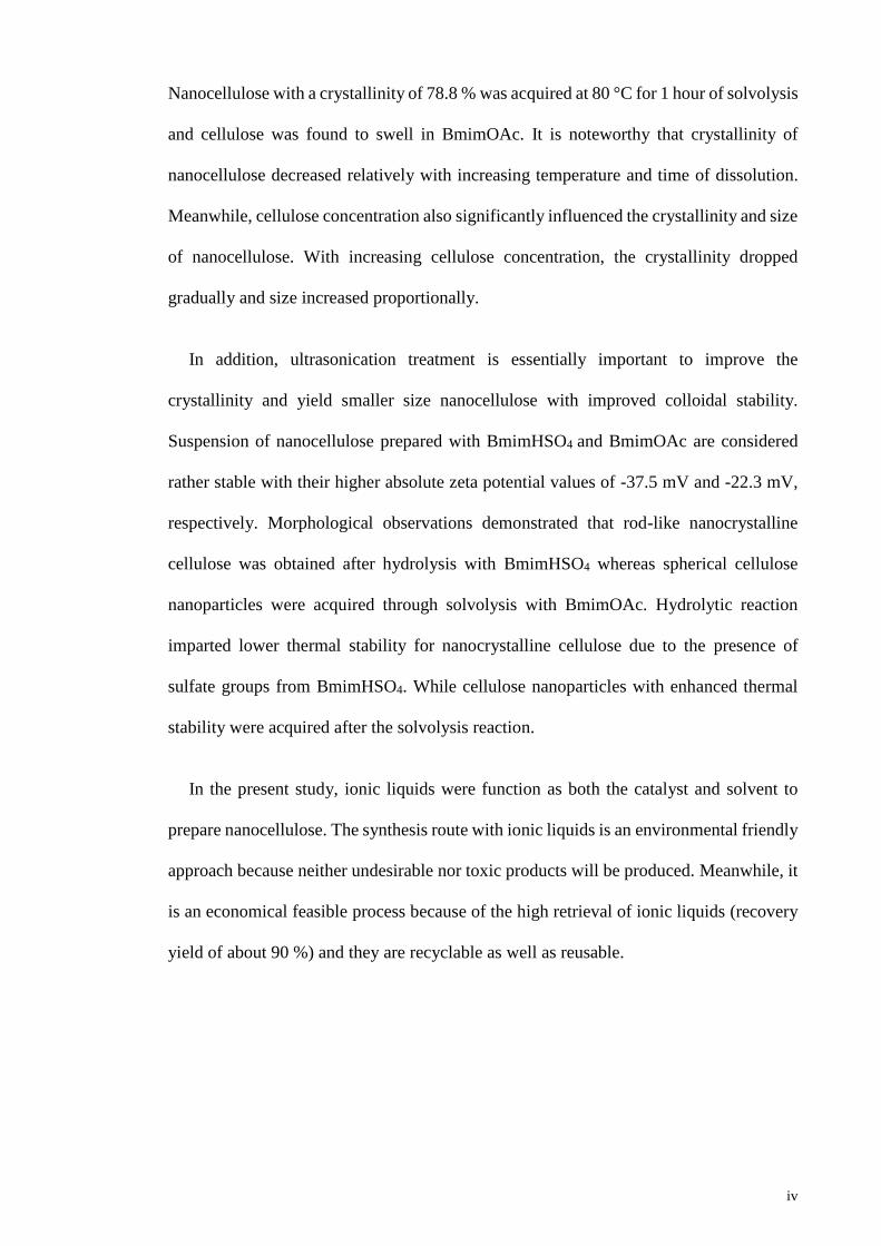
Nanocellulose with a crystallinity of 78.8 % was acquired at 80 °C for 1 hour of solvolysis
and cellulose was found to swell in BmimOAc. It is noteworthy that crystallinity of
nanocellulose decreased relatively with increasing temperature and time of dissolution.
Meanwhile, cellulose concentration also significantly influenced the crystallinity and size
of nanocellulose. With increasing cellulose concentration, the crystallinity dropped
gradually and size increased proportionally.
In addition, ultrasonication treatment is essentially important to improve the
crystallinity and yield smaller size nanocellulose with improved colloidal stability.
Suspension of nanocellulose prepared with BmimHSO4 and BmimOAc are considered
rather stable with their higher absolute zeta potential values of -37.5 mV and -22.3 mV,
respectively. Morphological observations demonstrated that rod-like nanocrystalline
cellulose was obtained after hydrolysis with BmimHSO4 whereas spherical cellulose
nanoparticles were acquired through solvolysis with BmimOAc. Hydrolytic reaction
imparted lower thermal stability for nanocrystalline cellulose due to the presence of
sulfate groups from BmimHSO4. While cellulose nanoparticles with enhanced thermal
stability were acquired after the solvolysis reaction.
In the present study, ionic liquids were function as both the catalyst and solvent to
prepare nanocellulose. The synthesis route with ionic liquids is an environmental friendly
approach because neither undesirable nor toxic products will be produced. Meanwhile, it
is an economical feasible process because of the high retrieval of ionic liquids (recovery
yield of about 90 %) and they are recyclable as well as reusable.
v
ABSTRAK
Nanoselulosa merupakan suatu bahan yang menjanjikan dan juga menarik perhatian
oleh kerana ciri-cirinya yang istimewa seperti memiliki permukaan yang luas, kekuatan
dan kekukuhan serta kemampuan untuk biopemerosotan. Dalam kajian ini, 1-butyl-3-
methylimidazolium hydrogen sulfate (BmimHSO4) dan 1-butyl-3-methylimidazolium
acetate (BmimOAc) cecair ionik telah digunakan sebagai pemangkin dan pelarut untuk
menghasilkan nanoselulosa daripada selulosa. Walaupun mempunyai kation yang sama,
keasidan BmimHSO4 dicadangkan menyumbang kepada hidrolisis. Sementara itu,
BmimOAc dengan keelektronegatifan yang tinggi lebih cenderung dalam proses solvolisis
untuk menghasilkan nanoselulosa. Kajian yang komprehensif dalam parameter sintesis
yang berlainan seperti suhu, masa, kepekatan (jisim tambahan MCC) dan rawatan
sonikasi telah dijalankan untuk mengawal sifat-sifat tertentu nanoselulosa.
Peningkatan hablur dengan pengekalan struktur hablur selulosa I dalam nanoselulosa
telah berjaya dihasilkan dengan menggunakan BmimHSO4. Berbanding dengan MCC,
hidrolisis pada suhu 90 ºC dalam masa 1.5 jam telah menyumbang kepada penghasilan
nanoselulosa dengan sifat hablur yang tinggi (CrI 92.2) serta berdiameter kecil (~15 nm).
Nanoselulosa dengan sifat hablur yang tinggi serta saiz yang lebih kecil dapat dihasilkan
dengan meningkatkan suhu dan masa reaksi hidrolitik. Walau bagaimanapun,
pemerosotan hablur nanoselulosa telah berlaku dengan meningkatkan lagi suhu dan masa
melebihi daripada keadaan optimum. Selain itu, kepekatan selulosa (dalam wt%) juga
menpunyai kesan yang agak besar dalam mempengaruhi hablur dan saiz nanoselulosa.
Dalam hal ini, saiz nanoselulosa telah bertambah berikutan dengan penambahan
kepekatan selulosa.
Manakala itu, transformasi struktur hablur dari selulosa I kepada II telah berlaku dalam
nanoselulosa diperolehi daripada reaksi solvolisis dengan BmimOAc. Nanoselulosa
vi
dengan 78.8 hablur telah berjaya diperolehkan menerui solvolisis pada suhu 80 ºC dalam
masa 1 jam. Selulosa didapati mengembang dalam BmimOAc. Pengurangan hablur dalam
nanoselulosa telah diperhatikan sewaktu dalam usaha meningkatkan suhu dan masa untuk
pencairan. Sementara itu, kepekatan selulosa juga didapati mempengaruhi hablur dan saiz
nanoselulosa. Hablur nanoselulosa telah merosot manakala saiz nanoselulosa mengalami
pertingkatan berikutan dengan penambahan kepekatan selulosa.
Selain itu, rawatan ultrasonikasi adalah penting untuk mengukuhkan hablur dan
menghasilkan nanoselulosa yang bersaiz kecil dengan kestabilan koloid. Larutan
nanoselulosa yang diperolehi dengan BmimHSO4 dan BmimOAc adalah dianggap agak
stabil dengan mempunyai nilai mutlak zeta potential yang tinggi, iaitu -37.5 mV dan -
22.3 mV masing-masing. Pemerhatian morfologi menunjukkan hablur nanoselulosa
dalam bentuk seperti rod telah berjaya dihasilkan menerui hidrolisis dengan BmimHSO4
manakala zarah nanoselulosa berbentuk bulat didapatkan melalui proses solvolisis
dengan BmimOAc. Hablur nanoselulosa mempunyai kestabilan haba yang rendah
disebabkan penambahan kumpulan sulfat menerusi proses hidrolisis. Sedangkan itu,
zarah nanoselulosa yang dihasilkan dalam proses solvolisis memiliki kestabilan haba.
Dalam kajian ini, cecair ionik berfungsi sebagai pemangkin dan pelarut untuk
menghasilkan nanoselulosa. Penghasilan nanoselulosa dengan menggunakan cecair ionik
merupakan suatu kaedah yang mesra alam kerana tiada bahan-bahan buangan berbahaya
dan toksik dijanakan dalam proses ini. Sementara itu, kaedah ini berfaedah dari segi
ekonomi disebabkan kadar pemulihan cecair ionik yang agak tinggi (sebanyak 90 %) dan
ia dapat dikitar serta boleh digunakan semula.

vii
ACKNOWLEDGEMENTS
First and foremost, I would like to express my sincere gratitude to my supervisors,
Prof. Dr. Sharifah Bee Abd Hamid and Dr. Lai Chin Wei for spending their precious time
to provide me with excellent guidance, motivation as well as invaluable suggestion
throughout my research journey.
In addition, I would like to convey grateful appreciation to all the research assistant
and friends in NANOCAT. The success of my research would not been made possible
without their support and kind assistance. Besides that, I would like to thank Ministry of
Higher Education Malaysia for funding my master program which provided me with the
opportunity to inevitably broaden my perspectives and knowledge.
Last but not least, deepest gratitude is convey to my family and friends who give me
unconditional support and encouragement throughout the time in completing this
research. Special thanks dedicated to my friends who inspired me and lent me a hand
whenever I faced difficulties throughout the master study.
Thank you.
viii
TABLE OF CONTENTS
Abstract ............................................................................................................................ iii
Abstrak .............................................................................................................................. v
Acknowledgements ......................................................................................................... vii
Table of Contents ........................................................................................................... viii
List of Figures ................................................................................................................. xii
List of Tables................................................................................................................... xv
List of Symbols and Abbreviations ................................................................................ xvi
List of Appendices .......................................................................................................... xx
CHAPTER 1: INTRODUCTION .................................................................................. 1
1.1 Introduction.............................................................................................................. 1
1.2 Problem Statement ................................................................................................... 6
1.3 Objectives of Research ............................................................................................ 8
1.4 Scope of Study ......................................................................................................... 9
1.5 Outline of thesis ..................................................................................................... 10
CHAPTER 2: LITERATURE REVIEW .................................................................... 11
2.1 Introduction............................................................................................................ 11
2.2 Cellulose ................................................................................................................ 11
2.3 Nanocellulose ........................................................................................................ 17
2.3.1 Types of Nanocellulose ............................................................................ 17
2.3.2 Advantageous properties of nanocellulose ............................................... 22
2.3.3 Potential applications of nanocellulose .................................................... 23
2.3.3.1 Improvement of nanocomposite mechanical properties ............ 23
2.3.3.2 Improved barrier properties of nanocomposites ........................ 25
ix
2.3.3.3 Biomedical applications ............................................................ 26
2.3.3.4 Drug delivery ............................................................................. 28
2.3.3.5 Nanopaper for flexible electronics ............................................ 29
2.4 Potential techniques for synthesizing nanocellulose ............................................. 30
2.4.1 Acid hydrolysis ......................................................................................... 31
2.4.2 Oxidation synthesis .................................................................................. 33
2.4.3 Steam explosion ....................................................................................... 35
2.4.4 Mechanical treatments .............................................................................. 37
2.4.5 Enzymatic hydrolysis ............................................................................... 41
2.5 Ionic Liquids .......................................................................................................... 43
2.5.1 Introduction to Ionic Liquids (ILs) ........................................................... 44
2.5.2 Ionic liquids for cellulose dissolution....................................................... 46
2.5.3 Acidic Ionic Liquid for Hydrolysis and Dissolution of Cellulose ............ 53
2.5.4 Acetate based Ionic Liquid for Cellulose Dissolution .............................. 56
2.5.5 Regeneration of Cellulose from Ionic Liquid Suspension ....................... 59
2.5.6 Recovery of Ionic Liquids ........................................................................ 62
CHAPTER 3: MATERIALS AND METHODOLOGY ............................................ 66
3.1 Introduction and Overview .................................................................................... 66
3.2 Raw Materials and Chemicals selections .............................................................. 66
3.2.1 Microcrystalline cellulose (MCC) ............................................................ 66
3.2.2 1-butyl-3-methylimidazolium hydrogen sulfate (BmimHSO4) ................ 67
3.2.3 1-butyl-3-methylimidazolium acetate (BmimOAc) ................................. 67
3.2.4 Deionized water (DI water) ...................................................................... 67
3.3 Methodology of Experiment .................................................................................. 67
3.3.1 Synthesis of nanocellulose by BmimHSO4 .............................................. 67
3.3.2 Synthesis of nanocellulose by BmimOAc ................................................ 68
x
3.3.3 Recovery of Ionic Liquid ......................................................................... 69
3.4 Characterization Techniques ................................................................................. 70
3.4.1 X-ray diffraction (XRD) ........................................................................... 71
3.4.2 Zeta sizer .................................................................................................. 72
3.4.3 Fourier transform infrared spectroscopy (FTIR) ...................................... 73
3.4.4 Field emission scanning electron microscopy (FESEM) ......................... 74
3.4.5 Atomic force microscope (AFM) ............................................................. 75
3.4.6 High resolution transmission electron microscope (HRTEM) ................. 76
3.4.7 Thermogravimetric analysis (TGA) ......................................................... 77
CHAPTER 4: RESULTS AND DISCUSSION .......................................................... 78
4.1 Introduction............................................................................................................ 78
4.2 Synthesis of Nanocrystalline Cellulose (NCC) by BmimHSO4 ............................ 78
4.2.1 Mechanism of NCC Synthesis with BmimHSO4 ..................................... 78
4.2.2 X-ray Diffraction Analysis ....................................................................... 84
4.2.3 Hydrodynamic Size and Zeta Potential Analysis ..................................... 93
4.2.4 Fourier Transform Infrared Spectroscopy (FTIR) Analysis ................... 100
4.2.5 Morphological analysis by Field Emission Scanning Electron Microscopy
(FESEM), Atomic Force Microscope (AFM) and Transmission Electron
Microscopy (TEM) ................................................................................. 106
4.2.6 Thermal properties analysis by Thermogravimetric Analysis (TGA) .... 116
4.3 Synthesis of Cellulose Nanoparticles (CNPs) by BmimOAc .............................. 124
4.3.1 Mechanism of CNPs Synthesis with BmimOAc .................................... 124
4.3.2 X-ray Diffraction Analysis ..................................................................... 130
4.3.3 Hydrodynamic Size and Zeta Potential Analysis ................................... 139
4.3.4 Fourier Transform Infrared Spectroscopy (FTIR) Analysis ................... 144
xi
4.3.5 Morphological analysis by Field Emission Scanning Electron Microscopy
(FESEM), Atomic Force Microscope (AFM) and Transmission Electron
Microscopy (TEM) ................................................................................. 149
4.3.6 Thermal properties analysis by Thermogravimetric Analysis (TGA) .... 159
CHAPTER 5: CONCLUSION AND RECOMMENDATIONS ............................. 163
5.1 Conclusion ........................................................................................................... 163
5.2 Recommendations for Future Work .................................................................... 166
References ..................................................................................................................... 168
List of Publications and Papers Presented .................................................................... 188
Appendix ....................................................................................................................... 189
xii
LIST OF FIGURES
Figure 2.1: Structure of cellulose polymer chain (Pinkert, Marsh, Pang, & Staiger, 2009).
......................................................................................................................................... 13
Figure 2.2: Intermolecular and intramolecular hydrogen bonding in cellulose (Pinkert et
al., 2009). ........................................................................................................................ 14
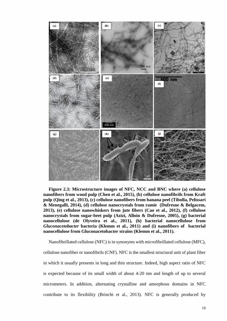
Figure 2.3: Microstructure images of NFC, NCC and BNC where (a) cellulose nanofibers
from wood pulp (Chen et al., 2015), (b) cellulose nanofibrils from Kraft pulp (Qing et al.,
2013), (c) cellulose nanofibers from banana peel (Tibolla, Pelissari & Menegalli, 2014),
(d) cellulose nanocrystals from ramie (Dufresne & Belgacem, 2013), (e) cellulose
nanowhiskers from jute fibers (Cao et al., 2012), (f) cellulose nanocrystals from sugar-
beet pulp (Azizi, Alloin & Dufresne, 2005), (g) bacterial nanocellulose (de Olyveira et
al., 2011), (h) bacterial nanocellulose from Gluconacetobacter bacteria (Klemm et al.,
2011) and (i) nanofibers of bacterial nanocellulose from Gluconacetobacter strains
(Klemm et al., 2011). ...................................................................................................... 19
Figure 2.4: Examples of cations and anions for ILs with their molecular structures and
abbreviations (Gericke et al., 2012). ............................................................................... 46
Figure 2.5: Proposed dissolution mechanism of cellulose in [Bmim]Cl (Pinkert et al.,
2009). .............................................................................................................................. 48
Figure 2.6: Chemical structure of [Bmim]HSO4 (Mao, Osorio-Madrazo, & Laborie,
2013). .............................................................................................................................. 55
Figure 2.7: Chemical structure of [Bmim]OAc (Wang et al., 2012; Zhang, 2013). ....... 57
Figure 2.8: Process involved in regeneration of cellulose fibers (Cao et al., 2009). ...... 61
Figure 3.1: Overview flow chart for nanocellulose synthesis methodology. .................. 70
Figure 4.1: Proposed mechanism on formation of NCC by BmimHSO4 mediated
hydrolysis. ....................................................................................................................... 81
Figure 4.2: XRD patterns of MCC and NCC optimized with different parameters (a)
temperature, (b) time, (c) cellulose concentration (mass loading of MCC), and (d)
ultrasonication conditions. .............................................................................................. 86
Figure 4.3: Observation of color of cellulose/BmimHSO4 suspension during the reaction
at (a) temperature below 100 °C and (b) temperature of 110 °C. ................................... 89
Figure 4.4: FTIR spectra of NCC optimized with different parameters (a) temperature,
(b) time, (c) cellulose concentration (mass loading of MCC), and (d) ultrasonication
conditions. ..................................................................................................................... 101
xiii
Figure 4.5: FTIR spectra of pure BmimHSO4 and regenerated BmimHSO4. .............. 105
Figure 4.6: FESEM images of (a) MCC, (b) NCC synthesized at temperatures of (b) 70
°C, (c) 80 °C, (d) 90 °C and (e) 100 °C. Insets are the images of cellulose fibres at 15,000x
magnification................................................................................................................. 107
Figure 4.7: FESEM images of NCC obtained at different hydrolysis time: (a) 0.5 h, (b)
1.0 h, (c) 1.5 h and (d) 2.0 h. ......................................................................................... 109
Figure 4.8: AFM height images and peak force error images of NCC obtained at different
synthesis time: (a) 0.5 h, (b) 1 h and (c) 1.5 h.............................................................. 112
Figure 4.9: Size distribution (diameter) of NCC based on AFM analysis obtained at
different synthesis time: (a) 0.5 h, (b) 1 h and (c) 1.5 h. .............................................. 113
Figure 4.10: HRTEM images of NCC synthesized at (a) 1 h, (b) 1.5 h and (c) 2 h. .... 115
Figure 4.11: TGA and DTG curves of MCC and NCC optimized with different parameters
(a) temperature, (b) time, (c) cellulose concentration (mass loading of MCC), and (d)
ultrasonication conditions. ............................................................................................ 117
Figure 4.12: Proposed mechanism of formation of CNPs with BmimOAc solvolysis. 126
Figure 4.13: XRD patterns of MCC and CNPs optimized with different parameters (a)
temperature, (b) time, (c) cellulose concentration (mass loading of MCC), and (d)
ultrasonication conditions. ............................................................................................ 131
Figure 4.14: Observation of color of cellulose/BmimOAc suspension during the reaction
at (a) temperature below 100 °C and (b) temperature of 110 °C. ................................. 135
Figure 4.15: FTIR spectra of CNPs optimized with different parameters (a) temperature,
(b) time, (c) cellulose concentration (mass loading of MCC), and (d) ultrasonication
conditions. ..................................................................................................................... 144
Figure 4.16: FTIR spectra of pure BmimOAc and regenerated BmimOAc. ................ 147
Figure 4.17: FESEM images of CNPs synthesized at temperatures of (a) 70 °C, (b) 80 °C,
(c) 90 °C, (d) 100 °C and (e) 110 °C with BmimOAc. Insets are the images of cellulose
fibres at 15,000x magnification..................................................................................... 149
Figure 4.18: FESEM images of CNPs obtained at different dissolution time: (a) 0.5 h, (b)
1.0 h, (c) 1.5 h and (d) 2.0 h. ......................................................................................... 152
Figure 4.19: AFM height images and peak force error images of CNPs obtained at
different synthesis time: (a) 0.5 h, (b) 1 h and (c) 1.5 h. .............................................. 154
xiv
Figure 4.20: Size distribution (diameter) of CNPs based on AFM analysis obtained at
different synthesis time: (a) 0.5 h, (b) 1 h and (c) 1.5 h. .............................................. 155
Figure 4.21: HRTEM images of CNPs synthesized at (a) 1 h, (b) 1.5 h and (c) 2 h. ... 158
Figure 4.22: TGA and DTG curves of MCC and CNPs optimized with different
parameters (a) temperature, (b) time, (c) cellulose concentration (mass loading of MCC),
and (d) ultrasonication conditions. ................................................................................ 159
xv
LIST OF TABLES
Table 2.1: Thermodynamic characteristics of various cellulose allomorphs (Ioelovich,
2016). .............................................................................................................................. 16
Table 2.2: Types of nanocellulose (Klemm et al., 2011). ............................................... 18
Table 2.3: Mechanical properties of PS (Plasticized starch) and PS/NCC nanocomposites
obtained from tensile test (Cao, Chen, Chang, Muir & Falk, 2008). .............................. 24
Table 2.4: Advantages and disadvantages of steam explosion method for the processing
of lignocellulosic biomass (Brodeur et al., 2011). .......................................................... 36
Table 2.5: Application of Ionic Liquids (ILs) for cellulose dissolution (Chowdhury et al.,
2014). .............................................................................................................................. 52
Table 2.6: Comparison of main properties of ILs relevant to dissolution. ..................... 58
Table 2.7: Summary of methods used for recovery and recycling of ILs (Mai, Ahn, &
Koo, 2014). ..................................................................................................................... 63
Table 3.1: List of raw materials and chemicals used for the synthesis of nanocellulose.
......................................................................................................................................... 66
Table 4.1: Crystallinity index and crystallite size of NCC. ............................................ 87
Table 4.2: Hydrodynamic size, polydispersity index and zeta potential of NCC. .......... 95
Table 4.3: Average length, diameter and aspect ratio of NCC determined from AFM
analysis. ......................................................................................................................... 110
Table 4.4: Onset decomposition temperature, maximum decomposition temperature and
char yield of MCC and NCC. ........................................................................................ 119
Table 4.5: Crystallinity index and crystallite size of CNPs. ......................................... 133
Table 4.6: Hydrodynamic size, polydispersity index and zeta potential of CNPs. ....... 140
Table 4.7: Onset decomposition temperature, maximum decomposition temperature and
char yield of CNPs. ....................................................................................................... 161
xvi
LIST OF SYMBOLS AND ABBREVIATIONS
α : Alpha
Å : Angstrom
β : Beta
cm : Centimeter
g : Gram
GPa : Gigapascal
h : Hour
kg : Kilogram
kJ : Kilojoule
kV : Kilovolt
kWh : Kilowatt hour
m : Meter
mA : Milliampere
mg : Milligram
min : Minutes
mL : Milliliter
mPa : Millipascal
MPa : Megapascal
nm : Nanometer
N : Normality
rpm : Rotation per minute
s : Second
µm : Micrometer
wt% : Weight percentage
xvii
°C : Degree Celsius
θ : Diffraction angle
λ : Wavelength
AFM : Atomic force microscope
AGU : Anhydroglucose
[Amim]Cl : 1-allyl-3-methylimidazolium chloride
BF4- : Tetrafluoroborate anion
[Bmim]+ : 1-butyl-3-methylimidazolium cation
[Bmim]Br : 1-butyl-3-methylimidazolium bromide
BmimCl : 1-butyl-3-methylimidazolium chloride
BmimHSO4 : 1-butyl-3-methylimidazolium hydrogen sulfate
BmimOAc : 1-butyl-3-methylimidazolium acetate
[Bmim]SCN : 1-butyl-3-methylimidazolium sulfocyanate
BNC : Bacterial nanocellulose
CNC : Cellulose nanocrystals
CNF : Cellulose nanofiber or nanofibrils
CNPs : Cellulose II nanoparticles
CNW : Cellulose nanowhiskers
CrI : Crystallinity index
CTAB : Cetyl trimethylammonium bromide
[C6mim]Cl : 1-hexyl-3-methylimidazolium chloride
[C8mim]Cl : 1-octyl-3-methylimidazolium chloride
DI : Deionized
DLS : Dynamic-light scattering
DMAc : N,N-dimethylacetamide
DMF : N,N-dimethylformamide
xviii
DMI : 1,3-dimethyl-2-imidazolidinone
DMSO : Dimethyl sulfoxide
DTG : Differential thermogravimetric
EDA : Electron donor-electron acceptor
EFB : Empty fruit bunches
ELS : Electrophoretic light scattering
EmimOAc : 1-ethyl-3-methylimidazolium acetate
FESEM : Field emission scanning electron microscope
FTIR : Fourier transform infrared spectroscopy
[H+] : Proton
[H3O]+ : Hydronium ion
HMF : Hydroxymethylfurfural
HPC : Hydroxypropylated cellulose
HRTEM : High resolution transmission electron microscope
[HSO4]- : Hydrogen sulfate anion
IL : Ionic liquid
ILs : Ionic liquids
LD50 : Median lethal dose
MCC : Microcrystalline cellulose
MFC : Microfibrillated cellulose
NCC : Nanocrystalline cellulose
NFC : Nanofibrillated cellulose
NMMO : N-methylmorpholine oxide
[OAc]- : Acetate anion
OECD : Organization for Economic Co-operation and Development
OH : Hydroxyl group
xix
PDI : Polydispersity index
PLA : Polylactic acid
PSD : Particle size distribution
RTILs : Room temperature ionic liquids
SO3H : Sulfonic acid
[SO4]2- : Sulfate
T0 : Onset decomposition temperature
TBAF : Tetrabutylammonium fluoride
TEMPO : 2,2,6,6-tetramethylpiperidinyl-1-oxyl
TGA : Thermogravimetric analysis
Tmax : Maximum thermal decomposition temperature
TSILs : Task-specific ionic liquids
XRD : X-ray diffraction

xx
LIST OF APPENDICES
Appendix A: Published Paper (Biomass and Bioenergy)……………………….. 189
1
CHAPTER 1: INTRODUCTION
1.1 Introduction
In recent decades, modern society is habituated to a high degree of mobility, fast
communication and daily comfort, all of which require considerable energy input. People
nowadays are getting increasing awareness on the issues regarding energy crisis. Indeed,
our modern world is heavily dependent on fossil fuels (85% of the world energy
consumption), mainly goes to commercial and industrial uses. Thus, one of the steps taken
by many countries to aid the earth and minimizing environmental problems is applying
renewable and sustainable development for energy resources. It seems that cellulose
which obtained from the biomass residues would be a great potential of renewable and
sustainable energy resources as a promising alternative to fossil-derived products. The
utilization of biomass materials is suggested able to decrease the dependence away from
the non-renewable fossil-fuels. In urbanization world, renewable and sustainable
development for energy resources is a strategic goal of modern society reflecting
contemporary demand for economic, social, political and environmental development.
Therefore, the finding of alternative clean energy source is crucial in leading a high
quality of life, which is in harmony with nature.
Recently, enormous interest has been focused on biomass as a potential alternative
resource for fuels and petrochemicals due to the depletion of fossil fuels and global
warming issues (Mehmood et al., 2015). In this case, biomass resources such as
lignocellulosic feedstock that derived from the commodity crops and residues from
industries and forestry have prompted the prospect of biomass as a promising substitute
for fossil fuel to secure the future supply of a clean and sustainable energy. Therefore,
establishment of new methods and technologies are required for the beneficial utilization
of biomass (Kilpeläinen et al., 2007).
2
It is a well-known fact that biomass is sustainable and abundance in availability.
According to the literatures, there is more than 150 million tons of biomass are being
generated annually as the side-product from industrial and agricultural wastes globally
(Ang, Ngoh, Chua, & Lee, 2012; Tadesse & Luque, 2011; Zhao et al., 2007). The sources
of biomass are generally from wood chips, agricultural residues, domestic waste, effluent
sludge and animal wastes. These inedible crop residues are usually cost effective due to
abundance and available in high quantity (Ang et al., 2012; Lee, Hamid, & Zain, 2014).
In general, biomass composed of three major components, such as cellulose,
hemicellulose and lignin. Particularly, cellulose is the most abundant biopolymer, which
consists of about half to one-third (30-50%) of the total biomass constituents. In fact,
cellulose is an almost inexhaustible source of material and became the most abundant
renewable resource available in nature as well as present in all living organisms. As a
matter of fact, biomass is an increasingly important material for the requirement of
environmental friendly, biocompatible as well as biodegradable products in the current
hunt for greener technologies materials (Zhang, Li, Yu, & Hsieh, 2010).
Cellulose is a polysaccharide under the carbohydrate groups. It is the most abundant
biopolymer available on earth, which can be derived from plant or animal origins.
Cellulose is biocompatible, biodegradable and non-toxic, so that it can be used in
biomedical and drug delivery applications. For instance, it can be used as antimicrobial
agents or drug carriers. This proves the biocompatibility of cellulose in which it functions
as the chelating agent to bind with other biomolecules such as proteins, cells and metal
ions to perform targeted applications. In terms of degradation, cellulose is readily
biodegraded by organisms that utilize cellulase enzymes (Puls, Wilson & Hölter, 2011).
For instance, cellulose composites in the packaging materials will enable them to slowly
3
degradable in the environment. Therefore, it is sensible to judge the biocompatibility as
well as biodegradability of cellulose due to its biomolecular structure.
Nowadays, cellulose has been widely applied as the source of energy and for the
building materials, including textiles, paper and clothing, in the form of wood as well as
the plant fibers. The aforementioned cellulose utilization was benign from its hierarchical
structure to provide sufficient strength and stiffness to support the materials. Furthermore,
cellulose played an important role in material engineering system as strength supporting
for the composite materials. However, several obvious hindrances to the widespread use
of cellulose as reinforcements in polymers are its incompatibility with hydrophobic
polymer matrix, tendency of aggregation during processing and prone to swell in water.
In addition, the mechanical properties can be influenced by the type of cellulose, parts of
a plant as well as different plants (Brinchi, Cotana, Fortunati, & Kenny, 2013).
On the other hand, high durability property of cellulose has emerged as the leading
candidate as efficient building blocks, especially the elementary unit of nanocellulose. In
this context, nanocellulose is the cellulose structure that exists in nanometer scale,
precisely range from 1 to 100 nm for at least one of its dimensions. It is the basic
reinforcement unit that constitutes for the strength and stiffness to structure of plants and
trees. Besides, nanocellulose has an exceptional mechanical performance, such that its
outstanding axial Young’s modulus is approximate to the one derived from theoretical
chemistry. Nanocellulose is also comparable stronger than that of Kevlar and steel as well
as within the range of other reinforcement materials (Brinchi et al., 2013).
The Young’s modulus of nanocellulose with a density of 1.6 g cm-3 is approximate
167.5 GPa. It is comparably stronger than Kevlar with Young’s modulus of 60-125 GPa
(density around 1.45 g cm-3) and potentially stronger than steel (200-220 GPa, density
around 8 g cm-3) (Lemaitre et al., 2009). Indeed, the specific Young’s modulus, which is
4
the ratio between the Young’s modulus and the density, of nanocellulose is around
65 J g−1 for microfibrils and 85 J g−1 for nanocrystals whereas it is around 25 J g−1 for
steel (Dufresne, 2013). This signifies that nanocellulose is about 8 times stronger than
that of steel, in terms of the ratio between strength and weight. Therefore, crystalline
nanocellulose has interesting mechanical properties for use in material applications.
Apart from that, nanocellulose also has many unique characteristics such as
lightweight, low density, high surface area, high aspect ratio and even modifiable surface
properties due to the reactive OH functional groups. Furthermore, evaluation of its
biodegradable properties in aqueous environment has been conducted according to the
OECD standard. In fact, biodegradation rate of nanocellulose is much faster than that of
their macroscopic counterpart, whereas other type of carbon-based nanomaterials such as
fullerenes and carbon nanotubes are not providing any biodegradation property (Brinchi
et al., 2013). The main reason of fast biodegradation rate is attributed to its natural
structure which derived from and constitute for the macroscopic cellulose.
The processing of cellulose into nanocellulose for better utilization and exploration its
new functional properties still remains as a great challenge. The reasons might be
attributed to the non-thermal plastic behavior and insolubility of cellulose in water as well
as most common organic solvents (Kilpeläinen et al., 2007). Moreover, hydrolysis of
cellulose molecules is difficult as their molecules are not readily hydrolyzed under
ambient conditions. Usually, harsh condition such as highly acidic or alkaline conditions
or the use of high pressure systems is required for transforming cellulose into
nanocellulose.
Generally, cellulose are recalcitrant to chemical treatments because their molecules
are embedded within the complex structure of lignocellulosic matrix in order to protect
them from chemical and biological attacks (Karatzos, Edye, & Doherty, 2012). Therefore,
5
well-designed natural structure of cellulose is still matter of debate in dissolving the
cellulose without any chemical modification or derivatization. The reasons might be
attributed to the rigidity of long-chain as well as strong inter- and intramolecular hydrogen
bonding that constitute for the stiff cellulose structure such that it is impermeable to most
of the molecules. Hence, development and identification of new efficient approach and
solvent systems prompted the need for prospect utilization and application of cellulose
(Zhang et al., 2010). Many scientists and researchers believed that improvement of
hydrophilicity with excellent biocompatibility as well as their biodegradability properties
would broaden the interest of nanocellulose as a promising smart and green material in
the field of composite, packaging as well as biomedical applications.
In this research studies, nanocellulose was synthesized from cellulose precursor by
using ionic liquids (ILs) function as both the catalyst and solvent. Ionic liquid (IL) is
suggested to perform dual functions in the present study: (1) catalyzed the hydrolysis by
cleavage of glycosidic linkage and meanwhile, (2) as a reaction medium for the solvolysis
or dissolution of cellulose to disrupt the hydrogen bonding. The selected ILs in this study
were 1-butyl-3-methylimidazolium hydrogen sulfate [BmimHSO4] (acidity) and 1-butyl-
3-methylimidazolium acetate [BmimOAc] (high electronegativity).
As a matter of fact, both imidazolium-based ILs could be provided good interaction
with cellulose by disrupting the hydrogen bonds due to the coordination effect of anion
groups of ILs with the hydroxyl groups of cellulose (Yuan et al., 2015). Thus, this
research was conducted to investigate the influence of synthesis parameters such as
reaction temperature, time, concentration in term of mass loading of microcrystalline
cellulose (MCC) and different sonication treatments in the preparation of nanocellulose.
The crystallinity, morphology and thermal properties of nanocellulose by controlling
aforementioned processing parameters will be investigated in detail. The low complexity
6
of process, less time consuming and improved properties of nanocellulose produced are
strongly augmented by using ILs as catalyst and solvent, thus making the project
attractive and profitable.
Preparation of nanocellulose is of significant importance to expand the potential
applications in vast industries as the stiff structure of cellulose hindered its exploitations.
This approach is believed to be an environmental friendly route because ILs are known
as green solvent for cellulose processing as they are recyclable and reusable. The high
crystallinity and enhanced thermal properties of nanocellulose produced are of great
interest as it can be further composites with other materials and used in reinforcement
purposes.
1.2 Problem Statement
In general, processing and derivatization of cellulose are rather difficult due to the
complex structure of biopolymeric network and presence of numerous noncovalent
interactions between cellulose molecules. Besides that, partially crystalline, highly
ordered structure as well as the stiff molecules results from the closely-packed chains via
extended network of inter- and intramolecular hydrogen bonds and rigid linkages lead to
insolubility of cellulose in water and other common organic solvents (Kokol, Božič,
Vogrinčič, & Mathew, 2015; Xu, Wang, & Wang, 2010). In this case, these constitutes a
serious problem for cellulose dissolution and activity as well as obstruct the application
of cellulose. A number of methods such as acid hydrolysis, steam explosion, mechanical
process, TEMPO-mediated oxidation, thermal hydrolysis and pyrolysis have been used
to breakdown the cellulose molecule into nanocellulose. However, these methods not
only destroy the pristine structure of cellulose but also added impurities such as halide
and metal ions into the final products and are difficult controllable processes as there are
propensity of over degradation of cellulose (Tang, Huang, Ou, Chen, & Chen, 2011).
7
To date, numerous solvent systems have been identified such as N-methylmorpholine
oxide (NMMO), N,N-dimethylacetamide/ lithium chloride (DMAc/LiCl), N,N-
dimethylformamide/nitrous tetroxide (DMF/N2O4), dimethyl
sulfoxide/tetrabutylammonium fluoride (DMSO/TBAF), 1,3-dimethyl-2-
imidazolidinone/ lithium chloride (DMI/LiCl) and some molten salt hydrates such as
LiClO4·3H2O, LiSCN·2H2O and other aqueous metal complexes are able to dissolve
cellulose effectively (Tian, Han, Lu, Zhang, & Yuan, 2014; Xu, Zhang, Zhao, & Wang,
2013). Those methods and solvents systems proposed are worthy to mention several
shortcomings might be occurred including toxicity, high cost, volatility, generation of
poisonous gas, difficulty in solvent recovery and process instability (Lan, Liu, Yue, Sun,
& Kennedy, 2011). In addition, multistep treatment and complicated separation process
are usually required, followed by prolonged reaction time up to several hours to days.
One of the most common methods adopted in the manufacture of nanocellulose is acid
hydrolysis. Acid hydrolysis is by far the most popular method for nanocellulose
production known for its effectiveness, since last decades. However, this method is not
recommended because it usually requires relatively harsh conditions such as extreme
acidic condition and often using hazardous chemicals such as concentrated acids and
alkaline and hence, is not environmental friendly. Furthermore, acid hydrolysis might be
contributed to serious environmental threats due to the acid recovery and reuse issues
once discharged into the effluent streams. For a commercial scale production, large
quantity of acid is used and prolonged usage of acid will cause the corrosion of reactor
and other equipment which are expensive and cumbersome (Harmsen et al., 2010; Lee,
Hamid & Zain, 2014; Sun & Cheng, 2002).
In the limelight of this issue, the demand for the implementation of “green” processes
for cellulose processing is getting more considerable attentions, with increasing

8
governmental regulations in the industries. The use of ILs for cellulose processing is
suggested to be a green technology with their non-toxic and non-harsh characters under
mild conditions because ILs are able to recyclable and reusable, thereby effectively
reduced solvent consumption. ILs are essentially green solvents for cellulose attributed
to their 2-3 lower magnitude orders of acidity than the conventional concentrated mineral
acids. ILs can be employed as substitution to the traditional volatile organic solvents and
conventional acids in the production of nanocellulose. ILs are capable to solubilize
cellulose by causing the disruption to the hydrogen bonding network of cellulose. Besides
that, atmospheric solvent loss and flammability hazard can be minimized via the
application of ILs because they have low vapour pressure and better thermal stability.
Moreover, they are ease of recovering and the ability to recycle and reusable contribute
to the lower operating cost of the catalytic process. In overall, the substitution of acids by
ILs can act as an environmentally friendly approach to synthesis the nanocellulose.
Development of new materials and green processes are driven by the goals to achieve
green chemistry, sustainability and eco-efficiency. To date, environmental concerns have
gained considerable attention and cannot be neglected. Therefore, the establishment of
recycle-based and sustainable communities is becoming increasingly important. A strong
driving force is initiated toward the exploitations of nanocellulose as a promising
renewable, sustainable and biodegradable material into the synthetic development of
environmental friendly and biocompatible products (Lin et al., 2014).
1.3 Objectives of Research
The objectives of this study are listed as follow:
To develop methodology for the development of nanocellulose aided by ionic
liquid by controlling synthesis parameters (temperature, time, mass loading of
MCC and ultrasonication conditions).
9
To characterize the nanocellulose obtained for its morphology, physical, chemical
and thermal properties for better understanding in method development.
1.4 Scope of Study
There are three main sections within the present research study. First stage of study is
about the preparation of nanocrystalline cellulose (NCC) by using 1-butyl-3-
methylimidazolium hydrogen sulfate (BmimHSO4) to catalyze the hydrolysis of β-
glycosidic linkage. On the other hand, second stage of study is about the preparation of
cellulose II nanoparticles (CNPs) in 1-butyl-3-methylimidazolium acetate (BmimOAc)
through dissolution and regeneration approaches. The synthesis procedures of
nanocellulose were carried out at different reaction temperatures, times, concentrations
and subjected to different sonication treatments. The resultant nanocellulose that
regenerated from IL was then further isolated by using repeated centrifugations and
washing steps to remove the residual IL. Nanocellulose was allowed to freeze dry prior
performing characterizations.
In the last stage of research study, multiple characterization techniques were used to
characterize the resultant nanocellulose in terms of morphological, physical, chemical,
structural and thermal properties. The phase structure and crystallinity of nanocellulose
were characterized by X-ray diffraction (XRD) and chemical functional groups was
investigated by Fourier Transform Infrared Spectroscopy (FTIR). In addition, the size and
surface charges of nanocellulose were examined by Zeta Sizer. Moreover, Field Emission
Scanning Electron Microscope (FESEM), Atomic Force Microscope (AFM) and High
Resolution Transmission Electron Microscope (HRTEM) were used to illustrate the
morphology, size and shape of the resultant nanocellulose samples. Other than that,
thermal decomposition behavior of nanocellulose was determined by Thermogravimetric
10
Analysis (TGA). The basic principle and details of each characterization techniques will
be discussed in Chapter 3-Research Methodology.
1.5 Outline of thesis
This thesis is organized into five chapters consecutively. Chapter 1 provides a brief
introduction of this research study, problem statement, objectives, scope of study and
thesis overview. Chapter 2 presents the structure of cellulose, different types of
nanocellulose, properties and applications of nanocellulose as well as methods of
production of nanocellulose. Moreover, literature review on ILs such as properties,
applications, dissolution of cellulose in ILs are also included in Chapter 2. Chapter 3
describes the specification of raw materials and chemicals used, experimental methods
and each of the characterization techniques adopted in this research. Chapter 4 presents
the results obtained from the characterizations and subsequent data analysis. Discussion
and explanation based on the results are included in this chapter. Lastly, Chapter 5
summarizes the conclusion of the study as well as several suggestions and
recommendations for the future work.
11
CHAPTER 2: LITERATURE REVIEW
2.1 Introduction
Nowadays, public concern about the environment, climate change and limited fossil
fuel resources have given rise to the urgent need of fostering development in the area of
renewable energies, which are inexhaustible and non-polluting. Cellulose is one of the
most promising prospects for efficient renewable resources due to its high abundance and
availability in nature. Recent studies have indicated that “nano” sized cellulose has
emerged as the excellent candidate to composite with other materials because of its
superior mechanical properties and biocompatibility. In order to further improve the
shortcomings of cellulose, considerable effort has to be exerted to increase the utilization
of cellulose by applying ILs as catalyst and dissolution solvent. Lately, interesting and
unique features of “nanocellulose” have gained much attention and became favorite
research matter among various groups of scientists.
The relationship between cellulose and ILs was still a matter of debate and remains
unclear. It was noted that the properties of this “nanocellulose” primarily depend on the
nature of the preparation method and the role of optimum ILs incorporated into the
cellulose. Therefore, the development of an efficient nanocellulose synthesis technique
remains to be determined. In the subsequent section, the introduction of cellulose and
nanocellulose as well as methods and ILs that commonly used by researchers with regards
to the nanocellulose will be reviewed in detail.
2.2 Cellulose
As early as 1838, cellulose was first isolated from plant matter and being discovered
by Anselme Payen on his research on ligneous matter. He reported that ligneous matter
was considered a single substance, which contained two chemically distinct materials.
One of these, the French chemist declared that it had the same chemical composition as
12
starch, but differed in structure and properties. Prior to its polymeric structure being
identified and recognized, cellulose has been utilized as a precursor for chemical
modification. In this context, the wood cell wall polymers were chemically modified due
to the abundant number of hydroxyl groups. It is this hydroxyl groups contribute to the
site for reactivity in the polymers and most reaction schemes have been based on the
reaction of hydroxyl groups for hydroxyl substitutions. Because the properties of wood
result from the chemistry of the cell wall components, the basic properties of a wood can
be changed by modifying the basic chemistry of the cell wall polymers. A major step
came in 1920 with the discovery of the polymeric structure of cellulose determined by
Hermann Staudinger (Klemm, Schmauder, & Heinze, 2005).
Nowadays, cellulose remains as the most abundant natural biopolymers on earth with
a remarkable annual production expected to be over 7.5 × 10¹⁰ tons (Habibi, Lucia, &
Rojas, 2010). It is one of the important polysaccharide because it is available widely in
higher plants (such as trees and wood), in annual crops (such as wheat straw, corn and
rice husks), in several aquatic animals (such as tunicates) and a lesser degree found in
simpler microorganisms including algae, bacteria, fungi, amoeba and invertebrates
(Klemm et al., 2011). In fact, cellulose is a major component that can be extracted from
these sources in which it comprising about 40 to 50 % of the total biomass components.
Therefore, it is a potential inexhaustible source of renewable bioenergy to replace the
dependence on fossil-based resources.
The cellulose obtained from higher plant, crops and aquatic animals such as tunicates
consist mainly of Iβ. Whereas cellulose produced by algae is rich in Iα form. However,
higher cellulose content with great strength, closeness of texture and durability can be
acquired from higher plants. Moreover, the cellulose fibers are long and exhibit
strengthen properties to provide sufficient support to the higher plants. On contrary, lower
13
plants such as crops contained lower cellulose content and the fibers are much shorter and
smaller with limited tensile strength as compared to the higher plants.
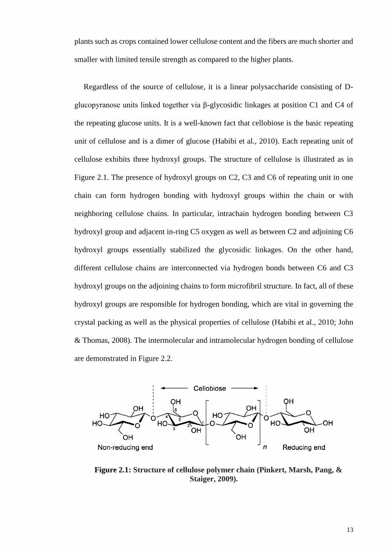
Regardless of the source of cellulose, it is a linear polysaccharide consisting of D-
glucopyranose units linked together via β-glycosidic linkages at position C1 and C4 of
the repeating glucose units. It is a well-known fact that cellobiose is the basic repeating
unit of cellulose and is a dimer of glucose (Habibi et al., 2010). Each repeating unit of
cellulose exhibits three hydroxyl groups. The structure of cellulose is illustrated as in
Figure 2.1. The presence of hydroxyl groups on C2, C3 and C6 of repeating unit in one
chain can form hydrogen bonding with hydroxyl groups within the chain or with
neighboring cellulose chains. In particular, intrachain hydrogen bonding between C3
hydroxyl group and adjacent in-ring C5 oxygen as well as between C2 and adjoining C6
hydroxyl groups essentially stabilized the glycosidic linkages. On the other hand,
different cellulose chains are interconnected via hydrogen bonds between C6 and C3
hydroxyl groups on the adjoining chains to form microfibril structure. In fact, all of these
hydroxyl groups are responsible for hydrogen bonding, which are vital in governing the
crystal packing as well as the physical properties of cellulose (Habibi et al., 2010; John
& Thomas, 2008). The intermolecular and intramolecular hydrogen bonding of cellulose
are demonstrated in Figure 2.2.
Figure 2.1: Structure of cellulose polymer chain (Pinkert, Marsh, Pang, &
Staiger, 2009).
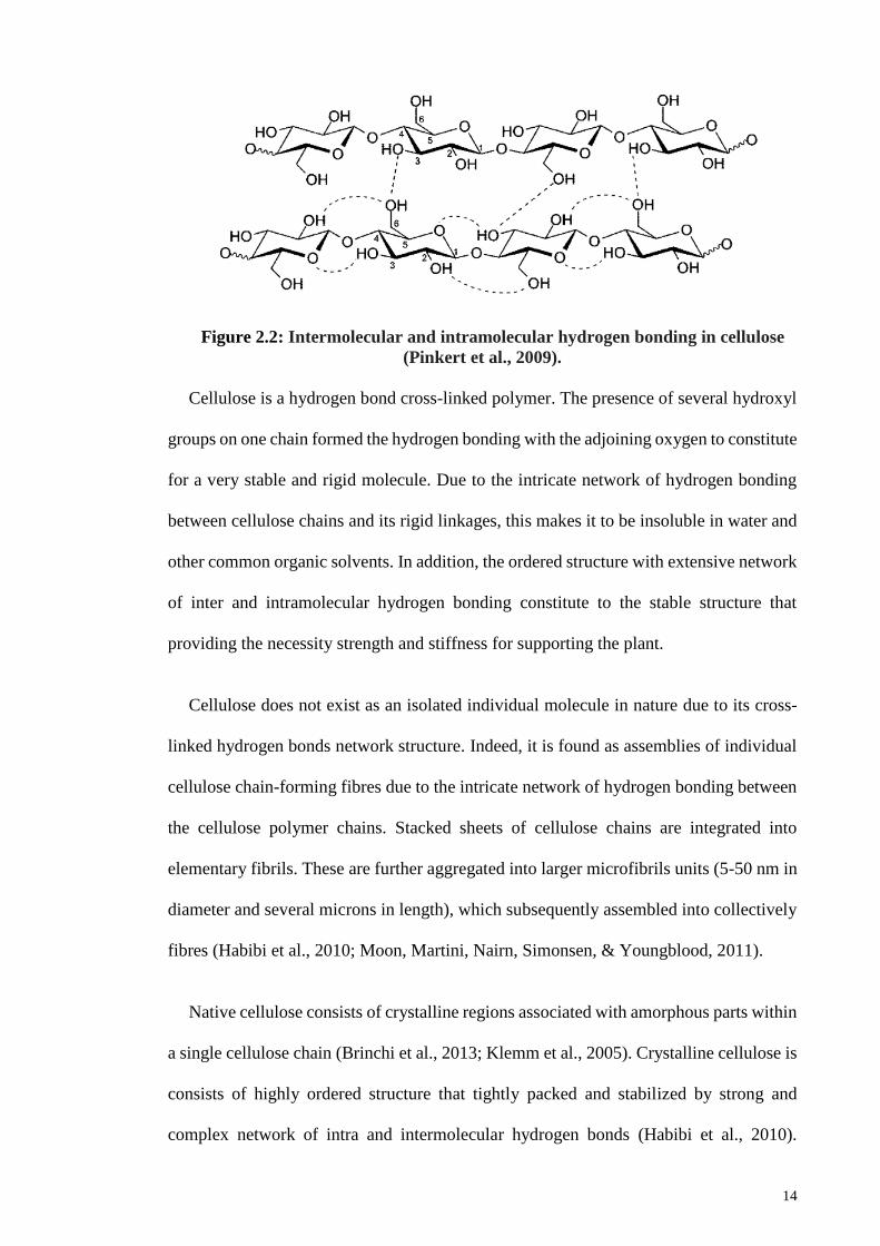
14
Figure 2.2: Intermolecular and intramolecular hydrogen bonding in cellulose
(Pinkert et al., 2009).
Cellulose is a hydrogen bond cross-linked polymer. The presence of several hydroxyl
groups on one chain formed the hydrogen bonding with the adjoining oxygen to constitute
for a very stable and rigid molecule. Due to the intricate network of hydrogen bonding
between cellulose chains and its rigid linkages, this makes it to be insoluble in water and
other common organic solvents. In addition, the ordered structure with extensive network
of inter and intramolecular hydrogen bonding constitute to the stable structure that
providing the necessity strength and stiffness for supporting the plant.
Cellulose does not exist as an isolated individual molecule in nature due to its cross-
linked hydrogen bonds network structure. Indeed, it is found as assemblies of individual
cellulose chain-forming fibres due to the intricate network of hydrogen bonding between
the cellulose polymer chains. Stacked sheets of cellulose chains are integrated into
elementary fibrils. These are further aggregated into larger microfibrils units (5-50 nm in
diameter and several microns in length), which subsequently assembled into collectively
fibres (Habibi et al., 2010; Moon, Martini, Nairn, Simonsen, & Youngblood, 2011).
Native cellulose consists of crystalline regions associated with amorphous parts within
a single cellulose chain (Brinchi et al., 2013; Klemm et al., 2005). Crystalline cellulose is
consists of highly ordered structure that tightly packed and stabilized by strong and
complex network of intra and intermolecular hydrogen bonds (Habibi et al., 2010).
15
Crystalline cellulose are strong and inaccessible structural elements. As against,
amorphous cellulose consists of disordered structure with chain segments that having the
length as short as the order of one cellobiose unit. Amorphous regions are weak and
accessible places of the fibrils which held by van der Waals forces (Ioelovich, 2016).
They are distinct from crystalline domains such that small molecules can easily penetrate
into the disordered region and access to the inner cellulose molecules. Expectedly,
amorphous cellulose is more chemically reactive and much prone to hydrolysis than the
corresponding crystalline parts reasonably due to the difference in their structural
arrangement (Moon et al., 2011; Pengfei, Kobayashi, & Fukuoka, 2011).
In addition, both inter- and intramolecular interactions and the molecular orientations
within the crystalline regions played a crucial role in differentiate the polymorphs or
allomorphs of cellulose. Cellulose exists in different crystal polymorphs in terms of unit
cell dimensions and hydrogen bonding patterns. Native cellulose generally consists of
cellulose I and it exists in two suballomorphs of cellulose Iα and Iβ. The existence of
cellulose I allomorphs is dependent on the origin of cellulose where Iα is widespread in
algae and bacterial cellulose, whereas Iβ is predominantly found in higher plants (Habibi
et al., 2010).
Cellulose I can be converted to other polymorphs such as cellulose II or III because it
is thermodynamically metastable. Cellulose II is the most stable structure because the
transformation of cellulose I to II is suggested to be irreversible (Pengfei et al., 2011).
This implies the difference in the hydrogen bonding patterns which give rise to different
crystalline structure. Cellulose II is more stable as the cellulose chains are stacking
alternatively in antiparallel modes between different hydrogen bonding planes. In
contrast, the parallel arrangement of cellulose chains aligned in the same direction caused
cellulose I to be less stable. Cellulose III and IV can be produced by treated with various
16
chemicals such as liquid ammonia or aqueous sodium hydroxide (Moon et al., 2011;
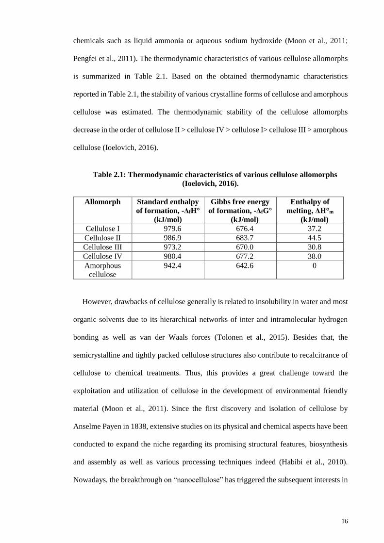
Pengfei et al., 2011). The thermodynamic characteristics of various cellulose allomorphs
is summarized in Table 2.1. Based on the obtained thermodynamic characteristics
reported in Table 2.1, the stability of various crystalline forms of cellulose and amorphous
cellulose was estimated. The thermodynamic stability of the cellulose allomorphs
decrease in the order of cellulose II > cellulose IV > cellulose I> cellulose III > amorphous
cellulose (Ioelovich, 2016).
Table 2.1: Thermodynamic characteristics of various cellulose allomorphs
(Ioelovich, 2016).
Allomorph Standard enthalpy
of formation, -ΔfH°
(kJ/mol)
Gibbs free energy
of formation, -ΔfG°
(kJ/mol)
Enthalpy of
melting, ΔH°m
(kJ/mol)
Cellulose I 979.6 676.4 37.2
Cellulose II 986.9 683.7 44.5
Cellulose III 973.2 670.0 30.8
Cellulose IV 980.4 677.2 38.0
Amorphous
cellulose
942.4 642.6 0
However, drawbacks of cellulose generally is related to insolubility in water and most
organic solvents due to its hierarchical networks of inter and intramolecular hydrogen
bonding as well as van der Waals forces (Tolonen et al., 2015). Besides that, the
semicrystalline and tightly packed cellulose structures also contribute to recalcitrance of
cellulose to chemical treatments. Thus, this provides a great challenge toward the
exploitation and utilization of cellulose in the development of environmental friendly
material (Moon et al., 2011). Since the first discovery and isolation of cellulose by
Anselme Payen in 1838, extensive studies on its physical and chemical aspects have been
conducted to expand the niche regarding its promising structural features, biosynthesis
and assembly as well as various processing techniques indeed (Habibi et al., 2010).
Nowadays, the breakthrough on “nanocellulose” has triggered the subsequent interests in
17
research by scientists and researchers from all over the world on nanocellulose and made
it as an important component in many practical applications.
2.3 Nanocellulose
2.3.1 Types of Nanocellulose
In general, nanocellulose is defined as cellulosic materials with at least one of its
dimension (length, width or diameter) within nanometer scale. Basically, nanocellulose
can be categorized into three main types accordingly with respect to its dimension,
preparation methods and functions, which in turn depend primarily on the sources and
processing conditions. Nevertheless, the nomenclature for nanocellulose has not yet
standardize and this presents ambiguities in the naming of cellulose particles with
dimensions in nanometer range (Klemm et al., 2011).
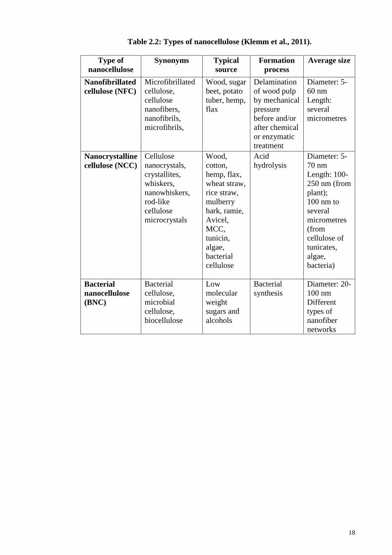
Nanocellulose can be divided into nanofibrillated cellulose (NFC), nanocrystalline
cellulose (NCC) and bacterial nanocellulose (BNC). NFC and NCC usually are obtained
via top-down methods such as chemical, enzymatic or physical methodologies for
isolation and extraction from higher plants, crops, forest or agricultural residues. In
contrast, BNC is synthesized from glucose by a family of bacteria (Gluconoacetobacter
xylinius) through bottom-up approach (Klemm et al., 2011). The summary on the types
of nanocellulose and respective details are tabulated in Table 2.2. Figure 2.3 illustrated
the microstructure images of NFC, NCC and BNC that obtained from respective sources,
viewed under electron microscope.
18
Table 2.2: Types of nanocellulose (Klemm et al., 2011).
Type of
nanocellulose
Synonyms Typical
source
Formation
process
Average size
Nanofibrillated
cellulose (NFC)
Microfibrillated
cellulose,
cellulose
nanofibers,
nanofibrils,
microfibrils,
Wood, sugar
beet, potato
tuber, hemp,
flax
Delamination
of wood pulp
by mechanical
pressure
before and/or
after chemical
or enzymatic
treatment
Diameter: 5-
60 nm
Length:
several
micrometres
Nanocrystalline
cellulose (NCC)
Cellulose
nanocrystals,
crystallites,
whiskers,
nanowhiskers,
rod-like
cellulose
microcrystals
Wood,
cotton,
hemp, flax,
wheat straw,
rice straw,
mulberry
bark, ramie,
Avicel,
MCC,
tunicin,
algae,
bacterial
cellulose
Acid
hydrolysis
Diameter: 5-
70 nm
Length: 100-
250 nm (from
plant);
100 nm to
several
micrometres
(from
cellulose of
tunicates,
algae,
bacteria)
Bacterial
nanocellulose
(BNC)
Bacterial
cellulose,
microbial
cellulose,
biocellulose
Low
molecular
weight
sugars and
alcohols
Bacterial
synthesis
Diameter: 20-
100 nm
Different
types of
nanofiber
networks
19
Figure 2.3: Microstructure images of NFC, NCC and BNC where (a) cellulose
nanofibers from wood pulp (Chen et al., 2015), (b) cellulose nanofibrils from Kraft
pulp (Qing et al., 2013), (c) cellulose nanofibers from banana peel (Tibolla, Pelissari
& Menegalli, 2014), (d) cellulose nanocrystals from ramie (Dufresne & Belgacem,
2013), (e) cellulose nanowhiskers from jute fibers (Cao et al., 2012), (f) cellulose
nanocrystals from sugar-beet pulp (Azizi, Alloin & Dufresne, 2005), (g) bacterial
nanocellulose (de Olyveira et al., 2011), (h) bacterial nanocellulose from
Gluconacetobacter bacteria (Klemm et al., 2011) and (i) nanofibers of bacterial
nanocellulose from Gluconacetobacter strains (Klemm et al., 2011).
Nanofibrillated cellulose (NFC) is in synonyms with microfibrillated cellulose (MFC),
cellulose nanofiber or nanofibrils (CNF). NFC is the smallest structural unit of plant fiber
in which it usually presents in long and thin structure. Indeed, high aspect ratio of NFC
is expected because of its small width of about 4-20 nm and length of up to several
micrometers. In addition, alternating crystalline and amorphous domains in NFC
contribute to its flexibility (Brinchi et al., 2013). NFC is generally produced by
20
delamination of wood pulp due to mechanical pressure before and/or after chemical or
enzymatic treatment. However, very high consumption energy of over 25,000 kWh per
ton is required for defibrillation of NFC due to intensive mechanical treatment which
makes the production method not energy efficient (Brinchi et al., 2013; Khalil et al., 2014;
Klemm et al., 2011).
It is important to note the differences between NFC and NCC of comparable
dimensions. Nanocrystalline cellulose (NCC), usually denoted as cellulose nanocrystals
(CNC), cellulose nanowhiskers (CNW), whiskers or rod-like cellulose microcrystals. In
fact, NCC typically exhibits as elongated crystallite in rigid rod-like shape. It is usually
obtained through acid hydrolysis of cellulose sources to extract the crystalline regions
remained intact in NCC. In contrast to NFC, NCC has very limited flexibility because the
amorphous regions are removed. NCC has relatively lower aspect ratio with 5-70 nm
wide and 50-500 nm in length. The particles are 100 % cellulose and highly crystalline
(Moon et al., 2011). Nevertheless, the dimensions of NCC are not uniform due to random
cleaving of cellulose chains during hydrolysis process (Habibi et al., 2010; Leung, Lam,
Chong, Hrapovic, & Luong, 2013).
Factors such as cellulosic source, preparation technique and conditions are mainly
influencing the dimensions, morphologies as well as degree of crystallinity of NCC
(Habibi et al., 2010). Cellulose can be obtained from different sources whether it is from
higher plants, crops, bacteria or algae and aquatic animals. The cellulose that obtained
from a particular source may inherent specific intrinsic properties such as morphology
and crystallinity. For example, cellulose that obtained from higher plants usually exhibit
long and thin fibers with higher crystallinity. On the other hand, the preparation technique
and condition employed also significantly affected the dimensions, morphology and
crystallinity of NCC. For instance, NFC is often produced after defibrillation process of
21
mechanical treatments such as homogenizing whereas NCC is usually obtained after acid
hydrolysis process (Brinchi et al., 2013). Besides that, duration of hydrolysis also affects
the dimensions of crystals, whereby a shorter crystal is produced upon hydrolyzed at a
longer time. Despite that, larger dimension NCC is derived from tunicate and bacterial
cellulose as compared to those from the higher plants. This is reasonably due to highly
crystalline structure of tunicate and bacterial cellulose that results in cleavage of lower
fractions of amorphous regions (Klemm et al., 2011).
There is another type of nanocellulose known as bacterial nanocellulose (BNC).
Terminology such as bacteria cellulose, microbial cellulose or biocellulose are refer to
BNC. In particular, it is derived from aerobic bacteria of genus Gluconacetobacter, which
is a kind of acetic acid bacteria that can be found widespread in nature on the occurrence
of fermentation of sugars and plant carbohydrates. BNC is actually derived through the
bottom-up approach. It is formed through a more natural way as BNC is bio-fabricated
from low molecular weight carbon precursor such as glucose and assembled into a
polymer nanomaterial. BNC is formed as exopolysaccharide at the interface to the air and
exists in stable hydrogel form. It is consists of network of nanofiber with diameter range
20-100 nm enclosed by high proportion of water. On contrary to NC, BNC has very high
purity and high molecular weight with highly crystalline and mechanically stable
structure. In addition, BNC is capable to have a controllable shape and structure of the
nanofiber during biosynthesis with fermentation technology. Thus, this creates an
exciting alternative to fabricate cellulose through bottom-up approach (Klemm et al.,
2011).
Thus, optimum content of cellulose and possible formation of nanocellulose are
important issues to address in order to form an ideal nanoscale cellulose with desired
properties.
22
2.3.2 Advantageous properties of nanocellulose
Nanocellulose is a novel biomaterial to base a new biopolymer composites industry
due to its promising and surpass physical and chemical properties. Indeed, it can utilized
in myriad of potential applications attributed to its nanoscale dimension. As mentioned
previously, nanocellulose has many advantageous properties such as low density (1.6
g/cm3) (Moon et al., 2011), large surface area (150–250 m2/g), high aspect ratio (70)
(Lam, Male, Chong, Leung, & Luong, 2012) and modifiable surface properties. Besides
that, the surface of nanocellulose can be functionalized to tailor particle surface chemistry
due to presence of many hydroxyl groups on the surface. For instance, surface
functionalization enables self-assembly, modification to alter hydrophilicity, to control
dispersion within other matrix polymers, to enable anchoring of metallic nanoparticles on
stabilized matrix or to prepare for targeted applications (Lam et al., 2012; Leung et al.,
2013).
Besides that, nanocellulose exhibits extraordinary mechanical properties such that its
axial Young’s modulus is as high as 167.5 GPa which is close to the value derived
theoretically and potentially stronger than steel and comparable to Kevlar, within the
range of other reinforcement materials. Elastic modulus of nanocellulose from cotton and
tunicate could reach up to 105 and 143 GPa, respectively. Meanwhile, the tensile strength
of nanocellulose is estimated to be 0.8-10 GPa (Moon et al., 2011). As one of the strongest
and stiffest biomaterial, it contains only a small number of defects. Therefore,
nanocellulose can be used as reinforcement material in composite to provide superior
mechanical performance.
The surpass characteristic of nanocellulose not only in their physical and chemical
properties, but also in thermal properties. Nanocellulose exhibits very low coefficient of
thermal expansion. It is a well-known fact that nanocellulose has inherent renewability,
23
sustainability as well as biodegradability and biocompatibility. Furthermore,
biodegradability of nanocellulose has been evaluated according to OECD standard 301B
(Organization for Economic Co-operation and Development) at where cellulose
nanoparticles were degraded in aqueous environment much faster than that its
corresponding macroscopic structure (Brinchi et al., 2013; Kümmerer et al., 2011). On
the other hand, other carbon-based nanomaterials such as fullerenes and carbon nanotubes
are non-biodegradable at all. Therefore, nanocellulose can be applied as environmental
friendly material such as biodegradable plastic to replace the dependence on
petrochemical-based products.
Nanocellulose behaves differently at the nanometer level as compared to its bulk
counterparts, presumably can be explained by the laws of atomic physics (Brinchi et al.,
2013; Moon et al., 2011). Indeed, nanocellulose is the fundamental building blocks for
macroscopic cellulose. Therefore, in the present work, nanocellulose is fabricated with
the aim to provide an environmental friendly materials as an alternative to plastics
materials, in addition to its excellent physical, chemical as well as thermal properties.
2.3.3 Potential applications of nanocellulose
Nanocellulose has gained tremendous attentions during the past decades owing to its
nanoscale dimension and many others promising physical as well chemical properties. A
broad range of applications of nanocellulose exist and summarized as below:
2.3.3.1 Improvement of nanocomposite mechanical properties
Nanocellulose can be used as filler in nanocomposites applications widely to improve
the mechanical properties. Since its introduction over the past decades, the applications
of nanocellulose as reinforcement materials have been increased exponentially due to its
outstanding strength and high stiffness.
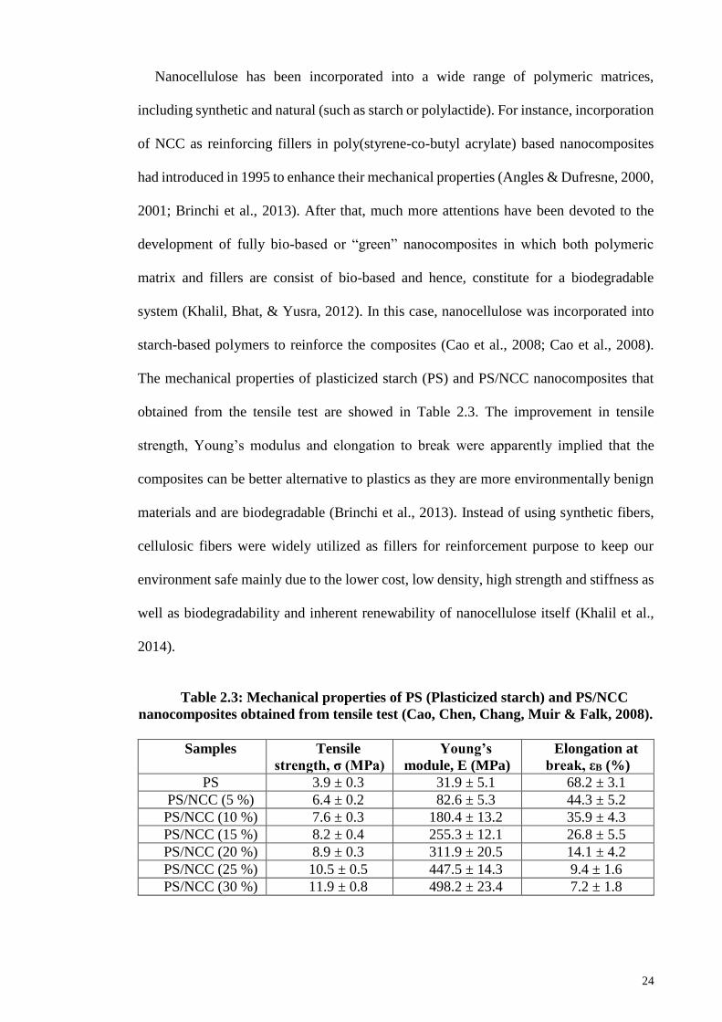
24
Nanocellulose has been incorporated into a wide range of polymeric matrices,
including synthetic and natural (such as starch or polylactide). For instance, incorporation
of NCC as reinforcing fillers in poly(styrene-co-butyl acrylate) based nanocomposites
had introduced in 1995 to enhance their mechanical properties (Angles & Dufresne, 2000,
2001; Brinchi et al., 2013). After that, much more attentions have been devoted to the
development of fully bio-based or “green” nanocomposites in which both polymeric
matrix and fillers are consist of bio-based and hence, constitute for a biodegradable
system (Khalil, Bhat, & Yusra, 2012). In this case, nanocellulose was incorporated into
starch-based polymers to reinforce the composites (Cao et al., 2008; Cao et al., 2008).
The mechanical properties of plasticized starch (PS) and PS/NCC nanocomposites that
obtained from the tensile test are showed in Table 2.3. The improvement in tensile
strength, Young’s modulus and elongation to break were apparently implied that the
composites can be better alternative to plastics as they are more environmentally benign
materials and are biodegradable (Brinchi et al., 2013). Instead of using synthetic fibers,
cellulosic fibers were widely utilized as fillers for reinforcement purpose to keep our
environment safe mainly due to the lower cost, low density, high strength and stiffness as
well as biodegradability and inherent renewability of nanocellulose itself (Khalil et al.,
2014).
Table 2.3: Mechanical properties of PS (Plasticized starch) and PS/NCC
nanocomposites obtained from tensile test (Cao, Chen, Chang, Muir & Falk, 2008).
Samples Tensile
strength, σ (MPa)
Young’s
module, E (MPa)
Elongation at
break, εB (%)
PS 3.9 ± 0.3 31.9 ± 5.1 68.2 ± 3.1
PS/NCC (5 %) 6.4 ± 0.2 82.6 ± 5.3 44.3 ± 5.2
PS/NCC (10 %) 7.6 ± 0.3 180.4 ± 13.2 35.9 ± 4.3
PS/NCC (15 %) 8.2 ± 0.4 255.3 ± 12.1 26.8 ± 5.5
PS/NCC (20 %) 8.9 ± 0.3 311.9 ± 20.5 14.1 ± 4.2
PS/NCC (25 %) 10.5 ± 0.5 447.5 ± 14.3 9.4 ± 1.6
PS/NCC (30 %) 11.9 ± 0.8 498.2 ± 23.4 7.2 ± 1.8

25
In contrast to micro-size composites, nanoscale composites are more advantageous
because they exhibit lower defects for reinforcement. More recently, extensive
applications of NFC have showed to improve the tensile performance of phenolic resin,
hydroxypropylated cellulose (HPC), amylopectin, polyurethane and melamine
formaldehyde composites (Khalil et al., 2014). The reinforced tensile strength and
modulus of nanocomposites are attributed to the percolation of NFC that formed a rigid
network of cellulose nanofibrils that entangled themselves within the polymer matrix of
nanocomposites. Therefore, high mechanical properties of nanocomposites could be
expected than that of the neat polymer in its elastomeric state. Besides that, incorporation
of NFC with polylactic acid (PLA) has increased the storage and tensile modulus of
elasticity of the composite materials based on dynamic mechanical analyser (DMA). As
mentioned previously, the stiffness and high Young’s modulus of nanocellulose (167.5
GPa) can significantly reinforced the tensile strength of the composite materials (Khalil
et al., 2014; Lee, Aitomäki, Berglund, Oksman, & Bismarck, 2014). Since then, several
attempts have been made to explore the mechanical performance of hybrid system of
nanocellulose with a variety of polymers and particles.
2.3.3.2 Improved barrier properties of nanocomposites
According to recent literatures, nanocellulose can be acted as a potential packaging
material in light of its superior mechanical performance because it is safe and
biodegradable (Leung et al., 2013). As a matter of fact, packaging appeared as the largest
industry for plastic materials consumption and this situation also responsible for main
sources of problems regarding waste disposal (Brinchi et al., 2013). In general, food
packaging materials are usually derived from petrochemical based polymers which are
non-degradable and this poses a serious threat to the environment. In recent years, safe
and environemtally friendly bio-based materials have getting increasing demand to use
as food packaging materials. Therefore, this driven the discovery of nanocellulose as
26
desirable bio-based nanocomposites is required due to its inherent abundance and
renewability. Besides that, problems with waste disposal and pollution can be reduced
consecutively because of the biodegradable property of nanocellulose.
In addition to mechanical strength, packaging materials for food are particularly
required barrier for oxygen molecules, moisture migration, flavour as well as aroma
control (Khalil et al., 2014). For instance, incorporation of nanocellulose into
xylan/sorbitol films to form biodegradable barrier membranes have been significantly
reduced the water transmission and oxygen permeability as compared to ethylene vinyl
alcohol that commonly used in barrier plastics. The main reason might be attributed to
the highly crystalline structure and establishment of integated matrix (dense percolating
network) through the formation of intricate and rigid network of hydrogen bonding with
polymer matrix to improve the barrier properties of nanocellulose film. The extensive
network of cross-linking thereby, hindered the mobility and diffusivity of the water vapor
and gas molecules. As a matter of fact, nanocellulose film or membranes practically
composed of good barrier properties because interactions between the particles and
polymer molecules are strong enough by integrating smaller size particles with the
surrounding polymer material (Belbekhouche et al., 2011; Minelli et al., 2010; Rojas,
Bedoya, & Ciro, 2015). Thereby, the mobility of chain segments and penetrant diffusivity
of water vapour and oxygen molecules are significantly reduced (Dufresne, 2013).
2.3.3.3 Biomedical applications
Nowadays, nanocellulose has a wide variety of applications in biomedical fields arises
from its safety and efficacy. By far, toxicity tests have been proved that nanocellulose is
non-toxic to human cell and it also does not cause any serious environmental concerns.
Furthermore, nanocellulose has been demonstrated that utility for enzyme
immobilization, biosensors as well as bioimaging applications. In fact, NCC is a potential
27
carrier inside cells due to its nanoscale dimension, benign nature composition as well as
its modifiable and hydrophilic surface which enable the binding of various cell markers,
protein molecules or receptors for targeted applications. It has been labelled with
fluorescent moiety fluorescin-5-isothiocyanate in fluorescence bioassay which used as an
indicator for in vivo localization and quantification of nanoparticles within a cell (Brinchi
et al., 2013). Due to the benign nature, high available surface area, smoothness and
reduced porosity, CNF have been reported to be a potential substrates for biosensors.
These biosensors are typically prepared by binding peptides or proteins to the support
matrix. Activated CNF substrates have been shown to bind to bovine serum albumin
(BSA), providing for diagnostics.
Besides that, nanocellulose can be used as substrate or supporting matrix for enzyme
immobilization arises from its large surface area and nonporous structure. For instance,
immobilized peroxidase on NCC through activation with cyanogen bromide has been
improved the removal of chlorinated phenolic compounds in aqueous solutions. The
immobilization provides enzyme deactivation effects by precipitated the products
induced by conjugating the amino groups. NCC and gold nanoparticles formed a
nanocomposite which serves as a matrix for enzyme immobilization, in particular
cyclodextrin glycosyl transferase (CGTase) and alcohol oxidase that immobilized on
NCC (Lam et al., 2012).
In addition, nanocellulose appeared as a potential detection and biosensing system due
to its benign characteristic. The hydroxyl groups on nanocellulose can be further modified
and conjugated with different biological moieties and to bind with other molecules such
as peptides or proteins to the support matrix for targeted applications. For instance,
cellulose can integrated with DNA molecules via bifunctional oxirane 1,4-butanediol
diglycidyl ether to form DNA/cellulose hybrid. Consequently, purification of
28
complimentary mRNA from total poly(A)-enriched RNA can be conducted by the
hybrids by affinity chromatography.
Apart from that, NCC were act as surface for the synthesis of metal nanoparticles
through a reduction method. Due to their chiral characteristics and structuring ability,
NCC can be used to promote the alignment of inorganic particles. In this context, the
metal nanoparticles were reduced on the surface of NCC with the use of cationic
surfactants CTAB in order to achieve a controllable dimensions of the nanoparticles.
Moreover, electrical detection of DNA hybridization can be probed by metal
nanoparticle/NCC nanocomposites to identify the complementary target DNA sequence
when Ag/carboxylated NCC nanocomposites or Ag-Pd/carboxylated NCC materials are
used as labels (Lam et al., 2012). Nanocellulose-based biosensors can be used to detect
human neutrophil elastase by peptide conjugation. Non-porous cellulosic films can be
provided for diagnostics by conjugation of NFC substrates with bovine serum albumin
(BSA). On the other hand, bacterial nanocellulose already exists in applications regarding
medical devices and tissue engineering such as wound dressing, implants including
cardio-vascular graft, as well as scaffolds for tissue replacement (Brinchi et al., 2013;
Salas, Nypelö, Rodriguez-Abreu, Carrillo, & Rojas, 2014). These required for the
properties of high crystallinity and purity of cellulose nanofibrils excreted by
Gluconacterobacter xylinum bacteria which are apparently low cytotoxicity and
genotoxicity.
2.3.3.4 Drug delivery
Apart from that, nanocellulose has been proposed for suitably targeted drug delivery
arises from its exceptional physical properties such as nanoscale dimension, high surface
area and modifiable surface properties. The hydroxyl groups on the surface of
nanocellulose can be tailored for functionalization to enable modification and grafting.
29
Besides that, nanocellulose show no cytotoxicity which makes it suitable for
internalization within cells. Chemically modified nanocellulose offers as targeted drug
delivery excipient for both hydrophilic ionisable water soluble antibiotics and
hydrophobic anticancer drugs. Likewise, the surface of NCC was coated with CTAB in
order to bind significant quantities of hydrophobic anticancer drugs such as paclitaxel,
etoposide and docetaxel, so as to release the drugs in a controlled manner over a period
of several days (Brinchi et al., 2013; Jackson et al., 2011). Moreover, ionisable drugs
tetracycline and doxorubicin are integrated with commercial softwood NCC to release the
drugs rapidly (Lam et al., 2012). Additionally, NCC was proposed as a carrier in targeted
delivery of therapeutics in which the delivery of folic acid for targeting mammalian brain
cancer tumors can be beneficial from the shape of NCC (Salas et al., 2014).
2.3.3.5 Nanopaper for flexible electronics
Other than that, nanostructure cellulose can be utilized to form cellulose nanopaper.
Cellulose nanopaper refers to a network constructed by intertwining nanofibrils
predominantly of high aspect ratio with random surface orientation. In this manner,
nanopaper can be beneficial from its high strength and transparency with low coefficient
of thermal expansion as well as less defects characteristics. It then can be processed to
use as a template for electronic materials (Khalil et al., 2014). As such, minimal porosity
and densely packing within the solid film enhanced the conductivity of nanocellulose
film. With the benign character of nanocellulose such as lightweight and good chemical
and thermal stability, it became the promising candidate for developing flexible electronic
devices.
Recently, CNF showed promising applications to work as precursor of carbon
nanofibers which can be used as anode material in sodium-ion batteries (Luo et al., 2013).
Moreover, it was shown that carbon fibers derived from CNF exhibit superior
30
reversibility, the rate capability required for fast charging and excellent cycling capacity
(Nystrom et al., 2010). Similarly, CNF-based supercapacitors have been prepared in
combination with various electroconductive materials such as graphene, carbon nanotube
or indium tin oxide (Gao et al., 2013; Legnani et al., 2008). CNF-based electroactive
composites was showed to be conductive, electroactive and suitable for energy storage
and electrochemically controlled separation. The above-mentioned described the
advances of nanocellulose in flexible electronics and also highlighted the applications of
nanocellulose as a supporting or active materials for flexible electronic devices (Li et al.,
2013). Thus, nanocellulose can be served as an efficient supercapacitor to enable the
formation of conductive composite when conjugated with conductive materials (Salas et
al., 2014).
2.4 Potential techniques for synthesizing nanocellulose
It is a well-known fact that depolymerisation of cellulose into nanocellulose is
generally difficult under mild conditions attributed to the stiff structure and extensive
network of inter and intramolecular hydrogen bonding in cellulose (Yuan et al., 2015).
The crystallites domains having three-dimensional order are strong and inaccessible
structural elements. The ordered structure and stiffness of chains within the crystalline
regions are attributed to the intricate and extensive network of hydrogen bonding which
constitutes for its recalcitrant to the hydrolysis (Ioelovich, 2016). Practically,
recalcitrance of crystalline cellulose to hydrolysis caused the hydrolysis process often
required harsh conditions such as the use of highly concentrated acids, intensive
mechanical treatments, high temperature and pressure systems are required to cleave the
strong bonds in order to open its structure to expose the reactive groups (Lee, Hamid, &
Zain, 2014). These appeared as major obstacle for its further exploitation.
31
Lately, there are a number of existing methods for the production of nanocellulose,
either from its raw materials such as wood pulp, wheat straw, sugar-cane bagasse or other
modal compounds including microcrystalline cellulose (MCC) and filter papers (Ahmadi,
Madadlou, & Sabouri, 2015; Li et al., 2012; Palme, Theliander, & Brelid, 2016;
Satyamurthy & Vigneshwaran, 2013). In fact, these methods employed could be divided
into chemical reactions (acid hydrolysis, oxidation), mechanical (high pressure
homogenization, grinding, microfluidization, cyrocrushing) and biological treatment
(enzymatic hydrolysis). Generally, numerous studies have been proved that top-down
approaches were successfully applied and subjected to destruct and disintegrate the
cellulosic fibres into nanostructure by controlling their dimensions and crystallinity
structure accordingly (Khalil et al., 2014; Lee, Hamid & Zain, 2014; Qing et al., 2013).
In the following section, the most commonly employed techniques are briefly reviewed.
2.4.1 Acid hydrolysis
Among various preparation techniques available as mentioned, acid hydrolysis has
been commonly used technique for the preparation of NCC or cellulose nanocrystals
(CNCs). Mineral acids such as sulfuric, hydrochloric and phosphoric acids are usually
used for hydrolysis process (Habibi et al., 2010). Typically, amorphous regions of
cellulose are preferentially hydrolyzed during hydrolysis because they have higher
accessibility to hydronium ions generated by the acids and more susceptible to hydrolytic
cleavage (Lee et al., 2014). As a consequence, individual crystallites were liberated after
the hydrolysis process for synthesizing high crystalline structure of NCC or CNCs (Palme
et al., 2016).
In summary, it was found that there are some important parameters should be taken
into consideration during hydrolysis including reaction temperature, time, agitation, acid
to cellulose ratio, types and concentration of acids. These factors played an important role
32
in controlling the dimension and properties of synthesized nanocellulose. In fact, typical
hydrolysis reaction was quenched by dilution of excess water and followed by washing
with repeated centrifugations. Additional steps such as dialysis against water were
required in order to remove free acid molecules from the suspension. Lastly, a mechanical
treatment, typically sonication is applied to disperse the NCC homogenously for size
uniformity (Klemm et al., 2011).
Nevertheless, the types of acid used for the hydrolysis will influenced the surface
properties of resulting NCC. For instance, improved colloidal stability of NCC
suspension has been acquired with sulfuric acid hydrolysis due to the effect of anionic
stabilization via electrostatic repulsion of negatively charged sulfate groups. The
esterification of surface hydroxyl groups of cellulose eventually caused the grafting of
anionic sulfate ester groups which resulted in improving the dispersion of NCC in water.
However, thermal stability of NCC was compromised due to the presence of sulfate
groups (Dufresne, 2013). On the other hand, NCC suspension formed by hydrochloric
acid hydrolysis has limited dispersion and likely flocculate, which explained its poor
colloidal stability.
Typically, the sulfuric acid hydrolysis for synthesizing nanocellulose were performed
at a concentration of about 64-70 wt%, with temperature range from room temperature
up to 70 °C and varied the reaction time from 30 minutes to overnight depending on the
temperature employed. In contrary, hydrochloric acid hydrolysis usually were conducted
at reflux temperature with acid concentration of 2.5-4 N at varied time of reaction,
depending on the cellulosic sources. It was found that smaller dimension (less than 10 nm
width) NCC was acquired at higher temperature of 72 °C and prolonged hydrolysis up to
2 hours with 63.5 wt% sulfuric acid (Elazzouzi-Hafraoui et al., 2007; Habibi et al., 2010).
In general, the higher the temperature and the longer the time of reaction will resulted in
33
the production of nanocellulose with significantly lower crystallinity as well as lower
yield. This is because the progressive and uncontrollable hydrolysis conditions caused the
removal of both amorphous and crystalline domains of cellulose which consequently
lower the cellulose content of the materials.
Despite its effectiveness in preparation of NCC, acid hydrolysis still remains a crucial
drawback that is excessive usage of concentrated acids. In this case, massive concentrated
acids waste will be produced after the hydrolysis process, thus, proper waste management
and disposal is required. Generally, hazardous concentrated acid waste usually is high
toxicity that will eventually cause severe environmental issues. In addition of that,
concentrated acids created serious corrosion to synthesis equipment and reactor. This
drawbacks make the production cost of acid hydrolysis process relatively expensive due
to the extra efforts and money is required in order to establish a high corrosion resistant
reactor. The other drawback of this method is the wastage of solution after the hydrolysis
process. The acid waste is difficult to regenerate and recover after the hydrolysis,
manifesting that the process is economically infeasible.
Low yield of NCC is another drawback when applying the acid hydrolysis method in
the production of nanocellulose. This is reasonably due to uncontrollable hydrolysis under
harsh condition (concentrated acid and high temperature system) would lead to over
degradation of cellulose into reducing sugars and other undesirable side products (Brinchi
et al., 2013; Lee at al., 2014). Therefore, intensive care and strictly controlled of acid
hydrolysis processing conditions are demanding to generate desirable NCC.
2.4.2 Oxidation synthesis
Oxidation synthesis or TEMPO-mediated oxidation is a most prominent and well-
established process to form nanocellulose using 2,2,6,6- tetramethylpiperidinyl-1-oxyl
(TEMPO) radicals. During the process, C6 hydroxyls of cellulose acted as the primary
34
alcohol groups and are selectively oxidized to carboxylate. Subsequently, presence of
significance numbers of carboxylate and aldehyde groups triggered from regioselective
oxidation imposed a higher density of negatively charged groups on the surface of
cellulose chains. This condition could generates sufficient interfibrillar repulsion between
the fibrils for effective fibrillation to overcome the adhesion between the cellulose fibrils
(Masruchin et al., 2015; Rattaz, Mishra, Chabot, & Daneault, 2011). In fact, TEMPO-
mediated oxidation could be modified the surface properties of nanocellulose toward
carboxylated to prevent the establishment of strong interfibrillar hydrogen bonds (Qing
et al., 2013; Rattaz, Mishra, Chabot, & Daneault, 2011; Saito, Kimura, Nishiyama, &
Isogai, 2007). Thereby, the interfibrillar repulsion forces generated via TEMPO-mediated
oxidation facilitated the isolation of nanofibers by inducing defibrillation and
disintegration of cellulose microfibrils.
Many literatures reported that TEMPO-mediated oxidation actually retained the
crystallinity and fibrous morphology of nanocellulose (Khalil et al., 2014; Rattaz et al.,
2011). In this case, the oxidation reaction happens only at the surface of cellulose fibrils
instead of penetrate into the crystallites interior. Besides that, the oxidation process could
give rise to the individual very small width fibrils (3-5 nm) with intact crystallinity
(Johnson, Zink-Sharp, & Glasser, 2011). The reasons might be attributed to establish
repulsion between the negatively charged cellulose fibrils resulted in effective
defibrillation of fibrils to isolate the individual nanofibers. In addition, oxidation
synthesis is a highly selective catalytic process which contributed to the high rate of
reaction and yield of nanofibers (Rattaz et al., 2011).
Despite the prospective effectiveness of TEMPO oxidation, there are some
shortcomings with respect to environmental concerns caused by TEMPO radicals and
high production cost. In addition, it must be taken into account that TEMPO radicals are
35
relatively expensive and toxic (Brinchi et al., 2013). Furthermore, the oxidation process
was typically carried out under alkaline condition of pH > 12 and the process must be
accompanied by the use of halide-based reagents such as sodium hypochlorite (NaClO)
and sodium bromide (NaBr) as the co-oxidizing agents (Khalil et al., 2014). This situation
caused a serious impact to our environmental due to highly basic condition and
detrimental effect of halide-based reagents to our aquatic ecosystem. Although the
process has been introduced since 14 years ago, however, it is still not yet
commercialization in operation up to today due to the above mentioned concerns (Rattaz
et al., 2011).
2.4.3 Steam explosion
Steam explosion is also widely employed due to its capability in separating nanofibers
directly from lignocellulosic biomass (Cherian et al., 2008). The process typically
requires high pressure steaming followed by fast decompression to depolymerize and
defibrillate the cellulose microfibrils with the ultimate goal to produce nanofibers or NFC.
Before the process is carried out, biomass sample is first milled into smaller components.
Then, the dry material is saturated with steam at elevated temperature and pressure for a
particular reaction time. The pressure adopted for steam explosion is typically range 14-
16 bar, with a temperature range of 200-270 °C for a relatively short time (20 s to 20 min)
(Brinchi et al., 2013). The pressure in digester is then released immediately by opening
the steam to allow the material to expose to normal atmospheric pressure. Consequently,
flash evaporation of water will be happen due to sudden dropped of pressure generates a
thermo-mechanical force on the cellulosic material, which caused cellulose to rupture
(Cherian et al., 2010). Indeed, the lignocellulosic structure is broken down arises from
the explosion.
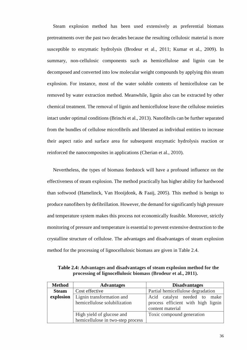
36
Steam explosion method has been used extensively as preferential biomass
pretreatments over the past two decades because the resulting cellulosic material is more
susceptible to enzymatic hydrolysis (Brodeur et al., 2011; Kumar et al., 2009). In
summary, non-cellulosic components such as hemicellulose and lignin can be
decomposed and converted into low molecular weight compounds by applying this steam
explosion. For instance, most of the water soluble contents of hemicellulose can be
removed by water extraction method. Meanwhile, lignin also can be extracted by other
chemical treatment. The removal of lignin and hemicellulose leave the cellulose moieties
intact under optimal conditions (Brinchi et al., 2013). Nanofibrils can be further separated
from the bundles of cellulose microfibrils and liberated as individual entities to increase
their aspect ratio and surface area for subsequent enzymatic hydrolysis reaction or
reinforced the nanocomposites in applications (Cherian et al., 2010).
Nevertheless, the types of biomass feedstock will have a profound influence on the
effectiveness of steam explosion. The method practically has higher ability for hardwood
than softwood (Hamelinck, Van Hooijdonk, & Faaij, 2005). This method is benign to
produce nanofibers by defibrillation. However, the demand for significantly high pressure
and temperature system makes this process not economically feasible. Moreover, strictly
monitoring of pressure and temperature is essential to prevent extensive destruction to the
crystalline structure of cellulose. The advantages and disadvantages of steam explosion
method for the processing of lignocellulosic biomass are given in Table 2.4.
Table 2.4: Advantages and disadvantages of steam explosion method for the
processing of lignocellulosic biomass (Brodeur et al., 2011).
Method Advantages Disadvantages
Steam
explosion
Cost effective Partial hemicellulose degradation
Lignin transformation and
hemicellulose solubilization
Acid catalyst needed to make
process efficient with high lignin
content material
High yield of glucose and
hemicellulose in two-step process
Toxic compound generation
37
2.4.4 Mechanical treatments
In general, NFC can be obtained through defibrillation of cellulose as the intensive
mechanical treatments effectively diminished the cellulose fibers into nanofibers. Among
various preparation techniques available as mentioned earlier, mechanical treatments
been commonly used technique for the preparation of NFC by applying high-pressure
homogenization, refining, microfluidization, grinding and cryocrushing (Khalil et al.,
2014; Qing et al., 2013). These mechanical approaches can be operated solely or in
combination of several intensive mechanical forces for effective disintegration of fibers
into nanofibers. Indeed, these process caused irreversible changes in the resultant
nanofibers (Nakagaito & Yano, 2004).
Typically, high-pressure homogenization is one of the most promising prospects for
mechanical treatment to form NFC. Within this process, high velocity cellulose slurry is
passed across a vessel with a very small nozzle to generate a high pressure stream. High
velocity and pressure as well as impact and shear forces on fluid generate shear rates in
the stream that decrease the size of fibers to nanoscale (Frone et al., 2011). During the
high temperature and pressurized treatment, the carbohydrate linkage within cellulose
fractions was subjected to break down and rendered the release of shorter cellulose chains
with lower degree of polymerization in the nanofibers (Brodeur et al., 2011). With
homogenization, uniform size NFC with smaller diameter (2-5 nm) is usually formed and
this method accounted for its high efficiency and simplicity without the use of any organic
solvents (Jonoobi, Niska, Harun, & Misra, 2009; Khalil et al., 2014).
Despite its efficiency, a crucial problem associated with clogging is likely to occur
during homogenization process due to smaller diameter size of orifice (Jonoobi et al.,
2009). Hence, the fibers required to be diminished before subjected to homogenization
38
stage in order to prevent the clogging issue. Nevertheless, the homogenization process is
encouraged to apply with other mechanical treatments to further improve its
effectiveness. The number of passes or cycles and applied pressure are strongly dependent
on the nature of biomass sample. It was reported that the number of cycles is varied from
10-15 cycles with pressure 30-50 MPa (Habibi, Mahrouz, & Vignon, 2009). Similar to
homogenization, microfluidization also involves an integrated pressurized system within
the chamber to generate impact and shear forces on cellulosic fibers for defibrillation due
to interaction between colliding streams and channel walls (Ferrer, Filpponen, Rodríguez,
Laine, & Rojas, 2012; Qua, Hornsby, Sharma, & Lyons, 2011).
Apart from that, refining is another potential method to defibrillate nanofibers. In this
case, refining involves a combination of mechanical shearing as well as frictional forces
in the fibrillation process. Production of NFC is usually beneficial upon the breakage of
hydrogen bonding by mechanical forces and meanwhile, the abrasive actions generate
frictional forces on the surface of fibers for repulsion. Generally, softwood is much easier
to refine than hardwood ascribed to variation of cellulose content. Stelte and Sanadi
(2009) have applied refining and homogenization at 50 MPa on hardwood and softwood
pulps to fibrillate the nanofibers. They revealed the diameter of obtained nanofibers was
around 10-25 nm for both pulps. However, excessive refining and homogenization lead
to decrease in the failure strain and strength of softwood nanocellulose fibers.
Furthermore, they reported that refining process was more efficient and faster for
softwood compared to hardwood pulp as the structure of softwood was broken after 25
passes while hardwood fibers were remained intact after 75 cycles. This is reasonably
attributed to the internal and external fibrillation phenomenon of cellulose in hardwoods
and softwoods (Stelte & Sanadi, 2009). Internal fibrillation took place as a result of
hydrogen bonds breakage due to mechanical actions, while external fibrillation occurred
39
on the surface of fibers by abrasive actions (Khalil et al., 2014). Thus, the effectiveness
of refining process is determined by the number of cycles passing through the refiner.
A report in 2011 by Karande and co-workers suggested that the increase in the number
of passes or cycles through refining or grinding were practically decreased the size,
crystallinity, degree of polymerization as well as decreased the failure strain and strength
of the nanofibers. The reason might be attributed to breaking of hydrogen bonding within
the crystalline domains of cellulose after intensive mechanical actions. Besides that,
combination of frictional and shear forces provide defibrillation effects on the surface of
cellulose fibers by abrasive action (Karande, Bharimalla, Hadge, Mhaske, &
Vigneshwaran, 2011; Khalil et al., 2014). The results showed that there is a distinct
dependency of the number of cycles of refining on crystallinity, physical and mechanical
properties of nanofibers.
In addition, defibrillation of nanofibers can be achieved by cryocrushing. In this
process, crushing action generates a high impact force on the immediate frozen cellulosic
fibers. It is suggested that pressure exerted by crushing of ice crystals is the main reason
for the collapse of cellulosic structure. As a result, individual nanofibers were liberated
from the bundles of microfibrils as separate entities (Siró & Plackett, 2010). Defibrillation
process of cellulosic fibers involves combination of intensive mechanical shearing forces
as well as frictional forces. These fibers are diminished due to the breakdown of hydrogen
bonds and cell wall structures by impact shear forces and individualization of microfibrils
to nanoscale fibers by repulsion of surface fibers attributed to the frictional forces
generated through the abrasive actions (Siró & Plackett, 2010).
In summary, the aforementioned mechanical approaches have their own distinct
features over others. The research finding indirectly showed the importance role of the
homogenization and microfluidization adopted with high pressure system to generate
40
higher shear and impact to produce homogenous size distribution of smaller width NFC
(Spence, Venditti, Rojas, Pawlak, & Hubbe, 2011). On the other hand, refining and
grinding effectively caused defibrillation through abrasive actions in which the heat is
generated as frictional forces for the fibrillar repulsion (Siró & Plackett, 2010). Therefore,
different mechanical treatments have different shear mechanisms and varied intensity of
shear forces, which results in different effects on the characterization of NFC (size,
crystallinity and degree of polymerization) (Qing et al., 2013).
In overall, the above discussed mechanical treatments contribute to several drawbacks.
The most critical issue is primarily associated with high consumption of energy for
production of NFC from mechanical defibrillation process (Dufresne, 2013). Besides that,
dramatic reduction of yield and aggregation and entanglement of nanofibers are apparent
for this process. Moreover, monitoring as well as higher number of cycles which often
consists of several paths through the disintegration device are practically essential for
production of NFC. In addition, clogging of fiber suspensions within the reaction chamber
is often occurred for high pressure homogenization and microfluidization (Qing et al.,
2013). These problems present serious obstacle which hindered the successful
commercialization for production of nanocellulose.
Nevertheless, combination of several potential mechanical treatments with chemical
or enzymatic routes will be ideal for nanocellulose production (Khalil et al., 2014; Lee et
al., 2014). The defibrillation of cellulose fibers through mechanical treatments required
intensive energy and high power consumption. Chemical or enzymatic pretreatments are
often took place before mechanical fibrillation (Chauhan & Chakrabarti, 2012). It is worth
noting that appropriate pretreatments of cellulosic fibers could promote the accessibility
of hydroxyl groups, increase the inner surface, alter the crystallinity, and break the
cellulose hydrogen bonds and therefore, boost the reactivity of the fibers towards
41
subsequent mechanical fibrillation (Khalil et al., 2014; Szczęsna-Antczak, Kazimierczak
& Antczak, 2012). The synergistic effect can be benign from the efficiency and
effectiveness of the combined methods which will compromise for the energy demand
for effective isolation of smaller and uniform size nanocellulose (Khalil et al., 2014; Lee
et al., 2014).
2.4.5 Enzymatic hydrolysis
Based on the literatures, biological treatment such as enzymatic hydrolysis has been
shown its effectiveness in preparation of NFC (Duran, Lemes, Duran, Freer, & Baeza,
2011). Contrary to acid hydrolysis, it is much more benign from the environment point
of view. Therefore, cellulase enzyme has been widely used for the pretreatment of
biomass materials as well as hydrolysis of cellulose due to its high selectivity and mild
reaction condition. It is a well-known fact that the enzyme is capable of modify and
degrade the hemicellulose and lignin components, with the retention of cellulose
domains. In the meantime, restrictive and selective hydrolysis of specified component in
the cellulosic fibers also induced by the enzyme cellulase (Janardhan & Sain, 2007).
In general, enzymatic hydrolysis of cellulose is a multistep catalytic process in which
the cellulase enzyme is initially adsorbed onto the cellulose surface. The cellulose is
subsequently breakdown via synergistic action of multiple components of cellulase. The
reason attributed to a single enzyme component is not possible to degrade the fiber due
to the presence of different organic compounds within the complex structure of cellulose.
Lastly, desorption of cellulase from the cellulose residue render its release into the
supernatant (Kuo & Lee, 2009). It is believed that the synergistic action of
endoglucanases and exoglucanases/cellobiohydrolases are responsible for the enzymatic
hydrolysis of cellulose. Initial cleavage of internal bonds (non-covalent interaction)
within the disordered structure was done by endoglucanases because they tend to attack
42
the amorphous regions. Afterwards, exoglucanases/cellobiohydrolases attack on the
exposed glycosdic end of cellulose chains generated by endoglucanases to release shorter
cellulose chains. Subsequently, this ease the further breakdown of glycosidic linkages of
shorter chains by cellobiases/betaglucosidases into formation of nanocellulose where
defibrillation presumably occurred (Lee et al., 2014).
Enzymatic hydrolysis is usually carried out in combination with mechanical shearing
or acid hydrolysis in order to improve its efficiency (Brinchi et al., 2013). For instance,
bleached softwood pulp has been treated at combined mild enzymatic hydrolysis with
homogenization and refining to prepare NFC (Pääkkö et al., 2007). During the mechanical
treatment, the NFC were defibrillated through the combination of mechanical shearing
forces and frictional forces to break down the tight packing arrangement within the
cellulose crystalline domains. By doing so, this can increased the exposed surface area of
cellulose polymer chains that are accessible for adsorption by cellulose enzyme for further
hydrolysis. Nevertheless, the rate of cellulose hydrolysis is determined by the
accessibility of cellulase to bind with adsorption sites on crystalline cellulose, which is a
rate limiting step (Kuo & Lee, 2009). The rate of enzymatic hydrolysis is typically
dependent on the structural features or organization and cellulase accessibility for the
adsorption sites, which in turn dependent on the purity of cellulose (Igarashi et al., 2011).
The rate of enzymatic hydrolysis is relatively slow. Studies have demonstrated that
significant enzymatic activity was noticeable only after 5 days of incubation. This
confirmed the mild enzymatic hydrolysis that usually carried out over a period of 7 to 14
days of incubation. The size of nanocellulose is reduced as the time progressed due to the
continued hydrolysis catalyzed by the enzymes. Also, Satyamurthy and Vigneshwaran
have reported on the steady reduction in the degree of polymerization of nanocellulose
with the progress of hydrolysis over the period of 15 days. Despite that, there was a
43
significant increase in the crystallinity of the nanocellulose to 81 % as compared to 69 %
crystallinity of MCC. The increase in crystallinity is reasonably attributed to preferential
hydrolysis of the amorphous regions of MCC by cellulase enzymes (Satyamurthy &
Vigneshwaran, 2013).
The hydrolysis induced by enzyme cellulase give rise to the potentially higher yield,
higher aspect ratio of NFC and higher selectivity under milder synthesis conditions than
chemical route (Siddiqui, Mills, Gardner, & Bousfield, 2011). This process is beneficial
in terms of lower energy costs and more environmental benign. However, such technique
is impeded by the drawbacks of economical aspect associated with costly cellulase
enzyme and technical with respect to long processing time is usually required for cellulose
degradation (rate limiting step). These issues remain its primary obstacle from
commercialization of nanocellulose synthesis, although it offers a numbers of potential
benefits (Lee et al., 2014).
2.5 Ionic Liquids
Design and development of nanostructure cellulose has gained significant scientific
interest and become the most studied material as it exhibits promising physical,
mechanical and functional properties. An obvious hindrance to the widespread use of
cellulose is its compact and semi-crystalline structure which caused it to recalcitrant to
hydrolysis and has low solubility in water (George & Sabapathi, 2015; Kalia et al., 2011).
Therefore, considerable efforts have been exerted to form nanocellulose via green route
by following our governmental regulations and policies. Thus, utilization of ionic liquids
(ILs) in nanocellulose synthesis stage is essential that lead to the higher efficiency of
hydrolysis and dissolution (solvolysis) process in forming nano-sized cellulose (Dong,
Holm, & Lassi, 2015). In the subsequent section, the introduction regarding to the ILs,
advantageous properties as well as ILs for cellulose processing will be reviewed in detail.
44
2.5.1 Introduction to Ionic Liquids (ILs)
In this section, the brief introduction of ILs are reviewed. It is a well-known fact that
ILs are molten salts with relatively low melting temperature (<100 °C) and typically exist
in liquid form at low temperature (below or around 100 °C) (Liu, Sale, Holmes, Simmons,
& Singh, 2009; Pinkert et al., 2009). They comprise entirely of cations and anions at
where all of the ions are poorly coordinated. In fact, the cations of ILs consist of bulky,
unsymmetrical organic ions with a delocalised charge whereas the anions of ILs can
varied from halide ions to linear or branch organic ions. It can be understood that the
bulkiness of ions, low internal symmetrical structure between cations and anions with
charge delocalization as well as the poor coordination of the ions are responsible for the
lower melting temperature of ILs (Pinkert et al., 2009).
Contrary to ILs, most salts or ionic compounds have high melting points because the
electrostatic interactions that hold the compounds together are very strong. A good
example to be presented is that sodium chloride (NaCl) with high melting point of 801
°C and it exists as solid at room temperature. The strong coulomb attraction between
cations and anions give rise to the formation of strong and stable crystal lattice structure.
However, this phenomenon is not favorable for ILs due to the sufficiently distortion to
the ordered ions structure that required for lower melting temperature of ILs.
In 1914, ILs were firstly introduced by Walden. The innovation by Walden produced
the first viable IL was ethylammonium nitrate, featuring a melting point of 12 °C, which
has been revolutionized the ILs industry (Walden, 1914). Then, ILs with alkyl-substituted
imidazolium and pyridinium based cations were introduced in 1970s and 1980s
(Herrmann, 2015). However, numerous researches on ILs potential utilization was only
extensively studied and reported over the past few decades.
45
Recent studies have indicated that ILs can act as promising solvents, which can benefit
from their remarkable physiochemical properties including high thermal and chemical
stability, negligible vapour pressure, non-flammability, wide electrochemical window,
low volatility, large liquid range and high solvency power to dissolve various inorganic
and organic substances (Wang, Gurau, & Rogers, 2012). ILs have gained much attention
and became favourite research matter among various groups of scientists due to their
above-mentioned interesting and unique features. As a matter of principle, ILs have been
known as promising “green” solvents as an alternative source to replace conventional
volatile organic solvents (Hu et al., 2013). ILs have myriad of potential applications
including electrochemistry, catalysis, organic synthesis, polymer chemistry as well as
extraction (Liu et al., 2010; Pinkert et al., 2009).
The cations of ILs are often featuring an aromatic or cyclic structure and long alkyl
chain. Common cations are alkylimidazolium, alkylpyridinium and quaternary
ammonium based. Whereas the anions of ILs can be varied from chloride, formate,
acetate, bromide, tetrafluoroborate, hexafluorophosphate or methanesulfonate (Gericke,
Fardim, & Heinze, 2012). Some common examples of cations and anions for ILs with
their molecular structures are presented in Figure 2.4. In fact, various combinations of
cations and anions will contribute to the variety types of ILs. As such, task-specific ILs
(TSILs) are arises for particular applications with desirable properties. Also, room-
temperature ILs (RTILs) are liquid ILs which have melting temperature below room
temperature.
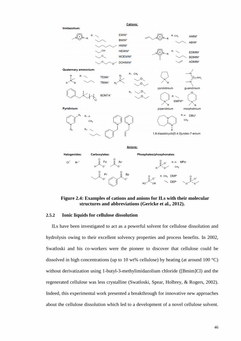
46
Figure 2.4: Examples of cations and anions for ILs with their molecular
structures and abbreviations (Gericke et al., 2012).
2.5.2 Ionic liquids for cellulose dissolution
ILs have been investigated to act as a powerful solvent for cellulose dissolution and
hydrolysis owing to their excellent solvency properties and process benefits. In 2002,
Swatloski and his co-workers were the pioneer to discover that cellulose could be
dissolved in high concentrations (up to 10 wt% cellulose) by heating (at around 100 °C)
without derivatization using 1-butyl-3-methylimidazolium chloride ([Bmim]Cl) and the
regenerated cellulose was less crystalline (Swatloski, Spear, Holbrey, & Rogers, 2002).
Indeed, this experimental work presented a breakthrough for innovative new approaches
about the cellulose dissolution which led to a development of a novel cellulose solvent.
47
Since then, utilization of ILs for biomass processing has been growing exponentially and
ILs were discovered to be effective in cellulose dissolution, as a reaction medium for
functionalization and chemical reactions, to form cellulose composite materials and to
produce cellulose films, fibers or beads. Besides that, ILs were capable to dissolve raw
biomass materials such as wood, shrimp shell, empty fruit bunches (EFB), rice husks and
many others (Wang et al., 2012).
In 2002, Swatloski and co-workers postulated that the dissolution of cellulose in
[Bmim]Cl was mainly attributed to the interaction between anions of IL with cellulose
(Swatloski et al., 2002). In particular, the solvation of cellulose might be attributed to the
coordination of chloride ions with the hydroxyl protons of cellulose. Then, the poorly
coordinated chloride ions were disintegrated themselves from [Bmim]Cl upon heat
treatment stage. Subsequently, the free chloride ions can interact with the hydroxyl groups
of cellulose by causing disruption to the hydrogen bonding within and between adjoining
cellulose polymer chains. The dissolution of cellulose in IL is believed due to the
hydrogen bonding interaction between chloride anions and hydroxyl protons of cellulose
(Wang et al., 2012).
In order to prove the hypothesis and gain insight into cellulose dissolution by ILs,
Zhang and coworkers performed dissolution of cellobiose (the repeating unit of cellulose)
in 1-ethyl-3-methylimidazolium acetate ([Emim]OAc) for 45-60 minutes at around 100
°C. It has been noted that formation of hydrogen bonding between acetate anions ([OAc]-
) and hydroxyls of cellobiose contributed to the solvation. In this case, acetate anions
favored the formation of hydrogen bonding with hydroxyl protons of cellobiose (Zhang
et al., 2010). The authors have been concluded that formation of hydrogen bonding
between anions of ILs and hydroxyl protons of cellulose are responsible for dissolution
of cellulose. As a consequence, the intermolecular hydrogen bonds between cellulose
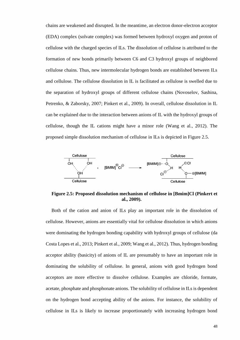
48
chains are weakened and disrupted. In the meantime, an electron donor-electron acceptor
(EDA) complex (solvate complex) was formed between hydroxyl oxygen and proton of
cellulose with the charged species of ILs. The dissolution of cellulose is attributed to the
formation of new bonds primarily between C6 and C3 hydroxyl groups of neighbored
cellulose chains. Thus, new intermolecular hydrogen bonds are established between ILs
and cellulose. The cellulose dissolution in IL is facilitated as cellulose is swelled due to
the separation of hydroxyl groups of different cellulose chains (Novoselov, Sashina,
Petrenko, & Zaborsky, 2007; Pinkert et al., 2009). In overall, cellulose dissolution in IL
can be explained due to the interaction between anions of IL with the hydroxyl groups of
cellulose, though the IL cations might have a minor role (Wang et al., 2012). The
proposed simple dissolution mechanism of cellulose in ILs is depicted in Figure 2.5.
Figure 2.5: Proposed dissolution mechanism of cellulose in [Bmim]Cl (Pinkert et
al., 2009).
Both of the cation and anion of ILs play an important role in the dissolution of
cellulose. However, anions are essentially vital for cellulose dissolution in which anions
were dominating the hydrogen bonding capability with hydroxyl groups of cellulose (da
Costa Lopes et al., 2013; Pinkert et al., 2009; Wang et al., 2012). Thus, hydrogen bonding
acceptor ability (basicity) of anions of IL are presumably to have an important role in
dominating the solubility of cellulose. In general, anions with good hydrogen bond
acceptors are more effective to dissolve cellulose. Examples are chloride, formate,
acetate, phosphate and phosphonate anions. The solubility of cellulose in ILs is dependent
on the hydrogen bond accepting ability of the anions. For instance, the solubility of
cellulose in ILs is likely to increase proportionately with increasing hydrogen bond
49
accepting ability of the anions (Itoh, 2014). The higher the hydrogen bond basicity or
accepting ability, the higher the ability of that particular anion of ILs to dissolve cellulose.
Anions with higher hydrogen bond basicity are necessity for cellulose dissolution because
the high basicity can practically weakens the inter and intramolecular hydrogen bonding
of cellulose. In addition, ILs containing non-coordinating or symmetry anions such as
BF4- and PF6
- cannot solubilize the cellulose (Pinkert et al., 2009; Wang et al., 2012).
Therefore, careful selection of potential anions to dissolve the cellulose is a challenging
task.
ILs with aromatic imidazolium and pyridinium cations are more effective to dissolve
cellulose when associated with a strong hydrogen bond basicity anions. Imidazolium and
pyridinium based cations are commonly used for cellulose dissolution because of their
aromatic nature and the ability to screen the solvate anion/cellulose polymer (EDA)
complexes. The aromatic rings structure contributes to lower relative interaction strengths
(lower symmetry) between the cations and anions due to reduction of electrostatic
strength. Moreover, the lower internal symmetry character essentially promotes charge
delocalization within the aromatic rings to make them more polarizable (Fernandes et al.,
2011). As such, anions are easier to form hydrogen bonds with cellulose (Wang et al.,
2012).
Apart from that, the length of alkyl chain substituents in the cations also influenced
the solubility of cellulose. Cellulose has lower solubility for longer alkyl chain length of
ILs cations. For example, [Bmim]Cl is more efficient to dissolve cellulose as compared
to 1-hexyl-3-methylimidazolium chloride, ([C6mim]Cl) and 1-octyl-3-
methylimidazolium chloride, ([C8mim]Cl). ILs containing longer alkyl chain length
cations tend to have lower solvation power to dissolve cellulose because of reduced
effective anion concentration or the hydrophobic interactions between cations which
50
likely reduce their ability to shield the solvate anion/cellulose complexes. Thus, cellulose
dissolution is lowered with the reduced effective anion concentration of ILs (Swatloski et
al., 2002; Wang et al., 2012). Alkyl groups in the cations of ILs are indirectly affected the
viscosity and dissolution of cellulose. The greater the number of alkyl chain length, the
more viscous the ILs and therefore, the reduced solubility of cellulose in ILs.
On the other hand, 1-alkyl-3-methylimidazolium-based ILs with even-numbered alkyl
chains, particularly below six carbon units exhibit higher capacities to solubilize cellulose
compared to odd-numbered alkyl chains. For instance, it is reported that cellulose is
practically unable to dissolve in [C3mim]Cl and [C5mim]Cl. While, ILs with even
numbered alkyl chain substituent cations such as [Bmim]Cl and [C6mim]Cl are far more
effective for cellulose dissolution. This phenomenon is attributed to odd-even effect
which contributes to additional polarity in the heteroatomic substituents on the
imidazolium ring and is more apparent for small alkyl chains (Pinkert et al., 2009; Wang
et al., 2012; Zhang, 2013).
Therefore, small and even-numbered alkyl chain substituent cations ILs that paired
with stronger hydrogen bond basicity anions are preferable in solvation of cellulose. The
roles of cations seems like to assist in solvating and separate the cellulose polymer chains
by interfere with hydrogen bonding of cellulose (Wang et al., 2012; Zhang, 2013). In
addition, cations also function to shield the solvate anion/cellulose polymer (EDA)
complexes in moieties.
Interestingly, ILs containing halide anions, especially anions, are particularly effective
for solvation of cellulose. It has been reported that cellulose has a solubility of 10 wt%
by conventional heating in [Bmim]Cl while with a greater solubility of up to 25 wt%
achieved by microwave heating (Rinaldi & Dwiatmoko, 2012). On the other hand,
although bromide anions ([Bmim]Br) and sulfocyanate anions ([Bmim]SCN)
51
alkyimidazolium based ILs are capable of dissolving cellulose, but their solubility is less
than half of [Bmim]Cl. Recent studies showed that different ILs including 3-methyl-N-
butylpyridinium chloride, 1-butyl-3-methylimidazolium chloride ([Bmim]Cl) and 1-
allyl-3-methylimidazolium chloride ([Amim]Cl) were capable to solubilize cellulose of
up to 39, 25 and 14.5 % wt% respectively (Dadi, Schall, & Varanasi, 2007; Isik, Sardon,
& Mecerreyes, 2014). The greater capability of chloride anions based ILs to dissolve a
high amount of cellulose are attributed to their high chloride concentration in ILs to
disrupt and extensively break down the hydrogen bonding in cellulose, without caused
derivatization (Cao et al., 2009).
Despite their effectiveness for dissolution of cellulose, ILs with chloride anions
possess several drawbacks. One of the main problem is relatively high viscosity of
chloride anions-based ILs which brought difficulties during dissolution process (Isik et
al., 2014). In general, dissolution of cellulose is strongly affected by viscosity of ILs.
High viscosity ILs practically unfavorable for dissolution and mass transfer due to
probable inefficiency mixing process (Gericke et al., 2012). [Bmim]Cl is known to exhibit
high viscosity of 11,000 MPa and melting point at 65 °C. The relatively high melting
point and viscosity of [Bmim]Cl caused dissolution of cellulose occurred at relatively
higher temperature (often above 80 C). Besides that, [Bmim]Cl is somewhat toxic and
exhibits high corrosive character. Its toxicity has been evaluated in which its median
lethal dose (LD50) is range from 50 mg kg-1 to 300 mg kg-1 and it is toxic to mice at
maternally toxic dosages (Wang et al., 2012). Therefore, the uncertain toxicity and
potential hazardousness of this IL that will be brought into ecosystems and zoology after
ineluctable release into the environment as well as corrosiveness are seriously limit their
practical applications in cellulose processing and derivatization in the industrial scale
(Itoh, 2014). The combination of cations and anions of ILs should be screened with
careful selections and their properties should be investigated such that the potential
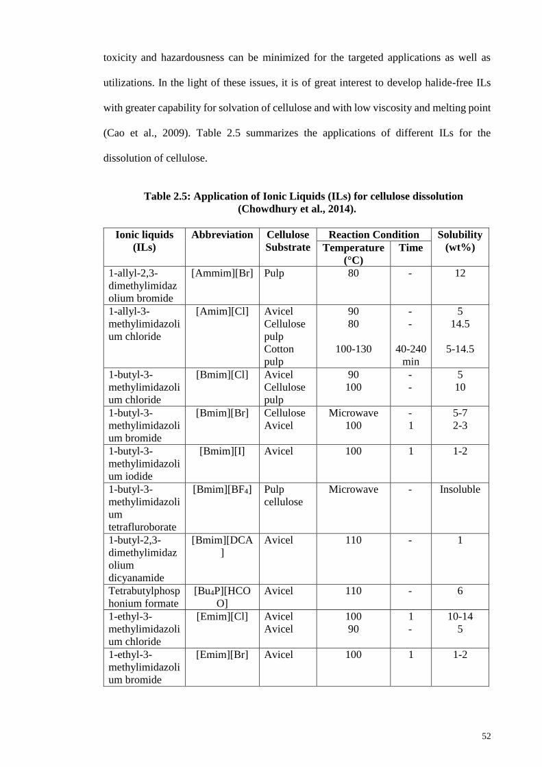
52
toxicity and hazardousness can be minimized for the targeted applications as well as
utilizations. In the light of these issues, it is of great interest to develop halide-free ILs
with greater capability for solvation of cellulose and with low viscosity and melting point
(Cao et al., 2009). Table 2.5 summarizes the applications of different ILs for the
dissolution of cellulose.
Table 2.5: Application of Ionic Liquids (ILs) for cellulose dissolution
(Chowdhury et al., 2014).
Ionic liquids
(ILs)
Abbreviation Cellulose
Substrate
Reaction Condition Solubility
(wt%) Temperature
(°C)
Time
1-allyl-2,3-
dimethylimidaz
olium bromide
[Ammim][Br] Pulp 80 - 12
1-allyl-3-
methylimidazoli
um chloride
[Amim][Cl] Avicel
Cellulose
pulp
Cotton
pulp
90
80
100-130
-
-
40-240
min
5
14.5
5-14.5
1-butyl-3-
methylimidazoli
um chloride
[Bmim][Cl] Avicel
Cellulose
pulp
90
100
-
-
5
10
1-butyl-3-
methylimidazoli
um bromide
[Bmim][Br] Cellulose
Avicel
Microwave
100
-
1
5-7
2-3
1-butyl-3-
methylimidazoli
um iodide
[Bmim][I] Avicel 100 1 1-2
1-butyl-3-
methylimidazoli
um
tetrafluroborate
[Bmim][BF4] Pulp
cellulose
Microwave - Insoluble
1-butyl-2,3-
dimethylimidaz
olium
dicyanamide
[Bmim][DCA
]
Avicel 110 - 1
Tetrabutylphosp
honium formate
[Bu4P][HCO
O]
Avicel 110 - 6
1-ethyl-3-
methylimidazoli
um chloride
[Emim][Cl] Avicel
Avicel
100
90
1
-
10-14
5
1-ethyl-3-
methylimidazoli
um bromide
[Emim][Br] Avicel 100 1 1-2
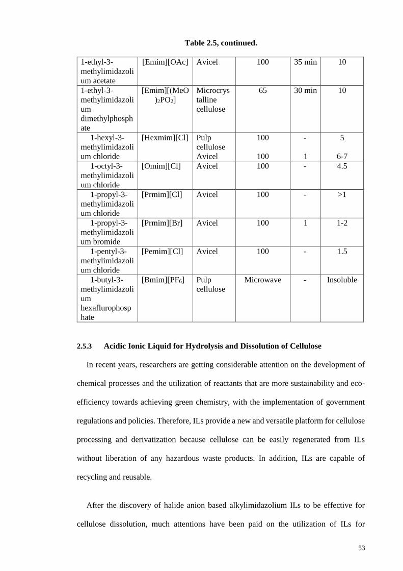
53
Table 2.5, continued.
1-ethyl-3-
methylimidazoli
um acetate
[Emim][OAc] Avicel 100 35 min 10
1-ethyl-3-
methylimidazoli
um
dimethylphosph
ate
[Emim][(MeO
)2PO2]
Microcrys
talline
cellulose
65 30 min 10
1-hexyl-3-
methylimidazoli
um chloride
[Hexmim][Cl] Pulp
cellulose
Avicel
100
100
-
1
5
6-7
1-octyl-3-
methylimidazoli
um chloride
[Omim][Cl] Avicel 100 - 4.5
1-propyl-3-
methylimidazoli
um chloride
[Prmim][Cl] Avicel 100 - >1
1-propyl-3-
methylimidazoli
um bromide
[Prmim][Br] Avicel 100 1 1-2
1-pentyl-3-
methylimidazoli
um chloride
[Pemim][Cl] Avicel 100 - 1.5
1-butyl-3-
methylimidazoli
um
hexaflurophosp
hate
[Bmim][PF6] Pulp
cellulose
Microwave - Insoluble
2.5.3 Acidic Ionic Liquid for Hydrolysis and Dissolution of Cellulose
In recent years, researchers are getting considerable attention on the development of
chemical processes and the utilization of reactants that are more sustainability and eco-
efficiency towards achieving green chemistry, with the implementation of government
regulations and policies. Therefore, ILs provide a new and versatile platform for cellulose
processing and derivatization because cellulose can be easily regenerated from ILs
without liberation of any hazardous waste products. In addition, ILs are capable of
recycling and reusable.
After the discovery of halide anion based alkylimidazolium ILs to be effective for
cellulose dissolution, much attentions have been paid on the utilization of ILs for
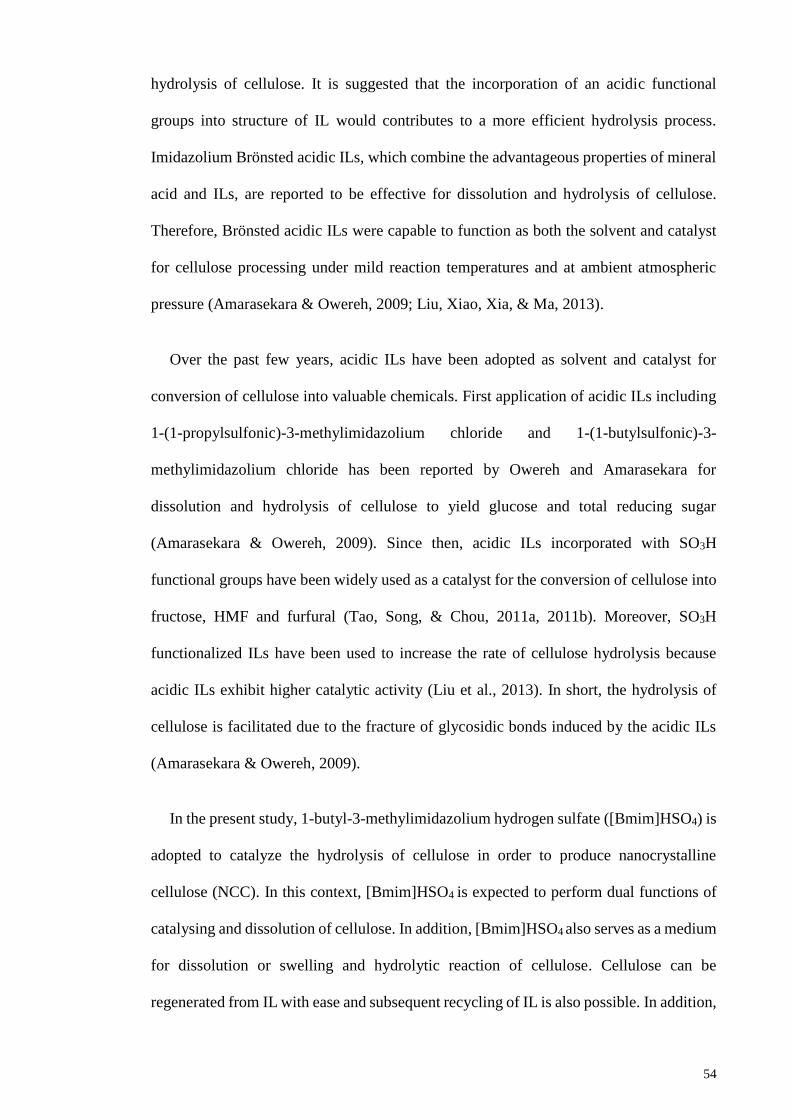
54
hydrolysis of cellulose. It is suggested that the incorporation of an acidic functional
groups into structure of IL would contributes to a more efficient hydrolysis process.
Imidazolium Brönsted acidic ILs, which combine the advantageous properties of mineral
acid and ILs, are reported to be effective for dissolution and hydrolysis of cellulose.
Therefore, Brönsted acidic ILs were capable to function as both the solvent and catalyst
for cellulose processing under mild reaction temperatures and at ambient atmospheric
pressure (Amarasekara & Owereh, 2009; Liu, Xiao, Xia, & Ma, 2013).
Over the past few years, acidic ILs have been adopted as solvent and catalyst for
conversion of cellulose into valuable chemicals. First application of acidic ILs including
1-(1-propylsulfonic)-3-methylimidazolium chloride and 1-(1-butylsulfonic)-3-
methylimidazolium chloride has been reported by Owereh and Amarasekara for
dissolution and hydrolysis of cellulose to yield glucose and total reducing sugar
(Amarasekara & Owereh, 2009). Since then, acidic ILs incorporated with SO3H
functional groups have been widely used as a catalyst for the conversion of cellulose into
fructose, HMF and furfural (Tao, Song, & Chou, 2011a, 2011b). Moreover, SO3H
functionalized ILs have been used to increase the rate of cellulose hydrolysis because
acidic ILs exhibit higher catalytic activity (Liu et al., 2013). In short, the hydrolysis of
cellulose is facilitated due to the fracture of glycosidic bonds induced by the acidic ILs
(Amarasekara & Owereh, 2009).
In the present study, 1-butyl-3-methylimidazolium hydrogen sulfate ([Bmim]HSO4) is
adopted to catalyze the hydrolysis of cellulose in order to produce nanocrystalline
cellulose (NCC). In this context, [Bmim]HSO4 is expected to perform dual functions of
catalysing and dissolution of cellulose. In addition, [Bmim]HSO4 also serves as a medium
for dissolution or swelling and hydrolytic reaction of cellulose. Cellulose can be
regenerated from IL with ease and subsequent recycling of IL is also possible. In addition,
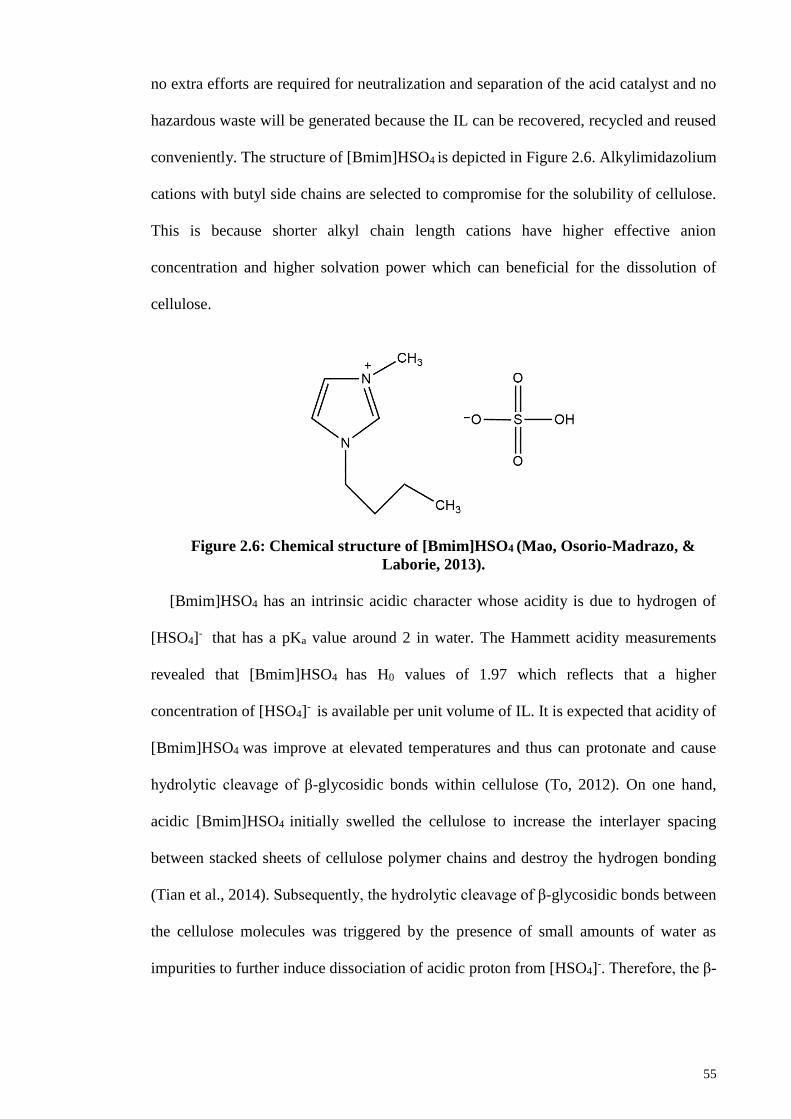
55
no extra efforts are required for neutralization and separation of the acid catalyst and no
hazardous waste will be generated because the IL can be recovered, recycled and reused
conveniently. The structure of [Bmim]HSO4 is depicted in Figure 2.6. Alkylimidazolium
cations with butyl side chains are selected to compromise for the solubility of cellulose.
This is because shorter alkyl chain length cations have higher effective anion
concentration and higher solvation power which can beneficial for the dissolution of
cellulose.
Figure 2.6: Chemical structure of [Bmim]HSO4 (Mao, Osorio-Madrazo, &
Laborie, 2013).
[Bmim]HSO4 has an intrinsic acidic character whose acidity is due to hydrogen of
[HSO4]- that has a pKa value around 2 in water. The Hammett acidity measurements
revealed that [Bmim]HSO4 has H0 values of 1.97 which reflects that a higher
concentration of [HSO4]- is available per unit volume of IL. It is expected that acidity of
[Bmim]HSO4 was improve at elevated temperatures and thus can protonate and cause
hydrolytic cleavage of β-glycosidic bonds within cellulose (To, 2012). On one hand,
acidic [Bmim]HSO4 initially swelled the cellulose to increase the interlayer spacing
between stacked sheets of cellulose polymer chains and destroy the hydrogen bonding
(Tian et al., 2014). Subsequently, the hydrolytic cleavage of β-glycosidic bonds between
the cellulose molecules was triggered by the presence of small amounts of water as
impurities to further induce dissociation of acidic proton from [HSO4]-. Therefore, the β-
56
glycosidic bonds dissolved in [Bmim]HSO4 can be easily attacked by hydronium ions
which facilitated the hydrolysis of cellulose (Mao et al., 2013).
Acidic [Bmim]HSO4 offers a promising approach for the synthesis of nanocellulose
from cellulose by performing cellulose dissolution with remarkable catalytic activity.
[Bmim]HSO4 showed apparent hydrolytic activity attributed to its stronger acidity (due
to proton from [HSO4]-). On the contrary, [Bmim]Cl exhibits lower catalytic capability
because it has limited acidity (Tian et al., 2014). Despite that, the highly viscous
[Bmim]Cl has a negative impact on the catalytic activity in which the mobility of ions are
restricted.
In summary, the structure, acidity and viscosity of ILs are important factors to
influence the catalytic capability of ILs. In particular, acidity and viscosity of ILs are
significantly affected the catalytic activity of ILs. The stronger acidity (active acid sites)
with low viscosity acidic ILs are well-suited and more effective for catalysing hydrolysis
of cellulose to accelerate the fracture of glycosidic linkages to produce more NCC (Liu
et al., 2013). The inherent acidic characteristic of acidic ILs which have dual functions of
swelling and catalyzing, makes them as a viable alternative approach in the production of
nanocellulose (Tian et al., 2014). Therefore, this approach could be developed into a
green, economical and efficient cellulose hydrolysis protocol for the production of
nanocellulose with the recycling of Brönsted acidic ILs after the reaction.
2.5.4 Acetate based Ionic Liquid for Cellulose Dissolution
A promising cellulose solvents ideal for dissolution should have most of the following
characteristics: (i) able to dissolve a high amount of cellulose at low temperature, (ii)
chemically and thermally stable, (iii) non-toxic and non-volatile, (iv) low melting point
and viscosity, (v) easy cellulose regeneration, (vi) no decomposition of cellulose, (vii)
high recovery of solvents and recyclable, and lastly (viii) cost effective process (Wang et
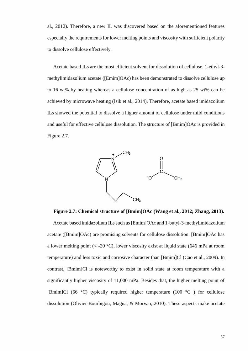
57
al., 2012). Therefore, a new IL was discovered based on the aforementioned features
especially the requirements for lower melting points and viscosity with sufficient polarity
to dissolve cellulose effectively.
Acetate based ILs are the most efficient solvent for dissolution of cellulose. 1-ethyl-3-
methylimidazolium acetate ([Emim]OAc) has been demonstrated to dissolve cellulose up
to 16 wt% by heating whereas a cellulose concentration of as high as 25 wt% can be
achieved by microwave heating (Isik et al., 2014). Therefore, acetate based imidazolium
ILs showed the potential to dissolve a higher amount of cellulose under mild conditions
and useful for effective cellulose dissolution. The structure of [Bmim]OAc is provided in
Figure 2.7.
Figure 2.7: Chemical structure of [Bmim]OAc (Wang et al., 2012; Zhang, 2013).
Acetate based imidazolium ILs such as [Emim]OAc and 1-butyl-3-methylimidazolium
acetate ([Bmim]OAc) are promising solvents for cellulose dissolution. [Bmim]OAc has
a lower melting point (< -20 °C), lower viscosity exist at liquid state (646 mPa at room
temperature) and less toxic and corrosive character than [Bmim]Cl (Cao et al., 2009). In
contrast, [Bmim]Cl is noteworthy to exist in solid state at room temperature with a
significantly higher viscosity of 11,000 mPa. Besides that, the higher melting point of
[Bmim]Cl (66 °C) typically required higher temperature (100 °C ) for cellulose
dissolution (Olivier-Bourbigou, Magna, & Morvan, 2010). These aspects make acetate
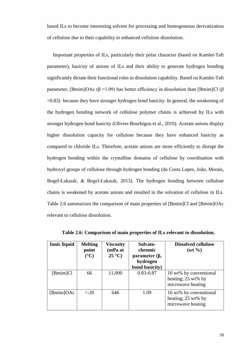
58
based ILs to become interesting solvent for processing and homogeneous derivatization
of cellulose due to their capability to enhanced cellulose dissolution.
Important properties of ILs, particularly their polar character (based on Kamlet-Taft
parameter), basicity of anions of ILs and their ability to generate hydrogen bonding
significantly dictate their functional roles in dissolution capability. Based on Kamlet-Taft
parameter, [Bmim]OAc (β =1.09) has better efficiency in dissolution than [Bmim]Cl (β
=0.83) because they have stronger hydrogen bond basicity. In general, the weakening of
the hydrogen bonding network of cellulose polymer chains is achieved by ILs with
stronger hydrogen bond basicity (Olivier-Bourbigou et al., 2010). Acetate anions display
higher dissolution capacity for cellulose because they have enhanced basicity as
compared to chloride ILs. Therefore, acetate anions are more efficiently to disrupt the
hydrogen bonding within the crystalline domains of cellulose by coordination with
hydroxyl groups of cellulose through hydrogen bonding (da Costa Lopes, João, Morais,
Bogel-Łukasik, & Bogel-Łukasik, 2013). The hydrogen bonding between cellulose
chains is weakened by acetate anions and resulted in the solvation of cellulose in ILs.
Table 2.6 summarizes the comparison of main properties of [Bmim]Cl and [Bmim]OAc
relevant to cellulose dissolution.
Table 2.6: Comparison of main properties of ILs relevant to dissolution.
Ionic liquid Melting
point
(°C)
Viscosity
(mPa at
25 °C)
Solvato-
chromic
parameter (β,
hydrogen
bond basicity)
Dissolved cellulose
(wt %)
[Bmim]Cl 66 11,000 0.83-0.87 10 wt% by conventional
heating; 25 wt% by
microwave heating
[Bmim]OAc <-20 646 1.09 16 wt% by conventional
heating; 25 wt% by
microwave heating
59
In this work, [Bmim]OAc is selected as the solvent for dissolution or solvolysis of
cellulose mainly due to its lower viscosity and melting point These features will enable
the dissolution much more favorable to conduct at lower temperature. Besides that, it is
suggested that a higher concentration of cellulose is able to dissolve well in [Bmim]OAc
because of its high dissolution capability and excellent solvency power for cellulose
(Olivier-Bourbigou et al., 2010). Nevertheless, [Bmim]OAc has less acidity in character
than [Bmim]HSO4 and this can explained why the hydrolysis reaction did not proceed
extensively in [Bmim]OAc (To, 2012). In fact, it has higher nucleophilicity and stronger
hydrogen bond basicity which makes it tends to accept hydrogen bonding for solvation
of cellulose.
On the other hand, [Bmim] cations are selected as the compromise to cost effective
and solubility of cellulose. It has been suggested that solubilization temperature of
cellulose are relevant to the length of alkyl side chains of imidazolium cations and
viscosity of ILs. Since viscosity of ILs are generally increases for longer alkyl side chains,
so do the increase in solubilization temperature (Itoh, 2014; Ohno & Fukaya, 2009).
Therefore, shorter alkyl side chains (butyl) imidazolium cations have decreased viscosity
of ILs which favoured for the dissolution of cellulose at lower temperature.
In short, [Bmim]OAc shows promise in the production of nanocellulose through
solvolysis or dissolution approach. With its excellent solvency power, it shows its
potential functional roles as solvents and catalysts for the efficient process of cellulose
dissolution.
2.5.5 Regeneration of Cellulose from Ionic Liquid Suspension
Regeneration of cellulose from cellulose/ILs mixture is possible by adding anti-
solvents to precipitate the cellulose. Typically, the ILs used to dissolve cellulose is water
soluble. The choices of anti-solvents are plenty such as water, ethanol, methanol, acetone,
60
acetonitrile and isopropanol (Wang et al., 2012). Nevertheless, water and ethanol are most
commonly used anti-solvents to precipitate the dissolved cellulose (Olivier-Bourbigou et
al., 2010). The usage of water as anti-solvent presents inexpensive and environmentally
benign approach because water is safe and do not pose any hazards. The criteria to be an
effective anti-solvent required that it is a polar solvent with the ability to form hydrogen
bond with anions of ILs more strongly than cellulose and solvate the charged species of
ILs (Wang et al., 2012).
Most ILs are recognized for their hygroscopic properties in which they have strong
absorbability for water. For instance, [Amim]Cl and [Bmim]Cl are completely miscible
with water in any ratio. Hence, the regenerated cellulose materials are easily obtained by
coagulations due to competitive hydrogen bonding between cellulose and water
molecules with ILs. In the regeneration process, ILs tend to form hydrogen bonding with
water molecules as O-H bonding has higher electronegativity than the C-H type hydrogen
bond (Han, Zhou, French, Han, & Wu, 2013). Despite that, the ions of ILs are extracted
into the aqueous phase through hydrogen bonding, dipolar and Coulombic forces in the
regeneration steps. Subsequently, the ions of ILs are surround by water molecules to form
hydrodynamic shells to shield the direct interactions of ILs ions with cellulose.
Consequently, the hydrogen bonds between cellulose and ILs are weakened while the
inter and intramolecular hydrogen bonds between cellulose chains are rebuilt and
reconnect. Therefore, this results in the precipitation of cellulose (Zavrel et al., 2008).
The regenerated cellulose can be isolated due to separation by centrifugation or filtration
process.
The regenerated cellulose can be obtained in various forms such as cellulose
membranes, fibres, films, beads and monoliths, depending on the processing conditions.
For instance, rapid mixing of water into ILs solution results in the precipitation of
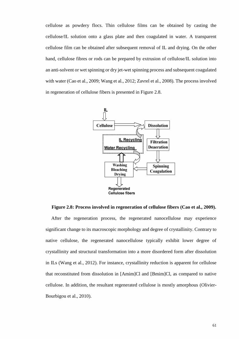
61
cellulose as powdery flocs. Thin cellulose films can be obtained by casting the
cellulose/IL solution onto a glass plate and then coagulated in water. A transparent
cellulose film can be obtained after subsequent removal of IL and drying. On the other
hand, cellulose fibres or rods can be prepared by extrusion of cellulose/IL solution into
an anti-solvent or wet spinning or dry jet-wet spinning process and subsequent coagulated
with water (Cao et al., 2009; Wang et al., 2012; Zavrel et al., 2008). The process involved
in regeneration of cellulose fibers is presented in Figure 2.8.
Figure 2.8: Process involved in regeneration of cellulose fibers (Cao et al., 2009).
After the regeneration process, the regenerated nanocellulose may experience
significant change to its macroscopic morphology and degree of crystallinity. Contrary to
native cellulose, the regenerated nanocellulose typically exhibit lower degree of
crystallinity and structural transformation into a more disordered form after dissolution
in ILs (Wang et al., 2012). For instance, crystallinity reduction is apparent for cellulose
that reconstituted from dissolution in [Amim]Cl and [Bmim]Cl, as compared to native
cellulose. In addition, the resultant regenerated cellulose is mostly amorphous (Olivier-
Bourbigou et al., 2010).
62
Apart from that, the yield of regenerated cellulose is affected by the types of anti-
solvents used. Water is most widely used as anti-solvents for cellulose regeneration
because the regeneration yield for water is higher than that in methanol, ethanol and
acetone. This is because water indeed, is a polar protic solvent and its easiness and
strength of the hydrogen bond formation between water and cellulose. Besides that,
mixture of anti-solvents such as ethanol and acetone results in the precipitation of
cellulose with porous appearance and enhanced dispersion in the solvents without
formation of gel (da Costa Lopes et al., 2013). On top of that, the anti-solvents selected
must be easily separated and removed from ILs. This can ensure that both components
can be recycled and reused from the environmentally benign standpoint and also a cost
effective way from the economical point of view.
2.5.6 Recovery of Ionic Liquids
Generally, ILs can be recovered after the regeneration of cellulose due to their
characteristic low vapor pressure and non-volatility. ILs can be removed from the
cellulose suspension through multiple washing with centrifugation steps. After the
repeated centrifugations, the ILs solution are isolated from the cellulose suspension as the
cellulose is precipitated as the sediment. Numerous methods have been investigated for
recovery of ILs, for example distillation, solvent extraction, adsorption, salting-out
process and membrane based methods such as nanofiltration and reverse osmosis. The
summary of each methods to recover ILs is provided in Table 2.7. The selection of
appropriate recovery process is actually depends on the characteristics of
systems/processes.
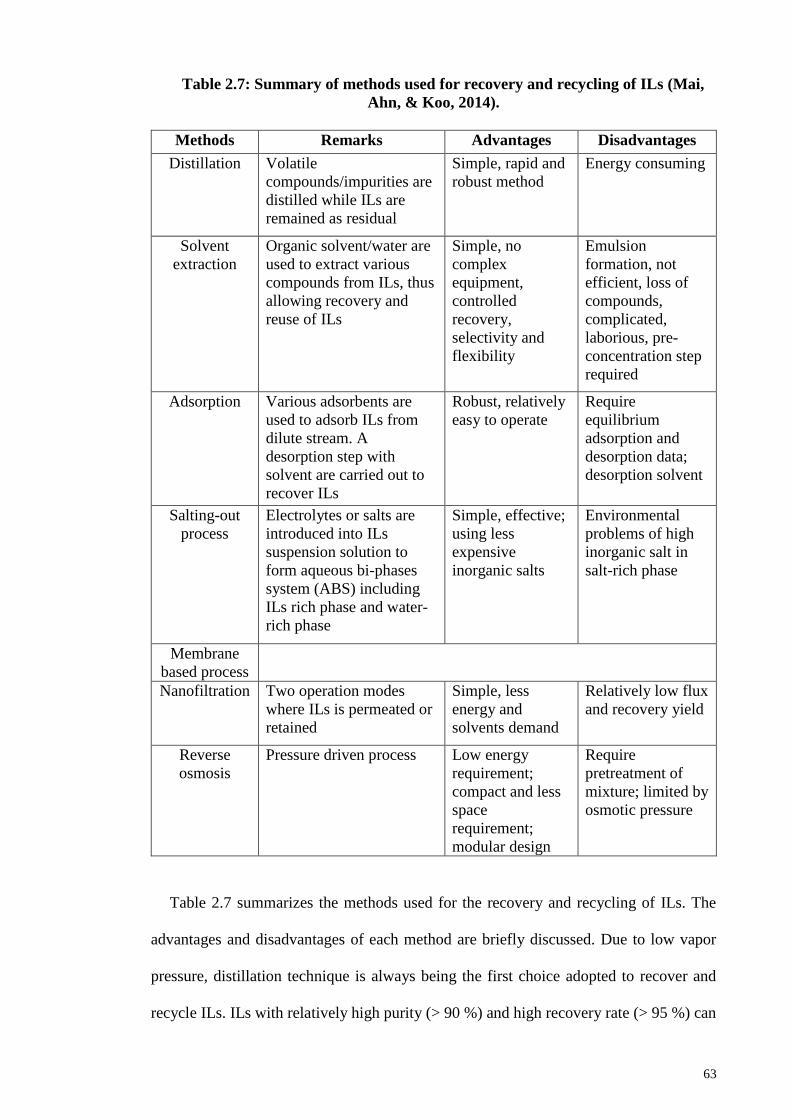
63
Table 2.7: Summary of methods used for recovery and recycling of ILs (Mai,
Ahn, & Koo, 2014).
Methods Remarks Advantages Disadvantages
Distillation Volatile
compounds/impurities are
distilled while ILs are
remained as residual
Simple, rapid and
robust method
Energy consuming
Solvent
extraction
Organic solvent/water are
used to extract various
compounds from ILs, thus
allowing recovery and
reuse of ILs
Simple, no
complex
equipment,
controlled
recovery,
selectivity and
flexibility
Emulsion
formation, not
efficient, loss of
compounds,
complicated,
laborious, pre-
concentration step
required
Adsorption Various adsorbents are
used to adsorb ILs from
dilute stream. A
desorption step with
solvent are carried out to
recover ILs
Robust, relatively
easy to operate
Require
equilibrium
adsorption and
desorption data;
desorption solvent
Salting-out
process
Electrolytes or salts are
introduced into ILs
suspension solution to
form aqueous bi-phases
system (ABS) including
ILs rich phase and water-
rich phase
Simple, effective;
using less
expensive
inorganic salts
Environmental
problems of high
inorganic salt in
salt-rich phase
Membrane
based process
Nanofiltration Two operation modes
where ILs is permeated or
retained
Simple, less
energy and
solvents demand
Relatively low flux
and recovery yield
Reverse
osmosis
Pressure driven process Low energy
requirement;
compact and less
space
requirement;
modular design
Require
pretreatment of
mixture; limited by
osmotic pressure
Table 2.7 summarizes the methods used for the recovery and recycling of ILs. The
advantages and disadvantages of each method are briefly discussed. Due to low vapor
pressure, distillation technique is always being the first choice adopted to recover and
recycle ILs. ILs with relatively high purity (> 90 %) and high recovery rate (> 95 %) can
64
be recovered via this method. Despite that, the extraction process yield about 89 % of
recover ILs. However, this method is associated with the emulsion formation, loss of
compounds, required laborious and pre-concentration steps which make the process
complicated and not efficient. Although 92 % of recovery rate can be achieved with
adsorption process, the requirement of adsorption/desorption data and additional
desorption solvents contribute to the sophisticated of the method. In addition, in the
continuous adsorption/desorption or chromatography method, the requirement of
complex chromatography equipment and pre-concentration step would make the methods
costly and laboriously. Besides that, salting-out methods are concerning with
environmental problems due to the presence of high inorganic salt which makes the
method become economical unfeasible, though its high recovery rate of about 96 %. Apart
from that, membrane-based process such as nanofiltration and reverse osmosis process
give rise to the relatively lower recovery rate of 14 % and 20 %, respectively.
Distillation is the most simple and conventional method employed for the recovery of
ILs in which the thermally stable ILs are retained through the removal of compounds with
low boiling point. Distillation of volatile compounds can be performed with vacuum
evaporation, molecular as well as column distillation. In the studies regarding to cellulose
processing, regenerated cellulose is firstly removed from ILs suspension by filtration.
Then, residual ILs in the filtrate is recovered by evaporation under reduced pressure and
distillation with subsequent drying in the vacuum conditions for 24 hours in order to
eliminate water (Mai et al., 2014). ILs can be recovered with high purity and recycled by
this method (Cao et al., 2009). Distillation method is considered to benefits from the
difference in boiling points between water (100 °C) and ILs (> 200 °C). Interestingly, ILs
have high recovery rate (> 95 %) and purity (> 90 %) (Cao et al., 2009; Swatloski et al.,
2002). The recovered ILs can be reused as the recycled ILs exhibit similar capability to
dissolve cellulose which reflects their durability in use.
65
On top of that, efficient recycling of ILs is key for cost effective process in industrial
application. Environmental concerns regarding to disposal issues such as toxicity and
biodegradation can be resolved to minimize with the recovery and recycling of ILs (Cao
et al., 2009). Therefore, application of ILs in the industry can be apparently promoted by
taking all the advantageous features of ILs especially recyclable and reusable.
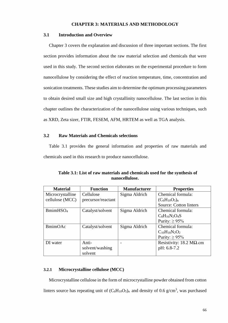
66
CHAPTER 3: MATERIALS AND METHODOLOGY
3.1 Introduction and Overview
Chapter 3 covers the explanation and discussion of three important sections. The first
section provides information about the raw material selection and chemicals that were
used in this study. The second section elaborates on the experimental procedure to form
nanocellulose by considering the effect of reaction temperature, time, concentration and
sonication treatments. These studies aim to determine the optimum processing parameters
to obtain desired small size and high crystallinity nanocellulose. The last section in this
chapter outlines the characterization of the nanocellulose using various techniques, such
as XRD, Zeta sizer, FTIR, FESEM, AFM, HRTEM as well as TGA analysis.
3.2 Raw Materials and Chemicals selections
Table 3.1 provides the general information and properties of raw materials and
chemicals used in this research to produce nanocellulose.
Table 3.1: List of raw materials and chemicals used for the synthesis of
nanocellulose.
Material Function Manufacturer Properties
Microcrystalline
cellulose (MCC)
Cellulose
precursor/reactant
Sigma Aldrich Chemical formula:
(C6H10O5)n
Source: Cotton linters
BmimHSO4 Catalyst/solvent Sigma Aldrich Chemical formula:
C8H16N2O4S
Purity: ≥ 95%
BmimOAc Catalyst/solvent Sigma Aldrich Chemical formula:
C10H18N2O2
Purity: ≥ 95%
DI water Anti-
solvent/washing
solvent
- Resistivity: 18.2 MΩ.cm
pH: 6.8-7.2
3.2.1 Microcrystalline cellulose (MCC)
Microcrystalline cellulose in the form of microcrystalline powder obtained from cotton
linters source has repeating unit of (C6H10O5)n and density of 0.6 g/cm3, was purchased
67
from Sigma-Aldrich. The CAS number is 9004-34-6. MCC is the starting raw materials
used in the present research study.
3.2.2 1-butyl-3-methylimidazolium hydrogen sulfate (BmimHSO4)
1-butyl-3-methylimidazolium hydrogen sulfate, ≥ 95% with a chemical formula of
C8H16N2O4S, has molecular weight of 236.29 g/mol and a density of 1.277 g/cm3, was
purchased from Sigma Aldrich. The CAS number is 262297-13-2.
3.2.3 1-butyl-3-methylimidazolium acetate (BmimOAc)
1-butyl-3-methylimidazolium acetate, ≥ 95% with a chemical formula of C10H18N2O2,
molecular weight = 198.26 g/mol, and exhibits a density of 1.055 g/cm3, was purchased
from Sigma Aldrich. The CAS number is 284049-75-8.
3.2.4 Deionized water (DI water)
DI water was used throughout the research study as the washing solvent to remove IL
after subsequent washing and centrifugation steps. It is also functions to dilute the IL in
the IL/cellulose suspension by quenched the reaction.
3.3 Methodology of Experiment
3.3.1 Synthesis of nanocellulose by BmimHSO4
In this stage, nanocellulose was primarily synthesized through IL-mediated hydrolysis
method. Microcrystalline cellulose (MCC) powder with different loading amounts was
added into BmimHSO4 and heated up to 70, 80, 90, 100 and 110 ºC respectively. The
mixture was then heating for different time varied from 0.5 h, 1.0 h, 1.5 h, 2.0 h and 2.5
h with continuous mechanical stirring at a speed of 400 rpm. The mass loading of MCC
was varied as 5 wt%, 10 wt%, 15 wt%, 20 wt% and 25 wt%. Several important synthesis
parameters including temperature, time and mass loading of MCC were studied and
optimized in order to produce smaller dimension nanocellulose with desired properties.
68
Nanocellulose can be precipitated from the mixture by adding DI water as anti-solvent.
The hydrolysis reaction was then quenched by addition of 20 mL cold DI water into the
mixture. Powdery flock were formed upon addition of water into the IL/cellulose mixture.
The mixture was allowed to stir for 5 minutes and then subjected to different sonication
treatments at room temperature. The sonication was carried out with high-intensity
ultrasonic probe (Hielscher Ultrasonic, UIP/1000hd) or low-intensity ultrasonic bath
(Thermoline, Thermo-6D) respectively. The suspension was subsequently washed with
DI water using repeated centrifugation at 7,500 rpm for 15 minutes interval until a stable
pH 5 suspension was achieved. The sediment was dried by using a freeze dryer
(Labconco, FreeZone 4.5 L Benchtop) for 2 days and kept in refrigerator at -4 ºC before
performing further characterization.
3.3.2 Synthesis of nanocellulose by BmimOAc
Preparation of nanocellulose by using BmimOAc was achieved via solvolysis or
dissolution approach. The synthesis procedures were similar to the methodology in the
previous section (Section 3.3.1) by using BmimHSO4 except for the IL used in the current
stage is BmimOAc.
Microcrystalline cellulose (MCC) powder at different mass loading amount was added
into BmimOAc in a round bottom flask and heated up to 70, 80, 90, 100 and 110 ºC
respectively. The mixture was heated for different times varied from 0.5 h, 1.0 h, 1.5 h,
2.0 h and 2.5 h, respectively in the oil-bath with continuous mechanical stirring at a speed
of 400 rpm. The mass loading of MCC was varied accordingly to 5 wt%, 10 wt%, 15
wt%, 20 wt% and 25 wt%. It is well-established that the properties and dimensional
features of nanocellulose can be controlled by variety of parameters, such as reaction
temperature, time and mass loading of MCC. It was observed that MCC started to dissolve
completely as the color of the mixture turned into amber and yield a transparent solution.
69
The dissolution was then quenched by rapid mixing of 20 mL cold DI water into the
mixture to precipitate the nanocellulose. Formation of gel-like off-white flocs were
observed upon rapid mixing of water with IL/cellulose mixture. The mixture was stirred
for 5 minutes before subjected to sonication treatment at room temperature. The mixture
was sonicated by using high-intensity ultrasonic probe (Hielscher Ultrasonic,
UIP/1000hd) or low-intensity ultrasonic bath (Thermoline, Thermo-6D) respectively. In
the present study, DI water was used to wash the resulting suspension with repeated
centrifugations at 7,500 rpm for 15 minutes interval in order to remove residual IL until
a stable pH 5 suspension was achieved. The sediment was freeze dried in Labconco,
FreeZone 4.5 L Benchtop freeze dryer for 2 days to yield white nanocellulose fine
powder. The nanocellulose was kept in refrigerator at -4 ºC prior performing
characterizations. The overview of nanocellulose synthesis methodology is illustrated in
Figure 3.1.
3.3.3 Recovery of Ionic Liquid
The supernatant that decanted from first two washing was collected for recovery of
ILs (recovered BmimHSO4 and BmimOAc) via distillation method. The ILs solution were
first concentrated under vacuum by evaporating the water in a rotary evaporator, where
the temperature of the water bath is 80 °C and subjected for 20 minutes. Subsequently,
the recovered ILs were dried in a vacuum freeze-dryer for 24 hours under the temperature
of -40 °C. The recovered ILs were characterized by FTIR to determine any changes to the
chemical groups. The recovery yield of ILs is given on Equation 3.1.
Recovery yield (%) = 𝑀𝑎𝑠𝑠 𝑜𝑓 𝑟𝑒𝑐𝑜𝑣𝑒𝑟 𝐼𝐿
𝑂𝑟𝑖𝑔𝑖𝑛𝑎𝑙 𝑚𝑎𝑠𝑠 𝑜𝑓 𝐼𝐿× 100% (Equation 3.1)
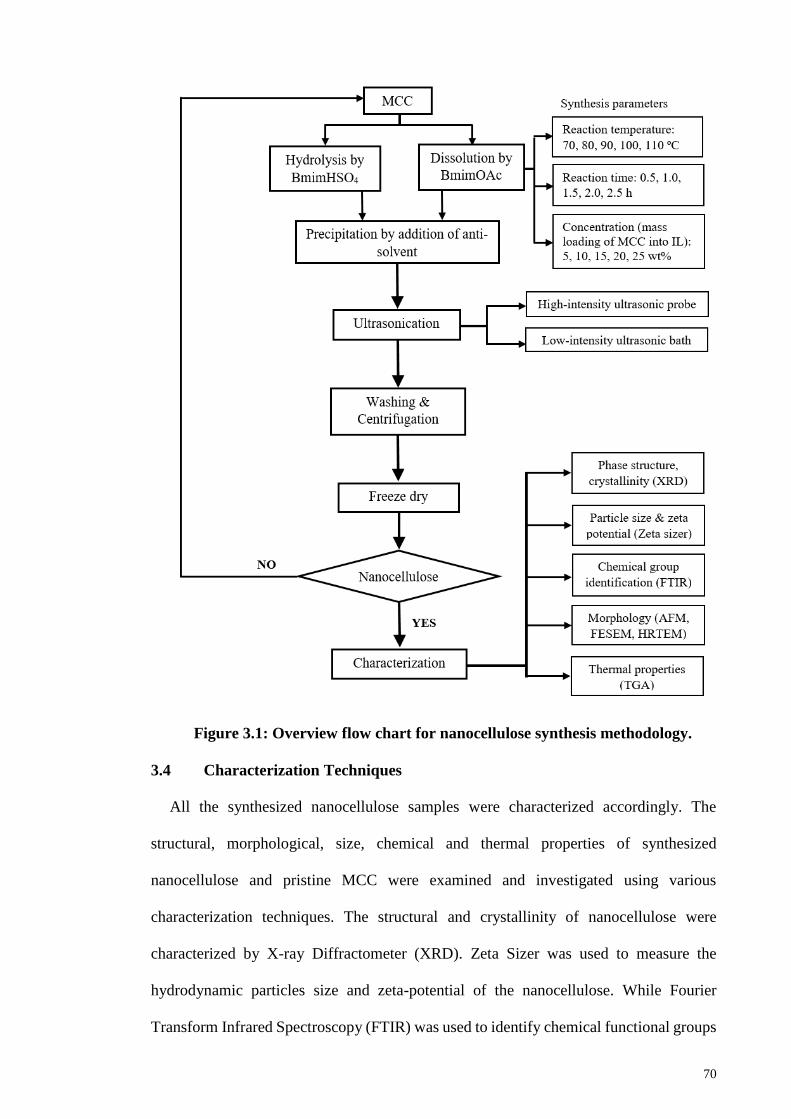
70
Figure 3.1: Overview flow chart for nanocellulose synthesis methodology.
3.4 Characterization Techniques
All the synthesized nanocellulose samples were characterized accordingly. The
structural, morphological, size, chemical and thermal properties of synthesized
nanocellulose and pristine MCC were examined and investigated using various
characterization techniques. The structural and crystallinity of nanocellulose were
characterized by X-ray Diffractometer (XRD). Zeta Sizer was used to measure the
hydrodynamic particles size and zeta-potential of the nanocellulose. While Fourier
Transform Infrared Spectroscopy (FTIR) was used to identify chemical functional groups
71
and track any chemical changes. The morphology was characterized using Atomic Force
Microscope (AFM), Field Emission Scanning Electron Microscopy (FESEM) and High-
resolution Transmission Electron Microscope (HRTEM). Thermogravimetric analysis
(TGA) was used to characterize the thermal properties of nanocellulose. In this section,
basic principles and information regarding the parameters set for the characterization
techniques were discussed briefly.
3.4.1 X-ray diffraction (XRD)
The crystallographic phase structure and crystallinity index of nanocellulose were
investigated by X-ray diffraction (XRD) analysis. The fundamental principle of XRD is
based on constructive interference of monochromatic X-rays radiation that directed
toward a crystalline material. The interaction of the incident rays with the sample
produces diffraction patterns or constructive interference. This conditions required to
satisfy Bragg’s Law (nλ=2d sin θ) where it provides the lattice spacing (or d-spacing) of
a crystalline material by relates the wavelength of electromagnetic radiation to the angles
of diffraction rays. By exposing the material to radiation that scan at a range of 2θ angles,
the diffracted X-rays are detected and all possible diffraction directions of lattice should
be attained due to the random orientation of the crystalline material. The diffraction peaks
and patterns can be indexed to provide information regarding the phase structure of the
material. Typically, identification and evaluation of phase structure of sample is achieved
by comparison of obtained XRD diffraction patterns with standard reference patterns
from International Centre for Diffraction Data (ICDD) database.
XRD patterns of pristine MCC and nanocellulose were analysed by Bruker D8
Advance X-ray Diffractometer equipped with Ni-filtered monochromatic Cu Kα radiation
(λ= 1.5418 Å). The operating voltage was 40 kV and the current was 40 mA. The
72
intensities were carried out in the range of 5 to 60º with a step size of 0.02º and a rate of
1º min-1.
The crystallinity index (CrI) of each sample was determined by referring to diffraction
intensity of crystalline and amorphous regions by using Segal’s method (Equation 3.2).
CrI = 𝐼200− 𝐼𝑎𝑚
𝐼200 (Equation 3.2)
where I200 is the maximum intensity of (200) lattice diffraction, representing the
crystalline component whereas Iam is the intensity of the baseline at 2θ = 18º, representing
the amorphous part of cellulose.
The crystallite size of each sample perpendicular to (200) planes, D (nm), was
calculated by using Debye Scherrer’s equation (Equation 3.3).
𝐷 = 𝐾𝜆
𝛽 cos 𝛳 (Equation 3.3)
where D is the mean diameter of ordered domains, K is the correction factor (0.94), λ
is the wavelength of X-ray radiation (0.154 nm), β is the corrected angular width in
radians at half maximum intensity of the (200) peak and θ is the Bragg angle.
3.4.2 Zeta sizer
Particle size distribution and zeta potential of samples were analyzed by Malvern
Zetasizer Nano ZS. Particle size distribution is determined based on principle of dynamic
light scattering (DLS). A laser beam caused illumination of the sample and subsequently
the resultant fluctuations of the scattered light at a known scattering angle were detected
by a fast photon detector. The Brownian motion of particles was measured by DLS and
the mean diameter of particles was calculated via the Stokes-Einstein equation (Equation
3.4). The particle size that measured is the diameter of the spherical particles diffused
73
instantaneously as particles being measured. On the other hand, the zeta potential of
cellulosic samples was measured by principle of electrophoretic light scattering (ELS).
The electrophoretic mobility of each nanocellulose suspension was identified using
Huckel approximation for the zeta potential.
D = 𝑘𝐵 𝑇
6 𝜋 𝜂 𝑟 (Equation 3.4)
where D is the diffusion constant, kB is Boltzmann’s constant, T is the absolute
temperature, η is the dynamic viscosity and r is the radius of the spherical particle.
In the present study, number-based particle size distribution (PSD), polydispersity
index (PDI) and zeta potential of nanocellulose were determined. Nanocellulose
suspensions were prepared at concentration of 0.05 wt% in DI water and subject to
sonication for 30 minutes prior to measurement in order to ensure well dispersibility of
the particles in DI water. The measurement was carried out at temperature of 25 C with
detection angle of 173. Water was used as dispersant in this study. The measurements
were carried out in triplicate for each sample and the average values were reported.
3.4.3 Fourier transform infrared spectroscopy (FTIR)
FTIR spectroscopy is a powerful characterization technique to provide information on
the chemical composition of the sample by identifies the functional group present within
molecules. It is based on the principle that absorption of light energy by most molecules
in the infrared region of electromagnetic spectrum is corresponds specifically to the bonds
present in the molecules. Each bond has characteristic vibration frequency to indicate the
presence of particular chemical bonds and functional groups specific to the molecules.
The infrared spectrum is displayed as the intensity of light transmitted through the sample
measured against the wavenumber. FTIR is particularly indispensable for identification
of organic molecular structure and compounds.
74
The changes in chemical structure were analysed by Fourier transform infrared
spectroscopy (FTIR). The FTIR spectra for cellulosic sample were recorded on a FTIR
spectrometer (Bruker-IFS 66) in the transmittance mode over the range of 4000 to 400
cm-1 with a resolution of 4 cm-1. The samples were prepared using KBr disk method in
which the weight ratio of samples to KBr was 1:100. About 2 mg of the dried cellulosic
samples were ground into powder by a fiber microtome, and then blended with KBr
powder before pressing into ultra-thin pellets. The pellets were then placed in FTIR
specimen holder for analysis and IR radiation was passed through it. The intensity of
transmittance due to IR radiation was collected as FTIR spectrum.
3.4.4 Field emission scanning electron microscopy (FESEM)
Field emission scanning electron microscopy (FESEM) is a non-destructive technique
that commonly used to visualize morphology and topography details on the surface of a
sample. The working principle of FESEM is relies on the liberation of electrons from a
field emission source and electrons are passed through a vacuum column under a high
electrical field gradient to focus on the surface of specimen. Deflection of primary
electrons by electronic lenses focused them to generate a narrow scan beam that bombards
the specimen. Consequently, the angle and velocity of secondary electrons attributed to
the interactions with specimen provide the information relate to the surface structure of
sample which then generates signals to be detected as sample image. The signals are
processed to produce SE or BSE images due to generation of electron beams including
secondary electrons (SE), backscattered electrons (BSE) and X-rays that scanned across
the surface of sample. FESEM is particularly useful to provide a great depth of field to
visualize the surface structure of a sample.
In this research, the surface morphology of nanocellulose was examined by FESEM
(JEOL JSM 7600-F) operated at an accelerating voltage of 5 kV and magnifications of 2,
75
15 and 50 kX were typically used for characterization. Prior to imaging, the freeze-dried
nanocellulose was mounted onto metal stubs with double sided electrically conductive
carbon tape and sputter coated with gold to prevent surface charging when exposed to the
electron beam.
3.4.5 Atomic force microscope (AFM)
In atomic force microscopy (AFM), a cantilever with a very sharp tip is equipped to
scan across the surface of the sample. It is able to measure microscopic surface
morphology and topography as well as to construct a 3-D image of the surface. When the
tip scans over a surface, interatomic forces between the tip and the surface induce the
displacement of the tip and deflection of cantilever towards the surface. As the cantilever
is lower to the surface which makes the tip makes contact with it, the repulsive force
caused the deflection of cantilever away from the surface. A laser beam is used to detect
orientation of cantilever by allowed it to transmit to and reflect from the cantilever. A
position-sensitive photo diode (PSPD) detector is used to track the changes on the
reflected laser beam and deflection of the cantilever. Thus, AFM can provides a
topographical image of the sample surface by scanning the cantilever over a region of
interest. AFM is a less intrusive surface morphological and topographical microscopic
technique where it produces quantitative and 3-D images with high resolution range from
a few microns to below 10 Angstroms.
The morphology of nanocellulose was visualized by AFM (Bruker Multimode 8) using
silicon cantilevers. A 0.005 wt% concentration of nanocellulose suspension in water was
prepared and allowed to sonicate for 10 minutes in order to ensure uniform dispersion of
nanocellulose. Before observation, a drop of the diluted nanocellulose suspension was
deposited on a freshly cleaved mica and allowed to dry for overnight at ambient condition.
The mica substrate was then glued onto a metal disk and attached to a magnetic sample
76
holder placed on the top of the scanner tube. The images were scanned with contact mode
in air at ambient conditions using silicon probes. The cantilever has a tip radius of 5 nm,
spring constant of 42 N/m and resonance frequency of 320 kHz. The scan rate for images
was 0.5 Hz and images resolution was 512 x 512 pixels. No image processing except
flattening was made. The morphology and size of nanocellulose were examined by
NanoScope Analysis 1.5 software provided by Bruker Company. The diameter of
nanocellulose was then plotted as in histogram for numerical diameter distribution after
measurement of 100 randomly selected nanocellulose.
3.4.6 High resolution transmission electron microscope (HRTEM)
Transmission electron microscopy (TEM) is an advanced microscopic technique to
visualize the sample material by using beam of high energy electrons (up to 300 kV
accelerating voltage). In TEM, beam of high velocity electrons are liberated from the
electron gun and accelerated under vacuum to focus the coherent beam onto the specimen
by condenser lens. The beam strikes the specimen while parts of it are transmitted and
focused by objective lens to enlarge and project into the image on phosphor screen or
captured by charge coupled device (CCD) camera. The images obtained is usually the
highly magnified view of the nano- and microstructure and present in high resolution
mode ultimately.
High resolution transmission electron miscroscope (HRTEM) is one of the important
tool for the morphological characterization of nanomaterials. The morphology and
dimension of nanocellulose were assessed using a HRTEM (JEOL JEM-2100F) operated
at 200 kV accelerating voltage with insertion of a GATAN camera. Prior to observation,
a drop of diluted suspension (0.002 wt%) of nanocellulose was loaded on a glow-
discharged carbon-coated copper grid (300-mesh, formvar-carbon, Ted Pella Inc). The
excess liquid was blotted with a filter paper and the grid was allowed to dry at ambient
77
temperature. The length and width or diameter of nanocellulose were measured using
Image J version 1.48 (National Institutes of Health, USA) and the results were reported
as the mean value of the data.
3.4.7 Thermogravimetric analysis (TGA)
Thermogravimetric analysis (TGA) is practically used to determine the thermal
stability of a material by measuring variation of mass with respect to temperature under
a controlled atmosphere. The basic principle of TGA is to determine changes of mass
profile when the sample is gradually heated in a furnace. The mass changes can used to
infer thermal stability of a sample or determine the composition of a material. Mass loss
is typical for thermal event and can be ascribed to evaporation, reduction, decomposition
or loss of volatile component. Other than that, a sample can retain or gain weight due to
oxidation or absorption. TGA can tracks the mass changes of the sample via a micro-
thermobalance. The plot of weight percentage of sample against temperature or time is
depicted as a thermogram to understand thermal transition behavior of the material.
TGA was carried out using Mettler Toledo (TGA/SDTA 851) in order to evaluate the
thermal stability of the cellulosic samples. Approximately 4 mg of each sample was
heated from room temperature to 600 °C at a heating rate of 10 °C/min. All measurements
were performed under a nitrogen atmosphere with a gas flow of 20 mL/min in order to
prevent any premature thermo-oxidative degradation and also fast pyrolysis of cellulosic
materials. The curve of percentage of weight loss against the temperature was plotted as
a thermogram. Derivative TG (DTG) curves expressed the weight-loss rate as a function
of time.
78
CHAPTER 4: RESULTS AND DISCUSSION
4.1 Introduction
This chapter presents the results obtained from the characterization of synthesized
nanocellulose and the discussion of the analysis. There are two main sections in this
chapter where the first section presents the results and data analysis regarding to the
formation of nanocrystalline cellulose (NCC) by using Bronsted acidic IL (1-butyl-3-
methylimidazolium hydrogen sulfate, BmimHSO4). While the second section
demonstrates the results and data analysis of regenerated cellulose nanoparticles (CNPs)
with 1-butyl-3-methylimidazolium acetate (BmimOAc). The influence of synthesis
parameters such as reaction temperature and time, cellulose concentration (mass loading
of MCC) as well as ultrasonication conditions are evaluated in detail on the formation
and properties of the synthesized nanocellulose. This study aims to optimize the synthesis
conditions such that highly crystalline nanocellulose with small size can be formed by
utilizing IL.
4.2 Synthesis of Nanocrystalline Cellulose (NCC) by BmimHSO4
4.2.1 Mechanism of NCC Synthesis with BmimHSO4
In the current section, the preparation of nanocrystalline cellulose (NCC) was
conducted with heat treatment with BmimHSO4. The process involves is a
thermochemical process, followed by mechanical treatment to ensure uniform dispersion
of NCC. The heat treatment is essentially to increase the rate of catalytic reaction
(hydrolysis) in BmimHSO4. Also, it is important to lower the viscosity of IL so that the
ions of IL could gained sufficient energy to promote the mobility as well as diffusion of
the ions into cellulose matrix for interaction. It is suggested that the temperature and time
of hydrolysis reaction as well as cellulose concentration (mass loading of MCC into IL)
are the main factors that strongly influenced the properties and dimensions of the
synthesized NCC. Therefore, the effects of these parameters will be discussed in details
79
within this chapter. The proposed mechanism of formation of NCC with BmimHSO4 is
discussed as below.
Acid hydrolysis is a frequently used method for the production of cellulose
nanocrystals (CNCs) or NCC. Instead of using concentrated sulfuric acid, acidic
BmimHSO4 is adopted as catalyst for cellulose hydrolysis in the present study owing to
the more advantageous properties of IL. Nevertheless, the hydrolysis of MCC by
BmimHSO4 is found to have some similarity with the working principle of acid hydrolysis
method (Man et al., 2011). In this context, BmimHSO4 is found to behave like an acid in
acid hydrolysis during its interaction with MCC. BmimHSO4 is a Bronsted acidic IL
because its [HSO4]- anions have a low pKa value of 2 in water. Besides that, the acid to
anhydroglucose molar ratio ([H+]/[AGU]) is 0.24 mol/mol within the acidic environment
(pH 1) of BmimHSO4. In contrast to strong acid hydrolysis (i.e. concentrated sulfuric acid
hydrolysis), BmimHSO4 mediated hydrolysis route has 2 to 3 lower magnitude orders.
This indicates BmimHSO4 enables a more efficient hydrolysis reaction in a mild acidic
condition (Mao et al., 2013).
In light of these, BmimHSO4 is suggested to distinctively perform dual functions by
initiating swelling of cellulose, and followed by catalysing the hydrolytic cleavage of β-
glycosidic linkages. This means that BmimHSO4 functions as both the solvent and
catalyst for cellulose hydrolysis (Tian et al., 2014). Despite that, it is able to be recovered,
recycled and eventually be reused after the reaction. In the present study, the yield of
BmimHSO4 recovered is about 90 % after removing excess water through evaporation
and subsequent concentrating under vacuum. This provides a green and environmental
friendly approach as compared to traditional strong acid hydrolysis because IL is highly
recoverable and recyclable as well as no discharge of any toxic or hazardous waste
products.
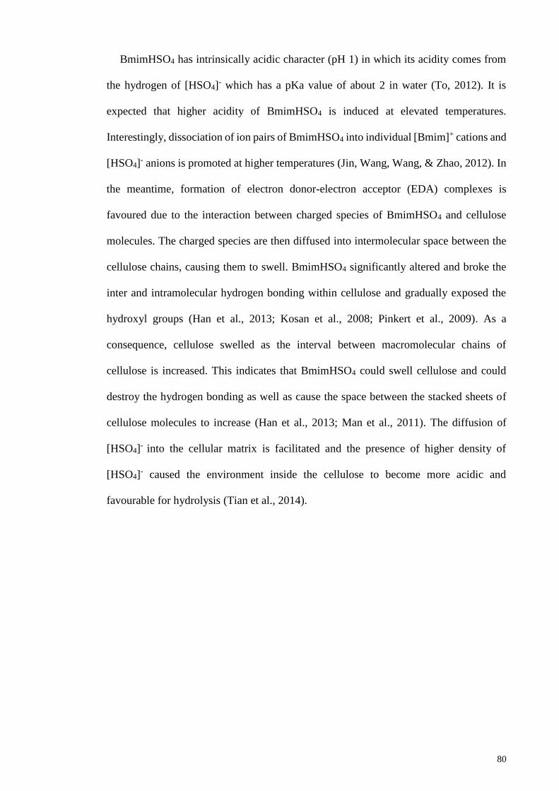
80
BmimHSO4 has intrinsically acidic character (pH 1) in which its acidity comes from
the hydrogen of [HSO4]- which has a pKa value of about 2 in water (To, 2012). It is
expected that higher acidity of BmimHSO4 is induced at elevated temperatures.
Interestingly, dissociation of ion pairs of BmimHSO4 into individual [Bmim]+ cations and
[HSO4]- anions is promoted at higher temperatures (Jin, Wang, Wang, & Zhao, 2012). In
the meantime, formation of electron donor-electron acceptor (EDA) complexes is
favoured due to the interaction between charged species of BmimHSO4 and cellulose
molecules. The charged species are then diffused into intermolecular space between the
cellulose chains, causing them to swell. BmimHSO4 significantly altered and broke the
inter and intramolecular hydrogen bonding within cellulose and gradually exposed the
hydroxyl groups (Han et al., 2013; Kosan et al., 2008; Pinkert et al., 2009). As a
consequence, cellulose swelled as the interval between macromolecular chains of
cellulose is increased. This indicates that BmimHSO4 could swell cellulose and could
destroy the hydrogen bonding as well as cause the space between the stacked sheets of
cellulose molecules to increase (Han et al., 2013; Man et al., 2011). The diffusion of
[HSO4]- into the cellular matrix is facilitated and the presence of higher density of
[HSO4]- caused the environment inside the cellulose to become more acidic and
favourable for hydrolysis (Tian et al., 2014).
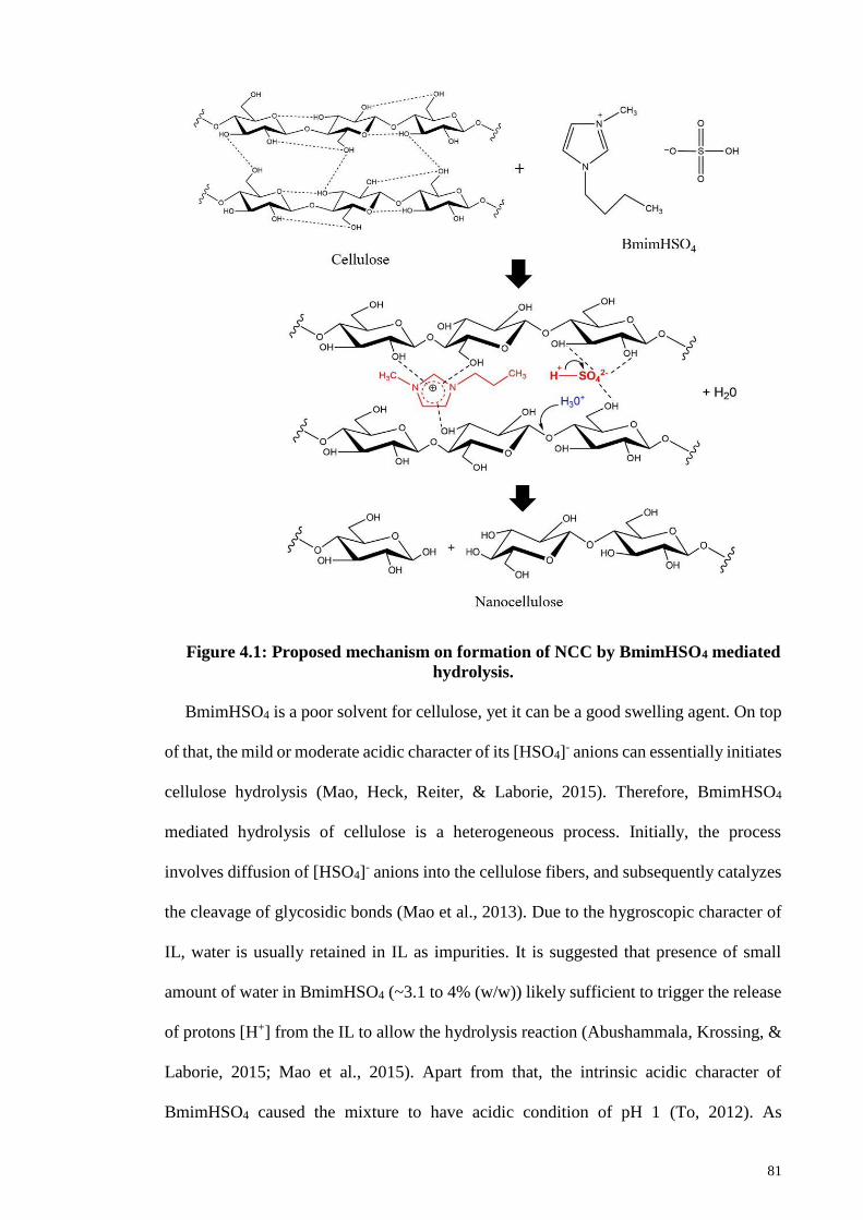
81
Figure 4.1: Proposed mechanism on formation of NCC by BmimHSO4 mediated
hydrolysis.
BmimHSO4 is a poor solvent for cellulose, yet it can be a good swelling agent. On top
of that, the mild or moderate acidic character of its [HSO4]- anions can essentially initiates
cellulose hydrolysis (Mao, Heck, Reiter, & Laborie, 2015). Therefore, BmimHSO4
mediated hydrolysis of cellulose is a heterogeneous process. Initially, the process
involves diffusion of [HSO4]- anions into the cellulose fibers, and subsequently catalyzes
the cleavage of glycosidic bonds (Mao et al., 2013). Due to the hygroscopic character of
IL, water is usually retained in IL as impurities. It is suggested that presence of small
amount of water in BmimHSO4 (~3.1 to 4% (w/w)) likely sufficient to trigger the release
of protons [H+] from the IL to allow the hydrolysis reaction (Abushammala, Krossing, &
Laborie, 2015; Mao et al., 2015). Apart from that, the intrinsic acidic character of
BmimHSO4 caused the mixture to have acidic condition of pH 1 (To, 2012). As
82
mentioned previously, the acidity is attributed to the hydrogen of [HSO4]- and have a pKa
of 2. Theoretically, pKa value greater than 0 indicates a weak acid. Thus, [HSO4]- is a
weaker acid than sulfuric acid (pKa -3) and will only partially dissociated into hydronium
ions [H3O]+ and sulfate anions [SO4]2-.The acidity is attributed to the electronegative
oxygens in drawing the electron density away from the hydrogen atom that is bonded to
one of the oxygens. Therefore, hydrogen are more partially positive and more reactive
(Tian et al., 2014). A small fractions of protons [H+] that dissociated from the [HSO4]-
are suggested to react with water molecules in the aqueous solution to facilitate the
formation of hydronium ions. As a consequence, most protons exist in the form of
hydronium ions in the system. The highly reactive hydronium ions are important to
further catalyze the hydrolytic cleavage of glycosidic bonds by causing the protonation
of glycosidic oxygen that consequently induced splitting of glycosidic bonds and resulted
in the release of shorter fragments of cellulose chains as NCC. Thus, hydrolysis of
cellulose is effectively catalyzed by Bronsted acid IL in the presence of trace amount of
water (Abushammala et al., 2015).
Protonation of glycosidic oxygen induced by hydronium ions initiated cellulose
hydrolysis and subsequently lead to splitting of glycosidic bonds. The hydrolytic cleavage
of glycosidic bonds between two anhydroglucose units results in the rearrangement of
interlinking chain ends, which rendered the release of internal strain (Man et al., 2011).
In native cellulose, amorphous regions have greater accessibility for [HSO4]- and,
therefore, are more susceptible to hydrolytic cleavage than the crystalline region
(Shimada, Takahashi, Hon, & Shiraishi, 1991). The amorphous regions are hydrolyzed at
a much faster rate due to their disordered structure and consist of weak van der Waals
forces. On contrary, a much slower rate for the reducing end and surface of residual
crystalline domains because they have stronger and ordered structure (Mao et al., 2013;
Millet, Moore, & Saeman, 1954). In other words, the crystalline regions are recalcitrant
83
to hydrolysis than the amorphous parts because of their intricate network of strong
hydrogen bonding. Consequently, the amorphous regions were selectively hydrolyzed
and dissolved in BmimHSO4 and leaving the crystalline regions remained intact in NCC
(Lu & Hsieh, 2010). However, controllable hydrolysis conditions is required in order to
prevent over degradation or complete hydrolysis of cellulose to its molecular units such
as glucose. In addition, mechanical treatment such as ultrasonication after the hydrolysis
reaction is essential to ensure the NCC was uniformly dispersed in the suspension and
also, to disintegrate the bulk cellulose aggregates into smaller dimensions with uniform
size distribution (Maiti et al., 2013; Man et al., 2011). Mechanical agitation is also
important to avoid the possible formation of aggregation and agglomeration of cellulose
crystallites.
In addition to chain scission, esterification of hydroxyl groups due to the [HSO4]- also
occurred during the cellulose hydrolysis catalyzed by BmimHSO4. The sulfate groups
were introduced along the surface of the cellulose crystallites which eventually imparted
a negative charge on the surface of NCC. This anionic stabilization via the electrostatic
repulsion forces of electrical double layers were able to effectively prevent the
aggregation of NCC driven by hydrogen bonding which accounted for the stability of the
colloidal suspension. Hydrogen bonding formed between the hydroxyl groups and greater
surface area of smaller NCC particles with high surface energy contributed to the
aggregation of NCC (Marchessault, Morehead & Koch, 1961; Lu & Hsieh, 2010).
After the hydrolysis and mechanical agitation process, regeneration of cellulose from
the cellulose/[BmimHSO4] suspension is possible with the addition of water as the anti-
solvent. Rapid mixing of water (a polar solvent) with cellulose in IL leads to the
precipitation of cellulose as powdery flocks. Hydrogen bonding properties are mainly
involves in the reconstruction of cellulose within the cellulose/water/BmimHSO4 system
84
(Swatloski et al., 2002). BmimHSO4 has strong water absorbability due to its preference
to form hydrogen bonding with water molecules as O-H bonding has higher
electronegativity and therefore, is much stronger than the corresponding C-H type
hydrogen bonds (Ding et al., 2012). The bonding between cellulose and BmimHSO4 is
then weakened due to the fast addition of water. Reconnection of hydrogen bonding
between cellulose chains during the regeneration process resulted in the reconstitution
and precipitation of highly ordered or crystalline structure of cellulose (Han et al., 2013).
In summary, BmimHSO4 is a Bronsted acidic IL which can performs dual function of
swelling and catalyzing cellulose hydrolysis. Therefore, the NCC produced by
BmimHSO4 mediated hydrolysis exhibit higher crystallinity and smaller size. Overall,
this synthesis route provided by BmimHSO4 offers a promising alternative to the
utilization of concentrated acid approach due to its more advantageous properties in terms
of higher yield and quality of NCC formed, recovery of IL and less toxic. This is an
environmental friendly approach in which the IL is recoverable and recyclable without
any hazardous waste products are being produced.
4.2.2 X-ray Diffraction Analysis
XRD analysis is conducted to determine the changes in crystallinity and crystallite size
of untreated MCC and NCC prepared by BmimHSO4. The crystallinity and crystallite
size of NCC are analysed based on Segal’s formula and Scherrer’s equation, respectively
and are summarized in Table 4.1. Figure 4.2 shows the XRD patterns of pristine MCC
and NCC that are synthesized with BmimHSO4 at different synthesis conditions. As
illustrated in the Figure, NCC exhibit similar diffraction peaks as MCC at 2θ= 14.7º,
16.5º, 22.5º and a small peak at 34.5º corresponding to the (-110), (110), (200) and (040)
crystallographic planes of typical cellulose I structure respectively. These characteristic
peaks are attributed to the monoclinic crystalline cellulose Iβ structure which consists of
85
repeating units of β-D-glucopyranose units joined together via strong intermolecular
hydrogen bonding (Lu et al., 2015). Crystalline cellulose I structure is preserved in the
synthesized NCC. During the hydrolysis catalyzed by BmimHSO4, cellulose amorphous
regions were selectively hydrolyzed and removed in IL. This resulted in the release of
internal strain as cellulose crystalline domains in which the crystallinity was retained or
improved. The crystalline structure was preserved as crystalline regions were
predominantly retained in NCC whereas the amorphous regions were removed and
dissolved in IL. Thereafter, the amorphous contents of NCC were significantly lowered
and yield in the production of highly crystalline structure of NCC.
The crystallinity index (CrI) of NCC is notably affected by hydrolysis temperature,
time, cellulose concentrations as well as ultrasonication conditions. A relatively ordered
structure with strong and resolve peak at position 22.5º (200) corresponds to the
crystallinity peak, and the CrI can be determined based on the intensity of this peak. A
sharper diffraction peak at 22.5º (200) indicates higher crystallinity and thus, higher CrI.
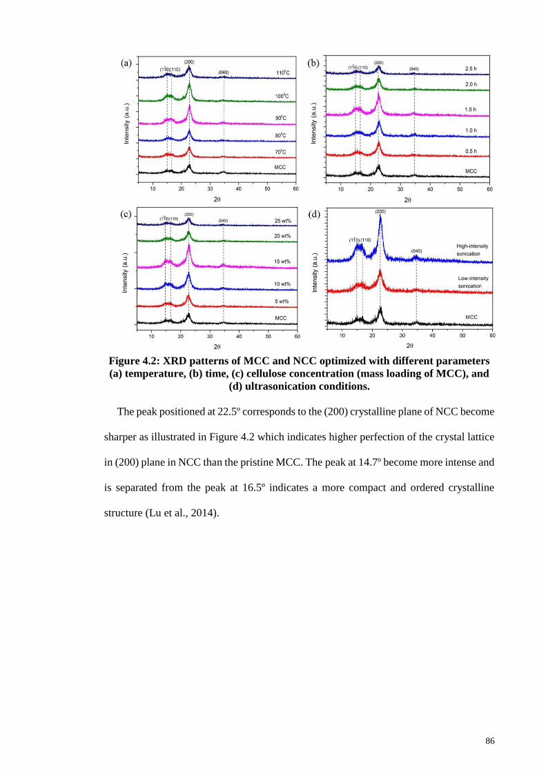
86
Figure 4.2: XRD patterns of MCC and NCC optimized with different parameters
(a) temperature, (b) time, (c) cellulose concentration (mass loading of MCC), and
(d) ultrasonication conditions.
The peak positioned at 22.5º corresponds to the (200) crystalline plane of NCC become
sharper as illustrated in Figure 4.2 which indicates higher perfection of the crystal lattice
in (200) plane in NCC than the pristine MCC. The peak at 14.7º become more intense and
is separated from the peak at 16.5º indicates a more compact and ordered crystalline
structure (Lu et al., 2014).
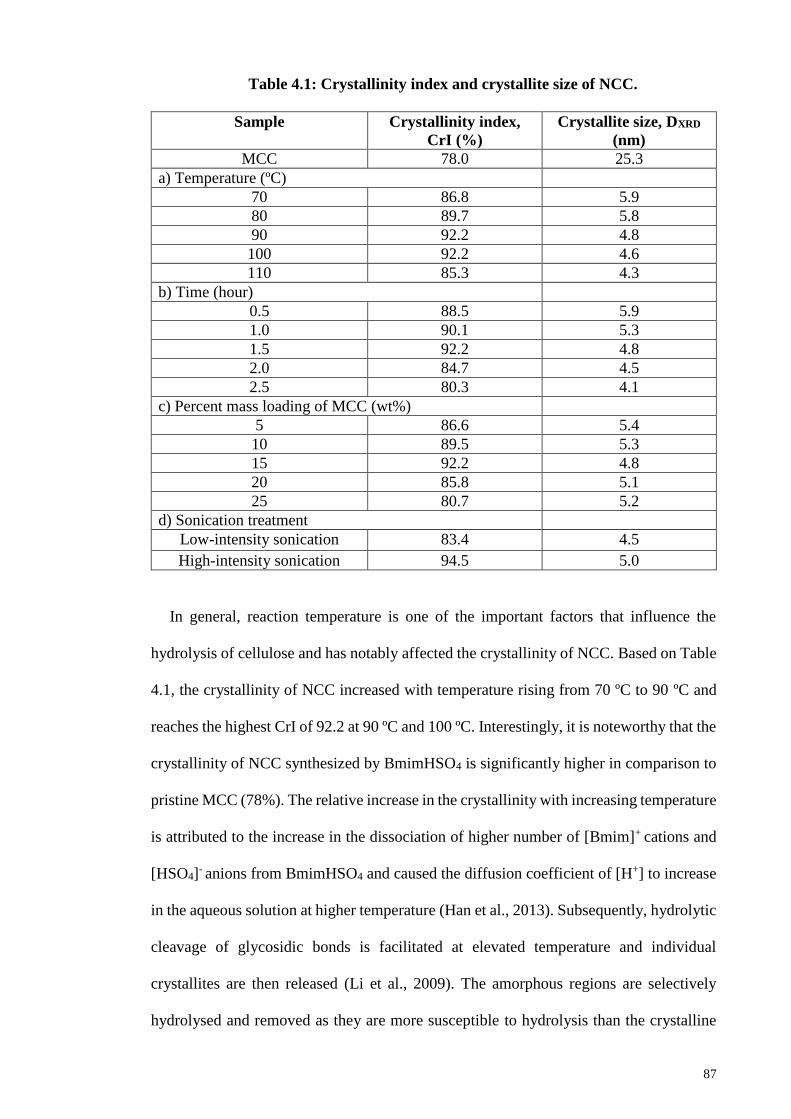
87
Table 4.1: Crystallinity index and crystallite size of NCC.
Sample Crystallinity index,
CrI (%)
Crystallite size, DXRD
(nm)
MCC 78.0 25.3
a) Temperature (ºC)
70 86.8 5.9
80 89.7 5.8
90 92.2 4.8
100 92.2 4.6
110 85.3 4.3
b) Time (hour)
0.5 88.5 5.9
1.0 90.1 5.3
1.5 92.2 4.8
2.0 84.7 4.5
2.5 80.3 4.1
c) Percent mass loading of MCC (wt%)
5 86.6 5.4
10 89.5 5.3
15 92.2 4.8
20 85.8 5.1
25 80.7 5.2
d) Sonication treatment
Low-intensity sonication 83.4 4.5
High-intensity sonication 94.5 5.0
In general, reaction temperature is one of the important factors that influence the
hydrolysis of cellulose and has notably affected the crystallinity of NCC. Based on Table
4.1, the crystallinity of NCC increased with temperature rising from 70 ºC to 90 ºC and
reaches the highest CrI of 92.2 at 90 ºC and 100 ºC. Interestingly, it is noteworthy that the
crystallinity of NCC synthesized by BmimHSO4 is significantly higher in comparison to
pristine MCC (78%). The relative increase in the crystallinity with increasing temperature
is attributed to the increase in the dissociation of higher number of [Bmim]+ cations and
[HSO4]- anions from BmimHSO4 and caused the diffusion coefficient of [H+] to increase
in the aqueous solution at higher temperature (Han et al., 2013). Subsequently, hydrolytic
cleavage of glycosidic bonds is facilitated at elevated temperature and individual
crystallites are then released (Li et al., 2009). The amorphous regions are selectively
hydrolysed and removed as they are more susceptible to hydrolysis than the crystalline
88
counterparts. Therefore, higher crystallinity of NCC is obtained at high temperature
hydrolysis reaction with BmimHSO4 because the removal of amorphous parts essentially
increased the crystallinity of NCC (Man et al., 2011).
On the other hand, crystallinity of NCC gradually decreased from 92.2 to 85.3 when
heated beyond 100 ºC. This is presumably due to the hydrolysis of crystalline components
taking place at higher temperature after the removal of amorphous fractions. In addition,
excessive hydrolysis of cellulose into glucose monomers suggesting loss of NCC, and
indirectly can lead to decrease in its crystallinity. This is further evidenced by the
darkening of cellulose suspension due to dehydration and carbonization or the existence
of some coloring matter arising from the glucose as temperature rose to 110 ºC, as
illustrated in Figure 4.3 (Fan & Li, 2002; Lu, Gui, Zheng, & Liu, 2013). Moreover, the
broadening of the crystalline peaks indicates the decrease in the crystallite size of NCC
as the reaction temperature increased. Cellulose with loose structure is favorable for the
changes in the crystallite size during hydrolysis. It has been suggested that the growth of
the defective crystallites and degradation of smaller crystallites influenced the crystallite
size during hydrolysis (Liu et al., 2013). Thus, smaller crystallite size NCC is formed at
high temperature as the degradation of crystallites took place to a large extent during
hydrolysis. At higher reaction temperature, the hydrolytic cleavage of glycosidic linkages
catalyzed by IL took place extensively and performed at a higher rate. Consequently, this
phenomenon induced the progressive reduction of cellulose crystallites and eventually
degradation of crystalline domains that resulted in the production of smaller crystallite
sizes of NCC at higher operating temperature. In summary, 90 ºC is selected to be the
optimal reaction temperature owing to higher crystallinity of NCC produced and also to
avoid excessive hydrolysis and possible degradation of cellulose, leading to lower NCC
yield and lower crystallinity. Excessive hydrolysis can be essentially avoided by adopting
a low-to-moderate reaction temperature for hydrolysis.
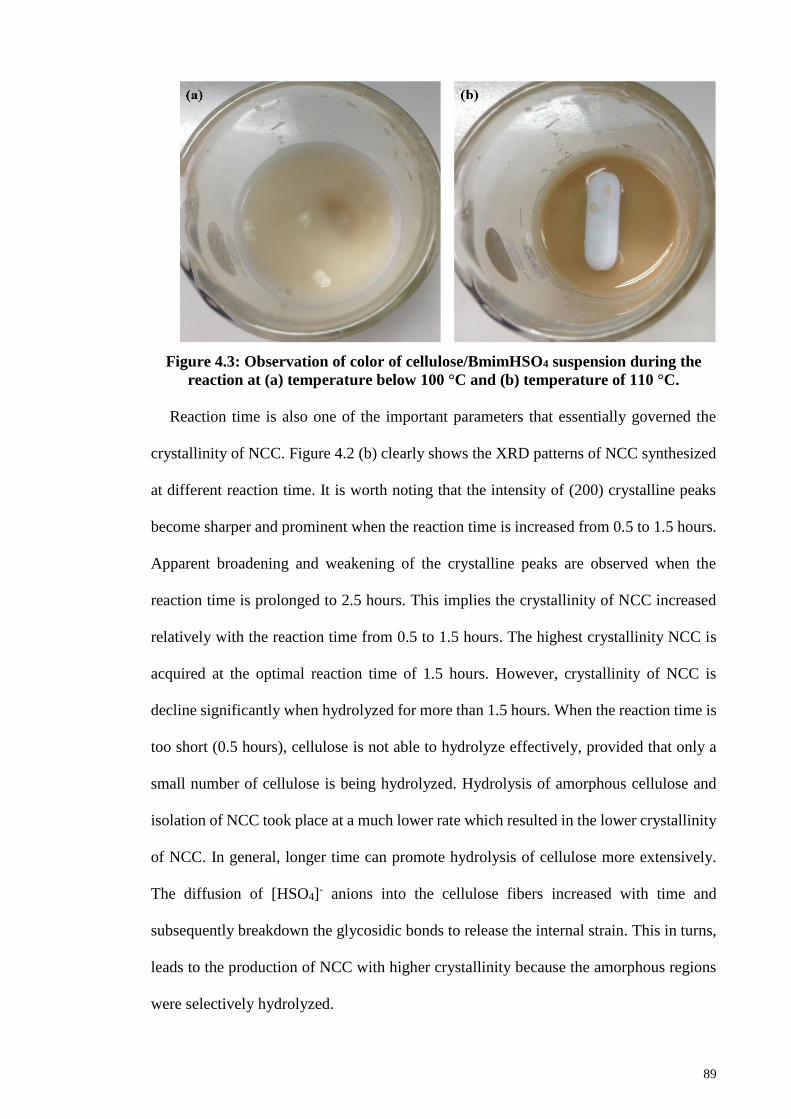
89
Figure 4.3: Observation of color of cellulose/BmimHSO4 suspension during the
reaction at (a) temperature below 100 °C and (b) temperature of 110 °C.
Reaction time is also one of the important parameters that essentially governed the
crystallinity of NCC. Figure 4.2 (b) clearly shows the XRD patterns of NCC synthesized
at different reaction time. It is worth noting that the intensity of (200) crystalline peaks
become sharper and prominent when the reaction time is increased from 0.5 to 1.5 hours.
Apparent broadening and weakening of the crystalline peaks are observed when the
reaction time is prolonged to 2.5 hours. This implies the crystallinity of NCC increased
relatively with the reaction time from 0.5 to 1.5 hours. The highest crystallinity NCC is
acquired at the optimal reaction time of 1.5 hours. However, crystallinity of NCC is
decline significantly when hydrolyzed for more than 1.5 hours. When the reaction time is
too short (0.5 hours), cellulose is not able to hydrolyze effectively, provided that only a
small number of cellulose is being hydrolyzed. Hydrolysis of amorphous cellulose and
isolation of NCC took place at a much lower rate which resulted in the lower crystallinity
of NCC. In general, longer time can promote hydrolysis of cellulose more extensively.
The diffusion of [HSO4]- anions into the cellulose fibers increased with time and
subsequently breakdown the glycosidic bonds to release the internal strain. This in turns,
leads to the production of NCC with higher crystallinity because the amorphous regions
were selectively hydrolyzed.
90
Amorphous regions are first and more easily decomposed than crystalline domains
because they consist of disordered microstructure and therefore, more susceptible to
hydrolysis reaction. However, prolonged exposure to hydrolysis can further improved the
cellulose hydrolysis which resulted in significant reduction in the crystallinity of NCC.
The lower crystallinity is attributed to the crystalline domains are increasingly subject to
extensive hydrolysis, and thus NCC is partly destroyed. The crystalline domain of
cellulose is gradually hydrolyzed and eventually degrade into glucose monomers after
prolonged hydrolysis treatment (Ahmadi et al., 2015; Fan & Li, 2012). Besides that,
crystallite size of NCC is decreased relatively with increasing reaction time. The
crystallite size is larger at short hydrolysis time (0.5 hour) which presumably is attributed
to the growth of defective crystallites during regeneration process. The size reduction of
crystallites was resulted from the excessive hydrolysis and eventually, the degradation of
the crystalline domains due to the prolonged exposure to hydrolysis. As a consequence,
the crystalline domains are increasingly subjected to hydrolytic cleavage and resulted in
the formation of smaller cellulose crystallites with lower crystallinity contents. Therefore,
1.5 hours of reaction time is chosen as the optimization of synthesis conditions.
Based on Table 4.1, cellulose concentrations (expressed in mass loading of MCC into
IL) also significantly influenced the crystallinity of NCC. Crystallinity of NCC increased
from 86.6 to 92.2 when the mass loading of MCC increased from 5 to 15 wt% and the
highest crystallinity is obtained at 15 wt%. The crystallinity declines drastically with the
further increase in the concentration of MCC beyond 15 wt%. The initial increase in the
crystallinity of NCC is attributed to the increase in the diffusion of [HSO4]- anions into
the cellulose fibers. The increase in the catalytic activity of BmimHSO4 caused the
hydrolytic cleavage of glycosidic bonds as there are greater number of BmimHSO4
molecules present in the system. Additionally, better solvating and catalytic power of
BmimHSO4 are suggested at lower cellulose concentrations. Lower cellulose
91
concentration implies the presence of higher concentrations of IL within the system. In
this context, the proportionate higher concentration of IL signifies the existence of greater
amount of ions of IL which able to acquire higher mobility to diffuse into the cellulose
polymer chains to initiate the hydrolysis and solvation of cellulose. Hence, BmimHSO4
is capable to perform dual functions of catalyzing and dissolution effectively at lower
cellulose concentration state.
On the other hand, gradual reduction of crystallinity is mainly due to the weakening of
the catalytic as well as solvation ability of BmimHSO4 with increasing cellulose
concentration. Besides that, the number of BmimHSO4 molecules present in the system
is much lower with respect to increasing cellulose concentrations which slow down the
diffusion of anions into the fibers. Consequently, the hydrolysis reaction took place at a
lower rate that essentially lead to lower crystallinity. This implies that BmimHSO4 is in
supersaturation state such that increasing cellulose concentration most likely slow down
or halted the hydrolysis reaction (Sun, Chen, Jiang, & Lynch, 2015).
Moreover, when the cellulose concentration is beyond the optimum concentration (>
15 wt% of MCC), apparent lower crystallinity in NCC has been observed because some
portion of MCC cannot dissolve completely in BmimHSO4 and become critical for the
hydrolysis (Lu et al., 2013). The viscosity of cellulose/BmimHSO4 suspension is
increased apparently with relatively higher cellulose concentrations. The viscous
suspension renders difficulty to the stirring process as well as effective mixing of
cellulose with BmimHSO4. As a consequence, hydrolysis of cellulose is seriously
impacted. The dimensions of the crystals related to the (200) plane is increased probably
attributed to the recrystallization of the amorphous regions in the border of the crystalline
regions of NCC (Nascimento et al., 2014). It is also suggested that the nucleation and
growth of crystallite is accelerated due to supersaturation effect (Sun et al., 2015). In
92
summary, lower cellulose concentration favored the hydrolysis and beneficial to the
production of highly crystalline NCC.
The crystallinity of NCC produced with low-intensity ultrasonic bath and high-
intensity ultrasonic probe are 83.4% and 94.5%, respectively. This signifies that high-
intensity ultrasonication produced higher crystallinity NCC due to stronger cavitation
effect. High-intensity ultrasonication allows the formation, growth and collapse of the
cavitation bubbles took place instantaneously (in-situ) and more intense in aqueous
solution because the ultrasonic probe is immersed directly into the cellulose suspension.
As a consequence, a greater intensity of cavitation and higher amplitude of vibrational
energy are generated (Hamid, Zain, Das & Centi, 2016). However, weaker cavitation
effect is produced by ultrasonic bath as the suspension is inserted into the water bath and
thereby, generating lower vibrational energy. The resulting ultrasound energy is
transferred to the cellulose chains through cavitation process. Consequently, the
mechanical shearing forces of higher intensity due to the collapse of the cavitation
bubbles generally break down the interaction forces that hold the cellulose microfibrils
after hydrolysis, which in turns facilitated the disintegration of cellulosic fibres into
nanofibers. In addition, it is suggested that greater cavitation effect due to high-intensity
ultrasonication selectively damaged the amorphous and defective crystalline regions
(Guo, Guo, Wang & Yin, 2016). Crystallinity enhancement for NCC with high-intensity
ultrasonication is more profound as the (200) crystalline peaks become sharper and more
resolve. This indicates the formation of a more compact and ordered crystalline structure
NCC with higher crystal lattice perfection is successful with the utilization of high-
intensity ultrasonication. Therefore, it can be concluded that high-intensity
ultrasonication shows more favorable effect on crystallinity enhancement as well as
beneficial to the isolation of NCC following hydrolysis.
93
Apart from that, high-intensity ultrasonication give rise to the formation of slightly
bigger cellulose crystallites because the treatment promotes the growth of the crystallites
and meanwhile, enhanced the crystallinity of NCC. On the contrary, low-intensity
sonication was less effectively in promoting the crystallites growth after the hydrolysis
process. Hence, the crystallites were comparably smaller due to the less profound
vibrational forces generated by the cavitation.
Overall, it is confirmed that BmimHSO4-mediated hydrolysis predominantly liberates
cellulose I structure with higher crystallinity than the precursor materials under mildly
acidic conditions. The synthesis conditions are optimized at 90 ºC for 1.5 hours with a
cellulose concentration of 15 wt% MCC. The controllable synthesis conditions are
essentially to avoid serious degradation of cellulose into sugar molecules and prolonged
hydrolysis will be not productive. The hydrolysis conditions can be controlled through
have a good monitoring and controllable parameters such as reaction temperature and
time. Low operating temperature and moderate reaction time are adopted for efficient
production of NCC through the hydrolysis process.
4.2.3 Hydrodynamic Size and Zeta Potential Analysis
Table 4.2 shows the hydrodynamic size, polydispersity index (PDI) and zeta potential
measurement of NCC synthesized with different reaction parameters. It is clearly
illustrated that synthesis parameters such as temperature, time, cellulose concentrations
as well as ultrasonication conditions are influenced the hydrodynamic size of NCC.
Apparently, the average hydrodynamic size of NCC is strongly dependent on the
temperature and time of synthesis. Hydrodynamic size of NCC decrease relatively with
increasing synthesis temperature and time. The size of NCC is decreased from 89.07 to
36.78 nm when the temperature is increased from 70 to 110 ºC. Meanwhile, drastic
reduction of hydrodynamic size from 85.69 to 33.71 nm is observed as the reaction time
94
is increased from 0.5 to 2.5 hours. This signifies that large and poorly dispersed
aggregation of NCC is formed with high polydispersity index (PDI) at low temperature
and shorter time. In other words, higher temperature and longer reaction time essentially
favored hydrolysis of cellulose by promoting the cleavage of glycosidic bonds.
Consequently, the size of NCC is diminished with more uniform size distribution. Hence,
small size NCC can be dispersed well in water to acquire a stable colloidal suspension
(Ahmadi et al., 2015).
Apart from that, cellulose concentration is also known to play a crucial role in cellulose
hydrolysis. Cellulose concentration yield significant impacts on the uniformity of NCC
particles. In this context, a lower cellulose concentration meaning a higher proportionate
concentration of IL in the system. The ions of IL capable to acquire higher mobility and
diffusion of ions into the cellulose matrix is performed at a higher rate due to the presence
of higher concentration of ions of IL. As a consequence, higher number of ions of IL are
able to surround and interact with the cellulose molecules for the hydrolysis reaction and
consequently resulted in the formation of NCC with smaller dimensional size and
enhanced uniformity. On contrary, higher cellulose concentration suggested that there are
lower concentration of IL in the system. The scarcity of ions of IL caused the interaction
between IL and cellulose less likely to be performed at an optimum rate. Therefore, some
of the cellulose molecules were effectively hydrolyzed into smaller particles, while some
part of it would retained bulk and aggregate sizes of NCC.
The hydrodynamic size of NCC obviously increased with increasing cellulose
concentrations. Interestingly, the PDI values also increased gradually. This manifests that
NCC formed under higher cellulose concentration have larger size with less uniformity
due to the weakening of the catalytic as well as solvation ability of BmimHSO4 at its
supersaturation state. Eventually, BmimHSO4 acquired a supersaturation state at the
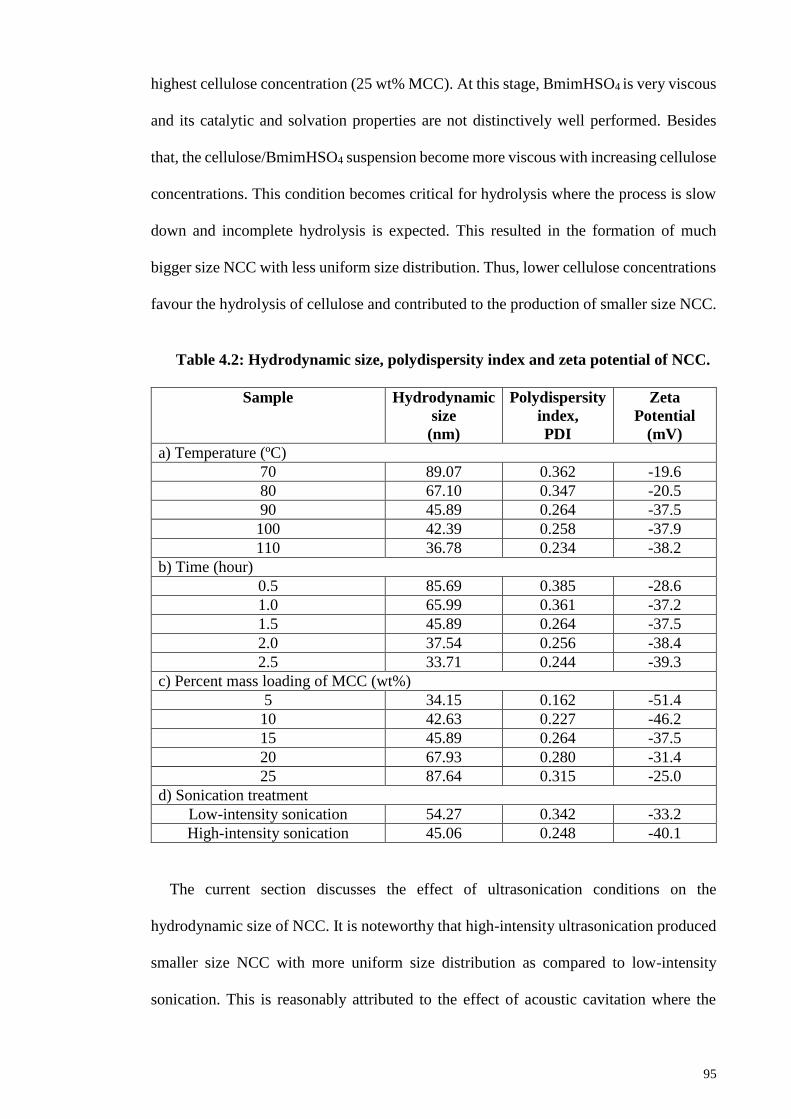
95
highest cellulose concentration (25 wt% MCC). At this stage, BmimHSO4 is very viscous
and its catalytic and solvation properties are not distinctively well performed. Besides
that, the cellulose/BmimHSO4 suspension become more viscous with increasing cellulose
concentrations. This condition becomes critical for hydrolysis where the process is slow
down and incomplete hydrolysis is expected. This resulted in the formation of much
bigger size NCC with less uniform size distribution. Thus, lower cellulose concentrations
favour the hydrolysis of cellulose and contributed to the production of smaller size NCC.
Table 4.2: Hydrodynamic size, polydispersity index and zeta potential of NCC.
Sample Hydrodynamic
size
(nm)
Polydispersity
index,
PDI
Zeta
Potential
(mV)
a) Temperature (ºC)
70 89.07 0.362 -19.6
80 67.10 0.347 -20.5
90 45.89 0.264 -37.5
100 42.39 0.258 -37.9
110 36.78 0.234 -38.2
b) Time (hour)
0.5 85.69 0.385 -28.6
1.0 65.99 0.361 -37.2
1.5 45.89 0.264 -37.5
2.0 37.54 0.256 -38.4
2.5 33.71 0.244 -39.3
c) Percent mass loading of MCC (wt%)
5 34.15 0.162 -51.4
10 42.63 0.227 -46.2
15 45.89 0.264 -37.5
20 67.93 0.280 -31.4
25 87.64 0.315 -25.0
d) Sonication treatment
Low-intensity sonication 54.27 0.342 -33.2
High-intensity sonication 45.06 0.248 -40.1
The current section discusses the effect of ultrasonication conditions on the
hydrodynamic size of NCC. It is noteworthy that high-intensity ultrasonication produced
smaller size NCC with more uniform size distribution as compared to low-intensity
sonication. This is reasonably attributed to the effect of acoustic cavitation where the

96
highly intense ultrasound energy is transmitted to the cellulose chains and caused
delamination to diminish the interaction forces between the cellulose chains (Guo et al.,
2016). High vibrational energy and violent shock waves generated by ultrasonication
consequently break down the glycosidic bonds and interlayer hydrogen bonding between
cellulose polymer chains which resulted in the delamination of the adjoining cellulose
chains for the liberation of smaller size NCC. In summary, high-intensity ultrasonication
provides a more profound cavitation effect and thereby, effectively disintegrates cellulose
aggregates into individual smaller size NCC (Guo et al., 2016; Lu et al., 2014). NCC is
observed to disperse well in water and have uniform size distribution when high-intensity
ultrasonication treatment is present. On the other hand, weak cavitation provided by low-
intensity sonication only breaks the intermolecular and van der Waals forces between the
cellulose chains and therefore, is not benign to the isolation of smaller size NCC. In
contrast to high-intensity ultrasonication, low-intensity sonication tend to give rise to the
formation of bigger particulates NCC with less uniform size distribution.
Therefore, it can be concluded that higher temperature, longer reaction time, lower
cellulose concentrations as well as with the aid of high-intensity ultrasonication
significantly resulted in the production of uniform and smaller size NCC. Nevertheless,
the hydrodynamic radius of spherical particles dispersed in a solution is indicated based
on dynamic-light scattering (DLS) technique adopted by zeta sizer. The approximate sizes
of NCC can be revealed by DLS. Apart from that, DLS was used to measure the sizes of
nanocellulose and acts as an indicator to compare the different size of NCC prepared by
varied reaction parameters in order to obtain a clear illustration on the process.
In DLS, rod-like cellulosic fibres are always treated as spherical particles, which are
clearly far from reality. The principle of DLS is based on the measurement of the scattered
light intensity due to Brownian motion of particles in a solvent without an applied electric
97
field. Hence, the particle size evaluated likely to be larger than the real dimensions of
particles and the sizes obtained from DLS are also likely to be deviated from those
measured by image analysis such as TEM and AFM (Qua, Hornsby, Sharma & Lyons,
2011). In addition, DLS technique is more biased to larger particles because they are
prone to mask the intensity of light scattered over the smaller particles. Hence, smaller
particles are difficult to be detected by the particle size analyzer (Satyamurthy &
Vigneshwaran, 2013). Furthermore, cellulose molecules are known to be highly
hydrophilic and very likely to swell in water. These resulted in significantly larger size of
NCC is measured with DLS technique of the zetasizer.
Zeta potential can be used to indicate the stability of a colloidal suspension. Basically,
it is derived from the measurement on mobility distribution of charged particles when
they are subjected to an electric field. The formation of the electric double layer between
a solid substrate and a liquid electrolyte or its dispersion medium result in zeta potential.
In general, a colloidal suspension is said to have a good stability accounts for its high zeta
potential value which larger than ±30 mV. Moderate zeta potential colloidal suspension
ranging from ±10 to ±30 mV has incipient instability, and the suspension with low zeta
potential ranging from 0 to ±5 mV will rapidly flocculate (Li, Zhang, Zhang, Xiu, & He,
2015). This means that higher zeta potential values indicate higher capacity to disperse in
water, while lower values indicate low dispersion (Lu et al., 2014).
Polar solvents have large dipole moments (partial charges) as the atoms with different
electronegativities are bonded together. In this context, DI water is adopted as the solvent
for cellulose suspension because it is a polar solvent. NCC spontaneously acquired a
negatively surface electrical charges when brought into contact with the DI water. The
greater surface charges of NCC suspension is attributed to the presence of higher number
of negatively charged sulfate groups that contribute to the electrostatic repulsion between
98
the cellulose chains to prevent the agglomeration and aggregation of NCC. The stability
of NCC suspension is evidenced by the high absolute values of the zeta potential.
Zeta potential values decreased progressively with increasing reaction temperature and
time. The zeta potential values decreased from -19.6 to -38.2 mV when the temperature
increased from 70 to 110 ºC. While the zeta potential is decreased from -28.6 to -39.3 mV
with increasing hydrolysis time from 0.5 to 2.5 hours. NCC synthesized at higher
temperature and prolonged exposure to hydrolysis have higher dispersion capability in
water and therefore, higher colloidal stability. The suspension of NCC is considered
stable as the absolute value is higher than 25 mV. The higher zeta potential values indicate
NCC suspension is stabilized by electrostatic repulsion due to BmimHSO4-mediated
hydrolysis that induced grafting of sulfate groups that randomly adsorbed on the surface
of NCC. Consequently, a negative electrostatic layer is formed on the surface of NCC
due to the negatively charged sulfate groups that repelled the adjoining cellulose chains.
Hence, the NCC suspension is stabilized with a greater mean surface charge. NCC
suspension will neither precipitate nor flocculate due to the electrostatic repulsion
between the negatively charged particles (Guo et al., 2016). The prevalent acidic
contribution is due to the proton of the hydrogen sulfate groups that help in catalyzing the
hydrolysis of cellulose. A greater number of BmimHSO4 molecules are dissociated into
individual charged species at higher temperature and leads to more sulfate groups being
adsorbed on the surface of NCC (Ahmadi et al., 2015). The aggregation of particles are
overcome due to the sufficient repulsive forces between the negatively charged particles.
Besides that, NCC suspension is more stable after prolonged exposure to hydrolysis
because more thorough hydrolysis reaction is favored at longer time. Larger size particles
(low surface area per unit mass) with a lower mean surface charge favored the interaction
between particles due to inadequate cellulose hydrolysis as only a small number of sulfate
99
groups were attached on the surface of NCC. Expectedly, increase in the surface charges
of NCC is evidenced with the gradual reduction of zeta potential values with increasing
hydrolysis time. The stabilization of NCC by electrostatic repulsion is due to the presence
of higher number of sulfate groups from IL to adsorb on the NCC. Ideally, the
electrostatic repulsion forces are strong enough to prevent the aggregation of NCC while
some agglomeration might dominated by van der Waals forces of attraction. Thus, NCC
suspension is considered stable because neither precipitation nor flocculation will be
occurred.
On the other hand, the zeta potential values of NCC increased proportionally with
increasing cellulose concentrations. This signifies the lower stability of NCC suspension
because incomplete or slower rate of hydrolysis presumably happen at higher cellulose
concentration due to the weakening of catalytic as well as solvation power of BmimHSO4
at highly viscous condition. NCC with a lower mean surface charges is formed because
there are only a limited number of sulfate groups present on the surface of NCC due to
ineffective hydrolysis. Weak repulsive forces lead to the agglomeration and aggregation
of particles in water which caused the suspension to become less stable.
Lastly, high-intensity ultrasonication confers the NCC suspension with higher
colloidal stability due to the stabilization of NCC by entanglement and steric hindrance
generated from the mechanical process (Abraham et al., 2013). Ultrasound energy can be
transferred to the cellulose chains through a cavitation process. Due to the acosutic
cavitation effect generated by high-intensity ultrasonication, it is postulates that intense
mechanical shearing forces generally breakdown the interaction forces between the
cellulose chains which subsequently caused delamination and disintegration of cellulose
into smaller particles (Bittmann, Haupert & Schlarb, 2009; Filson & Dawson-Andoh,
2009). Thereafter, smaller size particles with higher surface area would exhibit higher
100
mean surface charge that essentially contributed to the electrostatic repulsion accounted
for their colloidal stability.
4.2.4 Fourier Transform Infrared Spectroscopy (FTIR) Analysis
FTIR is one of the most important analytical techniques that is being widely used
nowadays. It is an indispensable technique to determine the chemical composition of
pristine MCC and changes in the chemical structure of NCC obtained after hydrolysis. In
general, changes in the chemical structure of cellulose can be inferred by disappearance
or increase/reduction in intensity of certain IR bands of the crystalline domains of
cellulose (Jonoobi, Khazaeian, Tahir, Azry, & Oksman, 2011; Shankar & Rhim, 2016).
Figure 4.4 demonstrates the FTIR spectra of pristine MCC and synthesized NCC at
different reaction parameters. As shown in Figure 4.4, NCC exhibit similar FTIR spectra
as MCC, indicates that preservation of chemical structure of cellulose I took place, which
is in good accordance with XRD results reported in the Section 4.2.2.
Both MCC and NCC exhibit relatively strong broad band in 3300-3450 cm-1
corresponds to stretching vibration of hydroxyl groups (-OH) in cellulose molecules
(Maiti et al., 2013; Pang et al., 2015). The band shifted to lower wavenumber after
hydrolysis, indicates that hydrolysis weakened the hydrogen bonding of cellulose.
Intricate network of hydrogen bonding between cellulose chains produced strong
crystalline structure of cellulose molecules that organized in the form of microfibrils. The
hydroxyl groups are responsible for the establishment of various inter and intramolecular
hydrogen bonding.
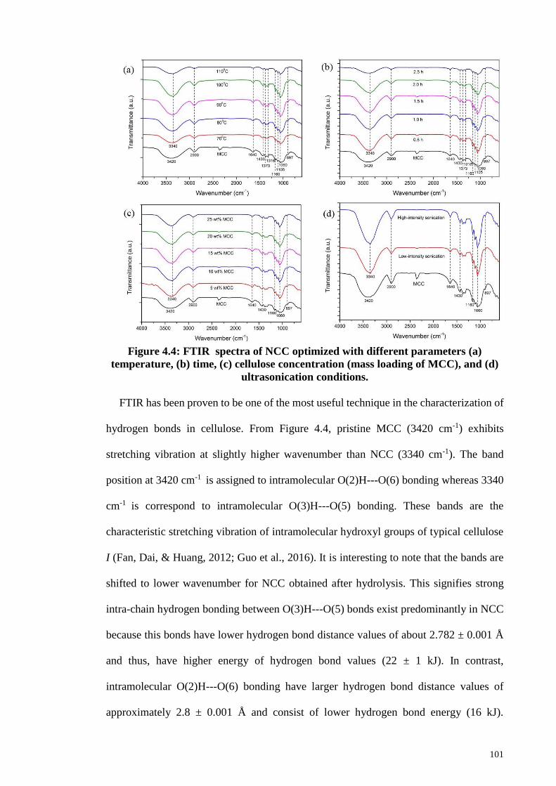
101
Figure 4.4: FTIR spectra of NCC optimized with different parameters (a)
temperature, (b) time, (c) cellulose concentration (mass loading of MCC), and (d)
ultrasonication conditions.
FTIR has been proven to be one of the most useful technique in the characterization of
hydrogen bonds in cellulose. From Figure 4.4, pristine MCC (3420 cm-1) exhibits
stretching vibration at slightly higher wavenumber than NCC (3340 cm-1). The band
position at 3420 cm-1 is assigned to intramolecular O(2)H---O(6) bonding whereas 3340
cm-1 is correspond to intramolecular O(3)H---O(5) bonding. These bands are the
characteristic stretching vibration of intramolecular hydroxyl groups of typical cellulose
I (Fan, Dai, & Huang, 2012; Guo et al., 2016). It is interesting to note that the bands are
shifted to lower wavenumber for NCC obtained after hydrolysis. This signifies strong
intra-chain hydrogen bonding between O(3)H---O(5) bonds exist predominantly in NCC
because this bonds have lower hydrogen bond distance values of about 2.782 ± 0.001 Å
and thus, have higher energy of hydrogen bond values (22 ± 1 kJ). In contrast,
intramolecular O(2)H---O(6) bonding have larger hydrogen bond distance values of
approximately 2.8 ± 0.001 Å and consist of lower hydrogen bond energy (16 kJ).
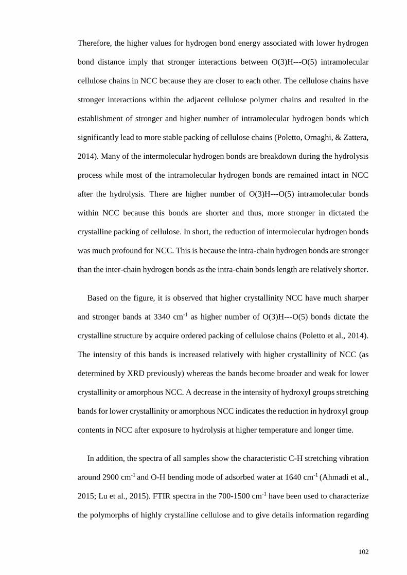
102
Therefore, the higher values for hydrogen bond energy associated with lower hydrogen
bond distance imply that stronger interactions between O(3)H---O(5) intramolecular
cellulose chains in NCC because they are closer to each other. The cellulose chains have
stronger interactions within the adjacent cellulose polymer chains and resulted in the
establishment of stronger and higher number of intramolecular hydrogen bonds which
significantly lead to more stable packing of cellulose chains (Poletto, Ornaghi, & Zattera,
2014). Many of the intermolecular hydrogen bonds are breakdown during the hydrolysis
process while most of the intramolecular hydrogen bonds are remained intact in NCC
after the hydrolysis. There are higher number of O(3)H---O(5) intramolecular bonds
within NCC because this bonds are shorter and thus, more stronger in dictated the
crystalline packing of cellulose. In short, the reduction of intermolecular hydrogen bonds
was much profound for NCC. This is because the intra-chain hydrogen bonds are stronger
than the inter-chain hydrogen bonds as the intra-chain bonds length are relatively shorter.
Based on the figure, it is observed that higher crystallinity NCC have much sharper
and stronger bands at 3340 cm-1 as higher number of O(3)H---O(5) bonds dictate the
crystalline structure by acquire ordered packing of cellulose chains (Poletto et al., 2014).
The intensity of this bands is increased relatively with higher crystallinity of NCC (as
determined by XRD previously) whereas the bands become broader and weak for lower
crystallinity or amorphous NCC. A decrease in the intensity of hydroxyl groups stretching
bands for lower crystallinity or amorphous NCC indicates the reduction in hydroxyl group
contents in NCC after exposure to hydrolysis at higher temperature and longer time.
In addition, the spectra of all samples show the characteristic C-H stretching vibration
around 2900 cm-1 and O-H bending mode of adsorbed water at 1640 cm-1 (Ahmadi et al.,
2015; Lu et al., 2015). FTIR spectra in the 700-1500 cm-1 have been used to characterize
the polymorphs of highly crystalline cellulose and to give details information regarding
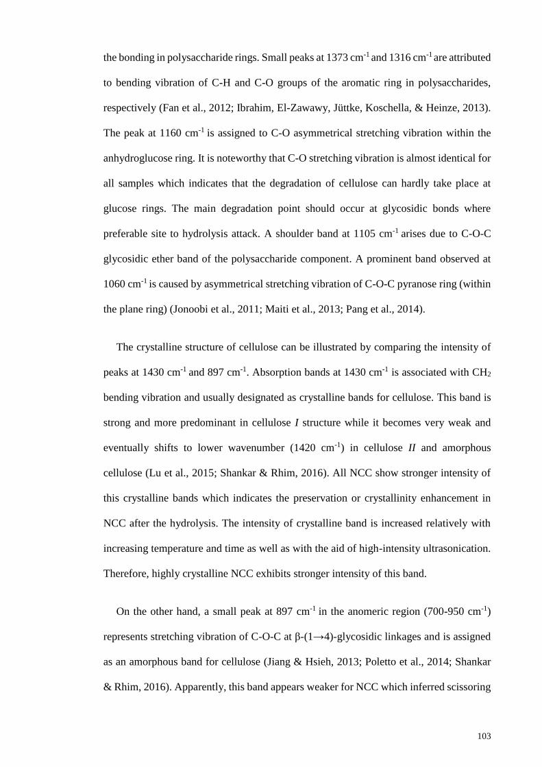
103
the bonding in polysaccharide rings. Small peaks at 1373 cm-1 and 1316 cm-1 are attributed
to bending vibration of C-H and C-O groups of the aromatic ring in polysaccharides,
respectively (Fan et al., 2012; Ibrahim, El-Zawawy, Jüttke, Koschella, & Heinze, 2013).
The peak at 1160 cm-1 is assigned to C-O asymmetrical stretching vibration within the
anhydroglucose ring. It is noteworthy that C-O stretching vibration is almost identical for
all samples which indicates that the degradation of cellulose can hardly take place at
glucose rings. The main degradation point should occur at glycosidic bonds where
preferable site to hydrolysis attack. A shoulder band at 1105 cm-1 arises due to C-O-C
glycosidic ether band of the polysaccharide component. A prominent band observed at
1060 cm-1 is caused by asymmetrical stretching vibration of C-O-C pyranose ring (within
the plane ring) (Jonoobi et al., 2011; Maiti et al., 2013; Pang et al., 2014).
The crystalline structure of cellulose can be illustrated by comparing the intensity of
peaks at 1430 cm-1 and 897 cm-1. Absorption bands at 1430 cm-1 is associated with CH2
bending vibration and usually designated as crystalline bands for cellulose. This band is
strong and more predominant in cellulose I structure while it becomes very weak and
eventually shifts to lower wavenumber (1420 cm-1) in cellulose II and amorphous
cellulose (Lu et al., 2015; Shankar & Rhim, 2016). All NCC show stronger intensity of
this crystalline bands which indicates the preservation or crystallinity enhancement in
NCC after the hydrolysis. The intensity of crystalline band is increased relatively with
increasing temperature and time as well as with the aid of high-intensity ultrasonication.
Therefore, highly crystalline NCC exhibits stronger intensity of this band.
On the other hand, a small peak at 897 cm-1 in the anomeric region (700-950 cm-1)
represents stretching vibration of C-O-C at β-(1→4)-glycosidic linkages and is assigned
as an amorphous band for cellulose (Jiang & Hsieh, 2013; Poletto et al., 2014; Shankar
& Rhim, 2016). Apparently, this band appears weaker for NCC which inferred scissoring
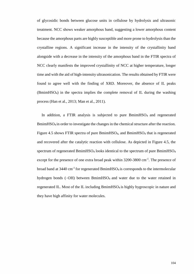
104
of glycosidic bonds between glucose units in cellulose by hydrolysis and ultrasonic
treatment. NCC shows weaker amorphous band, suggesting a lower amorphous content
because the amorphous parts are highly susceptible and more prone to hydrolysis than the
crystalline regions. A significant increase in the intensity of the crystallinity band
alongside with a decrease in the intensity of the amorphous band in the FTIR spectra of
NCC clearly manifests the improved crystallinity of NCC at higher temperature, longer
time and with the aid of high-intensity ultrasonication. The results obtained by FTIR were
found to agree well with the finding of XRD. Moreover, the absence of IL peaks
(BmimHSO4) in the spectra implies the complete removal of IL during the washing
process (Han et al., 2013; Man et al., 2011).
In addition, a FTIR analysis is subjected to pure BmimHSO4 and regenerated
BmimHSO4 in order to investigate the changes in the chemical structure after the reaction.
Figure 4.5 shows FTIR spectra of pure BmimHSO4, and BmimHSO4 that is regenerated
and recovered after the catalytic reaction with cellulose. As depicted in Figure 4.5, the
spectrum of regenerated BmimHSO4 looks identical to the spectrum of pure BmimHSO4
except for the presence of one extra broad peak within 3200-3800 cm-1. The presence of
broad band at 3440 cm-1 for regenerated BmimHSO4 is corresponds to the intermolecular
hydrogen bonds (–OH) between BmimHSO4 and water due to the water retained in
regenerated IL. Most of the IL including BmimHSO4 is highly hygroscopic in nature and
they have high affinity for water molecules.
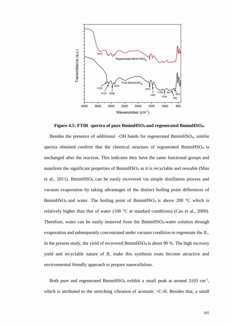
105
Figure 4.5: FTIR spectra of pure BmimHSO4 and regenerated BmimHSO4.
Besides the presence of additional –OH bands for regenerated BmimHSO4, similar
spectra obtained confirm that the chemical structure of regenerated BmimHSO4 is
unchanged after the reaction. This indicates they have the same functional groups and
manifests the significant properties of BmimHSO4 as it is recyclable and reusable (Man
et al., 2011). BmimHSO4 can be easily recovered via simple distillation process and
vacuum evaporation by taking advantages of the distinct boiling point differences of
BmimHSO4 and water. The boiling point of BmimHSO4 is above 200 °C which is
relatively higher than that of water (100 °C at standard conditions) (Cao et al., 2009).
Therefore, water can be easily removed from the BmimHSO4-water solution through
evaporation and subsequently concentrated under vacuum condition to regenerate the IL.
In the present study, the yield of recovered BmimHSO4 is about 90 %. The high recovery
yield and recyclable nature of IL make this synthesis route become attractive and
environmental friendly approach to prepare nanocellulose.
Both pure and regenerated BmimHSO4 exhibit a small peak at around 3103 cm-1,
which is attributed to the stretching vibration of aromatic =C-H. Besides that, a small
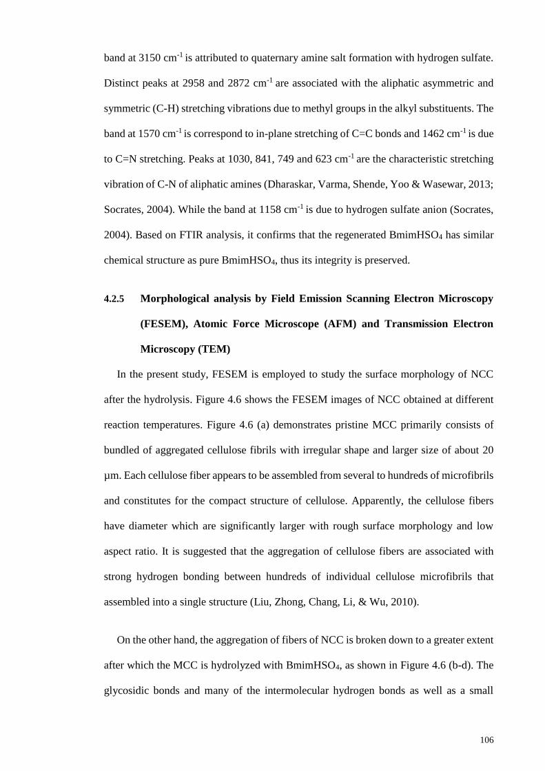
106
band at 3150 cm-1 is attributed to quaternary amine salt formation with hydrogen sulfate.
Distinct peaks at 2958 and 2872 cm-1 are associated with the aliphatic asymmetric and
symmetric (C-H) stretching vibrations due to methyl groups in the alkyl substituents. The
band at 1570 cm-1 is correspond to in-plane stretching of C=C bonds and 1462 cm-1 is due
to C=N stretching. Peaks at 1030, 841, 749 and 623 cm-1 are the characteristic stretching
vibration of C-N of aliphatic amines (Dharaskar, Varma, Shende, Yoo & Wasewar, 2013;
Socrates, 2004). While the band at 1158 cm-1 is due to hydrogen sulfate anion (Socrates,
2004). Based on FTIR analysis, it confirms that the regenerated BmimHSO4 has similar
chemical structure as pure BmimHSO4, thus its integrity is preserved.
4.2.5 Morphological analysis by Field Emission Scanning Electron Microscopy
(FESEM), Atomic Force Microscope (AFM) and Transmission Electron
Microscopy (TEM)
In the present study, FESEM is employed to study the surface morphology of NCC
after the hydrolysis. Figure 4.6 shows the FESEM images of NCC obtained at different
reaction temperatures. Figure 4.6 (a) demonstrates pristine MCC primarily consists of
bundled of aggregated cellulose fibrils with irregular shape and larger size of about 20
µm. Each cellulose fiber appears to be assembled from several to hundreds of microfibrils
and constitutes for the compact structure of cellulose. Apparently, the cellulose fibers
have diameter which are significantly larger with rough surface morphology and low
aspect ratio. It is suggested that the aggregation of cellulose fibers are associated with
strong hydrogen bonding between hundreds of individual cellulose microfibrils that
assembled into a single structure (Liu, Zhong, Chang, Li, & Wu, 2010).
On the other hand, the aggregation of fibers of NCC is broken down to a greater extent
after which the MCC is hydrolyzed with BmimHSO4, as shown in Figure 4.6 (b-d). The
glycosidic bonds and many of the intermolecular hydrogen bonds as well as a small
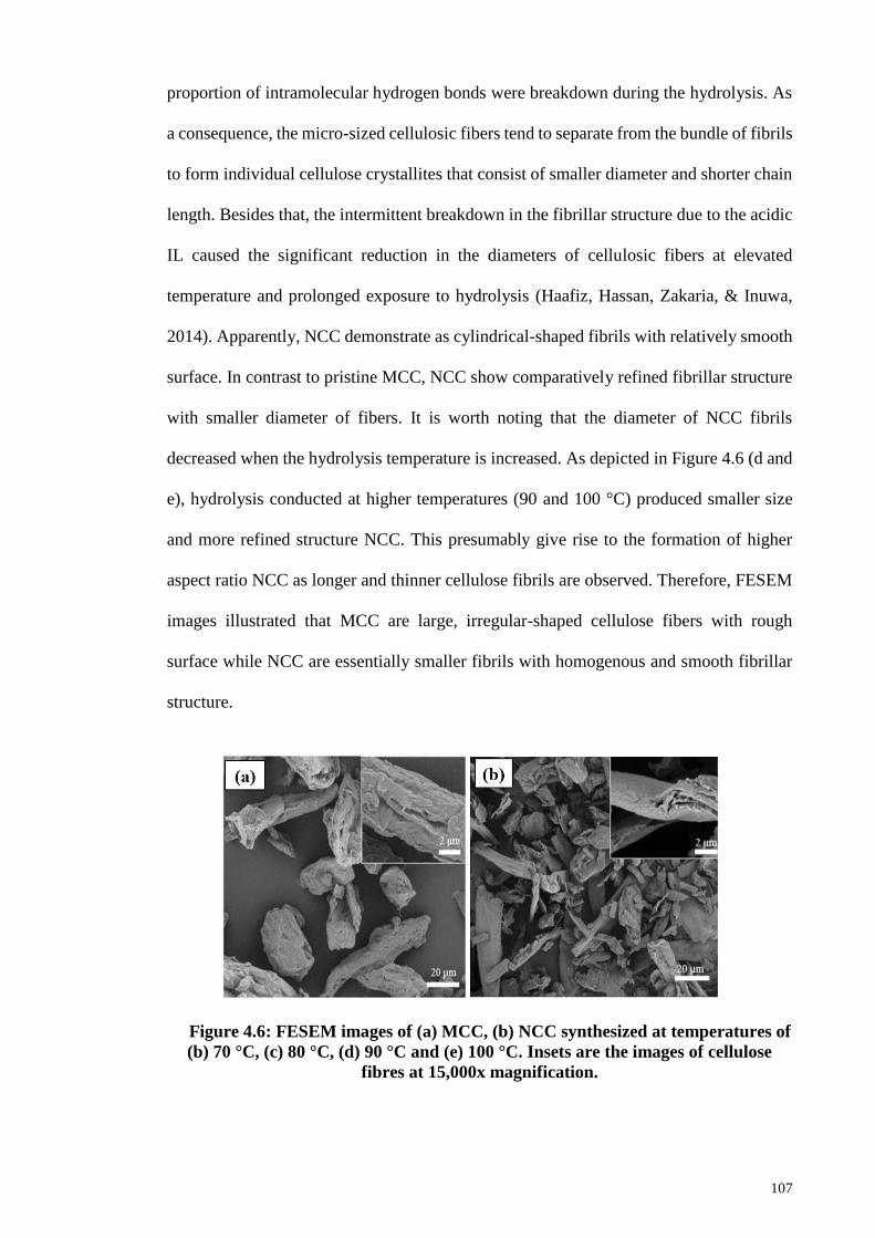
107
proportion of intramolecular hydrogen bonds were breakdown during the hydrolysis. As
a consequence, the micro-sized cellulosic fibers tend to separate from the bundle of fibrils
to form individual cellulose crystallites that consist of smaller diameter and shorter chain
length. Besides that, the intermittent breakdown in the fibrillar structure due to the acidic
IL caused the significant reduction in the diameters of cellulosic fibers at elevated
temperature and prolonged exposure to hydrolysis (Haafiz, Hassan, Zakaria, & Inuwa,
2014). Apparently, NCC demonstrate as cylindrical-shaped fibrils with relatively smooth
surface. In contrast to pristine MCC, NCC show comparatively refined fibrillar structure
with smaller diameter of fibers. It is worth noting that the diameter of NCC fibrils
decreased when the hydrolysis temperature is increased. As depicted in Figure 4.6 (d and
e), hydrolysis conducted at higher temperatures (90 and 100 °C) produced smaller size
and more refined structure NCC. This presumably give rise to the formation of higher
aspect ratio NCC as longer and thinner cellulose fibrils are observed. Therefore, FESEM
images illustrated that MCC are large, irregular-shaped cellulose fibers with rough
surface while NCC are essentially smaller fibrils with homogenous and smooth fibrillar
structure.
Figure 4.6: FESEM images of (a) MCC, (b) NCC synthesized at temperatures of
(b) 70 °C, (c) 80 °C, (d) 90 °C and (e) 100 °C. Insets are the images of cellulose
fibres at 15,000x magnification.
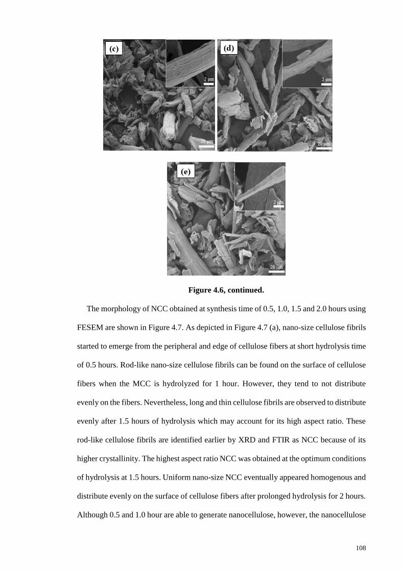
108
Figure 4.6, continued.
The morphology of NCC obtained at synthesis time of 0.5, 1.0, 1.5 and 2.0 hours using
FESEM are shown in Figure 4.7. As depicted in Figure 4.7 (a), nano-size cellulose fibrils
started to emerge from the peripheral and edge of cellulose fibers at short hydrolysis time
of 0.5 hours. Rod-like nano-size cellulose fibrils can be found on the surface of cellulose
fibers when the MCC is hydrolyzed for 1 hour. However, they tend to not distribute
evenly on the fibers. Nevertheless, long and thin cellulose fibrils are observed to distribute
evenly after 1.5 hours of hydrolysis which may account for its high aspect ratio. These
rod-like cellulose fibrils are identified earlier by XRD and FTIR as NCC because of its
higher crystallinity. The highest aspect ratio NCC was obtained at the optimum conditions
of hydrolysis at 1.5 hours. Uniform nano-size NCC eventually appeared homogenous and
distribute evenly on the surface of cellulose fibers after prolonged hydrolysis for 2 hours.
Although 0.5 and 1.0 hour are able to generate nanocellulose, however, the nanocellulose
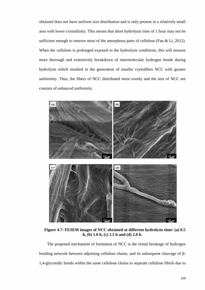
109
obtained does not have uniform size distribution and is only present in a relatively small
area with lower crystallinity. This means that short hydrolysis time of 1 hour may not be
sufficient enough to remove most of the amorphous parts of cellulose (Fan & Li, 2012).
When the cellulose is prolonged exposed to the hydrolysis conditions, this will ensures
more thorough and extensively breakdown of intermolecular hydrogen bonds during
hydrolysis which resulted in the generation of smaller crystallites NCC with greater
uniformity. Thus, the fibers of NCC distributed more evenly and the size of NCC are
consists of enhanced uniformity.
Figure 4.7: FESEM images of NCC obtained at different hydrolysis time: (a) 0.5
h, (b) 1.0 h, (c) 1.5 h and (d) 2.0 h.
The proposed mechanism of formation of NCC is the initial breakage of hydrogen
bonding network between adjoining cellulose chains, and its subsequent cleavage of β-
1,4-glycosidic bonds within the same cellulose chains to separate cellulose fibrils due to
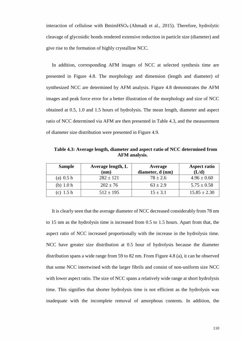
110
interaction of cellulose with BmimHSO4 (Ahmadi et al., 2015). Therefore, hydrolytic
cleavage of glycosidic bonds rendered extensive reduction in particle size (diameter) and
give rise to the formation of highly crystalline NCC.
In addition, corresponding AFM images of NCC at selected synthesis time are
presented in Figure 4.8. The morphology and dimension (length and diameter) of
synthesized NCC are determined by AFM analysis. Figure 4.8 demonstrates the AFM
images and peak force error for a better illustration of the morphology and size of NCC
obtained at 0.5, 1.0 and 1.5 hours of hydrolysis. The mean length, diameter and aspect
ratio of NCC determined via AFM are then presented in Table 4.3, and the measurement
of diameter size distribution were presented in Figure 4.9.
Table 4.3: Average length, diameter and aspect ratio of NCC determined from
AFM analysis.
Sample Average length, L
(nm)
Average
diameter, d (nm)
Aspect ratio
(L/d)
(a) 0.5 h 282 ± 121 78 ± 2.6 4.96 ± 0.60
(b) 1.0 h 202 ± 76 63 ± 2.9 5.75 ± 0.58
(c) 1.5 h 512 ± 195 15 ± 3.1 15.85 ± 2.30
It is clearly seen that the average diameter of NCC decreased considerably from 78 nm
to 15 nm as the hydrolysis time is increased from 0.5 to 1.5 hours. Apart from that, the
aspect ratio of NCC increased proportionally with the increase in the hydrolysis time.
NCC have greater size distribution at 0.5 hour of hydrolysis because the diameter
distribution spans a wide range from 59 to 82 nm. From Figure 4.8 (a), it can be observed
that some NCC intertwined with the larger fibrils and consist of non-uniform size NCC
with lower aspect ratio. The size of NCC spans a relatively wide range at short hydrolysis
time. This signifies that shorter hydrolysis time is not efficient as the hydrolysis was
inadequate with the incomplete removal of amorphous contents. In addition, the
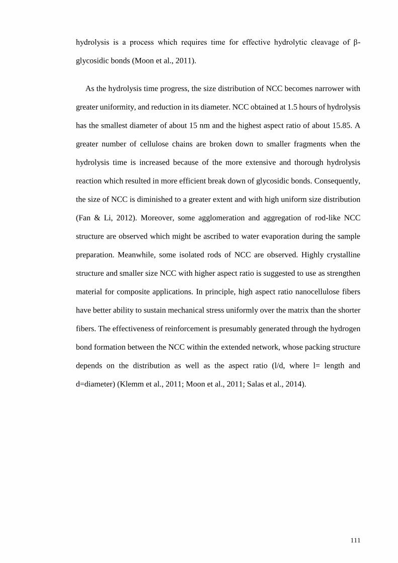
111
hydrolysis is a process which requires time for effective hydrolytic cleavage of β-
glycosidic bonds (Moon et al., 2011).
As the hydrolysis time progress, the size distribution of NCC becomes narrower with
greater uniformity, and reduction in its diameter. NCC obtained at 1.5 hours of hydrolysis
has the smallest diameter of about 15 nm and the highest aspect ratio of about 15.85. A
greater number of cellulose chains are broken down to smaller fragments when the
hydrolysis time is increased because of the more extensive and thorough hydrolysis
reaction which resulted in more efficient break down of glycosidic bonds. Consequently,
the size of NCC is diminished to a greater extent and with high uniform size distribution
(Fan & Li, 2012). Moreover, some agglomeration and aggregation of rod-like NCC
structure are observed which might be ascribed to water evaporation during the sample
preparation. Meanwhile, some isolated rods of NCC are observed. Highly crystalline
structure and smaller size NCC with higher aspect ratio is suggested to use as strengthen
material for composite applications. In principle, high aspect ratio nanocellulose fibers
have better ability to sustain mechanical stress uniformly over the matrix than the shorter
fibers. The effectiveness of reinforcement is presumably generated through the hydrogen
bond formation between the NCC within the extended network, whose packing structure
depends on the distribution as well as the aspect ratio (l/d, where l= length and
d=diameter) (Klemm et al., 2011; Moon et al., 2011; Salas et al., 2014).
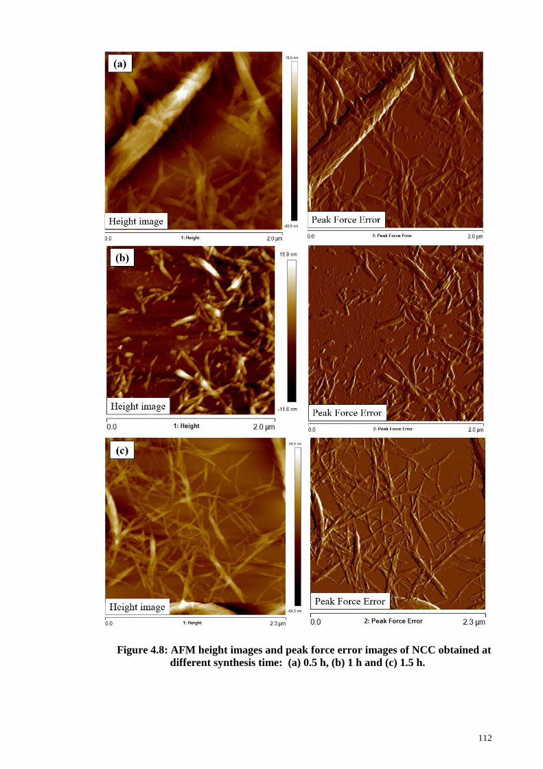
112
Figure 4.8: AFM height images and peak force error images of NCC obtained at
different synthesis time: (a) 0.5 h, (b) 1 h and (c) 1.5 h.
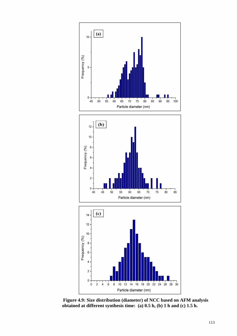
113
Figure 4.9: Size distribution (diameter) of NCC based on AFM analysis
obtained at different synthesis time: (a) 0.5 h, (b) 1 h and (c) 1.5 h.
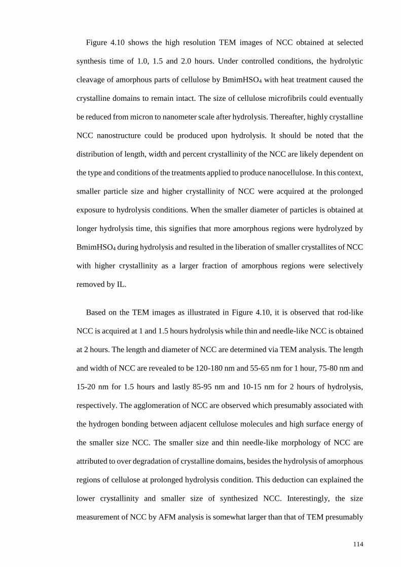
114
Figure 4.10 shows the high resolution TEM images of NCC obtained at selected
synthesis time of 1.0, 1.5 and 2.0 hours. Under controlled conditions, the hydrolytic
cleavage of amorphous parts of cellulose by BmimHSO4 with heat treatment caused the
crystalline domains to remain intact. The size of cellulose microfibrils could eventually
be reduced from micron to nanometer scale after hydrolysis. Thereafter, highly crystalline
NCC nanostructure could be produced upon hydrolysis. It should be noted that the
distribution of length, width and percent crystallinity of the NCC are likely dependent on
the type and conditions of the treatments applied to produce nanocellulose. In this context,
smaller particle size and higher crystallinity of NCC were acquired at the prolonged
exposure to hydrolysis conditions. When the smaller diameter of particles is obtained at
longer hydrolysis time, this signifies that more amorphous regions were hydrolyzed by
BmimHSO4 during hydrolysis and resulted in the liberation of smaller crystallites of NCC
with higher crystallinity as a larger fraction of amorphous regions were selectively
removed by IL.
Based on the TEM images as illustrated in Figure 4.10, it is observed that rod-like
NCC is acquired at 1 and 1.5 hours hydrolysis while thin and needle-like NCC is obtained
at 2 hours. The length and diameter of NCC are determined via TEM analysis. The length
and width of NCC are revealed to be 120-180 nm and 55-65 nm for 1 hour, 75-80 nm and
15-20 nm for 1.5 hours and lastly 85-95 nm and 10-15 nm for 2 hours of hydrolysis,
respectively. The agglomeration of NCC are observed which presumably associated with
the hydrogen bonding between adjacent cellulose molecules and high surface energy of
the smaller size NCC. The smaller size and thin needle-like morphology of NCC are
attributed to over degradation of crystalline domains, besides the hydrolysis of amorphous
regions of cellulose at prolonged hydrolysis condition. This deduction can explained the
lower crystallinity and smaller size of synthesized NCC. Interestingly, the size
measurement of NCC by AFM analysis is somewhat larger than that of TEM presumably
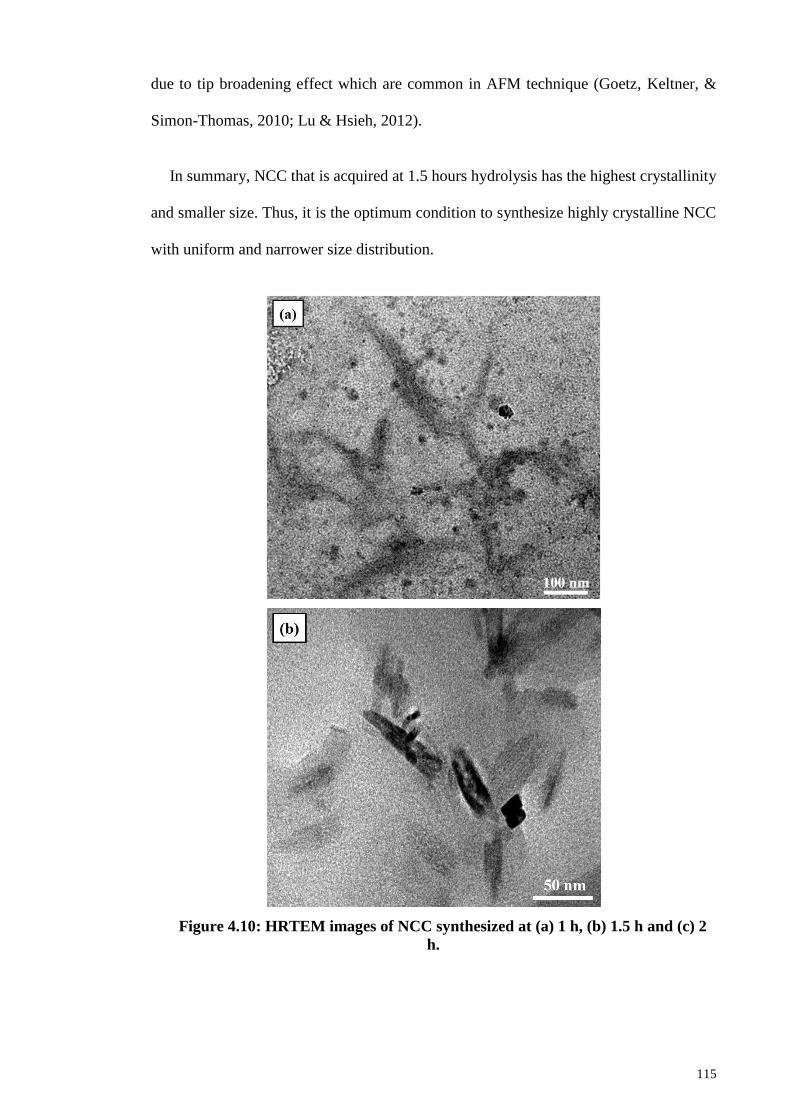
115
due to tip broadening effect which are common in AFM technique (Goetz, Keltner, &
Simon-Thomas, 2010; Lu & Hsieh, 2012).
In summary, NCC that is acquired at 1.5 hours hydrolysis has the highest crystallinity
and smaller size. Thus, it is the optimum condition to synthesize highly crystalline NCC
with uniform and narrower size distribution.
Figure 4.10: HRTEM images of NCC synthesized at (a) 1 h, (b) 1.5 h and (c) 2
h.
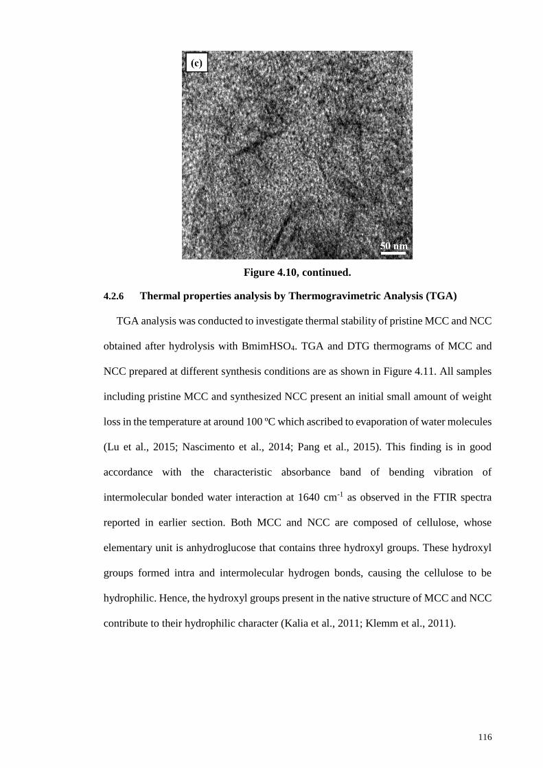
116
Figure 4.10, continued.
4.2.6 Thermal properties analysis by Thermogravimetric Analysis (TGA)
TGA analysis was conducted to investigate thermal stability of pristine MCC and NCC
obtained after hydrolysis with BmimHSO4. TGA and DTG thermograms of MCC and
NCC prepared at different synthesis conditions are as shown in Figure 4.11. All samples
including pristine MCC and synthesized NCC present an initial small amount of weight
loss in the temperature at around 100 ºC which ascribed to evaporation of water molecules
(Lu et al., 2015; Nascimento et al., 2014; Pang et al., 2015). This finding is in good
accordance with the characteristic absorbance band of bending vibration of
intermolecular bonded water interaction at 1640 cm-1 as observed in the FTIR spectra
reported in earlier section. Both MCC and NCC are composed of cellulose, whose
elementary unit is anhydroglucose that contains three hydroxyl groups. These hydroxyl
groups formed intra and intermolecular hydrogen bonds, causing the cellulose to be
hydrophilic. Hence, the hydroxyl groups present in the native structure of MCC and NCC
contribute to their hydrophilic character (Kalia et al., 2011; Klemm et al., 2011).
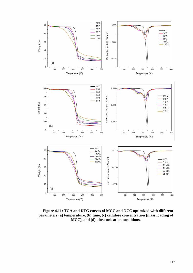
117
Figure 4.11: TGA and DTG curves of MCC and NCC optimized with different
parameters (a) temperature, (b) time, (c) cellulose concentration (mass loading of
MCC), and (d) ultrasonication conditions.
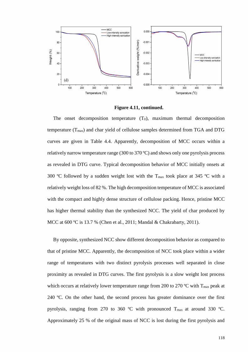
118
Figure 4.11, continued.
The onset decomposition temperature (T0), maximum thermal decomposition
temperature (Tmax) and char yield of cellulose samples determined from TGA and DTG
curves are given in Table 4.4. Apparently, decomposition of MCC occurs within a
relatively narrow temperature range (300 to 370 ºC) and shows only one pyrolysis process
as revealed in DTG curve. Typical decomposition behavior of MCC initially onsets at
300 ºC followed by a sudden weight lost with the Tmax took place at 345 ºC with a
relatively weight loss of 82 %. The high decomposition temperature of MCC is associated
with the compact and highly dense structure of cellulose packing. Hence, pristine MCC
has higher thermal stability than the synthesized NCC. The yield of char produced by
MCC at 600 ºC is 13.7 % (Chen et al., 2011; Mandal & Chakrabarty, 2011).
By opposite, synthesized NCC show different decomposition behavior as compared to
that of pristine MCC. Apparently, the decomposition of NCC took place within a wider
range of temperatures with two distinct pyrolysis processes well separated in close
proximity as revealed in DTG curves. The first pyrolysis is a slow weight lost process
which occurs at relatively lower temperature range from 200 to 270 ºC with Tmax peak at
240 ºC. On the other hand, the second process has greater dominance over the first
pyrolysis, ranging from 270 to 360 ºC with pronounced Tmax at around 330 ºC.
Approximately 25 % of the original mass of NCC is lost during the first pyrolysis and
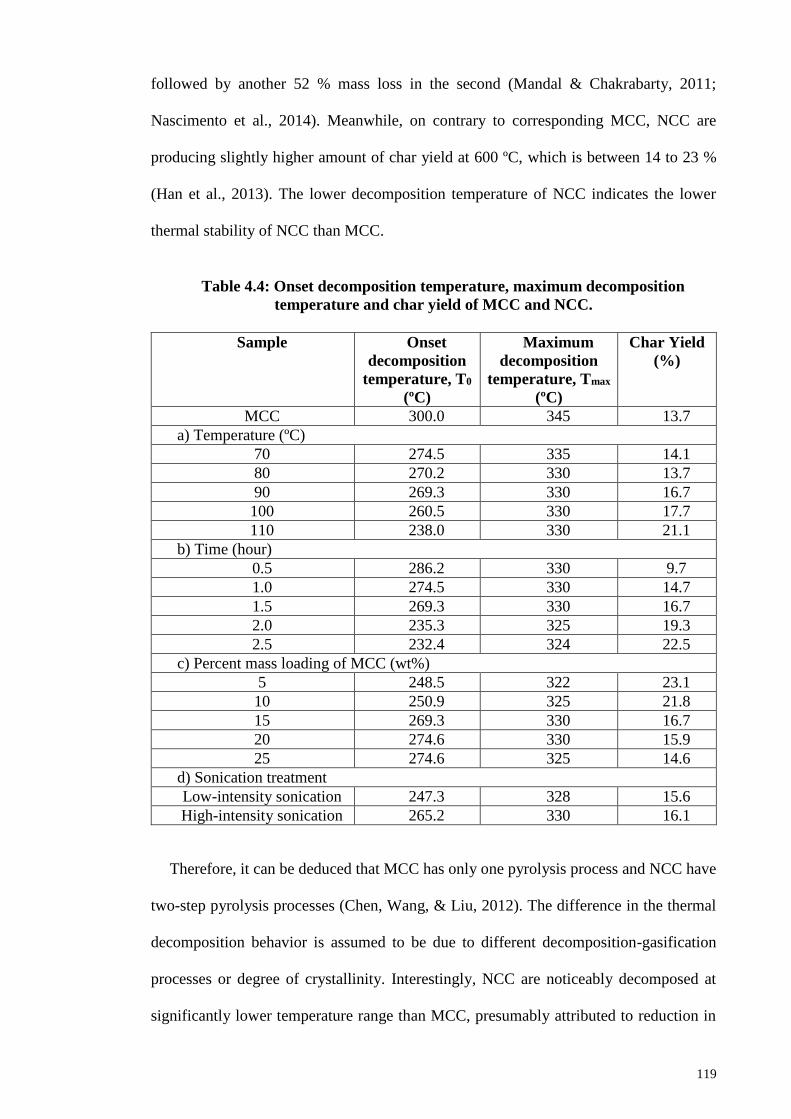
119
followed by another 52 % mass loss in the second (Mandal & Chakrabarty, 2011;
Nascimento et al., 2014). Meanwhile, on contrary to corresponding MCC, NCC are
producing slightly higher amount of char yield at 600 ºC, which is between 14 to 23 %
(Han et al., 2013). The lower decomposition temperature of NCC indicates the lower
thermal stability of NCC than MCC.
Table 4.4: Onset decomposition temperature, maximum decomposition
temperature and char yield of MCC and NCC.
Sample Onset
decomposition
temperature, T0
(ºC)
Maximum
decomposition
temperature, Tmax
(ºC)
Char Yield
(%)
MCC 300.0 345 13.7
a) Temperature (ºC)
70 274.5 335 14.1
80 270.2 330 13.7
90 269.3 330 16.7
100 260.5 330 17.7
110 238.0 330 21.1
b) Time (hour)
0.5 286.2 330 9.7
1.0 274.5 330 14.7
1.5 269.3 330 16.7
2.0 235.3 325 19.3
2.5 232.4 324 22.5
c) Percent mass loading of MCC (wt%)
5 248.5 322 23.1
10 250.9 325 21.8
15 269.3 330 16.7
20 274.6 330 15.9
25 274.6 325 14.6
d) Sonication treatment
Low-intensity sonication 247.3 328 15.6
High-intensity sonication 265.2 330 16.1
Therefore, it can be deduced that MCC has only one pyrolysis process and NCC have
two-step pyrolysis processes (Chen, Wang, & Liu, 2012). The difference in the thermal
decomposition behavior is assumed to be due to different decomposition-gasification
processes or degree of crystallinity. Interestingly, NCC are noticeably decomposed at
significantly lower temperature range than MCC, presumably attributed to reduction in
120
molecular weight of NCC. Consistent with onset decomposition temperature, Tmax of
NCC are lowered to 330 ºC. Pyrolysis process of NCC has been confirmed by the
evidence of two distinct decomposition peak as revealed in the DTG curves. The first
pyrolysis took place at lower temperature (240 ºC) corresponds to the decomposition of
highly accessible and sulfated amorphous regions which are less thermally stable whereas
the second pyrolysis occurred at higher temperature of 330 ºC represents the breakdown
of crystal interior due to thermal degradation of cellulosic materials (Lu et al., 2013). The
second pyrolysis indicates the cleavage of glycosidic linkage within cellulose which
usually falls within the temperature range from 275 to 350 ºC (Kim, Kim, Kim, & Yang,
2006).
Reduction in thermal stability of NCC could be ascribed to several reasons. First,
surface sulfation of NCC because the adhesion of sulfate groups from BmimHSO4 caused
the activation energy required for thermal decomposition was lowered. Sulfate groups
that adsorbed on the cellulose chains will catalyzes the thermal degradation of cellulose
at lower temperature by promoting dehydration reactions. Hence, the presence of sulfate
groups are responsible to the reduced thermal stability of NCC as the thermal
decomposition is occurred at lower temperature (de Morais Teixeira et al., 2010; Li, Yue
& Liu, 2012). Expectedly, NCC are decomposed at lower temperature (Man et al., 2011;
Morais et al., 2013; Xiong et al., 2012). Second, thermal stability diminished because of
the higher surface area of smaller NCC fiber dimensions as compared to macroscopic
cellulose which increased the exposure to heat that presumably contributes to lower
decomposition temperature (Han et al., 2013; Jiang & Hsieh, 2013). A greater exposed
surface area indicates that many of the sulfate groups adsorbed on the surface of cellulose
chains and act as catalyst to promote the degradation of cellulose at lower temperature.
Besides that, the increased exposure of surface sulfate groups to heat presumably
121
contributes to the lower decomposition temperature and therefore, lowered the thermal
stability of NCC.
Despite that, the yield of char residue produced by NCC (14-23 %) are greater than
MCC (13.7 %). Theoretically, pyrolysis of cellulose should produce an ideal yield of
44.4% of carbonaceous residuals during which the dehydration reaction will release all
hydrogen and oxygen in the form of H2O through dehydration reaction. However,
pyrolysis reaction may involves other complex reactions such as decarbonylation to CO,
decarboxylation to CO2, as well as reactions to form gaseous products, for instance, H2
and CH4 (Jiang & Hsieh, 2013). The pyrolysis of cellulose generally involves
dehydration, which occurs below 300 °C, followed by char formation. Dehydration and
char formation were predominantly occurred for cellulose with lower degree of
crystallinity and more favorable in the amorphous regions or disordered structure. The
pyrolytic reaction of cellulose usually proceeds with dehydration, which is the loss of
water molecules from the cellulose components. After the dehydration, there was
subsequent breakdown of the anhydrocellulose into the carbonaceous residuals at higher
temperature which known as char yield residuals. The formation of char yield is
accelerated by dehydration reaction which lead to a higher amount of char remnants,
whose structures are not further degraded into low volatile compounds (Broido & Nelson,
1975; Shafizadeh, 2012).
The higher char yield residues of NCC can be attributed to a few reasons. First, the
dehydration effect catalyzed by sulfate groups would facilitates the formation of char
residues. Moreover, the smaller dimensional size of NCC induced a greater amount of
free end chains. The formation of char residues is favorable as the decomposition of free
end chains took place at a lower temperature (Wang, Ding, & Cheng, 2007). During
hydrolysis, the cleavage of glycosidic linkages and breakdown of the inter and intra
122
hydrogen bonds resulted in the formation of NCC fragments with reduced chain length
and smaller dimensional size. Lower chain length NCC possess an increase in the exposed
surface area with sulfate bearing groups as well as increase in the number of free end
chains. Hence, the formation of char residues of NCC is favorable due to the dehydration
effect catalyzed by the sulfate groups at lower temperature and constitutes for the
increased char yield content (Li, Yue & Liu, 2012). Additionally, the intrinsically flame
resistant characteristic of the sulfated amorphous and crystalline regions of cellulose
caused an increase in char remnant for NCC (Johar, Ahmad, & Dufresne, 2012; Mandal
& Chakrabarty, 2011).
On top of that, decreasing trend of onset decomposition temperature is observed for
NCC that synthesized with higher reaction temperature and longer time. When the
temperature and time increased, onset decomposition temperature of NCC decreased
gradually with Tmax remain unchanged. This presumably attributed to a greater number of
sulfate groups adsorbed onto the surface of NCC with exposure to hydrolysis conditions
at higher temperature and prolonged reaction. Thereafter, surface sulfation of NCC
occurred more readily at higher temperature and prolonged time which eventually lead to
lower onset decomposition temperature (Xiong et al., 2012). Nonetheless, the highly
crystalline NCC fabricated at temperature of 90 ºC may compromise the effect of surface
sulfation and the onset decomposition temperature did not decrease drastically. Highly
crystalline NCC constitutes of more closely packed structure and ordered arrangement
of cellulose chains due to existence of higher number of hydrogen bonding established
between adjoining cellulose chains that resulted in a more thermally stable structure
(Ashori et al., 2006; Yang et al., 2007). Furthermore, the cellulose molecules of NCC are
stabilized by intramolecular hydrogen bonds to suppress thermal expansion along the
cellulose chains to enhance the thermal stability (Hidaka, Kim, & Wada, 2010).
Therefore, thermal stability is improved for higher crystallinity NCC because the closed
123
packed structure makes it difficult for heat transfer. The higher crystallinity cellulose
domains act as barriers for the heat transfer as well as thermal degradation (Poletto et al.,
2014). As a consequence, the onset decomposition temperature of NCC is not drastically
decrease to significantly lower temperature. This finding is supported by work of Kim
and his colleagues which demonstrated that high crystallinity cellulose has shifted the
thermal decomposition to higher temperatures (Kim, Eom, & Wada, 2010).
However, on further increasing the reaction temperature and time, many of the
hydrogen bonding and parts of the molecular cellulose chains are ruptured and eventually
degraded which lead to less thermally stable structure that started to decompose at
apparently lower temperature (Chen et al., 2012). In addition, onset decomposition
temperature of NCC is shifted to lower temperature for 5 wt% cellulose concentration
because there is a greater amount of sulfate groups are available for surface sulfation.
Meanwhile, ultrasonication conditions have no significant impact on the thermal
properties of NCC. Similar finding was reported by Chen and his co-workers on 2011.
On the other hand, char yield of NCC is increased proportionally with increasing
reaction time. The surface sulfation of cellulose chains took place extensively with
prolonged exposure to the hydrolysis conditions because reasonably a greater number of
sulfate groups adsorbed onto the surface of NCC and then the char formation is favorable.
It is the sulfate groups that catalyzed the dehydration reaction which contribute to the
formation of char remnants. In addition, the smaller dimensional size of NCC obtained
through longer hydrolysis time would induced a greater number of free end chains. As a
consequence, the formation of char residues is favorable. Less sulfation phenomenon
occurred on the cellulose chains with increasing cellulose concentration as restriction of
ion mobility at viscous suspension and consequently resulted in reduction of char yield
residues. It is well-known that sulfate groups have flame-retardants characteristics
124
because they prompt to form char and act as a catalyst in accelerating dehydration and
desaturation of pyranose rings (Johar et al., 2012; Kokol et al., 2015).
4.3 Synthesis of Cellulose Nanoparticles (CNPs) by BmimOAc
4.3.1 Mechanism of CNPs Synthesis with BmimOAc
The synthesis of regenerated cellulose nanoparticles (CNPs) in 1-butyl-3-
methylimidazolium acetate (BmimOAc) is investigated. Besides that, the influence of
different synthesis parameters on the preparation of CNPs are extensively studied.
Practically, cellulose is difficult to dissolve in water and most common organic solvents
due to its compact crystalline structure. The insolubility of cellulose is mainly attributed
to the stiff molecules as well as closed chain packing via van der Waals forces and
intricate network of inter- and intramolecular hydrogen bonds (Blackley, 2012; Dadi,
Schall, & Varanasi, 2007; Damodaran, Parkin, & Fennema, 2007). However, IL has been
proven to be effective in dissolving complex macromolecules and polymeric components
of lignocellulosic biomass by breaking the hydrogen bond network which in turns,
facilitated the dissolution of cellulose (Swatloski et al., 2002). The details of the proposed
working principle or mechanism is discussed as below.
Generally, dissolution of cellulose in IL is influenced by the interactions between
hydroxyl groups of cellulose and anion of the IL. The anion of the IL functions as
hydrogen-bond acceptor in dissolution process during which it interacts specifically with
hydroxyl proton of the cellulose. In particular, physicochemical properties such as
hydrogen-bonding basicity (β) and viscosity of IL determined the extent and ability of
anion of IL to accept the hydrogen bonding. Acetate anion has strong hydrogen-bonding
basicity (1.09) based on the Kamlet-Taft parameters of [Bmim] salts (MacFarlane et al.,
2006; Ohno & Fukaya, 2009) and BmimOAc has a relatively low viscosity of 646 mPa
(at 25 ºC) (Bonhote et al., 1996; Olivier-Bourbigou et al., 2010). As a result, BmimOAc
125
can be an excellent candidate to dissolve cellulose due to the ability of acetate anion to
accept and established the hydrogen bonding with cellulose. In addition, the low viscosity
of BmimOAc can essentially promotes the dissolution process due to the higher ions
mobility (Ha, Mai, An, & Koo, 2011).
Generally, IL is composed entirely of cations and anions. The ionic species in
BmimOAc dissociated into respective [Bmim]+ cations and [OAc]- anions (acetate anion)
as the interactions between charged species within BmimOAc are weakened at elevated
temperature. The charged species interact specifically with hydroxyl proton and oxygen
of cellulose molecules to form electron donor-electron acceptor complexes (EDA)
(Pinkert et al., 2009). On one hand, the dissociated charged species diffuse into the space
between the cellulose chains. Free acetate anions are then interact specifically with
hydroxyl proton of cellulose while [Bmim]+ cations preferably to associate with hydroxyl
oxygen of cellulose (Ha et al., 2011). These interactions result in the separation of
hydroxyl groups and increase the interlayer spacing between the adjoining cellulose
chains which consequently lead to swelling behavior of cellulose fibrils. Thus, cellulose
dissolution is facilitated (Kosan et al., 2008). The proposed mechanism of formation of
CNPs with BmimOAc solvolysis approach is illustrated in Figure 4.12.
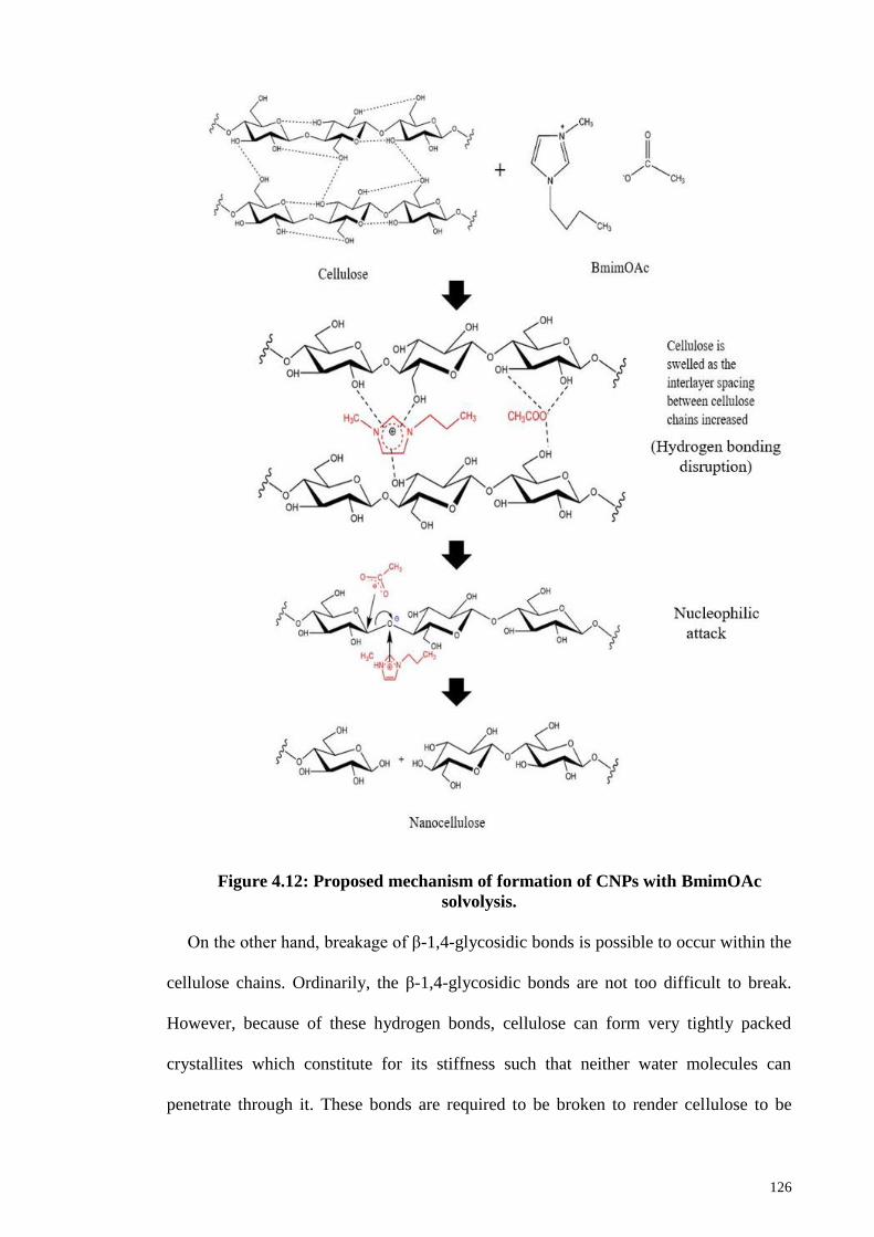
126
Figure 4.12: Proposed mechanism of formation of CNPs with BmimOAc
solvolysis.
On the other hand, breakage of β-1,4-glycosidic bonds is possible to occur within the
cellulose chains. Ordinarily, the β-1,4-glycosidic bonds are not too difficult to break.
However, because of these hydrogen bonds, cellulose can form very tightly packed
crystallites which constitute for its stiffness such that neither water molecules can
penetrate through it. These bonds are required to be broken to render cellulose to be
127
inactively strong such that high electronegativity molecules have good affinity on it
(Otulugbu, 2012; Soni, Hayes, & Srivastava, 2004). Acetate anions are good nucleophile
but they are not sufficiently strong to substitute the hydroxyl groups. The carbon atom of
β-1,4-glycosidic bonds is attacked by acetate anions whereas the glycosidic oxygen atom
is interacted with the electron-rich aromatic π system of [Bmim]+ via non-bonding or π
electrons. As a result, network of intermolecular hydrogen bonding between neighbouring
cellulose chains and β-1,4-glycosidic bonds within the same cellulose chains are broken
down which lead to dissolution and degradation of cellulose into molecular level (Han et
al., 2013; Kosan et al., 2008). It is suggested that the acetate anions are particularly
playing major role in governing the effective dissolution of cellulose. The dissolution of
cellulose in BmimOAc is inferred by the establishment of strong hydrogen bonding
between acetate anions with hydroxyl groups of cellulose.
On contrary, [Bmim]+ cations only weakly interact with hydroxyl groups through
hydrogen-bonding (Jiang et al., 2012). The existence of aromatic imidazolium ring of
[Bmim]+ cations are to promote electron delocalization in order to enhance the
electronegativity of the anions which in turns, dominate the hydrogen bonds forming
capability with the cellulose. Joint interactions of both [Bmim]+ cations and acetate anions
with hydroxyl groups contributed to the dissolution of cellulose in IL (Ha et al., 2011).
As a consequence, pristine hydrogen bonding network within cellulose is extensively
disrupted while the formation of hydrogen bonds between cellulose and BmimOAc is
facilitated and more established. The disruption on inter and intra hydrogen bonding
within cellulose is attributed to the swelling of cellulose induced by IL. Consequently,
the hydrogen bonds within the pristine cellulose get disrupted in such a way that they lose
their tightly, rigid and ordered packed structure. Meanwhile, intercalation of ions of IL
increased the interlayer spacing between cellulose and disrupted the hydrogen bonding
128
within the crystalline domains. Hence, the cellulose dissolution is accompanied with
swelling of cellulose with certain degree of structural disruption.
Apparently, the regenerated CNPs are swelled after the dissolution process, in
comparison to pristine MCC. The diffusion of charged species of BmimOAc into the
matrix of cellulose chains caused the interlayer spacing between chains to increase. This
contributed to the swelling behavior of regenerated CNPs that subsequently enhanced the
dissolution in BmimOAc (Brandt et al., 2010). As a result, CNPs that regenerated from
BmimOAc dissolution experience certain degree of swelling at different synthesis
conditions (Ang et al., 2012). It is noteworthy that dissolution and swelling of cellulose
in BmimOAc are mainly temperature and time-dependent. High temperature and
prolonged dissolution likely facilitate the dissolution and swelling of cellulose. Thus,
hydrogen bonding within cellulose are disrupted and break down due to the interactions
of acetate anions at higher temperature as rapid dissolution occurred and are intensified
over time (Wang, Li, Cao, & Tang, 2011).
Hydrogen bonding properties also plays a crucial role in the regeneration and
reconstruction of cellulose structure (Swatloski et al., 2002). CNPs can be easily
reconstituted from the BmimOAc/cellulose mixture by the addition of anti-solvent such
as water. Water is functions as an anti-solvent because it benign for the withdrawal of
acetate anions from cellulose by means of competitive hydrogen bonding that results in
the precipitation of cellulose. The hydrogen bonding between BmimOAc and cellulose is
weakened and get disrupted when excess water is added into the system as BmimOAc
has higher affinity to water and more preferentially to form hydrogen bonding with water
molecules.
The interaction energies between BmimOAc and cellulose prone to decrease as more
water is present in the system. On the other hand, the number of hydrogen bonding
129
increases because of the preferentially of BmimOAc to form hydrogen bonding with
water molecules. As a consequence, the interaction energies and number of hydrogen
bonds between BmimOAc and water molecules are increase when more water is present
in BmimOAc/cellulose system. It is evidenced that OH…O type hydrogen bonds are
much stronger than the CH…O type hydrogen bonds as O-H bonding has higher
electronegativity than the corresponding C-H type hydrogen bonding. Hence, the number
of OH…O type hydrogen bonds are higher than CH…O type hydrogen bonds and formed
readily with the addition of water. It is suggested that interaction energies for BmimOAc-
water are much higher than that of BmimOAc-cellulose when two or more water
molecules interact specifically with BmimOAc. This signifies precipitation of cellulose
is due to preference of water molecules to interact with BmimOAc. Therefore,
reconnection of hydrogen bonding between loose cellulose molecules caused the
cellulose to precipitate as the regenerated cellulose and contributes to the changes in the
crystalline structure of CNPs (Ding et al., 2012; Han et al., 2013; Swatloski et al., 2002).
Furthermore, anions of IL proven to be of critical importance in dictate the interaction
of IL with water molecules while the cations are of less significance (Cammarata,
Kazarian, Salter, & Welton, 2001). The absolute value of interaction energy is 5.24
kJ/mol when there is only one water molecule interacts with acetate-based IL through the
cationic component. In opposite, the absolute value of interaction energy is 75.05 kJ/mol
when the interaction is based on anion parts for one water molecule. This implies that
anions of IL interact more specifically with water molecules than the cations. The strength
of interactions of ionic species of IL with water molecules has been reported to follow
the trend of anion–H2O > cation–H2O (Ding et al., 2012).
In summary, CNPs can be acquired from BmimOAc via a solvolysis approach in which
the effective dissolution of cellulose is prompted and meantime, it catalyzes the
130
nucleophilic attacks to the glycosidic bonds. The significance of interactions between IL
and hydroxyl groups of cellulose lead to the breakage of hydrogen bonding between the
cellulose chains. Meanwhile, the cleavage of β-glycosidic bonds caused the formation of
molecular level cellulose at nanometer scale.
4.3.2 X-ray Diffraction Analysis
XRD analysis is adopted to study the changes in the crystalline structure, crystallinity
as well as crystallite size of CNPs after the dissolution and regeneration in BmimOAc.
Figure 4.13 presents the XRD diffraction patterns of CNPs obtained at different synthesis
parameters. Interestingly, XRD patterns of CNPs are apparently different from MCC as
the diffraction peaks were shifted to lower Bragg angle. MCC exhibits a sharp and
prominent crystalline peak at 2θ=22.5º which attributed to the characteristic cellulose I
structure. However, a less crystalline structure of CNPs is confirmed by XRD analysis
with the existence of a broad crystalline peak at position 20.2º corresponds to the
characteristic cellulose II structure. The diffraction peaks of CNPs at 2θ= 12.2º, 20.2º and
21.7º are corresponding to (-110), (110) and (020) crystallographic planes of typical
cellulose II structure respectively (Lu et al., 2015).
In comparison to the diffraction pattern of pristine MCC (cellulose I), a new peak is
evidenced at 12.2º and the (200) crystalline peak of cellulose I is split into two broad and
overlapped peaks around 20.2º and 21.7º. The shift of the main peaks to lower Bragg
angle manifests the increase in the space between stacked sheets of cellulose molecules
and the broader peak indicates smaller crystallites structure of CNPs. This implies that
transformation of crystalline structure from initial cellulose I to cellulose II has took place
after the dissolution and regeneration (solvolysis) in BmimOAc (Han et al., 2013). It is
suggested that the dissolution of cellulose in BmimOAc reasonably caused the swelling
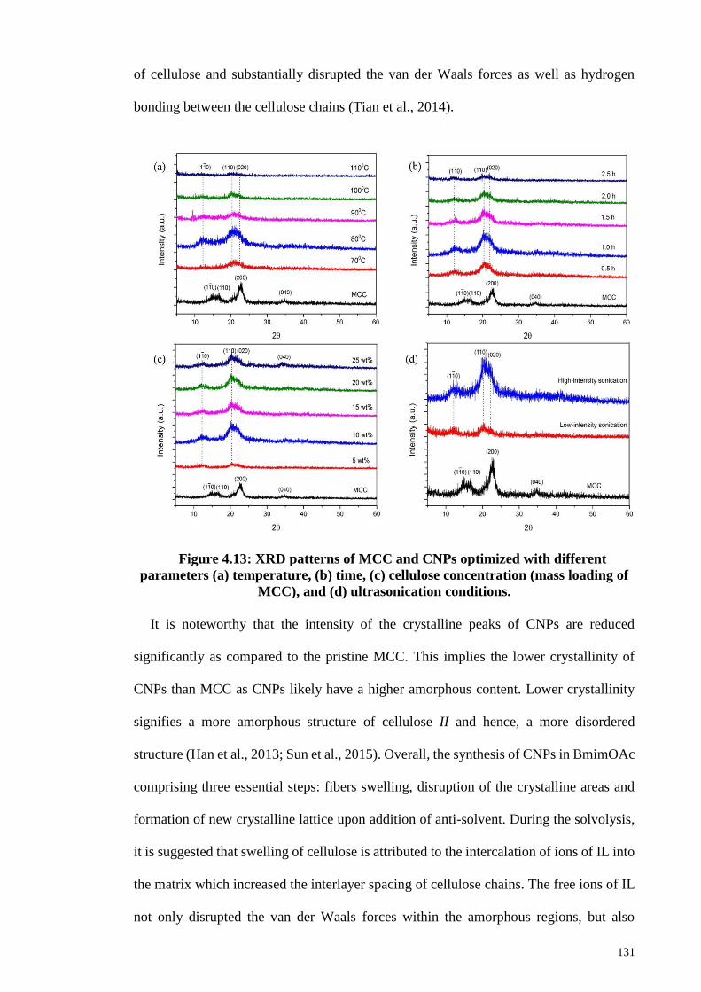
131
of cellulose and substantially disrupted the van der Waals forces as well as hydrogen
bonding between the cellulose chains (Tian et al., 2014).
Figure 4.13: XRD patterns of MCC and CNPs optimized with different
parameters (a) temperature, (b) time, (c) cellulose concentration (mass loading of
MCC), and (d) ultrasonication conditions.
It is noteworthy that the intensity of the crystalline peaks of CNPs are reduced
significantly as compared to the pristine MCC. This implies the lower crystallinity of
CNPs than MCC as CNPs likely have a higher amorphous content. Lower crystallinity
signifies a more amorphous structure of cellulose II and hence, a more disordered
structure (Han et al., 2013; Sun et al., 2015). Overall, the synthesis of CNPs in BmimOAc
comprising three essential steps: fibers swelling, disruption of the crystalline areas and
formation of new crystalline lattice upon addition of anti-solvent. During the solvolysis,
it is suggested that swelling of cellulose is attributed to the intercalation of ions of IL into
the matrix which increased the interlayer spacing of cellulose chains. The free ions of IL
not only disrupted the van der Waals forces within the amorphous regions, but also
132
induced the breakdown of interconnected hydrogen bonds within the crystallite domains.
The cellulose loses its stiffness and bonds were reconnected to form a more amorphous
structure of CNPs. Transition of the crystalline form probably due to the easy association
and rearrangement in intermolecular hydrogen bonding or aggregation of cellulose chains
in BmimOAc upon dissolution and regeneration (Pang et al., 2015). The lower
crystallinity of CNPs are ascribed to the disruption of hydrogen bonding primarily within
the crystalline domains of cellulose chains by interaction with BmimOAc that resulted in
higher amorphousity of CNPs.
Table 4.5 shows the crystallinity index and crystallite size of CNPs calculated based
on Segal’s formula and Scherrer’s equation, respectively from XRD results. Temperature
of reaction is known to be the main factor to influence the crystallinity of CNPs. The
crystallinity of CNPs initially increased from 59.4 to 78.8 when the reaction temperature
is increased from 70 ºC to 80 ºC. However, crystallinity of CNPs dropped tremendously
from 78.8 to 39.2 with elevating temperature from 80 ºC to 110 ºC. Interestingly, the
highest crystallinity CNPs is successfully obtained at a moderate temperature of 80 ºC.
The proportional initial increase in the crystallinity of CNPs is postulated due to
degradation of internal bonds (non-covalent interactions) in the amorphous parts of
cellulose chains. On the other hand, crystallinity dropped with increasing temperature is
due to the breakage of hydrogen bonds within the crystalline domains of cellulose chains
after the degradation of amorphous fractions (Mohkami & Talaeipour, 2011). Low
temperature is just able to overcome the weak van der Waals forces within the amorphous
or disordered regions. While high temperature is essentially required to break the intricate
network of hydrogen bonding within the crystalline domains.
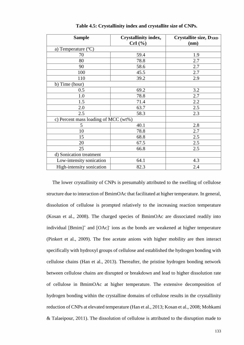
133
Table 4.5: Crystallinity index and crystallite size of CNPs.
Sample Crystallinity index,
CrI (%)
Crystallite size, DXRD
(nm)
a) Temperature (ºC)
70 59.4 1.9
80 78.8 2.7
90 58.6 2.7
100 45.5 2.7
110 39.2 2.9
b) Time (hour)
0.5 69.2 3.2
1.0 78.8 2.7
1.5 71.4 2.2
2.0 63.7 2.5
2.5 58.3 2.3
c) Percent mass loading of MCC (wt%)
5 40.1 2.8
10 78.8 2.7
15 68.8 2.5
20 67.5 2.5
25 66.8 2.5
d) Sonication treatment
Low-intensity sonication 64.1 4.3
High-intensity sonication 82.3 2.4
The lower crystallinity of CNPs is presumably attributed to the swelling of cellulose
structure due to interaction of BmimOAc that facilitated at higher temperature. In general,
dissolution of cellulose is prompted relatively to the increasing reaction temperature
(Kosan et al., 2008). The charged species of BmimOAc are dissociated readily into
individual [Bmim]+ and [OAc]- ions as the bonds are weakened at higher temperature
(Pinkert et al., 2009). The free acetate anions with higher mobility are then interact
specifically with hydroxyl groups of cellulose and established the hydrogen bonding with
cellulose chains (Han et al., 2013). Thereafter, the pristine hydrogen bonding network
between cellulose chains are disrupted or breakdown and lead to higher dissolution rate
of cellulose in BmimOAc at higher temperature. The extensive decomposition of
hydrogen bonding within the crystalline domains of cellulose results in the crystallinity
reduction of CNPs at elevated temperature (Han et al., 2013; Kosan et al., 2008; Mohkami
& Talaeipour, 2011). The dissolution of cellulose is attributed to the disruption made to
134
the intricate network of hydrogen bonding in cellulose. Cellulose loses its stiffness as well
as structure rigidity and bonded to the free ions of IL to become a soluble complex. Hence,
BmimOAc with electronegativity character is benign for the dissolution of cellulose.
In addition, apparent darkening of the cellulose suspension at temperature ≥100 ºC
suggested the complete dissolution and possible degradation of cellulose into glucose
monomer units. This was found in good agreement with the results reported by Wang et
al. (2011) where swelling happened at a temperature of 70 ºC and dissolution at 100 ºC.
Similar finding was obtained for increased dissolution rate with increasing temperature.
Thus, dissolution and swelling of cellulose in IL is temperature-dependent and dissolution
is promoted at temperature beyond 100 ºC. At higher temperature (≥100 ºC), dissolution
rate is the fastest which evidenced by the disappearance or complete dissolution of MCC
after only 10 minutes of addition and cellulose was observed to dissolve completely in
BmimOAc in a shorter time. Moreover, cellulose was started to degrade at a higher
temperature of 110 ºC which illustrated by the formation of yellowish-brown color
suspension as shown in Figure 4.14. This phenomenon indicates the possible occurrence
of degradation into glucose monomers. Therefore, experimental conditions of 80 ºC was
selected as the optimum temperature of synthesis of CNPs in BmimOAc as a compromise
to allow slow dissolution of cellulose at a moderate pace while avoiding the possibility
of cellulose degradation.
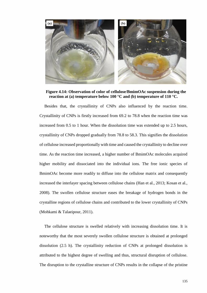
135
Figure 4.14: Observation of color of cellulose/BmimOAc suspension during the
reaction at (a) temperature below 100 °C and (b) temperature of 110 °C.
Besides that, the crystallinity of CNPs also influenced by the reaction time.
Crystallinity of CNPs is firstly increased from 69.2 to 78.8 when the reaction time was
increased from 0.5 to 1 hour. When the dissolution time was extended up to 2.5 hours,
crystallinity of CNPs dropped gradually from 78.8 to 58.3. This signifies the dissolution
of cellulose increased proportionally with time and caused the crystallinity to decline over
time. As the reaction time increased, a higher number of BmimOAc molecules acquired
higher mobility and dissociated into the individual ions. The free ionic species of
BmimOAc become more readily to diffuse into the cellulose matrix and consequently
increased the interlayer spacing between cellulose chains (Han et al., 2013; Kosan et al.,
2008). The swollen cellulose structure eases the breakage of hydrogen bonds in the
crystalline regions of cellulose chains and contributed to the lower crystallinity of CNPs
(Mohkami & Talaeipour, 2011).
The cellulose structure is swelled relatively with increasing dissolution time. It is
noteworthy that the most severely swollen cellulose structure is obtained at prolonged
dissolution (2.5 h). The crystallinity reduction of CNPs at prolonged dissolution is
attributed to the highest degree of swelling and thus, structural disruption of cellulose.
The disruption to the crystalline structure of CNPs results in the collapse of the pristine
136
cellulose ordered structure after the solvolysis due to the breakdown of hydrogen bonding
network (Lu et al., 2015). Interestingly, CNPs with higher crystallinity could be obtained
at 1 hour of dissolution in which the highest crystallinity is obtained throughout the time
frame studied. Shorter dissolution time and moderate reaction temperature were
preferably selected to allow sufficient dissolution while minimizing the possibility of
extensive disruption caused to the crystalline structure. During the regeneration process,
the crystallites most likely have incomplete growth and results in the increase of the
amorphous content of CNPs. Crystallite size decrease is inferred by an increase in peak
broadening with the incomplete growth of the crystallites.
In the current section, the effect of cellulose concentration (expressed in terms of mass
loading of MCC into IL) on the crystallinity of CNPs is discussed. The lowest cellulose
concentration (5 wt% MCC) indicates the presence of higher number of BmimOAc
molecules in the mixture which resulted in the formation of CNPs with very low
crystallinity (40.1). This is reflected by an apparent quite flat diffraction pattern for CNPs
with very low crystallinity and higher amorphous content. Lower cellulose concentration
favoured the dissolution of cellulose because of the greater solvating power of BmimOAc
at low viscosity condition as the free ions acquired higher mobility. The lower viscosity
of IL/cellulose suspension would beneficial for the mass transfer as well as promote the
mobility of the ions. The ions in a lower viscosity medium can acquired higher kinetic
energy and travelled faster for a reaction to take place. The increased diffusion of ions
into the gap between the hydrogen bonded sheets of cellulose chains lead to more
intercalation of ions into the cellulose lattice and subsequently disrupted the ordered
lattice. After a complete swollen state is formed, the ions diffused into the inside of the
crystals and underwent a thorough reaction with cellulose molecules. The breakdown of
hydrogen bonds initiated cellulose dissolution and swelling of cellulose induced the
137
crystalline structure disruption which in turns, resulted in crystallinity reduction of CNPs
after the solvolysis (Chen et al., 2012; Cheng et al., 2012; Sun et al., 2015).
Nevertheless, crystallinity decreased gradually for increasing cellulose concentrations
from 10 to 25 wt%. The dissolution of MCC in BmimOAc becomes less effective with
increasing cellulose concentrations. This is attributed to the decrease solvating power of
BmimOAc with the viscosity increase of the cellulose suspension. Low viscosity will
essentially promote the dissolution, presumably due to higher mobility of ions. On
contrary, higher cellulose concentration contributes to viscosity build up that dramatically
restrict the mobility of ions and eventually retard the ion diffusion to impede cellulose
dissolution. The complete dissolution of 25 wt% MCC will not happen because
BmimOAc is saturated with cellulose molecules and cannot effectively dissolve a higher
quantity of cellulose. Consequently, less disruption is caused to the crystalline structure
and lead to no drastic reduction in the crystallinity of CNPs. In addition, a small portions
of cellulose I structure remained intact in CNPs after dissolution in BmimOAc at 25 wt%
MCC. This is revealed by the presence of a distinct small peak at 34.5º (040) which
signifies the coexistence of cellulose I and II structure with higher amorphous content.
BmimOAc is in the supersaturation state with cellulose molecules. In summary, lower
cellulose concentration is more favorable for the effective dissolution of cellulose.
Apart from that, different ultrasonication treatments also significantly affected the
crystallinity of CNPs. On contrary to low-intensity sonication, high-intensity
ultrasonication contributes to the formation of higher crystallinity CNPs. Improvement in
crystallinity is evidenced by the existence of sharper and prominent of the (020)
crystalline peak (Tang et al., 2013). This is because high-intensity ultrasonication
produced a very strong and intense mechanical oscillating power due to acoustic
cavitation phenomenon. During the process, cavitation leads to the formation, growth and
138
implosion of bubbles occurred continuously as the molecules absorb the ultrasound
energy transmitted to the cellulose chains (Filson & Dawson-Andoh, 2009; Li, Yue, &
Liu, 2012; Suslick et al., 1991). The collapse of these bubbles generates a large amount
of energy and violent mechanical shock wave is given to the cellulose chains (Hamid et
al., 2016). Consequently, high amplitude of vibrational energy due to high-intensity
ultrasound transmits mechanical shearing forces to break down the interconnected
network of hydrogen bonds and caused damage to the van der Waals forces between the
cellulose chains within amorphous regions. Sonochemistry is the energy generated by
cavitation and it is approximately 10−100 kJ/mol, which is within the hydrogen bond
energy scale (Tischer, Sierakowski, Westfahl, & Tischer, 2010). Hence, the disintegration
of macro-cellulosic fibres into nanofibers or nanoparticles is facilitated and beneficial for
isolation of CNPs with higher crystallinity as the amorphous regions is seriously disrupted
due to impact of ultrasonic. On the other hand, less profound cavitation effect and
vibrational energy generated by ultrasonic bath only breaks the weak bonds of amorphous
cellulose, while the inter layers of cellulose are remained partially disentangled and
disintegrated. High-intensity ultrasonication deduced to be effective in the production of
higher crystallinity of CNPs (Li et al., 2015).
In conclusion, XRD analysis verified the fact that native cellulose experienced a
structural transformation from its initial cellulose I to terminated phase of cellulose II in
CNPs with a remarkable reduction in crystallinity after the solvolysis process in
BmimOAc. The hydrogen bonds in cellulose II are found between sheets, which
manifesting a three-dimensional (3D) network whereas hydrogen bonds in cellulose I are
among chains that exist merely in the same sheet. Therefore, CNPs achieved a more stable
crystal structure due to reorganization and rearrangement of hydrogen bonds between
cellulose (Pang et al., 2014; Pang et al., 2015).
139
4.3.3 Hydrodynamic Size and Zeta Potential Analysis
Table 4.6 shows the hydrodynamic size, PDI and zeta potential values of CNPs
obtained at different synthesis parameters. The hydrodynamic size of CNPs increased
dramatically with the increase in temperature and time, alongside with increase in PDI.
This signifies that CNPs synthesized at higher temperature and exposed to prolong
dissolution exhibit larger sizes with less uniformity in size distribution. Interaction
between cation and anion of BmimOAc was weakened at higher temperature and with
over time, so as the dissociation of ion pairs into individual charged species increased.
The free ions acquired greater mobility at higher temperature and their motions become
intensified over time. The diffusion of ions into the cellulose chains is then increased and
subsequently, more intercalation of ions into the lattice resulted in the swelling of the
cellulose structure (Zhao et al., 2012). The larger hydrodynamic sizes of CNPs at higher
temperature and prolonged dissolution are inferred by extensive swelling of cellulose
structure. The size distribution also becomes less uniform with increasing temperature
and time, likely attributed to different degree of swelling of cellulose particles and hence,
yield different hydrodynamic size when dissolved in water. Therefore, dissolution and
swelling of cellulose in BmimOAc are both temperature and time-dependent. It is
important to have controllable experiment parameters such that the extensive swelling of
cellulose can be avoided by performed the solvolysis or dissolution process in shorter
duration of time at relatively low-to-moderate reaction temperature.
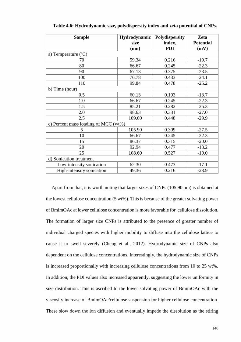
140
Table 4.6: Hydrodynamic size, polydispersity index and zeta potential of CNPs.
Sample Hydrodynamic
size
(nm)
Polydispersity
index,
PDI
Zeta
Potential
(mV)
a) Temperature (ºC)
70 59.34 0.216 -19.7
80 66.67 0.245 -22.3
90 67.13 0.375 -23.5
100 76.78 0.433 -24.1
110 99.84 0.478 -25.2
b) Time (hour)
0.5 60.13 0.193 -13.7
1.0 66.67 0.245 -22.3
1.5 85.21 0.282 -25.3
2.0 98.63 0.331 -27.0
2.5 109.00 0.448 -29.9
c) Percent mass loading of MCC (wt%)
5 105.90 0.309 -27.5
10 66.67 0.245 -22.3
15 86.37 0.315 -20.0
20 92.94 0.477 -13.2
25 108.60 0.527 -10.0
d) Sonication treatment
Low-intensity sonication 62.30 0.473 -17.1
High-intensity sonication 49.36 0.216 -23.9
Apart from that, it is worth noting that larger sizes of CNPs (105.90 nm) is obtained at
the lowest cellulose concentration (5 wt%). This is because of the greater solvating power
of BmimOAc at lower cellulose concentration is more favorable for cellulose dissolution.
The formation of larger size CNPs is attributed to the presence of greater number of
individual charged species with higher mobility to diffuse into the cellulose lattice to
cause it to swell severely (Cheng et al., 2012). Hydrodynamic size of CNPs also
dependent on the cellulose concentrations. Interestingly, the hydrodynamic size of CNPs
is increased proportionally with increasing cellulose concentrations from 10 to 25 wt%.
In addition, the PDI values also increased apparently, suggesting the lower uniformity in
size distribution. This is ascribed to the lower solvating power of BmimOAc with the
viscosity increase of BmimOAc/cellulose suspension for higher cellulose concentration.
These slow down the ion diffusion and eventually impede the dissolution as the stiring
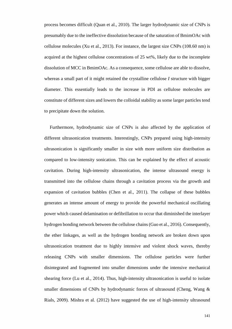
141
process becomes difficult (Quan et al., 2010). The larger hydrodynamic size of CNPs is
presumably due to the ineffective dissolution because of the saturation of BmimOAc with
cellulose molecules (Xu et al., 2013). For instance, the largest size CNPs (108.60 nm) is
acquired at the highest cellulose concentrations of 25 wt%, likely due to the incomplete
dissolution of MCC in BmimOAc. As a consequence, some cellulose are able to dissolve,
whereas a small part of it might retained the crystalline cellulose I structure with bigger
diameter. This essentially leads to the increase in PDI as cellulose molecules are
constitute of different sizes and lowers the colloidal stability as some larger particles tend
to precipitate down the solution.
Furthermore, hydrodynamic size of CNPs is also affected by the application of
different ultrasonication treatments. Interestingly, CNPs prepared using high-intensity
ultrasonication is significantly smaller in size with more uniform size distribution as
compared to low-intensity sonication. This can be explained by the effect of acoustic
cavitation. During high-intensity ultrasonication, the intense ultrasound energy is
transmitted into the cellulose chains through a cavitation process via the growth and
expansion of cavitation bubbles (Chen et al., 2011). The collapse of these bubbles
generates an intense amount of energy to provide the powerful mechanical oscillating
power which caused delamination or defibrillation to occur that diminished the interlayer
hydrogen bonding network between the cellulose chains (Guo et al., 2016). Consequently,
the ether linkages, as well as the hydrogen bonding network are broken down upon
ultrasonication treatment due to highly intensive and violent shock waves, thereby
releasing CNPs with smaller dimensions. The cellulose particles were further
disintegrated and fragmented into smaller dimensions under the intensive mechanical
shearing force (Lu et al., 2014). Thus, high-intensity ultrasonication is useful to isolate
smaller dimensions of CNPs by hydrodynamic forces of ultrasound (Cheng, Wang &
Rials, 2009). Mishra et al. (2012) have suggested the use of high-intensity ultrasound
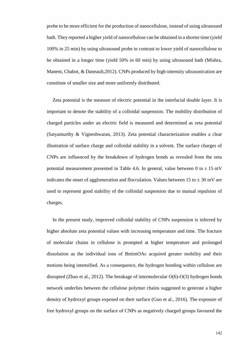
142
probe to be more efficient for the production of nanocellulose, instead of using ultrasound
bath. They reported a higher yield of nanocellulose can be obtained in a shorter time (yield
100% in 25 min) by using ultrasound probe in contrast to lower yield of nanocellulose to
be obtained in a longer time (yield 50% in 60 min) by using ultrasound bath (Mishra,
Manent, Chabot, & Daneault,2012). CNPs produced by high-intensity ultrasonication are
constitute of smaller size and more uniformly distributed.
Zeta potential is the measure of electric potential in the interfacial double layer. It is
important to denote the stability of a colloidal suspension. The mobility distribution of
charged particles under an electric field is measured and determined as zeta potential
(Satyamurthy & Vigneshwaran, 2013). Zeta potential characterization enables a clear
illustration of surface charge and colloidal stability in a solvent. The surface charges of
CNPs are influenced by the breakdown of hydrogen bonds as revealed from the zeta
potential measurement presented in Table 4.6. In general, value between 0 to ± 15 mV
indicates the onset of agglomeration and flocculation. Values between 15 to ± 30 mV are
used to represent good stability of the colloidal suspension due to mutual repulsion of
charges.
In the present study, improved colloidal stability of CNPs suspension is inferred by
higher absolute zeta potential values with increasing temperature and time. The fracture
of molecular chains in cellulose is prompted at higher temperature and prolonged
dissolution as the individual ions of BmimOAc acquired greater mobility and their
motions being intensified. As a consequence, the hydrogen bonding within cellulose are
disrupted (Zhao et al., 2012). The breakage of intermolecular O(6)-O(3) hydrogen bonds
network underlies between the cellulose polymer chains suggested to generate a higher
density of hydroxyl groups exposed on their surface (Guo et al., 2016). The exposure of
free hydroxyl groups on the surface of CNPs as negatively charged groups favoured the
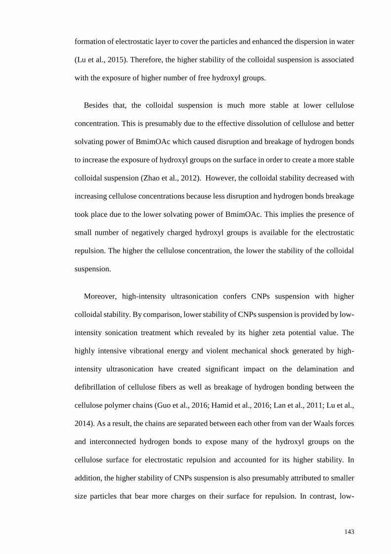
143
formation of electrostatic layer to cover the particles and enhanced the dispersion in water
(Lu et al., 2015). Therefore, the higher stability of the colloidal suspension is associated
with the exposure of higher number of free hydroxyl groups.
Besides that, the colloidal suspension is much more stable at lower cellulose
concentration. This is presumably due to the effective dissolution of cellulose and better
solvating power of BmimOAc which caused disruption and breakage of hydrogen bonds
to increase the exposure of hydroxyl groups on the surface in order to create a more stable
colloidal suspension (Zhao et al., 2012). However, the colloidal stability decreased with
increasing cellulose concentrations because less disruption and hydrogen bonds breakage
took place due to the lower solvating power of BmimOAc. This implies the presence of
small number of negatively charged hydroxyl groups is available for the electrostatic
repulsion. The higher the cellulose concentration, the lower the stability of the colloidal
suspension.
Moreover, high-intensity ultrasonication confers CNPs suspension with higher
colloidal stability. By comparison, lower stability of CNPs suspension is provided by low-
intensity sonication treatment which revealed by its higher zeta potential value. The
highly intensive vibrational energy and violent mechanical shock generated by high-
intensity ultrasonication have created significant impact on the delamination and
defibrillation of cellulose fibers as well as breakage of hydrogen bonding between the
cellulose polymer chains (Guo et al., 2016; Hamid et al., 2016; Lan et al., 2011; Lu et al.,
2014). As a result, the chains are separated between each other from van der Waals forces
and interconnected hydrogen bonds to expose many of the hydroxyl groups on the
cellulose surface for electrostatic repulsion and accounted for its higher stability. In
addition, the higher stability of CNPs suspension is also presumably attributed to smaller
size particles that bear more charges on their surface for repulsion. In contrast, low-
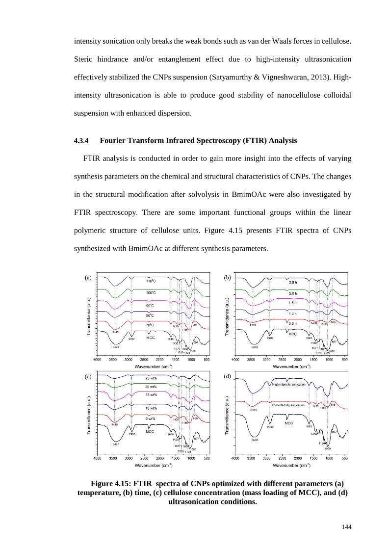
144
intensity sonication only breaks the weak bonds such as van der Waals forces in cellulose.
Steric hindrance and/or entanglement effect due to high-intensity ultrasonication
effectively stabilized the CNPs suspension (Satyamurthy & Vigneshwaran, 2013). High-
intensity ultrasonication is able to produce good stability of nanocellulose colloidal
suspension with enhanced dispersion.
4.3.4 Fourier Transform Infrared Spectroscopy (FTIR) Analysis
FTIR analysis is conducted in order to gain more insight into the effects of varying
synthesis parameters on the chemical and structural characteristics of CNPs. The changes
in the structural modification after solvolysis in BmimOAc were also investigated by
FTIR spectroscopy. There are some important functional groups within the linear
polymeric structure of cellulose units. Figure 4.15 presents FTIR spectra of CNPs
synthesized with BmimOAc at different synthesis parameters.
Figure 4.15: FTIR spectra of CNPs optimized with different parameters (a)
temperature, (b) time, (c) cellulose concentration (mass loading of MCC), and (d)
ultrasonication conditions.
145
In general, all cellulose samples including pristine MCC and synthesized CNPs exhibit
broad band in the region of 3000 to 3600 cm-1 which is attributed to the stretching
vibration of O-H due to existence of hydroxyl groups. In particular, the band at 3420 and
3445 cm-1 are mainly related to the intramolecular O(2)H--O(6) bonding within the
cellulose chains (Chen et al., 2012; Fan et al., 2012; Poletto et al., 2014). Interestingly,
the free O-H stretching of CNPs is shifted to slightly higher wavenumber as compared to
corresponding MCC. In addition, the apparent much weaker and broader bands signify
the weakened hydrogen bonding within CNPs after solvolysis in BmimOAc. This implies
disruption of hydrogen bonding network occurred in CNPs during dissolution process.
Besides that, it is likely that the number of hydrogen bonding in CNPs is diminished
which caused the bands to shift from 3420 cm-1 in pristine MCC to a higher wavenumber
of 3445 cm-1 in CNPs.
The band at 2900 cm-1 and 1640 cm-1 are assigned to the characteristic C-H stretching
vibration and O-H bending vibration of absorbed water, respectively (Han et al., 2013).
Cellulose II is observed to exhibit higher intensity of this peak as it reflects a greater
number of surface hydroxyl groups on CNPs. The hydroxyl groups on CNPs are capable
to form ion-dipole and/or dipole-dipole interactions with water molecules which in turns,
draw more hydroxyl groups into contact with the surrounding water molecules and makes
them more hydrophilic (Ahmadi et al., 2015). Despite that, sufficient space and freedom
is provided for each glucose monomer to rotate and bend around the ether linkage for
maximizing favorable molecule sub-phase interactions in the amorphous regions of
cellulose. While on increasing the crystallinity, the chain flexibility is reduced and render
the reorientation of the monomer segments as well as the hydrophilic/water interactions
preferentially maximized at the near surface. Small peaks at 1377 cm-1 and 1320 cm-1 are
associated with bending vibration of C-H and CH2 wagging of the polysaccharide
aromatic ring, respectively. The presence of a small peak at 1156 cm-1 is due to the C-O
146
anti-symmetric bridge stretching, whereas a sharp band at 1066 cm-1 is attributed to C-O-
C pyranose ring skeletal vibration (Maiti et al., 2013; Kokol et al., 2015).
The absorption bands at 1420 cm-1 and 895 cm-1 can be used to study the polymorphs
form of crystalline cellulose and crystallinity changes as FTIR spectrum of cellulose I is
different from that of cellulose II and amorphous cellulose in terms of intensity and
position of particular bands (Nelson & O’Connor, 1964a). On contrary to cellulose I, the
bands at 1430 cm-1 is disappeared and eventually shifted to lower wavenumber of 1420
cm-1 in cellulose II. The band corresponds to –CH2 bending vibration and is the
characteristic crystalline band for cellulose II and amorphous cellulose (Pang et al., 2015).
It is worth noting that CNPs exhibited lower intensity of crystalline band as compared to
pristine MCC to evidence the crystallinity reduction in CNPs. Extensive disruption to the
hydrogen bonding network of cellulose is due to the interaction with BmimOAc during
dissolution which is responsible for the structural transformation and lower crystallinity
of CNPs.
The reduced intensity of crystalline bands is simultaneously followed with an increase
in the intensity of amorphous band at 895 cm-1. The band at 895 cm-1 is the characteristic
C-O-C stretching vibration of β-glycosidic linkages and is known as the amorphous band
of typical cellulose. This band is more outstanding in cellulose II and amorphous cellulose
because it is more sensitive to the amorphous content in cellulose. Apparently, the
broadening and more resolved of this band signify the higher amorphousity of CNPs as
in cellulose II structure. Thus, synthesized CNPs are more amorphous than the
corresponding MCC in which demonstrates a higher content of disordered structure. The
disordered structure of CNPs is likely results from deformation vibration of β-glycosidic
linkages as well as the rearrangement in hydrogen bonding during regeneration process.
A significant reduction in the intensity of the crystallinity band alongside with an increase
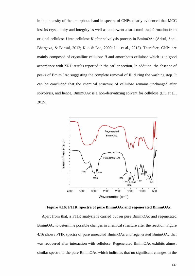
147
in the intensity of the amorphous band in spectra of CNPs clearly evidenced that MCC
lost its crystallinity and integrity as well as underwent a structural transformation from
original cellulose I into cellulose II after solvolysis process in BmimOAc (Adsul, Soni,
Bhargava, & Bansal, 2012; Kuo & Lee, 2009; Liu et al., 2015). Therefore, CNPs are
mainly composed of crystalline cellulose II and amorphous cellulose which is in good
accordance with XRD results reported in the earlier section. In addition, the absence of
peaks of BmimOAc suggesting the complete removal of IL during the washing step. It
can be concluded that the chemical structure of cellulose remains unchanged after
solvolysis, and hence, BmimOAc is a non-derivatizing solvent for cellulose (Liu et al.,
2015).
Figure 4.16: FTIR spectra of pure BmimOAc and regenerated BmimOAc.
Apart from that, a FTIR analysis is carried out on pure BmimOAc and regenerated
BmimOAc to determine possible changes in chemical structure after the reaction. Figure
4.16 shows FTIR spectra of pure unreacted BmimOAc and regenerated BmimOAc that
was recovered after interaction with cellulose. Regenerated BmimOAc exhibits almost
similar spectra to the pure BmimOAc which indicates that no significant changes in the
148
chemical structure of regenerated BmimOAc after the reaction. However, the presence of
an additional broad band in the range 3200-3700 cm-1 is distinct for the regenerated
BmimOAc. The band is the characteristic stretching vibration of –OH that is present in
water due to water absorption by IL because IL is highly hygroscopic. Both pure and
regenerated BmimOAc exhibit almost similar functional groups and this signifies the
potential usage of BmimOAc as a catalyst cum solvent for cellulose depolymerisation
because it is recyclable and reusable (Man et al., 2011). In the present study, the yield of
recovered BmimOAc is about 86 %. The highly recoverable yield and recyclable
properties of IL contribute to the attractive and environmental benign synthetic approach
to produce nanocellulose.
From Figure 4.16, a small band at 3150 cm-1 is observed in both pure and regenerated
BmimOAc, which is attributed to quaternary amine salt formation with acetate anion.
Besides that, small peak at around 3103 cm-1 is related to the stretching vibration of
aromatic =C-H. Two distinct peaks of close proximity at 2955 and 2869 cm -1 are
associated with the aliphatic asymmetric and symmetric (C-H) stretching vibrations due
to methyl groups (Pielesz & Biniaś, 2010). The presence of an absorption band at 1570
cm-1 corresponds to in-plane stretching of C=C bonds and 1460 cm-1 is due to C=N
stretching. A strong band appeared at 1171 cm-1 is attributed to the in-plane bending
vibration due to methyl groups (CH2). The existence of several bands at 890-1000 cm-1 is
the bending vibration of =C-H. Furthermore, peaks at 749 and 623 cm-1 are associated
with C-N stretching vibration (Dharaskar et al., 2013; Dharaskar et al., 2013; Socrates,
2004). Moreover, the confirmation of acetate anion is evidenced by the presence of bands
at 1650 cm−1 (C=O stretching) and 1368 cm−1 [C-H in –O(C=O)-CH3] (Adebajo, Frost,
Kloprogge, & Kokot, 2006; Spagnol et al., 2012; Sun, Sun, & Sun, 2002). FTIR analysis
reveals that regenerated BmimOAc has similar chemical structure as pure BmimOAc.
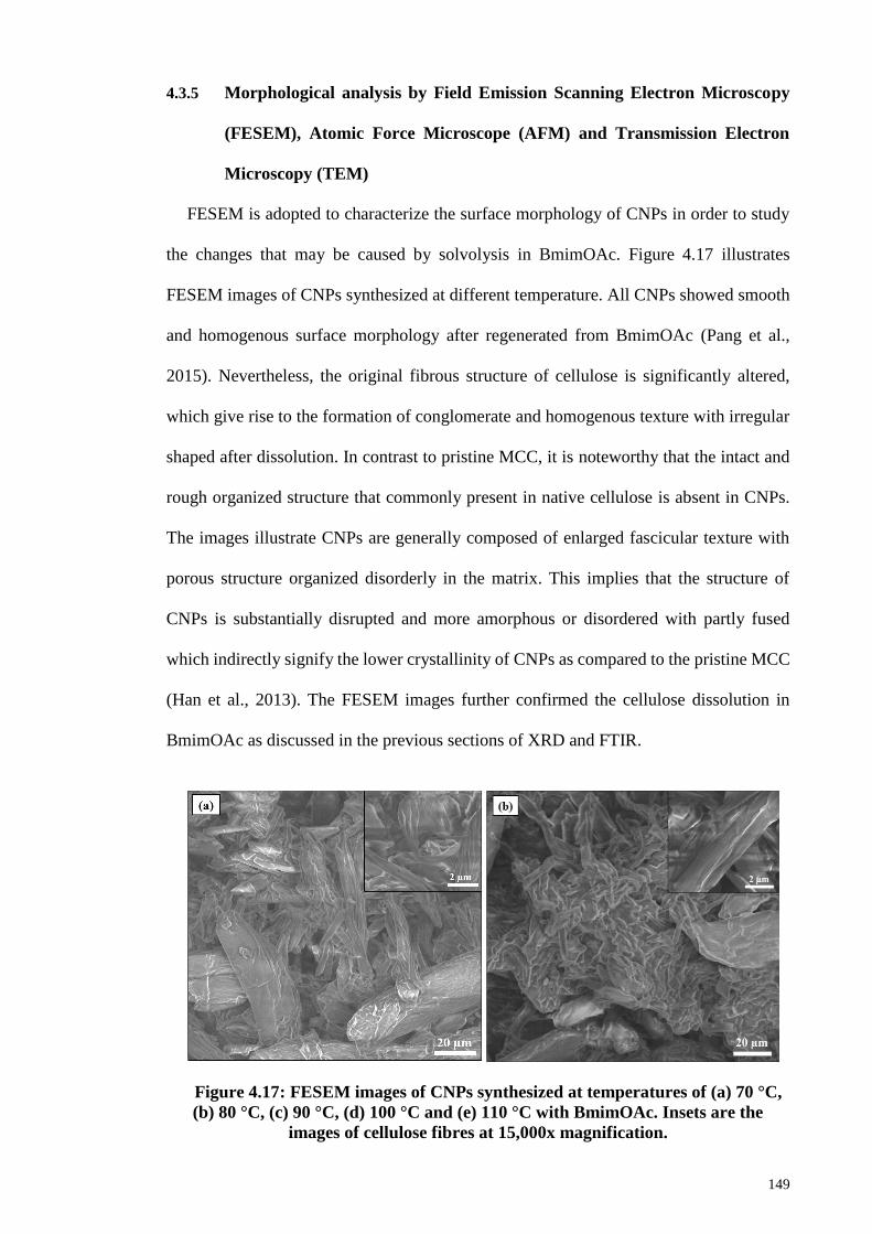
149
4.3.5 Morphological analysis by Field Emission Scanning Electron Microscopy
(FESEM), Atomic Force Microscope (AFM) and Transmission Electron
Microscopy (TEM)
FESEM is adopted to characterize the surface morphology of CNPs in order to study
the changes that may be caused by solvolysis in BmimOAc. Figure 4.17 illustrates
FESEM images of CNPs synthesized at different temperature. All CNPs showed smooth
and homogenous surface morphology after regenerated from BmimOAc (Pang et al.,
2015). Nevertheless, the original fibrous structure of cellulose is significantly altered,
which give rise to the formation of conglomerate and homogenous texture with irregular
shaped after dissolution. In contrast to pristine MCC, it is noteworthy that the intact and
rough organized structure that commonly present in native cellulose is absent in CNPs.
The images illustrate CNPs are generally composed of enlarged fascicular texture with
porous structure organized disorderly in the matrix. This implies that the structure of
CNPs is substantially disrupted and more amorphous or disordered with partly fused
which indirectly signify the lower crystallinity of CNPs as compared to the pristine MCC
(Han et al., 2013). The FESEM images further confirmed the cellulose dissolution in
BmimOAc as discussed in the previous sections of XRD and FTIR.
Figure 4.17: FESEM images of CNPs synthesized at temperatures of (a) 70 °C,
(b) 80 °C, (c) 90 °C, (d) 100 °C and (e) 110 °C with BmimOAc. Insets are the
images of cellulose fibres at 15,000x magnification.
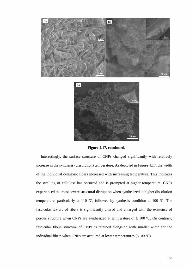
150
Figure 4.17, continued.
Interestingly, the surface structure of CNPs changed significantly with relatively
increase in the synthesis (dissolution) temperature. As depicted in Figure 4.17, the width
of the individual cellulosic fibers increased with increasing temperature. This indicates
the swelling of cellulose has occurred and is prompted at higher temperature. CNPs
experienced the most severe structural disruption when synthesized at higher dissolution
temperature, particularly at 110 ºC, followed by synthesis condition at 100 ºC. The
fascicular texture of fibers is significantly altered and enlarged with the existence of
porous structure when CNPs are synthesized at temperature of ≥ 100 ºC. On contrary,
fascicular fibers structure of CNPs is retained alongside with smaller width for the
individual fibers when CNPs are acquired at lower temperatures (<100 ºC).
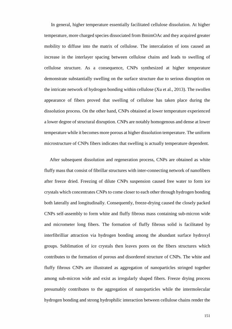
151
In general, higher temperature essentially facilitated cellulose dissolution. At higher
temperature, more charged species dissociated from BmimOAc and they acquired greater
mobility to diffuse into the matrix of cellulose. The intercalation of ions caused an
increase in the interlayer spacing between cellulose chains and leads to swelling of
cellulose structure. As a consequence, CNPs synthesized at higher temperature
demonstrate substantially swelling on the surface structure due to serious disruption on
the intricate network of hydrogen bonding within cellulose (Xu et al., 2013). The swollen
appearance of fibers proved that swelling of cellulose has taken place during the
dissolution process. On the other hand, CNPs obtained at lower temperature experienced
a lower degree of structural disruption. CNPs are notably homogenous and dense at lower
temperature while it becomes more porous at higher dissolution temperature. The uniform
microstructure of CNPs fibers indicates that swelling is actually temperature dependent.
After subsequent dissolution and regeneration process, CNPs are obtained as white
fluffy mass that consist of fibrillar structures with inter-connecting network of nanofibers
after freeze dried. Freezing of dilute CNPs suspension caused free water to form ice
crystals which concentrates CNPs to come closer to each other through hydrogen bonding
both laterally and longitudinally. Consequently, freeze-drying caused the closely packed
CNPs self-assembly to form white and fluffy fibrous mass containing sub-micron wide
and micrometer long fibers. The formation of fluffy fibrous solid is facilitated by
interfibrilliar attraction via hydrogen bonding among the abundant surface hydroxyl
groups. Sublimation of ice crystals then leaves pores on the fibers structures which
contributes to the formation of porous and disordered structure of CNPs. The white and
fluffy fibrous CNPs are illustrated as aggregation of nanoparticles stringed together
among sub-micron wide and exist as irregularly shaped fibers. Freeze drying process
presumably contributes to the aggregation of nanoparticles while the intermolecular
hydrogen bonding and strong hydrophilic interaction between cellulose chains render the
152
agglomeration (Chen et al., 2011a; Chirayil, Mathew, & Thomas, 2014a). Cellulose in a
suspension tends to form aggregation of bulk structure instead of isolated nanofibers
(Jiang & Hsieh, 2013).
Figure 4.18: FESEM images of CNPs obtained at different dissolution time: (a)
0.5 h, (b) 1.0 h, (c) 1.5 h and (d) 2.0 h.
Figure 4.18 shows FESEM images regarding microstructure morphology of CNPs
fibers obtained at different dissolution time, viewed at 50,000x magnification. From
Figure 4.18 (a), there is a small number of cellulosic fibers in nanometer scale diameter
present on the surface of microfibrils. However, most of the fibers tend to intervene
themselves with microfibers and still exist in aggregation. The fibers have non-uniform
size and distribute disorderly on the surface. Dissolution time of 1 hour is capable to
produce narrower width of cellulosic fibers which can discerned and separated from the
microfibers. The long and thin fibrilliar structure signifies lower degree of swelling took
153
place at short dissolution time and thus, less structural disruption happened. This in turns,
leads to the formation of smaller dimension CNPs with retained crystallinity.
Nevertheless, the intact fibrillar structure is rigorously altered and more porous structure
is obtained at prolonged dissolution. The enlargement of fibers is demonstrated at 1.5
hours of dissolution due to serious swelling of cellulose structure. Cellulose swelled to a
greater extent, and eventually the fibrilliar structure is completely destroyed after 2 hours
of dissolution. This manifests that substantially surface and structural disruption are
impacted on CNPs due to prolonged dissolution of cellulose in BmimOAc. CNPs
regenerated after 2 hours experienced the most severe swollen structure and appearance
with the existence of porous structure which responsible for their high amorphous content
and lower crystallinity as well.
FESEM images are in good consensus with XRD findings in which lower crystallinity
and amorphous cellulose structure are obtained after prolonged dissolution. The longer
the dissolution time, the lower the crystallinity of CNPs and therefore, the more
amorphous the cellulose become. As a matter of fact, the surface of cellulose II fibers is
observed to be not so homogenous as compared to that of cellulose I, presumably related
to different cellulose crystal structure.
AFM height images and peak force error of CNPs synthesized at 0.5, 1 and 1.5 hours
of dissolution are presented in Figure 4.19. In general, CNPs have a typical granular or
spherical shape (Pang et al., 2015). The sizes of granules/particles were measured, as
shown in Figure 4.20. The mean particle size is obtained from it. There are some
aggregations of particles observed, which presumably is due to the evaporation of water
during sample preparation. Meanwhile, some individual CNPs particles are apparently
clear in some images. At 0.5 hour, CNPs span larger size distribution where most of them
have sizes range from 40 to 60 nm with mean particle size of 53 ± 4.8 nm. Besides that,
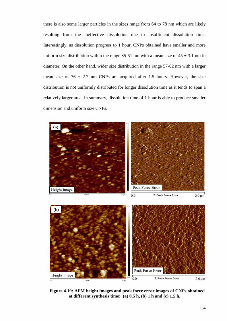
154
there is also some larger particles in the sizes range from 64 to 78 nm which are likely
resulting from the ineffective dissolution due to insufficient dissolution time.
Interestingly, as dissolution progress to 1 hour, CNPs obtained have smaller and more
uniform size distribution within the range 35-51 nm with a mean size of 45 ± 3.1 nm in
diameter. On the other hand, wider size distribution in the range 57-82 nm with a larger
mean size of 76 ± 2.7 nm CNPs are acquired after 1.5 hours. However, the size
distribution is not uniformly distributed for longer dissolution time as it tends to span a
relatively larger area. In summary, dissolution time of 1 hour is able to produce smaller
dimension and uniform size CNPs.
Figure 4.19: AFM height images and peak force error images of CNPs obtained
at different synthesis time: (a) 0.5 h, (b) 1 h and (c) 1.5 h.
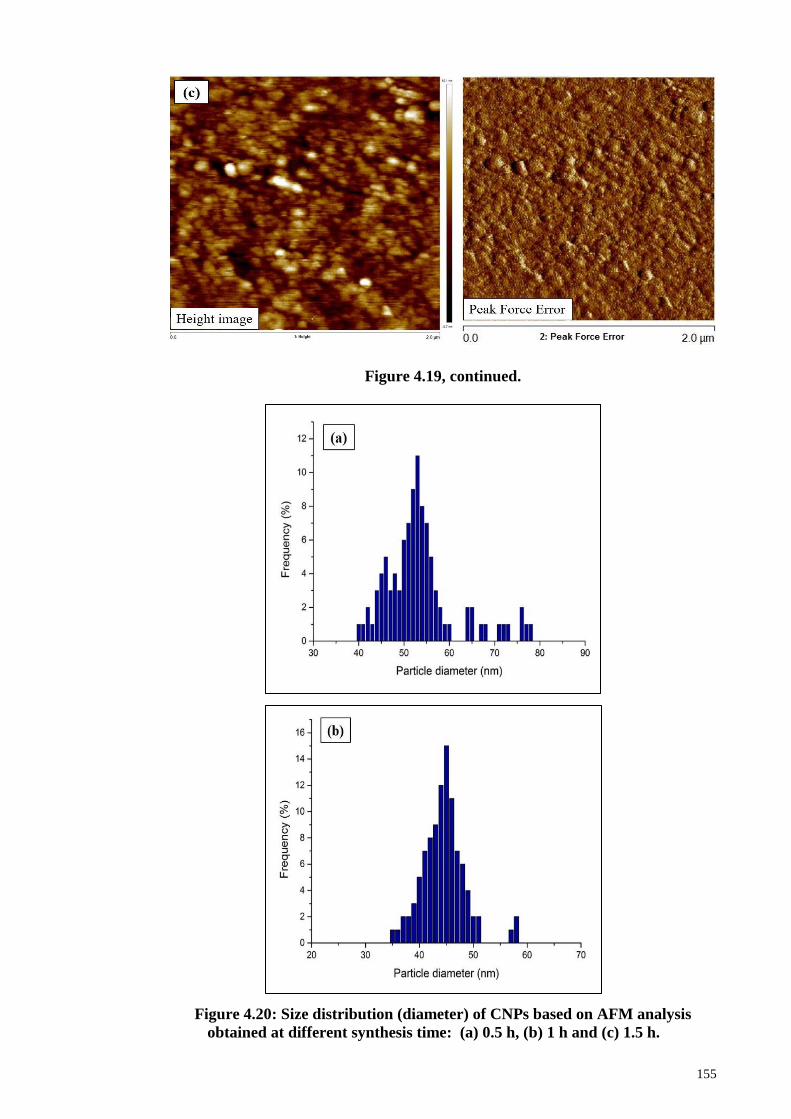
155
Figure 4.19, continued.
Figure 4.20: Size distribution (diameter) of CNPs based on AFM analysis
obtained at different synthesis time: (a) 0.5 h, (b) 1 h and (c) 1.5 h.
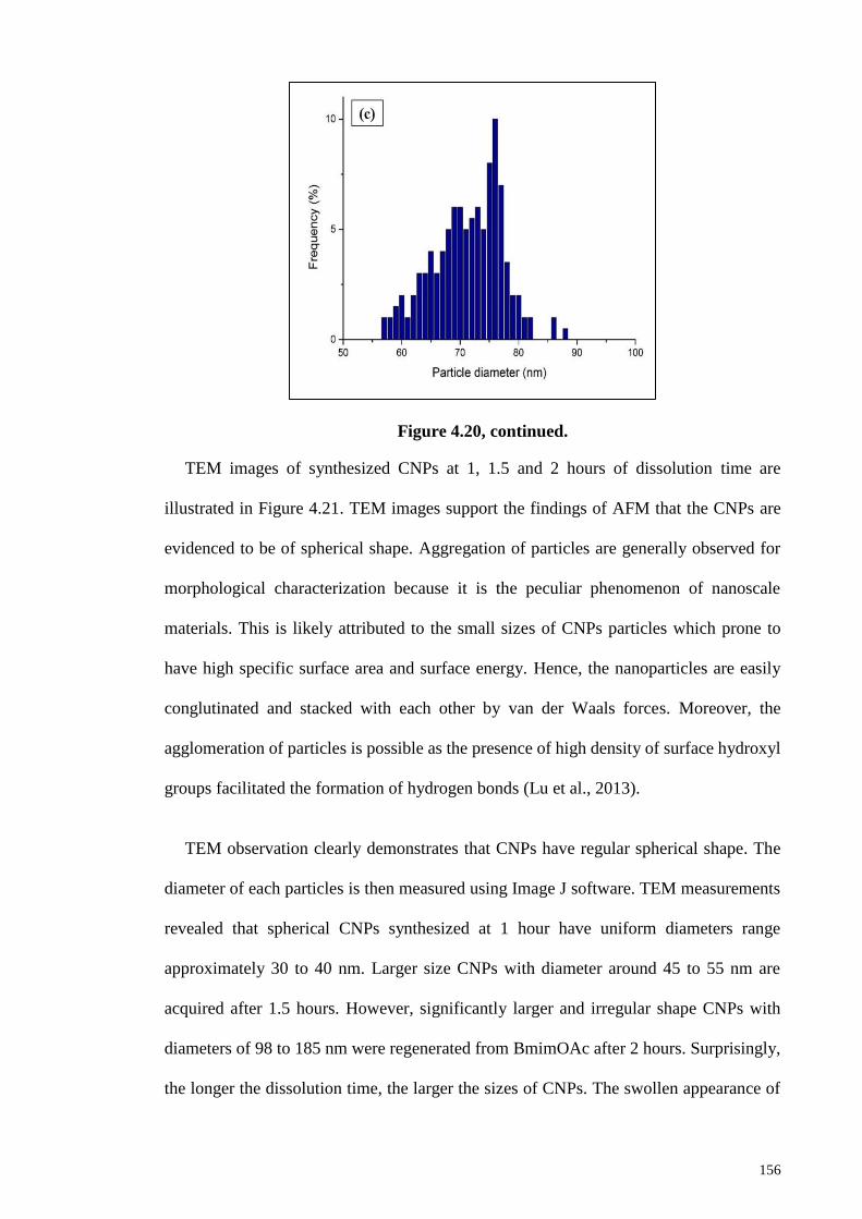
156
Figure 4.20, continued.
TEM images of synthesized CNPs at 1, 1.5 and 2 hours of dissolution time are
illustrated in Figure 4.21. TEM images support the findings of AFM that the CNPs are
evidenced to be of spherical shape. Aggregation of particles are generally observed for
morphological characterization because it is the peculiar phenomenon of nanoscale
materials. This is likely attributed to the small sizes of CNPs particles which prone to
have high specific surface area and surface energy. Hence, the nanoparticles are easily
conglutinated and stacked with each other by van der Waals forces. Moreover, the
agglomeration of particles is possible as the presence of high density of surface hydroxyl
groups facilitated the formation of hydrogen bonds (Lu et al., 2013).
TEM observation clearly demonstrates that CNPs have regular spherical shape. The
diameter of each particles is then measured using Image J software. TEM measurements
revealed that spherical CNPs synthesized at 1 hour have uniform diameters range
approximately 30 to 40 nm. Larger size CNPs with diameter around 45 to 55 nm are
acquired after 1.5 hours. However, significantly larger and irregular shape CNPs with
diameters of 98 to 185 nm were regenerated from BmimOAc after 2 hours. Surprisingly,
the longer the dissolution time, the larger the sizes of CNPs. The swollen appearance of
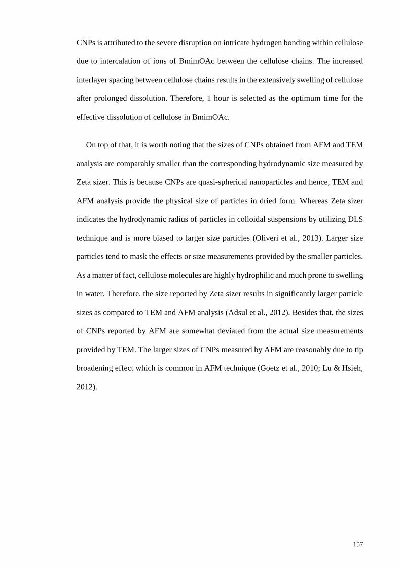
157
CNPs is attributed to the severe disruption on intricate hydrogen bonding within cellulose
due to intercalation of ions of BmimOAc between the cellulose chains. The increased
interlayer spacing between cellulose chains results in the extensively swelling of cellulose
after prolonged dissolution. Therefore, 1 hour is selected as the optimum time for the
effective dissolution of cellulose in BmimOAc.
On top of that, it is worth noting that the sizes of CNPs obtained from AFM and TEM
analysis are comparably smaller than the corresponding hydrodynamic size measured by
Zeta sizer. This is because CNPs are quasi-spherical nanoparticles and hence, TEM and
AFM analysis provide the physical size of particles in dried form. Whereas Zeta sizer
indicates the hydrodynamic radius of particles in colloidal suspensions by utilizing DLS
technique and is more biased to larger size particles (Oliveri et al., 2013). Larger size
particles tend to mask the effects or size measurements provided by the smaller particles.
As a matter of fact, cellulose molecules are highly hydrophilic and much prone to swelling
in water. Therefore, the size reported by Zeta sizer results in significantly larger particle
sizes as compared to TEM and AFM analysis (Adsul et al., 2012). Besides that, the sizes
of CNPs reported by AFM are somewhat deviated from the actual size measurements
provided by TEM. The larger sizes of CNPs measured by AFM are reasonably due to tip
broadening effect which is common in AFM technique (Goetz et al., 2010; Lu & Hsieh,
2012).
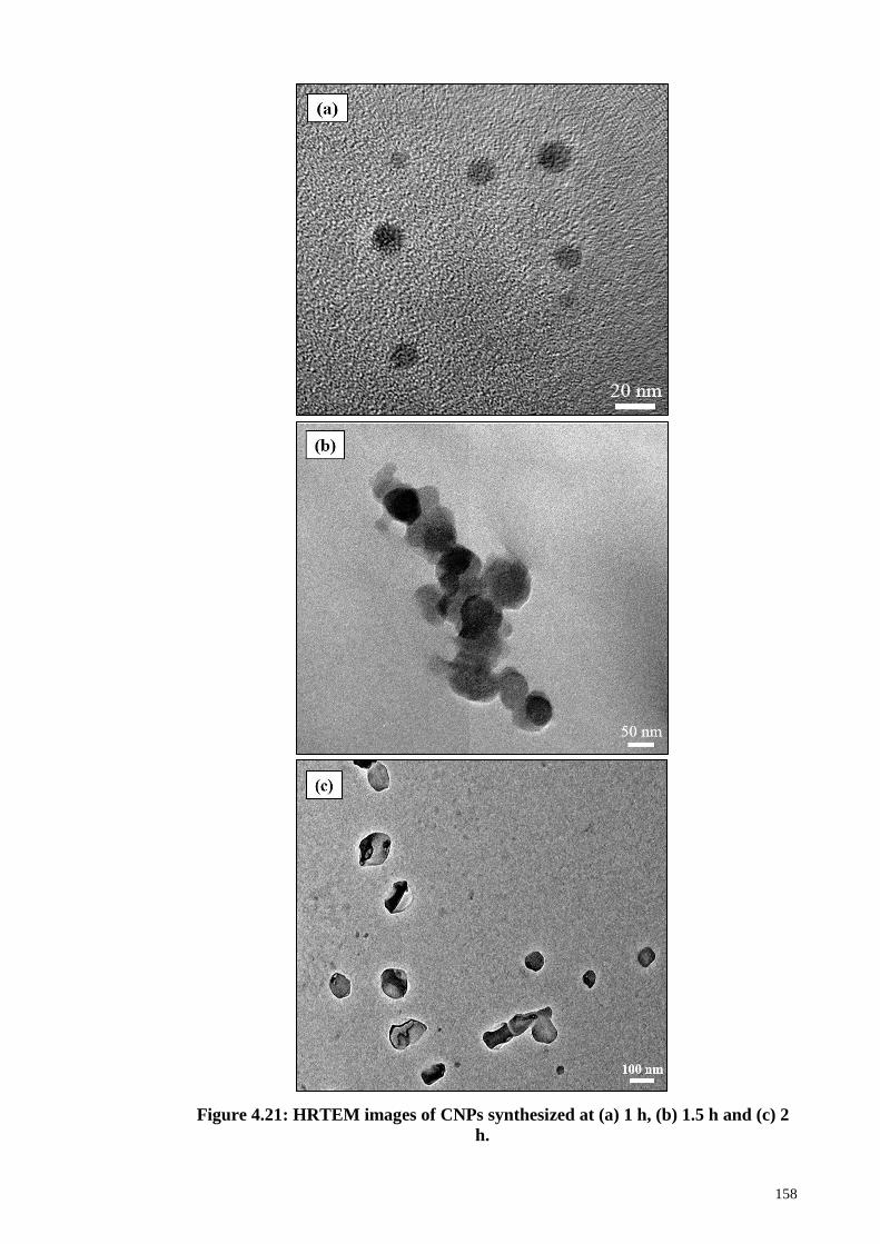
158
Figure 4.21: HRTEM images of CNPs synthesized at (a) 1 h, (b) 1.5 h and (c) 2
h.
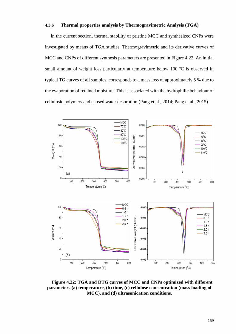
159
4.3.6 Thermal properties analysis by Thermogravimetric Analysis (TGA)
In the current section, thermal stability of pristine MCC and synthesized CNPs were
investigated by means of TGA studies. Thermogravimetric and its derivative curves of
MCC and CNPs of different synthesis parameters are presented in Figure 4.22. An initial
small amount of weight loss particularly at temperature below 100 ºC is observed in
typical TG curves of all samples, corresponds to a mass loss of approximately 5 % due to
the evaporation of retained moisture. This is associated with the hydrophilic behaviour of
cellulosic polymers and caused water desorption (Pang et al., 2014; Pang et al., 2015).
Figure 4.22: TGA and DTG curves of MCC and CNPs optimized with different
parameters (a) temperature, (b) time, (c) cellulose concentration (mass loading of
MCC), and (d) ultrasonication conditions.
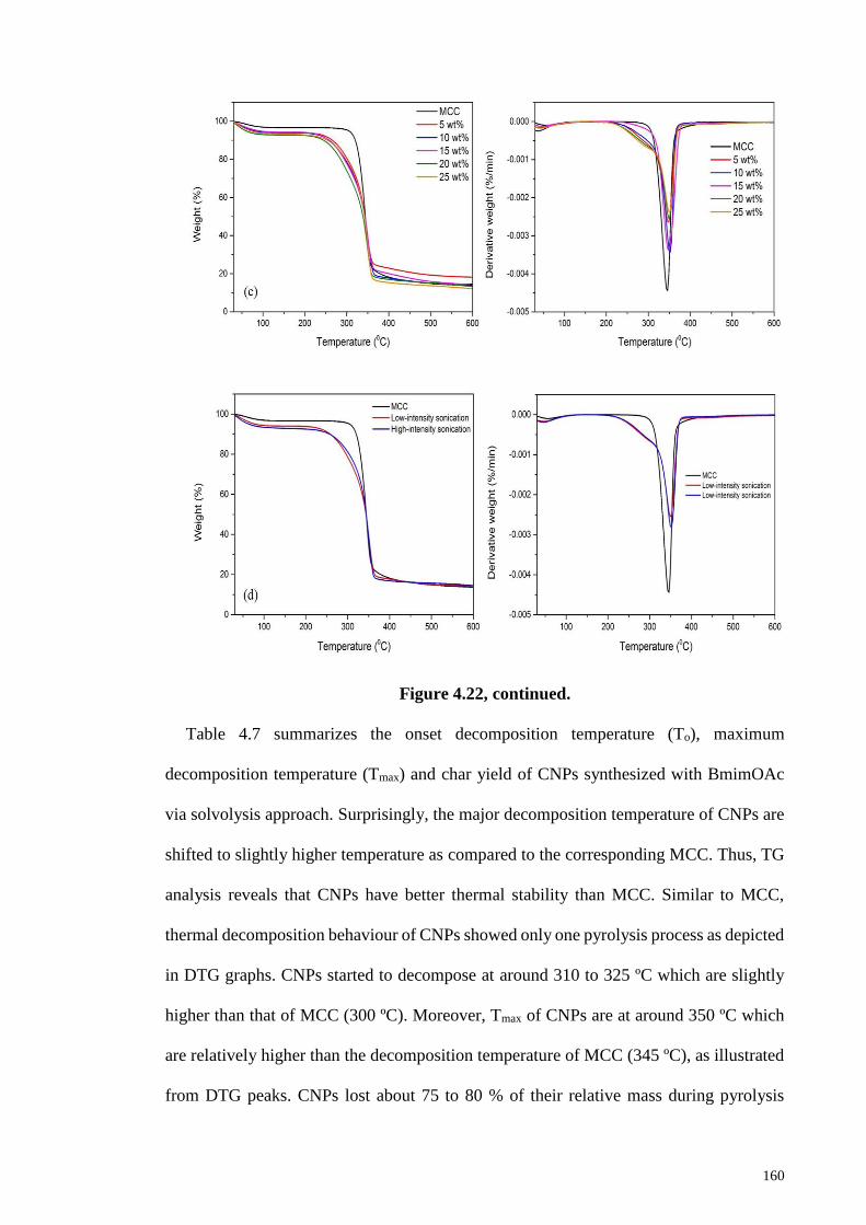
160
Figure 4.22, continued.
Table 4.7 summarizes the onset decomposition temperature (To), maximum
decomposition temperature (Tmax) and char yield of CNPs synthesized with BmimOAc
via solvolysis approach. Surprisingly, the major decomposition temperature of CNPs are
shifted to slightly higher temperature as compared to the corresponding MCC. Thus, TG
analysis reveals that CNPs have better thermal stability than MCC. Similar to MCC,
thermal decomposition behaviour of CNPs showed only one pyrolysis process as depicted
in DTG graphs. CNPs started to decompose at around 310 to 325 ºC which are slightly
higher than that of MCC (300 ºC). Moreover, Tmax of CNPs are at around 350 ºC which
are relatively higher than the decomposition temperature of MCC (345 ºC), as illustrated
from DTG peaks. CNPs lost about 75 to 80 % of their relative mass during pyrolysis
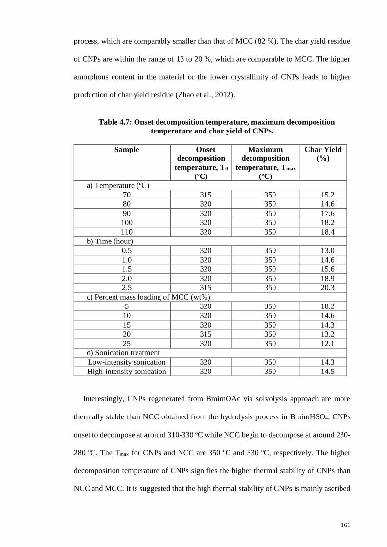
161
process, which are comparably smaller than that of MCC (82 %). The char yield residue
of CNPs are within the range of 13 to 20 %, which are comparable to MCC. The higher
amorphous content in the material or the lower crystallinity of CNPs leads to higher
production of char yield residue (Zhao et al., 2012).
Table 4.7: Onset decomposition temperature, maximum decomposition
temperature and char yield of CNPs.
Sample Onset
decomposition
temperature, T0
(ºC)
Maximum
decomposition
temperature, Tmax
(ºC)
Char Yield
(%)
a) Temperature (ºC)
70 315 350 15.2
80 320 350 14.6
90 320 350 17.6
100 320 350 18.2
110 320 350 18.4
b) Time (hour)
0.5 320 350 13.0
1.0 320 350 14.6
1.5 320 350 15.6
2.0 320 350 18.9
2.5 315 350 20.3
c) Percent mass loading of MCC (wt%)
5 320 350 18.2
10 320 350 14.6
15 320 350 14.3
20 315 350 13.2
25 320 350 12.1
d) Sonication treatment
Low-intensity sonication 320 350 14.3
High-intensity sonication 320 350 14.5
Interestingly, CNPs regenerated from BmimOAc via solvolysis approach are more
thermally stable than NCC obtained from the hydrolysis process in BmimHSO4. CNPs
onset to decompose at around 310-330 ºC while NCC begin to decompose at around 230-
280 ºC. The Tmax for CNPs and NCC are 350 ºC and 330 ºC, respectively. The higher
decomposition temperature of CNPs signifies the higher thermal stability of CNPs than
NCC and MCC. It is suggested that the high thermal stability of CNPs is mainly ascribed
162
to the cellulose II crystalline structure as there are rearrangement of hydrogen bonding
within CNPs during regeneration process. As a matter of fact, cellulose II nanoparticles
have improved thermal stability than cellulose I nanocrystals because the chains in
cellulose II are in antiparallel arrangement. In contrast to cellulose I, this arrangement
yields more hydrogen bonds and thus, leads to a more stable structure (Lu et al., 2015;
Moon et al., 2011).
Moreover, the high thermal stability of CNPs is also due to higher flexibility and
hence, the higher possibility of entanglements of cellulose nanofibrils that consequently
results in more thermodynamically stable structure (Maiti et al., 2013). Furthermore, the
absence of sulfate groups in BmimOAc caused no sulfation to take place on the cellulose
chains. Therefore, activation energy required for the thermal decomposition of cellulose
will not been significantly lowered. The high thermal stability of cellulose II nanoparticles
has large potential applications in composite materials which demand for thermal
stability.
Char yield residue indicates the residual mass of non-volatile carbonaceous material
produced at the end of pyrolysis process, usually at high temperature. CNPs produced
about 13-20 % char yield on pyrolysis. Parameters such as temperature, time as well as
cellulose concentration are influenced the char yield production. Char yield of CNPs
increased gradually when temperature is increased from 80 to 110 ºC. Besides that,
increase in char yield is also obvious with increasing dissolution time from 0.5 to 2.5
hours. This is likely due to substantially crystallinity reduction and increase in the
amorphous content of CNPs. On the other hand, it is worth noting that CNPs synthesized
at higher cellulose concentration are responsible for the lower char yield content. The
variations in thermal decomposition temperature and char yield of CNPs are qualitatively
dependent on the crystal structure as well as quantitative crystallinity of CNPs.
163
CHAPTER 5: CONCLUSION AND RECOMMENDATIONS
5.1 Conclusion
The present study was mainly focus to investigate the methodology for the synthesis
of nanocellulose from cellulose aided by the catalytic and solvation properties of ILs. 1-
butyl-3-methylimidazolium hydrogen sulfate (BmimHSO4) and 1-butyl-3-
methylimidazolium acetate (BmimOAc) ILs were adopted to perform dual functions as
catalyst and solvent for the effective conversion of cellulose into nanocellulose. Despite
having the same alkylmethylimidazolium cations, it is suggested that hydrogen sulfate
anions exhibit higher acidic properties to induce hydrolysis reaction. On the other hand,
higher electronegativity of acetate anions much prone to cause solvolysis of cellulose to
produce nanocellulose. In summary, the target objectives for this thesis have been
achieved. Synthesis parameters including reaction temperature, time, cellulose
concentration (mass loading of MCC) and sonication treatments have been optimized in
order to acquire controllable specific architecture and properties of nanocellulose.
In the first stage of research study, improved crystallinity of nanocellulose with the
preservation of crystalline cellulose I structure has been accomplished with acidic
BmimHSO4. The intrinsic acidic character of BmimHSO4 (pH 1) is attributed to the
acidity from the hydrogen of [HSO4]- which has a pKa value of about 2 in water.
Nanocrystalline cellulose with essentially high crystallinity (CrI 92.2) and small size (15
nm) was successfully synthesized via BmimHSO4-mediated hydrolysis at a temperature
of 90 ºC for 1.5 hours. It was found that increasing the temperature and time of the
hydrolytic reaction predominantly increased the crystallinity and produced smaller size
nanocellulose. However, crystallinity dropped gradually with further increased in the
temperature and time beyond the optimum conditions. The crystallinity improved is due
to the preferential hydrolysis of amorphous regions. While crystallinity reduction is
attributed to the uncontrollable hydrolysis of crystalline domains after exposure to higher
164
temperature and prolonged hydrolysis. Hydrolytic cleavage of glycosidic linkages and
selectively removal of amorphous regions in the cellulose chains catalyzed by acidic
BmimHSO4 render the release of crystalline domains in nanocellulose which resulted in
the higher crystallinity and smaller dimension size. Apart from that, cellulose
concentration (in wt%) also has significant impact on the crystallinity as well as size of
nanocellulose. Interestingly, the size of nanocellulose increase proportionally with
increasing cellulose concentration, reasonably attributed to ineffective hydrolysis
reaction at viscous environment.
On the other hand, regenerated cellulose nanoparticles from BmimOAc experienced
structural transformation from native cellulose I into cellulose II. Solvolysis is suggested
to take place in BmimOAc where it caused significant disruption to the hydrogen bonding
network. Meanwhile, the IL also attacked the glycosidic bonds for fragmentation of
cellulose chains. Cellulose nanoparticles with a crystallinity of 78.8 was acquired at 80
°C with 1 hour of solvolysis and cellulose was found to swell in BmimOAc. It is
noteworthy the crystallinity of cellulose nanoparticles decreased apparently with
increasing temperature and time of dissolution. Disruption caused to the original
crystalline structure and breakage of hydrogen bonding between cellulose chains during
solvolysis presumably lower the crystallinity of cellulose nanoparticles. Consequently,
reconstruction of antiparallel mode hydrogen bonding during regeneration leads to the
formation of more thermally and structurally stable structure of cellulose II. Moreover,
the swelling of cellulose was evidenced by the proportional increase in sizes of cellulose
nanoparticles with increasing temperature and time, as illustrated by AFM and TEM
analysis.
The application of high-intensity ultrasonication aided the formation of higher
crystallinity and smaller size nanocellulose in both hydrolysis and solvolysis approach.
165
Ultrasonication treatment was important because it is responsible for the defibrillation,
defragmentation of fibers while extensively degraded the cellulose amorphous regions. It
is important to essentially enhance the crystallinity and disintegrate the cellulose
molecules into smaller particles in order to yield smaller size nanocellulose with
improved dispersion stability. Suspension of nanocellulose prepared with BmimHSO4
and BmimOAc were considered rather stable with their high absolute zeta potential values
of -37.5 mV and -22.3 mV, respectively.
FTIR analysis have revealed the weakened intramolecular hydrogen bonding for
nanocellulose due to the interaction and coordination of charged species of ILs with the
hydroxyl groups of cellulose. Besides that, FTIR further confirmed the crystallinity of
nanocellulose as obtained from the XRD analysis. Moreover, AFM and TEM
observations demonstrated that nanoscale cellulose can be obtained after hydrolysis
reaction in BmimHSO4 and solvolysis in BmimOAc respectively. The size measurements
revealed that the dimension (length or width) of nanocellulose is actually less than 100
nm, though some agglomeration can be observed. Rod-like nanocrystalline cellulose was
obtained after hydrolysis with BmimHSO4 whereas spherical cellulose nanoparticles
were acquired after solvolysis with BmimOAc, as illustrated from TEM and AFM
observations. Furthermore, nanocrystalline cellulose exhibit lower thermal stability
although they have remarkable crystallinity due to the presence of sulfate groups from
BmimHSO4. The sulfate groups lowered the thermal stability of nanocrystalline cellulose
by accelerated thermal degradation of cellulose and promote the dehydration reaction to
be occurred at lower temperature. On the other hand, enhanced thermal stability of
cellulose nanoparticles is attributed to the antiparallel arrangement and higher number of
hydrogen bonding in cellulose II which makes it more thermally stable.
166
In summary, determination of optimum synthesis conditions is of critical importance
to avoid uncontrollable depolymerisation of cellulose into monomer units. The formation
of nanocellulose has been eased by the application of ILs as both the solvent and catalyst.
The advantageous properties of ILs such as recyclability and reusability have been
manifested by their high recovery yield (86-90 %). The method adopted in this study is a
green and environmental friendly approach because it did not produce any toxic and
hazardous waste products. The ILs solution that collected after the reaction were able to
recover and regenerate by evaporated the water content and re-concentrated in a vacuum
freeze dryer. Meanwhile, it is an economical feasible process because the retrieval of
ionic liquids is high, so as they are recyclable and reusable.
5.2 Recommendations for Future Work
An ideal method of production of nanocellulose should encompass green and
environmentally benign character to conform with the principle of green chemistry
emphasized in governmental regulations and policies. ILs are generally known as green
solvents for cellulose processing because they are recyclable and reusable. In the present
work, ILs have been utilized as the solvent and catalyst for the manufacture of
nanocellulose. The methodology has greatly contributed to the identification of optimum
synthesis conditions on the production of highly crystalline structure of nano-dimensional
cellulose. The successful formation of nanocellulose through hydrolysis and solvolysis
approach with ILs evidenced effectiveness of methods employed which is suggested to
replace the conventional acid hydrolysis as well as volatile cellulose solvents system.
Nonetheless, there are several recommendations and suggestions should be
noteworthy for future work as proposed below:
i. To investigate the hydrolysis and solvolysis by using alternative cellulosic
sources such as Avicel, biomass materials including hard and soft wood,
167
coconut husk, palm empty fruit bunch (EFB), cotton linter, sugarcane
bagasse and wheat straw. However, factors such as availability and cost of
raw materials need to be taken into consideration to select suitable materials
for further investigation.
ii. The utilization of different anions-based ILs (e.g., phosphate, formate and
sulfonate) and vary the cations (e.g., pyridinium) as well as different side
chain (alkyl chain length) of alkylimidazolium cations (e.g., ethyl-, butyl-
and hexyl-) for hydrolysis and solvolysis of cellulose and their influences
on the properties of nanocellulose. Once again, the accessibility and price
of ILs are important elements to be considered for their roles as catalyst and
solvents, so that the process is economically feasible.
iii. To investigate the regeneration of nanocellulose from cellulose/IL mixture
by using other types of anti-solvents such as ethanol, acetone or their
combination and their influences on the specific architecture and properties
of nanocellulose. The maximum yield and remarkable properties of
regenerated nanocellulose such as crystallinity will decide upon the
applicability of anti-solvents used.
iv. The nanocellulose can be characterized extensively for their degree of
depolymerization (DP) by Gel Permeation Chromatography (GPC)
technique with suitable column and dissolving medium to determine the
changes in molecular weight after the reaction in future research.
168
REFERENCES
Abraham, E., Deepa, B., Pothen, L. A., Cintil, J., Thomas, S., John, M. J., ... & Narine,
S. S. (2013). Environmental friendly method for the extraction of coir fibre and
isolation of nanofibre. Carbohydrate polymers, 92(2), 1477-1483.
Abushammala, H., Krossing, I., & Laborie, M.-P. (2015). Ionic liquid-mediated
technology to produce cellulose nanocrystals directly from wood. Carbohydrate
polymers, 134, 609-616.
Adebajo, M. O., Frost, R. L., Kloprogge, J. T., & Kokot, S. (2006). Raman spectroscopic
investigation of acetylation of raw cotton. Spectrochimica Acta Part a: molecular
and biomolecular spectroscopy, 64(2), 448-453.
Adsul, M., Soni, S. K., Bhargava, S. K., & Bansal, V. (2012). Facile approach for the
dispersion of regenerated cellulose in aqueous system in the form of nanoparticles.
Biomacromolecules, 13(9), 2890-2895.
Ahmadi, M., Madadlou, A., & Sabouri, A. A. (2015). Isolation of micro-and nano-
crystalline cellulose particles and fabrication of crystalline particles-loaded whey
protein cold-set gel. Food chemistry, 174, 97-103.
Amarasekara, A. S., & Owereh, O. S. (2009). Hydrolysis and decomposition of cellulose
in Bronsted acidic ionic liquids under mild conditions. Industrial & Engineering
Chemistry Research, 48(22), 10152-10155.
Ang, T. N., Ngoh, G. C., Chua, A. S. M., & Lee, M. G. (2012). Elucidation of the effect
of ionic liquid pretreatment on rice husk via structural analyses. Biotechnology for
biofuels, 5(1), 1.
Angles, M. N., & Dufresne, A. (2000). Plasticized starch/tunicin whiskers
nanocomposites. 1. Structural analysis. Macromolecules, 33(22), 8344-8353.
Angles, M. N., & Dufresne, A. (2001). Plasticized starch/tunicin whiskers nanocomposite
materials. 2. Mechanical behavior. Macromolecules, 34(9), 2921-2931.
Ashori, A., Harun, J., Raverty, W. D., & Yusoff, M. N. M. (2006). Chemical and
morphological characteristics of Malaysian cultivated kenaf (Hibiscus
cannabinus) fiber. Polymer-Plastics Technology and Engineering, 45(1), 131-
134.
169
Azizi Samir, M. A. S., Alloin, F., & Dufresne, A. (2005). Review of recent research into
cellulosic whiskers, their properties and their application in nanocomposite field.
Biomacromolecules, 6(2), 612-626.
Belbekhouche, S., Bras, J., Siqueira, G., Chappey, C., Lebrun, L., Khelifi, B., Marais, S.,
& Dufresne, A. (2011). Water sorption behavior and gas barrier properties of
cellulose whiskers and microfibrils films. Carbohydrate Polymers, 83(4), 1740-
1748.
Bittmann, B., Haupert, F., & Schlarb, A. K. (2009). Ultrasonic dispersion of inorganic
nanoparticles in epoxy resin. Ultrasonics sonochemistry, 16(5), 622-628.
Blackley, D. C. (2012). Polymer Latices: Science and Technology Volume 3:
Applications of Latices. Springer Science & Business Media.
Bonhote, P., Dias, A. P., Papageorgiou, N., Kalyanasundaram, K., & Grätzel, M. (1996).
Hydrophobic, highly conductive ambient-temperature molten salts. Inorganic
chemistry, 35(5), 1168-1178.
Broido, A., & Nelson, M. A. (1975). Char yield on pyrolysis of cellulose. Combustion
and Flame, 24, 263-268.
Brandt, A., Hallett, J. P., Leak, D. J., Murphy, R. J., & Welton, T. (2010). The effect of
the ionic liquid anion in the pretreatment of pine wood chips. Green Chemistry,
12(4), 672-679.
Brinchi, L., Cotana, F., Fortunati, E., & Kenny, J. (2013). Production of nanocrystalline
cellulose from lignocellulosic biomass: technology and applications.
Carbohydrate polymers, 94(1), 154-169.
Brodeur, G., Yau, E., Badal, K., Collier, J., Ramachandran, K. B., & Ramakrishnan, S.
(2011). Chemical and physicochemical pretreatment of lignocellulosic biomass: a
review. Enzyme research, 2011.
Cammarata, L., Kazarian, S. G., Salter, P. A., & Welton, T. (2001). Molecular states of
water in room temperature ionic liquids. Physical Chemistry Chemical Physics,
3(23), 5192-5200.
Cao, X., Chen, Y., Chang, P. R., Stumborg, M., & Huneault, M. A. (2008). Green
composites reinforced with hemp nanocrystals in plasticized starch. Journal of
Applied Polymer Science, 109(6), 3804-3810.
170
Cao, X., Chen, Y., Chang, P. R., Muir, A. D., & Falk, G. (2008). Starch-based
nanocomposites reinforced with flax cellulose nanocrystals. Express Polym Lett,
2(7), 502-510.
Cao, X., Ding, B., Yu, J., & Al-Deyab, S. S. (2012). Cellulose nanowhiskers extracted
from TEMPO-oxidized jute fibers. Carbohydrate polymers, 90(2), 1075-1080.
Cao, Y., Wu, J., Zhang, J., Li, H., Zhang, Y., & He, J. (2009). Room temperature ionic
liquids (RTILs): a new and versatile platform for cellulose processing and
derivatization. Chemical Engineering Journal, 147(1), 13-21.
Chauhan, V. S., & Chakrabarti, S. K. (2012). Use of nanotechnology for high
performance cellulosic and papermaking products. Cellulose Chemistry and
Technology, 46(5), 389.
Chen, H. Z., Wang, N., & Liu, L. Y. (2012). Regenerated cellulose membrane prepared
with ionic liquid 1‐butyl‐3‐methylimidazolium chloride as solvent using wheat
straw. Journal of Chemical Technology and Biotechnology, 87(12), 1634-1640.
Chen, W., Li, Q., Cao, J., Liu, Y., Li, J., Zhang, J., Luo, S. & Yu, H. (2015). Revealing
the structures of cellulose nanofiber bundles obtained by mechanical
nanofibrillation via TEM observation. Carbohydrate polymers, 117, 950-956.
Chen, W., Yu, H., Liu, Y., Chen, P., Zhang, M., & Hai, Y. (2011). Individualization of
cellulose nanofibers from wood using high-intensity ultrasonication combined
with chemical pretreatments. Carbohydrate Polymers, 83(4), 1804-1811.
Chen, W., Yu, H., Liu, Y., Hai, Y., Zhang, M., & Chen, P. (2011). Isolation and
characterization of cellulose nanofibers from four plant cellulose fibers using a
chemical-ultrasonic process. Cellulose, 18(2), 433-442.
Cheng, G., Varanasi, P., Arora, R., Stavila, V., Simmons, B. A., Kent, M. S., & Singh, S.
(2012). Impact of ionic liquid pretreatment conditions on cellulose crystalline
structure using 1-ethyl-3-methylimidazolium acetate. The Journal of Physical
Chemistry B, 116(33), 10049-10054.
Cheng, Q., Wang, S., & Rials, T. G. (2009). Poly (vinyl alcohol) nanocomposites
reinforced with cellulose fibrils isolated by high intensity ultrasonication.
Composites Part A: Applied Science and Manufacturing, 40(2), 218-224.
171
Cherian, B. M., Leão, A. L., de Souza, S. F., Thomas, S., Pothan, L. A., & Kottaisamy,
M. (2010). Isolation of nanocellulose from pineapple leaf fibres by steam
explosion. Carbohydrate Polymers, 81(3), 720-725.
Cherian, B. M., Pothan, L. A., Nguyen-Chung, T., Mennig, G., Kottaisamy, M., &
Thomas, S. (2008). A novel method for the synthesis of cellulose nanofibril
whiskers from banana fibers and characterization. Journal of agricultural and
food chemistry, 56(14), 5617-5627.
Chirayil, C. J., Mathew, L., & Thomas, S. (2014). Review of recent research in nano
cellulose preparation from different lignocellulosic fibers. Rev Adv Mater Sci,
37(1-2), 20-28.
Chowdhury, Z. Z., Zain, S. M., Hamid, S. B. A., & Khalid, K. (2014). Catalytic role of
ionic liquids for dissolution and degradation of biomacromolecules.
BioResources, 9(1), 1787-1823.
Cintas, P., & Luche, J.-L. (1999). Green chemistry. The sonochemical approach. Green
Chem., 1(3), 115-125.
da Costa Lopes, A. M., João, K. G., Morais, A. R. C., Bogel-Łukasik, E., & Bogel-
Łukasik, R. (2013). Ionic liquids as a tool for lignocellulosic biomass
fractionation. Sustain Chem Process, 1(1), 3.
Dadi, A. P., Schall, C. A., & Varanasi, S. (2007). Mitigation of cellulose recalcitrance to
enzymatic hydrolysis by ionic liquid pretreatment Applied Biochemistry and
Biotecnology (pp. 407-421): Springer.
Damodaran, S., Parkin, K. L., & Fennema, O. R. (Eds.). (2007). Fennema's food
chemistry. CRC press.
de Morais Teixeira, E., Corrêa, A. C., Manzoli, A., de Lima Leite, F., de Oliveira, C. R.,
& Mattoso, L. H. C. (2010). Cellulose nanofibers from white and naturally colored
cotton fibers. Cellulose, 17(3), 595-606.
de Olyveira, G. M., Costa, L. M. M., Basmaji, P., & Xavier Filho, L. (2011). Bacterial
nanocellulose for medicine regenerative. Journal of Nanotechnology in
Engineering and Medicine, 2(3), 034001.
Dharaskar, S. A., Varma, M. N., Shende, D. Z., Yoo, C. K., & Wasewar, K. L. (2013).
Synthesis, characterization and application of 1-butyl-3 methylimidazolium
172
chloride as green material for extractive desulfurization of liquid fuel. The
Scientific World Journal, 2013.
Dharaskar, S. A., Wasewar, K. L., Varma, M. N., Shende, D. Z., & Yoo, C. (2013).
Synthesis, characterization and application of 1-butyl-3-methylimidazolium
tetrafluoroborate for extractive desulfurization of liquid fuel. Arabian Journal of
Chemistry.
Ding, Z.-D., Chi, Z., Gu, W.-X., Gu, S.-M., Liu, J.-H., & Wang, H.-J. (2012). Theoretical
and experimental investigation on dissolution and regeneration of cellulose in
ionic liquid. Carbohydrate polymers, 89(1), 7-16.
Dong, Y., Holm, J., & Lassi, U. (2015). Dissolution and Hydrolysis of Lignocellulosic
Biomass using Tailored Ionic Liquids.
Dufresne, A. (2013). Nanocellulose: a new ageless bionanomaterial. Materials Today,
16(6), 220-227.
Dufresne, A., & Belgacem, M. N. (2013). Cellulose-reinforced composites: from micro-
to nanoscale. Polímeros, 23(3), 277-286.
Duran, N., Lemes, A. P., Duran, M., Freer, J., & Baeza, J. (2011). A minireview of
cellulose nanocrystals and its potential integration as co-product in bioethanol
production. Journal of the Chilean Chemical Society, 56(2), 672-677.
Elazzouzi-Hafraoui, S., Nishiyama, Y., Putaux, J.-L., Heux, L., Dubreuil, F., & Rochas,
C. (2007). The shape and size distribution of crystalline nanoparticles prepared by
acid hydrolysis of native cellulose. Biomacromolecules, 9(1), 57-65.
Fan, J. S., & Li, Y. H. (2012). Maximizing the yield of nanocrystalline cellulose from
cotton pulp fiber. Carbohydrate Polymers, 88(4), 1184-1188.
Fan, M., Dai, D., & Huang, B. (2012). Fourier transform infrared spectroscopy for natural
fibres. Fourier transform–materials analysis. InTech.
Fernandes, A. M., Rocha, M. A., Freire, M. G., Marrucho, I. M., Coutinho, J. A., &
Santos, L. M. (2011). Evaluation of cation− anion interaction strength in ionic
liquids. The Journal of Physical Chemistry B, 115(14), 4033-4041.
Ferrer, A., Filpponen, I., Rodríguez, A., Laine, J., & Rojas, O. J. (2012). Valorization of
residual Empty Palm Fruit Bunch Fibers (EPFBF) by microfluidization:
173
production of nanofibrillated cellulose and EPFBF nanopaper. Bioresource
technology, 125, 249-255.
Filson, P. B., & Dawson-Andoh, B. E. (2009). Sono-chemical preparation of cellulose
nanocrystals from lignocellulose derived materials. Bioresource Technology,
100(7), 2259-2264.
Frone, A. N., Panaitescu, D. M., Donescu, D., Spataru, C. I., Radovici, C., Trusca, R., &
Somoghi, R. (2011). Preparation and characterization of PVA composites with
cellulose nanofibers obtained by ultrasonication. BioResources, 6(1), 487-512.
Gao, K., Shao, Z., Li, J., Wang, X., Peng, X., Wang, W., & Wang, F. (2013). Cellulose
nanofiber–graphene all solid-state flexible supercapacitors. Journal of Materials
Chemistry A, 1(1), 63-67.
George, J., & Sabapathi, S. N. (2015). Cellulose nanocrystals: synthesis, functional
properties, and applications. Nanotechnology, science and applications, 8, 45.
Gericke, M., Fardim, P., & Heinze, T. (2012). Ionic liquids—promising but challenging
solvents for homogeneous derivatization of cellulose. Molecules, 17(6), 7458-
7502.
Goetz, J. L., Keltner, D., & Simon-Thomas, E. (2010). Compassion: an evolutionary
analysis and empirical review. Psychological bulletin, 136(3), 351.
Guo, J., Guo, X., Wang, S., & Yin, Y. (2016). Effects of ultrasonic treatment during acid
hydrolysis on the yield, particle size and structure of cellulose nanocrystals.
Carbohydrate polymers, 135, 248-255.
Ha, S. H., Mai, N. L., An, G., & Koo, Y. M. (2011). Microwave-assisted pretreatment of
cellulose in ionic liquid for accelerated enzymatic hydrolysis. Bioresource
technology, 102(2), 1214-1219.
Haafiz, M. M., Hassan, A., Zakaria, Z., & Inuwa, I. M. (2014). Isolation and
characterization of cellulose nanowhiskers from oil palm biomass
microcrystalline cellulose. Carbohydrate polymers, 103, 119-125.
Habibi, Y., Lucia, L. A., & Rojas, O. J. (2010). Cellulose nanocrystals: chemistry, self-
assembly, and applications. Chemical reviews, 110(6), 3479-3500.
174
Habibi, Y., Mahrouz, M., & Vignon, M. R. (2009). Microfibrillated cellulose from the
peel of prickly pear fruits. Food Chemistry, 115(2), 423-429.
Hamelinck, C. N., Van Hooijdonk, G., & Faaij, A. P. (2005). Ethanol from lignocellulosic
biomass: techno-economic performance in short-, middle-and long-term. Biomass
and bioenergy, 28(4), 384-410.
Hamid, S. B. A., Zain, S. K., Das, R., & Centi, G. (2016). Synergic effect of
tungstophosphoric acid and sonication for rapid synthesis of crystalline
nanocellulose. Carbohydrate Polymers, 138, 349-355.
Han, J., Zhou, C., French, A. D., Han, G., & Wu, Q. (2013). Characterization of cellulose
II nanoparticles regenerated from 1-butyl-3-methylimidazolium chloride.
Carbohydrate polymers, 94(2), 773-781.
Harmsen, P., Huijgen, W., Bermudez, L., & Bakker, R. (2010). Literature review of
physical and chemical pretreatment processes for lignocellulosic biomass. Energy
Research Centre of the Netherlands, 10-13.
Herrmann, S. (2015). New Synthetic Routes to Polyoxometalate Containing Ionic
Liquids: An Investigation of Their Properties: Springer.
Hidaka, H., Kim, U. J., & Wada, M. (2010). Synchrotron X-ray fiber diffraction study on
the thermal expansion behavior of cellulose crystals in tension wood of Japanese
poplar in the low-temperature region. Holzforschung, 64(2), 167-171.
Hu, X., Xiao, Y., Niu, K., Zhao, Y., Zhang, B., & Hu, B. (2013). Functional ionic liquids
for hydrolysis of lignocellulose. Carbohydrate polymers, 97(1), 172-176.
Ibrahim, M. M., El-Zawawy, W. K., Jüttke, Y., Koschella, A., & Heinze, T. (2013).
Cellulose and microcrystalline cellulose from rice straw and banana plant waste:
preparation and characterization. Cellulose, 20(5), 2403-2416.
Igarashi, K., Uchihashi, T., Koivula, A., Wada, M., Kimura, S., Okamoto, T., . . .
Samejima, M. (2011). Traffic jams reduce hydrolytic efficiency of cellulase on
cellulose surface. Science, 333(6047), 1279-1282.
Ioelovich, M (2016). Study of thermodynamic properties of various allomorphs of
cellulose. ChemXpress, 9(3), 259-265.
175
Isik, M., Sardon, H., & Mecerreyes, D. (2014). Ionic liquids and cellulose: Dissolution,
chemical modification and preparation of new cellulosic materials. International
journal of molecular sciences, 15(7), 11922-11940.
Itoh, T. (2014). Design of Ionic Liquids for Cellulose Dissolution Production of Biofuels
and Chemicals with Ionic Liquids (pp. 91-106): Springer.
Jackson, J. K., Letchford, K., Wasserman, B. Z., Ye, L., Hamad, W. Y., & Burt, H. M.
(2011). The use of nanocrystalline cellulose for the binding and controlled release
of drugs. International journal of nanomedicine, 6, 321.
Janardhnan, S., & Sain, M. M. (2007). Isolation of cellulose microfibrils–an enzymatic
approach. Bioresources, 1(2), 176-188.
Jiang, F., & Hsieh, Y. L. (2013). Chemically and mechanically isolated nanocellulose and
their self-assembled structures. Carbohydrate Polymers, 95(1), 32-40.
Jiang, G., Huang, W., Wang, B., Zhang, Y., & Wang, H. (2012). The changes of
crystalline structure of cellulose during dissolution in 1-butyl-3-
methylimidazolium chloride. Cellulose, 19(3), 679-685.
Jin, Z., Wang, S., Wang, J., & Zhao, M. (2012). Effects of plasticization conditions on
the structures and properties of cellulose packaging films from ionic liquid
[BMIM] Cl. Journal of Applied Polymer Science, 125(1), 704-709.
Johar, N., Ahmad, I., & Dufresne, A. (2012). Extraction, preparation and characterization
of cellulose fibres and nanocrystals from rice husk. Industrial Crops and
Products, 37(1), 93-99.
John, M. J., & Thomas, S. (2008). Biofibres and biocomposites. Carbohydrate polymers,
71(3), 343-364.
Johnson, R. K., Zink-Sharp, A., & Glasser, W. G. (2011). Preparation and
characterization of hydrophobic derivatives of TEMPO-oxidized nanocelluloses.
Cellulose, 18(6), 1599-1609.
Jonoobi, M., Khazaeian, A., Tahir, P. M., Azry, S. S., & Oksman, K. (2011).
Characteristics of cellulose nanofibers isolated from rubberwood and empty fruit
bunches of oil palm using chemo-mechanical process. Cellulose, 18(4), 1085-
1095.
176
Jonoobi, M., Niska, K. O., Harun, J., & Misra, M. (2009). Chemical composition,
crystallinity, and thermal degradation of bleached and unbleached kenaf bast
(Hibiscus cannabinus) pulp and nanofibers. BioResources, 4(2), 626-639.
Kalia, S., Dufresne, A., Cherian, B. M., Kaith, B. S., Avérous, L., Njuguna, J., &
Nassiopoulos, E. (2011). Cellulose-based bio-and nanocomposites: a review.
International Journal of Polymer Science, 2011.
Karande, V., Bharimalla, A., Hadge, G., Mhaske, S., & Vigneshwaran, N. (2011).
Nanofibrillation of cotton fibers by disc refiner and its characterization. Fibers
and Polymers, 12(3), 399-404.
Karatzos, S. K., Edye, L. A., & Doherty, W. O. S. (2012). Sugarcane bagasse
pretreatment using three imidazolium-based ionic liquids; mass balances and
enzyme kinetics. Biotechnology for biofuels, 5(1), 1.
Khalil, H. A., Bhat, A., & Yusra, A. I. (2012). Green composites from sustainable
cellulose nanofibrils: a review. Carbohydrate Polymers, 87(2), 963-979.
Khalil, H. A., Davoudpour, Y., Islam, M. N., Mustapha, A., Sudesh, K., Dungani, R., &
Jawaid, M. (2014). Production and modification of nanofibrillated cellulose using
various mechanical processes: a review. Carbohydrate polymers, 99, 649-665.
Kilpeläinen, I., Xie, H., King, A., Granstrom, M., Heikkinen, S., & Argyropoulos, D. S.
(2007). Dissolution of wood in ionic liquids. Journal of agricultural and food
chemistry, 55(22), 9142-9148.
Kim, H. S., Kim, S., Kim, H. J., & Yang, H. S. (2006). Thermal properties of bio-flour-
filled polyolefin composites with different compatibilizing agent type and
content. Thermochimica Acta, 451(1), 181-188.
Kim, U. J., Eom, S. H., & Wada, M. (2010). Thermal decomposition of native cellulose:
influence on crystallite size. Polymer Degradation and Stability, 95(5), 778-781.
Klemm, D., Kramer, F., Moritz, S., Lindström, T., Ankerfors, M., Gray, D., & Dorris, A.
(2011). Nanocelluloses: A New Family of Nature‐Based Materials. Angewandte
Chemie International Edition, 50(24), 5438-5466.
Klemm, D., Schmauder, H. P., & Heinze, T. (2005). Cellulose. Biopolymers online.
177
Kokol, V., Božič, M., Vogrinčič, R., & Mathew, A. P. (2015). Characterisation and
properties of homo-and heterogenously phosphorylated nanocellulose.
Carbohydrate polymers, 125, 301-313.
Kosan, B., Michels, C., & Meister, F. (2008). Dissolution and forming of cellulose with
ionic liquids. Cellulose, 15(1), 59-66.
Kumar, P., Barrett, D. M., Delwiche, M. J., & Stroeve, P. (2009). Methods for
pretreatment of lignocellulosic biomass for efficient hydrolysis and biofuel
production. Industrial & engineering chemistry research, 48(8), 3713-3729.
Kümmerer, K., Menz, J., Schubert, T., & Thielemans, W. (2011). Biodegradability of
organic nanoparticles in the aqueous environment. Chemosphere, 82(10), 1387-
1392.
Kuo, C.-H., & Lee, C.-K. (2009). Enhancement of enzymatic saccharification of cellulose
by cellulose dissolution pretreatments. Carbohydrate Polymers, 77(1), 41-46.
Lam, E., Male, K. B., Chong, J. H., Leung, A. C., & Luong, J. H. (2012). Applications of
functionalized and nanoparticle-modified nanocrystalline cellulose. Trends in
biotechnology, 30(5), 283-290.
Lan, W., Liu, C.-F., Yue, F.-X., Sun, R.-C., & Kennedy, J. F. (2011). Ultrasound-assisted
dissolution of cellulose in ionic liquid. Carbohydrate polymers, 86(2), 672-677.
Lee, H., Hamid, S., & Zain, S. (2014). Conversion of lignocellulosic biomass to
nanocellulose: structure and chemical process. The Scientific World Journal,
2014.
Lee, K.-Y., Aitomäki, Y., Berglund, L. A., Oksman, K., & Bismarck, A. (2014). On the
use of nanocellulose as reinforcement in polymer matrix composites. Composites
Science and Technology, 105, 15-27.
Legnani, C., Vilani, C., Calil, V. L., Barud, H. S., Quirino, W. G., Achete, C. A., Ribeiro,
S.J.L. & Cremona, M. (2008). Bacterial cellulose membrane as flexible substrate
for organic light emitting devices. Thin Solid Films, 517(3), 1016-1020.
Lemaitre, J., Chaboche, J. L., Benallal, A., & Desmorat, R. (2009). Mécanique des
matériaux solides-3eme édition. Dunod.
178
Leung, A. C., Lam, E., Chong, J., Hrapovic, S., & Luong, J. H. (2013). Reinforced plastics
and aerogels by nanocrystalline cellulose. Journal of nanoparticle research,
15(5), 1-24.
Li, F., Biagioni, P., Bollani, M., Maccagnan, A., & Piergiovanni, L. (2013). Multi-
functional coating of cellulose nanocrystals for flexible packaging applications.
Cellulose, 20(5), 2491-2504.
Li, J., Wei, X., Wang, Q., Chen, J., Chang, G., Kong, L., . . . Liu, Y. (2012). Homogeneous
isolation of nanocellulose from sugarcane bagasse by high pressure
homogenization. Carbohydrate polymers, 90(4), 1609-1613.
Li, J., Zhang, X., Zhang, M., Xiu, H., & He, H. (2015). Ultrasonic enhance acid hydrolysis
selectivity of cellulose with HCl–FeCl3 as catalyst. Carbohydrate polymers, 117,
917-922.
Li, M. C., Wu, Q., Song, K., Lee, S., Qing, Y., & Wu, Y. (2015). Cellulose nanoparticles:
structure–morphology–rheology relationships. ACS Sustainable Chemistry &
Engineering, 3(5), 821-832.
Li, W., Yue, J., & Liu, S. (2012). Preparation of nanocrystalline cellulose via ultrasound
and its reinforcement capability for poly (vinyl alcohol) composites. Ultrasonics
sonochemistry, 19(3), 479-485.
Lin, J., Yu, L., Tian, F., Zhao, N., Li, X., Bian, F., & Wang, J. (2014). Cellulose
nanofibrils aerogels generated from jute fibers. Carbohydrate polymers, 109, 35-
43.
Liu, D., Zhong, T., Chang, P. R., Li, K., & Wu, Q. (2010). Starch composites reinforced
by bamboo cellulosic crystals. Bioresource Technology, 101(7), 2529-2536.
Liu, H., Sale, K. L., Holmes, B. M., Simmons, B. A., & Singh, S. (2010). Understanding
the interactions of cellulose with ionic liquids: a molecular dynamics study. The
Journal of Physical Chemistry B, 114(12), 4293-4301.
Liu, Y., Xiao, W., Xia, S., & Ma, P. (2013). SO3H-functionalized acidic ionic liquids as
catalysts for the hydrolysis of cellulose. Carbohydrate polymers, 92(1), 218-222.
Lu, H., Gui, Y., Zheng, L., & Liu, X. (2013). Morphological, crystalline, thermal and
physicochemical properties of cellulose nanocrystals obtained from sweet potato
residue. Food research international, 50(1), 121-128.
179
Lu, P., & Hsieh, Y.-L. (2010). Preparation and properties of cellulose nanocrystals: rods,
spheres, and network. Carbohydrate Polymers, 82(2), 329-336.
Lu, Q. L., Tang, L. R., Wang, S., Huang, B., Chen, Y. D., & Chen, X. R. (2014). An
investigation on the characteristics of cellulose nanocrystals from Pennisetum
sinese. Biomass and Bioenergy, 70, 267-272.
Lu, Q., Lin, W., Tang, L., Wang, S., Chen, X., & Huang, B. (2015). A mechanochemical
approach to manufacturing bamboo cellulose nanocrystals. Journal of Materials
Science, 50(2), 611-619.
Lu, Q., Tang, L., Lin, F., Wang, S., Chen, Y., Chen, X., & Huang, B. (2014). Preparation
and characterization of cellulose nanocrystals via ultrasonication-assisted FeCl3-
catalyzed hydrolysis. Cellulose, 21(5), 3497-3506.
Lu, Q.-l., Tang, L.-r., Wang, S., Huang, B., Chen, Y.-d., & Chen, X.-r. (2014). An
investigation on the characteristics of cellulose nanocrystals from Pennisetum
sinese. Biomass and Bioenergy, 70, 267-272.
Lu, Z., Fan, L., Zheng, H., Lu, Q., Liao, Y., & Huang, B. (2013). Preparation,
characterization and optimization of nanocellulose whiskers by simultaneously
ultrasonic wave and microwave assisted. Bioresource technology, 146, 82-88.
Luo, W., Schardt, J., Bommier, C., Wang, B., Razink, J., Simonsen, J., & Ji, X. (2013).
Carbon nanofibers derived from cellulose nanofibers as a long-life anode material
for rechargeable sodium-ion batteries. Journal of Materials Chemistry A, 1(36),
10662-10666.
MacFarlane, D. R., Pringle, J. M., Johansson, K. M., Forsyth, S. A., & Forsyth, M. (2006).
Lewis base ionic liquids. Chemical communications, (18), 1905-1917.
Mai, N. L., Ahn, K., & Koo, Y.-M. (2014). Methods for recovery of ionic liquids—A
review. Process Biochemistry, 49(5), 872-881.
Maiti, S., Jayaramudu, J., Das, K., Reddy, S. M., Sadiku, R., Ray, S. S., & Liu, D. (2013).
Preparation and characterization of nano-cellulose with new shape from different
precursor. Carbohydrate polymers, 98(1), 562-567.
Man, Z., Muhammad, N., Sarwono, A., Bustam, M. A., Kumar, M. V., & Rafiq, S. (2011).
Preparation of cellulose nanocrystals using an ionic liquid. Journal of Polymers
and the Environment, 19(3), 726-731.
180
Mandal, A., & Chakrabarty, D. (2011). Isolation of nanocellulose from waste sugarcane
bagasse (SCB) and its characterization. Carbohydrate Polymers, 86(3), 1291-
1299.
Mao, J., Heck, B., Reiter, G., & Laborie, M.-P. (2015). Cellulose nanocrystals’ production
in near theoretical yields by 1-butyl-3-methylimidazolium hydrogen sulfate
([Bmim]HSO4)–mediated hydrolysis. Carbohydrate polymers, 117, 443-451.
Mao, J., Osorio-Madrazo, A., & Laborie, M.-P. (2013). Preparation of cellulose I
nanowhiskers with a mildly acidic aqueous ionic liquid: reaction efficiency and
whiskers attributes. Cellulose, 20(4), 1829-1840.
Marchessault, R., Morehead, F., & Koch, M. J. (1961). Some hydrodynamic properties
of neutral suspensions of cellulose crystallites as related to size and shape. Journal
of Colloid Science, 16(4), 327-344.
Masruchin, N., Park, B. D., Causin, V., & Um, I. C. (2015). Characteristics of TEMPO-
oxidized cellulose fibril-based hydrogels induced by cationic ions and their
properties. Cellulose, 22(3), 1993-2010.
Mehmood, N., Husson, E., Jacquard, C., Wewetzer, S., Büchs, J., Sarazin, C., & Gosselin,
I. (2015). Impact of two ionic liquids, 1-ethyl-3-methylimidazolium acetate and
1-ethyl-3-methylimidazolium methylphosphonate, on Saccharomyces cerevisiae:
metabolic, physiologic, and morphological investigations. Biotechnology for
biofuels, 8(1), 1.
Millet, M. A., Moore, W. E., & Saeman, J. E. (1954). Preparation and properties of
hydrocelluloses. Industrial & Engineering Chemistry, 46(7), 1493-1497.
Minelli, M., Baschetti, M. G., Doghieri, F., Ankerfors, M., Lindström, T., Siró, I., &
Plackett, D. (2010). Investigation of mass transport properties of microfibrillated
cellulose (MFC) films. Journal of Membrane Science, 358(1), 67-75.
Mishra, S. P., Manent, A.-S., Chabot, B., & Daneault, C. (2012). The use of sodium
chlorite in post-oxidation of TEMPO-oxidized pulp: Effect on pulp characteristics
and nanocellulose yield. Journal of Wood Chemistry and Technology, 32(2), 137-
148.
Mohkami, M., & Talaeipour, M. (2011). Investigation of the chemical structure of
carboxylated and carboxymethylated fibers from waste paper via XRD and FTIR
analysis. BioResources, 6(2), 1988-2003.
181
Moon, R. J., Martini, A., Nairn, J., Simonsen, J., & Youngblood, J. (2011). Cellulose
nanomaterials review: structure, properties and nanocomposites. Chemical
Society Reviews, 40(7), 3941-3994.
Morais, J. P. S., de Freitas Rosa, M., Nascimento, L. D., do Nascimento, D. M., &
Cassales, A. R. (2013). Extraction and characterization of nanocellulose structures
from raw cotton linter. Carbohydrate polymers, 91(1), 229-235.
Nakagaito, A., & Yano, H. (2004). The effect of morphological changes from pulp fiber
towards nano-scale fibrillated cellulose on the mechanical properties of high-
strength plant fiber based composites. Applied Physics A, 78(4), 547-552.
Nascimento, D. M., Almeida, J. S., Dias, A. F., Figueirêdo, M. C. B., Morais, J. P. S.,
Feitosa, J. P., & Rosa, M. D. F. (2014). A novel green approach for the preparation
of cellulose nanowhiskers from white coir. Carbohydrate polymers, 110, 456-463.
Nelson, M. L., & O'Connor, R. T. (1964). Relation of certain infrared bands to cellulose
crystallinity and crystal lattice type. Part II. A new infrared ratio for estimation of
crystallinity in celluloses I and II. Journal of Applied Polymer Science, 8(3), 1325-
1341.
Novoselov, N., Sashina, E., Petrenko, V., & Zaborsky, M. (2007). Study of dissolution of
cellulose in ionic liquids by computer modeling. Fibre Chemistry, 39(2), 153-158.
Nystrom, G., Mihranyan, A., Razaq, A., Lindstrom, T., Nyholm, L., & Strømme, M.
(2010). A nanocellulose polypyrrole composite based on microfibrillated
cellulose from wood. The Journal of Physical Chemistry B, 114(12), 4178-4182.
Ohno, H., & Fukaya, Y. (2009). Task specific ionic liquids for cellulose technology.
Chemistry letters, 38(1), 2-7.
Oliveri, A. F., Elliott, E. W., Carnes, M. E., Hutchison, J. E., & Johnson, D. W. (2013).
Elucidating inorganic nanoscale species in solution: complementary and
corroborative approaches. ChemPhysChem, 14(12), 2655-2661.
Olivier-Bourbigou, H., Magna, L., & Morvan, D. (2010). Ionic liquids and catalysis:
recent progress from knowledge to applications. Applied Catalysis A: General,
373(1), 1-56.
Otulugbu, K. (2012). Production of ethanol from cellulose (sawdust).
182
Pääkkö, M., Ankerfors, M., Kosonen, H., Nykänen, A., Ahola, S., Österberg, M., . . .
Ikkala, O. (2007). Enzymatic hydrolysis combined with mechanical shearing and
high-pressure homogenization for nanoscale cellulose fibrils and strong gels.
Biomacromolecules, 8(6), 1934-1941.
Palme, A., Theliander, H., & Brelid, H. (2016). Acid hydrolysis of cellulosic fibres:
Comparison of bleached kraft pulp, dissolving pulps and cotton textile cellulose.
Carbohydrate polymers, 136, 1281-1287.
Pang, J. H., Liu, X., Wu, M., Wu, Y. Y., Zhang, X. M., & Sun, R. C. (2014). Fabrication
and characterization of regenerated cellulose films using different ionic liquids.
Journal of Spectroscopy, 2014.
Pang, J., Wu, M., Zhang, Q., Tan, X., Xu, F., Zhang, X., & Sun, R. (2015). Comparison
of physical properties of regenerated cellulose films fabricated with different
cellulose feedstocks in ionic liquid. Carbohydrate polymers, 121, 71-78.
Pengfei, Y., Kobayashi, H., & Fukuoka, A. (2011). Recent developments in the catalytic
conversion of cellulose into valuable chemicals. Chinese Journal of Catalysis,
32(5), 716-722.
Pielesz, A., & Biniaś, W. (2010). Cellulose acetate membrane electrophoresis and FTIR
spectroscopy as methods of identifying a fucoidan in Fucusvesiculosus Linnaeus.
Carbohydrate research, 345(18), 2676-2682.
Pinkert, A., Marsh, K. N., Pang, S., & Staiger, M. P. (2009). Ionic liquids and their
interaction with cellulose. Chemical Reviews, 109(12), 6712-6728.
Poletto, M., Ornaghi, H. L., & Zattera, A. J. (2014). Native cellulose: structure,
characterization and thermal properties. Materials, 7(9), 6105-6119.
Puls, J., Wilson, S. A., & Hölter, D. (2011). Degradation of cellulose acetate-based
materials: a review. Journal of Polymers and the Environment, 19(1), 152-165.
Qing, Y., Sabo, R., Zhu, J., Agarwal, U., Cai, Z., & Wu, Y. (2013). A comparative study
of cellulose nanofibrils disintegrated via multiple processing approaches.
Carbohydrate polymers, 97(1), 226-234.
Qua, E., Hornsby, P., Sharma, H., & Lyons, G. (2011). Preparation and characterisation
of cellulose nanofibres. Journal of materials science, 46(18), 6029-6045.
183
Rattaz, A., Mishra, S. P., Chabot, B., & Daneault, C. (2011). Cellulose nanofibres by
sonocatalysed-TEMPO-oxidation. Cellulose, 18(3), 585-593.
Rinaldi, N., & Dwiatmoko, A. A. (2012). Hydrolysis process of cellulose and palm stem
into total reducing sugars (TRS) over solid acid in 1-n-butyl-3-
methylimidazolium chloride: a preliminary study. International Journal of
Engineering & Technology, 12, 26-29.
Rojas, J., Bedoya, M., & Ciro, Y. (2015). Current Trends in the Production of Cellulose
Nanoparticles and Nanocomposites for Biomedical Applications.
Saito, T., Kimura, S., Nishiyama, Y., & Isogai, A. (2007). Cellulose nanofibers prepared
by TEMPO-mediated oxidation of native cellulose. Biomacromolecules, 8(8),
2485-2491.
Salas, C., Nypelö, T., Rodriguez-Abreu, C., Carrillo, C., & Rojas, O. J. (2014).
Nanocellulose properties and applications in colloids and interfaces. Current
Opinion in Colloid & Interface Science, 19(5), 383-396.
Saraswathy, M., & Gong, S. (2014). Recent developments in the co-delivery of siRNA
and small molecule anticancer drugs for cancer treatment. Materials Today, 17(6),
298-306.
Satyamurthy, P., & Vigneshwaran, N. (2013). A novel process for synthesis of spherical
nanocellulose by controlled hydrolysis of microcrystalline cellulose using
anaerobic microbial consortium. Enzyme and microbial technology, 52(1), 20-25.
Shafizadeh, F. (Ed.). (2012). Thermal uses and properties of carbohydrates and lignins.
Elsevier.
Shankar, S., & Rhim, J. W. (2016). Preparation of nanocellulose from micro-crystalline
cellulose: The effect on the performance and properties of agar-based composite
films. Carbohydrate polymers, 135, 18-26.
Shimada, M., Takahashi, M., Hon, D., & Shiraishi, N. (1991). Wood and cellulosic
chemistry (pp. 621): Marcel. Dekker, New York.
Siddiqui, N., Mills, R. H., Gardner, D. J., & Bousfield, D. (2011). Production and
characterization of cellulose nanofibers from wood pulp. Journal of Adhesion
Science and Technology, 25(6-7), 709-721.
184
Siró, I., & Plackett, D. (2010). Microfibrillated cellulose and new nanocomposite
materials: a review. Cellulose, 17(3), 459-494.
Socrates, G. (2004). Infrared and Raman characteristic group frequencies: tables and
charts. John Wiley & Sons.
Soni, B., Hayes, T., & Srivastava, V. (2004). U.S. Patent Application No. 11/011,837.
Spagnol, C., Rodrigues, F. H., Neto, A. G., Pereira, A. G., Fajardo, A. R., Radovanovic,
E., ... & Muniz, E. C. (2012). Nanocomposites based on poly (acrylamide-co-
acrylate) and cellulose nanowhiskers. European Polymer Journal, 48(3), 454-463.
Spence, K. L., Venditti, R. A., Rojas, O. J., Pawlak, J. J., & Hubbe, M. A. (2011). Water
vapor barrier properties of coated and filled microfibrillated cellulose composite
films. Bioresources, 6(4), 4370-4388.
Stelte, W., & Sanadi, A. R. (2009). Preparation and characterization of cellulose
nanofibers from two commercial hardwood and softwood pulps. Industrial &
engineering chemistry research, 48(24), 11211-11219.
Sun, L., Chen, J. Y., Jiang, W., & Lynch, V. (2015). Crystalline characteristics of
cellulose fiber and film regenerated from ionic liquid solution. Carbohydrate
polymers, 118, 150-155.
Sun, Y., & Cheng, J. (2002). Hydrolysis of lignocellulosic materials for ethanol
production: a review. Bioresource technology, 83(1), 1-11.
Suslick, K. S., Choe, S. B., Cichowlas, A. A., & Grinstaff, M. W. (1991). Sonochemical
synthesis of amorphous iron. nature, 353(6343), 414-416.
Swatloski, R. P., Spear, S. K., Holbrey, J. D., & Rogers, R. D. (2002). Dissolution of
cellose with ionic liquids. Journal of the American Chemical Society, 124(18),
4974-4975.
Szczęsna-Antczak, M., Kazimierczak, J., & Antczak, T. (2012). Nanotechnology-
Methods of Manufacturing Cellulose Nanofibres. Fibres & Textiles in Eastern
Europe, 8-12.
185
Tadesse, H., & Luque, R. (2011). Advances on biomass pretreatment using ionic liquids:
an overview. Energy & Environmental Science, 4(10), 3913-3929.
Tang, L., Huang, B., Lu, Q., Wang, S., Ou, W., Lin, W., & Chen, X. (2013).
Ultrasonication-assisted manufacture of cellulose nanocrystals esterified with
acetic acid. Bioresource technology, 127, 100-105.
Tang, L.-r., Huang, B., Ou, W., Chen, X.-r., & Chen, Y.-d. (2011). Manufacture of
cellulose nanocrystals by cation exchange resin-catalyzed hydrolysis of cellulose.
Bioresource technology, 102(23), 10973-10977.
Tao, F., Song, H., & Chou, L. (2011a). Dehydration of fructose into 5-
hydroxymethylfurfural in acidic ionic liquids. RSC Advances, 1(4), 672-676.
Tao, F., Song, H., & Chou, L. (2011b). Hydrolysis of cellulose in SO 3 H-functionalized
ionic liquids. Bioresource technology, 102(19), 9000-9006.
Tian, D., Han, Y., Lu, C., Zhang, X., & Yuan, G. (2014). Acidic ionic liquid as “quasi-
homogeneous” catalyst for controllable synthesis of cellulose acetate.
Carbohydrate polymers, 113, 83-90.
Tibolla, H., Pelissari, F. M., & Menegalli, F. C. (2014). Cellulose nanofibers produced
from banana peel by chemical and enzymatic treatment. LWT-Food Science and
Technology, 59(2), 1311-1318.
Tischer, P. C. F., Sierakowski, M. R., Westfahl Jr, H., & Tischer, C. A. (2010).
Nanostructural reorganization of bacterial cellulose by ultrasonic treatment.
Biomacromolecules, 11(5), 1217-1224.
To, T. Q. (2012). Investigation of the acidity and nucleophilicity of ionic liquid mixtures
and their effects on reaction rates. Imperial College London.
Tolonen, L. K., Bergenstrahle-Wohlert, M., Sixta, H., & Wohlert, J. (2015). Solubility of
cellulose in supercritical water studied by molecular dynamics simulations. The
Journal of Physical Chemistry B, 119(13), 4739-4748.
Walden, P. (1914). Ueber die Molekulargrosse und elektrische Leitfahigkeit einiger
geschmolzenen Salze. Известия Российской академии наук. Серия
математическая, 8(6), 405-422.
186
Wang, H., Gurau, G., & Rogers, R. D. (2012). Ionic liquid processing of cellulose.
Chemical Society Reviews, 41(4), 1519-1537.
Wang, N., Ding, E., & Cheng, R. (2007). Thermal degradation behaviors of spherical
cellulose nanocrystals with sulfate groups. Polymer, 48(12), 3486-3493.
Wang, X., Li, H., Cao, Y., & Tang, Q. (2011). Cellulose extraction from wood chip in an
ionic liquid 1-allyl-3-methylimidazolium chloride (AmimCl). Bioresource
technology, 102(17), 7959-7965.
Xiong, R., Zhang, X., Tian, D., Zhou, Z., & Lu, C. (2012). Comparing microcrystalline
with spherical nanocrystalline cellulose from waste cotton fabrics. Cellulose,
19(4), 1189-1198.
Xu, A., Wang, J., & Wang, H. (2010). Effects of anionic structure and lithium salts
addition on the dissolution of cellulose in 1-butyl-3-methylimidazolium-based
ionic liquid solvent systems. Green chemistry, 12(2), 268-275.
Xu, A., Zhang, Y., Zhao, Y., & Wang, J. (2013). Cellulose dissolution at ambient
temperature: role of preferential solvation of cations of ionic liquids by a
cosolvent. Carbohydrate polymers, 92(1), 540-544.
Yang, H., Yan, R., Chen, H., Lee, D. H., & Zheng, C. (2007). Characteristics of
hemicellulose, cellulose and lignin pyrolysis. Fuel, 86(12), 1781-1788.
Yuan, Z., Zhang, J., Jiang, A., Lv, W., Wang, Y., Geng, H., . . . Qin, M. (2015).
Fabrication of cellulose self-assemblies and high-strength ordered cellulose films.
Carbohydrate polymers, 117, 414-421.
Zavrel, M., Schmidt, T., Michalik, C., Ansorge‐Schumacher, M., Marquardt, W., Büchs,
J., & Spiess, A. C. (2008). Mechanistic kinetic model for symmetric
carboligations using benzaldehyde lyase. Biotechnology and bioengineering,
101(1), 27-38.
Zhang, J., Zhang, H., Wu, J., Zhang, J., He, J., & Xiang, J. (2010). NMR spectroscopic
studies of cellobiose solvation in EmimAc aimed to understand the dissolution
mechanism of cellulose in ionic liquids. Physical Chemistry Chemical Physics,
12(8), 1941-1947.
Zhang, S., Li, F.-X., Yu, J.-y., & Hsieh, Y.-L. (2010). Dissolution behaviour and
solubility of cellulose in NaOH complex solution. Carbohydrate polymers, 81(3),
668-674.
187
Zhang, Z. C. (2013). Catalytic transformation of carbohydrates and lignin in ionic liquids.
Wiley Interdisciplinary Reviews: Energy and Environment, 2(6), 655-672.
Zhao, D., Li, H., Zhang, J., Fu, L., Liu, M., Fu, J., & Ren, P. (2012). Dissolution of
cellulose in phosphate-based ionic liquids. Carbohydrate Polymers, 87(2), 1490-
1494.
Zhao, H., Kwak, J. H., Zhang, Z. C., Brown, H. M., Arey, B. W., & Holladay, J. E. (2007).
Studying cellulose fiber structure by SEM, XRD, NMR and acid hydrolysis.
Carbohydrate polymers, 68(2), 235-241.
188
LIST OF PUBLICATIONS AND PAPERS PRESENTED
ISI-Cited Publications:
1. Xiao Yun Tan, Sharifah Bee Abd Hamid, Chin Wei Lai (2015). Preparation of
high crystallinity cellulose nanocrystals (CNCs) by ionic liquid solvolysis.
Biomass and Bioenergy, 81, 584-591.
2. Xiao Yun Tan, Chin Wei Lai, Sharifah Bee Abd Hamid (2015). Facile
Preparation of Highly Crystalline Nanocellulose by using Ionic Liquid. Advanced
Materials Research, 1087, 106-110.
Conference Proceedings:
1. Xiao Yun Tan, Chin Wei Lai, Sharifah Bee Abd Hamid (2015). Facile
Preparation of Highly Crystalline Nanocellulose by using Ionic Liquid.
Proceeding of International Conference on X-Rays and Related Technique in
Research & Industry 2014 (ICXRI 14), 11-13th August 2014, KSL Resort Hotel,
Johor Bahru, Malaysia.
2. Xiao Yun Tan, Chin Wei Lai, Sharifah Bee Abd Hamid (2014). Preliminary
Study on Preparation and Characterization of Nanosize Cellulose. Proceeding of
6th International Conference on Postgraduate Education 2014 (ICPE-6), 17-18th
December 2014, Main Hall, Universiti Teknikal Malaysia Melaka (UTeM),
Malacca, Malaysia.
189
APPENDIX
APPENDIX A: Published Paper (Biomass and Bioenergy)