a 1,3-dipolar cycloaddition protocol to porphyrin ... · nano res 1 a 1,3-dipolar cycloaddition...
TRANSCRIPT
Nano Res
1
A 1,3-Dipolar Cycloaddition Protocol to
Porphyrin-Functionalized Reduced Graphene Oxide
with a Push-Pull Motif
Aijian Wang,1 Wang Yu,1 Zhengguo Xiao,2 Yinglin Song,2 Marie P. Cifuentes,3 Mark G. Humphrey,3 and Chi
Zhang*1
Nano Res., Just Accepted Manuscript • DOI: 10.1007/s12274-014-0569-x
http://www.thenanoresearch.com on Aughst 25, 2014
© Tsinghua University Press 2014
Just Accepted
This is a “Just Accepted” manuscript, which has been examined by the peer-review process and has been
accepted for publication. A “Just Accepted” manuscript is published online shortly after its acceptance,
which is prior to technical editing and formatting and author proofing. Tsinghua University Press (TUP)
provides “Just Accepted” as an optional and free service which allows authors to make their results available
to the research community as soon as possible after acceptance. After a manuscript has been technically
edited and formatted, it will be removed from the “Just Accepted” Web site and published as an ASAP
article. Please note that technical editing may introduce minor changes to the manuscript text and/or
graphics which may affect the content, and all legal disclaimers that apply to the journal pertain. In no event
shall TUP be held responsible for errors or consequences arising from the use of any information contained
in these “Just Accepted” manuscripts. To cite this manuscript please use its Digital Object Identifier (DOI®),
which is identical for all formats of publication.
Nano Research
DOI 10.1007/s12274-014-0569-x
1
Graphical Table of Contents
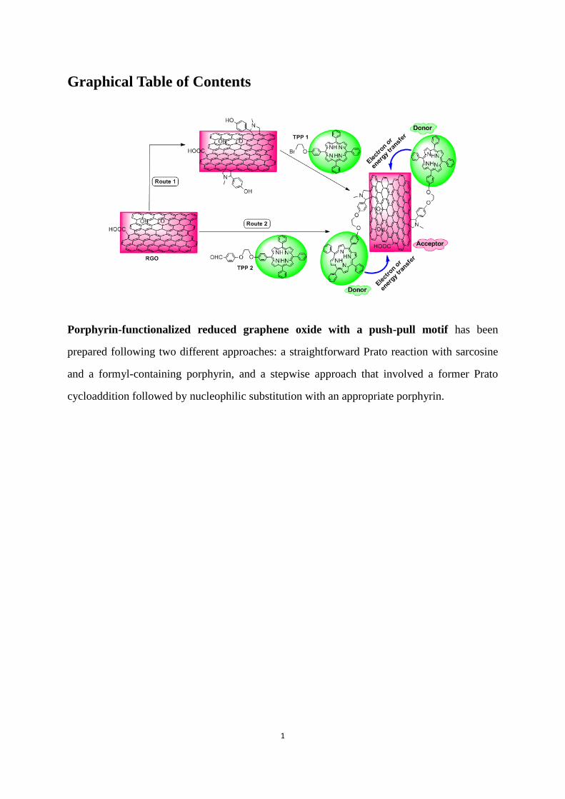
Porphyrin-functionalized reduced graphene oxide with a push-pull motif has been
prepared following two different approaches: a straightforward Prato reaction with sarcosine
and a formyl-containing porphyrin, and a stepwise approach that involved a former Prato
cycloaddition followed by nucleophilic substitution with an appropriate porphyrin.
2
A 1,3-Dipolar Cycloaddition Protocol to Porphyrin-Functionalized
Reduced Graphene Oxide with a Push-Pull Motif
Aijian Wang,1 Wang Yu,1 Zhengguo Xiao,2 Yinglin Song,2 Marie P. Cifuentes,3 Mark G.
Humphrey,3 and Chi Zhang*1
1 China-Australia Joint Research Center for Functional Molecular Materials, School of
Chemical and Material Engineering, Jiangnan University, Wuxi 214122, P.R. China
2 School of Physical Science and Technology, Soochow University, Suzhou 215006, P.R. China
3 Research School of Chemistry, Australian National University, Canberra, ACT 0200,
Australia
Address correspondence to Chi Zhang, [email protected]
3
Abstract: Reduced graphene oxide (RGO) has been covalently functionalized with
porphyrin moieties by two methods: a straightforward Prato reaction (i.e. a 1,3-dipolar
cycloaddition) with sarcosine and a formyl-containing porphyrin, and a stepwise method that
involves a 1,3-dipolar cycloaddition to the RGO surface using 4-hydroxybenzaldehyde,
followed by nucleophilic substitution with an appropriate porphyrin. The chemical bonding of
porphyrins to the RGO’s surface has been confirmed by ultraviolet/visible absorption,
fluorescence, Fourier-transform infrared, and Raman spectroscopies, X-ray powder diffraction
and X-ray photoelectron spectroscopy, transmission electron and atomic force microscopies,
and thermogravimetric analysis; this chemical attachment assures efficient electron/energy
transfer between RGO and the porphyrin, and affords improved optical nonlinearities
compared to those of the RGO precursor and the pristine porphyrin.
Keywords: porphyrin ∙ cycloaddition ∙ reduced graphene oxide ∙ nonlinear optics
4
1. Introduction
Graphene is a single layer two-dimensional planar sheet of sp2 hybridized carbon atoms
arranged in a hexagonal lattice, the basic structural element for graphite and carbon
nanotubes.[1] First produced in the lab by Novoselov et al. in 2004,[2] graphene is considered
to be one of the most robust nano-scale materials. The novel and unique electronic properties
exhibited by graphene and its derivatives that result from the presence of extended and
delocalized π-electron systems make them excellent candidates for applications in the area of
optoelectronics, energy storage and photovoltaic devices.[3-5] The easy of processing of
graphene is of critical importance in facilitating its integration with substrates and
materials.[6,7] Many reports have focused on the chemical modification of graphene with
specific functionalities via covalent or non-covalent methods for tuning its chemical and
physical properties,[8] while the resultant graphene materials can facilitate charge transfer
when graphene is combined with electron donors, such as porphyrin[9] or phthalocyanine;[10]
graphene is a particularly efficient electron acceptor. Modification of the carbon network by
grafting organic moieties is important in the design of graphene-based nanoelectronics due to
the fact that this may provide a means to dope the material.[11,12]
Porphyrins have many potential uses in optoelectronics, nonlinear optics, solar cell
applications, and photodynamic therapies, because of strong excited-state absorption, high
triplet yields, long excited-state lifetimes, and delocalizable electron density.[13-16]
Carbon-nanotube-porphyrin and C60-porphyrin nanohybrids have attracted widespread
attention and have been explored in a number of potential applications.[17-19] Because of these
precedents, and the similarity of graphene, carbon nanotubes and C60, nanohybrids combining
5
graphene with porphyrin may be useful for a diverse range of potential applications in biology,
catalysis, sensors and solar cells, etc.[5] Thus far, reports of graphene-based hybrid
nanomaterials are largely restricted to graphene oxide (GO), which has various chemically
reactive oxygen-containing functionalities (e.g. carboxyl, epoxy, and hydroxyl groups).[20-23]
In contrast to GO, the electrical conductivity and electron/hole transporting properties can be
recovered following the reduction step to afford reduced graphene oxide (RGO),[24,25] and
organic moieties can be chemically grafted to the surface of RGO with retention of the
structural integrity and electronic structure of the RGO framework. As such, chemical
functionalization could potentially pave the way towards the use of RGO in practical
applications. However, reports on functionalized RGO systems are scarce, especially for those
involving covalent attachment;[26,27] to some extent, this can be attributed to the irreversible
aggregation of RGO which ensues in the absence of electrostatic or steric protection,
rendering further processing more difficult. It is therefore critically important to design and
prepare RGO-based readily-processed nanohybrid materials for optoelectronic and photonic
devices.
Encouraged by these considerations, we wondered if the combination of RGO and
optoelectronic porphyrin molecules would afford species that possess not only the intrinsic
properties of RGO and porphyrins, but also novel functions resulting from the mutual π
interaction between the RGO and the porphyrins; multifunctional nanometer-scale systems for
optical and optoelectronic applications may thereby be generated. However, to the best of our
knowledge, there is no report in the literature of the fabrication of RGO-porphyrin conjugates.
In this contribution, we present the first study of the preparation of dispersible
6
RGO-porphyrin nanohybrids through 1,3-dipolar cycloaddition reaction of RGO, sarcosine
and appropriately functionalized porphyrin; this reaction has been previously used for the
chemical modification of carbon nanotubes and fullerenes.[28,29] A stepwise approach to
achieve the porphyrin functionalized RGO at minimum synthetic cost has also been explored;
this widely applicable approach affords functionalized RGO in which the electronic structure
is preserved. The hybrid materials thus prepared are stable in solution and have been
characterized by a number of spectroscopic and microscopy techniques. In particular, we
complement our work with a detailed photophysical investigation on ground- and
excited-state RGO-porphyrin interactions, as well as the third-order nonlinear optical (NLO)
performance of these nanohybrids in the nanosecond regime at 532 nm; the hybrids exhibit
enhanced NLO responses in comparison with the individual RGO and porphyrins.
2. Results and Discussion
2.1. Syntheses
1,3-Dipolar cycloaddition has proven to be an effective method for functionalizing
conjugated π systems: convenient synthetic applications of 1,3-dipolar cycloadditions to
fullerenes, carbon nanotubes, onions and nanohorns have led to many applications in areas
such as drug delivery, nanoelectronic devices, solar cells and biotechnology.[30-32] The
carbon-based nanohybrids usually act as electron acceptors when they are appropriately
interfaced with an electron donor moiety. Although the reactivity of graphene differs from that
of fullerenes and carbon nanotubes, the 1,3-dipolar cycloaddition can be efficiently performed
and affords a highly functionalized hybrid with reaction taking place not only at the edges, but
also at the C=C bonds in the center of graphene sheets.[33-37] Inspired by these considerations,
7
we decided to explore this class of reactions with graphene surfaces. In the present study,
RGO was successfully functionalized with porphyrin molecules by 1,3-dipolar cycloaddition
reactions. Although functionalization of graphene employing unstable 1,3-dipolar azomethine
ylides (obtained from condensation of an aldehyde with an α-amino acid) has been reported
previously,[33,36] the functionalization of RGO sheets with porphyrin units is not
straightforward. Two possible routes toward functionalizing RGO with porphyrin were
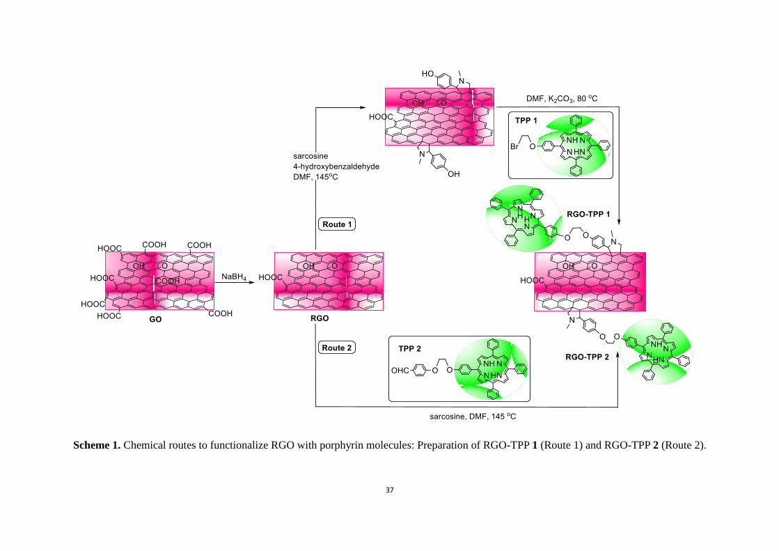
envisaged. Scheme 1 summarizes these procedures, employed for the preparation of the
nanohybrids RGO-TPP 1 and RGO-TPP 2. The first route (Route 1, Scheme 1) involves
initial reaction of sarcosine and 4-hydroxybenzaldehyde with RGO, followed by nucleophilic
substitution of 5-[4-(2-bromoethoxy)phenyl]-10,15,20-triphenylporphyrin (TPP 1) by the
derivatized RGO. This method is advantageous in that chemical modification of graphene
usually requires a large excess of the reactants;[34] in this particular case, one reactant is the
inexpensive 4-hydroxybenzaldehyde. Another important consideration is that hydroxyl groups
can be attached to the RGO surfaces by a single reaction without degrading the RGO
electronic properties, permitting a high grafting density of organic units and potentially a
wider application as an optoelectronic nanohybrid material. The subsequent nucleophilic
substitution reaction of TPP 1 and the functionalized RGO bearing pendent OH groups can, in
principle, proceed under nearly stoichiometric conditions. A potential setback of this route lies
in the fact that it is very difficult to control the number of OH units that are used in
nucleophilic substitution with TPP 1. As a consequence, we explored a second method (Route
2 in Scheme 1) that involves preparing a formyl-containing porphyrin
5-[4-(2-(4-formylphenoxy)ethyloxy)phenyl]-10,15,20-triphenylporphyrin (TPP 2). The
8
advantage of this method is that the formyl-containing TPP 2 can be attached to RGO in a
straightforward fashion, through the formation of the corresponding azomethine ylide
(Scheme 1). After only a few hours, the color of the solution had changed to dark brown,
implying that reaction had occurred. The crude product was isolated by filtration and washed
with deionized water and several organic solvents; UV-Vis spectroscopy and thin layer
chromatography (TLC) were then used to confirm that the supernatant layer contained no
unreacted TPP 2 following the final washing.
(Please insert Scheme 1 here)
2.2. Linear Optical Absorption and Fluorescence Spectroscopy Analysis
The fingerprint for porphyrin-functionalized RGO nanohybrids is their electronic
behavior; the linear absorption and fluorescence spectra are sensitive to the presence of
donor/acceptor units that can influence such spectra by photo-induced energy and/or electron
transfer.[38] DMSO was used to achieve RGO dispersion, and thereby facilitate comparison of
the electronic properties of RGO-TPP 1 and RGO-TPP 2 with those of TPP 1 and TPP 2,
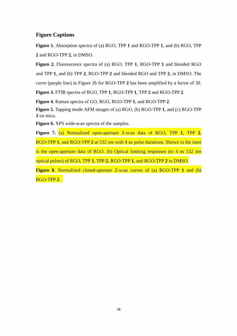
respectively. RGO exhibits a broad absorption with continuously decreasing intensity
extending to 700 nm, while the electronic absorption spectrum of TPP 1 is characterized by a
strong Soret absorption at 420 nm, together with four relatively weaker Q bands between 500
and 700 nm (Figure 1a). Covalent grafting of TPP 1 onto RGO results in the reduction in
intensity of the absorption of the porphyrin moieties in the RGO-TPP 1 nanohybrid compared
to those in the absorption spectrum of TPP 1 in DMSO, although no obvious spectral shift is
observed. A noteworthy broadening of the Soret band is observed compared with that of the
free porphyrin moiety, which may be due to electronic interactions between the RGO and
9
porphyrin units in this system (RGO is usually regarded as an electron acceptor and TPP 1 as
an electron donor). For RGO-TPP 2, a broad absorption at 423 nm, corresponding to a small
red shift of 3 nm compared to TPP 2 (Figure 1b), is observed. . Upon comparing the spectra of
the two porphyrin-functionalized RGOs (RGO-TPP 1 and RGO-TPP 2), three observations
can be made (Figure 1). First, the overall intensity of the porphyrin-centered transitions
increases on proceeding from RGO-TPP 2 to RGO-TPP 1. The intensity of the Soret band of
RGO-TPP 2 is diminished significantly, while the G-bands in RGO-TPP 2 are not as
prominent as those in RGO-TPP 1. This suggests that the porphyrin content in the two
RGO-TPP nanohybrids declines on proceeding from RGO-TPP 1 to RGO-TPP 2 due to the
different functionalization procedures. Secondly, a red-shift of 3 nm (i.e. from 420 to 423 nm)
in the absorption maximum is seen for RGO-TPP 2 in comparison with that of RGO-TPP 1,
indicating the effect of different reaction conditions on the absorption spectra. Thirdly, the
RGO characteristics in both RGO-TPP nanohybrids are concomitantly reduced in absorption
intensity. The ground-state absorption spectra of RGO-TPP 1 and RGO-TPP 2 contain
features of both the RGO and porphyrin, confirming the formation of RGO-TPP nanohybrids.
Remarkably, similar effects rules have also been observed in the UV/Vis absorption spectra of
porphyrin-functionalized multi-walled carbon nanotubes (MWCNTs),[18] corresponding to the
different degree of functionalization.[39]
(Please insert Figure 1 here)
A more compelling test of the interactions between the porphyrins and RGO is available
from fluorescence experiments focusing on the fluorescence features of the porphyrin
moieties. An intramolecular donor-acceptor structure usually allows charge-transfer
10
interaction or the highest occupied molecular orbital (HOMO)-lowest unoccupied molecular
orbital (LUMO) transitions to occur,[24,40] and this is evident from the fluorescence spectra of
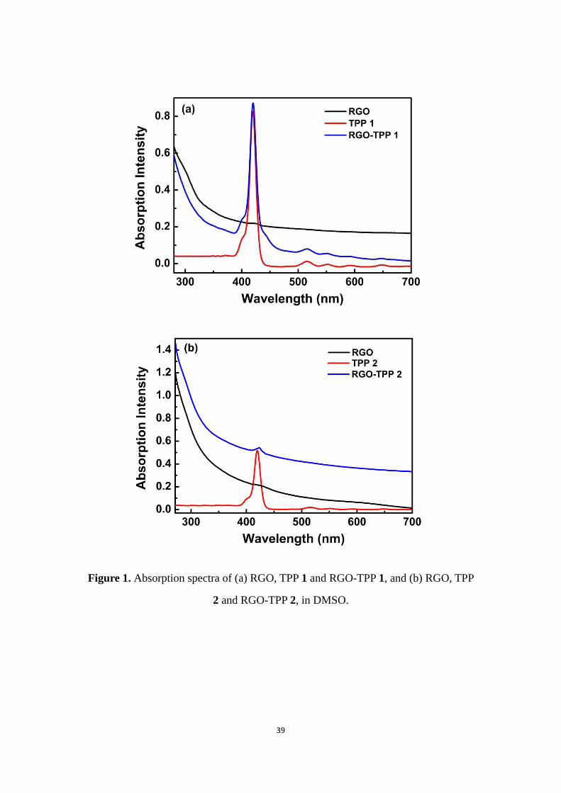
RGO-TPP 1 and RGO-TPP 2, the results being shown in Figure 2. Upon excitation at 420 nm,
RGO-TPP 1 displays fluorescence features with a maximum and a shoulder at 654 and 719
nm, respectively. Because RGO does not show a fluorescence signal, the fluorescence of
RGO-TPP 1 must be from the porphyrin moieties, which unequivocally confirms the presence
of the porphyrin units in RGO-TPP 1. Following excitation at the same wavelength, a
physically blended sample of RGO and TPP 1 (1:1 weight ratio, a control sample with the
same absorbance value as that of the nanohybrids) exhibits similar emission bands with 15%
quenching of fluorescence emission in comparison with TPP 1. However, compared to TPP 1,
a significant fluorescence quenching (58%) is observed for RGO-TPP 1, a result likely to
arise from a through-bond mechanism because of the unique direct linkage of the porphyrin
moieties and the RGO, suggesting the presence of photo-induced electron/energy transfer
between the excited states of the TPP unit and the RGO moiety. The physically blended
sample of RGO with TPP 2 (1:1 weight ratio) shows similar fluorescence quenching (13%) to
the mixture of RGO and TPP 1. In contrast, RGO-TPP 2 exhibits a vastly different
fluorescence quantum yield from RGO-TPP 1. A quenching factor of 133 was determined for
RGO-TPP 2 (compared with TPP 2), which is much larger than the value of 2.3 for RGO-TPP
1 (compared with TPP 1). In addition, the weak fluorescence emission for RGO-TPP 2 was
observed at 656 nm, corresponding to a red shift of 2 nm and a significantly decrease in
intensity compared with that of RGO-TPP 1. These observations are consistent with both the
formation of RGO-TPP nanohybrids and the tunability of optical properties through different
11
covalent functionalization methods. Similar fluorescence quenching has been observed in the
MWCNT-TPP hybrids, for which photo-induced energy/electron transfer was suggested to
explain the mechanism of their fluorescence quenching.[18] The functionalized porphyrin
moieties presumably act as an electron transporting antenna when covalently linked to RGO,
while the RGO acts as an electron acceptor unit, leading to the observed fluorescence
quenching and energy release. In the present RGO-TPP nanohybrids, the RGO significantly
quenches the photoluminescence of the porphyrin moieties, which confirms the close
proximity of RGO and porphyrin, and their strong interaction. Both nanohybrids have
potential as the active materials for various optoelectronic applications, such as the sources
for solar energy harvesting in solar cells.
(Please insert Figure 2 here)
2.3. FTIR Spectroscopy Analysis
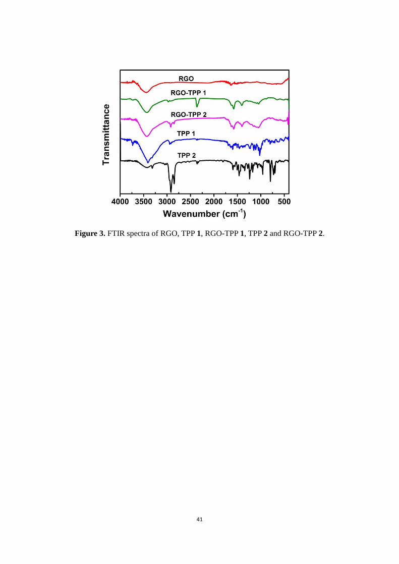
Further evidence in support of covalent functionalization of RGO is available from FTIR
spectroscopy. Figure 3 displays FTIR spectra of RGO, TPP 1, TPP 2 and the nanohybrid
materials (RGO-TPP 1 and RGO-TPP 2). The FTIR spectrum of RGO is almost featureless
with weak skeletal vibration of the aromatic domains around 1634 cm-1, which is consistent
with previous observations[41,42] and with removal or transformation of much of the
oxygen-containing functional groups in GO on its conversion into RGO. For the RGO-TPP 1
and RGO-TPP 2 nanohybrids, some bands that are characteristic of porphyrin are observed,
which are coincident with those displayed by TPP 1 and TPP 2. The disappearance of the
aldehyde stretching-band centred at 1705 cm-1, in proceeding from RGO-TPP 1 to RGO-TPP
2, is consistent with reaction of the aldehyde units and thereby covalent attachment of the
12
porphyrin to the RGO. The absorption bands in the region 2850-2960 cm-1, attributed to the
C-H stretching vibrations of the aromatic rings, further confirm the existence of the porphyrin
moieties on RGO. All features in the FTIR spectra are consistent with the formation of
RGO-TPP nanohybrids. Although some absorption peaks characteristic of porphyrins were
observed in the FTIR spectra, it should be noted that some difference was found for the
RGO-TPP nanohybrids, ascribed to the presence of residual phenol units in RGO-TPP 1
resulting from incomplete reaction of hydroxyl groups in the
4-hydroxybenzaldehyde-functionalized RGO hybrid with TPP 1. Consistent with the clear
distinctions apparent in the UV/Vis and fluorescence measurements, these observations
illustrate the effect of the reaction conditions on the spectroscopic properties of the two
nanohybrids prepared via the different routes.
(Please insert Figure 3 here)
2.4. Raman Spectroscopy Analysis
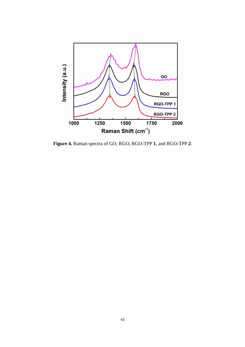
The significant structural changes in proceeding from GO to RGO, and then to
RGO-TPP 1 and RGO-TPP 2, are also reflected in their Raman spectra (Figure 4). GO
exhibits a prominent band at 1370 cm-1, corresponding to the breathing mode of κ-point
phonons of A1g symmetry (the D-band). There is also an intense tangential mode at 1602 cm-1,
which arises from the first-order scattering of E2g phonons from sp2 carbon atoms (the G-band
or E2g mode).[43] After treatment by NaBH4, the D- and G-bands of RGO are located at 1343
and 1577 cm-1, respectively, confirming the reduction of GO during the chemical treatment.
The Raman spectrum of RGO-TPP 1 displays two bands at 1341 and 1584 cm-1. The
down-shift of the D-band and the up-shift of the G-band for RGO-TPP 1, in comparison to
13
RGO, are consistent with previously reported functionalization of RGO.[25] However, the D
and G bands of the RGO-TPP 2 hybrid, which appear at 1348 and 1587 cm-1, respectively, are
both up-shifted compared to those of RGO. These results suggest a strong interaction between
porphyrin and the RGO sheets, and are consistent with the fluorescence emission results. The
intensity ratio of the D/G bands provides a measure of the disorder/defects in graphene, a
smaller intensity ratio ID/IG being ascribed to a larger average size and fewer sp3
defects/disorders of the in-plane graphitic crystallite sp2 domains. The D-band is a measure of
the degree of covalent functionalization, which transforms sp2 to sp3 sites, whereas the
G-band has been used to estimate the distribution of this modification.[44-46] We have not
found clear evidence of any peaks characteristic of the functionalizing moieties, due to the
fact that functionalization is weak. However, the intensity ratio of the D band to the
corresponding G band increased from 0.98 for RGO to 1.03 for RGO-TPP 1 and 1.02 for
RGO-TPP 2, which is consistent with the introduction of a considerable amount of structural
defects on the lattice following functionalization, suggesting that the newly formed graphitic
domains are smaller in size, but more numerous in number. Reduction in the sp2 hybridization
is expected following the successful cycloaddition reactions, consistent with the results of
FTIR spectroscopy discussed above.
(Please insert Figure 4 here)
2.5. XRD Studies
The attachment of porphyrins to RGO is also supported by powder XRD patterns, the
results being shown in Figure S4. The inter-planar d-spacing of GO is 0.78 nm (2θ = 11.2°),
consistent with the presence of oxygen-containing functional groups attached to the surface of
14
GO and the atomic-scale roughness arising from structural defects.[47,48] A shoulder with a
small broad peak was observed at around 22o, indicating that a fraction of GO was not fully
intercalated. After reduction of GO by NaBH4, the (002) peak of GO disappears, whereas a
broad diffraction peak was observed extending from 15o to 30o, assigned to exfoliation of
RGO into monolayers or several-layer species. The decreased interlayer spacing can be
attributed to the removal of oxygen-based functional groups on the basal plane following
reduction of the GO, resulting in a tighter RGO. After functionalization, as depicted in Figure
S4, the (002) peaks of RGO-TPP 1 and RGO-TPP 2 were shifted to smaller 2 values
compared to RGO, which is indicative of the grafting of porphyrin moieties onto the surface
of the RGO, and their functions as “spacers” for the RGO platelets, resulting in increased
interlayer spacing.[49] The broad peaks imply that the samples are very poorly ordered along
the stacking direction,[46] while the increased intensity ratio (ID/IG) of the D-band intensity (ID)
compared to the G-band intensity (IG) is consistent with a lower degree of crystallinity of
these RGO materials (Figure 4). Both nanohybrids have a pattern similar to RGO, so the
functionalization process does not destroy the layered structure of RGO.
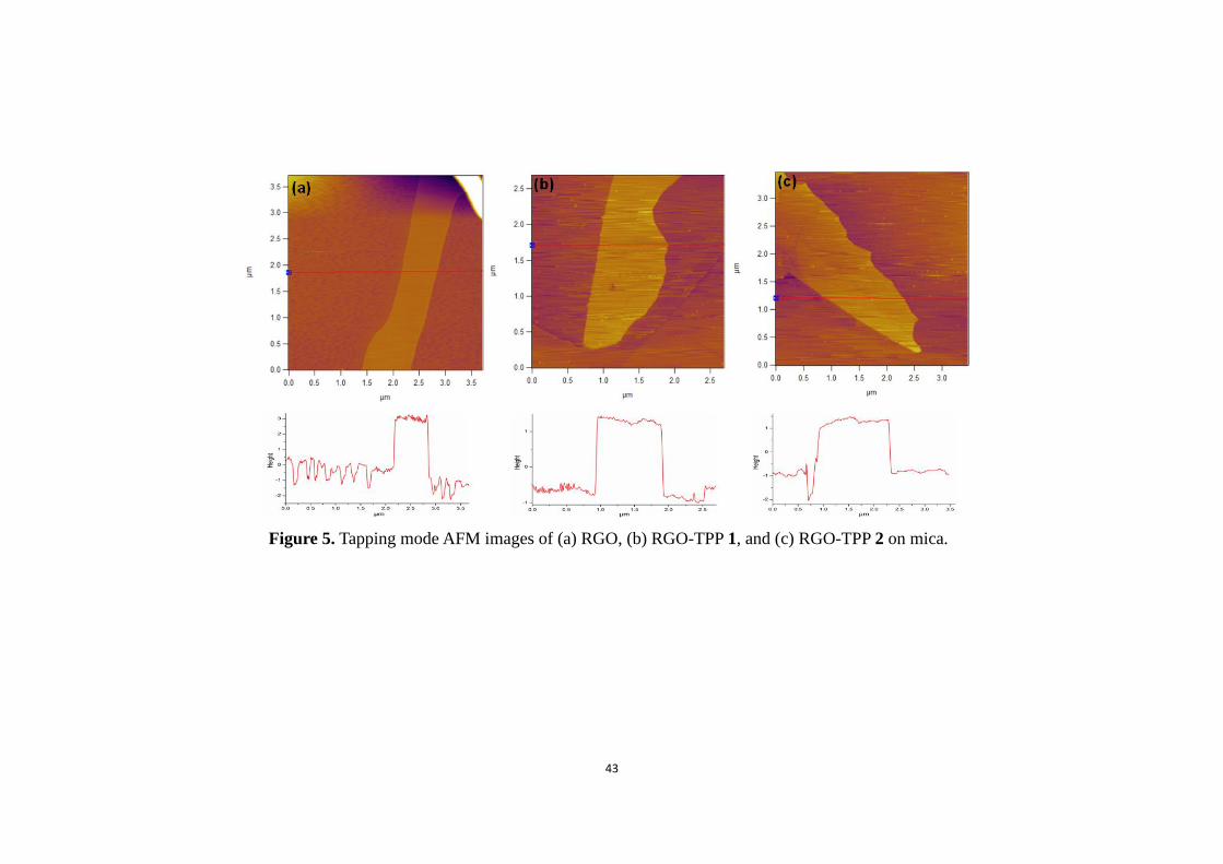
2.6. AFM and TEM Studies
The distribution of porphyrin on the RGO nanosheets of the RGO-TPP nanohybrids and
the height profile in selected locations were examined by AFM. As shown in Figure 5a, the
average thickness of the RGO aggregations was determined to be about 3.6 nm because of the
poor dispersion of the RGO nanosheets in methanol and π-π stacking during reduction,
resulting in the formation of several-layer graphene. Considering that the thickness of a clean
graphene nanosheet is approximately 0.95 nm,[50] this suggests ca. 4 layers of RGO nanosheet.
15
By comparison, when RGO was functionalized with TPP 1, the average thickness of a
RGO-TPP 1 aggregation was measured to be about 2.1 nm (Figure 5b), a ca. 1.15 nm increase
over the thickness of a graphene sheet. As the thickness of one porphyrin molecule is about
0.5 nm,[9] it can be concluded that porphyrin molecules are most likely to be grafted onto the
RGO surface of both sides. Moreover, as shown in Figure 5b, the distribution line of the
height profile of RGO-TPP 1 is relatively flat, suggesting that the porphyrin molecules should
be uniformly distributed on the surface of RGO. Similar results were also observed for
RGO-TPP 2 (Figure 5c). The structural change of RGO before and after functionalization was
further investigated by TEM. In the case of RGO, the lack of functional groups in the centre
of the sheets induces significant aggregation (Figure S5a), consistent with a flake-like shape
of RGO.[34] From the TEM images of RGO-TPP 1 and RGO-TPP 2 (Figures S5b and S5c),
the presence of transparent single- and several-layer RGO flakes can be observed due to their
thin nature, which indicates that the compatibility between the RGO and organic solvents was
significantly improved following functionalization,[51] and that the porphyrin-functionalized
RGO can be uniformly dispersed in DMSO.
(Please insert Figure 5 here)
2.7. Thermogravimetric Analysis
The presence of organic groups on the RGO sheets was further confirmed by TGA,
which has been widely used to characterize the covalent chemical functionalization of
graphene.[52] Figure S6 displays TGA thermograms of weight loss as a function of
temperature for RGO, RGO-TPP 1 and RGO-TPP 2, as measured under a nitrogen
atmosphere. The TGA curve of RGO reveals significant thermal stability following the
16
removal of the labile oxygen functional groups by the chemical reduction. Apart from a slight
mass loss below 150 oC, which can be attributed to the loss of adsorbed water and the organic
impurities, no significant mass loss (approximately 5.2%) was detected when the material was
heated up to 800 oC. RGO-TPP 1 and RGO-TPP 2 display approximately 27% and 30%
weight loss, respectively, between 50 and 800 oC. It is likely that the significantly poorer
thermal stability of RGO-TPP 1 and RGO-TPP 2 nanohybrids over the investigated
temperature range is due to the loss of the porphyrin units, which therefore affords further
evidence of the success in incorporating porphyrin moieties onto the RGO surfaces. The
weight loss that occurred above 500 oC can be ascribed to the thermal decomposition of
defects created at sites where the RGO functionalization occurred. The degradation behavior
of RGO-TPP 1 and RGO-TPP 2 is complicated in comparison with that of RGO because
porphyrin is a char-forming substance, which leaves a certain amount of residual material at
high temperature in TGA measurements;[18] thus, the exact porphyrin content of both hybrids
is likely to be larger than that derived from the weight loss in the TGA measurements, and
consistent with their solubility in common organic solvents such as DMF and DMSO.[53]
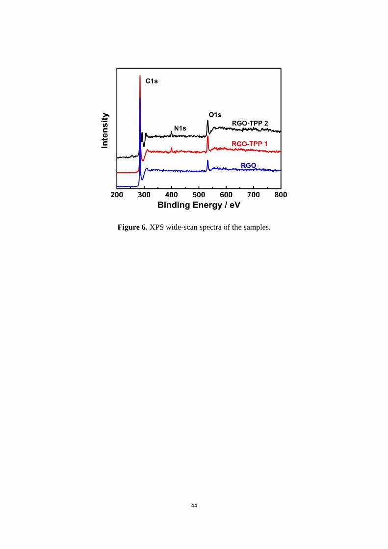
2.8. X-ray Photoelectron Spectroscopy
XPS is a surface analysis technique that determines relative atomic composition,[54] in
the present case providing confirmation of the covalent attachment of the porphyrin moieties
onto the surface of the RGO. From Figure 6, it can be clearly seen that after reduction by
NaBH4, a weak O1s signal is observed in the XPS spectrum of RGO, which is indicative of
the presence of residual oxygen functionalities on the RGO. After functionalization with

17
porphyrin, the spectra of RGO-TPP 1 and RGO-TPP 2 show a new peak that is ascribed to N
1s, confirming the formation of porphyrin grafted RGO.
(Please insert Figure 6 here)
Detailed analysis of the XPS spectra provides clear evidence that the grafting of
porphyrin to RGO has been achieved. The C 1s XPS spectrum of RGO (Figure S7a) displays
three peaks at around 284.5, 285.3 and 286.0 eV, corresponding to a strong sp2 C signal from
the C=C bond and a weak sp3 C signal from the C-O bond,[25] and consistent with the absence
of significant oxygen-containing functional groups, i.e. most of the oxygen functionalities in
the GO have been removed via reduction. After coupling porphyrin to the RGO, new
contributions appear in the C 1s spectra of RGO-TPP 1 and RGO-TPP 2 (Figures S7b and
S7c), corresponding to the functionalized porphyrins. For example, Figures S7b and S7c
include two new peaks, located at around 285.8 and 288.2 eV, which were attributed to the
C-N and C=N units, respectively.[55] The N 1s core-level XPS spectra afford further
information about the presence of the porphyrin moieties on the functionalized RGO (Figures
S7d and S7e). The N 1s spectrum of RGO-TPP 1 displays three peaks at 398.1 (the N atoms
in the C-N bonds), 399.3 (the N atoms in the N-H bonds), and 400.2 eV (the N atoms in the
C=N bonds).[18] In contrast to RGO-TPP 1, the binding energies of the N 1s of RGO-TPP 2
are 398.8, 399.7 and 400.7 eV, and there is a considerable intensity increase of the N 1s peak
due to the different chemical environments. From analysis of the various spectroscopic and
structural data above, we conclude that (i) the 1,3-dipolar cycloaddition reaction has
efficiently introduced porphyrin moieties onto the surface of RGO; and (ii) the peak
separation of the N 1s spectra of RGO-TPP 1 and RGO-TPP 2 confirms the different nitrogen
18
species on the surface of both nanohybrids, and thereby the success of the porphyrin grafting
process.
2.9. Optical Nonlinearities Studies
Because of their ultrafast carrier dynamics and good incident light absorption capabilities,
graphene and its derivatives have become the benchmark standards for optical limiting
studies,[56] so it was of significant interest to assess the optical nonlinearities of the RGO-TPP
1 and RGO-TPP 2 nanohybrids. The RGO, TPP 1, TPP 2, RGO-TPP 1, and RGO-TPP 2
samples were prepared by dissolution in DMSO, followed by 0.5 h of ultrasonic processing.
Z-scan is a simple technique that is used to measure the on-axis phase change of a laser beam
as the beam propagates through nonlinear media.[57] The nonlinear media experience the
maximum laser light intensity at the focal point, with the intensity decreasing on moving
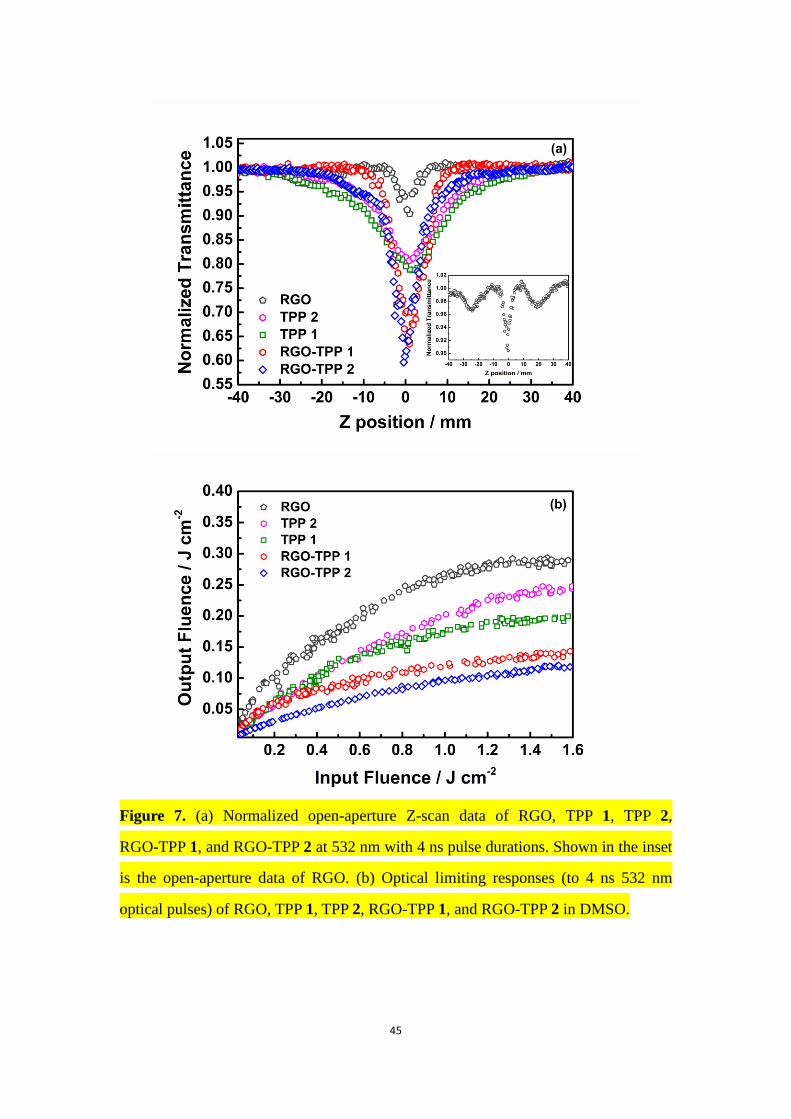
away from the focal point. The open-aperture Z-scan results of the tested samples at 532 nm
are presented in Figure 7a. As shown in Figure 1, the linear absorption spectra of RGO-TPP 1
and RGO-TPP 2 display very low linear absorption at 532 nm, suggesting low intensity loss
and little temperature change by photon absorption during the nonlinear optical
measurements.[58] A broad transparency range is of significant importance for NLO
applications, especially those in waveguide form.[59] For ease of comparison of the Z-scan
results, all samples were adjusted to have the same linear transmittance (71% at 532 nm) by
varying their concentration in DMSO. For an optical limiter, the depth of the valley in the
open-aperture Z-scan curves is related to the extent of OL.[60] As shown in Figure 7a, both
RGO-TPP nanohybrids exhibit better NLO response than RGO or the porphyrin precursors
(TPP 1 and TPP 2), suggesting a significant effect from covalent grafting. A similar
19
observation was made for the porphyrin-covalently-functionalized C60 and MWCNTs, which
exhibited enhanced NLO properties compared with their precursors.[18,39,61] The valley-shaped
curves are indicative of the occurrence of positive nonlinear absorption, and an OL
behavior.[60] The optical nonlinearities of the solvent DMSO and the empty quartz cells were
also measured under the same experimental conditions, but no NLO response was detected at
the measurement wavelength, so their effects on the experimental results can be neglected.
Comparing the nonlinear absorption performance of the RGO-TPP nanohybrids and the
C60-TPP system in Ref. [61], it is seen that both RGO-TPP nanohybrids (RGO-TPP 1 and
RGO-TPP 2) possess larger effective nonlinear absorption responses. However, different
phenomena are believed responsible for the observed NLO behavior of the RGO-TPP
nanohybrids and the MWCNT-TPP systems,[18] rendering further comment unwarranted. The
normalized transmittance of RGO-TPP 2 is very similar to that of the previously reported
ZnTPP-functionalized MWCNTs (ZnTPP = 5-[4-{(4-formylphenyl)ethynyl}phenyl]-
10,15,20-triphenyl-porphinatozinc(II)) upon excitation at 532 nm with 4 ns pulses, with the
largest dip of normalized transmittance valley observed at the focal point, while the changes
of normalized transmittance of both RGO-TPP nanohybrids are clearly greater than the
MWCNT-TPP system (TPP = 5-[4-{2-(4-formylphenoxy)ethyloxy}phenyl]-10,15,20-
triphenylporphyrin). The tetraphenylporphyrin-functionalized MWCNTs prepared by the
reaction of MWCNTs with in situ generated porphyrin diazonium compounds exhibit a
weaker nonlinear absorption performance at 532 nm with 5.6 ns pulse duration compared with
those of both RGO-TPP nanohybrids in the present case,[39] though the differing experimental
geometries render such comparison necessarily cautious.
20
The curve for RGO clearly shows reverse saturable absorption (RSA) behavior followed
by saturable absorption (SA) and then a reversion to RSA (Figure 7a), consistent with the
NLO performance of GO.[62] Fast relaxation from higher excited states Sn and slower
relaxation from the first singlet excited state S1 play an important factor in switching the
nonlinear optical absorption behavior from RSA to SA and back to RSA.[63] The S1 state is
populated both by relaxation from the higher excited states and by ground-state absorption.
The longer lifetime of the S1 state results in RGO exhibiting saturation in the intensity region
0.58-0.92 μJ. The Sn states are populated by the ESA corresponding to the S1 to Sn transition
as well as two-photon absorption, but these relax to the lower excited state very quickly. At
still higher intensities, two-photon absorption may dominate for RGO, resulting in the
observed RSA effect.[26] The intensity-dependent switching of nonlinear absorption implies
that materials such as RGO are potential candidates for ultrafast nonlinear optical switching
applications.
Figure S8 displays closed-aperture Z-scan curves of dispersions of RGO, TPP 1, and TPP
2, measured with nanosecond pulses. As depicted in Figure S8a, the DMSO suspension of the
RGO nanosheets was found to exhibit a valley-peak transmission configuration, indicative of
positive nonlinear refraction, and corresponding to self-focusing behavior. In contrast, the
solutions of TPP 1 and TPP 2 exhibited a peak-valley transmission configuration under visible
excitation (Figures S8b and S8c), suggesting negative nonlinear refraction and corresponding
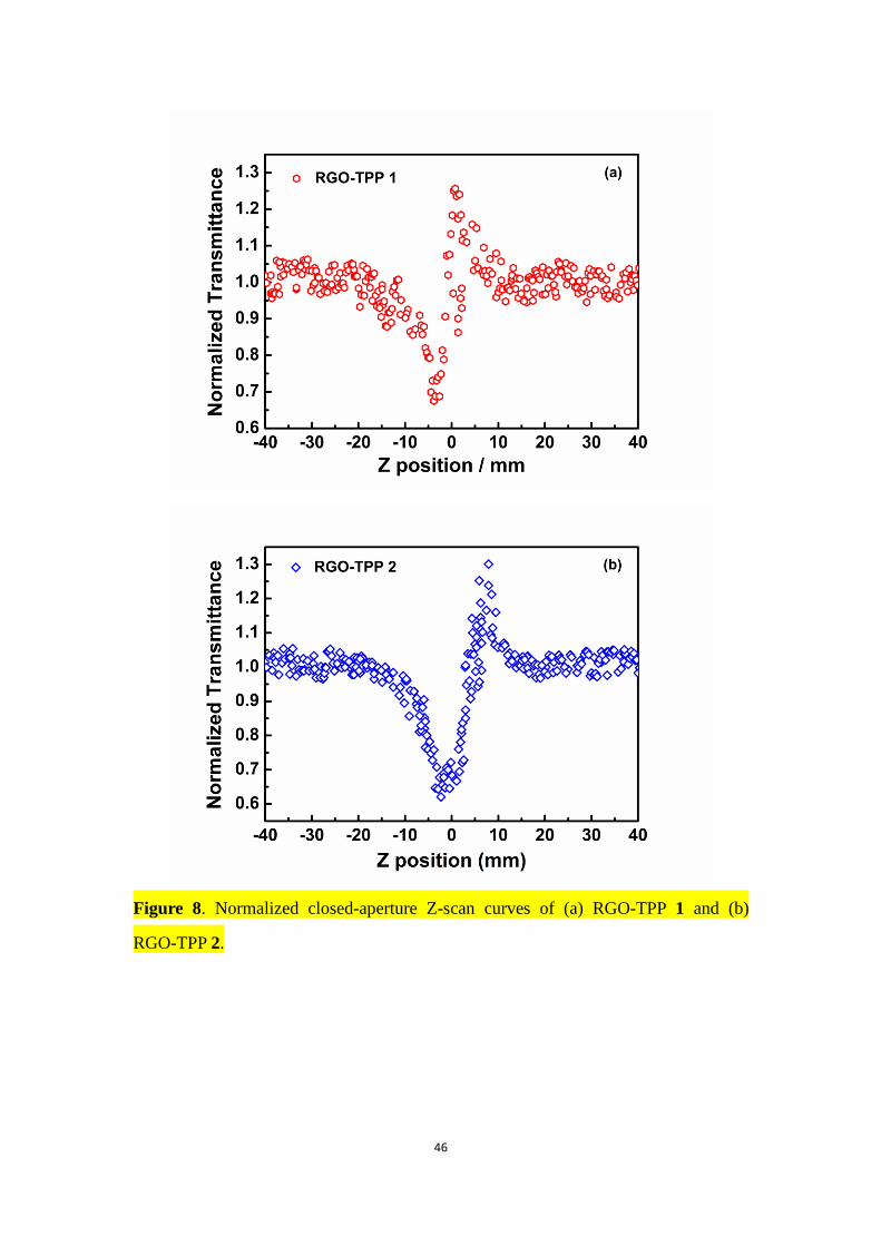
to self-defocusing behavior. Similar to RGO, the nonlinear refraction Z-scan curves of both
nanohybrids (RGO-TPP 1 and RGO-TPP 2) were also characterized by a valley followed by a
peak as shown in Figures 8a and 8b, implying that the behavior of the RGO nanosheets is
21
dominant in determining the nonlinear refraction performance of the RGO-TPP nanohybrids.
However, both RGO-TPP nanohybrids exhibit different normalized transmittance values and
peak-valley positions in comparison with those of RGO, TPP 1, and TPP 2, presumably a
result of their covalent linkage.
Optical limiting, the optical analogy of an electrical surge protector, is of significant
importance for protecting human eyes and optical sensors against damaging sources of
light.[64] Any materials (optical limiters) which have high transmittance to low-intensity light
but low transmittance to intense light, and that thereby possess an efficient nonlinear
absorption effect as a function of input fluence, are of great interest for OL. With the aim of
investigating the OL behavior of all samples, input fluence versus output fluence plots were
constructed as given in Figure 7b. At low input fluence, there is no appreciable change in the
output fluence, the linear relationship obeying Beer’s Law. As the incident fluence increases,
the output fluence decreases and deviates from linearity, consistent with an OL response. The
decrease in output fluence of RGO-TPP 1 and RGO-TPP 2 with increasing fluence exceeds
that of the other three samples in the present study. The extent of the decrease in output
fluence (RGO-TPP 2 > RGO-TPP 1 > TPP 1 > TPP 2 > RGO) suggests that the OL
performance of the RGO-TPP nanohybrids is better than RGO, TPP 1 and TPP 2, consistent
with the results of open-aperture Z-scan. It should be pointed out that the NLO response of
RGO may partially arise from nonlinear scattering (NLS), which is consistent with the
behaviour of graphene dispersions,[65] while that of porphyrin is dominated by RSA.
Accordingly, the improved OL effect of both RGO-TPP nanohybrids can be partially
attributed to the combination of NLS and RSA.[66] The defect-induced states present in both
22
nanohybrids, which contribute to interband transitions through ESA, may also play a role in
the enhanced OL.[67,68] The presence of defects has been confirmed by the increased ID/IG ratio
observed following covalent linkage between porphyrin and RGO, which implies an increase
in the number of defects in the samples. In the RGO-TPP system, porphyrin is a favorable
electron donor and RGO is an electron acceptor when the two moieties are connected directly.
Thus, as in the C60- and MWCNT-porphyrin systems,[18,61] the intramolecular donor-acceptor
interaction between the two moieties of TPP and RGO in our RGO-TPP nanohybrids may
result in charge transfer from the photo-excited singlet TPP to the RGO moiety, resulting in
fluorescence quenching and energy release. Similarly, photo-induced electron transfer has also
been observed in the GO-TPP and MWCNT-TPP systems.[23,39] It is well-known that
photo-induced electron transfer can lead to an increased NLO response,[69] as observed in the
single-walled carbon nanotube (SWCNT)-TPP system.[70] Therefore, the photo-induced
energy/electron transfer between RGO and the porphyrin moieties may also be responsible for
the increased OL response. Finally, the porphyrin units may assist the homogeneous
dispersion of the RGO-TPP nanohybrids in organic solvents such as DMF and DMSO, which
will improve the utility of these OL effects, and consistent with the results of OL properties of
functionalized SWCNT.[71]
(Please insert Figure 7 here)
3. Conclusions
Porphyrin-RGO hybrids with a push-pull motif have been satisfactorily prepared
following the Prato protocol, via a 1,3-dipolar cycloaddition reaction of appropriate formyl
derivatives with sarcosine. Stepwise and “one-pot” procedures have been explored. The
23
former involves 1,3-dipolar cycloaddition of a large excess of 4-hydroxybenzaldehyde and the
sarcosine with RGO, affording the OH functionalized RGO, followed by a nucleophilic
substitution reaction between the functional RGO and TPP 1, giving the nanohybrid material
RGO-TPP 1. The latter affords RGO-TPP 2 by means of a cycloaddition reaction of TPP 2
bearing a formyl group. This method solves the difficulty in controlling the degree of
nucleophilic substitution and is presented as a one-step reaction that affords the same
nanohybrid systems. UV/Vis, fluorescence, FTIR, and Raman spectroscopies, XRD, XPS,
TEM, AFM and TGA clearly confirm that the porphyrins are covalently grafted to the RGO,
affording hybrid materials that combine the advantages of both the RGO and the porphyrin in
the OL response. The interaction between RGO and the porphyrins was followed by
fluorescence spectroscopy, and significant fluorescence quenching was observed for both
nanohybrids, indicating the presence of efficient electron/energy transfer. When compared to
RGO and porphyrins (TPP 1 and TPP 2), both hybrids display better OL performance,
implying a synergistic effect between two components arising from the covalent linkage. In
concert with the unique structures and excellent electronic features of the nanohybrids, the
approach toward versatile nanoconjugates with new and improved properties described herein
opens the way to novel chemical and optoelectronic systems.
4. Experimental Section
4.1 Materials and Reagents
All reactions were carried out under a nitrogen (N2) atmosphere with the use of standard
Schlenk techniques. All reagents were of chemical or analytical grade. Dimethyl formamide
(DMF) was dried over CaH2 and distilled before use. Purified natural graphite was purchased
24
from Qingdao Zhongtian Co. Ltd. Other chemicals were purchased from commercial
suppliers and used as received unless otherwise stated. Reactions that required anhydrous
conditions were carried out under N2 in oven-dried glassware.
5-[4-(2-(4-Formylphenoxy)ethyloxy)-phenyl]-10,15,20-triphenylporphyrin (TPP 2),
5-[4-(2-bromoethoxy)phenyl]-10,15,20-triphenylporphyrin (TPP 1), and RGO were obtained
according to literature procedures.[72-74]
4.2 Instruments and Measurements
Fourier transform infrared (FTIR) spectra were measured with a MB154S-FTIR
spectrometer (Canada) between 400 and 4000 cm-1. The powdered samples were mixed with
KBr and pressed into thin pellets for FTIR study. All spectra were recorded by accumulating
32 scans at a spectral resolution of 4 cm-1 at room temperature. The UV/Vis absorption
spectra were recorded in the range between 200 and 700 nm by using a JASCO V-570
spectrophotometer. Steady-state fluorescence spectra were acquired using a Fluoro-Max-P
instrument; samples were dissolved in dry dimethylsulfoxide (DMSO), and the resultant
solutions were filtered, transferred to a long quartz cell, and then capped and deoxygenated by
bubbling with N2 before measurement. Raman spectra were performed on a Renishaw inVia
Raman Microscope using the 532 nm line of an Ar+ ion laser for excitation with a
backscattering geometry. X-ray powder diffraction (XRD) studies of all samples were carried
out at room temperature on a XD-3 diffractometer (Beijing Purkinje General Instrument Co.
Ltd, China) by using Cu Kα radiation (λ = 0.15418 nm). Atomic force microscopy (AFM)
measurements were carried out with an AFM XE-100 (Park System) in the tapping mode of
dropping the sample solution onto the freshly exfoliated mica substrate. Transmission electron
25
microscopy (TEM) experiments were conducted using a JEM-2100 (JEOL) instrument
working at 200 kV. Samples for TEM imaging were prepared by placing a drop of a dilute
dispersion of the as-prepared products on amorphous carbon-coated copper grids and then
drying in air before transfer to the TEM sample chamber. The chemical nature and elemental
composition of RGO and its composites were characterized by X-ray photoelectron
spectroscopy (XPS), which was performed on a RBD upgraded PHI-5000C ESCA
(Perkin-Elmer) electron spectrometer with a Mg Kα line at 280 eV. Thermogravimetric
analysis (TGA) was run on a Perkin-Elmer Pyris 1 system from 50 to 800 oC under a N2 purge
and with a heating rate of 10 °C/min. Samples of about 1.5 mg were measured in an alumina
crucible.
4.3 Nonlinear Optical Measurements
The nonlinear absorption responses of all samples (including RGO, TPP 1, TPP 2,
RGO-TPP 1 and RGO-TPP 2) were measured by using open-aperture Z-scan; nonlinear
refraction Z-scan curves of the samples were obtained by dividing the closed-aperture curves
by the corresponding open-aperture Z-scan curves. The Z-scan experimental setup has been
described previously.[75] Linearly polarized 4 ns pulsed 532 nm light generated from a
mode-locked Nd:YAG laser with a repetition rate of 2 Hz was used as the light source for the
nanosecond experiments of all samples. The laser beam was focused by a 40 cm focal length
plano-convex lens. DMSO solutions of all samples were placed in a quartz cell of 2 mm
thickness, which was controlled by a computer, and then moved along the z-axis of the
incident beam. The incident and transmitted laser pulses were recorded by two energy
detectors (Rjp-765 energy probe), which were linked to an energy meter (Rj-7620 ENERGY
26
RATIOMETER, Laserprobe).
The nonlinear transmission measurements, i.e. the OL performances of all samples, were
performed by the Z-scan technique with the same laser system as in the nonlinear absorption
experiments. A variable beam splitter was used to vary the intensity of the incident energy.
The input laser intensity was varied systematically and the whole output energy was captured
by a large aperture photodetector maintained at a 10 cm distance from the OL materials. The
output fluence transmitted by the sample was measured as a function of the input fluence. In
order to avoid the influence of cumulative thermal effects, the laser pulses were fired at a low
frequency of 2 Hz, to confirm that each pulse of light encountered fresh sample.
4.4 Preparation of Nanohybrid RGO-TPP 1
The previously-prepared purified RGO (40 mg) was sonicated for 0.5 h in DMF (40 mL).
4-Hydroxybenzaldehyde (500 mg) was added to the suspension, and the mixture was heated
at 145 oC. Sarcosine (900 mg) was added in portions (3 × 300 mg every 24 h) over a period of
6 days. After this period, 100 mL of deionized water was added to the mixture. The crude
product was filtered through a 0.45 μm nylon membrane to isolate the carbon-based material,
which was washed with deionized water, methanol and then ethanol. The filtrate was
sonicated in DMF for 2 h and then centrifuged. The supernatant was separated and the solvent
was evaporated to afford the 4-hydroxybenzaldehyde-functionalized RGO hybrid as a black
solid, which was thoroughly vacuum-dried at room temperature for 24 h. The procedure for
the preparation of RGO-TPP 1 is shown in Scheme 1 (Route 1). A 50 mL flask was charged
with anhydrous DMF (25 mL), 4-hydroxybenzaldehyde-functionalized RGO (25 mg), TPP 1
(30 mg), and K2CO3 (100 mg). The mixture was stirred at 80 oC under N2 for 4 days. The
27
resulting product was isolated by filtration, and the black solid was thoroughly washed with
deionized water, CH2Cl2, methanol and ethanol, and then dried under vacuum overnight to
give 30 mg of RGO-TPP 1.
4.5 Functionalization of RGO with TPP 2 (RGO-TPP 2)
A 1,3-dipolar cycloaddition reaction was carried out with the sarcosine and TPP 2 (Route
2 in Scheme 1), the experimental procedure being as follows: In a typical experiment, RGO
(30 mg) was added to DMF (30 mL) in a 100 mL round-bottomed flask. The resulting
solution was sonicated in an ultrasonic bath for 0.5 h. Sarcosine (50 mg) and TPP 2 (40 mg)
were then added, and the mixture was stirred at 145 °C under N2 for 6 days. After the reaction
was finished, it was allowed to cool to room temperature over a 2 h period while stirring was
maintained. The resultant solution was poured into iced water (100 mL) and then filtered
through a 0.45 μm nylon membrane. The black solid on the nylon film was collected and
washed with water, methanol, CH2Cl2 and anhydrous diethyl ether, after which the filtrate
became colorless. The crude product was subsequently sonicated in DMF and centrifuged.
The supernatant was separated, the solvent evaporated, and the resultant solid residue was
washed with methanol to give 26 mg of the desired RGO-TPP 2 hybrid material as a black
powder.
Acknowledgements
Financial support from the National Natural Science Foundation of China (50925207,
51172100), the Ministry of Science and Technology of China for the International Science
Linkages Program (2009DFA50620, 2011DFG52970), the Ministry of Education of China for
28
the Changjiang Innovation Research Team (IRT1064), the Ministry of Education and the State
Administration of Foreign Experts Affairs for the 111 Project (B13025), and Jiangsu
Innovation Research Team are gratefully acknowledged. M.G.H and M.P.C. thank the
Australian Research Council (ARC) for support.
Electronic Supplementary Material: Supplementary material (details of ultraviolet/visible
absorption, fluorescence, and Fourier-transform infrared spectra of GO, XRD spectra, TEM
images, normalized TGA plots, deconvoluted experimental XPS spectra and closed-aperture
Z-scan curves of related materials) is available in the online version of this article at
http://dx.doi.org/10.1007/****** and is accessible free of charge.
29
References
[1] Meyer, J. C.; Geim, A. K.; Katsnelson, M. I.; Novoselov, K. S.; Booth, T. J.; Roth, S. The
structure of suspended graphene sheets. Nature 2007, 446, 60-63.
[2] Novoselov, K. S.; Geim, A. K.; Morozov, S. V.; Jiang, D.; Zhang, Y.; Dubonos, S. V.;
Grigorieva, I. V.; Firsov, A. A. Electric field effect in atomically thin carbon films.
Science 2004, 306, 666-669.
[3] Xu, Y. X.; Lin, Z. Y.; Huang, X. Q.; Liu, Y.; Huang, Y.; Duan, X. F. Flexible solid-state
supercapacitors based on three-dimensional graphene hydrogel films. ACS Nano 2013, 7,
4042-4049.
[4] Georgakilas, V.; Otyepka, M.; Bourlinos, A. B.; Chandra, V.; Kim, N.; Kemp, K. C.;
Hobza, P.; Zboril, R.; Kim, K. S. Functionalization of graphene: Covalent and
non-covalent approaches, derivatives and applications. Chem. Rev. 2012, 112, 6156-6214.
[5] Zhang, X.; Hou, L.; Cnossen, A.; Coleman, A. C.; Ivashenko, O.; Rudolf, P.; van Wees, B.
J.; Browne, W. R.; Feringa, B. L. One-pot functionalization of graphene with porphyrin
through cycloaddition reactions. Chem. Eur. J. 2011, 17, 8957-8964.
[6] Chua, C. K.; Pumera, M. Covalent chemistry on graphene. Chem. Soc. Rev. 2013, 42,
3222-3233.
[7] Ragoussi, M. -E.; Casado, S.; Ribeiro-Viana, R.; de la Torre, G.; Rojo, J.; Torres, T.
Selective carbohydrate-lectin interactions in covalent graphene- and SWCNT-based
molecular recognition systems. Chem. Sci. 2013, 4, 4035-4041.
[8] Wu, Q.; Xu, Y. X.; Yao, Z. Y.; Liu, A. R.; Shi, G. Q. Supercapacitors based on flexible
graphene/polyaniline nanofiber composite films. ACS Nano 2010, 4, 1963-1970.
[9] Xu, Y. X.; Zhao, L.; Bai, H.; Hong, W. J.; Li, C.; Shi, G. Q. Chemically converted
graphene induced molecular flattening of 5,10,15,20-tetrakis(1-methyl-4-
pyridinio)porphyrin and its application for optical detection of cadmium(II) ions. J. Am.
Chem. Soc. 2009, 131, 13490-13497.
[10] Li, Y. X.; Zhu, J. H.; Chen, Y.; Zhang, J. J.; Wang, J.; Zhang, B.; He, Y.; Blau, W. J.
Synthesis and strong optical limiting response of graphite oxide covalently functionalized
with gallium phthalocyanine. Nanotechnology 2011, 22, 205704.
30
[11] Westervelt, R. M. Graphene nanoelectronics. Science 2008, 320, 324-325.
[12] Chen, W.; Chen, S.; Qi, D. C.; Gao, X. Y.; Wee, A. T. S. Surface transfer p-type doping of
epitaxial graphene. J. Am. Chem. Soc. 2007, 129, 10418-10422.
[13] Arnold, D. P.; Worthington, E. I.; Sakata, Y.; Sugiura, K. meso-ŋ1-Metalloporphyrins:
Preparation of palladio- and platinioporphyrins and the crystal structure of
5-[bromo-1,2-bis(diphenylphosphino)ethanepalladio(II)]-10,20-diphenylporphyrin. Chem.
Commun. 1998, 2331-2332.
[14] Merhi, A.; Drouet, S.; Kerisit, N.; Paul-Roth, C. O. Linear porphyrin dimers with
fluorenyl arms linked by an ethynyl bridge. Tetrahedron 2013, 69, 7112-7124.
[15] Saito, S.; Osuka, A. Expanded porphyrins: Intriguing structures, electronic properties, and
reactivities. Angew. Chem. Int. Ed. 2011, 50, 4342-4373.
[16] Esdaile, L. J.; Jensen, P.; McMurtrie, J. C.; Arnold, D. P. Azoporphyrin: the porphyrin
analogue of azobenzene. Angew. Chem. Int. Ed. 2007, 46, 2090-2093.
[17] Guldi, D. M.; Rahman, G. M. A.; Sqobba, V.; Ehli. C. Multifunctional molecular carbon
materials-from fullerenes to carbon nanotubes. Chem. Soc. Rev. 2006, 35, 471-487.
[18] Wang, A. J.; Fang, Y.; Long, L. L.; Song, Y. L.; Yu, W.; Zhao, W.; Cifuentes, M. P.;
Humphrey, M. G.; Zhang, C. Facile synthesis and enhanced nonlinear optical properties
of porphyrin-functionalized multi-walled carbon nanotubes. Chem. Eur. J. 2013, 19,
14159-14170.
[19] Wang, A. J.; Fang, Y.; Yu, W.; Long, L. L.; Song, Y. L.; Zhao, W.; Cifuentes, M. P.;
Humphrey, M. G.; Zhang, C. Allyoxyporphyrin-functionalized multiwalled carbon
nanotubes: Synthesis by radical polymerization and enhanced optical-limiting properties.
Chem. Asian J. 2014, 9, 639-648.
[20] Zhu, J. H.; Li, Y. X.; Chen, Y.; Wang, J.; Zhang, B.; Zhang, J. J.; Blau, W. J. Graphene
oxide covalently functionalized with zinc phthalocyanine for broadband optical limiting.
Carbon 2011, 1900-1905.
[21] Liu, Y. S.; Zhou, J. Y.; Zhang, X. L.; Liu, Z. B.; Wan, X. J.; Tian, J. G.; Wang, T.; Chen, Y.
S. Synthesis, characterization and optical limiting property of covalently
oligothiophene-functionalized graphene material. Carbon 2009, 47, 3113-3121.
[22] Nalla, V.; Polavarapu, L.; Manga, K. K.; Goh, B. M.; Loh, K. P.; Xu, Q. H.; Ji, W.
31
Transient photoconductivity and femtosecond nonlinear optical properties of a conjugated
polymer-graphene oxide composite. Nanotechnology 2010, 21, 415203.
[23] Wang, A. J.; Long, L. L.; Zhao, W.; Song, Y. L.; Humphrey, M. G.; Cifuentes, M. P.; Wu,
X.; Fu, Y.; Zhang, D. D.; Li, X.; Zhang, C. Increased optical nonlinearities of graphene
nanohybrids covalently functionalized by axially-coordinated porphyrins. Carbon 2013,
53, 327-338.
[24] Kamat, P. V. Graphene-based nanoassemblies. J. Phys. Chem. Lett. 2011, 2, 242-251.
[25] Zhang, B.; Chen, Y.; Liu, G.; Xu, L. Q.; Chen, J.; Zhu, X. X.; Neoh, K. G.; Kang, E. T.
Push-pull archetype of reducede graphene oxide functionalized with polyfluorene for
nonvolatile rewritable memory. J. Polym. Sci., Part A: Polym. Chem. 2012, 50, 378-387.
[26] Zhou, Y.; Bao, Q.; Tang, L. A. L.; Zhong, Y.; Loh, K. P. Hydrothermal dehydration for the
“green” reduction of exfoliated graphene oxide to graphene and demonstration of tunable
optical limiting properties. Chem. Mater. 2009, 21, 2950-2956.
[27] Li, P. P.; Chen, Y.; Zhu, J. H.; Feng, M.; Zhuang, X. D.; Lin, Y.; Zhan, H. B.
Charm-bracelet-type poly(N-vinylcarbazole) functionalized with reduced graphene oxide
for broadband optical limiting. Chem. Eur. J. 2011, 17, 780-785.
[28] Maggini, M.; Scorrano, G.; Prato, M. Addition of azomethine ylides to C60: Synthesis,
characterization, and functionalization of fullerene pyrrolidines. J. Am. Chem. Soc. 1993,
115, 9798-9799.
[29] Georgakilas, V.; Kordatos, K.; Prato, M.; Guldi, D. M.; Holzinger, M.; Hirsch, A. Organic
functionalization of carbon nanotubes. J. Am. Chem. Soc. 2002, 124, 760-761.
[30] Ballesteros, B.; de la Torre, G.; Ehli, C.; Rahman, G. M. A.; Agulló-Rueda, F.; Guldi, D.
M.; Torres, T. Single-wall carbon nanotubes bearing covalently linked phthalocyanines -
photoinduced electron transfer. J. Am. Chem. Soc. 2007, 129, 5061-5068.
[31] Dreyer, D. R.; Park, S.; Bielawski, C. W.; Ruoff, R. S. The chemistry of graphene oxide.
Chem. Soc. Rev. 2010, 39, 228-240.
[32] Segura, J. L.; Martin, N.; Guldi, D. M. Materials for organic solar cells: the
C60/π-conjugated oligomer approach. Chem. Soc. Rev. 2005, 34, 31-47.
[33] Cao, Y.; Houk, K. N. Computational assessment of 1,3-dipolar cycloadditions to graphene.
J. Mater. Chem. 2011, 21, 1503-1508.
32
[34] Ou, B.; Zhou, Z.; Liu, Q.; Liao, B.; Yi, S.; Ou, Y.; Zhang, X.; Li, D. Covalent
functionalization of graphene with poly(methyl methacrylate) by atom transfer
polymerization at room temperature. Polym. Chem. 2012, 3, 2768-2775.
[35] Quintana, M.; Spyrou, K.; Grzelczak, M.; Browne, W. R.; Rudolf, P.; Prato, M.
Functionalization of graphene via 1,3-dipolar cycloaddition. ACS Nano 2010, 4,
3527-3533.
[36] Quintana, M.; Vazquez, E.; Prato, M. Organic functionalization of graphene in
dispersions. Acc. Chem. Res. 2013, 46, 138-148.
[37] Quintana, M.; Montellano, A.; Castillo, A. E. D. R.; Tendeloo, G. V.; Bittencourt, C.;
Prato, M. Selective organic functionalization of graphene bulk or graphene edges. Chem.
Commun. 2011, 47, 9330-9332.
[38] Castelaín, M.; Martínez, G.; Merino, P.; Martín-Gago, J. Á.; Segura, J. L.; Ellis, G.;
Salavagione, H. J. Graphene functionalisation with a conjugated poly(fluorene) by click
coupling: Striking electronic properties in solution. Chem. Eur. J. 2012, 18, 4965-4973.
[39] Liu, Z. B.; Guo, Z.; Zhang, X. L.; Zheng, J. Y.; Tian, J. G. Increased optical nonlinearities
of multi-walled carbon nanotubes covalently functionalized with porphyrin. Carbon 2013,
51, 419-426.
[40] Balapanuru, J.; Yang, J. -X.; Xiao, S.; Bao, Q.; Jahan, M.; Polavarapu, L.; Wei, J.; Xu, Q.
-H.; Loh, K. P. A graphene oxide-organic dye ionic complex with DNA-sensing and
optical-limiting properties. Angew. Chem. Int. Ed. 2010, 49, 6549-6553.
[41] Li, Z.; Chen, Y. J.; Du, Y. K.; Wang, X. M.; Yang, P.; Zheng, J. W.
Triphenylamine-functionalized graphene decorated with Pt nanoparticles and its
application in photocatalytic hydrogen production. Int. J. Hydrogen Energy 2012, 37,
4880-4888.
[42] Fan, W.; Lai, Q.; Zhang, Q.; Wang, Y. Nanocomposites of TiO2 and reduced graphene
oxide as efficient photocatalysts for hydrogen evolution. J. Phys. Chem. C 2011, 115,
10694-10701.
[43] Stankovich, S.; Dikin, D. A.; Piner, R. D.; Kohlhaas, K. A.; Kleinhammes, A.; Jia, Y.; Wu,
Y.; Nguyes, S. T.; Ruoff, R. S. Synthesis of graphene-based nanosheets via chemical
reduction of exfoliated graphite oxide. Carbon 2007, 45, 1558-1565.
33
[44] Georgakilas, V.; Bourlinos, A. B.; Zboril, R.; Steriotis, T. A.; Dallas, P.; Stubos, A. K.;
Trapalis, C. Organic functionalisation of graphenes. Chem. Commun. 2010, 46,
1766-1768.
[45] Gong, F.; Xu, X.; Zhou, G.; Wang, Z. S. Enhanced charge transportation in a polypyrrole
counter electrode via incorporation of reduced graphene oxide sheets for dye-sensitized
solar cells. Phys. Chem. Chem. Phys. 2013, 15, 546-552.
[46] Hsiao, M. C.; Liao, S. H.; Yen, M. Y.; Liu, O. I.; Pu, N. W.; Wang, C. A.; Ma, C. C. M.
Preparation of covalently functionalized graphene using residual oxygen-containing
functional groups. ACS Appl. Mater. Inter. 2010, 2, 3092-3099.
[47] Jeong, H. K.; Lee, Y. P.; Lahaye, R. J. W. E.; Park, M. H.; An, K. H.; Kim, I. J.; Yang, C.
W.; Park, C. Y.; Ruoff, R. S.; Lee, Y. H. Evidence of graphitic AB stacking order of
graphite oxides. J. Am. Chem. Soc. 2008, 130, 1362-1366.
[48] Bourlinos, A. B.; Gournis, D.; Petridis, D.; Szabó, T.; Dékány, I. Graphite oxide:
Chemical reduction to graphite and surface modification with primary aliphatic amines
and amino acids. Lamgmuir 2003, 19, 6050-6055.
[49] Han, Y.; Lu, Y. Preparation and characterization of graphite oxide/polypyrrole
composites. Carbon 2007, 45, 2394-2399.
[50] Guo, C. X.; Yang, H. B.; Sheng, Z. M.; Lu, Z. S.; Song, Q. L.; Li, C. M. Layered
graphene/quantum dots for photovoltaic devices. Angew. Chem. Int. Ed. 2010, 49,
3014-3017.
[51] Shen, J.; Li, N.; Shi, M.; Hu, Y.; Ye, M. Covalent synthesis of organophilic chemically
functionalized graphene sheets. J. Colloid. Interf. Sci 2010, 348, 377-383.
[52] Stankovich, S.; Piner, R. D.; Nguyen, S. T.; Ruoff, R. S. Synthesis and exfoliation of
isocyanate-treated graphene oxide nanoplatelets. Carbon 2006, 44, 3342-3347.
[53] Chang, C. M.; Liu, Y. L. Functionalization of multi-walled carbon nanotubes with furan
and maleimide compounds through Diels-Alder cycloaddition. Carbon 2009, 47,
3041-3049.
[54] Yang, K.; Gu, M. Y.; Guo, Y. P.; Pan, X. F.; Mu, G. H. Effects of carbon nanotube
functionalization on the mechanical and thermal properties of epoxy composites. Carbon
2009, 47, 1723-1737.
34
[55] Shao, H. Z.; Shi, Z. X.; Fang, J. H.; Yin, J. One pot synthesis of multiwalled carbon
nanotubes reinforced polybenzimidazole hybrids: Preparation, characterization and
properties. Polymer 2009, 50, 5987-5995.
[56] Wei, W.; He, T.; Teng, X.; Wu, S.; Ma, L.; Zhang, H.; Ma, J.; Yang, Y.; Chen, H.; Han, Y.;
Sun, H.; Huang, L. Nanocomposites of graphene oxide and upconversion rare-earth
nanocrystals with superior optical limiting performance. Small 2012, 8, 2271-2276.
[57] Shao, P.; Li, Y. J.; Sun, W. F. Cyclometalated platinum(II) complex with strong and
broadband nonlinear optical response. J. Phys. Chem. A 2008, 112, 1172-1179.
[58] Su, X.; Guang, S.; Li, C.; Xu, H.; Liu, X.; Wang, X.; Song, Y. Molecular hybrid optical
limiting materials from polyhedral oligomer silsequioxane: Preparation and relationship
between molecular structure and properties. Macromolecules 2010, 43, 2840-2845.
[59] Hou, H. W.; Song, Y. L.; Xu, H.; Wei, Y. L.; Fan, Y. T.; Zhu, Y.; Li, L. K.; Du, C. X.
Polymeric complexes with “piperazine-pyridine” building blocks: Synthesis, network
structures, and third-order nonlinear optical properties. Macromolecules 2003, 36,
999-1008.
[60] Wang, J.; Blau,W. J. Solvent effect on optical limiting properties of single-walled carbon
nanotube dispersions. J. Phys. Chem. C 2008, 112, 2298-2303.
[61] Filidou, V.; Chatzikyriakos, G.; Lliopoulos, K.; Couris, S.; Bonifazi, D. The effect of
charge transfer on the NLO response of some porphyrin [60]fullerene dyads. AIP Conf.
Proc. 2010, 1288, 188-191.
[62] Argouarch, G.; Veillard, R.; Roisnel, T.; Amar, A.; Meghezzi, H.; Boucekkine, A.; Hugues,
V.; Mongin, O.; Blanchard-Desce, M.; Paul, F. Triaryl-1,3,5-triazinane-2,4,6-triones
(isocyanurates) peripherally functionalized by donor groups: Synthesis and study of their
linear and nonlinear optical properties. Chem. Eur. J. 2012, 18, 11811-11827.
[63] Liu, Z.; Wang, Y.; Zhang, X.; Xu, Y.; Chen, Y.; Tian, J. Nonlinear optical properties of
graphene oxide in nanosecond and picosecond regimes. Appl. Phys. Lett. 2009, 94,
021902.
[64] Krishna, M. B. M.; Venkatramaiah, N.; Venkatesan, R.; Rao, D. N. Nonlinear optical
properties of graphene-(OH, Sn) porphyrin composites in picosecond regime. AIP Conf.
35
Proc. 2011, 1391, 680-682.
[65] Wang, J.; Hernandez, Y.; Lotya, M.; Coleman, J. N.; Blau, W. J. Broadband nonlinear
optical response of graphene dispersions. Adv. Mater. 2009, 21, 2430-2435.
[66] Xu, X.; Chen, J.; Luo, X.; Lu, J.; Zhou, H.; Wu, W.; Zhan, H.; Dong, Y.; Yan, S.; Qin, J.;
Li, Z. Poly(9,9’-diheylfluorene carbazole) functionalized with reduced graphene oxide:
Convenient synthesis using nitrogen-based nucleophiles and potential applications in
optical limiting. Chem. Eur. J. 2012, 18, 14384-14391.
[67] Kavitha, M. K.; John, H.; Gopinath, P.; Philip, R. Synthesis of reduced graphene
oxide-ZnO hybrid with enhanced optical limiting properties. J. Mater. Chem. C 2013, 1,
3669-3676.
[68] Anand, B.; Kaniyoor, A.; Sai, S. S. S.; Philip, R.; Ramaprabhu, S. Enhanced optical
limiting in functionalized hydrogen exfoliated graphene and its metal hybrids. J. Mater.
Chem. C 2013, 1, 2773-2780.
[69] Liu, Z. B.; Xu, Y. F.; Zhang, X. Y.; Zhang, X. L.; Chen, Y. S.; Tian, J. G. Porphyrin and
fullerene covalently functionalized graphene hybrid materials with large nonlinear optical
properties. J. Phys. Chem. B 2009, 113, 9681-9686.
[70] Liu, Z. B.; Tian, J. G.; Guo, Z.; Du, F.; Zheng, J. Y.; Chen, Y. S. Enhanced optical limiting
effects in porphyrin-covalently functionalized single-walled carbon nanotubes. Adv.
Mater. 2008, 20, 511-515.
[71] Li, P. P.; Niu, L. J.; Chen, Y.; Wang, J.; Liu, Y.; Zhang, J. J.; Blau, W. J. In situ synthesis
and optical limiting response of poly(N-vinylcarbazole) functionalized single-walled
carbon nanotubes. Nanotechnology 2011, 22, 015204.
[72] D’Souza, F.; Gadde, S.; Zandler, M. E.; Arkady, K.; El-Khouly, M. E.; Fujitsuka, M.; Ito,
O. Studies on covalently linked porphyrin-C60 dyads: Stabilization of charge-separated
states by axial coordination. J. Phys. Chem. A 2002, 106, 12393-12404.
[73] Bhyrappa, P.; Krishnan, V. Covalently linked bisporphyrins bearing tetraphenylporphyrin
and perbromoporphyrin units: Synthesis and their properties. J. Chem. Sci. 2004, 116,
71-78.
[74] Shen, J. F.; Hu, Y. Z.; Li, C.; Qin, C.; Ye, M. X. Synthesis of amphiphilic graphene
nanoplatelets. Small 2009, 5, 82-85.
36
[75] Yoon, Z. S.; Cho, D. G.; Kim, K. S.; Sessler, J. L.; Kim, D. Nonlinear optical properties
as a guide to aromaticity in congeneric pentapyrrolic expanded porphyrins: Pentaphyrin,
sapphyrin, isosmaradyrin, and orangarin. J. Am. Chem. Soc. 2008, 130, 6930-6931.
37
Scheme 1. Chemical routes to functionalize RGO with porphyrin molecules: Preparation of RGO-TPP 1 (Route 1) and RGO-TPP 2 (Route 2).
38
Figure Captions
Figure 1. Absorption spectra of (a) RGO, TPP 1 and RGO-TPP 1, and (b) RGO, TPP
2 and RGO-TPP 2, in DMSO.
Figure 2. Fluorescence spectra of (a) RGO, TPP 1, RGO-TPP 1 and blended RGO
and TPP 1, and (b) TPP 2, RGO-TPP 2 and blended RGO and TPP 2, in DMSO. The
curve (purple line) in Figure 2b for RGO-TPP 2 has been amplified by a factor of 30.
Figure 3. FTIR spectra of RGO, TPP 1, RGO-TPP 1, TPP 2 and RGO-TPP 2.
Figure 4. Raman spectra of GO, RGO, RGO-TPP 1, and RGO-TPP 2.
Figure 5. Tapping mode AFM images of (a) RGO, (b) RGO-TPP 1, and (c) RGO-TPP
2 on mica.
Figure 6. XPS wide-scan spectra of the samples.
Figure 7. (a) Normalized open-aperture Z-scan data of RGO, TPP 1, TPP 2,
RGO-TPP 1, and RGO-TPP 2 at 532 nm with 4 ns pulse durations. Shown in the inset
is the open-aperture data of RGO. (b) Optical limiting responses (to 4 ns 532 nm
optical pulses) of RGO, TPP 1, TPP 2, RGO-TPP 1, and RGO-TPP 2 in DMSO.
Figure 8. Normalized closed-aperture Z-scan curves of (a) RGO-TPP 1 and (b)
RGO-TPP 2.
39
Figure 1. Absorption spectra of (a) RGO, TPP 1 and RGO-TPP 1, and (b) RGO, TPP
2 and RGO-TPP 2, in DMSO.
40
Figure 2. Fluorescence spectra of (a) RGO, TPP 1, RGO-TPP 1 and blended RGO
and TPP 1, and (b) TPP 2, RGO-TPP 2 and blended RGO and TPP 2, in DMSO. The
curve (purple line) in Figure 2b for RGO-TPP 2 has been amplified by a factor of 30.
41
Figure 3. FTIR spectra of RGO, TPP 1, RGO-TPP 1, TPP 2 and RGO-TPP 2.
42
Figure 4. Raman spectra of GO, RGO, RGO-TPP 1, and RGO-TPP 2.
43
Figure 5. Tapping mode AFM images of (a) RGO, (b) RGO-TPP 1, and (c) RGO-TPP 2 on mica.
44
Figure 6. XPS wide-scan spectra of the samples.
45
Figure 7. (a) Normalized open-aperture Z-scan data of RGO, TPP 1, TPP 2,
RGO-TPP 1, and RGO-TPP 2 at 532 nm with 4 ns pulse durations. Shown in the inset
is the open-aperture data of RGO. (b) Optical limiting responses (to 4 ns 532 nm
optical pulses) of RGO, TPP 1, TPP 2, RGO-TPP 1, and RGO-TPP 2 in DMSO.
46
Figure 8. Normalized closed-aperture Z-scan curves of (a) RGO-TPP 1 and (b)
RGO-TPP 2.