re-emerging human viral hemorrhagic fevers
TRANSCRIPT
American Journal of Infectious Diseases and Microbiology, 2016, Vol. 4, No. 4, 79-90 Available online at http://pubs.sciepub.com/ajidm/4/4/3 ©Science and Education Publishing DOI:10.12691/ajidm-4-4-3
Re-Emerging Human Viral Hemorrhagic Fevers: A Review
Joshua B. Owolabi*, Chinenye M. Mamah, Cynthia C. Okoro, Chinemerem A. Iheanacho
Department of Microbiology & Immunology, All Saints University College of Medicine, Kingstown, St. Vincent & the Grenadines *Corresponding author: [email protected]
Abstract The explosive introduction of Chikungunya in the Caribbean and imported cases in the United States, the rapid spread of Ebola in West Africa, the clusters of birth defects linked to an epidemic of Zika virus in the Americas, and the increasing endemic spread in parts of West Africa and recent imported cases of Lassa fever in the United Kingdom and Germany illustrate how quickly local outbreaks of infectious diseases can become threats to international public health. Unfortunately, with the exception of a few, there are no drugs or vaccines available for treatment or prevention of re-emerging human viral hemorrhagic infections, thus, surveillance remains the best approach for the early detection of outbreaks. Although, molecular diagnostics continues to evolve very rapidly, and its impact in the diagnosis of infectious viral diseases is undeniable, multiple challenges still remain for the widespread use of cost-effective, validated, and commercially available molecular tools. In this review, we briefly describe the epidemiology and global expansion of selected re-emerging hemorrhagic fever viruses in recent years, then focus on the clinical features and the laboratory testing for the diseases. In particular, we reflect on new learnings during recent outbreak investigations, discuss ongoing research and development priorities and identify further interventions needed for effective control and prevention of these re-emerging viral diseases.
Keywords: Chikungunya, Ebola, Lassa, Zika, global health security
Cite This Article: Joshua B. Owolabi, Chinenye M. Mamah, Cynthia C. Okoro, and Chinemerem A. Iheanacho, “Re-Emerging Human Viral Hemorrhagic Fevers: A Review.” American Journal of Infectious Diseases and Microbiology, vol. 4, no. 4 (2016): 79-90. doi: 10.12691/ajidm-4-4-3.
1. Introduction Despite the initial success in combating infectious
diseases, first by vaccination, and later with antimicrobial agents, emerging and re-emerging infections still kill 15 million people each year [1]. Emerging disease is a term used with increasing frequency to describe the appearance of an as yet unrecognized infection, or a previously recognized infection that has expanded into a new ecological niche or geographical zone and often accompanied by a significant change in pathogenicity [2]. Approximately, 70 emerging and re-emerging pathogens comprising viruses, bacteria, protozoa and prions have been identified during the last 50 years [3].
Some of the conditions favoring emergence/re-emergence include ecologic changes, such as deforestation (e.g., movement of raccoons into the suburbs, increasing Rabies transmission), and flooding (e.g. increased populations of vector species resulting in resurgence of Dengue virus), vector spread and enhanced vector competence (e.g. Chikungunya virus), civil disturbance or human displacement (e.g. Marburg viral infection in Angola), globalization/travel patterns (e.g. the rapid spread of the severe acute respiratory syndrome (SARS) virus), importation of infected animals (e.g. Monkey pox virus), and scientific advances enabling discovery and characterization of microbes, microbial evolution, surveillance, and health policy, among others [4].
Although, infectious diseases continue to emerge/re-emerge, except for a few (e.g. Lassa fever, yellow fever), there are no drugs or vaccines available for treatment or prevention of most re-emerging viral infections, thus, surveillance remains the best approach for the early detection of outbreaks and clinical microbiology laboratory scientists continue to play a very important role in establishing diagnosis.
The following sections describe the epidemiology, global expansion, clinical features, laboratory testing, treatment (if any), prevention and control of Lassa fever, Chikungunya, Ebola and Zika hemorrhagic fevers.
2. Human Viral Hemorrhagic Fevers The viral hemorrhagic fever syndrome is caused by a
diverse group of viruses, the common features of which are a single-stranded, ribonucleic acid (RNA) genome and a lipid envelope [5]. The viruses belong to the families Arenaviridae (e.g., Lassa, Junin and Machupo), Bunyaviridae (e.g., Hantaviruses, Rift Valley Fever and Crimean Congo Hemorrhagic Fever), Togaviridae (e.g., Chikungunya), Filoviridae (e.g., Ebola, Marburg) and Flaviviridae (e.g., Dengue, Yellow fever and Zika).
2.1. Lassa fever (LF) LF, an acute viral illness, is the best known of the
human hemorrhagic fevers caused by an arenavirus [6].
American Journal of Infectious Diseases and Microbiology 80
The illness was discovered in 1969 and the virus was named after the town in Nigeria where the first cases occurred [7]. Other agents, such as Junin and Machupo viruses, cause similar syndromes, respectively, in Argentina and Bolivia [6].
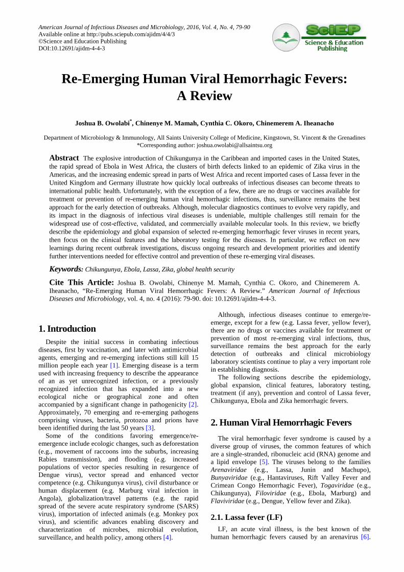
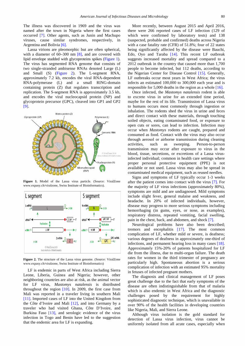
Lassa virions are pleomorphic but are often spherical, with a diameter of 60–300 nm [8], and are covered with lipid envelope studded with glycoproteins spikes (Figure 1). The virus has segmented RNA genome that consists of two single-stranded ambisense RNAs denoted Large (L) and Small (S) (Figure 2). The L-segment RNA, approximately 7.2 kb, encodes the viral RNA-dependent RNA-polymerase (L) and a small RING-domain containing protein (Z) that regulates transcription and replication. The S-segment RNA is approximately 3.5 kb, and encodes the viral nucleocapsid protein (NP) and glycoprotein precursor (GPC), cleaved into GP1 and GP2 [9].
Figure 1. Model of the Lassa virus particle. (Source: ViralZone www.expasy.ch/viralzone, Swiss Institute of Bioinformatics).
Figure 2. The structure of the Lassa virus genome. (Source: ViralZone www.expasy.ch/viralzone, Swiss Institute of Bioinformatics)
LF is endemic in parts of West Africa including Sierra Leone, Liberia, Guinea and Nigeria; however, other neighboring countries are also at risk, as the animal vector for LF virus, Mastomys natalensis is distributed throughout the region [10]. In 2009, the first case from Mali was reported in a traveler living in southern Mali [11]. Imported cases of LF into the United Kingdom from the Côte d’Ivoire and Mali [12], and into Germany by a traveler who had visited Ghana, Côte D’Ivoire, and Burkina Faso [13], and serologic evidence of the virus infection in Togo and Benin have led to the suggestion that the endemic area for LF is expanding.
More recently, between August 2015 and April 2016, there were 266 reported cases of LF infection (129 of which were confirmed by laboratory tests) and 138 (suspected, probable and confirmed) deaths across Nigeria with a case fatality rate (CFR) of 51.8%; four of 22 states being significantly affected by the disease were Bauchi, Edo, Oyo and Taraba [14]. This recent LF outbreak suggests increased mortality and spread compared to a 2012 outbreak in the country that caused more than 1,700 people to become infected, but 112 deaths, according to the Nigerian Center for Disease Control [15]. Generally, LF outbreaks occur most years in West Africa; the virus infects an estimated 100,000 to 300,000 each year and is responsible for 5,000 deaths in the region as a whole [16].
Once infected, the Mastomys natalensis rodent is able to excrete virus in urine for an extended time period, maybe for the rest of its life. Transmission of Lassa virus to humans occurs most commonly through ingestion or inhalation. The rodents shed the virus in urine and feces and direct contact with these materials, through touching soiled objects, eating contaminated food, or exposure to open cuts or sores, can lead to infection. Infection may occur when Mastomys rodents are caught, prepared and consumed as food. Contact with the virus may also occur through aerosol or airborne transmission during cleaning activities, such as sweeping. Person-to-person transmission may occur after exposure to virus in the blood, tissue, secretions, or excretions of a Lassa virus-infected individual; common in health care settings where proper personal protective equipment (PPE) is not available or not used. Lassa virus may also be spread in contaminated medical equipment, such as reused needles.
Signs and symptoms of LF typically occur 1-3 weeks after the patient comes into contact with the virus [7]. For the majority of LF virus infections (approximately 80%), symptoms are mild and are undiagnosed. Mild symptoms include slight fever, general malaise and weakness, and headache. In 20% of infected individuals, however, disease may progress to more serious symptoms including hemorrhaging (in gums, eyes, or nose, as examples), respiratory distress, repeated vomiting, facial swelling, pain in the chest, back, and abdomen, and shock [7].
Neurological problems have also been described; tremors and encephalitis [17]. The most common complication of LF, whether mild or severe, is deafness; various degrees of deafness in approximately one-third of infections, and permanent hearing loss in many cases [18]. Approximately 15%-20% of patients hospitalized for LF die from the illness, due to multi-organ failure. The death rates for women in the third trimester of pregnancy are particularly high. Spontaneous abortion is a serious complication of infection with an estimated 95% mortality in fetuses of infected pregnant mothers.
The diagnosis and clinical management of LF poses great challenge due to the fact that early symptoms of the disease are often indistinguishable from that of malaria which is also endemic in West Africa and the diagnostic challenges posed by the requirement for highly sophisticated diagnostic technique, which is unavailable in over 90% of the health facilities in developing countries like Nigeria, Mali, and Sierra Leone.
Although virus isolation is the gold standard for detection of Lassa virus infection, virus cannot be uniformly isolated from all acute cases, especially when
81 American Journal of Infectious Diseases and Microbiology
the sample is obtained late in the course of illness [19]. In addition, the viral cultivation period is long (7 to 10 days) and this procedure requires a high containment laboratory with good laboratory practices.
The combined enzyme-linked immunosorbent serologic (ELISA)/Ag assay which detects IgM and IgG antibodies as well as Lassa antigen appears to be highly sensitive and specific for the diagnosis of LF [19]. The antigen detection assay offers a particular advantage in providing early diagnosis as well as prognostic information. However, breaks in the cold chain during storage of the virus in serum have resulted in the deterioration of Lassa virus while the more heat-stable antibodies remained. This has resulted in apparent false-positive serologic results [19].
Sensitive reverse transcription-polymerase chain reaction (RT-PCR) assays exist and have application in the early stage of disease, however, issues of strain variation, cross contamination, and expense pose practical problems for use in the developing countries where LF is endemic [20].
Ribavirin, an antiviral drug, has been used with success in LF patients, especially, when given early in the course of the illness [20]. However, drug administration with ribavirin poses significant stress to the physician as it requires the use of several ampoules of the drug, depending on the weight of the patient. Patients also receive supportive care consisting of maintenance of appropriate fluid and electrolyte balance, oxygenation and blood pressure, as well as treatment of any other complicating infections.
Primary transmission of the Lassa virus from its host to humans can be prevented by avoiding contact with Mastomys rodents, putting food away in rodent-proof containers, discouraging rodents from entering homes and not using them as a food source. When caring for patients with LF, nosocomial routes can be avoided by taking preventive precautions against contact with patient secretions. These precautions include wearing protective clothing, such as masks, gloves, gowns, and goggles; using infection control measures, such as complete equipment sterilization; and isolating infected patients from contact with unprotected persons until the disease has run its course.
Currently, there is no licensed vaccine for humans against the Lassa virus and research and development have been hampered by the high cost of non-human primate (NHP) animal models and by biocontainment requirements (BSL-4). In addition, genetic diversity among Lassa virus strains is the highest among the Arenaviridae, and causes a great challenge for vaccine development. Although progress in vaccine development has been less impressive, at least three vaccine platforms (recombinant vaccinia, Mopeia/Lassa reassortant (ML29), and recombinant vesicular stomatitis virus (VSV); VSVΔG/LASVGP vaccine, provided vaccine candidates that were successful when tested in NHP animals [21], the only relevant model for human LF, under the Animal Rule [22]. However, vaccinia virus is no longer acceptable in Africa with high prevalence of HIV (due to the potential safety issues associated with the immunosuppressive phenotype of the vector), but Mopeia/Lassa reassortant (ML29) and rVSV vaccines have some room for further development, in spite of safety issues surrounding their potential use [21].
Although mice do not accurately model human LF disease, they can provide an economical assay to determine vaccine potency via the capacity of vaccine candidates to elicit protective cell-mediated immune (CMI) responses. Evaluation of Lassa virus vaccine immunogenicity in the CBA/J-ML29 mouse model has been reported recently [23]. A single intraperitoneal immunization of CBA/J mice with ML29 protected animals against a lethal homologous intracerebral challenge with 588 LD. While ML29 is currently the most advanced Lassa vaccine candidate inducing sterilizing, cross-protective immunity and acting as a post-exposure countermeasure, at least 5 years will be required for clinical development of this vaccine [21].
2.2. Chikungunya Fever Chikungunya fever, an arthropod-borne virus (arbovirus)
disease, is acute febrile illness often with severe, debilitating poly-arthralgias [24]. In the Makonde language, “Chikungunya” meaning “that which bends up” [25] refers to the crippling arthritis associated with serious disease caused by infection with the virus.
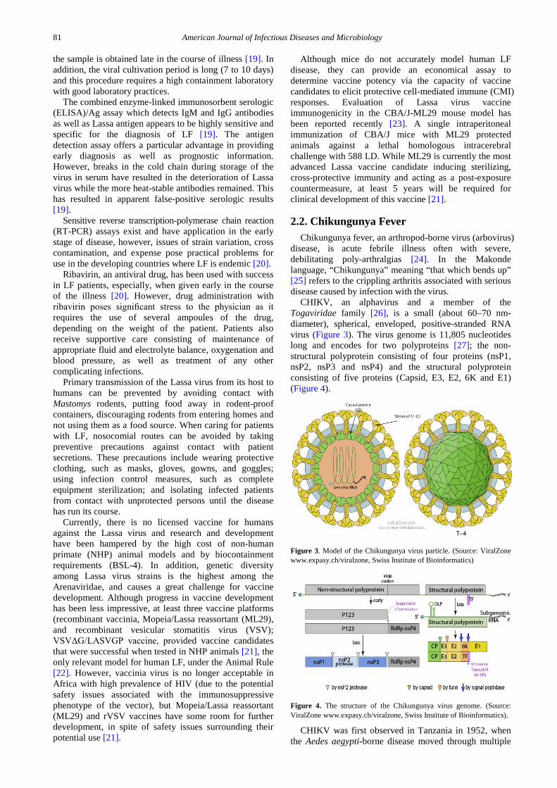
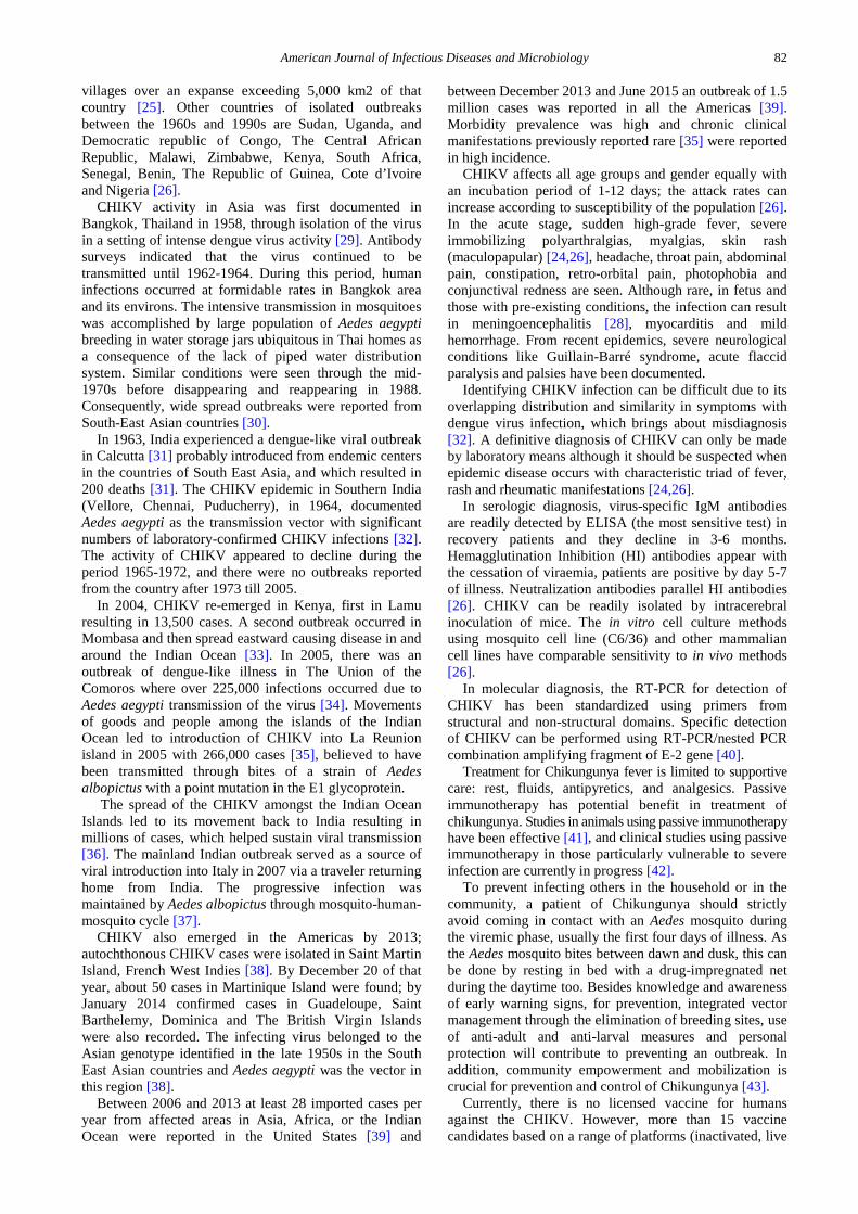
CHIKV, an alphavirus and a member of the Togaviridae family [26], is a small (about 60–70 nm-diameter), spherical, enveloped, positive-stranded RNA virus (Figure 3). The virus genome is 11,805 nucleotides long and encodes for two polyproteins [27]; the non-structural polyprotein consisting of four proteins (nsP1, nsP2, nsP3 and nsP4) and the structural polyprotein consisting of five proteins (Capsid, E3, E2, 6K and E1) (Figure 4).
Figure 3. Model of the Chikungunya virus particle. (Source: ViralZone www.expasy.ch/viralzone, Swiss Institute of Bioinformatics)
Figure 4. The structure of the Chikungunya virus genome. (Source: ViralZone www.expasy.ch/viralzone, Swiss Institute of Bioinformatics).
CHIKV was first observed in Tanzania in 1952, when the Aedes aegypti-borne disease moved through multiple
American Journal of Infectious Diseases and Microbiology 82
villages over an expanse exceeding 5,000 km2 of that country [25]. Other countries of isolated outbreaks between the 1960s and 1990s are Sudan, Uganda, and Democratic republic of Congo, The Central African Republic, Malawi, Zimbabwe, Kenya, South Africa, Senegal, Benin, The Republic of Guinea, Cote d’Ivoire and Nigeria [26].
CHIKV activity in Asia was first documented in Bangkok, Thailand in 1958, through isolation of the virus in a setting of intense dengue virus activity [29]. Antibody surveys indicated that the virus continued to be transmitted until 1962-1964. During this period, human infections occurred at formidable rates in Bangkok area and its environs. The intensive transmission in mosquitoes was accomplished by large population of Aedes aegypti breeding in water storage jars ubiquitous in Thai homes as a consequence of the lack of piped water distribution system. Similar conditions were seen through the mid-1970s before disappearing and reappearing in 1988. Consequently, wide spread outbreaks were reported from South-East Asian countries [30].
In 1963, India experienced a dengue-like viral outbreak in Calcutta [31] probably introduced from endemic centers in the countries of South East Asia, and which resulted in 200 deaths [31]. The CHIKV epidemic in Southern India (Vellore, Chennai, Puducherry), in 1964, documented Aedes aegypti as the transmission vector with significant numbers of laboratory-confirmed CHIKV infections [32]. The activity of CHIKV appeared to decline during the period 1965-1972, and there were no outbreaks reported from the country after 1973 till 2005.
In 2004, CHIKV re-emerged in Kenya, first in Lamu resulting in 13,500 cases. A second outbreak occurred in Mombasa and then spread eastward causing disease in and around the Indian Ocean [33]. In 2005, there was an outbreak of dengue-like illness in The Union of the Comoros where over 225,000 infections occurred due to Aedes aegypti transmission of the virus [34]. Movements of goods and people among the islands of the Indian Ocean led to introduction of CHIKV into La Reunion island in 2005 with 266,000 cases [35], believed to have been transmitted through bites of a strain of Aedes albopictus with a point mutation in the E1 glycoprotein.
The spread of the CHIKV amongst the Indian Ocean Islands led to its movement back to India resulting in millions of cases, which helped sustain viral transmission [36]. The mainland Indian outbreak served as a source of viral introduction into Italy in 2007 via a traveler returning home from India. The progressive infection was maintained by Aedes albopictus through mosquito-human-mosquito cycle [37].
CHIKV also emerged in the Americas by 2013; autochthonous CHIKV cases were isolated in Saint Martin Island, French West Indies [38]. By December 20 of that year, about 50 cases in Martinique Island were found; by January 2014 confirmed cases in Guadeloupe, Saint Barthelemy, Dominica and The British Virgin Islands were also recorded. The infecting virus belonged to the Asian genotype identified in the late 1950s in the South East Asian countries and Aedes aegypti was the vector in this region [38].
Between 2006 and 2013 at least 28 imported cases per year from affected areas in Asia, Africa, or the Indian Ocean were reported in the United States [39] and
between December 2013 and June 2015 an outbreak of 1.5 million cases was reported in all the Americas [39]. Morbidity prevalence was high and chronic clinical manifestations previously reported rare [35] were reported in high incidence.
CHIKV affects all age groups and gender equally with an incubation period of 1-12 days; the attack rates can increase according to susceptibility of the population [26]. In the acute stage, sudden high-grade fever, severe immobilizing polyarthralgias, myalgias, skin rash (maculopapular) [24,26], headache, throat pain, abdominal pain, constipation, retro-orbital pain, photophobia and conjunctival redness are seen. Although rare, in fetus and those with pre-existing conditions, the infection can result in meningoencephalitis [28], myocarditis and mild hemorrhage. From recent epidemics, severe neurological conditions like Guillain-Barré syndrome, acute flaccid paralysis and palsies have been documented.
Identifying CHIKV infection can be difficult due to its overlapping distribution and similarity in symptoms with dengue virus infection, which brings about misdiagnosis [32]. A definitive diagnosis of CHIKV can only be made by laboratory means although it should be suspected when epidemic disease occurs with characteristic triad of fever, rash and rheumatic manifestations [24,26].
In serologic diagnosis, virus-specific IgM antibodies are readily detected by ELISA (the most sensitive test) in recovery patients and they decline in 3-6 months. Hemagglutination Inhibition (HI) antibodies appear with the cessation of viraemia, patients are positive by day 5-7 of illness. Neutralization antibodies parallel HI antibodies [26]. CHIKV can be readily isolated by intracerebral inoculation of mice. The in vitro cell culture methods using mosquito cell line (C6/36) and other mammalian cell lines have comparable sensitivity to in vivo methods [26].
In molecular diagnosis, the RT-PCR for detection of CHIKV has been standardized using primers from structural and non-structural domains. Specific detection of CHIKV can be performed using RT-PCR/nested PCR combination amplifying fragment of E-2 gene [40].
Treatment for Chikungunya fever is limited to supportive care: rest, fluids, antipyretics, and analgesics. Passive immunotherapy has potential benefit in treatment of chikungunya. Studies in animals using passive immunotherapy have been effective [41], and clinical studies using passive immunotherapy in those particularly vulnerable to severe infection are currently in progress [42].
To prevent infecting others in the household or in the community, a patient of Chikungunya should strictly avoid coming in contact with an Aedes mosquito during the viremic phase, usually the first four days of illness. As the Aedes mosquito bites between dawn and dusk, this can be done by resting in bed with a drug-impregnated net during the daytime too. Besides knowledge and awareness of early warning signs, for prevention, integrated vector management through the elimination of breeding sites, use of anti-adult and anti-larval measures and personal protection will contribute to preventing an outbreak. In addition, community empowerment and mobilization is crucial for prevention and control of Chikungunya [43].
Currently, there is no licensed vaccine for humans against the CHIKV. However, more than 15 vaccine candidates based on a range of platforms (inactivated, live
83 American Journal of Infectious Diseases and Microbiology
attenuated, live vectored, chimeric, virus-like particle [VLP], subunit protein, DNA) are currently in preclinical and clinical development [44,45]. Three of these Chikungunya vaccine candidates have reached phase-II clinical development status; TSI-GSD-218 (U.S. Army Medical Research Institute of Infectious Diseases/Salk Institute for Biological Studies), VRC-CHKVLP059-00-VP (National Institute of Allergy and Infectious Diseases (NIAID), and MV-CHIK (Themis Bioscience GmbH/Institut Pasteur) [44,45]. In a phase II trial, TSI-GSD-218, a live, attenuated virus, was reported to develop viral resistance in 98% of those tested after 28 days and 85% still showed resistance after one year [46]. However, ~10% of people reported transient joint pain, and attenuation was found to be due to two mutations in the E2 glycoprotein [47]. The vaccine candidate VRC-CHKVLP059-00-VP, a VLP, has been shown in a phase I trial in healthy adults to possess an excellent safety profile and strong immunogenicity for low (10 µg), medium (20 µg) and high (40 µg) dose cohorts (3 intramuscular [i.m.] immunizations each, without adjuvant, at 0, 4 and 20 weeks) [48]. Phase II studies (clinicaltrials.gov NCT02562482) on VRC-CHKVLP059-00-VP were scheduled to begin at multiple sites in the Caribbean in October, 2015 [44]; status update is pending study completion and data analysis. A Phase 1 dose-escalation trial of MV-CHIK vaccine candidate, a recombinant measles virus (Schwarz strain) expressing CHIKV structural proteins, was reported to be well tolerated in healthy adults, without significant effects on tolerability or immunogenicity associated with pre-existing immunity to measles virus [49]. Alternative vaccine strategies including IRES-based attenuation, chimeric CHIK vaccines based on other attenuated alphavirus backbones, or those based on VSV, adenovirus or poxvirus (MVA) vectors expressing CHIKV structural proteins show efficacy in mouse models [44,45] but further development of these candidates beyond the preclinical status appears uncertain.
2.3. Ebola Virus (EBOV) Ebola infection is caused by a virus of the family
Filoviridae, genus Ebolavirus [50]. Five virus species have been identified; Ebola virus (Zaire ebolavirus), Sudan Virus (Sudan ebolavirus), Taï Forest virus (Taï Forest ebolavirus; previously referred to as Côte d’Ivoire ebolavirus), and Bundibugyo virus (Bundibugyo ebolavirus); these four cause diseases in human. Reston virus (Reston ebolavirus), the fifth, has caused disease only in non-human primates.
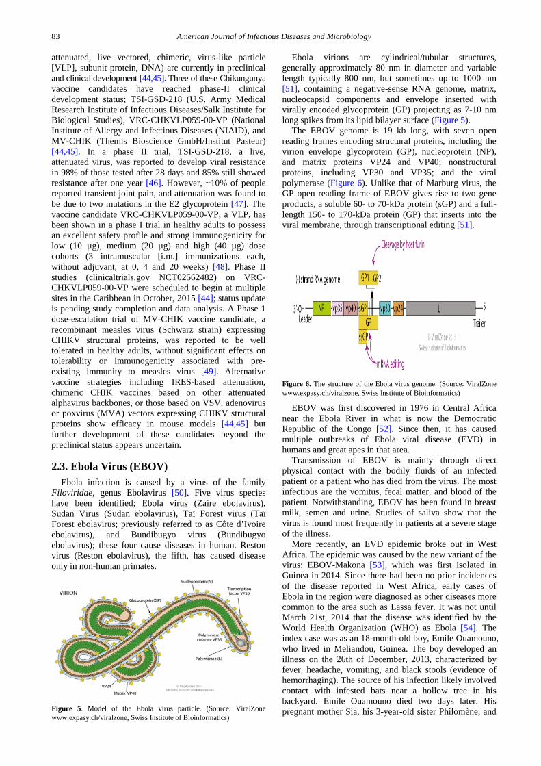
Figure 5. Model of the Ebola virus particle. (Source: ViralZone www.expasy.ch/viralzone, Swiss Institute of Bioinformatics)
Ebola virions are cylindrical/tubular structures, generally approximately 80 nm in diameter and variable length typically 800 nm, but sometimes up to 1000 nm [51], containing a negative-sense RNA genome, matrix, nucleocapsid components and envelope inserted with virally encoded glycoprotein (GP) projecting as 7-10 nm long spikes from its lipid bilayer surface (Figure 5).
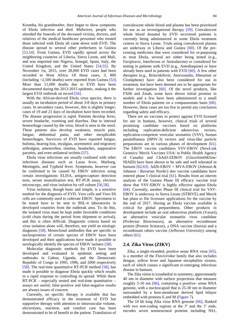
The EBOV genome is 19 kb long, with seven open reading frames encoding structural proteins, including the virion envelope glycoprotein (GP), nucleoprotein (NP), and matrix proteins VP24 and VP40; nonstructural proteins, including VP30 and VP35; and the viral polymerase (Figure 6). Unlike that of Marburg virus, the GP open reading frame of EBOV gives rise to two gene products, a soluble 60- to 70-kDa protein (sGP) and a full-length 150- to 170-kDa protein (GP) that inserts into the viral membrane, through transcriptional editing [51].
Figure 6. The structure of the Ebola virus genome. (Source: ViralZone www.expasy.ch/viralzone, Swiss Institute of Bioinformatics)
EBOV was first discovered in 1976 in Central Africa near the Ebola River in what is now the Democratic Republic of the Congo [52]. Since then, it has caused multiple outbreaks of Ebola viral disease (EVD) in humans and great apes in that area.
Transmission of EBOV is mainly through direct physical contact with the bodily fluids of an infected patient or a patient who has died from the virus. The most infectious are the vomitus, fecal matter, and blood of the patient. Notwithstanding, EBOV has been found in breast milk, semen and urine. Studies of saliva show that the virus is found most frequently in patients at a severe stage of the illness.
More recently, an EVD epidemic broke out in West Africa. The epidemic was caused by the new variant of the virus: EBOV-Makona [53], which was first isolated in Guinea in 2014. Since there had been no prior incidences of the disease reported in West Africa, early cases of Ebola in the region were diagnosed as other diseases more common to the area such as Lassa fever. It was not until March 21st, 2014 that the disease was identified by the World Health Organization (WHO) as Ebola [54]. The index case was as an 18-month-old boy, Emile Ouamouno, who lived in Meliandou, Guinea. The boy developed an illness on the 26th of December, 2013, characterized by fever, headache, vomiting, and black stools (evidence of hemorrhaging). The source of his infection likely involved contact with infested bats near a hollow tree in his backyard. Emile Ouamouno died two days later. His pregnant mother Sia, his 3-year-old sister Philomène, and
American Journal of Infectious Diseases and Microbiology 84
Koumba, his grandmother, then began to show symptoms of Ebola infection and died. Midwives, people who attended the funerals of the deceased victims, doctors, and relatives of the medical healthcare personnel who treated those infected with Ebola all came down with EVD. The disease spread to several other prefectures in Guinea [53,54]. From Guinea, EVD rapidly spread across the neighboring countries of Liberia, Sierra Leone, and Mali, and was imported into Nigeria, Senegal, Spain, Italy, the United Kingdom, and the United States [54,55]. By November 1st, 2015, over 28,000 EVD cases had been recorded in West Africa. Of those cases, 3, 800 (including >2,500 deaths) were reported from Guinea [53]. More than 11,000 deaths due to EVD have been documented during the 2013-2015 epidemic, making it the largest EVD outbreak on record [56].
With the African-derived Ebola virus species, there is usually an incubation period of about 3-8 days in primary cases. In secondary cases, however, this is slightly longer; cases of 19 and 21 days of incubation have been recorded. The disease progression is rapid. Patients develop fever, severe headache, vomiting and diarrhea. Due to internal hemorrhage caused by the virus, blood is seen in the stool. These patients also develop weakness, muscle pain, fatigue, abdominal pains, and other inexplicable hemorrhages. Survivors of EVD have reported fatigue, bulimia, hearing loss, myalgias, asymmetric and migratory arthralgias, amenorrhea, tinnitus, headaches, suppurative parotitis, and even unilateral orchitis [57].
Ebola virus infections are usually confused with other infectious diseases such as Lassa fever, Marburg, Meningitis, and Typhoid fever. Symptoms, however can be confirmed to be caused by EBOV infection using certain investigations: ELISA, antigen-capture detection tests, serum neutralization test, RT-PCR assay, electron microscopy, and virus isolation by cell culture [56,58].
Virus isolation, though basic and simple, is a sensitive method for the diagnosis of EVD. Vero cells and Vero E6 cells are commonly used to cultivate EBOV. Specimens to be tested have to be sent to BSL-4 laboratories in developed countries from the outbreak area. To do this, the isolated virus must be kept under favorable conditions (cold chain during the period from shipment to arrival), and this is often difficult. Diagnostic criteria based on virus isolation alone will, therefore, not yield an etiologic diagnosis [58]. Monoclonal antibodies that are specific to nucleoproteins of certain species of EBOV have been developed and their applications have made it possible to serologically identify the species of EBOV isolates [58].
Molecular diagnostic methods for EVD have been developed and evaluated in epidemic setting and outbreaks in Gabon, Uganda, and the Democratic Republic of Congo in 1995, 1996, and 2000 respectively [58]. The real-time quantitative RT-PCR method [58], has made it possible to diagnose Ebola quickly which results in a rapid response to controlling its spread. While these RT-PCR – especially nested and real-time quantitative – assays are useful, false-positive and false-negative results are always issues of concern.
Currently, no specific therapy is available that has demonstrated efficacy in the treatment of EVD but supportive therapy with attention to intravascular volume, electrolytes, nutrition, and comfort care has been demonstrated to be of benefit to the patient. Transfusion of
convalescent whole blood and plasma has been prioritized for use as an investigational therapy [59]. Convalescent whole blood donated by EVD recovered patients is currently being administered in some Ebola treatment centres in Sierra Leone. Trials using convalescent plasma are underway in Liberia and Guinea [60]. Of the pre-existing medicines that were considered for re-purposing to treat Ebola, several are either being tested (e.g., Favipiravir, Interferons or Amiodarone) or considered for testing in patients with EVD (e.g., Amodiaquine) or have already been used in patients with EVD [60]. A few other therapies (e.g., Brincidofovir, Atorvostatin, Irbesartan or Clomiphene) have also been considered for use in treatment, but have been deemed not to be appropriate for further investigation [60]. Of the novel products, like FX06 and Zmab, some have shown initial promise in models and a few have been administered to a small number of Ebola patients on a compassionate basis [60]. However, these cases are too few to permit any conclusion regarding safety and efficacy.
There are no vaccines to protect against EVD licensed for use in humans, however, clinical trials of several promising candidate vaccines based on platforms including replication-deficient adenovirus vectors, replication-competent vesicular stomatitis (VSV), human parainfluenza (HPIV-3) vectors and virus-like particle preparations are in various phases of development [61]. The EBOV vaccine candidates VSV-EBOV (NewLink Genetics/ Merck Vaccines USA/ in Public Health Agency of Canada) and ChAd3-ZEBOV (GlaxoSmithKline/ NIAID) have been shown to be safe and well tolerated in humans [62,63]. Ad26-EBOV + MVA-EBOV (Johnson & Johnson / Bavarian Nordic) duo vaccine candidates have entered phase I clinical trial [61]. Results from an interim analysis of the Guinea Phase III efficacy vaccine trial show that VSV-EBOV is highly effective against Ebola [64]. Currently, another Phase III clinical trial for VSV-EBOV is underway in Sierra Leone (STRIVE) and Merck has plans to file licensure applications for the vaccine by the end of 2017. Having an Ebola vaccine available is critical for global preparedness. Other products in development include an oral adenovirus platform (Vaxart), an alternative vesicular stomatitis virus candidate (Profectus Biosciences), an alternative recombinant protein (Protein Sciences), a DNA vaccine (Inovia) and a recombinant rabies vaccine (Jefferson University) among others [61].
2.4. Zika Virus (ZIKV) Zika, a single-stranded, positive sense RNA virus [65],
is a member of the Flaviviridae family that also includes dengue, yellow fever and Japanese encephalitis viruses, each of which causes a significant re-emerging infectious disease in humans.
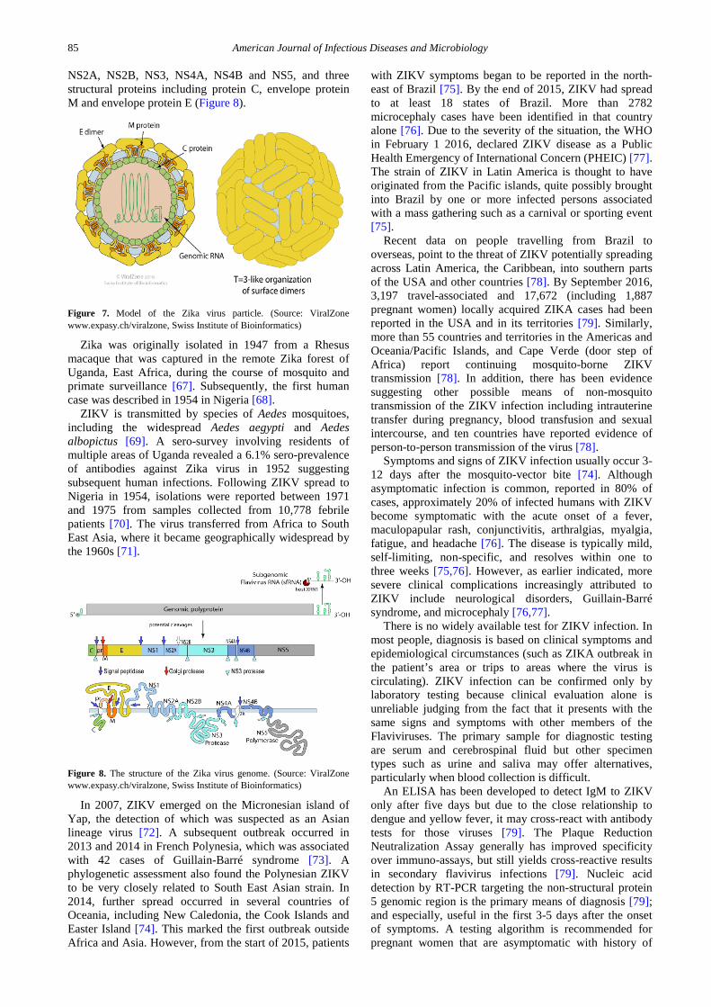
The Zika virion is icosahedral in symmetry, approximately 40 nm in diameter with surface projections that measure roughly 5-10 nm [66], containing a positive- sense RNA genome, with a nucleocapsid that is 25-30 nm in diameter surrounded by a host-membrane derived lipid bilayer embedded with proteins E and M (Figure 7).
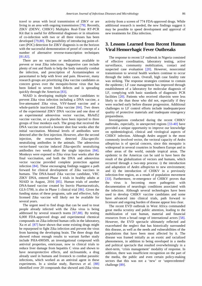
The 10 kb long Zika virus RNA genome [66], flanked with two non-coding regions at the 5′ and the 3′ ends, encodes seven nonstructural proteins including NS1,
85 American Journal of Infectious Diseases and Microbiology
NS2A, NS2B, NS3, NS4A, NS4B and NS5, and three structural proteins including protein C, envelope protein M and envelope protein E (Figure 8).
Figure 7. Model of the Zika virus particle. (Source: ViralZone www.expasy.ch/viralzone, Swiss Institute of Bioinformatics)
Zika was originally isolated in 1947 from a Rhesus macaque that was captured in the remote Zika forest of Uganda, East Africa, during the course of mosquito and primate surveillance [67]. Subsequently, the first human case was described in 1954 in Nigeria [68].
ZIKV is transmitted by species of Aedes mosquitoes, including the widespread Aedes aegypti and Aedes albopictus [69]. A sero-survey involving residents of multiple areas of Uganda revealed a 6.1% sero-prevalence of antibodies against Zika virus in 1952 suggesting subsequent human infections. Following ZIKV spread to Nigeria in 1954, isolations were reported between 1971 and 1975 from samples collected from 10,778 febrile patients [70]. The virus transferred from Africa to South East Asia, where it became geographically widespread by the 1960s [71].
Figure 8. The structure of the Zika virus genome. (Source: ViralZone www.expasy.ch/viralzone, Swiss Institute of Bioinformatics)
In 2007, ZIKV emerged on the Micronesian island of Yap, the detection of which was suspected as an Asian lineage virus [72]. A subsequent outbreak occurred in 2013 and 2014 in French Polynesia, which was associated with 42 cases of Guillain-Barré syndrome [73]. A phylogenetic assessment also found the Polynesian ZIKV to be very closely related to South East Asian strain. In 2014, further spread occurred in several countries of Oceania, including New Caledonia, the Cook Islands and Easter Island [74]. This marked the first outbreak outside Africa and Asia. However, from the start of 2015, patients
with ZIKV symptoms began to be reported in the north-east of Brazil [75]. By the end of 2015, ZIKV had spread to at least 18 states of Brazil. More than 2782 microcephaly cases have been identified in that country alone [76]. Due to the severity of the situation, the WHO in February 1 2016, declared ZIKV disease as a Public Health Emergency of International Concern (PHEIC) [77]. The strain of ZIKV in Latin America is thought to have originated from the Pacific islands, quite possibly brought into Brazil by one or more infected persons associated with a mass gathering such as a carnival or sporting event [75].
Recent data on people travelling from Brazil to overseas, point to the threat of ZIKV potentially spreading across Latin America, the Caribbean, into southern parts of the USA and other countries [78]. By September 2016, 3,197 travel-associated and 17,672 (including 1,887 pregnant women) locally acquired ZIKA cases had been reported in the USA and in its territories [79]. Similarly, more than 55 countries and territories in the Americas and Oceania/Pacific Islands, and Cape Verde (door step of Africa) report continuing mosquito-borne ZIKV transmission [78]. In addition, there has been evidence suggesting other possible means of non-mosquito transmission of the ZIKV infection including intrauterine transfer during pregnancy, blood transfusion and sexual intercourse, and ten countries have reported evidence of person-to-person transmission of the virus [78].
Symptoms and signs of ZIKV infection usually occur 3-12 days after the mosquito-vector bite [74]. Although asymptomatic infection is common, reported in 80% of cases, approximately 20% of infected humans with ZIKV become symptomatic with the acute onset of a fever, maculopapular rash, conjunctivitis, arthralgias, myalgia, fatigue, and headache [76]. The disease is typically mild, self-limiting, non-specific, and resolves within one to three weeks [75,76]. However, as earlier indicated, more severe clinical complications increasingly attributed to ZIKV include neurological disorders, Guillain-Barré syndrome, and microcephaly [76,77].
There is no widely available test for ZIKV infection. In most people, diagnosis is based on clinical symptoms and epidemiological circumstances (such as ZIKA outbreak in the patient’s area or trips to areas where the virus is circulating). ZIKV infection can be confirmed only by laboratory testing because clinical evaluation alone is unreliable judging from the fact that it presents with the same signs and symptoms with other members of the Flaviviruses. The primary sample for diagnostic testing are serum and cerebrospinal fluid but other specimen types such as urine and saliva may offer alternatives, particularly when blood collection is difficult.
An ELISA has been developed to detect IgM to ZIKV only after five days but due to the close relationship to dengue and yellow fever, it may cross-react with antibody tests for those viruses [79]. The Plaque Reduction Neutralization Assay generally has improved specificity over immuno-assays, but still yields cross-reactive results in secondary flavivirus infections [79]. Nucleic acid detection by RT-PCR targeting the non-structural protein 5 genomic region is the primary means of diagnosis [79]; and especially, useful in the first 3-5 days after the onset of symptoms. A testing algorithm is recommended for pregnant women that are asymptomatic with history of
American Journal of Infectious Diseases and Microbiology 86
travel to areas with local transmission of ZIKV or are living in an area with ongoing transmission [79]. Recently, ZIKV (DENV, CHIKV) Multiplex Real-Time RT-PCR Kit that is useful for differential diagnoses or in situations of co-infection with two or all three viruses has been developed [79,80]. The possibility of introducing point-of-care (POC) detection for ZIKV diagnosis is on the horizon with the successful demonstration of proof of concept of a number of alternative reverse-transcription techniques [81,82].
There are no vaccines or medications available to prevent or treat Zika infections. Supportive care include plenty of rest and fluids to help the body naturally combat the infection, and prescription of Acetaminophen or paracetamol to help with fever and pain. However, many research groups are prioritizing Zika vaccine candidates as concern grows over the mosquito-borne virus that has been linked to severe birth defects and is spreading quickly through the Americas [83].
NIAID is developing multiple vaccine candidates to prevent Zika virus infection including a DNA vaccine, a live-attenuated Zika virus, VSV-based vaccine and a whole-particle inactivated Zika vaccine [84]. Two doses of the experimental ZIKV DNA vaccine and one dose of an experimental adenovirus vector vaccine, RhAd52 vaccine vaccine, or a placebo have been injected to three groups of four monkeys each. The group that received the DNA vaccine received a booster shot four weeks after the initial vaccination. Minimal levels of antibodies were detected after the first injection. However, after the second injection, the researchers detected Zika-specific neutralizing antibodies in the animals. The adenovirus vector-based vaccine induced Zika-specific neutralizing antibodies two weeks after the single injection [84]. Monkeys were exposed to Zika virus four weeks after the final vaccination, and both the DNA and adenovirus vector vaccine provided complete protection against infection [84]. These encouraging findings suggest a path forward for clinical development of Zika vaccines in humans. The DNA-based Zika vaccine candidate, VRC ZIKV DNA, entered Phase 1 trials in healthy adults at NIAID in August, 2016 [85]. Human trials of another DNA-based vaccine created by Inovio Pharmaceuticals, GLS-5700, is also in Phase 1 clinical trial [86]. Given the funding status of these programs, safe and effective, fully licensed Zika vaccine will likely not be available for several years.
The urgent need to find drugs that can be used to treat people already infected with the Zika virus is being addressed by several research teams [87,88]. By testing 6,000 FDA-approved drugs and experimental chemical compounds on Zika-infected human cells in the laboratory, Xu et al. [87] have shown that some existing drugs might be repurposed to fight Zika infection and prevent the virus from harming the developing brain. The three drugs that showed robust enough results to warrant further study include PHA-690509, an investigational compound with antiviral properties, emricasan, now in clinical trials to reduce liver damage from hepatitis C virus and shown to have neuroprotective effects, and niclosamide, a drug already used in humans and livestock to combat parasitic infections, which worked as an antiviral agent in these experiments. In a similar study, Barrows et al. [88] identified over 20 compounds that showed anti-Zika virus
activity from a screen of 774 FDA-approved drugs. While additional research is needed, the new findings suggest it may be possible to speed development and approval of new treatments for Zika infection.
3. Lessons Learned from Recent Human Viral Hemorrhagic Fever Outbreaks
The response to recent LF outbreak in Nigeria consisted of effective coordination, laboratory testing, active surveillance, community mobilization, contact and suspected case evaluation [20]. However, nosocomial transmission to several health workers continue to occur through the index cases. Overall, high case fatality rate was striking. The response strategies continue to contain the epidemic; LF case management has improved through establishment of a laboratory for molecular diagnosis of LF, complying with basic standards of diagnostic PCR facilities [20]. Patients who received ribavirin were less likely to die than those who did not, especially if they were reached early before disease progression. Additional challenges to LF control efforts include inadequate/poor quality of protective materials and inadequate emergency preparedness.
Investigations conducted during the recent CHIKV outbreaks, especially, in unexpected regions [33-40] have provided a unique opportunity to improve our knowledge on epidemiological, clinical and virological aspects of CHIKV infection. Although Aedes aegypti is the most commonly involved vector, the vectoral capacity of Aedes albopictus is of special concern, since this mosquito is widespread in several countries in Southern Europe and in other areas of the world, outside tropical areas. The epidemic in the Americas can be said to have been the result of the globalization of vectors and humans, which occurred through a two-step process: i) the introduction and adaptation of Aedes albopictus to new environment; and ii) the introduction of CHIKV in a previously infection-free region, as a result of population movement [33]. Furthermore, re-emergence of CHIKV proves that the virus is becoming more pathogenic with documentation of neurologic conditions associated with the infection. Although several technologies have been used to develop CHIKV vaccine candidates and some have advanced into clinical trials, path forward to licensure and ongoing burden of disease appear less clear.
The recent EVD outbreak in West Africa commanded great media scrutiny and public attention, leading to the mobilization of vast human, material and financial resources from a broad range of international actors [58]. However, the EVD spectacle ultimately reflected and exacerbated the neglect that has historically surrounded this disease, as well as the needs and vulnerabilities of the populations that have been most affected by it. The disease was framed initially as an exotic and racialized phenomenon, in addition to being enveloped in a media and political spectacle that resulted overwhelmingly in a short-term, ‘crisis management’ modality of response. In addition, there was insufficient recognition on the part of the media, the public and even certain policy-making sectors that this was not a ‘new’ or ‘unprecedented’ challenge [89].

87 American Journal of Infectious Diseases and Microbiology
Due to the high fatality rate (50% -74%) for confirmed EVD cases, the rapid progression to death and the long time required to obtain laboratory results, rapid diagnosis was extremely important for timely triage and treatment of patients [90, 91]. A field validation of the dipstick immunoassay for EVD with 100% sensitivity and 92% specificity in both point-of-care (POC) and reference laboratory testing was only applicable for samples with high virus load [90]. Interestingly, recombinase polymerase amplification (RPA) which overcomes the technical difficulties posed by current amplification methods; no requirement for thermal denaturation of template, operation at a low and constant temperature, without reliance on expensive thermocycler, has been shown to have great potential for POC testing of EVD infections [91].
The increasing pressure to develop targeted therapeutic agents has triggered an intensive debate on the use of drug repurposing in EVD treatment [92]. Although, various readily available biologics (e.g., blood transfusions from EVD survivors) and drugs including antimalarial drug chloroquine and antiarrhythmic agents such as amiodarone, amongst many others have been examined as potential therapeutic agents targeting the Ebola virus life cycle or the associated immune reaction, scientific data on their efficacy is limited and additional investigations to justify their use in the specific treatment of EVD are necessary. Given the lack of a systematic assessment of current evidence, a protocol for a systematic review of therapeutic agents targeting the Ebola virus has been proposed [93].
ZIKV hitherto considered for many years to be of little clinical or epidemiological importance, has occurred now in such an explosive fashion throughout the Americas. In many ways, Zika is another example of a re-emerging infectious disease; an old disease presenting in large numbers and in a new context. Since the recent declaration by the World Health Organization (WHO) that ZIKV is a global health concern, rapid progress has been made to understand its pathogenesis and to develop human in vitro models and animal in vivo models [94-96]. Following clinical observations of ZIKV in fetal brains obtained from infected women [97], ZIKV has been reported to efficiently target human neural progenitor cells (hNPCs) and to attenuate their growth [94]. This finding provides a potential mechanism for ZIKV-induced microcephaly as hNPCs drive the development of the cortex in the human brain. Furthermore, several research groups have shown that ZIKV infection of brain organoids, which are used as three-dimensional (3D) cellular models of early human brain development, leads to reduced thickness of hNPC and neuronal layers, and an overall reduction in organoid [98,99], observations consistent with features of microcephaly [76]. These advancements in understanding how ZIKV causes developmental abnormalities and in preclinical studies have opened up opportunities to develop drug repurposing screens that have enabled discovery of effective compounds with potential to treat or prevent ZIKV infection [87,88].
Given the rapid geographic spread of ZIKV in recent years, a coordinated local, regional, and global effort is needed to generate sufficient resources and political traction to effectively halt and contain further expansion of the current outbreak. Inadequate mosquito control might be implicated as one contributing factor to the
ZIKV spread. The risk of local transmission of ZIKV and the threat of outbreaks is equal for all the countries where mosquitoes susceptible to or competent for ZIKV are found. Finally, ZIKV shows us, people spread mosquito-borne diseases too [78].
4. Conclusions Perhaps the good news from the unwitting importation
of Lassa to Europe, tragic EVD outbreak in West Africa, CHIKV infections in the Americas and ongoing ZIKV infections in Latin America, United States and US territories, and elsewhere is that they may have served as a wake-up call. While short term emergency management, such as vector control, early detection and management of symptoms, education of people traveling to endemic countries about the risk of infections and possible measures to prevent the diseases will hopefully reduce their importation into other countries, a major consideration should be implementation of research strategies for prevention of global pandemics. Although, many research groups and pharmaceutical companies are prioritizing vaccine candidates, there is no Lassa vaccine in the horizon, further clinical development of CHIKV vaccines appears uncertain, and ZIKV vaccines are in early stages of clinical development. Having an Ebola vaccine available is critical for global preparedness and Merck’s VSV-EBOV vaccine is on path to filing licensure applications. The increasing pressure to alleviate patients’ suffering has triggered the use of drug repurposing in the treatment of viral hemorrhagic fevers, and screening programs leading to discovery of potential drugs have emerged, however, a systematic assessment of current evidence is warranted to justify their use in specific treatment. In conclusion, we must do all we can to contain more infectious diseases, make investments in facilities, infrastructure, and personnel development in the field of virology, especially, in developing countries as these are usually the first areas of outbreaks. The world needs a global warning and response system for infectious disease outbreaks.
Statement of Competing Interests The authors have no competing interests.
References [1] Fauci, A. S., Morens, D. M., “The perpetual challenge of
infectious diseases.” N Engl J Med 366: 454-461. Feb. 2012. [2] Morse, S. S., Hughes, J. M., “Developing an integrated
epidemiologic approach to emerging infectious diseases.” Epidemiol Rev 18: 1-3. Feb. 1996.
[3] Olano, J. P., Walker, D. H., “Diagnosing emerging and reemerging infectious diseases. The pivotal role of the pathologist.” Arch Pathol Lab Med 135: 83-91. Jan. 2011.
[4] Dong, J., Olano, J. P., McBride, J. W., Walker, D. H., “Emerging pathogens: Challenges and successes of molecular diagnostics.” JMD 10(3): 185-197. May 2008.
[5] Cleri, D. J., Ricketti, A. J., Porwancher, R. B., Ramos-Bonner, L. S., Vernaleo, J. R., “Viral hemorrhagic fevers: Current status of endemic disease and strategies for control.” Infect Dis Clin North Am 20: 359-393. Jun. 2006.

American Journal of Infectious Diseases and Microbiology 88
[6] Bowen, M. D., Peters, C. J., Nichol, S. T., “Phylogenetic analysis of the Arenaviridae: Patterns of virus evolution and evidence for co-speciation between arenaviruses and their rodent hosts.” Mol Phylogenet Evol 8: 301-316. Dec. 1997.
[7] Frame, J. D., Baldwin, J. M., Gocke, D. J., Troup, J. M., “Lassa fever, a new virus disease of man from West Africa. I. Clinical description and pathological findings.” Am J Trop Med Hyg 19(4):670-676. Jul. 1970.
[8] Swiss Institute of Bioinformatics, “Viralzone: Arenavirus” Sep. 2016. Available: http://viralzone.expasy.org/viralzone/all_by_species/83.html [Accessed Sep. 15 2016].
[9] Lukashevich, I.S. Salvato, M.S. “Lassa Virus Genome.” Current Genomics 7: 351-379. Sep. 2006.
[10] Demby, A. H., Inapogui, A., Kargbo, A., Koninga, J., Kourouma, K., et al., “Lassa fever in Guinea: II. Distribution and prevalence of Lassa virus infection in small mammals.” Vector Borne Zoonotic Dis 1(4):283-297. Dec. 2001.
[11] Atkin, S., Anaraki, S., Gothard, P., Walsh, A., Brown, D., Gopal, R., Hand, J., Morgan, D., “The first case of Lassa fever imported from Mali to the United Kingdom, February 2009.” Euro Surveill 14(1):19145. Mar. 2009.
[12] Sogoba, N., Feldmann, H., Safronetz, D., “Lassa fever in West Africa: Evidence for an expanded region of endemicity.” Zoonoses Public Health 2: 43-47. Sep. 2012.
[13] Günther, S., Emmerich, P., Laue, T., Kühle, O., Asper, M., Jung, A., Grewing, T., ter Meulen, J., Schmitz, H., “Imported Lassa Fever in Germany: Molecular characterization of a new Lassa virus strain.” Emerg. Infect. Dis. 6(5): 466-476. Sep. 2000.
[14] World Health Organization, “WHO calls for early diagnostic tests for Lassa fever.” Emergencies preparedness, response Apr. 2016. Available: http://www.who.int/csr/disease/lassafever/early-diagnostic-lassa-fever/en/. [Accessed May 20, 2016].
[15] World Health Organization, (2012) “Lassa fever in Nigeria.” Global alert and response, Apr. 2012. Available: http://www.who.int/csr/don/2012_04_04/en/. [Accessed May 20, 2016].
[16] Ogbu, O., Ajuluchukwu, E, Uneke, C. J. “Lassa fever in West African sub-region: An overview.” J Vect Borne Dis 44: 1-11. Mar. 2007.
[17] Cummins, D., Bennett, D., Fisher-Hoch, S. P., Farrar, B., Machin, S. J., McCormick, J. B., “Lassa fever encephalopathy: Clinical and laboratory findings.” J Trop Med Hyg 95: 197-201. Jun. 1992.
[18] Cummins, D., McCormick, J. B., Bennet, D., Samba, J. A., Farrar, B., Machin, S. J., Fisher-Hoch, S. P., “Acute sensorineural deafness in Lassa fever.” JAMA 264(16): 2093-2096. Oct. 1990.
[19] Bausch, D. G., Rollin, P. E., Demby, A. H., Coulibaly, M., Kanu, J., Conteh, A. S., Wagoner, K. D., McMullan, L. K., Bowen, M. D., Peters, C. J., Ksiazek, T. G., “Diagnosis and clinical virology of Lassa fever as evaluated by enzyme-linked immunosorbent assay, indirect fluorescent-antibody test, and virus isolation.” J Clin Microbiol 38(7): 2670-2677. Jul. 2000.
[20] Asogun, D. A., Adomeh, D. I., Ehimuan, J., Odia, I., Hass, M., et al., “Molecular diagnostics for Lassa fever at Irrua specialist teaching hospital, Nigeria: Lessons learnt from two years of laboratory operation.” PLoS Negl Trop Dis. 6(9):e1839. Sep. 2012.
[21] Lukashevich, I. S., “Advanced vaccine candidates for Lassa fever.” Viruses 4: 2514-2557. Oct. 2012.
[22] Snoy, P.J., “Establishing efficacy of human products using animals” Veterinary Pathology Online 47: 774-778. Sep. 2010.
[23] Goicocheaa, M.A., Zapata, J.C., Bryant, J., Davis, H., Salvato, M.S., Lukashevich, I.S., “Evaluation of Lassa virus vaccine immuno-genicity in a CBA/J–ML29 mouse model.” Vaccine 30: 1445-1452. Feb. 2012.
[24] Robinson, M. C., “An epidemic of virus disease in Southern Province, Tanganyika Territory, in 1952-53. I. Clinical features.” Trans R Soc Trop Med Hyg 49(1): 28-32. Jan. 1955.
[25] Lumsden, W. H., “An epidemic of virus disease in Southern Province, Tanganyika Territory, in 1952-53 II: General description and epidemiology.” Trans R Soc Trop Med Hyg 49: 33-57. Jan. 1955.
[26] Chhabra, M., Mittal, V., Bhattacharya, D., Rana, U., Lal, S., “Chikungunya fever: A re-emerging viral infection.” Indian J Med Microbiol 26(1): 5-12. Jan. 2008.
[27] Swiss Institute of Bioinformatics, “Viralzone: Alphavirus” Sep. 2016. Available:
http://viralzone.expasy.org/viralzone/all_by_species/625.html [Accessed Sep 15 2016].
[28] Powers, A. M., Logue, H. C., “Changing patterns of chikungunya virus: Re-emerging zoonotic arbovirus.” J Gen Virol 88(9): 2363-2377. May 2007.
[29] Halstead, S. B., Nimmannitya, S., Margiotta, M. R., “Dengue and chikungunya virus infection in man in Thailand, 1962-1964, II: Observations on disease in outpatients.” Am J Trop Med Hyg 18: 972-783. Nov. 1969.
[30] Centers for Disease Control, “Chikungunya fever among US Peace Corps Volunteers - Republic of the Philippines.” MMWR Morb Mortal Wkly Rep 35: 573-574. Sep. 1986.
[31] Sarkar, J. K., Chatterjee, S. N., Chakravarty, S. K., “Haemorrhagic fever in Calcutta: Some epidemiological observations.” Indian J Med Res 52: 651-659. Jul. 1964.
[32] Sharma, H. M., Shanmugham, C. A., Iyer, S.P., Rao, A. R., Kuppuswami, S. A., “Report on a random survey conducted to assess the prevalence of a dengue-like illness in Madras city - 1964.” Indian J Med Res 53: 720-728. Aug. 1965.
[33] Staples, J. E., Breiman, R. F., Powers, A. M., “Chikungunya fever: An epidemiological review of a re-emerging infectious disease.” Clin Infect Dis 49: 942-948. Sep. 2009.
[34] Sang, R. C., Ahmed, O., Faye, O., Kelly, C. L., Yahaya, A. A., et al., “Entomologic investigations of a chikungunya virus epidemic in the Union of the Comoros, 2005.” Am J Trop Med Hyg 78: 77-82. Jan. 2008.
[35] Charrel, R. N., de Lamballerie, X., Raoult, D., “Chikungunya outbreaks - The globalization of vector borne diseases.” N Engl J Med 356(8): 769-771. Feb. 2007.
[36] Lahariya, C., Pradhan, S. K., “Emergence of chikungunya virus in India subcontinent after 32 years: A review.” J Vect Borne Dis 43:151-160. Dec. 2006.
[37] Angelini, R., Finarelli, A. C., Angelini, P., Po, C., Petropulacos, K., et al., “An outbreak of Chikungunya fever in the province of Ravenna, Italy.” Eurosurveillance. 12 (36): pii=3260. Sep. 2007.
[38] Goffart, I. L., Nougairede, A., Cassadou, S., Prat, C., de Lamballerie, X., “Chikungunya in the Americas.” The Lancet 383:514. Feb. 2014.
[39] Fischer, M., Staples, J. E., “Chikungunya virus spreads in the Americas -Caribbean and South America, 2013-2014.” CDC Morb Mortal Wkly Rep 63(22): 500-501. Jun. 2014.
[40] Pfeffer, M., Linssen, B., Parke, M. D., Kinney, R. M., “Specific detection of chikungunya virus using a RT-PCR/nested PCR combination.” J Vet Med B Infect Dis Vet Public Health 49: 49-54. Feb. 2002.
[41] Chu, H., Das, S. C., Fuchs, J. F., Suresh, M., Weaver, S. C., Stinchcomb, D. T., et al., “Deciphering the protective role of adaptive immunity to CHIKV/IRES a novel candidate vaccine against Chikungunya in the A129 mouse model.” Vaccine: 31(33):3353-3360. Jul. 2013.
[42] Hoen B., “Prevention of mother-to-child trans- mission of Chikungunya infection: Clinical evaluation of anti-CHIKV hyperimmune IVIg.” In: Gaps and opportunities in Chikungunya research: Expert consultation on Chikungunya disease in the Americas. [presentation] 11/03/2015; 2015. Available: https://respond.niaid.nih.gov/conferences/chikungunya/documents/9 hoen bruno modified.pdf.
[43] World Health Organization, Regional Office for South-East Asia, “Guidelines for prevention and control of chikungunya fever.” WHO Library Cataloguing-in-Publication data. New Delhi, India. 2009.
[44] Smalley, C., Erasmus J. H., Chesson C. B., Beasley D. W. C., “Status of research and development of vaccines for chikungunya” Vaccine 34: 2976-2981. Mar. 2016.
[45] Weaver, S. C., Osorio J. E., Livengood J. A., Chen R, Stinchcomb D. T., “Chikungunya virus and prospects for a vaccine.” Expert Rev Vaccines 11(9): 1087-1101. Sep. 2012.
[46] Edelman, R., Tacket, C. O., Wasserman, S. S., Bodison, S. A., Perry, J. G., Mangiafico, J. A., “Phase 2 safety and immunogenicity study of live chikungunya virus vaccine TSI-GSD-218.” Am. J. Trop. Med. Hyg. 62(6): 681–685. … 2000.
[47] Gorchakov, R., Wang, E., Leal, G., et al. “Attenuation of chikungunya virus vaccine strain 181/clone 25 is determined by two amino acid substitutions in the E2 envelope glycoprotein.” J. Virol. 86(11): 6084-6096. Jun. 2012.
[48] Chang, L. J., Dowd, K. A., Mendoza, F. H., Saunders, J. G., Sitar, S., Plummer, S. H., et al. “Safety and tolerability of chikungunya
89 American Journal of Infectious Diseases and Microbiology
virus-like particle vaccine in healthy adults: A phase 1 dose-escalation trial.” Lancet 384(9959): 2046-52. Dec. 2014.
[49] Ramsauer, K., Schwameis, M., Firbas, C., Mullner, M., Putnak, R. J., Thomas, S. J., et al. “Immunogenicity, safety, and tolerability of a recombinant measles-virus-based chikungunya vaccine: A randomised, double-blind, placebo-controlled, active-comparator, first-in-man trial. Lancet Infect Dis 15(5): 519-27. May 2015.
[50] Peters, C. J., LeDuc, J. W., “An introduction to Ebola: The virus and the disease.” J of Infect Dis 179(suppl.1): ix-xvi. Feb. 1999.
[51] Swiss Institute of Bioinformatics, “Viralzone: Ebolavirus” Sep. 2016. Available: http://viralzone.expasy.org/viralzone/all_by_species/207.html [Accessed Sep 15 2016].
[52] Johnson, K. M,, Lange, J. V., Webb, P. A., Murphy, F. A., “Isolation and characterization of a new virus (Ebola virus) causing acute hemorr-hagic fever in Zaire.” The Lancet 1: 569-571. Mar. 1977.
[53] Rico, A., Brody, D., Coronado, F., Rondy, M., Fiebig, L., et al., “Epidemiology of epidemic Ebola virus disease in Conakry and surrounding Prefectures, Guinea, 2014-2015.” CDC. 22(2) Feb. 2016. Available: http://wwwnc.cdc.gov/eid/article/22/2/15-1304_article. [Accessed May 22 2016].
[54] World Health Organization, “Emergencies preparedness, response. Origins of the 2014 Ebola epidemic. One year into the Ebola epidemic.” Jan. 2015. Available: http://www.who.int/csr/disease/ebola/one-year-report/virus-origin/en/ [Accessed May 21 2016].
[55] Park, D. J., Dudas, G., Wohl, S., Goba, A., Whitmer, S. L., et al., “Ebola virus epidemiology, transmission, and evolution during seven months in Sierra Leone.” Cell 161(7): 1516-1526. Jun. 2015.
[56] World Health Organization, “Ebola virus disease.” Media centre Fact sheet No 103. Updated Jan. 2016. Available: http://www.who.int/mediacentre/factsheets/fs103/en/. [Accessed 21 May 2016].
[57] King, J. W., Chandrasekar, P. H., “Ebola virus infection clinical presentation.” Medscape. Feb. 2015. Available: http://emedicine.medscape.com/article/216288-clinical [Accessed May 21 2016].
[58] Saijo, M., Niikura, M., Ikegami, T., Kurane, T., Kurata, G. Morikawa, S., “Laboratory diagnostic systems for Ebola and Marburg hemorrhagic fevers developed with recombinant proteins.” Clin Vaccine Immunol 13(4): 444-451. Apr. 2006.
[59] World Health Organization, “Experimental therapies: Growing interest in the use of whole blood or plasma from recovered Ebola patients (convalescent therapies).” Media centre. Ebola situation assessment. Sep. 2014. Available: http://www.who.int/mediacentre/news/ebola/26-september-2014/en/ [Accessed Sep. 18 2016].
[60] World Health Organization, “Ebola vaccines, therapies, and diagnostics.” Essential medicines and health products. Questions and Answers. Oct. 2015. Available: http://www.who.int/medicines/emp_ebola_q_as/en/. [Accessed Sep. 18 2016].
[61] World Health Organization, “Experimental Ebola vaccines.” Ebola situation assessment. Media centre, Oct. 2014. Available: http://www.who.int/mediacentre/news/ebola/01-october-2014/en/ [Accessed Sep. 18 2016].
[62] Agnandji, S.T., Huttner, A., Zinser M.E., Njuguna, P., Dahlke, C., et al., “Phase 1 trials of rVSV Ebola vaccine in Africa and Europe.” N Engl J Med 374: 1647-1660. Apr. 2016.
[63] Tapia, M.D., Sow, S.O., Lyke K. E., Haidara F. C., Diallo, F., et al., “Use of ChAd3-EBO-Z Ebola virus vaccine in Malian and US adults, and boosting of Malian adults with MVA-BN-Filo: a phase 1, single-blind, randomised trial, a phase 1b, open-label and double-blind, dose-escalation trial, and a nested, randomised, double-blind, placebo-controlled trial.” Lancet 16: 31-42. Jan. 2016.
[64] Henao-Restrepo, A.M., Longini, I.M., Egger, M., Dean, N. E., et al., “Efficacy and effectiveness of an rVSV-vectored vaccine expressing Ebola surface glycoprotein: interim results from the Guinea ring vaccination cluster-randomised trial.” www.thelancet.com Published online July 31, 2015.
[65] Kuno, G., Chang, G-J., “Full-length sequencing and genomic characterization of Bagaza, Kedougou, and Zika viruses.” Arch Virol 152: 687-689. Jan. 2007.
[66] Swiss Institute of Bioinformatics, “Viralzone: Zika virus” Sep. 2016. Available:
http://viralzone.expasy.org/viralzone/all_by_species/6756.html. [Accessed Sep 15 2016].
[67] Dick, G. W., Kitchen, S. F., Haddow, A. J., “Zika virus. I. Isolations and serological specificity.” Trans R Soc Trop Med Hyg 46:509-20. Sep. 1952.
[68] MacNamara, F., “Zika virus: a report on three cases of human infection during an epidemic of jaundice in Nigeria.” Trans R Soc Trop Med Hyg 48:139-45. Mar. 1954.
[69] Diallo, D., Sall, A. A, Diagne, C. T., Faye, O., Faye, O., et al., “Zika virus emergence in mosquitoes in southeastern Senegal, 2011.” PLoS One. 9:e109442. Oct. 2014.
[70] Fagbami, A. H., “Zika virus infections in Nigeria: Virological and sero-epidemiological investigations in Oyo State.” J Hyg (Lond) 83(2):213-219. Oct. 1979.
[71] Marchette, N., Garcia, R., Rudnick, A., “Isolation of Zika virus from Aedes aegypti mosquitoes in Malaysia”. Am J Trop Med Hyg 18: 411-415. May 1969.
[72] Lanciotti, R. S., Kosoy, O. L., Laven, J. J,, Velez, J. O., Lambert, A. J., et al., “Genetic and serologic properties of Zika virus associated with an epidemic, Yap State, Micronesia, 2007.” Emerg Infect Dis 14: 1232-1239. Aug. 2008.
[73] Cao-Lormeau, V. M., Roche, C., Teissier, A., Robin, E., Berry, A. L., et al., “Zika virus, French Polynesia, South Pacific, 2013.” Emerg Infect Dis 20: 1085-1086. Jun. 2014.
[74] Alera, M. T., Hermann, L., Tac-An, I. A., Klungthong, C., Rutvisuttinunt, W., et al., “Zika virus infection, Philippines, 2012.” Emerg Infect Dis 21(4): 722-7242. Apr. 2015.
[75] Zanluca, C., de Melo, V. C. A., Mosimann, A. L. P, dos Santos, G. I. V., dos Santos, C. N. D., Luz, K, “First report of autochthonous transmission of Zika virus in Brazil.” Mem Inst Oswaldo Cruz 110: 569-572. Jun. 2015.
[76] Dyer, O.,”Zika virus spreads across America as concerns mount over birth defects.” BMJ.2015; h6983. Dec. 2015.
[77] World Health Organization, “WHO Director-General summarizes the outcome of the Emergency Committee regarding clusters of microcephaly and Guillain-Barré syndrome.” Media centre Feb. 2016. Available: http://www.who.int/mediacentre/news/statements/2016/emergency-committee-zika-microcephaly/en/ [Accessed May 21 2016].
[78] Centers for Disease Control, “Zika virus disease in the United States, 2015–2016.” Apr. 2016. Available: http://www.cdc.gov/zika/geo/united-states.html. [Accessed May 21 2016].
[79] Centers for Disease Control, “Zika virus: Case counts in the US.” Sep. 2016. Available: http://www.cdc.gov/zika/geo/united-states.html. [Accessed Sep 19 2016].
[80] Parreira, R., Centeno-Lima, S., Lopes, A., Portugal-Calisto, D., Constantino, A., Nina, J., “Dengue virus serotype 4 and chikungunya virus coinfection in a traveler returning from Luanda, Angola, January 2014.” Euro Surveill 19(10): pii=20730. Mar. 2014.
[81] Song, J., Mauk, M.G., Hackett, B.A., Cherry, S., Bau, H.H., Liu, C., “Instrument-free point-of-care molecular detection of Zika virus.” Anal. Chem. 2016, 88: 7289-7294. Jun. 2016.
[82] Pardee, K., Green, A. A., Takahashi, M. K., Braff, D., Lambert, G., et al., “Rapid, low-cost detection of Zika virus sing programmable biomolecular components.” Cell 165: 1255-1266. May 2016.
[83] Dawes, B.E., Smalley, C. A., Tiner, B. L., Beasley, D.W.C., Mil, G. N., “Research and development of Zika virus vaccines.” npj Vaccines 1: 16007. Jul. 2016.
[84] Abbink, P., Larocca, R.A., De La Barrera, R. A., Bricault, C. A., Moseley E. T., et al. “Protective efficacy of multiple platforms against Zika virus challenge in rhesus monkeys.” Science 10.1126/science.aah6157. Aug. 2016.
[85] National Institute of Health, “NIH begins testing investigational Zika vaccine in humans.” News Releases. Available: https://www.nih.gov/news-events/news-releases/nih-begins-testing-investigational-zika-vaccine-humans Aug. 2016. [Accessed Sep. 18 2016].
[86] novio Pharmaceuticals, “Inovio Pharmaceuticals doses first subject in Zika vaccine clinical trial.” Globe Newswire, Jul. 2016. Available: http://ir.inovio.com/news/news-releases/news-releases-details/2016/Inovio-Pharmaceuticals-Doses-First-Subject-in-Zika-Vaccine-Clinical-Trial/default.aspx. [Accessed Sep 20 2016].
[87] Xu, M., Lee, E. M., Wen, Z., Cheng, Y., Huang, W. K., et al. “Identification of small-molecule inhibitors of Zika virus infection
American Journal of Infectious Diseases and Microbiology 90
and induced neural cell death via a drug repurposing screen.” Nat Med. 2016 Aug 29.
[88] Barrows, N. J., Campos, R. K., Powell, S. T., Prasanth, K. R., Schott-Lerner, G., et al., “A screen of FDA-approved drugs for inhibitors of Zika virus infection.” Cell Host Microbe 20(2): 259-270. Jul. 2016.
[89] Nunes, J., “Ebola and the production of neglect in global health.” Third World Quarterly 37:542-556. Mar. 2016.
[90] Broadhurst, M. J., Kelly, J. D., Miller, A., Semper, A., Bailey, D., et al., “ReEBOV Antigen Rapid Test kit for point-of-care and laboratory-based testing for Ebola virus disease: a field validation study.” Lancet 386(9996): 867-874. Jun. 2015.
[91] Yang, M., Ke, Y., Wang, X., Ren, H., Liu, W., et al., “Development and evaluation of a rapid and sensitive EBOV-RPA test for rapid diagnosis of Ebola virus disease.” Sci. Rep. 6: 26943. Jun. 2016.
[92] Enserink M. “Debate erupts on repurposed drugs for Ebola.” Science 345: 718-719. Aug. 2014.
[93] Sweiti, H., Ekwunife, O., Jaschinski,T., Lhachimi, S. K., “Repurposed therapeutic agents targeting the Ebola virus: A protocol for a systematic review.” Systematic Reviews 4:171- Nov. 2015.
[94] Tang, H., Hammack, C., Ogden, S. C., Wen, Z., Qian X., et al. “Zika virus infects human cortical neural progenitors and attenuates their growth.” Cell Stem Cell 18: 587-590. May 2016.
[95] Rossi, S. L., Tesh, R. B., Azar, S. R., Muruato, A. E., Hanley, K. A., et al. “Characterization of a novel murine model to study Zika virus.” Am. J. Trop. Med. Hyg. 94: 1362-1369. Mar. 2016.
[96] Li, C., Xu, D., Ye, Q., Hong S., Jiang Y., et al. “Zika virus disrupts neural progenitor development and leads to microcephaly in mice.” Cell Stem Cell 19: 120-126. Jul 2016.
[97] Driggers, R.W., Ho, C.-Y., Korhonen, E.M. Kuivanen, S., Jääskeläinen A.J., et al. “Zika virus infection with prolonged maternal viremia and fetal brain abnormalities.” N. Engl. J. Med. 374: 2142-2151 Jun. 2016.
[98] Qian, X., Nguyen, H. N., Song, M. M., Hadiono, C., Ogden, S. C., et al. “Brain-region-specific organoids using mini-bioreactors for modeling ZIKV exposure.” Cell 165: 1238-1254. May 2016.
[99] Dang, J., Tiwari, S.K., Lichinchi, G., Qin, Y., Patil V. S., et al. “Zika virus depletes neural progenitors in human cerebral organoids through activation of the innate immune receptor TLR3.” Cell Stem Cell 19: 258-265. Jul. 2016.