rdna transcription and cardiac hypertrophy
TRANSCRIPT
rDNA Transcription and Cardiac Hypertrophy WenQin Xie and Lawrence I. Rothblum
A common result of the various stimuli that alter the growth rates of cells is a parallel alteration of the rate of ribosome biogenesis. In many cell types, this mandates an altered rate of ribosomal RNA synthesis. Preliminary studies indicate that rDNA transcription may in fact be regulated at more than one level or step. Our understanding of the process of transcription and the mechanisms by which it is regulated are discussed. (Trends Cardiovasc Med 1993;3:7-11)
Simplistically, cells can grow in one of two ways-they can increase in number or size. The ways in which the various cell types grow can determine the ability of an organ (or an organism) to carry out its physiologic role. For example, over- growth of cardiac fibroblasts or overpro- duction of collagen by these cells can alter the structure of the heart and decrease cardiac output (Chow et al. 1991). However, cardiac growth is a normal developmental process or physi- ologic response. Soon after birth, mam- malian cardiac myocytes complete their last rounds of mitotic growth and under- take growth through hypertrophy (Ra- kusan I984), that is, an increase in cell mass but not an increase in the number of cells. Similarly, in response to any one of numerous stimuli, the adult heart can also undergo hypertrophy. Unfortunately, this process, if uncontrolled, can in- crease the risks of unfavorable cardio- vascular events, decrease cardiac function, and, eventually, lead to cardiac failure.
The process of cardiomyocyte hypertro- phy has been studied in cultured neona- tal and adult cardiomyocytes and has been the subject of recent reviews (Mor- gan et al. 1987). Various stimuli that result in hypertrophy in whole animals or perfused organs also cause hypertro-
WenQin Xie and Lawrence I. Rothblum are at the Weis Center for Research, Geisinger Clinic, 100 North Academy Avenue, Danville, PA 17822-2618, USA.
phy and alter patterns of expression of myocyte contractile protein genes in cultured myocytes. Several mechanisms, involving specific intracellular signals or pathways, have been proposed to explain the altered patterns of gene expression associated with hypertrophic stimuli (Simpson 1989).
A common effect of different hypertro- phic stimuli is the net accumulation of protein. This accumulation is the result of an increased rate of protein synthesis, relative to the rate of degradation [re- viewed in Morgan et al. (1987)]. Associ- ated with the increased rate of protein synthesis is an accumulation of ribo- somes (Morgan et al. 1987). In cultured neonatal myocytes, the increase in ribo- some content, indicative of a greater capacity for protein synthesis, reflects an increased rate of ribosomal RNA (r-RNA) synthesis. Studies by McDermott et al. (1989 and 1991) demonstrated that the increased rate of r-RNA synthesis results horn an increased rate of transcription of the rRNA genes that encode the 45 S precursor of the 18 S, 5.8 S, and 28 S rRNAs (discussed below). Thus, the study of how r-RNA gene (rDNA) transcription is regulated during hypertrophy should provide information for unraveling the signal transduction pathways that result in cardiac hypertrophy.
The goals of this review are to intro- duce the reader to (a) what is known of the basic mechanism of transcription of the rDNA by RNA polymerase I; (b) our understanding of the mechanisms of
how rDNA transcription is regulated; and (c) application of these basic results to studies on skeletal and cardiac my- ocytes.
l Transcription by RNA Polymerase I
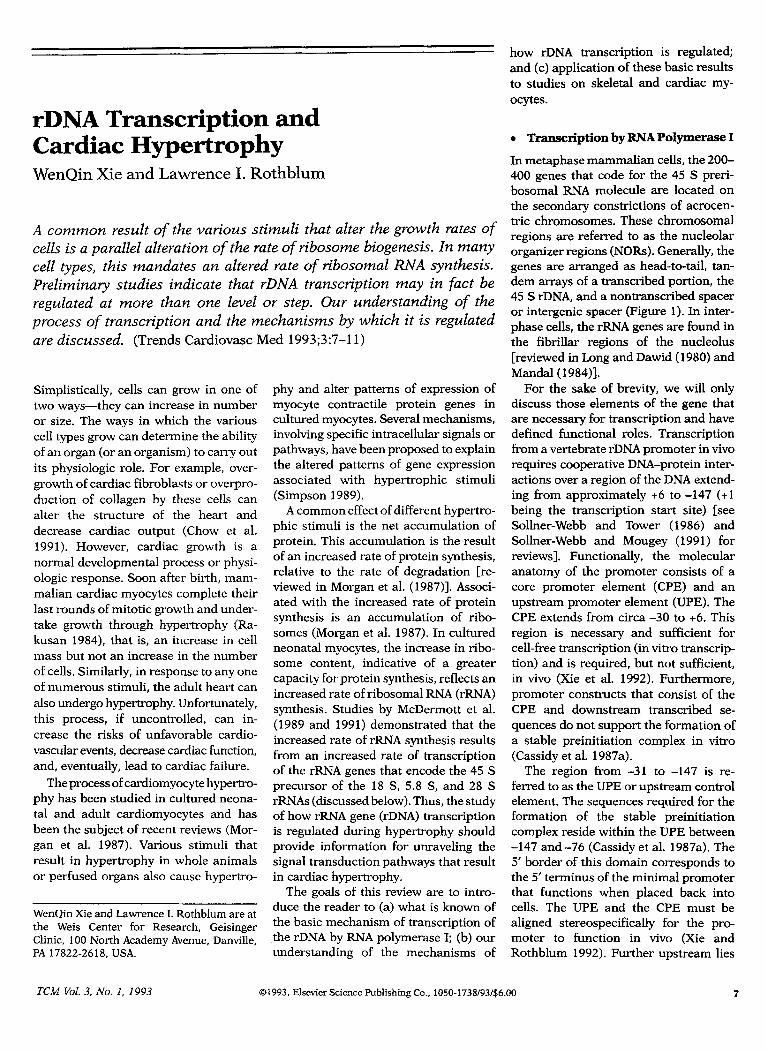
In metaphase mammalian cells, the 200- 400 genes that code for the 45 S preri- bosomal RNA molecule are located on the secondary constrictions of acrocen- tric chromosomes. These chromosomal regions are referred to as the nucleolar organizer regions (NORs). Generally, the genes are arranged as head-to-tail, tan- dem arrays of a transcribed portion, the 45 S rDNA, and a nontranscribed spacer or intergenic spacer (Figure 1). In inter- phase cells, the r-RNA genes are found in the fibrillar regions of the nucleolus [reviewed in Long and Dawid (1980) and Mandal(l984)].
For the sake of brevity, we will only discuss those elements of the gene that are necessary for transcription and have defined functional roles. Transcription from a vertebrate rDNA promoter in vivo requires cooperative DNA-protein inter- actions over a region of the DNA extend- ing from approximately +6 to -147 (+l being the transcription start site) [see Sollner-Webb and Tower (1986) and Sollner-Webb and Mougey (1991) for reviews]. Functionally, the molecular anatomy of the promoter consists of a core promoter element (CPE) and an upstream promoter element (UPE). The CPE extends from circa -30 to +6. This region is necessary and sufficient for cell-free transcription (in vitro transcrip- tion) and is required, but not sufficient, in vivo (Xie et al. 1992). Furthermore, promoter constructs that consist of the CPE and downstream transcribed se- quences do not support the formation of a stable preinitiation complex in vitro (Cassidy et al. 1987a).
The region from -31 to -147 is re- ferred to as the UPE or upstream control element. The sequences required for the formation of the stable preinitiation complex reside within the UPE between -147 and -76 (Cassidy et al. 1987a). The 5’ border of this domain corresponds to the 5’ terminus of the minimal promoter that functions when placed back into cells. The UPE and the CPE must be aligned stereospecifically for the pro- moter to function in vivo (Xie and Rothblum 1992). Further upstream lies
TCM Vol. 3, No. 1, 1993 01993, Elsetier Science Publishing Co., lOSO-1738/93/$6.00 ‘I
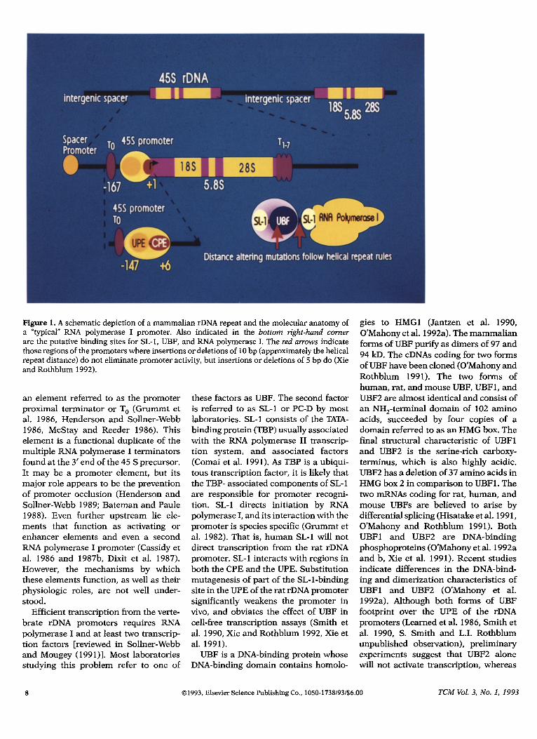
Figure 1. A schematic depiction of a mammalian I-DNA repeat and the molecular anatomy of a “typical” RNA polymerase I promoter. Also indicated in the bottom right-hand corner
are the putative binding sites for SL-1, UBF, and RNA polymerase I. The red arrows indicate those regions of the promoters where insertions or deletions of 10 bp (approximately the helical repeat distance) do not eliminate promoter activity, but insertions or deletions of 5 bp do (Xie and Rothblum 1992).
an element referred to as the promoter proximal terminator or T, (Grummt et al. 1986, Henderson and Sollner-Webb 1986, McStay and Reeder 1986). This element is a functional duplicate of the multiple RNA polymerase I terminators found at the 3’ end of the 45 S precursor. It may be a promoter element, but its major role appears to be the prevention of promoter occlusion (Henderson and Sollner-Webb 1989; Bateman and Paule 1988). Even further upstream lie ele- ments that function as activating or enhancer elements and even a second RNA polymerase I promoter (Cassidy et al. 1986 and 1987b, Dixit et al. 1987). However, the mechanisms by which these elements function, as well as their physiologic roles, are not well under- stood.
Efficient transcription from the verte- brate rDNA promoters requires RNA polymerase I and at least two transcrip- tion factors [reviewed in Sollner-Webb and Mougey (199 I)]. Most laboratories studying this problem refer to one of
these factors as UBF. The second factor is referred to as SL-1 or PC-D by most laboratories. SL-1 consists of the TATA- binding protein (TBP) usually associated with the RNA polymerase II transcrip- tion system, and associated factors (Comai et al. 1991). As TBP is a ubiqui- tous transcription factor, it is likely that the TBP- associated components of SL- 1 are responsible for promoter recogni- tion. SL-1 directs initiation by RNA polymerase I, and its interaction with the promoter is species specific (Grummt et al. 1982). That is, human SL-1 will not direct transcription from the rat rDNA promoter. SL-1 interacts with regions in both the CPE and the UPE. Substitution mutagenesis of part of the SL-l-binding site in the UPE of the rat rDNA promoter significantly weakens the promoter in vivo, and obviates the effect of UBF in cell-free transcription assays (Smith et al. 1990, Xie and Rothblum 1992, Xie et al. 1991).
UBF is a DNA-binding protein whose DNA-binding domain contains homolo-
gies to HMGl (Jantzen et al. 1990, O’Mahony et al. 1992a). The mammalian forms of UBF purify as dimers of 97 and 94 kD. The cDNAs coding for two forms of UBF have been cloned (O’Mahony and Rothblum 1991). The two forms of human, rat, and mouse UBF, UBFl, and UBF2 are almost identical and consist of an NH,-terminal domain of 102 amino acids, succeeded by four copies of a domain referred to as an HMG box. The final structural characteristic of UBFl and UBF2 is the serine-rich carboxy- terminus, which is also highly acidic. UBF2 has a deletion of 37 amino acids in HMG box 2 in comparison to UBFl . The two mRNAs coding for rat, human, and mouse UBFs are believed to arise by differential splicing (Hisatake et al. 199 1, O’Mahony and Rothblum 1991). Both UBFl and UBF2 are DNA-binding phosphoproteins (O’Mahony et al. 1992a and b, Xie et al. 1991). Recent studies indicate differences in the DNA-bind- ing and dimerization characteristics of UBFl and UBF2 (O’Mahony et al. 1992a). Although both forms of UBF footprint over the UPE of the rDNA promoters (Learned et al. 1986, Smith et al. 1990, S. Smith and L.I. Rothblum unpublished observation), preliminary experiments suggest that UBF2 alone will not activate transcription, whereas
8 01993, Elsevier Science Publishing Co., 1050-1738/93/$6.00 TCM Vol. 3, No. 1, 1993
UBF~ will (SD. Smith and L.I. Roth- blum unpublished observations). Unlike SL-1, UBF does not appear to be species specific. The mammalian homologues of UBF are interchangeable, and even forms of UBF isolated from organisms as dis- parate as Xenopus and rat are function- ally exchangeable, albeit weakly (Pikaard et al. 1990).
The addition of UBF to an in vitro transcription system enhances transcrip- tion (Smith et al. 1990, Learned et al. 1986). However, it is not clear whether UBF is an essential component of the transcription complex or whether UBF “only” facilitates the formation of the preinitiation complex. While our labora- tory has reported that extracts that are “UBF depleted” can form a stable preini- tiation complex and initiate transcrip- tion, others have reported that UBF is (McStay et al. 1991), or may be (Bell et al. 1988), required for transcription.
In addition to UBF and SL-1, rRNA synthesis requires a form of RNA poly- merase I that is capable of accurately initiating transcription. When purified from actively dividing cells, only a frac- tion of the total RNA polymerase I activity is capable of participating in specific transcription, as compared with the total activity measured on calf thy- mus DNA. This property has been as- cribed to a modification of one of the polymerase subunits or to the presence of a tightly associated (high-salt resis- tant) transcription initiation factor (dis- cussed below).
l Regulation of rDNA Transcription
The majority of the studies on the mech- anism of regulation of transcription have been carried out with the use of tumor cells grown in tissue culture, or cultures of the soil protozoan Acanthamoeba cas- tellanii that have been exposed to condi- tions, such as serum or nutrient deprivation, or agents, such as the pro- tein synthesis inhibitor cycloheximide, that cause a rapid cessation of rDNA transcription [reviewed in Sollner-Webb and Mougey (199 1 )].
Preliminary studies indicated that rapid cessation of transcription was due to a modification of a fraction of the RNA polymerase I molecules to a form that was incapable of accurately initiating transcription (Bateman and Paule 1986). Subsequent experiments from the labo-
ratories of Ingrid Grummt and Aubrey Thompson identified polymerase-associ- ated activities referred to as either TIF- 1A or TFIC, respectively, which were capable of reactivating inactive polym- erase (Buttgereit et al. 1985, Mahajan et al. 1990a). TFIC was separated from RNA polymerase I, purified to homogene- ity, and its effect on initiation has been studied in detail (Mahajan et al. 1990a and b, Gokal et al. 1990). TFIC is required for initiation and appears to function stoichiometrically, suggesting that the factor is somehow inactivated following a single round of initiation. Further experiments will be required to determine whether TIF- 1A and TFIC are equivalent, how they mediate the inter- action between the preinitiation com- plex and RNA polymerase I, and how TFIC is cycled between the active and inactive states.
In contrast, the transcriptionally ac- tive and inactive forms of Acanthamoeba RNA polymerase I have the same num- ber of subunits (Bateman and Paule 1986). Preliminary results indicate a posttranslational modification of one of the low molecular weight polymerase subunits that determines the ability of the polymerase to form a productive initiation complex (M. Paule, personal communication, 1992). The regulation of the ability of the polymerase itself to participate in transcription appears to be a common regulatory mechanism.
However, the regulation of rDNA tran- scription may also involve the other fac- tors involved in initiation. Our studies have demonstrated that UBF is a phosphoprotein and, when cells in tissue culture are serum starved, the phosphoryla- tion of UBF is decreased significantly, but the total mass of UBF in the cells does not decrease. Further, when the cells are refed serum, the phosphorylation of UBF recovers (O’Mahony et al. 1992b). These results led to the hypothesis that the activity of UBF depends upon its phosphorylation state. Studies with cell- free transcription confirmed this hy- pothesis. Dephosphorylation of UBF greatly reduced its ability to activate transcrip- tion. In indirect immunofluorescence ex- periments, we found that, when cells were serum starved, or postconfluent, UBF appeared to leave the nucleolus and as- sumed a diffuse, nucleo-cytoplasmic stain- ing pattern (O’Mahony et al. 1992b). These results suggest that cells might
regulate transcription by RNA polymerase I by altering the amount, activity, and/or subcellular localization of UBF.
l rRNA Synthesis in Myocytes
Skeletal myoblast differentiation pro- vides a good system to study the regula- tion of polymerase I transcription during a “normal” developmental process. Dur- ing the differentiation of mouse and rat myoblasts into myotubes (fibers), the rate of rRNA accumulation decreases by >75% (Bowman 1987, Larson et al. 1991). This decrease is due to a lower rate of transcription rather than in- creased rates of r-RNA degradation.
However, the total RNA polymerase I activity present in nuclear extracts pre- pared from either proliferating myo- blasts or terminally differentiated myotubes is equivalent (manuscript in
Figure 2. Differentiated L6 myotubes contain less UBF than nondifferentiated L6 myo- blasts. Equivalent amounts of protein from whole cell extracts of myoblasts and myo- tubes were fractionated by SDS-PAGE and analyzed by immunoblot for myosin heavy chain (MHC, top panel) and UBF (bottom panel). The samples shown are from the beginning and end point of the experiments. At intermediate points, where there are re- duced levels (50%60%) of rDNA transcrip- tion, there are also reduced levels of UBF (manuscript in preparation). The methods used were described previously (O’Mahony et al. 1992b). The band identified as UBF was confirmed by electrophoresing a known sam- ple of UBF in an adjacent lane. Also, pre- stained molecular size standards were run in the last lanes of the gel to confirm the molecular sizes of the proteins.
Days in Diff. Medium
-MHC
UBFl -UBF2
TCM Vol. 3, No. 1, I993 01993, Elsevier Science Publishing Co., 1050-1738/93/$6.00
preparation). It is likely then that the reduced level of rDNA transcription can be attributed to the regulation of the factors required for transcription. In collaboration with Dr. Bruce Sells and his colleagues, we have found that less activity characteristic of SL-1 can be recovered from L6 myotubes, than from L6 myoblasts. Further, concomitant with the terminal differentiation of L6 myo- blasts into myotubes, there is a decrease in the amount of the transcription factor UBF (Figure 2).
The mechanisms that control ri- bosomal protein synthesis vary in dif- ferent cell types. For mouse muscle cells, the regulation of ribosomal proteins (S16 and L32) occurs at both transcrip- tional and translational levels (Agrawal and Bowman 1987), and a two- to sixfold reduction of r-protein gene transcription has been reported following terminal differentiation. However, for rat L6 cells, the rates of synthesis of r-protein mRNA and 5 S RNA do not change, and control is mainly at the translational and posttran- scription levels (Larson et al. 1991).
Cultured neonatal cardiac myocytes undergo hypertrophic growth in re- sponse to many of the factors that stimulate hypertrophy in the animal. The signals that mediate this hypertro- phic growth include growth factors, hormones, and mechanical force. Con- traction can also serve as a stimulus for
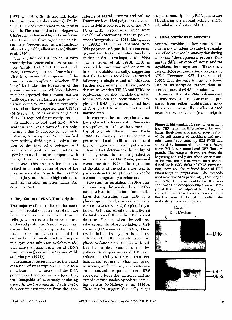
Figure 3. Contracting, cultured, neonatal cardiomyocytes contain more UBF than non- contracting (KCl-arrested) cardiomyocytes. The Western blot of whole cell extracts from contracting (B) and noncontracting ceils (N) was carried out as described previously (O’Ma- hony et al. 1992b). The bands in the photo- graph are UBFl and UBFZ. To control for the protein load, the blots were probed with an a-actin antibody (Sigma, St. Louis, MO). No significant differences in the amounts of actin per lane were observed. Ail other details are the same as in Figure 2.
growth. Neonatal cardiomyocytes cul- tured in serum-free, defined medium contract spontaneously, whereas cells grown in medium supplemented with 50 mM KC1 do not beat (McDermott et al. 1989). After 4 days in culture, the contracting cells accumulate 30% more protein and RNA than do the noncon- tracting cells. For the most part, this is accomplished by increasing the cellular rates of protein and RNA synthesis.
The synthesis of rRNA in this model of hypertrophy has been measured by both pulse and equilibrium labeling of total cellular RNA, and for the 18 S and 28 S rRNAs specifically. McDermott et al. (1989) demonstrated that the fractional rates (KS) of rRNA synthesis were in- creased 53%59% in beating myocytes and that, after 2 days of contracting, the synthesis of both 18 S and 28 S rRNA was accelerated twofold. The fractional rate of degradation of 18 S and 28 S t-RNA was virtually unchanged. These results suggested that the increased rate of t-RNA accumulation was due to either an increased rate of transcription or an increase in the fractional rate of proc- essing. Subsequent nuclear run-on ex- periments demonstrated that the increased rate of rRNA synthesis in contracting cells was due to a higher rate of transcrip- tion (McDermott et al. 199 1).
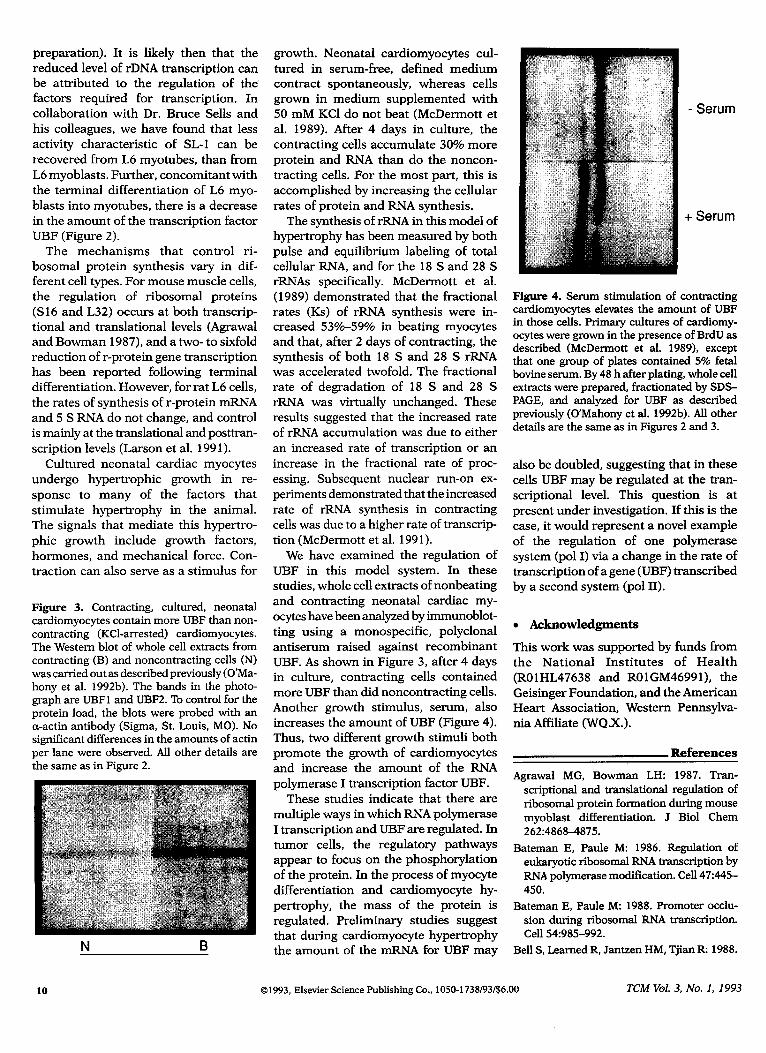
We have examined the regulation of UBF in this model system. In these studies, whole cell extracts of nonbeating and contracting neonatal cardiac my- ocytes have been analyzed by immunoblot- ting using a monospecific, polyclonal antiserum raised against recombinant UBF. As shown in Figure 3, after 4 days in culture, contracting cells contained more UBF than did noncontracting cells. Another growth stimulus, serum, also increases the amount of UBF (Figure 4). Thus, two different growth stimuli both promote the growth of cardiomyocytes and increase the amount of the RNA polymerase I transcription factor UBF.
These studies indicate that there are multiple ways in which RNA polymerase I transcription and UBF are regulated. In tumor cells, the regulatory pathways appear to focus on the phosphorylation of the protein. In the process of myocyte differentiation and cardiomyocyte hy- per-trophy, the mass of the protein is regulated. Preliminary studies suggest that during cardiomyocyte hypertrophy the amount of the mRNA for UBF may
+
Serum
Serum
Figure 4. Serum stimulation of contracting cardiomyocytes elevates the amount of UBF in those cells. Primary cultures of carciiomy- ocytes were grown in the presence of BrdU as described (McDermott et al. 1989), except that one group of plates contained 5% fetal bovine serum. By 48 h after plating, whole cell extracts were prepared, fractionated by SDS- PAGE, and analyzed for UBF as described previously (O’Mahony et al. 1992b). All other details are the same as in Figures 2 and 3.
also be doubled, suggesting that in these cells UBF may be regulated at the tran- scriptional level. This question is at present under investigation. If this is the case, it would represent a novel example of the regulation of one polymerase system (pal I) via a change in the rate of transcription of a gene (UBF) transcribed by a second system (pol II).
0 Acknowledgments
This work was supported by funds from the National Institutes of Health (ROlHL47638 and ROlGM46991), the Geisinger Foundation, and the American Heart Association, Western Pennsylva- nia Affiliate (WQX.).
Agrawal MG, Bowman LH: 1987. Tran- scriptional and transiational regulation of ribosomal protein formation during mouse myobiast differentiation. J Biol Chem 262:4868-4875.
Bateman E, Pauie M: 1986. Regulation of eukaryotic ribosomai RNA transcription by RNA poiymerase modification. Ceil 47:445- 450.
Bateman E, Paule M: 1988. Promoter occlu- sion during ribosomai RNA transcription. Ceil 54985-992.
Bell S, Learned R, Jantzen HM, Tjian R: 1988.
10 01993, ElsevierScience Publishing Co.,1050-1738/93/$6.00 TCM Vol. 3, No. I, 1993
Functional cooperativity between tran- nada K, Song C, Muramatsu M: 1991.
scription factors UBFl and SLl mediates Cloning and structural analysis of cDNA
human ribosomal RNA synthesis. Science and the gene for mouse transcription factor
241:1192-1197. UBF. Nucleic Acids Res 19:4631-4637.
Bowman LH: 1987. rDNA transcription and pre-t-RNA processing during the differentia- tion of a mouse myoblast cell line. Dev Bioi
119:152-163.
Buttgereit D, Pflugfelder G, Grummt I: 1985. Growth-dependent regulation of RRNA syn- thesis is mediated by a transcription initia- tion factor (TIF-1A). Nucleic Acids Res
138165-8180.
Cassidy B, Yang-Yen HF, Rothblum L: 1986.
Transcriptional role of the nontranscribed spacer of rat ribosomal DNA. Mol Cell Biol
612766-2733.
Jantzen HM, Admon A, Bell S. Tjian R: 1990.
Nucleolar transcription factor hUBF con- tains a DNA-binding motif with homology to HMG proteins. Nature 344:830-835.
Larson DE, Zahradka P, Sells BH: 1991. Control points in eukaryotic ribosome bio- genesis. Biochem Cell Biol69:5-22.
Learned R, Learned T, Haltiner M, Tjian R:
1986. Human RRNA transcription is modu- lated by the coordinate binding of two
factors to an upstream control element. Cell
45:847-857.
Cassidy B, Haglund R, Rothblum L: 1987a. Regions upstream from the core promoter of the rat ribosomal gene are required for the formation of a stable transcription initiation complex by RNA polymerase 1 in vitro. Biochim Biophys Acta 909: 133-l 44.
Cassidy B, Yang-Yen HF, Rothblum L: 1987b. Additional RNA polymerase 1 initiation site within the nontranscribed region of the rat rRNA gene. Mol Cell Biol 7:2388-2396.
Chow LH, Yee SP, Pawson T, McManus BM:
199 1. Progressive cardiac fibrosis and my-
ocyte injury in v-fps transgenic mice: a model for primary disorders of connective
tissue in the heart? Lab Invest 64:457-462.
Long E, Dawid I: 1980. Repeated genes in eukaryotes. Annu Rev Biochem 49:727-
764.
Mahajan P, Gokal P, Thompson E: 1990a.
Hormonal regulation of transcription of rDNA: the role of TFIC in formation of initiation complexes. JBiol Chem 265:16,244 16,247.
Mahajan P, Gokal P, Thompson E: 1990b. Hormonal regulation of transcription of rDNA. J Biol Chem 265:16,225-16,233.
Mandal R: 1984. The organization and tran-
scription of eukaryotic ribosomal RNA genes. Prog Nucleic Acids Res Mol Biol 3 1:115-
160.
Comai L, Tanese N, Tjian R: 1991. The
TATA-binding protein and associated fac-
tors are integral components of the RNA polymerase I transcription factor, SL-1.
Cell 68~965-976.
McDermott P, Rothblum L, Smith S, Morgan
H: 1989, Accelerated rates of ribosomal
RNA synthesis during growth of contract-
ing heart cells in culture. J Biol Chem
264:18,220-18,227.
Dixit A, Garg L, Chao W, Jacob S: 1987. An enhancer element in the far upstream spacer
region of rat ribosomal RNA gene. J Biol Chem 262:11,616-11,622.
McDermott P, Carl L. Conner K, Al10 S: 1991.
Transcriptional regulation of ribosomal RNA
synthesis during growth of cardiac my- ocytes in culture. J Biol Chem 266:4409-
4416. Gokal P, Mahajan P, Thompson E: 1990.
Hormonal regulation of transcription of
rDNA: formation of initiated complexes by RNA polymerase I in vitro. J Biol Chem 265:16,234-16,243.
McStay B, Reeder RH: 1986. A termination
site forxenopus RNA polymerase I also acts as an element of an adjacent promoter. Cell 47:913-920.
Grummt I, Roth E, Paule M: 1982. Ribo- somal RNA transcription in vitro is species-
specific. Nature 296:173-174.
Grummt I, Kuhn A, Bartsch I, Rosenbauer
H: 1986. A transcription terminator located upstream of the mouse rDNA initiation site affects rRNA synthesis. Cell 47:901-911.
Henderson S, Sollner-Webb B: 1986. A
transcriptional terminator is a novel ele-
ment of the promoter of the mouse ribo-
somal RNA gene. Cell 43:891-900.
Henderson S, Sollner-Webb B: 1989. The
promoter-proximal rDNA terminator aug-
ments initiation by preventing disruption of
the stable transcription complex caused by polymemse read-in. Genes Dev 3:2 12-223.
Hisatake K, Nishimura T, Maeda Y, Ha-
McStay B, Hu CH, Pikaard CS, Reeder RH:
199 1. XUBF and Rib1 are both required for formation of a stable polymerase I pro- moter complex in X. heuis. EMBO J 10:2297- 2303.
Morgan H, Gordon E, Kira Y, et al.: 1987. Biochemical mechanisms of cardiac hy-
I
Is this your subscription to
, CardiG%scular
Use the bound-in order ca to subscribe to TCM today!
pertrophy. Annu Rev Physiol49:533-543.
O’Mahony D, Rothblum L: 1991. Identifica-
tion of two forms of the RNA polymerase I
transcription factor UBF. Proc Natl Acad
Sci USA 88:3180-3184.
O’Mahony DJ, Smith SD, Xie WQ, Rothblum LI: 1992a. Analysis of the phosphorylation,
DNA-binding and dimerization properties and of the nucleolar fibrillar localization of the RNA polymerase I transcription factors UBFl and UBFZ. Nucleic AcidsRes 20:1301-
1308.
O’Mahony DJ, Xie WQ, Smith SD, Singer HA,
RothblumLI: 1992b. Differentialphosphoryla-
tion and localization of the transcription
factor UBF in vivo in response to serum deprivation. J Biol Chem 267:35-38.
Pikaard C, Pape L, Henderson S, et al.: 1990. Enhancers for RNA polymerase I in mouse ribosomal DNA. Mol Cell Biol 10:4816- 4825.
Rakusan K: 1984. Cardiac growth, matura- tion, and aging. In Zak R, ed. Growth of the Heart in Health and Disease. New York, Raven, pp 131-148.
Simpson PC: 1989. Molecular mechanisms in
myocardial hypertrophy. Heart Failure 5: 113- 129.
Smith S, Oriahi E, Yang-Yen HF, et al.: 1990. Characterization of factors that direct tran-
scription of rat ribosomal DNA. Mol Cell
Biol l&3105-3116.
Sollner-Webb B, Mougey EB: 1991. News for
the nucleolus: rDNA gene expression.
Trends Biochem 16:58-62.
Sollner-Webb B, Tower J: 1986. Transcription
of cloned eukaryotic ribosomal RNA genes.
Annu Rev Biochem 55:801-830.
Xie WQ, Rothblum L: 1992. Domains of the
rat rDNA promoter must be aligned stereo- specifically. Mol Cell Biol 12:1266-1275.
Xie WQ, O’Mahony D, Smith D, Rothblum L: 199 1. Complementary in vivo and in vitro
analyses of the interactions between the cis-acting elements of the rat rDNA pro-
moter. Mol Cell Biochem 104:127-136.
Xie WQ, O’Mahony DJ, Smith SD, Lowe D,
Rothblum L: 1992. Analysis of the rat
ribosomal DNA promoter: characterization of linker-scanning mutants and of the bind-
ing of UBF. Nucleic Acids Res 20:1587-
1592. TCM
TCM Vol. 3, No. I, I993 81993, Elsevier Science Publishing Co.. lOSO-1738193/$6.00