rbcs abnormal morphology. red blood cell morphology normal red blood cells
Post on 20-Dec-2015
238 views
TRANSCRIPT

RBCs Abnormal RBCs Abnormal morphologymorphology

RED BLOOD CELL RED BLOOD CELL MORPHOLOGYMORPHOLOGY
• NORMAL RED BLOOD CELLS


RED BLOOD CELL RED BLOOD CELL MORPHOLOGYMORPHOLOGY
Abnormal erythrocyte Abnormal erythrocyte morphology is found in morphology is found in pathological states that may pathological states that may
bebe : :
- -abnormalities in size abnormalities in size (anisocytosis)(anisocytosis) . .
- -In shape (poikilocytosis)In shape (poikilocytosis) . .
--In hemoglobin content or the In hemoglobin content or the presence of inclusion bodies presence of inclusion bodies in erythrocytein erythrocyte..

RED BLOOD CELL RED BLOOD CELL MORPHOLOGYMORPHOLOGY
HypochromicHypochromic:: A descriptive term applied to a red blood cell with a A descriptive term applied to a red blood cell with a decreased concentration of hemoglobindecreased concentration of hemoglobin..NormochromicNormochromic::
A descriptive term applied to a red blood cell with a A descriptive term applied to a red blood cell with a normal concentration of hemoglobinnormal concentration of hemoglobin . .NormocyticNormocytic::
A descriptive term applied to normal size of RBCA descriptive term applied to normal size of RBCMacrocyticMacrocytic::
A descriptive term applied to a larger than normal A descriptive term applied to a larger than normal red blood cellred blood cell..

11--Variation in erythrocyte Variation in erythrocyte size (anisocytosis)size (anisocytosis)
11--MicrocytosisMicrocytosis::
MorphologyMorphology::
- -Decrease in the red cell size. Red cells are smaller Decrease in the red cell size. Red cells are smaller than ± 7µm in diameter. The nucleus of a small than ± 7µm in diameter. The nucleus of a small lymphocyte (± 8,µm) is a useful guide to the size lymphocyte (± 8,µm) is a useful guide to the size of a red blood cellof a red blood cell..
Found inFound in::
- -Iron deficiency anemia.Iron deficiency anemia.- Thalassaemia.- Thalassaemia.- Sideroblastic anemia.- Sideroblastic anemia.- Lead poisoning.- Lead poisoning.- Anemia of chronic disease- Anemia of chronic disease..

11--Variation in erythrocyte Variation in erythrocyte size (anisocytosis)size (anisocytosis)
CommentComment:: Most erythrocytes Most erythrocytes presented in the picture presented in the picture are microcytes (compare are microcytes (compare with the small with the small lymphocyte). The degree lymphocyte). The degree of hemoglobinization is of hemoglobinization is sufficient. Normal sufficient. Normal platelets and single platelets and single ovalocytes are presentovalocytes are present . .Staining:Staining: MGG MGG MagnificationMagnification:: x 1000 x 1000
11 . .microcytemicrocyte 2. normocyte2. normocyte

11--Variation in erythrocyte Variation in erythrocyte size (anisocytosis)size (anisocytosis)
2-Macrocytosis: MorphologyMorphology::
Increase in the size of a red Increase in the size of a red cell. Red cells are larger than cell. Red cells are larger than 9µm in diameter. May be 9µm in diameter. May be round or oval in shape, the round or oval in shape, the diagnostic significance diagnostic significance being differentbeing different..
Found in:Found in:- Folate and B12 deficiencies - Folate and B12 deficiencies (oval)(oval)- Ethanol (round)- Ethanol (round)- Liver disease (round)- Liver disease (round)- Reticulocytosis (round- Reticulocytosis (round))

II-Variation in hemoglobin II-Variation in hemoglobin contentcontent
11--HypochromasiaHypochromasia::
Morphology:Morphology:Increase in the red cells' central Increase in the red cells' central pallor which occupies more than the pallor which occupies more than the
normal third of the red cell diameternormal third of the red cell diameter..
Found in:Found in: - - Iron deficiencyIron deficiency- Thalassaemia- Thalassaemia any of the conditions leading to any of the conditions leading to MicrocytosisMicrocytosis

II-Variation in hemoglobin II-Variation in hemoglobin contentcontent22 - -PolychromasiaPolychromasia::
MorphologyMorphology::
Red cells stain shades of blue-gray Red cells stain shades of blue-gray as a consequence of uptake of both as a consequence of uptake of both eosin (by hemoglobin) and basic dyes eosin (by hemoglobin) and basic dyes (by residual ribosomal RNA). Often (by residual ribosomal RNA). Often slightly larger than normal red cells slightly larger than normal red cells and round in shape - round and round in shape - round macrocytosismacrocytosis..
Found inFound in::
Any situation with reticulocytosis Any situation with reticulocytosis - for example bleeding, - for example bleeding, hemolysis or response to hemolysis or response to haemostatic factor replacementhaemostatic factor replacement..

III- Variation of red cells III- Variation of red cells shape (Poikilocytosis)shape (Poikilocytosis)
RBCs may have different RBCs may have different shapesshapes : :
11 - -SpherocytosisSpherocytosis:: MorphologyMorphology::
Red cells are more spherical. Lack Red cells are more spherical. Lack the central area of pallor on a stained the central area of pallor on a stained blood filmblood film..
Found in:Found in:- - Hereditary spherocytosisHereditary spherocytosis- Immune haemolytic anemia- Immune haemolytic anemia- Zieve's syndrome- Zieve's syndrome- Microangiopathic haemolytic - Microangiopathic haemolytic anemiaanemia

III- Variation of red cells III- Variation of red cells shape (Poikilocytosis)shape (Poikilocytosis)
22--Target CellsTarget Cells::
Morphology:Morphology:Red cells have an area of increased Red cells have an area of increased staining which appears in the area staining which appears in the area
of central pallorof central pallor..
Found in:Found in:--Obstructive liver diseaseObstructive liver disease- Severe iron deficiency- Severe iron deficiency- Thalassaemia- Thalassaemia- Haemoglobinopathies (S and C)- Haemoglobinopathies (S and C)- Post splenectomy- Post splenectomy

III- Variation of red cells III- Variation of red cells shape (Poikilocytosis)shape (Poikilocytosis)
33 - -OvalocytesOvalocytes::
Morphology:Morphology:oval shape red blood celloval shape red blood cell
Found inFound in::
- -Thalassaemia majorThalassaemia major..
- -Hereditary ovalocytosisHereditary ovalocytosis . .
- -Sickle cell anemiaSickle cell anemia

III- Variation of red cells III- Variation of red cells shape (Poikilocytosis)shape (Poikilocytosis)
44 - -ElliptocytosisElliptocytosis::MorphologyMorphology::
The red cells are oval or The red cells are oval or elliptical in shape. Long axis is elliptical in shape. Long axis is twice the short axistwice the short axis..
Found inFound in::
- -Hereditary elliptocytosisHereditary elliptocytosis- Megaloblastic anemia- Megaloblastic anemia- Iron deficiency - Iron deficiency - Thalassaemia- Thalassaemia- Myelofibrosis- Myelofibrosis

III- Variation of red cells III- Variation of red cells shape (Poikilocytosis)shape (Poikilocytosis)
55 - -Tear Drop CellsTear Drop Cells::MorphologyMorphology::
Red cells shaped like a tear Red cells shaped like a tear drop or peardrop or pear
Found inFound in::
- -Bone marrow fibrosisBone marrow fibrosis- Megaloblastic anemia- Megaloblastic anemia- Iron deficiency- Iron deficiency- Thalassaemia- Thalassaemia

III- Variation of red cells III- Variation of red cells shape (Poikilocytosis)shape (Poikilocytosis)
66 - -Blister cellBlister cell::
MorphologyMorphology:: Have accentric hallow areaHave accentric hallow area..
Found inFound in:: Microangiopathic hemolytic Microangiopathic hemolytic anemiaanemia

III- Variation of red cells III- Variation of red cells shape (Poikilocytosis)shape (Poikilocytosis)
77 - -SchistocytosisSchistocytosis::
MorphologyMorphology::
Fragmentation of the red Fragmentation of the red
cellscells..
Found in:Found in: - - DIC DIC - Micro angiopathic haemolytic - Micro angiopathic haemolytic anemiaanemia- Mechanical haemolytic anemia- Mechanical haemolytic anemia

III- Variation of red cells shape III- Variation of red cells shape (Poikilocytosis)(Poikilocytosis)
88 - -StomatocytosisStomatocytosis::Morphology:Morphology: Red cells with a central linear Red cells with a central linear slit or stoma. Seen as mouth-slit or stoma. Seen as mouth-shaped form in peripheral shaped form in peripheral smearsmear..
Found in:Found in:- - Alcohol excessAlcohol excess- Alcoholic liver disease- Alcoholic liver disease- Hereditary stomatocytosis- Hereditary stomatocytosis- Hereditary spherocytosis- Hereditary spherocytosis

III- Variation of red cells shape III- Variation of red cells shape (Poikilocytosis)(Poikilocytosis)
99 - -Burr (crenation ) cellBurr (crenation ) cell::
MorphologyMorphology::Red cell with uniformly spaced, Red cell with uniformly spaced, pointed projections on their pointed projections on their surfacesurface..
Found in:Found in:- - hemolytic anemiahemolytic anemia
- -UremiaUremia..
- -Megaloblastic anemiaMegaloblastic anemia

III- Variation of red cells shape III- Variation of red cells shape (Poikilocytosis)(Poikilocytosis)
1010 - -KeratocytesKeratocytes ( (horn horn cellcell))::Morphology:Morphology:Part of the cell fuses back leaving Part of the cell fuses back leaving two or three horn-like projections. two or three horn-like projections. The keratocyte is a fragile cell and The keratocyte is a fragile cell and remains in circulation for only a few remains in circulation for only a few hourshours..Found in:Found in:- - UraemiaUraemia- Severe burns- Severe burns- EDTA artifact- EDTA artifact- Liver disease- Liver disease
III- Variation of red cells shape III- Variation of red cells shape (Poikilocytosis)(Poikilocytosis)
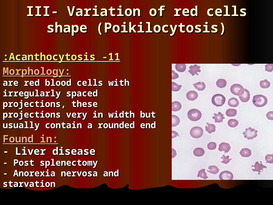
1111 - -AcanthocytosisAcanthocytosis::
Morphology:Morphology:are red blood cells with irregularly are red blood cells with irregularly spaced projections, these spaced projections, these projections very in width but projections very in width but usually contain a rounded endusually contain a rounded end
Found in:Found in:- - Liver disease Liver disease - Post splenectomy- Post splenectomy- Anorexia nervosa and starvation- Anorexia nervosa and starvation

III- Variation of red cells shape III- Variation of red cells shape (Poikilocytosis)(Poikilocytosis)
1212 - -Sickle CellsSickle Cells::
MorphologyMorphology::
Sickle shaped red cellsSickle shaped red cells Found inFound in::
Hb-S diseaseHb-S disease

III- Variation of red cells shape III- Variation of red cells shape (Poikilocytosis)(Poikilocytosis)
1313 - -Rouleaux FormationRouleaux Formation::MorphologyMorphology::
Stacks of RBC's resembling a Stacks of RBC's resembling a stack of coinsstack of coins..Found inFound in::
- -HyperfibrinogenaemiaHyperfibrinogenaemia- Hyperglobulinaemia- Hyperglobulinaemia

III- Variation of red cells shape III- Variation of red cells shape (Poikilocytosis)(Poikilocytosis)
1414 - -Red cell-agglutinationRed cell-agglutination::
MorphologyMorphology:: Irregular clumps of red cellsIrregular clumps of red cells
Found in:Found in:- - Cold agglutininsCold agglutinins- Warm autoimmune - Warm autoimmune hemolysishemolysis

III- Variation of red cells shape III- Variation of red cells shape (Poikilocytosis)(Poikilocytosis)
15- Nucleated red 15- Nucleated red blood cells.blood cells.
These red blood cells are These red blood cells are released from the bone released from the bone marrow early into the marrow early into the blood stream, due to the blood stream, due to the need for oxygen. Normal need for oxygen. Normal red blood cells do not red blood cells do not contain a nucleus on a contain a nucleus on a peripheral smear. peripheral smear.

IV -Erythrocyte inclusion IV -Erythrocyte inclusion bodiesbodies
11 - -Howell-Jolly Howell-Jolly BodiesBodies::Morphology:Morphology:Small round cytoplasmic Small round cytoplasmic red cell inclusion with red cell inclusion with same staining same staining characteristics as nucleicharacteristics as nuclei Found in:Found in:- - Post splenectomyPost splenectomy- Megaloblastic anemia- Megaloblastic anemia
IV -Erythrocyte inclusion IV -Erythrocyte inclusion bodiesbodies
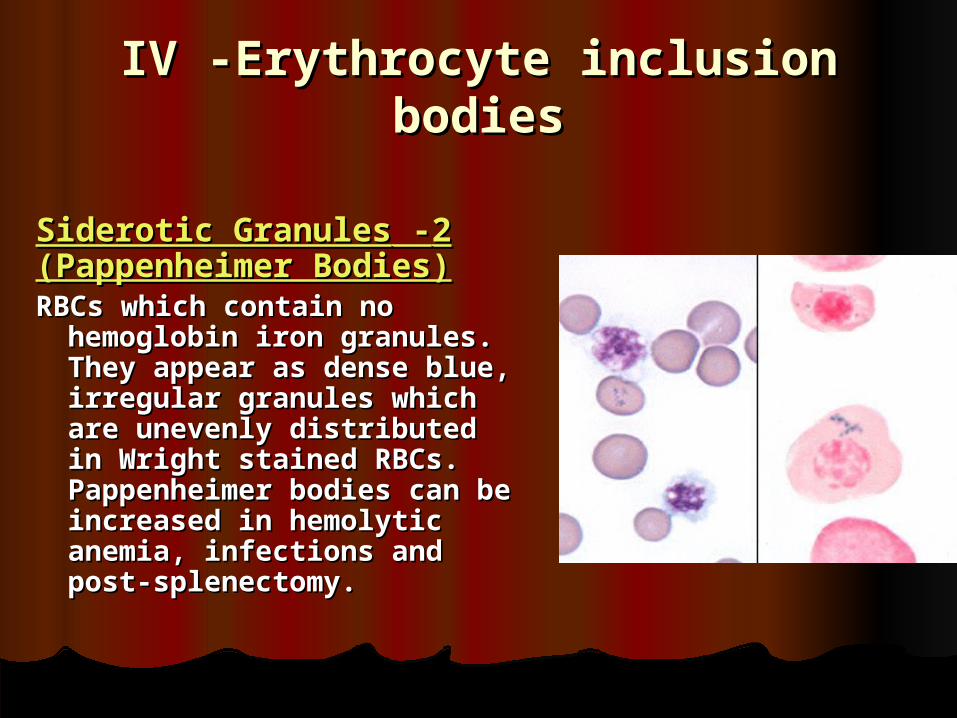
22 - -Siderotic Granules Siderotic Granules (Pappenheimer (Pappenheimer Bodies)Bodies)RBCs which contain no RBCs which contain no
hemoglobin iron granules. hemoglobin iron granules. They appear as dense blue, They appear as dense blue, irregular granules which are irregular granules which are unevenly distributed in unevenly distributed in Wright stained RBCs. Wright stained RBCs. Pappenheimer bodies can be Pappenheimer bodies can be increased in hemolytic increased in hemolytic anemia, infections and post-anemia, infections and post-splenectomy.splenectomy.

IV -Erythrocyte inclusion IV -Erythrocyte inclusion bodiesbodies
33 - -Basophilic stipplingBasophilic stippling:: Morphology:Morphology:Considerable numbers of Considerable numbers of small basophilic inclusions in small basophilic inclusions in red cellsred cells..
Found in:Found in:- - ThalassaemiaThalassaemia- Megaloblastic anemia- Megaloblastic anemia- Hemolytic anemia - Hemolytic anemia - Liver disease- Liver disease- Heavy metal poisoning- Heavy metal poisoning..
IV -Erythrocyte inclusion IV -Erythrocyte inclusion bodiesbodies
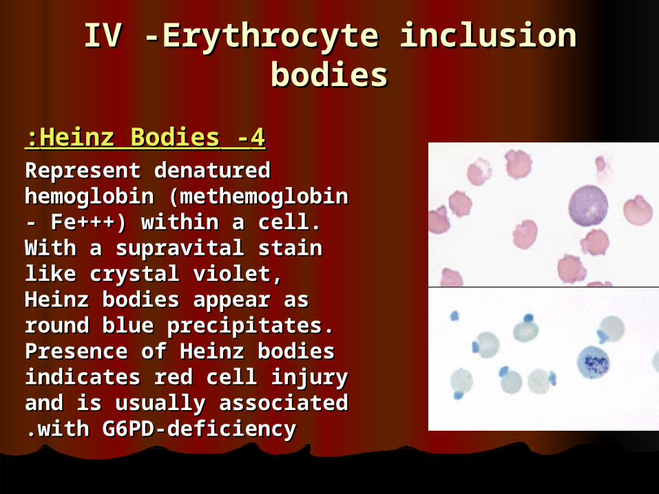
44 - -Heinz BodiesHeinz Bodies::Represent denatured hemoglobin Represent denatured hemoglobin (methemoglobin - Fe+++) (methemoglobin - Fe+++) within a cell. With a supravital within a cell. With a supravital stain like crystal violet, Heinz stain like crystal violet, Heinz bodies appear as round blue bodies appear as round blue precipitates. Presence of Heinz precipitates. Presence of Heinz bodies indicates red cell injury bodies indicates red cell injury and is usually associated with and is usually associated with G6PD-deficiencyG6PD-deficiency..

IV -Erythrocyte inclusion IV -Erythrocyte inclusion bodiesbodies
55 - -Cabot RingsCabot Rings:: Reddish-blue threadlike rings Reddish-blue threadlike rings in RBCs of severe anemia's. in RBCs of severe anemia's. These are remnants of the These are remnants of the nuclear membrane and nuclear membrane and appear as a ring or figure 8 appear as a ring or figure 8 pattern. Very rare finding in pattern. Very rare finding in patients with Megaloblastic patients with Megaloblastic anemia, severe anemia's, anemia, severe anemia's, lead poisoning, and lead poisoning, and dyserythropoiesisdyserythropoiesis..

IV -Erythrocyte inclusion IV -Erythrocyte inclusion bodiesbodies
66 - -Parasites of Red CellParasites of Red Cell::
are protozoan parasites are protozoan parasites which occur in many which occur in many species of birds and are species of birds and are the cause of avian the cause of avian malaria. Transmitted by malaria. Transmitted by mosquitoes, infection mosquitoes, infection with with PlasmodiumPlasmodium can be can be a cause of hemolytic a cause of hemolytic anemiaanemia

RBCs Abnormal RBCs Abnormal morphologymorphology
Depiction of red blood Depiction of red blood cell morphologies cell morphologies that may appear on that may appear on a peripheral smear, a peripheral smear, showingshowing : :
))AA ( (basophilic stippling, basophilic stippling, (B) Howell-Jolly bodies, (B) Howell-Jolly bodies, (C) Cabot's ring bodies (C) Cabot's ring bodies
and (D) Heinz's bodiesand (D) Heinz's bodies . .