rb intrinsically promotes erythropoiesis by coupling cell...
TRANSCRIPT
Rb intrinsically promotes erythropoiesisby coupling cell cycle exitwith mitochondrial biogenesisVijay G. Sankaran,1,2 Stuart H. Orkin,1,2,3,4 and Carl R. Walkley1,2
1Division of Hematology/Oncology, Children’s Hospital Boston, Harvard Medical School, Boston, Massachusetts 02115,USA; 2Department of Pediatric Oncology, Dana-Farber Cancer Institute, Harvard Stem Cell Institute, Harvard MedicalSchool, Boston, Massachusetts 02115, USA; 3Howard Hughes Medical Institute, Boston, Massachusetts 02115, USA
Regulation of the cell cycle is intimately linked to erythroid differentiation, yet how these processes arecoupled is not well understood. To gain insight into this coordinate regulation, we examined the role that theretinoblastoma protein (Rb), a central regulator of the cell cycle, plays in erythropoiesis. We found that Rbserves a cell-intrinsic role and its absence causes ineffective erythropoiesis, with a differentiation block at thetransition from early to late erythroblasts. Unexpectedly, in addition to a failure to properly exit the cell cycle,mitochondrial biogenesis fails to be up-regulated concomitantly, contributing to this differentiation block. Thelink between erythropoiesis and mitochondrial function was validated by inhibition of mitochondrialbiogenesis. Erythropoiesis in the absence of Rb resembles the human myelodysplastic syndromes, wheredefects in cell cycle regulation and mitochondrial function frequently occur. Our work demonstrates howthese seemingly disparate pathways play a role in coordinately regulating cellular differentiation.
[Keywords: Rb; cell cycle; cell differentiation; erythropoiesis; hematopoiesis; mitochondrial biogenesis]
Supplemental material is available at http://www.genesdev.org.
Received October 19, 2007; revised version accepted December 6, 2007.
The process of erythroid differentiation is intimatelycoupled with control of the cell cycle. The earliest com-mitted progenitor of the erythroid lineage is the burst-forming unit (BFU-E), which has a slow rate of prolifera-tion (Gregory and Eaves 1977, 1978). BFU-Es then ma-ture into the colony-forming unit erythroid (CFU-E)cells, and in the process undergo a rapid rise in prolifera-tion. A series of three to five cell divisions ensues andfurther maturation occurs through the proerythroblast,basophilic erythroblast, and polychromatophilic erythro-blast stages. Subsequently, cell cycle exit and post-mi-totic maturation through the orthochromatophilicerythroblast stage occurs. Enucleation gives rise to re-ticulocytes, which then loose remaining organelles tobecome mature erythrocytes that enter the circulation.
While erythroid cell cycle regulation has been exten-sively studied at a descriptive level, many aspects of themolecular control of this process are not well under-stood. Studies in cell lines have suggested a role for ery-throid transcription factors, such as GATA-1, in modu-lating the G1-phase arrest that occurs during erythroidmaturation (Rylski et al. 2003). However, in vivo studieson the role of cell cycle regulators in erythropoiesis have
been limited and are difficult to interpret in light of bothcell-type-intrinsic and -extrinsic roles that these genesmay have in erythropoiesis (Fero et al. 1996; Ciemerychet al. 2002). Insights into the molecular control of theerythroid cell cycle will further an understanding of howdifferentiation within this hematopoietic lineage pro-ceeds. Furthermore, modulation of the erythroid cellcycle with S-phase inhibitors is utilized clinically in at-tempts to increase fetal hemoglobin levels in patientswith hemoglobinopathies (Letvin et al. 1985; Stamatoy-annopoulos 2005). Knowledge pertaining to how the cellcycle is coupled to erythroid cell maturation may lead tomore efficacious therapies for sickle cell anemia and �-thalassemia.
The retinoblastoma protein (Rb) is a central regulatorof the G1-to-S-phase transition of the cell cycle (Classonand Harlow 2002). Rb is one member of the pocket pro-tein family of cell cycle regulators that also include p107and p130. Rb is phosphorylated by activated cyclin-de-pendent kinases (CDKs), and the resulting hyperphos-phorylated form is incapable of binding to its interactionpartners, promoting cell cycle progression. The bestcharacterized among these interaction partners are theE2F family of transcription factors that are bound by hy-pophosphorylated Rb. This interaction represses E2F tar-get genes that are necessary for cell cycle progression.
The role of Rb in erythropoiesis has been the subject ofcontroversy for >15 years. Rb-null mice die at approxi-
4Corresponding author.E-MAIL [email protected]; FAX (617) 632-4367.Article published online ahead of print. Article and publication date areonline at http://www.genesdev.org/cgi/doi/10.1101/gad.1627208.
GENES & DEVELOPMENT 22:463–475 © 2008 by Cold Spring Harbor Laboratory Press ISSN 0890-9369/08; www.genesdev.org 463
Cold Spring Harbor Laboratory Press on August 27, 2019 - Published by genesdev.cshlp.orgDownloaded from
mately E14.5 with several developmental defects, in-cluding a failure of erythroid cell maturation, causing ananemia proposed to be the likely cause of death (Clarkeet al. 1992; Jacks et al. 1992; Lee et al. 1992). Subsequentwork showed that in the context of chimeric mice, Rb-null cells contribute to mature erythrocytes, suggestingthat the red cell defects in the original knockout micewere non-cell-autonomous (Maandag et al. 1994; Wil-liams et al. 1994). This conclusion received additionalsupport from the finding that many embryonic defectscould be explained by a placental defect that occurs inthe absence of Rb with an accompanying deficiency infetal–maternal nutrient exchange (Wu et al. 2003; Wen-zel et al. 2007). Transplantation of fetal liver (FL) ery-throid progenitors demonstrated, however, that erythro-poiesis is not entirely normal in the absence of Rb (Hu etal. 1997), but the interpretation of this finding is com-plicated by possible defects in hematopoietic cells result-ing from impaired placental nutrient exchange. In sum,prior work has not clarified the intrinsic role, if any, ofRb in erythropoiesis.
Additional efforts to resolve these issues have yieldedconflicting results. It was hypothesized that Rb mighthave a cell-type-intrinsic, but non-cell-autonomous rolein erythropoiesis (Whyatt and Grosveld 2002), as appearsto be the case in mice that overexpress the GATA-1 pro-tein (Whyatt et al. 2000). Studies using chimeric andtransplant models suggested that Rb plays a role in stresserythropoiesis, but the gene appeared to be dispensablefor homeostatic erythropoiesis (Spike et al. 2004). Usingin vitro culture approaches, one group proposed that Rbserves an intrinsic role in erythroid maturation (Clark etal. 2004), while other investigators suggested that Rb isdispensible in erythroid cells, but necessary in macro-phages for the formation of an intact erythroid island invitro (Iavarone et al. 2004). To date, no direct experi-ments have been performed to assess an intrinsic role forRb in erythropoiesis in vivo.
Here, we utilize conditional gene inactivation to de-lete Rb specifically in the erythroid lineage, as well as inother hematopoietic compartments. We demonstratethat Rb has a cell-type-intrinsic and cell-autonomousrole in erythropoiesis. Rb loss in the erythroid compart-ment leads to a stable anemia that is attributable to in-effective erythropoiesis. Notably, the mild anemia of themice results from a large degree of in vivo compensationfor this ineffective erythropoiesis. Specifically, Rb is nec-essary for terminal maturation of erythroblasts that oc-curs concomitantly with cell cycle exit. We demonstratethat the intrinsic role for Rb in erythropoiesis is medi-ated by coupling the process of mitochondrial biogenesiswith cell cycle exit during erythropoiesis. Our findingsprovide an in vivo demonstration of how cellular differ-entiation may be efficiently controlled through the co-ordinate regulation of mitochondrial biogenesis with cel-lular proliferation. Additionally, this work gives new in-sight into how ineffective erythropoiesis can occur inhuman diseases like the myelodysplastic syndromes(MDS) and suggests new therapeutic strategies for theseconditions.
Results
Loss of Rb in the erythroid compartment resultsin a moderate anemia
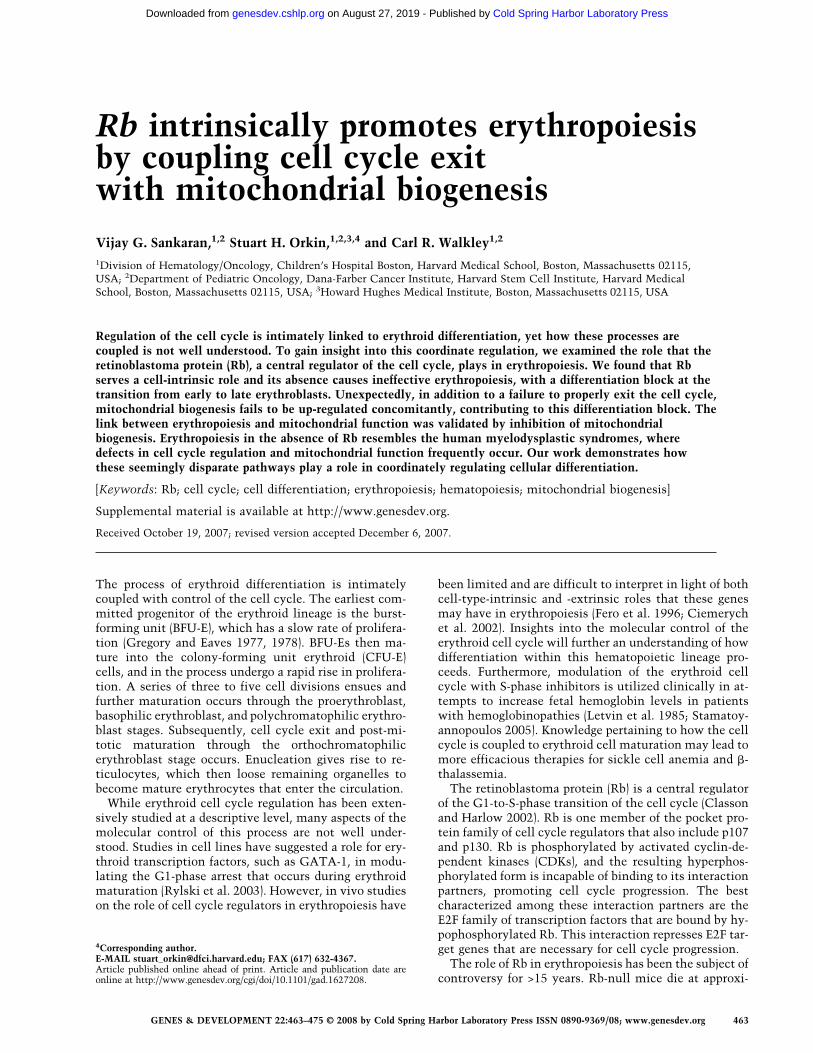
The Rb gene was deleted specifically in the erythroidcompartment by interbreeding pRbfl/fl mice (Sage et al.2003) with erythropoietin receptor GFPcre knock-in(EpoR-GFPcre) heterozygous mice (Heinrich et al. 2004).The EpoR-GFPcre allele has been shown previously todirect quantitative excision of floxed alleles in the ery-throid lineage both during development of the embryoand in the adult (Heinrich et al. 2004; Dumitriu et al.2006; Maetens et al. 2007). Analysis of RNA and DNAfrom sorted early erythroblasts (CD71high/Ter-119high
bone marrow [BM] cells) demonstrated efficient excisionof the floxed Rb allele (Fig. 1A; Supplemental Fig. S1).The expression of the other pocket proteins, p107 andp130, was not appreciably changed. Therefore, increasedexpression of these proteins does not occur as a compen-satory step in response to loss of Rb.
Animals lacking Rb in the erythroid compartmentwere born with expected Mendelian ratios and did notdisplay visible developmental abnormalities. Peripheralblood (PB) from control (EpoR-GFPcre/+; pRbfl/+, hereinpRb�/+) and pRb�/� animals was examined at multipletimes. The pRb�/� animals had occasional Howell-Jollybodies (nuclear remnants) present in most high-powerfields, which were not found in the control animals (Fig.1B, arrows). This indicates that there may have been ei-ther impaired splenic function or a more rapid terminalmaturation of erythroid cells. Moderate and stable ane-mia persisted in adult pRb�/� animals (Fig. 1C). Of note,the anemia was normocytic and normochromic, as as-sessed by blood smears and red cell indices (e.g., MCV:pRb�/+ 53.8 ± 1.13 fL, pRb�/� 55.6 ± 0.64 fL; 8 wk of life).Furthermore, peripheral counts of other lineages werenormal, indicating that the defect was restricted to theerythroid lineage.
Since prior work posited that macrophages in ery-throid islands may require Rb function to support nor-mal erythropoiesis (Iavarone et al. 2004), we deleted Rbin the myeloid lineage (macrophages and granulocytes)using Lysozyme-M-Cre, as has been described previously(Clausen et al. 1999; Walkley et al. 2007). In early adult-hood, no anemia was noted, but with age, an exceedinglymild anemia appeared (Fig. 1D). These findings suggestthat there either may be an additional age-dependent ex-trinsic role for Rb in erythropoiesis, or stochastic exci-sion eventually occurs in earlier hematopoietic progeni-tors, resulting in Rb-null erythroid cells, as has been de-scribed previously (Ye et al. 2003). In support of the latteridea, we found excision of Rb in erythroid progenitorsfrom some aged Lys-M-Cre; Rb fl/fl mice (SupplementalFig. S2). We then combined myeloid and erythroid exci-sion of Rb (using both Lys-M-Cre and EpoR-GFPcre) andfound that there was a similar degree of anemia as wasfound in the EpoR-GFPcre mutant alone (Fig. 1E). Fi-nally, complete excision of Rb in all hematopoietic lin-eages using the interferon-inducible Mx1-Cre (Kuhn etal. 1995; Walkley et al. 2007) revealed that somatic ex-
Sankaran et al.
464 GENES & DEVELOPMENT
Cold Spring Harbor Laboratory Press on August 27, 2019 - Published by genesdev.cshlp.orgDownloaded from
cision in the adult resulted in a similar degree of anemia(Fig. 1F) as with excision confined to the erythroid lin-eage. Therefore, these data indicate that Rb has a cell-intrinsic requirement in erythropoiesis.
We further explored the nature of the anemia that waspresent in erythroid pRb�/� animals, noting a 1.7-foldincrease in the percentage of circulating reticulocytes,which is consistent with a partial physiologic attempt tocompensate for the anemia (Fig. 1G). Erythropoietin lev-els were approximately twofold elevated in the pRb�/�
versus control animals (Fig. 1H), indicating that a sec-ondary response to the anemia was present in these ani-mals and that the compensation is adequate to maintaina stable red cell count throughout life. To evaluatewhether the anemia may in part be due to increased redcell destruction, we measured the life span of red bloodcells (RBCs) in the circulation by labeling these cellswith N-hydroxysuccinimide-biotin, and found that theRBC life span was completely preserved in the pRb�/�
mice (Supplemental Fig. S3). Therefore, these data areconsistent with a cell-intrinsic failure in maturation inthe course of erythropoiesis.
Late-stage ineffective erythropoiesis occursupon deletion of Rb
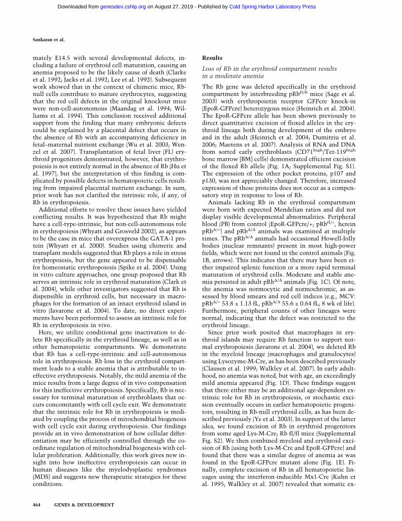
Given the inability of the Rb-null mice to compensategiven their degree of anemia, we then examined hema-topoiesis in the BM. We first noted that the cellularity ofthe BM in pRb�/� and pRb�/+ mice was similar (Supple-mental Fig. S4A). Phenotypic assessment by FACS analy-sis revealed a 1.7-fold increase in the absolute numbersof early erythroblasts (CD71high/Ter-119high) in the BM,but near normal numbers of late erythroblasts (CD71low/Ter-119high) (Fig. 2A). The post-mitotic transition be-tween polychromatophilic erythroblasts and orthochro-matophilic erythroblasts takes place around the transi-tion point between the CD71high and CD71low fractionsof Ter-119high cells (Socolovsky et al. 2001; Zhang et al.2003). Thus, it appears that a relative block in effectivematuration of erythroblasts at this transition is imposedby loss of Rb. Concomitant with the observed expansionof early erythroblasts in the BM, a slight secondary de-crease in BM progenitors of the other major lineages wasfound (Fig. 2B). Histological sections of the marrow con-
Figure 1. Deletion of Rb in the erythroid compartment causes a stable anemia. (A) qRT–PCR of RNA from sorted CD71high/Ter-119high BM cells from EpoR-GFPcre/+; pRbfl/+ (pRb�/+), and EpoR-GFPcre/+; pRbfl/fl (pRb�/�) 8- to 12-wk-old animals (n � 3). (**)P < 0.01. (B) Representative PB smears viewed at 600× magnification. Howell-Jolly bodies are seen in adult pRb�/� mice (arrows). (C–E)RBC counts in EpoR-GFPcre/+ animals (C), LysM-Cre animals (D), and EpoR-GFPcre and LysM-Cre animals (E) at multiple time pointsduring life. (*) P < 0.05; (**) P < 0.01. (F) RBC counts in Mx1-Cre adult animals following somatic deletion. (*) P < 0.05; (**) P < 0.01.(G,H) Reticulocyte percentages (G) and serum erythropoietin levels (H) in EpoR-GFPcre/+ animals (n � 4).
The role of Rb in erythropoiesis
GENES & DEVELOPMENT 465
Cold Spring Harbor Laboratory Press on August 27, 2019 - Published by genesdev.cshlp.orgDownloaded from
firmed the increase in the number of visually identifiableearly erythroblasts present (Fig. 2C). Comparison of earlyerythroid progenitors using methycellulose colony as-says did not reveal differences in progenitor numbers.The frequency of both BFU-E and megakaryocyte-ery-throid (MegE) colonies were similar between the pRb�/�
and pRb�/+ mice (Supplemental Fig. S4B). Furthermore,CFU-E numbers were comparable in the presence or ab-sence of Rb (Supplemental Fig. S4C). We also examinedthe number of earlier progenitors (CFU-GEMM) andfailed to note differences in Rb-null mice compared withcontrols (data not shown).
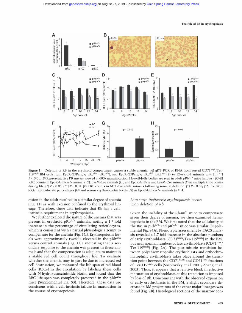
The spleens of the Rb-null mice were considerably en-larged. Spleen weights, as well as total cellularity, wereboth significantly increased (Fig. 3A,B). Single-cell sus-pensions prepared from the spleens were markedly dif-ferent from controls. A greater degree of hemoglobiniza-tion was evident in the single-cell suspensions preparedfrom the pRb�/� mice (Fig. 3C). This observation indi-cated that extensive extramedullary erythropoiesis oc-curs in the spleens of Rb-null animals. Phenotypic analy-sis revealed a 60-fold increase in the number of earlyerythroblasts (CD71high/Ter-119high) present in thespleen (Fig. 3D). The numbers of more mature post-mi-
totic erythroid precursors (CD71low/Ter-119high) wasalso increased by threefold (Fig. 3D). It is important tonote that the relative increase in the early erythroblastfraction in the spleens of the pRb�/� mice is not nearlyaccounted for by the smaller increase in terminal eryth-roblasts. The frequency of BFU-E colonies in the spleenwas slightly reduced (1.7-fold) in the pRb�/� mice com-pared with littermate controls (Supplemental Fig. S4D);however, when the 2.5-fold increase in cellularity istaken into account, this indicates that the number ofBFU-E colonies per spleen is slightly increased. In thecase of the CFU-E colonies from the spleen, a threefoldexpansion in the frequency of colonies was observed(Supplemental Fig. S4E). When the increase in cellularityis also accounted for, it appears that the expansion inerythroblasts in the spleen of the Rb-null mice occurs byincreased proliferation at and subsequent to the CFU-Estage. Examination of histological sections of the spleenwas consistent with this interpretation and revealed adramatic expansion of erythroblasts in the red pulp ofthe spleens of pRb�/� mice, with preservation of thewhite pulp architecture (Fig. 3E).
The data from the spleen and BM of the pRb�/� mice isconsistent with a phenotype of extensive ineffective
Figure 2. Erythroid-specific deletion of Rb causes ineffective erythropoiesis in the BM. (A) FACS profile and mean numbers of early(CD71high/Ter-119high) and late (CD71low/Ter-119high) erythroblasts in the BM of EpoR-GFPcre/+ animals (n � 5). (B) Percentages ofmajor lineages in the BM as assessed using phenotypic surface markers (n � 5). (*) P < 0.05; (**) P < 0.01. (C) Representative histo-logical sections of the BM stained with hemotoxylin and eosin (H&E) viewed at 400× magnification.
Sankaran et al.
466 GENES & DEVELOPMENT
Cold Spring Harbor Laboratory Press on August 27, 2019 - Published by genesdev.cshlp.orgDownloaded from
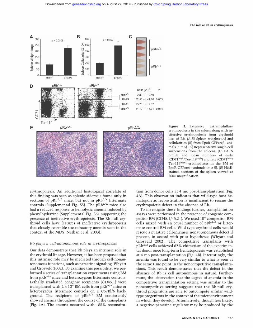
erythropoiesis. An additional histological correlate ofthis finding was seen as splenic siderosis found only insections of pRb�/� mice, but not in pRb�/+ littermatecontrols (Supplemental Fig. S5). The pRb�/� mice alsohad a reduced response to hemolytic anemia induced byphenylhydrazine (Supplemental Fig. S6), supporting thepresence of ineffective erythropoiesis. The Rb-null ery-throid cells have features of ineffective erythropoiesisthat closely resemble the refractory anemia seen in thecontext of the MDS (Nathan et al. 2003).
Rb plays a cell-autonomous role in erythropoiesis
Our data demonstrate that Rb plays an intrinsic role inthe erythroid lineage. However, it has been proposed thatthis intrinsic role may be mediated through cell-nonau-tonomous functions, such as paracrine signaling (Whyattand Grosveld 2002). To examine this possibility, we per-formed a series of transplantation experiments using BMfrom pRb�/� mice and heterozygous littermate controls.Lethally irradiated congenic recipients (CD45.1) weretransplanted with 2 × 10⁶ BM cells from pRb�/� mice orheterozygous littermate controls on a C57Bl/6 back-ground. The recipients of pRb�/� BM consistentlyshowed anemia throughout the course of the transplants(Fig. 4A). The anemia occurred with ∼88% reconstitu-
tion from donor cells at 4 mo post-transplantation (Fig.4A). This observation indicates that wild-type host he-matopoietic reconstitution is insufficient to rescue theerythropoietic defect in the absence of Rb.
To investigate these findings further, transplantationassays were performed in the presence of congenic com-petitor BM (CD45.1/45.2+). We used 106 competitor BMcells mixed with an equal number of pRb�/� or litter-mate control BM cells. Wild-type erythroid cells wouldrescue a putative cell-intrinsic nonautonomous defect ifpresent, in accord with prior hypotheses (Whyatt andGrosveld 2002). The competitive transplants withpRb�/� cells achieved 62% chimerism of the experimen-tal donor once long-term hematopoiesis was establishedat 4 mo post-transplantation (Fig. 4B). Interestingly, theanemia was found to be very similar to what is seen atthe same time point in the noncompetitive transplanta-tions. This result demonstrates that the defect in theabsence of Rb is cell autonomous in nature. Further-more, the observation that the degree of anemia in thecompetitive transplantation setting was similar to thenoncompetitive setting suggests that the Rb-null ery-throid progenitors are able to outcompete similar wild-type progenitors in the context of the microenvironmentin which they develop. Alternatively, though less likely,a negative paracrine regulator may be produced by the
Figure 3. Extensive extramedullaryerythropoiesis in the spleen along with in-effective erythropoiesis from erythroidloss of Rb. (A,B) Spleen weights (A) andcellularities (B) from EpoR-GFPcre/+ ani-mals (n � 5). (C) Representative single-cellsuspensions from the spleens. (D) FACSprofile and mean numbers of early(CD71high/Ter-119high) and late (CD71low/Ter-119high) erythroblasts in the BM ofEpoR-GFPcre/+ animals (n � 5). (E) H&E-stained sections of the spleen viewed at200× magnification.
The role of Rb in erythropoiesis
GENES & DEVELOPMENT 467
Cold Spring Harbor Laboratory Press on August 27, 2019 - Published by genesdev.cshlp.orgDownloaded from
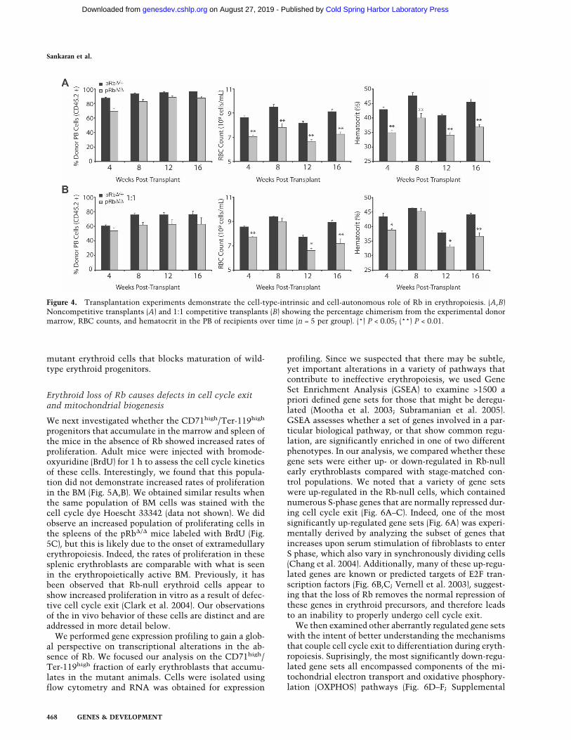
mutant erythroid cells that blocks maturation of wild-type erythroid progenitors.
Erythroid loss of Rb causes defects in cell cycle exitand mitochondrial biogenesis
We next investigated whether the CD71high/Ter-119high
progenitors that accumulate in the marrow and spleen ofthe mice in the absence of Rb showed increased rates ofproliferation. Adult mice were injected with bromode-oxyuridine (BrdU) for 1 h to assess the cell cycle kineticsof these cells. Interestingly, we found that this popula-tion did not demonstrate increased rates of proliferationin the BM (Fig. 5A,B). We obtained similar results whenthe same population of BM cells was stained with thecell cycle dye Hoescht 33342 (data not shown). We didobserve an increased population of proliferating cells inthe spleens of the pRb�/� mice labeled with BrdU (Fig.5C), but this is likely due to the onset of extramedullaryerythropoiesis. Indeed, the rates of proliferation in thesesplenic erythroblasts are comparable with what is seenin the erythropoietically active BM. Previously, it hasbeen observed that Rb-null erythroid cells appear toshow increased proliferation in vitro as a result of defec-tive cell cycle exit (Clark et al. 2004). Our observationsof the in vivo behavior of these cells are distinct and areaddressed in more detail below.
We performed gene expression profiling to gain a glob-al perspective on transcriptional alterations in the ab-sence of Rb. We focused our analysis on the CD71high/Ter-119high fraction of early erythroblasts that accumu-lates in the mutant animals. Cells were isolated usingflow cytometry and RNA was obtained for expression
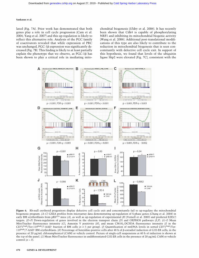
profiling. Since we suspected that there may be subtle,yet important alterations in a variety of pathways thatcontribute to ineffective erythropoiesis, we used GeneSet Enrichment Analysis (GSEA) to examine >1500 apriori defined gene sets for those that might be deregu-lated (Mootha et al. 2003; Subramanian et al. 2005).GSEA assesses whether a set of genes involved in a par-ticular biological pathway, or that show common regu-lation, are significantly enriched in one of two differentphenotypes. In our analysis, we compared whether thesegene sets were either up- or down-regulated in Rb-nullearly erythroblasts compared with stage-matched con-trol populations. We noted that a variety of gene setswere up-regulated in the Rb-null cells, which containednumerous S-phase genes that are normally repressed dur-ing cell cycle exit (Fig. 6A–C). Indeed, one of the mostsignificantly up-regulated gene sets (Fig. 6A) was experi-mentally derived by analyzing the subset of genes thatincreases upon serum stimulation of fibroblasts to enterS phase, which also vary in synchronously dividing cells(Chang et al. 2004). Additionally, many of these up-regu-lated genes are known or predicted targets of E2F tran-scription factors (Fig. 6B,C; Vernell et al. 2003), suggest-ing that the loss of Rb removes the normal repression ofthese genes in erythroid precursors, and therefore leadsto an inability to properly undergo cell cycle exit.
We then examined other aberrantly regulated gene setswith the intent of better understanding the mechanismsthat couple cell cycle exit to differentiation during eryth-ropoiesis. Suprisingly, the most significantly down-regu-lated gene sets all encompassed components of the mi-tochondrial electron transport and oxidative phosphory-lation (OXPHOS) pathways (Fig. 6D–F; Supplemental
Figure 4. Transplantation experiments demonstrate the cell-type-intrinsic and cell-autonomous role of Rb in erythropoiesis. (A,B)Noncompetitive transplants (A) and 1:1 competitive transplants (B) showing the percentage chimerism from the experimental donormarrow, RBC counts, and hematocrit in the PB of recipients over time (n = 5 per group). (*) P < 0.05; (**) P < 0.01.
Sankaran et al.
468 GENES & DEVELOPMENT
Cold Spring Harbor Laboratory Press on August 27, 2019 - Published by genesdev.cshlp.orgDownloaded from
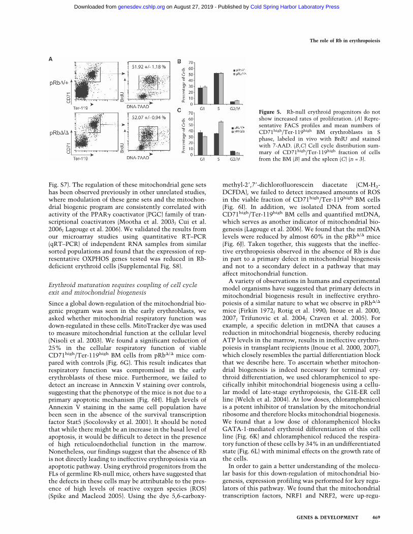
Fig. S7). The regulation of these mitochondrial gene setshas been observed previously in other unrelated studies,where modulation of these gene sets and the mitochon-drial biogenic program are consistently correlated withactivity of the PPAR� coactivator (PGC) family of tran-scriptional coactivators (Mootha et al. 2003; Cui et al.2006; Lagouge et al. 2006). We validated the results fromour microarray studies using quantitative RT–PCR(qRT–PCR) of independent RNA samples from similarsorted populations and found that the expression of rep-resentative OXPHOS genes tested was reduced in Rb-deficient erythroid cells (Supplemental Fig. S8).
Erythroid maturation requires coupling of cell cycleexit and mitochondrial biogenesis
Since a global down-regulation of the mitochondrial bio-genic program was seen in the early erythroblasts, weasked whether mitochondrial respiratory function wasdown-regulated in these cells. MitoTracker dye was usedto measure mitochondrial function at the cellular level(Nisoli et al. 2003). We found a significant reduction of25% in the cellular respiratory function of viableCD71high/Ter-119high BM cells from pRb�/� mice com-pared with controls (Fig. 6G). This result indicates thatrespiratory function was compromised in the earlyerythroblasts of these mice. Furthermore, we failed todetect an increase in Annexin V staining over controls,suggesting that the phenotype of the mice is not due to aprimary apoptotic mechanism (Fig. 6H). High levels ofAnnexin V staining in the same cell population havebeen seen in the absence of the survival transcriptionfactor Stat5 (Socolovsky et al. 2001). It should be notedthat while there might be an increase in the basal level ofapoptosis, it would be difficult to detect in the presenceof high reticuloendothelial function in the marrow.Nonetheless, our findings suggest that the absence of Rbis not directly leading to ineffective erythropoiesis via anapoptotic pathway. Using erythroid progenitors from theFLs of germline Rb-null mice, others have suggested thatthe defects in these cells may be attributable to the pres-ence of high levels of reactive oxygen species (ROS)(Spike and Macleod 2005). Using the dye 5,6-carboxy-
methyl-2�,7�-dichlorofluorescein diacetate (CM-H2-DCFDA), we failed to detect increased amounts of ROSin the viable fraction of CD71high/Ter-119high BM cells(Fig. 6I). In addition, we isolated DNA from sortedCD71high/Ter-119high BM cells and quantified mtDNA,which serves as another indicator of mitochondrial bio-genesis (Lagouge et al. 2006). We found that the mtDNAlevels were reduced by almost 60% in the pRb�/� mice(Fig. 6J). Taken together, this suggests that the ineffec-tive erythropoiesis observed in the absence of Rb is duein part to a primary defect in mitochondrial biogenesisand not to a secondary defect in a pathway that mayaffect mitochondrial function.
A variety of observations in humans and experimentalmodel organisms have suggested that primary defects inmitochondrial biogenesis result in ineffective erythro-poiesis of a similar nature to what we observe in pRb�/�
mice (Firkin 1972; Rotig et al. 1990; Inoue et al. 2000,2007; Trifunovic et al. 2004; Craven et al. 2005). Forexample, a specific deletion in mtDNA that causes areduction in mitochondrial biogenesis, thereby reducingATP levels in the marrow, results in ineffective erythro-poiesis in transplant recipients (Inoue et al. 2000, 2007),which closely resembles the partial differentiation blockthat we describe here. To ascertain whether mitochon-drial biogenesis is indeed necessary for terminal ery-throid differentiation, we used chloramphenicol to spe-cifically inhibit mitochondrial biogenesis using a cellu-lar model of late-stage erythropoiesis, the G1E-ER cellline (Welch et al. 2004). At low doses, chloramphenicolis a potent inhibitor of translation by the mitochondrialribosome and therefore blocks mitochondrial biogenesis.We found that a low dose of chloramphenicol blocksGATA-1-mediated erythroid differentiation of this cellline (Fig. 6K) and chloramphenicol reduced the respira-tory function of these cells by 34% in an undifferentiatedstate (Fig. 6L) with minimal effects on the growth rate ofthe cells.
In order to gain a better understanding of the molecu-lar basis for this down-regulation of mitochondrial bio-genesis, expression profiling was performed for key regu-lators of this pathway. We found that the mitochondrialtranscription factors, NRF1 and NRF2, were up-regu-
Figure 5. Rb-null erythroid progenitors do notshow increased rates of proliferation. (A) Repre-sentative FACS profiles and mean numbers ofCD71high/Ter-119high BM erythroblasts in Sphase, labeled in vivo with BrdU and stainedwith 7-AAD. (B,C) Cell cycle distribution sum-mary of CD71high/Ter-119high fraction of cellsfrom the BM (B) and the spleen (C) (n = 3).
The role of Rb in erythropoiesis
GENES & DEVELOPMENT 469
Cold Spring Harbor Laboratory Press on August 27, 2019 - Published by genesdev.cshlp.orgDownloaded from
lated (Fig. 7A). Prior work has demonstrated that bothgenes play a role in cell cycle progression (Cam et al.2004; Yang et al. 2007) and this up-regulation is likely toreflect this alternative role. Analysis of the PGC familyof coactivators revealed that while expression of PRCwas unchanged, PGC-1� expression was significantly de-creased (Fig. 7B). This finding is likely to at least partiallyexplain the phenotype that we observe, as PGC-1� hasbeen shown to play a critical role in mediating mito-
chondrial biogenesis (Uldry et al. 2006). It has recentlybeen shown that Cdk4 is capable of phosphorylatingNRF1 and inhibiting its mitochondrial biogenic activity(Wang et al. 2006). Additional post-translational modifi-cations of this type are also likely to contribute to thereduction in mitochondrial biogenesis that is seen con-comitantly with defective cell cycle exit. In support ofthis hypothesis, we found that levels of the ubiquitinligase Skp2 were elevated (Fig. 7C), consistent with the
Figure 6. Rb-null erythroid progenitors display defective cell cycle exit and concomitantly fail to up-regulate the mitochondrialbiogenesic program. (A–C) GSEA profiles from microarray data demonstrating up-regulation of S-phase genes (Chang et al. 2004) inearly BM erythroblasts from pRb�/� mice (A), as well as up-regulation of experimental (B) (Vernell et al. 2003) and predicted E2F(C)targets. (D–F) Down-regulation of genes involved in the electron transport chain (D) and OXPHOS pathways (E,F). (G–I) MeanMitoTracker fluorescence intensity (G), Annexin V positivity (H), and mean CM-H2-DCFDA fluorescence intensity (I) in theCD71high/Ter-119high/7-AAD- fraction of BM cells (n � 5 per group). (J) Quantification of mtDNA levels in sorted CD71high/Ter-119high/7-AAD- BM erythroblasts. (K) Percentage of benzidine-positive cells after 48 h of �-estradiol induction of G1E-ER cells, in thepresence of 20 µg/mL chloramphenicol (CAM) or vehicle control. Picture of single-cell suspensions at 60 h of induction is shown atthe top of the panel. (L) Mean MitoTracker fluorescence in undifferentiated G1E-ER cells in the presence of 20 µg/mL CAM or vehiclecontrol (n = 3).
Sankaran et al.
470 GENES & DEVELOPMENT
Cold Spring Harbor Laboratory Press on August 27, 2019 - Published by genesdev.cshlp.orgDownloaded from
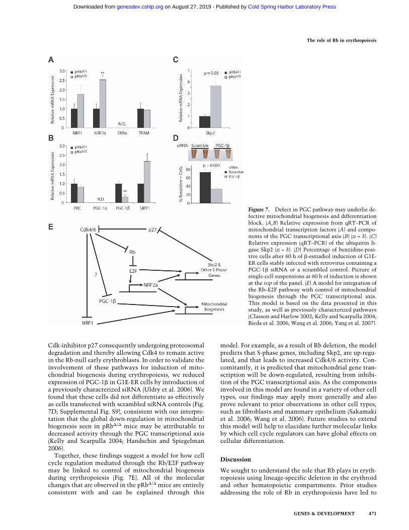
Cdk-inhibitor p27 consequently undergoing proteosomaldegradation and thereby allowing Cdk4 to remain activein the Rb-null early erythroblasts. In order to validate theinvolvement of these pathways for induction of mito-chondrial biogenesis during erythropoiesis, we reducedexpression of PGC-1� in G1E-ER cells by introduction ofa previously characterized siRNA (Uldry et al. 2006). Wefound that these cells did not differentiate as effectivelyas cells transfected with scrambled siRNA controls (Fig.7D; Supplemental Fig. S9), consistent with our interpre-tation that the global down-regulation in mitochondrialbiogenesis seen in pRb�/� mice may be attributable todecreased activity through the PGC transcriptional axis(Kelly and Scarpulla 2004; Handschin and Spiegelman2006).
Together, these findings suggest a model for how cellcycle regulation mediated through the Rb/E2F pathwaymay be linked to control of mitochondrial biogenesisduring erythropoiesis (Fig. 7E). All of the molecularchanges that are observed in the pRb�/� mice are entirelyconsistent with and can be explained through this
model. For example, as a result of Rb deletion, the modelpredicts that S-phase genes, including Skp2, are up-regu-lated, and that leads to increased Cdk4/6 activity. Con-comitantly, it is predicted that mitochondrial gene tran-scription will be down-regulated, resulting from inhibi-tion of the PGC transcriptional axis. As the componentsinvolved in this model are found in a variety of other celltypes, our findings may apply more generally and alsoprove relevant to prior observations in other cell types,such as fibroblasts and mammary epithelium (Sakamakiet al. 2006; Wang et al. 2006). Future studies to extendthis model will help to elucidate further molecular linksby which cell cycle regulators can have global effects oncellular differentiation.
Discussion
We sought to understand the role that Rb plays in eryth-ropoiesis using lineage-specific deletion in the erythroidand other hematopoietic compartments. Prior studiesaddressing the role of Rb in erythropoiesis have led to
Figure 7. Defect in PGC pathway may underlie de-fective mitochondrial biogenesis and differentiationblock. (A,B) Relative expression from qRT–PCR ofmitochondrial transcription factors (A) and compo-nents of the PGC transcriptional axis (B) (n = 3). (C)Relative expression (qRT–PCR) of the ubiquitin li-gase Skp2 (n = 3). (D) Percentage of benzidine-posi-tive cells after 60 h of �-estradiol induction of G1E-ER cells stably infected with retrovirus containing aPGC-1� siRNA or a scrambled control. Picture ofsingle-cell suspensions at 60 h of induction is shownat the top of the panel. (E) A model for integration ofthe Rb–E2F pathway with control of mitochondrialbiogenesis through the PGC transcriptional axis.This model is based on the data presented in thisstudy, as well as previously characterized pathways(Classon and Harlow 2002; Kelly and Scarpulla 2004;Bieda et al. 2006; Wang et al. 2006; Yang et al. 2007).
The role of Rb in erythropoiesis
GENES & DEVELOPMENT 471
Cold Spring Harbor Laboratory Press on August 27, 2019 - Published by genesdev.cshlp.orgDownloaded from
conflicting observations and interpretations. The in vivostudies we describe using conditional inactivation of Rbin the erythroid lineage avoid confounding experimentalproblems and provide novel insights into how Rb lossadversely affects this lineage. We found that Rb plays anintrinsic and cell-autonomous role in erythropoiesis.Unexpectedly, we found that this intrinsic role of Rb inpromoting cell cycle exit during late-stage erythropoiesisis coupled to differentiation through mitochondrial bio-genesis.
The role of Rb in erythropoiesis
Germline Rb knockout mice are embryonic lethal ataround E14.5 and display marked defects in erythropoi-esis, as well as in neurogenesis (Clarke et al. 1992; Jackset al. 1992; Lee et al. 1992). Subsequent work showedthat these early defects could be largely rescued by thepresence of a wild-type placenta (Wu et al. 2003; Wenzelet al. 2007). A major question posed by this work washow a defect in fetal–maternal nutrient exchange mightadversely affect particular organ systems, while allowingfor relatively normal development of various other celltypes. Our findings begin to explain these results. Wefound that the absence of Rb causes ineffective erythro-poiesis, which appears to be due in part to a defect inmitochondrial biogenesis. A cell type, such as the eryth-roblast, that is already metabolically challenged (fromdefective mitochondrial biogenesis) is likely to be moreadversely affected in the face of compromised nutrientexchange that would result from the Rb-null placenta.
It was hypothesized that the defect in erythropoiesis ofRb-null cells might also be explained by a cell-type-in-trinsic but non-cell-autonomous defect, such as througha paracrine effector of erythropoiesis (Whyatt and Gros-veld 2002). The results of our competitive transplantsdemonstrate that is not likely to be the case, as even inthe presence of high levels of hematopoietic chimerismby wild-type BM, the anemia resulting from pRb�/� cellsoccurs to a similar extent as is seen in the noncompeti-tive setting. Other work has suggested that Rb may playa role in promoting erythropoiesis in the context ofstress erythropoiesis and that Rb promotes erythroid dif-ferentiation by promoting enucleation and limiting theproduction of ROS (Spike et al. 2004; Spike and Macleod2005). Our findings are distinct from these results, sincewe show that Rb does indeed play a role in normal eryth-ropoiesis and enucleation defects are not observed in thepRb�/� mice. Furthermore, we did not detect increasedlevels of ROS in Rb-null erythroid progenitors. It shouldbe noted that the increased levels of ROS have only beenseen in the erythroid cells from the FL of germline Rb-null animals (Spike and Macleod 2005) and may repre-sent the consequence of multiple extrinsic defects.
Studies using ex vivo cell cultures have suggested thatRb may be necessary or dispensible for normal erythro-poiesis. One group found that Rb-null FL cells continuedto proliferate in a liquid culture system and failed toundergo proper differentiation, as compared with wild-type FL cells (Clark et al. 2004). Work using respiratory
chain-deficient cells has demonstrated that metaboli-cally compromised cells can survive in in vitro condi-tions due to a high level of nutrients, including pyruvateand uridine, that allow the requirement for OXPHOS tobe bypassed (King and Attardi 1989). It is likely that inthe nutrient-permissive conditions used in the FL cellculture experiments, Rb-null erythroid progenitors thatfailed to differentiate were able to survive, in contrast towhat occurs in vivo (Clark et al. 2004). Other workersused a coculture system that is meant to recapitulate theformation of an erythroid island, involving a supportingmacrophage nursing differentiating erythroid progeni-tors (Iavarone et al. 2004). Under these conditions, Rbactivity appeared to be necessary in macrophages, butdispensible in erythroid progenitors, for proper forma-tion of an in vitro erythroid island. Our results contrastwith this finding, since animals lacking Rb in macro-phages appear to have normal erythropoiesis, with only aslight anemia occurring with age in the animals. It islikely that Rb-null macrophages may behave differentlythan their wild-type counterparts in the context of theculture conditions used to create ex vivo erythroid is-lands (Iavarone et al. 2004), but this does not appear to bephysiologically relevant. Furthermore, intrinsic defectsthat can cause ineffective erythropoiesis in vivo may notbe detectable in this coculture system.
Cell cycle regulation, mitochondrial biogenesis,and differentiation
The role of cell cycle regulation in erythropoiesis hasbeen carefully studied at a descriptive level. However,the molecular control of this process in vivo and themechanisms by which this is coupled to differentiationare largely unknown. Our results point to a previouslyundescribed mechanism that links cell cycle exit to late-stage erythropoiesis. We found that mitochondrial bio-genic function is necessary at this transition in erythroidcell maturation, just as commitment to cell cycle exit isinitiated. This observation is consistent with the differ-entiation block seen from primary defects in mitochon-drial biogenesis (Firkin 1972; Inoue et al. 2000, 2007;Trifunovic et al. 2004; Craven et al. 2005). In the contextof the pRb�/� erythroid cells, down-regulation of genesinvolved in cell cycle progression does not occur prop-erly, and concomitantly there is a failure to up-regulatethe mitochondrial biogenic program. Late-stage erythro-poiesis is critically dependent on mitochondrial functionfor high-level heme biosynthesis, as well as elevatedATP production for globin gene transcription and trans-lation. In addition to general mitochondrial biogenesis,prior studies have demonstrated that changes in mito-chondrial composition occur during these stages oferythropoiesis, including increased transcription ofUCP2, ABC-me, and mitoferrin (Shirihai et al. 2000;Shaw et al. 2006). It is interesting to note that cell cycleexit has been decoupled from erythroid differentiation invitro (Rylski et al. 2003), suggesting that these processesmay be separable under some conditions. Mitochondrial
Sankaran et al.
472 GENES & DEVELOPMENT
Cold Spring Harbor Laboratory Press on August 27, 2019 - Published by genesdev.cshlp.orgDownloaded from
biogenesis is an attractive candidate as a mediator ofsuch a process.
The similarity of the ineffective erythropoiesis seen inthe pRb�/� mice to that of human MDS is striking. Notonly is the block in erythropoiesis similar to what is seenin MDS, but the finding that this anemia cannot be over-come even in a chimeric situation resembles the refrac-tory anemia of MDS that arises in a clonal manner (Co-rey et al. 2007). The molecular basis of MDS is not wellunderstood, although the most common known abnor-mality involves epigenetic silencing of CDKN2B (Boult-wood and Wainscoat 2007), a key negative regulator ofthe cell cycle. It is possible that further studies may findthat deregulation of other cell cycle components of theRb pathway contribute to MDS. At the same time, it isknown that deregulated mitochondrial function andstructure is frequently seen in MDS, particularly in theerythroid lineage (Greenberg et al. 2002). Our findingsshow that these seemingly disparate pathways may playkey roles in synergistically promoting differentiation. Itis likely that the coordination of cell cycle regulationand mitochondrial biogenesis will be important not onlyin erythropoiesis, but will also play a role in other casesof cellular differentiation. Indeed, a variety of observa-tions in cancer cells have shown that uncontrolled pro-liferation, which is nearly always driven in part by mu-tations in the Rb pathway (Classon and Harlow 2002),leads to reduced production of ATP by aerobic respira-tion and increased dependence upon anaerobic glycolysis(Warburg 1930; Fantin et al. 2006).
Importantly, our findings provide insight into thepathophysiology underlying ineffective erythropoiesisand suggest novel therapeutic modalities. Of note, weimplicated PGC-1� in the mitochondrial dysfunctionseen in erythroid precursors lacking Rb. Of particularinterest, PGC-1� is haploinsufficient in the 5q− subtypeof MDS (Boultwood et al. 2002). Modulation of mito-chondrial function is already being attempted in neuro-degenerative diseases. Similar approaches may benefithematologic diseases like MDS. Indeed, initial researchin this area has shown some promise (Greenberg et al.2002; Galili et al. 2007). While modulation of the cellcycle is a common therapeutic target in MDS (Corey etal. 2007), addition of agents that strike at the dysfunc-tional mitochondria of cells arising from surviving dys-plastic clones might be beneficial. This may explain inpart the demonstrated clinical efficacy of arsenic triox-ide, a potent inhibitor of the OXPHOS component suc-cinate dehydrogenase, in MDS (Schiller et al. 2006; Veyet al. 2006). Our findings on the molecular control oferythroid differentiation, through the coordination ofcell cycle exit and mitochondrial biogenesis, may haveimportant implications in attempting to develop thera-pies for a set of largely incurable diseases involving in-effective erythropoiesis.
Materials and methods
A detailed version of the Materials and Methods can be foundin the Supplemental Material.
Experimental animals
EpoR-GFPcre mice (Heinrich et al. 2004) were kindly providedby Drs. Ursula Klingmüller and Ben Neel. pRbfl/fl mutant mice(Sage et al. 2003), Mx1-Cre transgenic mice (Kuhn et al. 1995;Walkley and Orkin 2006), and Lys-M-Cre animals (Clausen etal. 1999) have been described previously (Walkley et al. 2007).All experiments were performed with the approval of the CHBInstitute Animal Ethics Committee.
Flow cytometry analysis
All antibodies and clone numbers are listed in the SupplementalMaterial. Flow cytometry was performed on a FACSCalibur,and sorting was performed on a FACS Aria; all data were ana-lyzed using Cell Quest Pro software (Becton Dickinson).
Cell culture
G1E-ER cells were cultured and synchronously differentiated asdescribed previously (Rylski et al. 2003). Details of these proce-dures can be found in the Supplemental Material.
Gene expression analysis
A detailed description of the methods used for gene expressionanalysis using qRT–PCR and microarray can be found in theSupplemental Material. The microarray data were deposited inthe Gene Expression Omnibus (http://www.ncbi.nlm.nih.gov/geo) under accession number GSE9717.
Statistical analyses
Statistical analyses were performed using the paired and un-paired Student’s t-test, with a P value �0.05 being consideredsignificant. Results are depicted as mean ± standard error of themean (SEM) for n given samples.
Acknowledgments
We are grateful to B. Spiegelman and W. Yang for providingreagents and helpful suggestions, and to U. Klingmüller, B.Neel, T. Jacks, B. Lowell, and M. Huntgeburth for providingmouse strains. We thank D. Nathan, L. Purton, J. Hirschhorn,M. Weiss, L. Zon, V. Mootha, and C. Sieff for valuable discus-sion and critical comments; the Children’s Animal Facility stafffor care of experimental animals; G. Losyev, J. Daley, and S.Lazo-Kallanian for assistance with FACS sorting; the DFCI Mi-croarray Core; and the DFCI/HCC Rodent Histology Core. Thiswork was supported in part by a Center of Excellence Award inMolecular Hematology from the NIH-NIDDK (S.H.O.). V.G.S.was supported by MSTP and NRSA awards from the NIH.C.R.W. is a Special Fellow of the Leukemia and LymphomaSociety and S.H.O. is an Investigator of the Howard HughesMedical Institute.
References
Bieda, M., Xu, X., Singer, M.A., Green, R., and Farnham, P.J.2006. Unbiased location analysis of E2F1-binding sites sug-gests a widespread role for E2F1 in the human genome. Ge-nome Res. 16: 595–605.
Boultwood, J. and Wainscoat, J.S. 2007. Gene silencing by DNAmethylation in haematological malignancies. Br. J. Haema-tol. 138: 3–11.
The role of Rb in erythropoiesis
GENES & DEVELOPMENT 473
Cold Spring Harbor Laboratory Press on August 27, 2019 - Published by genesdev.cshlp.orgDownloaded from
Boultwood, J., Fidler, C., Strickson, A.J., Watkins, F., Gama, S.,Kearney, L., Tosi, S., Kasprzyk, A., Cheng, J.F., Jaju, R.J., etal. 2002. Narrowing and genomic annotation of the com-monly deleted region of the 5q− syndrome. Blood 99: 4638–4641.
Cam, H., Balciunaite, E., Blais, A., Spektor, A., Scarpulla, R.C.,Young, R., Kluger, Y., and Dynlacht, B.D. 2004. A commonset of gene regulatory networks links metabolism andgrowth inhibition. Mol. Cell 16: 399–411.
Chang, H.Y., Sneddon, J.B., Alizadeh, A.A., Sood, R., West, R.B.,Montgomery, K., Chi, J.T., van de Rijn, M., Botstein, D., andBrown, P.O. 2004. Gene expression signature of fibroblastserum response predicts human cancer progression: Simi-larities between tumors and wounds. PLoS Biol. 2: E7. doi:10.1371/journal.pbio.0020007.
Ciemerych, M.A., Kenney, A.M., Sicinska, E., Kalaszczynska, I.,Bronson, R.T., Rowitch, D.H., Gardner, H., and Sicinski, P.2002. Development of mice expressing a single D-type cyc-lin. Genes & Dev. 16: 3277–3289.
Clark, A.J., Doyle, K.M., and Humbert, P.O. 2004. Cell-intrinsicrequirement for pRb in erythropoiesis. Blood 104:1324–1326.
Clarke, A.R., Maandag, E.R., van Roon, M., van der Lugt, N.M.,van der Valk, M., Hooper, M.L., Berns, A., and te Riele, H.1992. Requirement for a functional Rb-1 gene in murine de-velopment. Nature 359: 328–330.
Classon, M. and Harlow, E. 2002. The retinoblastoma tumoursuppressor in development and cancer. Nat. Rev. Cancer 2:910–917.
Clausen, B.E., Burkhardt, C., Reith, W., Renkawitz, R., and For-ster, I. 1999. Conditional gene targeting in macrophages andgranulocytes using LysMcre mice. Transgenic Res. 8: 265–277.
Corey, S.J., Minden, M.D., Barber, D.L., Kantarjian, H., Wang,J.C., and Schimmer, A.D. 2007. Myelodysplastic syndromes:The complexity of stem-cell diseases. Nat. Rev. Cancer 7:118–129.
Craven, S.E., French, D., Ye, W., de Sauvage, F., and Rosenthal,A. 2005. Loss of Hspa9b in zebrafish recapitulates the inef-fective hematopoiesis of the myelodysplastic syndrome.Blood 105: 3528–3534.
Cui, L., Jeong, H., Borovecki, F., Parkhurst, C.N., Tanese, N.,and Krainc, D. 2006. Transcriptional repression of PGC-1�
by mutant huntingtin leads to mitochondrial dysfunctionand neurodegeneration. Cell 127: 59–69.
Dumitriu, B., Patrick, M.R., Petschek, J.P., Cherukuri, S., Kling-muller, U., Fox, P.L., and Lefebvre, V. 2006. Sox6 cell-au-tonomously stimulates erythroid cell survival, proliferation,and terminal maturation and is thereby an important en-hancer of definitive erythropoiesis during mouse develop-ment. Blood 108: 1198–1207.
Fantin, V.R., St-Pierre, J., and Leder, P. 2006. Attenuation ofLDH-A expression uncovers a link between glycolysis, mi-tochondrial physiology, and tumor maintenance. CancerCell 9: 425–434.
Fero, M.L., Rivkin, M., Tasch, M., Porter, P., Carow, C.E., Firpo,E., Polyak, K., Tsai, L.H., Broudy, V., Perlmutter, R.M., et al.1996. A syndrome of multiorgan hyperplasia with features ofgigantism, tumorigenesis, and female sterility in p27(Kip1)-deficient mice. Cell 85: 733–744.
Firkin, F.C. 1972. Mitochondrial lesions in reversible erythro-poietic depression due to chloramphenicol. J. Clin. Invest.51: 2085–2092.
Galili, N., Sechman, E.V., Cerny, J., Mehdi, M., Mumtaz, M.,Westervelt, P., Maguire, J., and Raza, A. 2007. Clinical re-sponse of myelodysplastic syndromes patients to treatment
with coenzyme Q10. Leuk. Res. 31: 19–26.Greenberg, P.L., Young, N.S., and Gattermann, N. 2002. Myelo-
dysplastic syndromes. Hematology Am. Soc. Hematol.Educ. Program. 2002: 136–161.
Gregory, C.J. and Eaves, A.C. 1977. Human marrow cells ca-pable of erythropoietic differentiation in vitro: Definition ofthree erythroid colony responses. Blood 49: 855–864.
Gregory, C.J. and Eaves, A.C. 1978. Three stages of erythropoi-etic progenitor cell differentiation distinguished by a num-ber of physical and biologic properties. Blood 51: 527–537.
Handschin, C. and Spiegelman, B.M. 2006. Peroxisome prolif-erator-activated receptor � coactivator 1 coactivators, energyhomeostasis, and metabolism. Endocr. Rev. 27: 728–735.
Heinrich, A.C., Pelanda, R., and Klingmuller, U. 2004. A mousemodel for visualization and conditional mutations in theerythroid lineage. Blood 104: 659–666.
Hu, N., Gulley, M.L., Kung, J.T., and Lee, E.Y. 1997. Retino-blastoma gene deficiency has mitogenic but not tumorigeniceffects on erythropoiesis. Cancer Res. 57: 4123–4129.
Iavarone, A., King, E.R., Dai, X.M., Leone, G., Stanley, E.R., andLasorella, A. 2004. Retinoblastoma promotes definitiveerythropoiesis by repressing Id2 in fetal liver macrophages.Nature 432: 1040–1045.
Inoue, K., Nakada, K., Ogura, A., Isobe, K., Goto, Y., Nonaka, I.,and Hayashi, J.I. 2000. Generation of mice with mitochon-drial dysfunction by introducing mouse mtDNA carrying adeletion into zygotes. Nat. Genet. 26: 176–181.
Inoue, S., Yokota, M., Nakada, K., Miyoshi, H., and Hayashi, J.2007. Pathogenic mitochondrial DNA-induced respirationdefects in hematopoietic cells result in anemia by suppress-ing erythroid differentiation. FEBS Lett. 581: 1910–1916.
Jacks, T., Fazeli, A., Schmitt, E.M., Bronson, R.T., Goodell,M.A., and Weinberg, R.A. 1992. Effects of an Rb mutation inthe mouse. Nature 359: 295–300.
Kelly, D.P. and Scarpulla, R.C. 2004. Transcriptional regulatorycircuits controlling mitochondrial biogenesis and function.Genes & Dev. 18: 357–368.
King, M.P. and Attardi, G. 1989. Human cells lacking mtDNA:Repopulation with exogenous mitochondria by complemen-tation. Science 246: 500–503.
Kuhn, R., Schwenk, F., Aguet, M., and Rajewsky, K. 1995. In-ducible gene targeting in mice. Science 269: 1427–1429.
Lagouge, M., Argmann, C., Gerhart-Hines, Z., Meziane, H., Le-rin, C., Daussin, F., Messadeq, N., Milne, J., Lambert, P.,Elliott, P., et al. 2006. Resveratrol improves mitochondrialfunction and protects against metabolic disease by activat-ing SIRT1 and PGC-1�. Cell 127: 1109–1122.
Lee, E.Y., Chang, C.Y., Hu, N., Wang, Y.C., Lai, C.C., Herrup,K., Lee, W.H., and Bradley, A. 1992. Mice deficient for Rb arenonviable and show defects in neurogenesis and haemato-poiesis. Nature 359: 288–294.
Letvin, N.L., Linch, D.C., Beardsley, G.P., McIntyre, K.W.,Miller, B.A., and Nathan, D.G. 1985. Influence of cell cyclephase-specific agents on simian fetal hemoglobin synthesis.J. Clin. Invest. 75: 1999–2005.
Maandag, E.C., van der Valk, M., Vlaar, M., Feltkamp, C.,O’Brien, J., van Roon, M., van der Lugt, N., Berns, A., and teRiele, H. 1994. Developmental rescue of an embryonic-le-thal mutation in the retinoblastoma gene in chimeric mice.EMBO J. 13: 4260–4268.
Maetens, M., Doumont, G., Clercq, S.D., Francoz, S., Froment,P., Bellefroid, E., Klingmuller, U., Lozano, G., and Marine,J.C. 2007. Distinct roles of Mdm2 and Mdm4 in red cellproduction. Blood 109: 2630–2633.
Mootha, V.K., Lindgren, C.M., Eriksson, K.F., Subramanian, A.,Sihag, S., Lehar, J., Puigserver, P., Carlsson, E., Ridderstrale,
Sankaran et al.
474 GENES & DEVELOPMENT
Cold Spring Harbor Laboratory Press on August 27, 2019 - Published by genesdev.cshlp.orgDownloaded from
M., Laurila, E., et al. 2003. PGC-1�-responsive genes in-volved in oxidative phosphorylation are coordinately down-regulated in human diabetes. Nat. Genet. 34: 267–273.
Nathan, D.G., Orkin, S.H., Look, A.T., and Ginsburg, D. 2003.Nathan and Oski’s hematology of infancy and childhood.Saunders, Philadelphia, PA.
Nisoli, E., Clementi, E., Paolucci, C., Cozzi, V., Tonello, C.,Sciorati, C., Bracale, R., Valerio, A., Francolini, M.,Moncada, S., et al. 2003. Mitochondrial biogenesis in mam-mals: The role of endogenous nitric oxide. Science 299: 896–899.
Rotig, A., Cormier, V., Blanche, S., Bonnefont, J.P., Ledeist, F.,Romero, N., Schmitz, J., Rustin, P., Fischer, A., Saudubray,J.M., et al. 1990. Pearson’s marrow-pancreas syndrome. Amultisystem mitochondrial disorder in infancy. J. Clin. In-vest. 86: 1601–1608.
Rylski, M., Welch, J.J., Chen, Y.Y., Letting, D.L., Diehl, J.A.,Chodosh, L.A., Blobel, G.A., and Weiss, M.J. 2003. GATA-1-mediated proliferation arrest during erythroid maturation.Mol. Cell. Biol. 23: 5031–5042.
Sage, J., Miller, A.L., Perez-Mancera, P.A., Wysocki, J.M., andJacks, T. 2003. Acute mutation of retinoblastoma gene func-tion is sufficient for cell cycle re-entry. Nature 424: 223–228.
Sakamaki, T., Casimiro, M.C., Ju, X., Quong, A.A., Katiyar, S.,Liu, M., Jiao, X., Li, A., Zhang, X., Lu, Y., et al. 2006. CyclinD1 determines mitochondrial function in vivo. Mol. Cell.Biol. 26: 5449–5469.
Schiller, G.J., Slack, J., Hainsworth, J.D., Mason, J., Saleh, M.,Rizzieri, D., Douer, D., and List, A.F. 2006. Phase II multi-center study of arsenic trioxide in patients with myelodys-plastic syndromes. J. Clin. Oncol. 24: 2456–2464.
Shaw, G.C., Cope, J.J., Li, L., Corson, K., Hersey, C., Acker-mann, G.E., Gwynn, B., Lambert, A.J., Wingert, R.A., Traver,D., et al. 2006. Mitoferrin is essential for erythroid iron as-similation. Nature 440: 96–100.
Shirihai, O.S., Gregory, T., Yu, C., Orkin, S.H., and Weiss, M.J.2000. ABC-me: A novel mitochondrial transporter inducedby GATA-1 during erythroid differentiation. EMBO J. 19:2492–2502.
Socolovsky, M., Nam, H., Fleming, M.D., Haase, V.H., Brug-nara, C., and Lodish, H.F. 2001. Ineffective erythropoiesis inStat5a−/−5b−/− mice due to decreased survival of early eryth-roblasts. Blood 98: 3261–3273.
Spike, B.T. and Macleod, K.F. 2005. The Rb tumor suppressor instress responses and hematopoietic homeostasis. Cell cycle4: 42–45.
Spike, B.T., Dirlam, A., Dibling, B.C., Marvin, J., Williams, B.O.,Jacks, T., and Macleod, K.F. 2004. The Rb tumor suppressoris required for stress erythropoiesis. EMBO J. 23: 4319–4329.
Stamatoyannopoulos, G. 2005. Control of globin gene expres-sion during development and erythroid differentiation. Exp.Hematol. 33: 259–271.
Subramanian, A., Tamayo, P., Mootha, V.K., Mukherjee, S., Eb-ert, B.L., Gillette, M.A., Paulovich, A., Pomeroy, S.L., Golub,T.R., Lander, E.S., et al. 2005. Gene set enrichment analysis:A knowledge-based approach for interpreting genome-wideexpression profiles. Proc. Natl. Acad. Sci. 102: 15545–15550.
Trifunovic, A., Wredenberg, A., Falkenberg, M., Spelbrink, J.N.,Rovio, A.T., Bruder, C.E., Bohlooly, Y.M., Gidlof, S., Oldfors,A., Wibom, R., et al. 2004. Premature ageing in mice express-ing defective mitochondrial DNA polymerase. Nature 429:417–423.
Uldry, M., Yang, W., St-Pierre, J., Lin, J., Seale, P., and Spiegel-man, B.M. 2006. Complementary action of the PGC-1 coac-tivators in mitochondrial biogenesis and brown fat differen-tiation. Cell Metab. 3: 333–341.
Vernell, R., Helin, K., and Muller, H. 2003. Identification oftarget genes of the p16INK4A-pRB–E2F pathway. J. Biol.Chem. 278: 46124–46137.
Vey, N., Bosly, A., Guerci, A., Feremans, W., Dombret, H.,Dreyfus, F., Bowen, D., Burnett, A., Dennis, M., Ribrag, V., etal. 2006. Arsenic trioxide in patients with myelodysplasticsyndromes: A phase II multicenter study. J. Clin. Oncol. 24:2465–2471.
Walkley, C.R. and Orkin, S.H. 2006. Rb is dispensable for self-renewal and multilineage differentiation of adult hematopoi-etic stem cells. Proc. Natl. Acad. Sci. 103: 9057–9062.
Walkley, C.R., Shea, J.M., Sims, N.A., Purton, L.E., and Orkin,S.H. 2007. Rb regulates interactions between hematopoieticstem cells and their bone marrow microenvironment. Cell129: 1081–1095.
Wang, C., Li, Z., Lu, Y., Du, R., Katiyar, S., Yang, J., Fu, M.,Leader, J.E., Quong, A., Novikoff, P.M., et al. 2006. CyclinD1 repression of nuclear respiratory factor 1 integratesnuclear DNA synthesis and mitochondrial function. Proc.Natl. Acad. Sci. 103: 11567–11572.
Warburg, O. 1930. The metabolism of tumors. Arnold Con-stable, London, UK.
Welch, J.J., Watts, J.A., Vakoc, C.R., Yao, Y., Wang, H., Hardi-son, R.C., Blobel, G.A., Chodosh, L.A., and Weiss, M.J. 2004.Global regulation of erythroid gene expression by transcrip-tion factor GATA-1. Blood 104: 3136–3147.
Wenzel, P.L., Wu, L., de Bruin, A., Chong, J.L., Chen, W.Y.,Dureska, G., Sites, E., Pan, T., Sharma, A., Huang, K., et al.2007. Rb is critical in a mammalian tissue stem cell popu-lation. Genes & Dev. 21: 85–97.
Whyatt, D. and Grosveld, F. 2002. Cell-nonautonomous func-tion of the retinoblastoma tumour suppressor protein: Newinterpretations of old phenotypes. EMBO Rep. 3: 130–135.
Whyatt, D., Lindeboom, F., Karis, A., Ferreira, R., Milot, E.,Hendriks, R., de Bruijn, M., Langeveld, A., Gribnau, J., Gros-veld, F., et al. 2000. An intrinsic but cell-nonautonomousdefect in GATA-1-overexpressing mouse erythroid cells. Na-ture 406: 519–524.
Williams, B.O., Schmitt, E.M., Remington, L., Bronson, R.T.,Albert, D.M., Weinberg, R.A., and Jacks, T. 1994. Extensivecontribution of Rb-deficient cells to adult chimeric micewith limited histopathological consequences. EMBO J. 13:4251–4259.
Wu, L., de Bruin, A., Saavedra, H.I., Starovic, M., Trimboli, A.,Yang, Y., Opavska, J., Wilson, P., Thompson, J.C., Os-trowski, M.C., et al. 2003. Extra-embryonic function of Rb isessential for embryonic development and viability. Nature421: 942–947.
Yang, Z.F., Mott, S., and Rosmarin, A.G. 2007. The Ets tran-scription factor GABP is required for cell-cycle progression.Nat. Cell Biol. 9: 339–346.
Ye, M., Iwasaki, H., Laiosa, C.V., Stadtfeld, M., Xie, H., Heck, S.,Clausen, B., Akashi, K., and Graf, T. 2003. Hematopoieticstem cells expressing the myeloid lysozyme gene retainlong-term, multilineage repopulation potential. Immunity19: 689–699.
Zhang, J., Socolovsky, M., Gross, A.W., and Lodish, H.F. 2003.Role of Ras signaling in erythroid differentiation of mousefetal liver cells: Functional analysis by a flow cytometry-based novel culture system. Blood 102: 3938–3946.
The role of Rb in erythropoiesis
GENES & DEVELOPMENT 475
Cold Spring Harbor Laboratory Press on August 27, 2019 - Published by genesdev.cshlp.orgDownloaded from
10.1101/gad.1627208Access the most recent version at doi: originally published online February 7, 200822:2008, Genes Dev.
Vijay G. Sankaran, Stuart H. Orkin, and Carl R. Walkley with mitochondrial biogenesis
intrinsically promotes erythropoiesis by coupling cell cycle exitRb
Material
Supplemental
http://genesdev.cshlp.org/content/suppl/2008/01/29/gad.1627208.DC1
Related Content
Sci. Signal. February , 2008 1: ec73
Elizabeth M. AdlerCycling on with Decreased Mitochondrial Function
References
http://genesdev.cshlp.org/content/22/4/463.full.html#related-urls
Articles cited in:
http://genesdev.cshlp.org/content/22/4/463.full.html#ref-list-1This article cites 65 articles, 30 of which can be accessed free at:
License
ServiceEmail Alerting
click here.right corner of the article or
Receive free email alerts when new articles cite this article - sign up in the box at the top
Copyright © 2008, Cold Spring Harbor Laboratory Press
Cold Spring Harbor Laboratory Press on August 27, 2019 - Published by genesdev.cshlp.orgDownloaded from