rare infi ltrative lung diseases: a challenge for...
TRANSCRIPT

Thematic Review Series
Respiration 2004;71:431–443 DOI: 10.1159/000080625
Rare Infi ltrative Lung Diseases: A Challenge for Clinicians
Venerino Poletti a, b Ulrich Costabel c Gian Luca Casoni a Caterina Bigliazzi a Marjolein Drent d Dario Olivieri b a
Dipartimento di Malattie dell’Apparato Respiratorio e del Torace, Ospedale GB Morgagni, Forlì , and b
Section of Respiratory Diseases, Department of Clinical Sciences, University of Parma, Parma , Italy; c
Ruhrlandklinik, Abteilung Pneumologie/Allergologie, Essen , Germany; d Department of Pulmonology,
University Hospital, Maastricht , The Netherlands
cussed (pulmonary alveolar proteinosis, inherited lipido-ses, acute eosinophilic pneumonia, amyloidosis, pulmo-nary ossifi cation, pulmonary alveolar microlithiasis). The list is obviously not exhaustive and arbitrarily cho-sen. The intent is, however, to emphasize that in this dif-fi cult fi eld multidisciplinary expertise and the knowledge of the most recent pathogenetic mechanisms have the main role in diagnosis and treatment.
Copyright © 2004 S. Karger AG, Basel
Introduction
Rare infi ltrative lung diseases are a challenge for clini-cians, radiologists and lung pathologists for at least three reasons [1–4] : (a) their low incidence and prevalence hamper the acquisition of expertise and frequently the diagnosis is delayed; (b) therapeutic actions are mainly empirical and based on steroid use, and (c) pathogenetic events are diffi cult to explain and only recently new ther-apeutic measures taking advantage of innovative genet-ic and/or immunopathogenetic studies have been sug-gested.
In this article the clinical, pathologic and radiographic patterns of rare parenchymal lung disorders are concisely reported and the more recent pathogenetic advances are briefl y described.
Key Words Rare diffuse infi ltrative lung diseases � Alveolar proteinosis � Acute eosinophilic pneumonia � Inherited lipidoses � Pulmonary amyloidosis
Abstract Rare diffuse infi ltrative lung diseases are a challenge for clinicians, radiologists, and pathologists for at least three reasons: (a) their low incidence and prevalence hamper the acquisition of expertise and frequently the diagnosis is delayed; (b) therapeutic actions are mainly empirical and based on steroid use, and (c) pathogenetic events are diffi cult to explain and only recently new therapeutic measures taking advantage of innovative genetic and/or immunopathogenetic studies have been suggested. In this review rare diffuse lung disorders are briefl y dis-
Venerino Poletti, MD, FCCP Dipartimento di Malattie dell’Apparato Respiratorio e del Torace Ospedale GB Morgagni, Via C Forlanini 34 IT–47100 Forlì (Italy) Tel. +39 0543 735042 , Fax +39 0543 735882, E-Mail [email protected], [email protected]
© 2004 S. Karger AG, Basel 0025–7931/04/0715–0431$21.00/0
Accessible online at: www.karger.com/res
Fax +41 61 306 12 34E-Mail [email protected]
Previous articles in this series: 1. Zompatori M, Bnà C, Poletti V, Spaggiari E, Ormitti F, Calabrò E, Tognini G, Sverzellati N: Diagnostic imaging of diffuse infi ltrative disease of the lung. Respiration 2004;71:4–19. 2. Poletti V, Chilosi M, Olivieri D: Diagnostic invasive procedures in diffuse infi ltrative lung dis-eases. Respiration 2004;71:107–119. 3. Chetta A, Marangio E, Olivieri D: Pulmonary function testing in interstitial lung dis-eases. Respiration 2004;71:209–213. 4. Camus P, Fanton A, Bonniaud P, Camus C, Foucher P: Interstitial lung disease in-duced by drugs and radiation. Respiration 2004;71:301–326.
Poletti/Costabel/Casoni/Bigliazzi/Drent/Olivieri
Respiration 2004;71:431–443 432
Pulmonary Alveolar Proteinosis
This disorder is characterized by abundant accumula-tion of phospholipids and proteinaceous material in the alveoli and distal airways. The intra-alveolar material mainly represents pulmonary surfactant phospholipids and protein components. The disease was fi rst described by Rosen et al. [5] in 1958. It can be primary (idiopathic) or secondary, associated with pulmonary infections, ma-lignancies (lymphoma, leukemia), inhalation exposure (silica, metal dusts and chemicals), neutropenia observed after chemotherapy for hematologic neoplasms, and HIV infection. A congenital form of alveolar proteinosis oc-curs in full-term newborns. It is an autosomal recessive disorder and involves mutations in the SP-B or SP-C genes. Infants with this disease die within the fi rst year of life despite maximal medical therapy. Alveolar proteino-sis has also been reported in patients with lysinuric pro-tein intolerance. The secondary forms are much rarer than the primary ones. Form the morphologic point of view the alveoli are fi lled with a characteristic acellular, fi nely granular material that stains with periodic acid-Schiff (PAS) and is diastase-negative. The interstitial structures are free of infl ammatory infi ltrates. Type II pneumocytes may be hyperplastic. Electron microscopy reveals that the intra-alveolar material consists predomi-nantly of unusual tubular, myelin-like, multilamellated structures, which resemble the tubular myelin found in normal lungs but lack the intersecting membranes of nor-mal tubular myelin. Components that represent cell de-bris are also present. Lamellar bodies as those seen in normal lungs are only minor components [6] .
Biochemical analysis of bronchoalveolar lavage (BAL) fl uid has revealed that the content of total phospholipids is increased with a relative decrease in phosphatidylcho-line and phosphatidylglycerol, and a relative increase in sphingomyelin and phosphatidylinositol [7, 8] .
The concentrations of surfactant protein A, B and D are also increased. The relative abundance of surfactant protein A isoforms is different from that in normal BAL and varies markedly from patient to patient, suggesting heterogeneity in the severity of the condition at the level of the structure of the surfactant proteins [9, 11] .
The pathogenesis is not defi nitely proven. As surfac-tant and surfactant-like material are abundantly present, a derangement in the normal pathway of surfactant secre-tion, metabolism and reuse or degradation seems likely. Physiologically, surfactant and its corresponding apopro-teins are synthesized and released in the form of lamellar bodies by alveolar type II cells. Most of the secreted sur-
factant is recycled and taken up by the type II cells again, probably mediated by receptors for surfactant apoprotein A on type II cells. The remainder is cleared either through phagocytosis and degradation by macrophages or to a lesser degree via lymphatics or the airways mucociliary apparatus. In alveolar proteinosis, there may be either increased surfactant production which is not eliminated suffi ciently by defective alveolar macrophages, or an in-terruption of surfactant reuptake by type II cells. The al-veolar macrophages show several secondary functional defects, such as reduced mobility, impaired adherence and chemotaxis, reduced phagocytosis, and a decreased ability to kill ingested microorganisms [12] . This func-tional impairment may contribute to the increased risk of pulmonary infections in patients with alveolar pro-teinosis. Recently, animal models of alveolar proteinosis and human data have suggested a role for granulocyte-macrophage colony-stimulating factor (GM-CSF) in the pathogenesis. GM-CSF seems essential for normal sur-factant clearance by activating alveolar macrophages and increasing their rate of surfactant catabolism [12] . Mice lacking GM-CSF or the GM-CSF receptor develop a pul-monary abnormality that resembles human alveolar pro-teinosis [13, 14] . Local pulmonary epithelial cell expres-sion of GM-CSF, bone marrow transplantation [15] or aerosolized GM-CSF inhalation corrects alveolar pro-teinosis in GM-CSF-defi cient mice [13] . In congenital al-veolar proteinosis a defect in the GM-CSF receptor ex-pression has been observed [16] . In adult alveolar pro-teinosis, such a receptor defect was, however, not ob-served [17] . Furthermore, other studies showed increased serum and BAL GM-CSF levels and normal GM-CSF production by blood monocytes and alveolar macro-phages in adult alveolar proteinosis and a normal re-sponse of alveolar macrophages to GM-CSF in terms of tumor necrosis factor production. These data would rath-er exclude a lack of GM-CSF production as an etiological factor. An immunologic explanation for these observa-tions was revealed by the fact that BAL fl uid and serum from patients with idiopathic alveolar proteinosis carry a factor that inhibits GM-CSF and is postulated to be an autoantibody [18, 19] . Some adult patients with idiopath-ic alveolar proteinosis improved with administration of GM-CSF [18, 20, 21] . Taken together, these observations suggest that adult idiopathic alveolar proteinosis is a het-erogeneous disease that may be caused in some patients by a decreased functional availability of GM-CSF due to GM-CSF-blocking activity.
The disease occurs predominantly in men with a male:female ratio of about 3: 1. The true prevalence is unknown
Rare Diffuse Infi ltrative Lung Diseases Respiration 2004;71:431–443 433
with current understanding based on fewer than 500 re-ported cases [5, 9, 10, 22–25] . The peak age of onset is between 30 and 50 years but infants and children may also be affected. Familial occurrence is rare but has been reported. There is an increased incidence in smokers. The main presenting complaints are slowly increasing dys-pnea on exertion (80%) and cough (60%). Less common symptoms (20–30%) include fever, weight loss, fatigue, chest pain and hemoptysis. Physical examination is usu-ally inconclusive; inspiratory crackles are heard in a mi-nority of patients and clubbing occurs in 30–50%. Less frequent fi ndings include cyanosis and evidence of cor pulmonale. The chest radiograph may be distinctive, showing diffuse bilateral symmetrical alveolar infi ltrates with air bronchograms. The shadowing may be cloudy and butterfl y- or batwing-like [26] , as a result of the more prominent involvement of the perihilar regions. Less commonly, unilateral infi ltrates or a reticulonodular pat-tern may by seen. Lymphadenopathy and pleural lesions are rare. Kerley B lines are absent initially but may de-velop later. The high-resolution CT (HRCT) shows air-space fi lling in variable and patchy distribution. The dis-tinctive features are: ground-glass opacifi cation sharply demarcated from normal lung, creating a ‘geographical’ pattern; intra- and interlobular septal thickening, often in polygonal shapes, called ‘crazy paving’, and large areas of consolidation with air bronchograms surrounded by ground-glass opacifi cation [26] .
Lung function tests characteristically show a restric-tive pattern and a reduced diffusing capacity. Hypoxemia at rest is present in about one third and during exercise in more than one half of the patients. An increase in the shunt fraction while breathing 100% oxygen is seen in almost all patients. Laboratory markers show a nonspe-cifi c increase of serum lactate dehydrogenase in most pa-tients, which declines after therapeutic lavage or sponta-neous resolution; its isoenzyme pattern is normal. Eleva-tion of serum carcinoembryonic antigen and other tumor markers has been seen and proposed as a marker of dis-ease activity. Serum levels of surfactant protein A and D can also be increased but this is not specifi c for the dis-ease, since high levels are also seen in patients with idio-pathic pulmonary fi brosis. Serum levels of KL-6, a mu-cin-like glycoprotein, have been recognized to be ex-tremely high in alveolar proteinosis, higher than those in patients with other interstitial lung disease. Serological diagnosis of alveolar proteinosis by demonstration of au-toantibodies against GM-CSF is not yet a routine proce-dure, but may become available in the future [27] . The diagnosis of alveolar proteinosis should be considered in
a patient with chronic insidiously developing dyspnea, a ‘butterfl y’ pattern of acinar shadowing on the chest radio-graph and characteristic fi ndings on HRCT (geographical distribution of a ‘crazy paving’ pattern) along with elevat-ed serum lactate dehydrogenase and an increased shunt fraction.
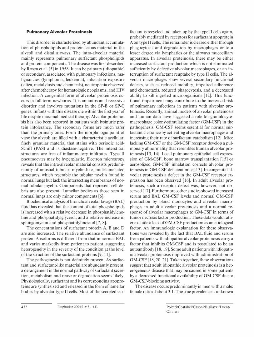
The diagnosis is usually established by BAL, obviating the need for transbronchial or open biopsy in many in-stances. On gross examination, the BAL fl uid has a char-acteristic milky appearance. On light microscopy the striking features are: acellular globules that are basophil-ic on May-Grünwald-Giemsa and positive with PAS staining; few and foamy macrophages, and large amounts of cell debris showing weak PAS staining. In cases associ-ated with protein intolerance and lysinuria cholesterol crystals may be prominent (pers. experience) ( fi g. 1 ). Elec-tron microscopy is not usually required to establish the diagnosis but if performed shows that the BAL sediment consists of characteristic myelin-like multilamellated structures and lamellar bodies.
Spontaneous remission occurs in up to one third of patients. Treatment is indicated when respiratory symp-toms impair the quality of life or when lung function de-teriorates. The treatment of choice is whole-lung lavage, which is almost always effective. Treatment with GM-CSF still has to be considered experimental. Anecdotal reports show responses to this therapy in some but not all
Fig. 1. BAL in a patient with secondary pulmonary alveolar pro-teinosis (lysinuric protein intolerance). A cholesterol crystal is evi-dent. May-Grünwald-Giemsa. ! 400.
Poletti/Costabel/Casoni/Bigliazzi/Drent/Olivieri
Respiration 2004;71:431–443 434
patients. Whole-lung lavage is safe when performed by an experienced team and under continuous monitoring of oxygen saturation, blood pressure, electrocardiography and lavage fl uid balance. The more severely affected lung is lavaged fi rst. The severity of respiratory impairment may be estimated by CT scan or by lung perfusion scan-ning. The second lung may be lavaged 3–7 days later. The procedure is performed under general anesthesia. The pa-tient is intubated with a double-lumen endotracheal tube (e.g. Carlens tube). After 15 min ventilation of both lungs with 100% oxygen to wash out the nitrogen, one lung is lavaged with isotonic saline at 37 ° C. The volume used for each fi lling is 500–1,000 ml. The lung is then allowed to drain by gravity. This fi lling and drainage is repeated un-til the effl uent is virtually clear which may require 10–40 liters.
The prognosis has improved considerably with the in-troduction of therapeutic lavage. Although there are no established response criteria for therapeutic lavage, sig-nifi cant clinical, physiologic, and radiologic improve-ments were claimed following the fi rst therapeutic lavage in 84% of the evaluable published cases [6, 12, 24, 25, 28–32] . In the literature cases, the interval between the diagnosis of pulmonary alveolar proteinosis and the fi rst application of therapeutic whole-lung lavage ranged from 0 (immediate lavage) to 210 months with a median of 2 months. The majority of patients who underwent lavage did so within 12 months of diagnosis (79%), but there was a continuing increase in the proportion of patients having received such therapy. In the era when lavage was avail-able after 1964, the likelihood of a patient with pulmo-nary alveolar proteinosis remaining free from therapeutic lavage was only 37% at 5 years [25] . In 55 instances of reported response to lavage, there was information pro-vided on the duration of the benefi t.
The median duration of clinical benefi t from lavage was 15 months with less than 20% of those patients fol-
lowed beyond 3 years remaining free of recurrent pulmo-nary alveolar proteinosis manifestations. Comparing the demographic and disease-related features of patients who did or did not respond to therapeutic lavage, there were no differences seen in gender, region of origin, duration of symptoms, smoking status, and time from diagnosis to lavage. When response rates to lavage were calculated within cohorts for age at diagnosis (20 years or less, 21–39 years, and 40 years or more), a signifi cant difference was observed: 58, 84 and 90%, respectively [25] . In four series totaling 64 patients there were no deaths related to alveo-lar proteinosis after 10–15 years’ experience with whole-lung lavage. Improvement may be long-lasting: 25–50% of patients achieve permanent remission after one lavage. In the others the procedure has to be repeated at intervals of 6–24 months. Alveolar proteinosis may be complicat-ed by infections such as nocardiosis, cryptococcosis, mu-cormycosis and others. In the era of therapeutic lavage these complications are rare. There have been single re-ports of progressive interstitial pulmonary fi brosis devel-oping in patients previously affected by alveolar proteino-sis. Lung transplantation may be an option for these pa-tients, although recurrence of disease has recently been reported in 1 patient following double lung transplanta-tion.
Inherited Lipidoses
The underlying defect in the inherited lipidoses is the accumulation of metabolites, including the glycolipids and sphingomyelin. The glycosphingolipids, which have a major structural function in many cells, are formed by the addition of various carbohydrates to a backbone of ceramide, an acylated sphingosine. In table 1 the molecu-lar genetics of Gaucher, Niemann-Pick, and Fabry dis-eases are depicted.
Table 1. Molecular genetics of Gaucher, Niemann-Pick, and Fabry diseases
Disease Chromosomeassignment
Molecular characteristics
Gaucher 1q21 cDNA, functional and pseudogenomic sequences, >200 mutant alleles known
Niemann-PickTypes A and B 11p15.1 to p15.4 cDNA , entire genomic sequence, >30 mutant alleles knownType C 18q11-q12 region cDNA, entire genomic sequence, >100 mutant alleles known
Fabry Xq22.1 cDNA, entire genomic sequences, >200 mutant alleles known
Rare Diffuse Infi ltrative Lung Diseases Respiration 2004;71:431–443 435
Gaucher disease is characterized by the deposition of glucosylceramide in cells of the macrophage-monocyte system. It was fi rst described by Gaucher in 1882 and the storage of glucocerebroside was fi rst recognized by Ep-stein in 1924. There are three clinical subtypes that are delineated by the absence or presence and progression of neurological involvement: type I or the adult, nonneu-ronopathic form; type II or the infantile or acute neurono-pathic form, and type III or the juvenile or Norrbotten form. All three subtypes are inherited as autosomal reces-sive traits and result from the defi cient activity of the ly-sosomal hydrolase acid ß -glucosidase. The pathologic dis-ease hallmark is the presence of the Gaucher cell in the macrophage-monocyte system, particularly in the bone marrow. These cells, which are 20–100 Ì m in diameter, have a characteristic wrinkled-paper appearance result-ing from intracytoplasmic substrate deposition and stain positively with PAS.
Four distinct patterns of pulmonary involvement by Gaucher cells have been described: intracapillary, patchy interstitial infi ltrates in a lymphatic distribution, massive interstitial thickening of alveolar septa, and intra-alveolar infi ltrates. Pulmonary involvement may be part of the broad spectrum of clinical expression among patients with type I disease. It is clinically evident in less than 5% of patients [33] . Dyspnea, diffuse and/or patchy lung in-fi ltrates, restrictive impairment and low single breath CO diffusing capacity represent the clinical disease profi le. About 10% of patients, although with normal physical examination and chest radiographs and with normal or nearly normal pulmonary function tests, may experience limitations in physical exertion and are easily fatigued. L444P homozygotes appear at major risk for developing pulmonary disease, even at earlier ages [34] . Pulmonary hypertension, strongly associated with splenectomy and female gender, may occur in subjects with non-N370S acid ß -glucosidase (GBA) gene mutation, positive family history, and ACE I gene polymorphism [35] .
Replacement therapy with recombinant acid ß -gluco-sidase has improved the pulmonary status; substrate re-duction therapy (the greatest experience has been with miglustat) is now an alternative in patients in whom the fi rst option is not suitable [36] .
Niemann-Pick disease types A and B result from defi -cient acid sphingomyelinase activity. In type C, the ge-netic defect involves the defective transport of choles-terol from the lysosome to the cytosol. Two different genes causing the altered cholesterol transport in type C disease were recently identifi ed, permitting more precise diagnosis, carrier detection, and prenatal diagnosis in af-
fected families. Pulmonary involvement is due to wide-spread infi ltration of both alveoli and interstitium by sea-blue histiocytes [37].
Clinically lung involvement in type B disease is chron-ic with a dry cough and exertional dyspnea, mild restric-tive impairment and minimal alteration of the diffusing capacity for carbon monoxide. In type C disease lung in-volvement may be pronounced, leading to early death caused by respiratory failure [38] .
Fabry disease is an X-linked inborn error of glyco-sphingolipid catabolism, which results from a defi ciency of lysosomal galactosidase activity. This results in an ab-normal accumulation of the glycosphingolipid ceramide trihexoside in vascular smooth muscle throughout the body, particularly in vessels of the skin, kidneys, heart, pulmonary vascular system, and neurological system. Pulmonary involvement has occasionally been reported: diffuse alveolar hemorrhage associated with renal failure or, more frequently, airfl ow obstruction due to the pres-ence of typical lamellar inclusion bodies within ciliated bronchial epithelial cells [39, 40] . Previously a univer-sally fatal disease, the recent development of human re-combinant · -galactosidase A, has been shown to reverse the clinical manifestations of the disease [41].
Hermansky-Pudlak Syndrome
It is a rare autosomal recessive disorder manifested by oculocutaneous albinism, a bleeding tendency, and in some cases ceroid-lipofuscin-lysosomal storage disease. The storage pool defect arises from defects in formation or traffi cking of lysosomes and related organelles, includ-ing melanosomes and platelet dense granules. The mo-lecular basis for Hermansky-Pudlak syndrome (HPS) is complex and heterogeneous, involving different genetic loci. HPS-causing mutations have been identifi ed in sev-eral human genes [42] . One of these genes encodes for the beta-3A subunit of AP-3, a protein complex that mediates signal-dependent traffi cking of integral membrane pro-teins to lysosomes and related organelles. Other genes are now identifi ed causing HPS in humans (HPS1, 3, 4, 5, 6). The HPS1, 3, 4, 5 and 6 proteins all have unknown func-tions [43–46] .
HPS1, HPS3 and HPS4 products are part of a stable protein complex named biogenesis of lysosome-related organelle complex (BLOC-2). HPS5 and HPS6 also inter-act and form BLOC-2. Linkage analysis of Puerto Rico families mapped the HPS1 gene to chromosome 10q23. The HPS1 gene has 20 exons, and it encodes a protein
Poletti/Costabel/Casoni/Bigliazzi/Drent/Olivieri
Respiration 2004;71:431–443 436
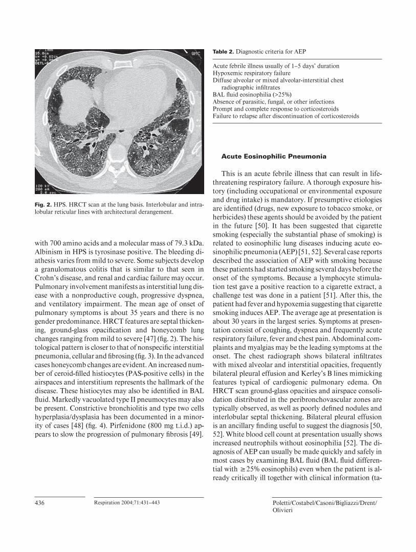
with 700 amino acids and a molecular mass of 79.3 kDa. Albinism in HPS is tyrosinase positive. The bleeding di-athesis varies from mild to severe. Some subjects develop a granulomatous colitis that is similar to that seen in Crohn’s disease, and renal and cardiac failure may occur. Pulmonary involvement manifests as interstitial lung dis-ease with a nonproductive cough, progressive dyspnea, and ventilatory impairment. The mean age of onset of pulmonary symptoms is about 35 years and there is no gender predominance. HRCT features are septal thicken-ing, ground-glass opacifi cation and honeycomb lung changes ranging from mild to severe [47] ( fi g. 2 ). The his-tological pattern is closer to that of nonspecifi c interstitial pneumonia, cellular and fi brosing ( fi g. 3 ). In the advanced cases honeycomb changes are evident. An increased num-ber of ceroid-fi lled histiocytes (PAS-positive cells) in the airspaces and interstitium represents the hallmark of the disease. These histiocytes may also be identifi ed in BAL fl uid. Markedly vacuolated type II pneumocytes may also be present. Constrictive bronchiolitis and type two cells hyperplasia/dysplasia has been documented in a minor-ity of cases [48] ( fi g. 4 ). Pirfenidone (800 mg t.i.d.) ap-pears to slow the progression of pulmonary fi brosis [49] .
Acute Eosinophilic Pneumonia
This is an acute febrile illness that can result in life-threatening respiratory failure. A thorough exposure his-tory (including occupational or environmental exposure and drug intake) is mandatory. If presumptive etiologies are identifi ed (drugs, new exposure to tobacco smoke, or herbicides) these agents should be avoided by the patient in the future [50] . It has been suggested that cigarette smoking (especially the substantial phase of smoking) is related to eosinophilic lung diseases inducing acute eo-sinophilic pneumonia (AEP) [51, 52]. Several case reports described the association of AEP with smoking because these patients had started smoking several days before the onset of the symptoms. Because a lymphocyte stimula-tion test gave a positive reaction to a cigarette extract, a challenge test was done in a patient [51] . After this, the patient had fever and hypoxemia suggesting that cigarette smoking induces AEP. The average age at presentation is about 30 years in the largest series. Symptoms at presen-tation consist of coughing, dyspnea and frequently acute respiratory failure, fever and chest pain. Abdominal com-plaints and myalgias may be the leading symptoms at the onset. The chest radiograph shows bilateral infi ltrates with mixed alveolar and interstitial opacities, frequently bilateral pleural effusion and Kerley’s B lines mimicking features typical of cardiogenic pulmonary edema. On HRCT scan ground-glass opacities and airspace consoli-dation distributed in the peribronchovascular zones are typically observed, as well as poorly defi ned nodules and interlobular septal thickening. Bilateral pleural effusion is an ancillary fi nding useful to suggest the diagnosis [50, 52] . White blood cell count at presentation usually shows increased neutrophils without eosinophilia [52] . The di-agnosis of AEP can usually be made quickly and safely in most cases by examining BAL fl uid (BAL fl uid differen-tial with 6 25% eosinophils) even when the patient is al-ready critically ill together with clinical information ( ta-
Fig. 2. HPS. HRCT scan at the lung basis. Interlobular and intra-lobular reticular lines with architectural derangement.
Table 2. Diagnostic criteria for AEP
Acute febrile illness usually of 1–5 days’ durationHypoxemic respiratory failureDiffuse alveolar or mixed alveolar-interstitial chest
radiographic infi ltratesBAL fl uid eosinophilia (>25%)Absence of parasitic, fungal, or other infectionsPrompt and complete response to corticosteroidsFailure to relapse after discontinuation of corticosteroids
Rare Diffuse Infi ltrative Lung Diseases Respiration 2004;71:431–443 437
ble 2 ). Moreover, lung biopsy is often not an option in acutely ill patients such as those with AEP. Furthermore, the presence of BAL fl uid fi ndings consistent with diffuse alveolar damage may strengthen the suspicion of AEP [53] . Intervention with corticosteroids results in rapid complete recovery without relapse.
Amyloidosis
This term stands for a heterogeneous group of diseases characterized by deposition of an insoluble ß -pleated fi -brillar protein in the extracellular matrix of involved tis-sues. A classifi cation of the various forms of amyloid is now based on the plasma precursors involved (immuno-globulins, light and heavy chains, serum amyloid A, trans-thyretin, fi brinogen, ß 2 -microglobulin, amyloid ß -protein precursor), the protein deposited in tissues and on the clinical profi le with which these deposits manifest them-selves ( table 3 ).
The diverse spectrum of amyloid-related diseases is now recognized to include Alzheimer’s disease, type II diabetes, and the transmissible spongiform encephalopa-
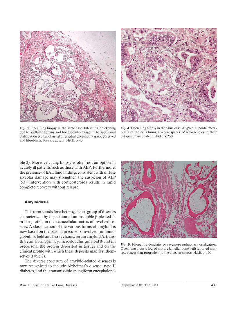
Fig. 3. Open lung biopsy in the same case. Interstitial thickening due to acellular fi brosis and honeycomb changes. The subpleural distribution typical of usual interstitial pneumonia is not observed and fi broblastic foci are absent. H&E. ! 40.
Fig. 4. Open lung biopsy in the same case. Atypical cuboidal meta-plasia of the cells lining alveolar spaces. Macrovacuoles in their cytoplasm are evident. H&E. ! 250.
Fig. 5. Idiopathic dendritic or racemose pulmonary ossifi cation. Open lung biopsy: foci of mature lamellar bone with fat-fi lled mar-row spaces that protrude into the alveolar spaces. H&E. ! 100.
Poletti/Costabel/Casoni/Bigliazzi/Drent/Olivieri
Respiration 2004;71:431–443 438
thies. Amyloidosis can be hereditary or acquired, local-ized or systemic, and potentially lethal or merely an inci-dental fi nding.
Respiratory Tract Amyloidosis Amyloid localized to the respiratory tract was recog-
nized by Lesser in 1877. Since then various classifi cations have been proposed based upon radiographic or broncho-scopic fi ndings [54, 55] . Inclusion of pulmonary vascular amyloidosis as a clinical syndrome is confusing since this is a histological fi nding that occurs to some extent in all its systemic forms.
There have been a few reports of systemic amyloidosis affecting the lungs but fi bril typing has generally been im-perfect [56–58 ] and all studies in which the fi bril protein has been sequenced identifi ed AL type [59–61] . Promi-nent lung disease is not a recognized feature of hereditary amyloidosis. In most situations, therefore, respiratory amyloidosis will be of the AL type although the presence of chronic infl ammatory disease or a family history or extreme old age should signal other possibilities.
Tracheobronchial Amyloidosis Tracheobronchial amyloidosis is an uncommon diag-
nosis [54] . It will not be reviewed in this article limited to diffuse disorders of the lung parenchyma.
Parenchymal Amyloidosis Amyloid involving the lung parenchyma is the most
frequently diagnosed respiratory amyloidosis syndrome. Amyloid nodules in the lung parenchyma are usually an incidental fi nding that needs to be distinguished from neoplasia. They are usually peripheral and subpleural, oc-cur more frequently in the lower lobes, may be bilateral,
and range in diameter from 0.4 to 15 cm. They grow slowly and frequently cavitate or calcify [62–64] . Larger nodules can occasionally produce space-occupying ef-fects. Diffuse alveolar septal amyloid is the least common form of isolated pulmonary amyloidosis and only a few cases have been reported [65] . Patients have dyspnea and a cough but rarely hemoptysis. Radiographically reticular and reticulonodular infi ltrates of varying severity are de-tected. CT scan fi ndings are small nodules, patchy ground-glass opacities or alveolar opacifi cation, thickening of the interlobular septa and irregular linear opacities. Honey-comb lung may occur later. Foci of calcifi cation in the nodules have also been observed. Pleural effusion may be present and occasionally dominate the clinical course. Multiple cysts and calcifi cation probably resulting from fragile alveolar walls as a consequence of amyloid deposi-tion both on alveolar walls and around capillaries have been described [66, 67] .
Autopsy series have confi rmed that diffuse parenchy-mal amyloid deposition is a common histological fi ndings in systemic amyloidosis [68] . Clinical involvement is rare but can be confused with pulmonary edema and/or fi bro-sis. Respiratory function tests may, but not always do, reveal a restrictive defect with impaired gas exchange, but it can be diffi cult to determine the relative contribution to symptoms of pulmonary versus cardiac amyloid which frequently coexist [56] . Pulmonary involvement is not a major contributor to death in systemic amyloidosis [68] and the median survival of patients with clinically overt lung deposition is about 16 months, similar to that for systemic amyloidosis in general [54] . The lymphatic sys-tem is frequently affected in systemic amyloidosis al-though predominant lymph node deposition is unusual [69, 70] . Hilar or mediastinal adenopathy is rarely associ-
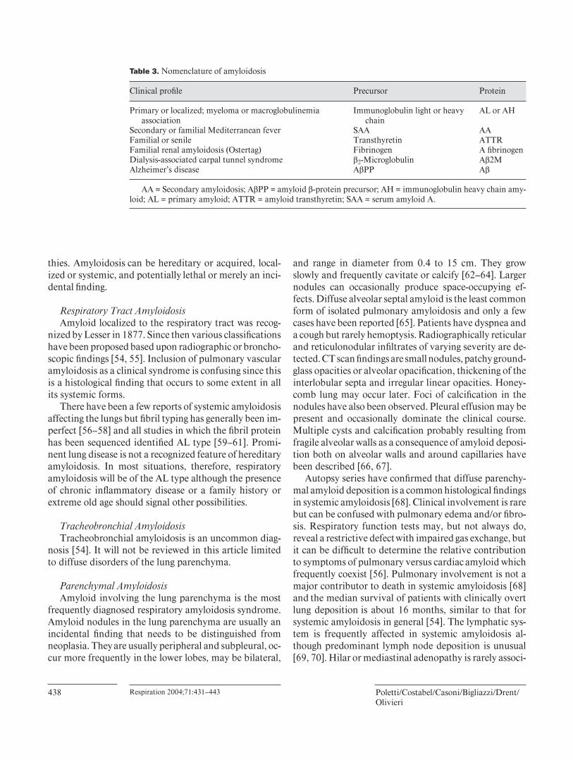
Table 3. Nomenclature of amyloidosis
Clinical profi le Precursor Protein
Primary or localized; myeloma or macroglobulinemia association
Immunoglobulin light or heavychain
AL or AH
Secondary or familial Mediterranean fever SAA AAFamilial or senile Transthyretin ATTRFamilial renal amyloidosis (Ostertag) Fibrinogen A fi brinogenDialysis-associated carpal tunnel syndrome ß2-Microglobulin Aß2MAlzheimer’s disease AßPP Aß
AA = Secondary amyloidosis; AßPP = amyloid ß-protein precursor; AH = immunoglobulin heavy chain amy-loid; AL = primary amyloid; ATTR = amyloid transthyretin; SAA = serum amyloid A.
Rare Diffuse Infi ltrative Lung Diseases Respiration 2004;71:431–443 439
ated with localized pulmonary amyloidosis [71] and its discovery should prompt a search for a systemic etiology. Amyloid lymphadenopathy can also represent localized AL deposition in association with solitary or multifocal B cell lymphomas. Pulmonary hypertension due to pul-monary artery occlusive involvement by amyloid depos-its has been described [54] . The diagnosis of amyloidosis usually requires histological confi rmation, and Congo red staining that produces green birefringence under crossed polarized light remains the gold standard [72] . Most tis-sue specimens, ranging from needle biopsies to open sur-gical resections, can be studied for amyloid although small biopsy specimens are open to signifi cant sampling error. Positive histology results for amyloid must be followed up by immunohistochemical analysis to determine the fi bril type [73] . Radiolabelled serum amyloid P compo-nent may help to localize specifi cally amyloid deposits in vivo; it is more sensitive for solid viscera such as the liv-er, kidneys and spleen [65, 74] . Intense fl uorodeoxyglu-cose activity on positron emission tomography has been described in amyloidosis [75] .
Once the diagnosis is clear, nodular parenchymal am-yloidosis rarely requires intervention [55] . In contrast, diffuse parenchymal amyloidosis is usually a systemic phenomenon with a poor prognosis [56] . Treatment with corticosteroids or irradiation does not infl uence its course [63] . However, assuming that amyloid deposits are of AL type chemotherapy to suppress the underlying plasma cell dyscrasia has to be considered [54, 76] .
Pulmonary Ossifi cation
Pulmonary calcifi cation and ossifi cation are relatively rare and often asymptomatic. Several predisposing condi-tions are associated with pulmonary parenchymal calcifi -cation with or without ossifi cation. These include hyper-calcemia, hyperphosphatemia, alkalosis, and lung injury in the presence or absence of conditions that result in an-giogenesis and increased pulmonary blood fl ow causing elevated vessel wall shear stress. The clinical states asso-ciated with pulmonary calcifi cation include other under-lying pulmonary diseases such as interstitial fi brosis, re-current bronchopneumonia, amyloidosis, or pulmonary edema (particularly with mitral stenosis), but also hyper-parathyroidism, chronic renal failure, hemodialysis, or-thotopic liver transplantation, granulomatous infection, infection by DNA viruses or parasites. It most common-ly affects the lower lobes. For some conditions, for ex-ample pulmonary fi brosis, the calcifi cation and ossifi ca-
tion may be a marker of disease severity and accelerated morbidity. It may also be idiopathic [77, 78] . Two histo-logical types of pulmonary ossifi cation have been de-scribed [79] : (1) a nodular circumscribed form and (2) a dendriform type.
The nodular form is characterized by lamellar deposits of calcifi ed osteoid material situated within the alveolar spaces often without marrow elements. The nodular form is typically associated with preexisting cardiac disorders that result in chronic pulmonary venous congestion such as mitral stenosis, chronic left ventricular failure, and id-iopathic hypertrophic subaortic stenosis [77, 78, 80, 81] . In contrast, dendriform ossifi cation refers to interstitial branching spicules of bone and marrow elements that may protrude into the alveoli [79] ( fi g. 5 ) and is usually idiopathic. Based on this simplifi ed morphologic classifi -cation for pulmonary ossifi cation, it is interesting to spec-ulate that some cases of IPO may represent a sequela of previously unidentifi ed lung injuries.
IPO is most often found in men over the age 60 but has also been reported in younger adults and in women. Familial clustering has been reported suggesting a genet-ic predisposition [82, 83] . Patients can be asymptomatic or have minimal complaints, and usually IPO represents an unexplained radiographic fi nding. Many cases are di-agnosed at autopsy. A restrictive pulmonary physiology with low diffusion capacity is present when disease is ex-tensive [82–84] . In the secondary forms, signs, symptoms, and the physiologic abnormalities are more likely due to
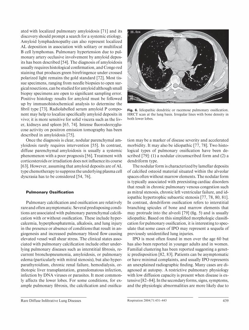
Fig. 6. Idiopathic dendritic or racemose pulmonary ossifi cation. HRCT scan at the lung basis. Irregular lines with bone density in both lower lobes.
Poletti/Costabel/Casoni/Bigliazzi/Drent/Olivieri
Respiration 2004;71:431–443 440
the accompanying disorder. It is uncommon for pulmo-nary ossifi cation to be seen on the chest radiograph. When present, it involves the lower lobes, appearing as nonspe-cifi c reticulonodular densities; CT scan fi ndings are more specifi c showing irregular lines with bone density preva-lent in the lower lobes [85] ( fi g. 6 ).
The pathogenesis of pulmonary ossifi cation is un-known. Serum calcium and phosphorus levels are usu-ally normal. Unlike heterotopic ossifi cation that occurs around joints in association with spinal cord injuries [86] , the serum alkaline phosphatase levels in pulmonary os-sifi cation are generally within normal limits, although this has not been consistently evaluated. In cases associated with pulmonary venous congestion, chronic intra-alveo-lar hemorrhage has been implicated as a predisposing fac-tor for subsequent fi brosis and ossifi cation [87] . Ossifi ca-tion is the sequela of a series of events beginning with degeneration of the arterial media, followed by infl am-mation and hyalinization of the perivascular tissue. Growth factors from cells involved in this extracellular matrix formation and resolution of infl ammation may also play a role in ossifi cation. Transforming growth fac-tor- ß is elaborated by infl ammatory macrophages and damaged epithelial cells and represents a critical growth factor for collagen and the extracellular matrix. Trans-forming growth factor- ß , strongly implicated in idiopath-ic pulmonary fi brosis and other fi brotic pulmonary dis-eases [88] , also stimulates osteoblast and chondrocyte proliferation. Another growth factor that may play an im-portant role in ectopic pulmonary ossifi cation is bone morphogenic protein, a member of the transforming growth factor- ß superfamily [89] . Bone morphogenic pro-tein, which is likely important in the development of fa-milial primary pulmonary hypertension [90] , induces ec-topic bone formation in the rat submandibular gland [91] . Interleukin-1 has also been shown to enhance bone mor-phogenic protein-induced hereotopic ossifi cation in labo-ratory animals [92] . The profi brotic cytokine interleu-kin-4, in conjunction with monocyte-colony-stimulating factor, may also transform human alveolar macrophages to osteoclasts, a cell important in bone remodeling [93] . Although the role of fi brogenic, angiogenic, and osteo-genic growth factors and cytokines in idiopathic and sec-ondary pulmonary ossifi cation has not been explored, their infl uence may potentially induce ossifi cation in fi -broproliferative pulmonary disorders such as idiopathic pulmonary fi brosis.
Pulmonary Alveolar Microlithiasis
Pulmonary alveolar microlithiasis (PAM), a rare dis-order of unknown etiology, is recognized by the intra-al-veolar accumulation of spherical calcifi ed concretions [94, 95] . Most patients are between 30 and 50 years of age when fi rst discovered. Although there is a familial asso-ciation in at least 50% of the cases, common environmen-tal factors could also account for this observation [96, 97] . This disease is especially prevalent in Turkey, represent-ing 33% of the world literature [98] . There is no evidence that infection plays a role. An isolated inborn error of calcium metabolism in the lungs has been proposed, but circulating calcium and phosphorus levels are consistent-ly normal in PAM. It is speculated that, due to an un-known stimulus, changes in the alveolar lining membrane or secretions result in greater alkalinity, promoting intra-alveolar precipitation of calcium phosphates and carbon-ates [99] .
Asymptomatic cases, even with extensive radiograph-ic involvement, are often discovered incidentally. Cough and dyspnea are the most common presenting symptoms and usually occur late in the course of the disease. Normal or mild restrictive pulmonary physiology may be present in the asymptomatic individual. With progressive disease, severe lung restriction may ensue with impairment of the diffusing capacity and gas exchange abnormalities. The chest radiograph shows bilateral, sand-like, micronodular calcifi ed densities known as microliths or calcispherites, which are usually less than 1 mm in diameter [100] . They appear concentrated in the lower two thirds of the lung, often obliterating the diaphragmatic, mediastinal, and cardiac borders. The greater radiographic density at the lung bases is likely due to the larger lower lobe volumes rather than selective predisposition. The predominant HRCT fi ndings are intra-alveolar calcifi cations with a sub-pleural posterior and lower lobe predominance. A peri-lobular and centrilobular distribution of the calcifi cations may be seen [100] . HRCT may in addition reveal ground-glass opacities that are interspersed with microcysts and the calcispherites [100] . In addition to the fi ne nodulation, HRCT may show polygonal-shaped calcifi ed densities caused by the accumulation of microliths in the periphery of the lobules rather than actual thickening or deposition of calcium within alveolar septa [100, 101] . Although 99m Tc bone scintigraphy can also help confi rm the calcifi c nature of the lesions, the standard chest radiograph is of-ten characteristic for PAM. The diagnosis is made on the basis of the characteristic chest radiographic and HRCT fi ndings; this usually obviates lung biopsy.

Rare Diffuse Infi ltrative Lung Diseases Respiration 2004;71:431–443 441
Identifi cation of microliths in expectorated sputum or BAL is diagnostic. Histologically, the lesion of PAM con-sists of intra-alveolar calcispherites, which represent lam-inated calcium phosphate concretions. This appearance is distinct from metastatic and dystrophic calcifi cations in which the calcifi cation is in the interstitial or vascular compartments. With progression, interstitial infl amma-tion and fi brosis will occur and result in signifi cantly di-minished lung volumes, sometimes fi nger clubbing and eventually right heart failure. There is no known therapy for PAM. Corticosteroids, chelating agents, and BAL
have demonstrated no benefi t, and the role for the use of bisphosphonates remains to be proven [102] . The few cases with response to corticosteroids are more likely to be related to attenuation of the accompanying interstitial disease [103] . In symptomatic cases, nasal continuous positive airway pressure improves gas exchange by de-creasing the physiologic intrapulmonary shunt [104] . Bi-lateral lung transplantation is a viable option for far ad-vanced cases.
References
1 Zompatori M, Bnà C, Poletti V, Spaggiari E, Ormitti F, Calabrò E, Tognini G, Sverzellati N: Diagnostic imaging of diffuse infi ltrative dis-ease of the lung. Respiration 2004;71:4–19.
2 Chetta A, Marangio E, Olivieri D: Pulmonary function testing in interstitial lung diseases. Respiration 2004;71:209–213.
3 Poletti V, Chilosi M, Olivieri D: Diagnostic invasive procedures in diffuse infi ltrative lung diseases. Respiration 2004;71:107–119.
4 Camus P, Fanton A, Bonniaud P, Camus C, Foucher P: Interstitial lung disease induced by drugs and radiation. Respiration 2004;71:301–326.
5 Rosen SH, Castleman B, Liebow AA: Pulmo-nary alveolar proteinosis. N Engl J Med 1958; 258: 1123–1142.
6 Costello JF, Moriarty DC, Branthwaite MA, Turner-Warwick M, Corrin B: Diagnosis and management of alveolar proteinosis: The role of electron microscopy. Thorax 1975; 30: 121–132.
7 Maygarden SJ, Iacocca MV, Funkhouser WK, Novotny DB: Pulmonary alveolar proteinosis: A spectrum of cytologic, histochemical, and ul-trastructural fi ndings in bronchoalveolar la-vage fl uid. Diagn Cytopathol 2001; 24: 389–395.
8 Prakash UB, Barham SS, Carpenter HA, Dines DE, Marsh MH: Pulmonary alveolar phospho-lipoproteinosis: Experience with 34 cases and a review. Mayo Clin Proc 1987; 62: 499–518.
9 Alberti A, Luisetti M, Braschi A, Rodi G, Lot-ti G, Sella D, Poletti V, Benori V, Baritussio A: Bronchoalveolar lavage fl uid composition in alveolar proteinosis. Early changes after thera-peutic lavage. Am J Respir Crit Care Med 1996; 154: 817–820.
10 Honda Y, Kuroki Y, Matsuura E, Nagae H, Takahashi H, Akino T, Abe S: Pulmonary sur-factant protein D in sera and bronchoalveolar lavage fl uids. Am J Respir Crit Care Med 1995; 152: 1860–1866.
11 Honda Y, Takahashi H, Shijubo N, Kuroki Y, Akino T: Surfactant protein-A concentration in bronchoalveolar lavage fl uids of patients with pulmonary alveolar proteinosis. Chest 1993; 103: 496–499.
12 Trapnell BC, Whitsett JA, Nakata K: Pulmo-nary alveolar proteinosis. N Engl J Med 2003; 349: 2527–2539.
13 Reed JA, Ikegami M, Cianciolo ER, Lu W, Cho PS, Hull W, Jobe AH, Whitsett JA: Aerosolized GM-CSF ameliorates pulmonary alveolar pro-teinosis in GM-CSF-defi cient mice. Am J Physiol 1999; 276:L556–L563.
14 Huffman JA, Hull WM, Dranoff G, Mulligan RC, Whitsett JA: Pulmonary epithelial cell ex-pression of GM-CSF corrects the alveolar pro-teinosis in GM-CSF-defi cient mice. J Clin In-vest 1996; 97: 649–655.
15 Nishinakamura R, Wiler R, Dirksen U, Mori-kawa Y, Arai K, Miyajima A, Burdach S, Mur-ray R: The pulmonary alveolar proteinosis in granulocyte macrophage colony-stimulating factor/interleukins 3/5 beta c receptor-defi -cient mice is reversed by bone marrow trans-plantation. J Exp Med 1996; 183: 2657–2662.
16 Dirksen U, Nishinakamura R, Groneck P, Hattenhorst U, Nogee L, Murray R, Burdach S: Human pulmonary alveolar proteinosis as-sociated with a defect in GM-CSF/IL-3/IL-5 receptor common beta chain expression. J Clin Invest 1997; 100: 2211–2217.
17 Bewig B, Wang XD, Kirsten D, Dalhoff K, Schafer H: GM-CSF and GM-CSF beta c re-ceptor in adult patients with pulmonary alveo-lar proteinosis. Eur Respir J 2000; 15: 350–357.
18 Kitamura T, Tanaka N, Watanabe J, et al: Id-iopathic pulmonary alveolar proteinosis as an autoimmune disease with neutralizing anti-body against granulocyte/macrophage colony-stimulating factor. J Exp Med 1999; 190: 875–880.
19 Bonfi eld TL, Russell D, Burgess S, Malur A, Kavuru MS, Thomassen MJ: Autoantibodies against granulocyte macrophage colony-stimu-lating factor are diagnostic for pulmonary al-veolar proteinosis. Am J Respir Cell Mol Biol 2002; 27: 481–486.
20 Kavuru MS, Sullivan EJ, Piccin R, Thomassen MJ, Stoller JK: Exogenous granulocyte-macro-phage colony-stimulating factor administra-tion for pulmonary alveolar proteinosis. Am J Respir Crit Care Med 2000; 161: 1143–1148.
21 Seymour JF, Presneill JJ, Schoch OD, et al: Therapeutic effi cacy of ganulocyte-macro-phage colony-stimulating factor in patients with idiopathic acquired alveolar proteinosis. Am J Respir Crit Care Med 2001; 163: 524–531.
22 Costabel U, Corrin B: Alveolar proteinosis; in Gibson GJ, Geddes DM, Costabel U, Sterk PJ, Corrin B (eds): Respiratory Medicine, ed 3. London, Saunders, 2003, pp 1676–1682.
23 Du Bois RM, McAllister WA, Branthwaite MA: Alveolar proteinosis: Diagnosis and treat-ment over a 10-year period. Thorax 1983; 38: 360–363.
24 Kariman K, Kylstra JA, Spock A: Pulmonary alveolar proteinosis: Prospective clinical expe-rience in 23 patients for 15 years. Lung 1984; 162: 223–231.
25 Seymour JF, Presneill JJ: Pulmonary alveolar proteinosis: Progress in the fi rst 44 years. Am J Respir Crit Care Med 2002; 166: 215–235.
26 Lee KN, Levin DL, Webb WR, Chen D, Stor-to ML, Golden JA: Pulmonary alveolar pro-teinosis: High-resolution CT, chest radio-graphic, and functional correlations. Chest 1997; 111: 989–995.
27 Kitamura T, Uchida K, Tanaka N, Tsuchiya T, Watanabe J, Yamada Y, Hanaoka K, Sey-mour JF, Schoch OD, Doyle I, Inoue Y, Saka-tani M, Kudoh S, Azuma A, Nukiwa T, To-mita T, Katagiri M, Fujita A, Kurashima A, Kanegasaki S, Nakata K: Serological diagnosis of idiopathic pulmonary alveolar proteinosis. Am J Respir Crit Care Med 2000; 162: 658–662.
28 Pattle RE: Properties, function and origin of the alveolar lining layer. Nature 1955; 175: 1125–1126.
29 Rogers RM, Levin DC, Gray BA, Moseley LW Jr: Physiologic effects of bronchopulmonary lavage in alveolar proteinosis. Am Rev Respir Dis 1978; 118: 255–264.
Poletti/Costabel/Casoni/Bigliazzi/Drent/Olivieri
Respiration 2004;71:431–443 442
30 Robertson HE: Pulmonary alveolar proteino-sis. Can Med Assoc J 1965; 93: 980–983.
31 Smith LJ, Katzenstein AL, Ankin MG, Shap-iro BA: Management of pulmonary alveolar proteinosis: Clinical conference in pulmonary disease from Northwest University McGaw Medical Center, Chicago. Chest 1980; 78: 765–770.
32 Kao D, Wasserman K, Costley D, Benfi eld JR: Advances in the treatment of pulmonary al-veolar proteinosis. Am Rev Respir Dis 1975; 111: 361–363.
33 Miller A, Brown LK, Pastores GM, Desnick RJ: Pulmonary involvement in type 1 Gaucher disease: Functional and exercise fi ndings in pa-tients with and without clinical interstitial lung disease. Clin Genet 2003; 63: 368–376.
34 Santamaria F, Parenti G, Guidi G, Filocamo M, Strisciuglio P, Grillo G, Farina V, Sarnelli P, Rizzolo MG, Rotondo A, Andria G: Pulmo-nary manifestations of Gaucher disease: An increased risk for L444P homozygotes? Am J Respir Crit Care Med 1998; 157: 985–989.
35 Mistry PK, Sirrs S, Chan A, Pritzker MR, Duffy TP, Grace ME, Meeker DP, Goldman ME: Pulmonary hypertension in type 1 Gau-cher’s disease: Genetic and epigenetic determi-nants of phenotype and response to therapy. Mol Genet Metab 2002; 77: 91–98.
36 Zimran A, Elstein D: Gaucher disease and the clinical experience with substrate reduction therapy. Philos Trans R Soc Lond B Biol Sci 2003; 358: 961–966.
37 Gonzalez-Reimers E, Sanchez-Perez MJ, Bo-nilla-Arjona A, Rodriguez-Gaspar M, Carras-co-Juan JL, Alvarez-Arguelles H, Santolaria-Fernandez F: Case report. Pulmonary involvement in an adult male affected by type B Niemann-Pick disease. Br J Radiol 2003; 76: 838–840.
38 Millat G, Chikh K, Naureckiene S, Sleat DE, Fensom AH, Higaki K, Elleder M, Lobel P, Vanier MT: Niemann-Pick disease type C: Spectrum of HE1 mutations and genotype/phenotype correlations in the NPC2 group. Am J Hum Genet 2001; 69: 1013–1021.
39 Brown LK, Miller A, Bhuptani A, Sloane MF, Zimmerman MI, Schilero G, Eng CM, Desnick RJ: Pulmonary involvement in Fabry disease. Am J Respir Crit Care Med 1997; 155: 1004–1010.
40 Kelly MM, Leigh R, McKenzie R, Kamada D, Ramsdale EH, Hargreave FE: Induced sputum examination: Diagnosis of pulmonary involve-ment in Fabry’s disease. Thorax 2000; 55: 720–721.
41 Desnick RJ, Brady R, Barranger J, Collins AJ, Germain DP, Goldman M, Grabowski G, Packman S, Wilcox WR: Fabry disease, an un-der-recognized multisystemic disorder: Expert recommendations for diagnosis, management, and enzyme replacement therapy. Ann Intern Med 2003; 138: 338–346.
42 Huizing M, Boissy RE, Gahl WA: Hermansky-Pudlak syndrome: Vesicle formation from yeast to man. Pigment Cell Res 2002; 15: 405–419.
43 Witkop CJ, Nunez Babcock M, Rao GH, Gaudier F, Summers CG, Shanahan F, Har-mon KR, Townsend D, Sedano HO, King RA, et al: Albinism and Hermansky-Pudlak syn-drome in Puerto Rico. Bol Asoc Med PR 1990; 82: 333–339.
44 Gahl WA, Brantly M, Kaiser-Kupfer MI, Iwa-ta F, Hazelwood S, Shotelersuk V, Duffy LF, Kuehl EM, Troendle J, Bernardini I: Genetic defects and clinical characteristics of patients with a form of oculocutaneous albinism (Her-mansky-Pudlak syndrome). N Engl J Med 1998; 338: 1258–1264.
45 Huizing M, Helip-Wooley A, Dorward H, Claassen D, Hess R, Gahl WA: IL-25 Herman-sky-Pudlak syndrome: A model for abnormal vesicle formation and traffi cking. Pigment Cell Res 2003; 16: 584.
46 Di Pietro SM, Falcon-Perez JM, Dell’Angelica EC: Characterization of BLOC-2, a complex containing the Hermansky-Pudlak syndrome proteins HPS3, HPS5 and HPS6. Traffi c 2004; 5: 276–283.
47 Avila NA, Brantly M, Premkumar A, Huizing M, Dwyer A, Gahl WA: Hermansky-Pudlak syndrome: Radiography and CT of the chest compared with pulmonary function tests and genetic studies. AJR Am J Roentgenol 2002; 179: 887–892.
48 Nakatani Y, Nakamura N, Sano J, et al: Inter-stitial pneumonia in Hermansky-Pudlak syn-drome: Signifi cance of fl orid foamy swelling/degeneration (giant lamellar body degenera-tion) of type II pneumocytes. Virchows Arch 2000; 437: 304–313.
49 Gahl WA, Brantly M, Troendle J, et al: Effect of pirfenidone on the pulmonary fi brosis of Hermansky-Pudlak syndrome. Mol Genet Metab 2002; 76; 234–242.
50 Pope-Harman AL, Davis WB, Allen ED, Christoforidis AJ, Allen JN: Acute eosinophil-ic pneumonia. A summary of 15 cases and re-view of the literature. Medicine (Baltimore) 1996; 75: 334.
51 Shintani H, Fujimura M, Yasui M, Ueda K, Kameda S, Noto M, Matsuda T, Kobayashi M: Acute eosinophilic pneumonia caused by ciga-rette smoking. Intern Med 2000; 39/1: 66–68.
52 Philit F, Etienne-Mastroianni B, Parrot A, Guerin C, Robert D, Cordier JF: Idiopathic acute eosinophilic pneumonia: A study of 22 patients. Am J Respir Crit Care Med 2002; 166: 1235–1239.
53 Trisolini R, Cancellieri A, Bonaccorsi A, Po-letti V: Bronchoalveolar lavage suggesting dif-fuse alveolar damage in a patient with acute eosinophilic pneumonia. Sarcoidosis Vasc Dif-fuse Lung Dis 2001; 18: 311–312.
54 Utz JP, Swensen SJ, Gertz MA: Pulmonary amyloidosis: The Mayo Clinic experience from 1980–1993. Ann Intern Med 1996; 124: 407–413.
55 Thompson PJ, Citron KM: Amyloid and the lower respiratory tract. Thorax 1983; 38: 84–87.
56 Smith RR, Hutchins GM, Moore GW, et al: Type and distribution of pulmonary parenchy-mal and vascular amyloid. Am J Med 1979; 66: 96–104.
57 Hui AN, Koss MH, Hochholzer L, Wehunt WD: Amyloidosis presenting in the lower respiratory tract. Clinicopathologic, radiolog-ic, immunohistochemical and histochemical studies on 48 cases. Arch Pathol Lab Med 1986; 110: 212–218.
58 Fukumura M, Mieno T, Suzuki T, et al: Pri-mary diffuse tracheobronchial amyloidosis treated by bronchoscopic Nd-YAG laser irra-diation. Jpn J Med 1990; 29: 620–622.
59 Schulz C, Hauck RW, Nathrath WB, et al: Combined amyloidosis of the upper and lower respiratory tract. Respiration 1995; 62: 163–166.
60 Troxler RF, Kane K, Berg AM, et al: Localised amyloidosis of the larynx: Evidence for light chain composition. Ann Otol Rhinol Laryngol 1993; 102: 884–889.
61 Toyoda M, Ebihara Y, Kato H, et al: Tracheo-bronchial AL amyloidosis: Histological, im-munohistochemical, ultrastructural and im-munoelectron microscopic observations. Hum Pathol 1993; 24: 970–976.
62 Himmelfarb E, Wells S, Rabinowitz JG: The radiologic spectrum of cardiopulmonary amy-loidosis. Chest 1977; 72: 327–332.
63 Rubinow A, Celli BR, Cohen AS, et al: Lo-calised amyloidosis of the lower respiratory tract. Am Rev Respir Dis 1978; 118: 603–611.
64 Ayuso MC, Gilabert R, Bombi JA, et al: CT appearance of localised pulmonary amyloido-sis. J Comput Assist Tomogr 1987; 11: 197–199.
65 Gillmore JD, Hawkins PH: Amyloidosis and the respiratory tract. Thorax 1999; 54: 444–451.
66 Pickford HA, Swensen SJ, Utz GP: Thoracic cross-sectional imaging of amyloidosis. AJR Am J Roentgenol 1997; 168: 351–355.
67 Ohdama S, Akagawa S, Matsubara O, Yoshiza-wa Y: Primary diffuse alveolar septal amyloi-dosis with multiple cysts and calcifi cation. Eur Respir J 1996; 7: 1569–1571.
68 Celli R, Rubinow A, Cohen AS, et al: Patterns of pulmonary involvement in systemic amyloi-dosis. Chest 1978; 74: 543–547.
69 Desai RA, Mahajan VK, Benjamin S, et al: Pul-monary amyloidoma and hilar adenopathy. Chest 1979; 76: 170–173.
70 Khan JA, Shamsi SH, Rana TA, et al: Pulmo-nary amyloidosis: A case with hilar and medi-astinal involvement and review of the litera-ture. Clin Pulm Med 1996; 2: 66–69.
71 Gallego FG, Canelas JC: Hilar enlargement in amyloidosis. N Engl J Med 1974; 291: 531.
72 Puchtler H, Sweat F, Levine M: On the binding of Congo red by amyloid. J Histochem Cyto-chem 1962; 10: 355–364.
73 Tan SY, Pepys MB: Amyloidosis. Histopathol-ogy 1994; 25: 403–414.
Rare Diffuse Infi ltrative Lung Diseases Respiration 2004;71:431–443 443
74 Hawkins PN, Myers MJ, Lavender JP, Pepys MB: Diagnostic radionuclide imaging of amy-loid: Biological targeting by circulating human serum amyloid P component. Lancet 1988;i:1413–1418.
75 Kung J, Zhuang H, Yu JQ, Duarte PS, Alavi A: Intense fl uorodeoxyglucose activity in pul-monary amyloidosis on positron emission to-mography. Clin Nucl Med 2003; 12: 975–976.
76 Gertz MA, Lacy MQ, Dispenzieri A: Immuno-globulin light-chain amyloidosis – Primary amyloidosis; in Greer JP, Foerster J, Lukens JN, Rodgers GM, Paraskevas F, Glader B (eds): Wintrobe’s Clinical Hematology, ed 11. Philadelphia, Lippincott Williams & Wilkins, 2004, pp 2637–2665.
77 Buja LM, Roberts WC: Pulmonary parenchy-mal ossifi c nodules in idiopathic hypertrophic subaortic stenosis. Am J Cardiol 1970; 25: 710–715.
78 Popelka CG, Kleinerman J: Diffuse pulmo-nary ossifi cation. Arch Intern Med 1977; 137: 523–525.
79 Ndimbie OK, Williams CR, Lee MW: Dendri-form pulmonary ossifi cation. Arch Pathol Lab Med 1987; 111: 1062–1064.
80 Galloway R, Epstein EJ, Coulshed N: Pulmo-nary ossifi c nodules in mitral valve disease. Br Heart J 1961; 23: 297–307.
81 Whitaker W, Black A, Warrack AJN: Pulmo-nary ossifi cation in patients with mitral steno-sis. J Fac Radiol 1955; 7: 29–34.
82 Azuma A, Miyamoto H, Enomoto T, Usuki J, Kudoh S: Familial clustering of dendriform pulmonary ossifi cation. Sarcoidosis Vasc Dif-fuse Lung Dis 2003; 20: 152–154.
83 Joines RW, Roggli VL: Dendriform pulmo-nary ossifi cation: Report of two cases with unique fi ndings. Am J Clin Pathol 1989; 91: 398–402.
84 Rajjoub S, Altmeyer RB: A case report of idio-pathic pulmonary ossifi cation. WV Med J 1998; 94: 143–145.
85 Gevenois PA, Abehsera M, Knoop C, Jacobo-vitz D, Estenne M: Disseminated pulmonary ossifi cation in end-stage pulmonary fi brosis: CT demonstration. AJR Am J Roentgenol 1994; 162: 1303–1304.
86 Kim SW, Charter RA, Chai CJ, Kim SK, Kim ES: Serum alkaline phosphatase and inorganic phosphorus values in spinal cord injury pa-tients with heterotopic ossifi cation. Paraplegia 1990; 28: 441–447.
87 Green JD, Harle TS, Greenberg SD, Weg JG, Nevin H, Jenkins DE: Disseminated pulmo-nary ossifi cation. Am Rev Respir Dis 1970; 101: 293–298.
88 Schwarz MI, King TE: Interstitial Lung Dis-ease, ed 3. Hamilton, Decker, 1998.
89 Maiti SK, Singh GR: Bone morphogenic pro-teins: Novel regulators of bone formation. In-dian J Exp Biol 1998; 36: 237–244.
90 Lane KB, Machado RD, Pauciulo MW, Thom-son JR, Phillips JA, Loyd JE, Nichols WC, Trembath RC: Heterozygous germline muta-tions in BMPR2, encoding a TGF-beta recep-tor, cause familial primary pulmonary hyper-tension. Nat Genet 2000; 26: 81–84.
91 Muramatsu T, Hamano H, Fukumashi K, Shi-igai T, Fujiseki M, Katayanagi T, Osada K, Inoue T, Shimono M: An experimental study of chondrogenesis and osteogenesis in rat sub-mandibular gland induced by implantation of demineralized dentin. Bull Tokyo Dent Coll 1993; 34: 15–22.
92 Mahy PR, Ursit MR: Experimental heterotop-ic bone formation induced by bone morphoge-netic protein and recombinant human inter-leukin-1B. Clin Orthop 1988; 237: 236–244.
93 Akagawa KS, Takasuka N, Nozaki Y, Komuro I, Azuma M, Ueda M, Naito M, Takahashi K: Generation of CD1+RelB+ dendritic cells and tartrate-resistant acid phosphatase-positive os-teoclast-like multi-nucleated giant cells from human monocytes. Blood 1996; 88: 4029–4039.
94 Barnard NJ, Crocker PR, Blainey AD, Davies RJ, Ell SR, Levison DA: Pulmonary alveolar microlithiasis: A new analytical approach. His-topathology 1987; 11: 639–645.
95 Castellana G, Lamorgese V: Pulmonary al-veolar microlithiasis. Respiration 2003; 70: 549–555.
96 Coetzee T: Pulmonary alveolar microlithiasis with involvement of the sympathetic nervous system and gonads. Thorax 1970; 25: 637–642.
97 Sosman MC, Dodd GD, Jones WD, Pillmore GU: The familial occurrence of pulmonary alveolar microlithiasis. AJR Am J Roentgen-ol 1957; 77: 947–1012.
98 Ucan ES, Keyf AI, Aydilek R, Yalcin Z, Sebit S, Kudu M, Ok U: Pulmonary alveolar micro-lithiasis: Review of Turkish reports. Thorax 1993; 48: 171–173.
99 Bogart SD: Disseminated pulmonary calcino-sis with pulmonary alveolar microlithiasis. NY State J Med 1980; 80: 1283–1284.
100 Cluzel P, Grenier P, Bernadac P, Laurent F, Picard JD: Pulmonary alveolar microlithia-sis: CT fi ndings. J Comput Assist Tomogr 1991; 15: 938–942.
101 Chai JL, Patz EF: CT of the lung: Patterns of calcifi cation and other high-attenuation ab-normalities. AJR Am J Roentgenol 1994; 162: 1063–1066.
102 Gocmen A, Toppare MF, Kiper N, Buyuk-pamukcu N: Treatment of pulmonary alveo-lar microlithiasis with a diphosphonate: Pre-liminary results of a case. Respiration 1992; 59: 250–252.
103 Richardson J, Slovis B, Miller G, Dummer S: Development of pulmonary alveolar micro-lithiasis in a renal transplant recipient. Trans-plantation 1995; 59: 1056–1057.
104 Freiberg DB, Young IH, Laks L, Regnis JA, Lehrhaft B, Sullivan CE: Improvement in gas exchange with nasal continuous positive air-way pressure in pulmonary alveolar micro-lithiasis. Am Rev Respir Dis 1992; 145: 1215–1216.