rapid, realtime icu and er eeg monitoring of california, riverside ca rapid, realtime icu and er eeg...
TRANSCRIPT
Kenneth G. Jordan, MD, FACP, FACNS, FAAN President, Jordan NeuroScience , Inc.
Assoc. Clin. Prof of Biomedical Science, Neurology University of California, Riverside CA
Rapid, Realtime ICU and ER EEG Monitoring
ASET Review Course 2007
• Principal shareholder of Jordan NeuroScience, Inc., a medical device manufacturer in the field of acute EEG monitoring
• Certain JNS proprietary products and methods are included in this presentation.
• No money or fees from outside sources
DisclosureDisclosure

What is Emergency EEG Monitoring (EmEEG)?
1. EEG that is done acutely for new onset or rapidly deteriorating brain dysfunction.
2. EEG done within a critical time-window to reverse, reduce or prevent brain damage.
3. EEG surveillance of intervention during acute or unstable brain injury.
“Dost thou love life? Then do not squander time, for that is the stuff life is made of.” Benjamin Franklin
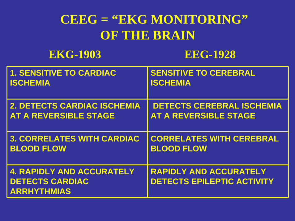
1. SENSITIVE TO CARDIAC ISCHEMIA
SENSITIVE TO CEREBRAL ISCHEMIA
2. DETECTS CARDIAC ISCHEMIA AT A REVERSIBLE STAGE
DETECTS CEREBRAL ISCHEMIA AT A REVERSIBLE STAGE
3. CORRELATES WITH CARDIAC BLOOD FLOW
CORRELATES WITH CEREBRAL BLOOD FLOW
4. RAPIDLY AND ACCURATELY DETECTS CARDIAC ARRHYTHMIAS
RAPIDLY AND ACCURATELY DETECTS EPILEPTIC ACTIVITY
EKG-1903 EEG-1928
CEEG = “EKG MONITORING”OF THE BRAIN
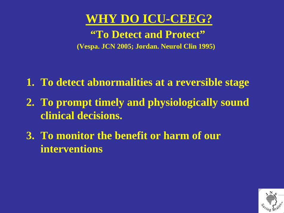
WHY DO ICU-CEEG?“To Detect and Protect”
(Vespa. JCN 2005; Jordan. Neurol Clin 1995)
1. To detect abnormalities at a reversible stage
2. To prompt timely and physiologically sound clinical decisions.
3. To monitor the benefit or harm of our interventions
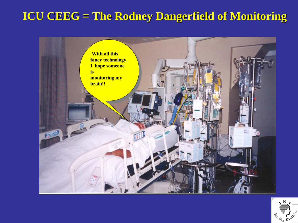
With all this fancy technology, I hope someone ismonitoring my brain!!
With all this fancy technology, I hope someone ismonitoring my brain!!
ICU CEEG = The Rodney Dangerfield of MonitoringICU CEEG = The Rodney Dangerfield of Monitoring
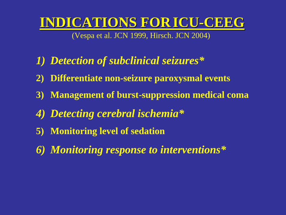
INDICATIONS FORINDICATIONS FOR ICUICU--CEEGCEEG(Vespa et al. JCN 1999, Hirsch. JCN 2004)
1) Detection of subclinical seizures*2) Differentiate non-seizure paroxysmal events
3) Management of burst-suppression medical coma
4) Detecting cerebral ischemia*5) Monitoring level of sedation
6) Monitoring response to interventions*
Detection of Detection of SubclincalSubclincal SeizuresSeizures
The The ““Critical Care EMUCritical Care EMU””
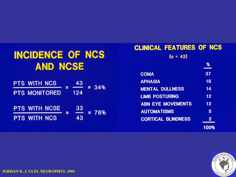
JORDAN K. J. CLIN. NEUROPHYS. 1993
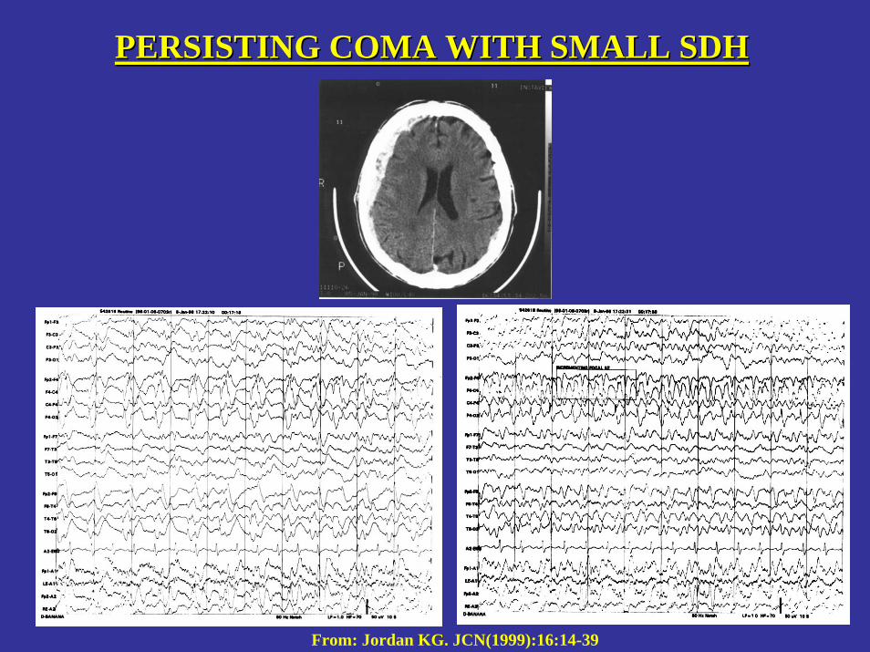
PERSISTING COMA WITH SMALL SDHPERSISTING COMA WITH SMALL SDH
From: Jordan KG. JCN(1999):16:14-39
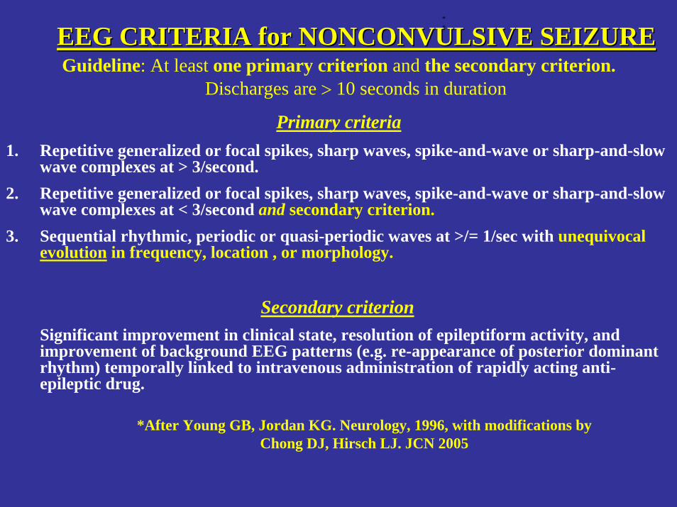
EEG CRITERIA for NONCONVULSIVE SEIZUREEEG CRITERIA for NONCONVULSIVE SEIZUREGuideline: At least one primary criterion and the secondary criterion.
Discharges are > 10 seconds in duration
Primary criteria1. Repetitive generalized or focal spikes, sharp waves, spike-and-wave or sharp-and-slow
wave complexes at > 3/second.2. Repetitive generalized or focal spikes, sharp waves, spike-and-wave or sharp-and-slow
wave complexes at < 3/second and secondary criterion.3. Sequential rhythmic, periodic or quasi-periodic waves at >/= 1/sec with unequivocal
evolution in frequency, location , or morphology.
Secondary criterionSignificant improvement in clinical state, resolution of epileptiform activity, and improvement of background EEG patterns (e.g. re-appearance of posterior dominant rhythm) temporally linked to intravenous administration of rapidly acting anti-epileptic drug.
**
*After Young GB, Jordan KG. Neurology, 1996, with modifications by Chong DJ, Hirsch LJ. JCN 2005
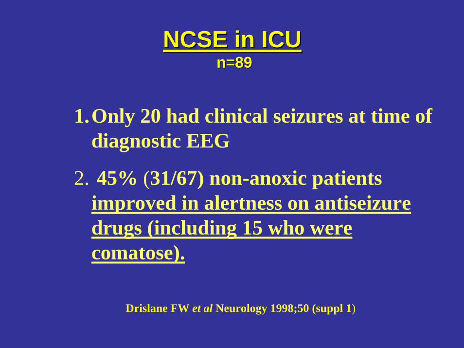
NCSE in ICUNCSE in ICUn=89n=89
1.Only 20 had clinical seizures at time of diagnostic EEG
2. 45% (31/67) non-anoxic patients improved in alertness on antiseizuredrugs (including 15 who were comatose).
Drislane FW et al Neurology 1998;50 (suppl 1)
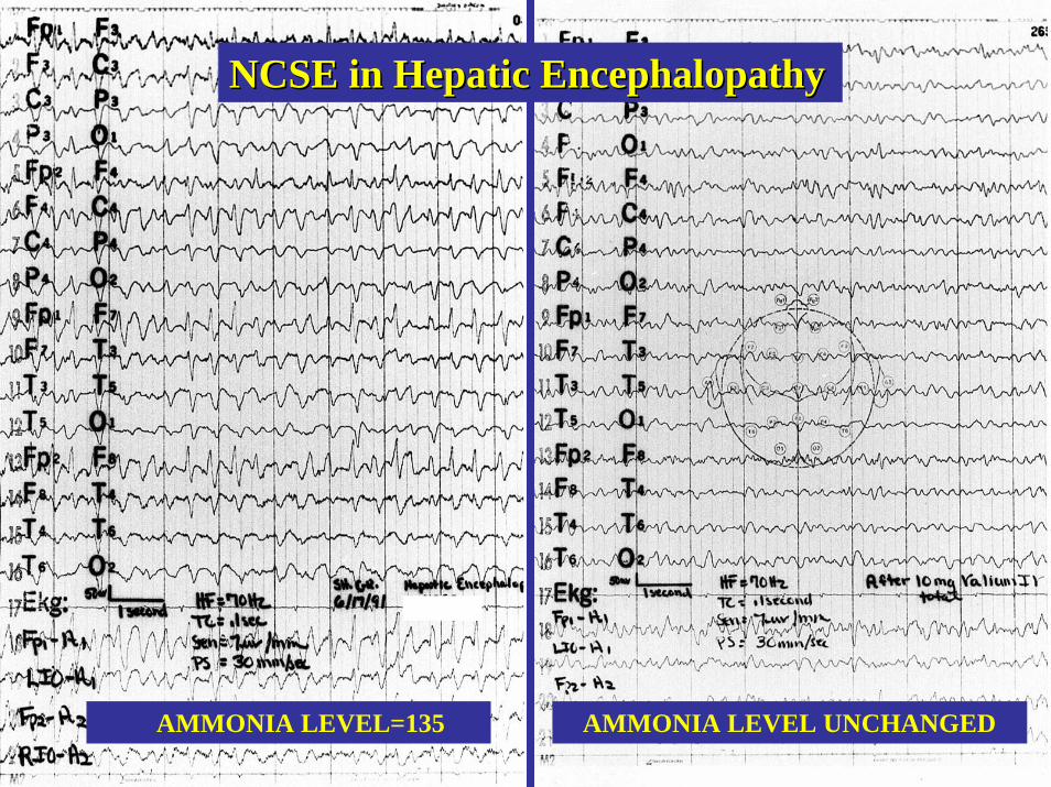
AMMONIA LEVEL=135 AMMONIA LEVEL UNCHANGED
NCSE in Hepatic EncephalopathyNCSE in Hepatic Encephalopathy
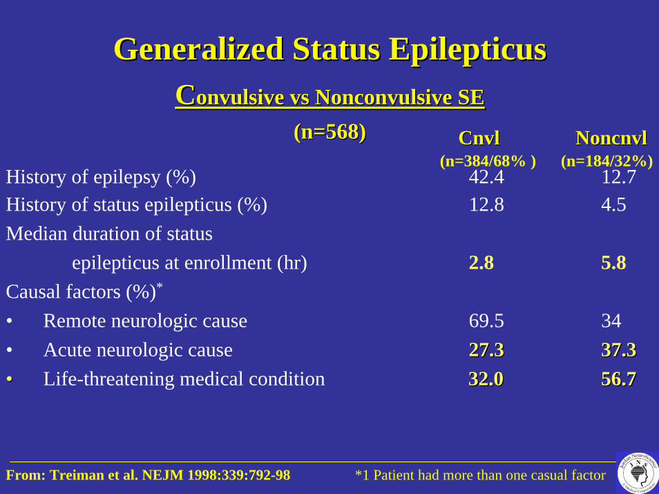
CnvlCnvl NoncnvlNoncnvl(n=384/68% ) (n=184/32%)
History of epilepsy (%) 42.4 12.7History of status epilepticus (%) 12.8 4.5Median duration of status
epilepticus at enrollment (hr) 2.8 5.8Causal factors (%)*
• Remote neurologic cause 69.5 34• Acute neurologic cause 27.327.3 37.337.3•• Life-threatening medical condition 32.032.0 56.756.7
From: Treiman et al. NEJM 1998:339:792-98 *1 Patient had more than one casual factor
Generalized Status Generalized Status EpilepticusEpilepticusCConvulsive onvulsive vsvs Nonconvulsive SE Nonconvulsive SE
(n=568)(n=568)

NCSE in Comatose PatientsNCSE in Comatose Patients((n = 610 patients)n = 610 patients)
1. 33% (40/119) of comatose children were in NCSE.
2. 22% (108/491) of comatose adults were in NCSE.
Hosain SA, (Abstr) American Epilepsy Society Meeting 2002

EKG
RESP
CHIN EMG
L EYE
R EYE
FP1-C3
C3-01
FP1-T3
T3-01
FP2-C4
C4-O2
FP2-T4
T4-O2
F7-FZ
FZ-F8
T3-CZ
CZ-T4
T5-PZ
PZ-T6
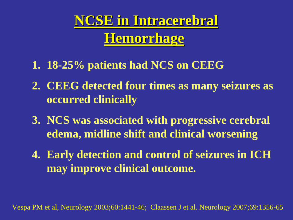
NCSE in NCSE in IntracerebralIntracerebralHemorrhageHemorrhage
1. 18-25% patients had NCS on CEEG
2. CEEG detected four times as many seizures as occurred clinically
3. NCS was associated with progressive cerebral edema, midline shift and clinical worsening
4. Early detection and control of seizures in ICH may improve clinical outcome.
Vespa PM et al, Neurology 2003;60:1441-46; Claassen J et al. Neurology 2007;69:1356-65
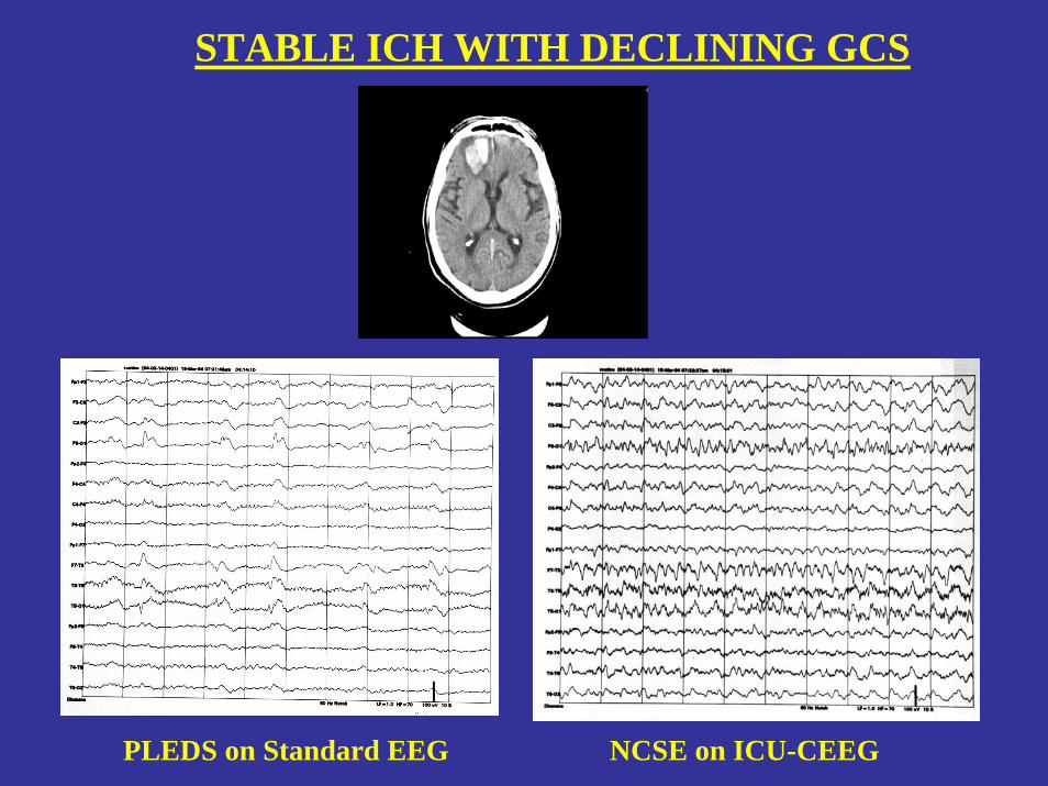
STABLE ICH WITH DECLINING GCS
PLEDS on Standard EEG NCSE on ICU-CEEG
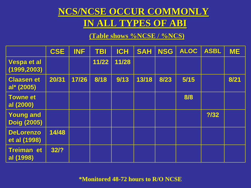
CSECSE INFINF
17/2620/31
14/48
32/?
TBITBI ICHICH SAHSAH NSGNSG ALOCALOC ASBLASBL MEMEVespa et al Vespa et al (1999,2003)(1999,2003)
11/22 11/28
8/23
Towne et Towne et al (2000)al (2000)
8/8
8/21
DeLorenzoDeLorenzoet al (1998)et al (1998)TreimanTreiman et et al (1998)al (1998)
?/32
5/15ClaasenClaasen et et al* (2005)al* (2005)
8/18 9/13 13/18
Young and Young and DoigDoig (2005)(2005)
NCS/NCSE OCCUR COMMONLY NCS/NCSE OCCUR COMMONLY IN ALL TYPES OF ABI IN ALL TYPES OF ABI
(Table shows %NCSE / %NCS)(Table shows %NCSE / %NCS)
*Monitored 48-72 hours to R/O NCSE
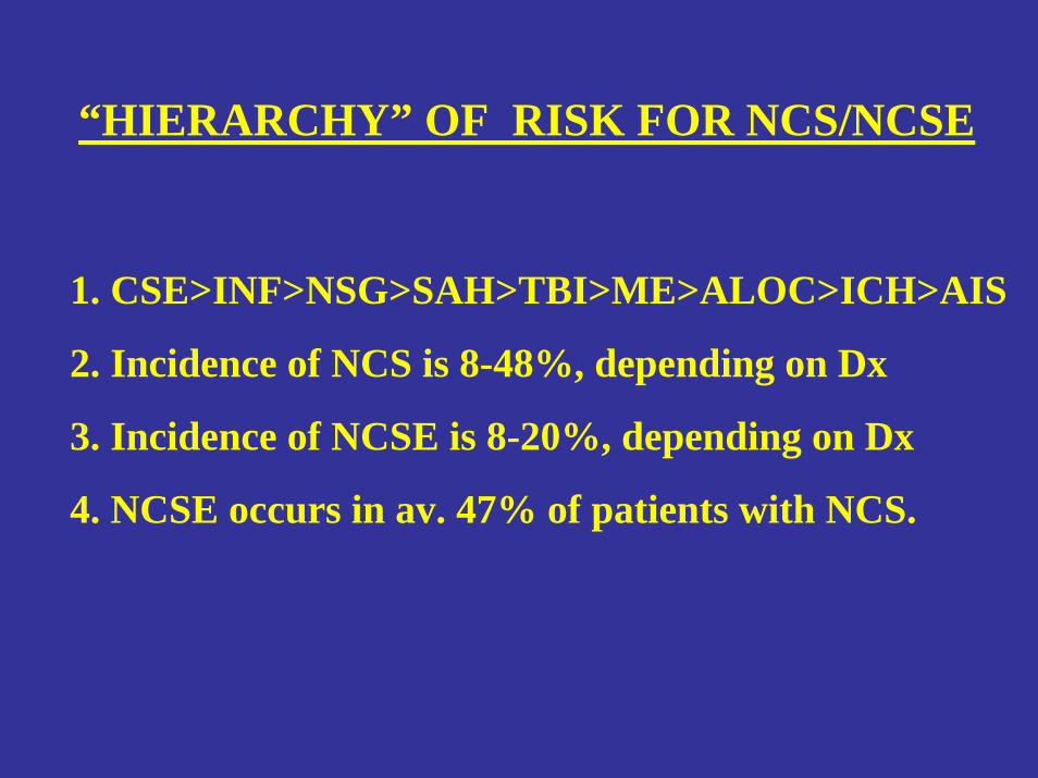
1. CSE>INF>NSG>SAH>TBI>ME>ALOC>ICH>AIS
2. Incidence of NCS is 8-48%, depending on Dx
3. Incidence of NCSE is 8-20%, depending on Dx
4. NCSE occurs in av. 47% of patients with NCS.
“HIERARCHY” OF RISK FOR NCS/NCSE
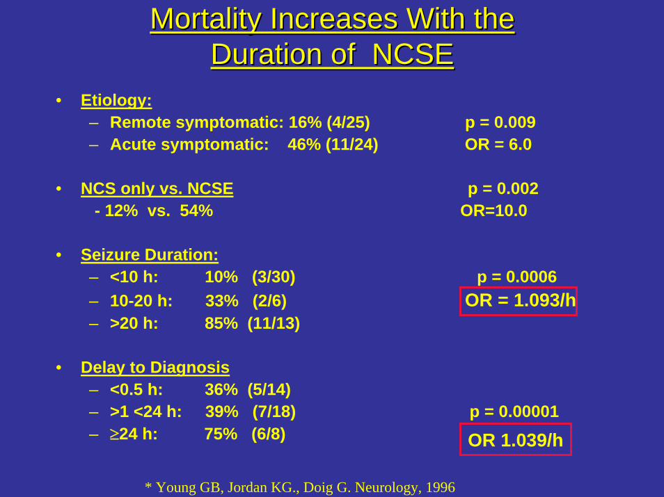
Mortality Increases With the Mortality Increases With the Duration of NCSEDuration of NCSE
• Etiology:– Remote symptomatic: 16% (4/25) p = 0.009– Acute symptomatic: 46% (11/24) OR = 6.0
• NCS only vs. NCSE p = 0.002- 12% vs. 54% OR=10.0
• Seizure Duration:– <10 h: 10% (3/30) p = 0.0006– 10-20 h: 33% (2/6) OR = 1.093/h– >20 h: 85% (11/13)
• Delay to Diagnosis– <0.5 h: 36% (5/14)– >1 <24 h: 39% (7/18) p = 0.00001– ≥24 h: 75% (6/8)
* Young GB, Jordan KG., Doig G. Neurology, 1996
OR 1.039/h
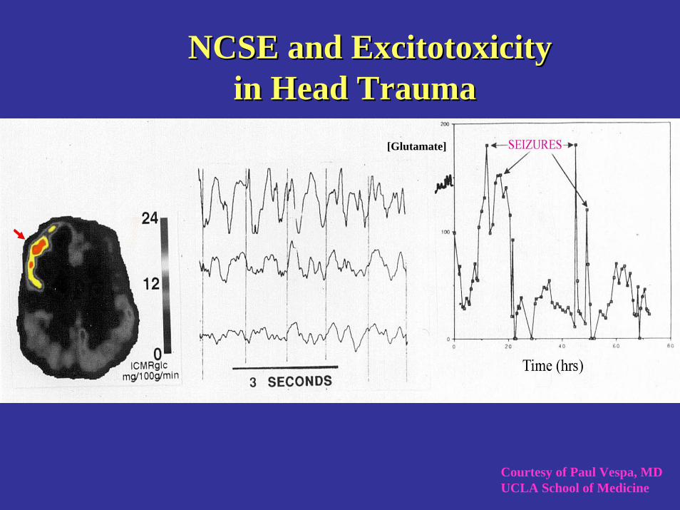
Courtesy of Paul Vespa, MD UCLA School of Medicine
NCSE and NCSE and ExcitotoxicityExcitotoxicityin Head Traumain Head Trauma
[Glutamate]
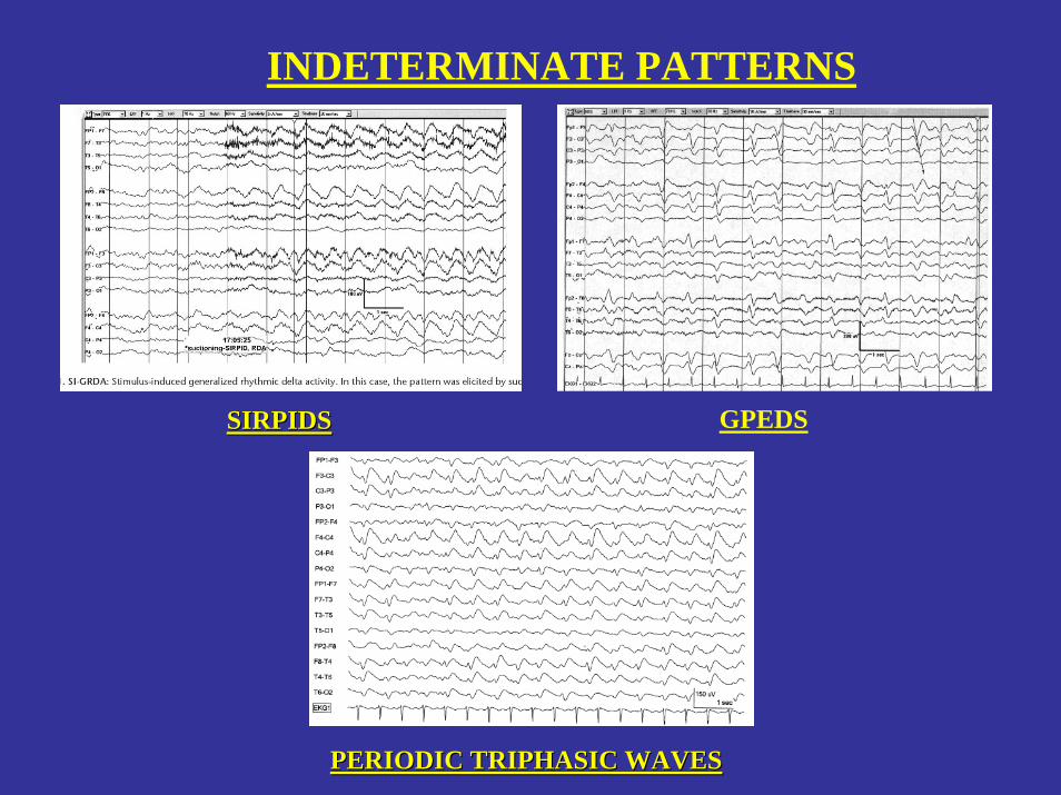
INDETERMINATE PATTERNS
SIRPIDSSIRPIDS GPEDS
PERIODIC TRIPHASIC WAVESPERIODIC TRIPHASIC WAVES
DETECTING AND MONITORING DETECTING AND MONITORING CEREBRAL ISCHEMIACEREBRAL ISCHEMIA
Based on: Jordan KG. JCN(2004);21:341-352)

1-70 Hz
Ischemic EEG Changes in Carotid Clamping
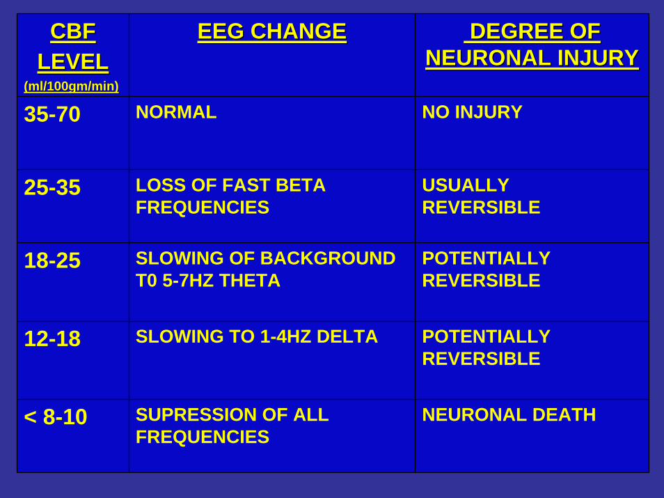
CBF CBF LEVELLEVEL
(ml/100gm/min)(ml/100gm/min)
EEG CHANGEEEG CHANGE DEGREE OF DEGREE OF NEURONAL INJURYNEURONAL INJURY
35-70 NORMAL NO INJURY
USUALLY REVERSIBLE
POTENTIALLY REVERSIBLE
POTENTIALLY REVERSIBLE
NEURONAL DEATH< 8-10 SUPRESSION OF ALL FREQUENCIES
25-35 LOSS OF FAST BETA FREQUENCIES
18-25 SLOWING OF BACKGROUND T0 5-7HZ THETA
12-18 SLOWING TO 1-4HZ DELTA
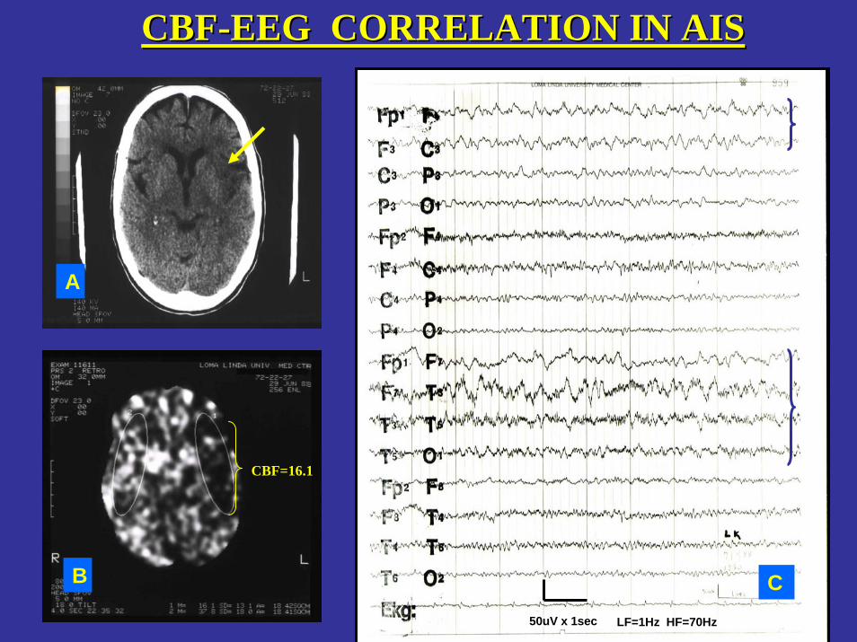
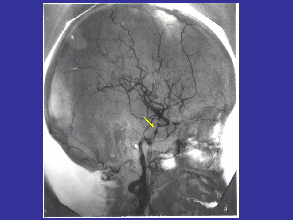
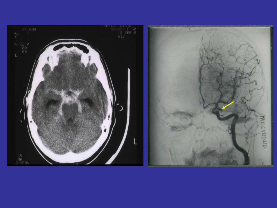
CBF=16.1
A
B C50uV x 1sec LF=1Hz HF=70Hz
CBFCBF--EEG CORRELATION IN AISEEG CORRELATION IN AIS
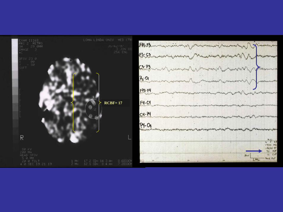
RCBF= 17
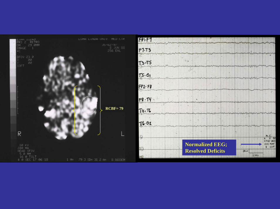
Normalized EEG; Normalized EEG; Resolved DeficitsResolved Deficits
RCBF= 79
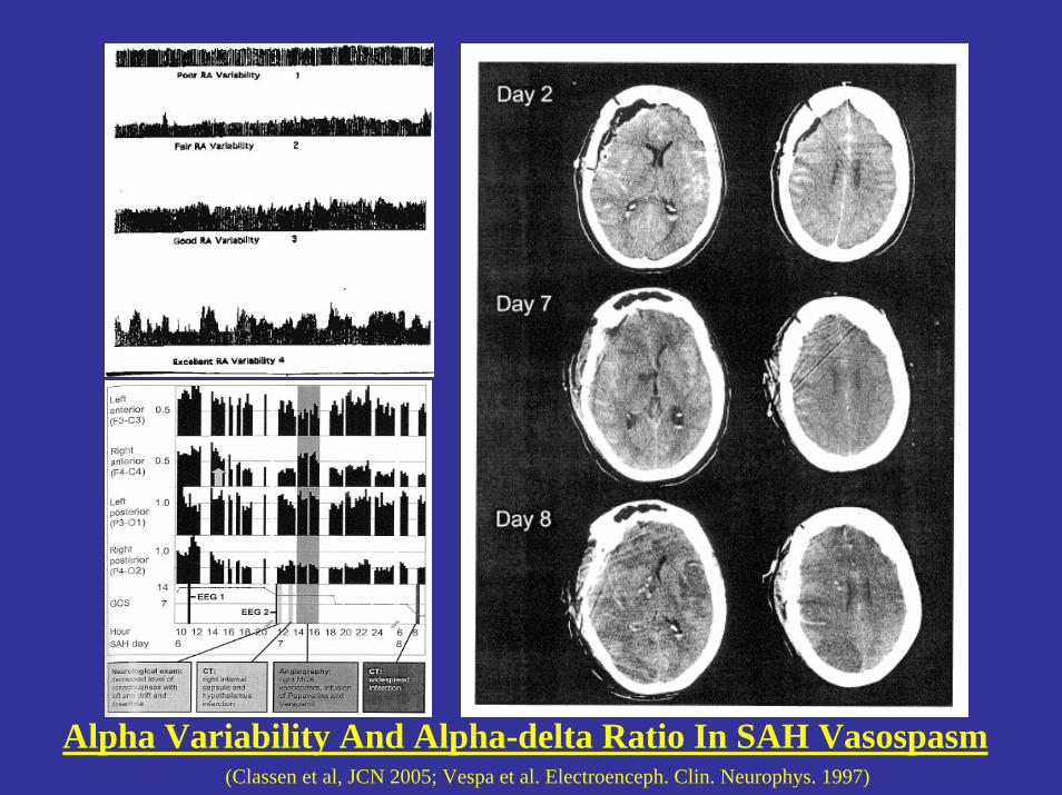
Alpha Variability And Alpha-delta Ratio In SAH Vasospasm(Classen et al, JCN 2005; Vespa et al. Electroenceph. Clin. Neurophys. 1997)
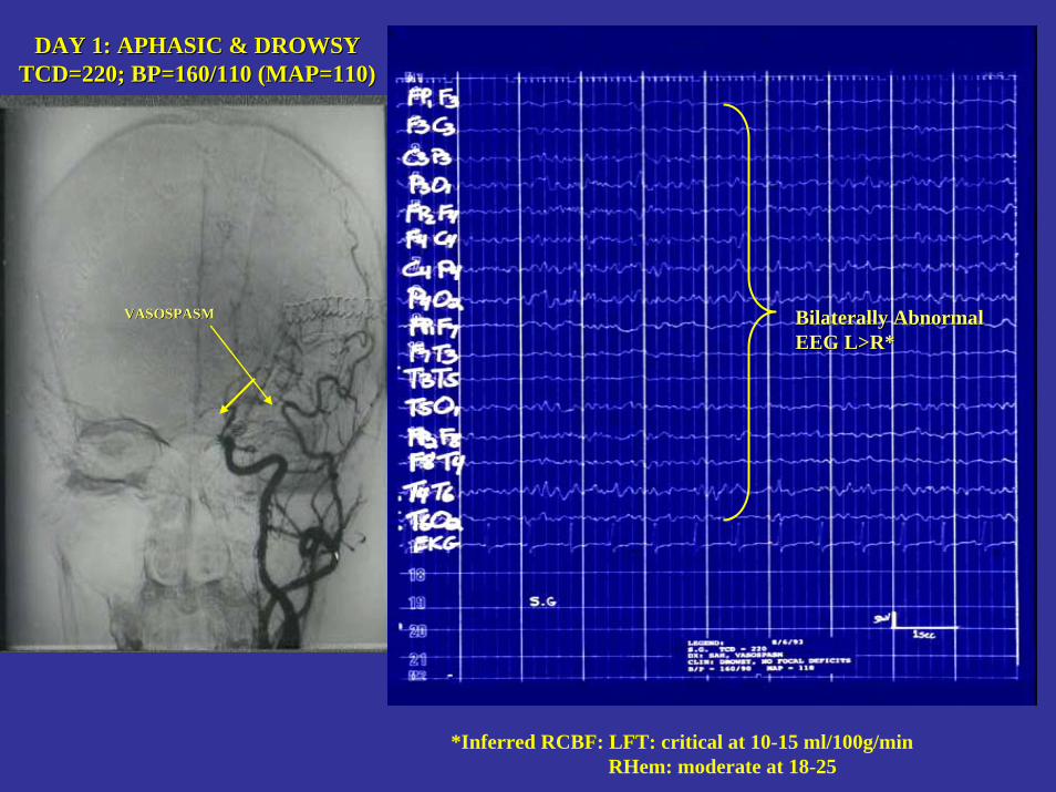
DAY 1: APHASIC & DROWSY TCD=220; BP=160/110 (MAP=110)
DAY 1: APHASIC & DROWSY TCD=220; BP=160/110 (MAP=110)
Bilaterally Abnormal Bilaterally Abnormal EEG L>R*EEG L>R*
VASOSPASMVASOSPASM
*Inferred RCBF: LFT: critical at 10-15 ml/100g/min RHem: moderate at 18-25
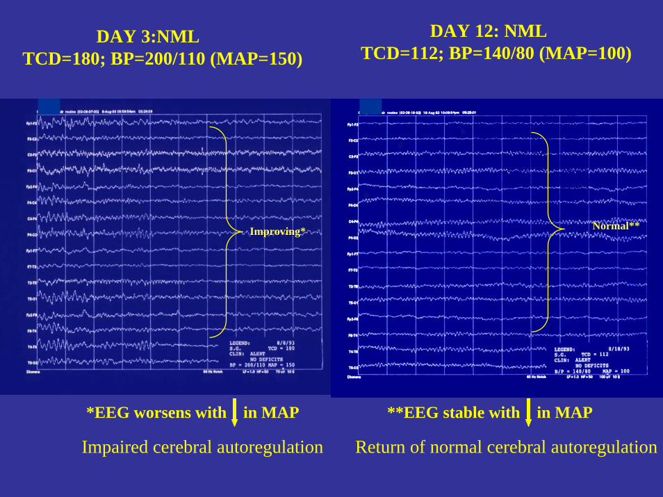
DAY 3:NML TCD=180; BP=200/110 (MAP=150)
DAY 12: NML TCD=112; BP=140/80 (MAP=100)
Improving*Improving* Normal**Normal**
Impaired cerebral autoregulation
*EEG worsens with in MAP **EEG stable with in MAP
Return of normal cerebral autoregulation
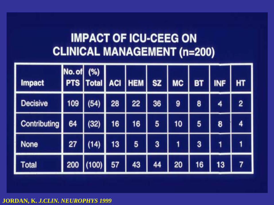
JORDAN, K. J.CLIN. NEUROPHYS 1999
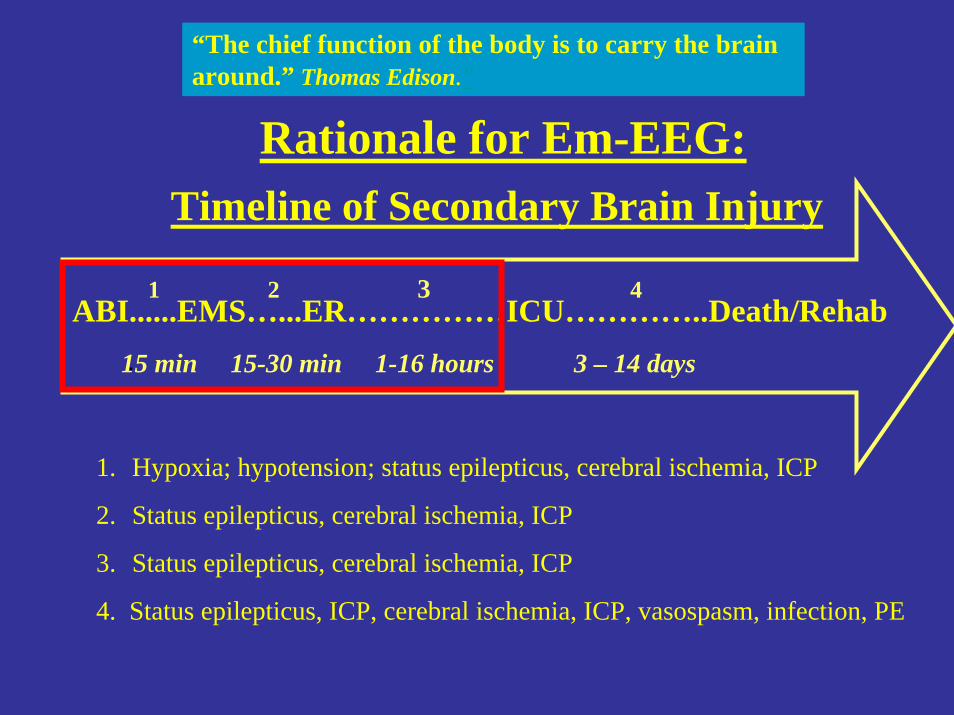
ABI......EMS…...ER……………ICU…………..Death/Rehab1 2 3 4
. 15 min 15-30 min 1-16 hours 3 – 14 days
1. Hypoxia; hypotension; status epilepticus, cerebral ischemia, ICP
2. Status epilepticus, cerebral ischemia, ICP
3. Status epilepticus, cerebral ischemia, ICP
4. Status epilepticus, ICP, cerebral ischemia, ICP, vasospasm, infection, PE
Timeline of Secondary Brain Injury
“The chief function of the body is to carry the brain around.” Thomas Edison."
Rationale for Em-EEG:
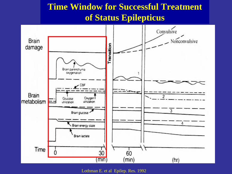
Time Window for Successful Treatment of Status Epilepticus
Lothman E. et al Epilep. Res. 1992
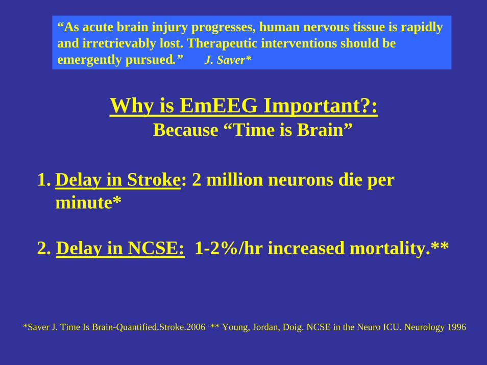
*Saver J. Time Is Brain-Quantified.Stroke.2006 ** Young, Jordan, Doig. NCSE in the Neuro ICU. Neurology 1996
1. Delay in Stroke: 2 million neurons die per minute*
2. Delay in NCSE: 1-2%/hr increased mortality.**
Why is EmEEG Important?: Because “Time is Brain”
“As acute brain injury progresses, human nervous tissue is rapidly and irretrievably lost. Therapeutic interventions should be emergently pursued.” J. Saver*
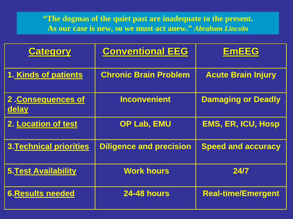
CategoryCategory Conventional EEGConventional EEG EmEEGEmEEG
1. Kinds of patients Chronic Brain Problem
Inconvenient
OP Lab, EMU
Diligence and precision
Work hours
24-48 hours
Acute Brain Injury
2 .Consequences of delay
Damaging or Deadly
EMS, ER, ICU, Hosp
Speed and accuracy
24/7
2. Location of test
3.Technical priorities
5.Test Availability
6.Results needed Real-time/Emergent
“The dogmas of the quiet past are inadequate to the present. As our case is new, so we must act anew.” Abraham Lincoln
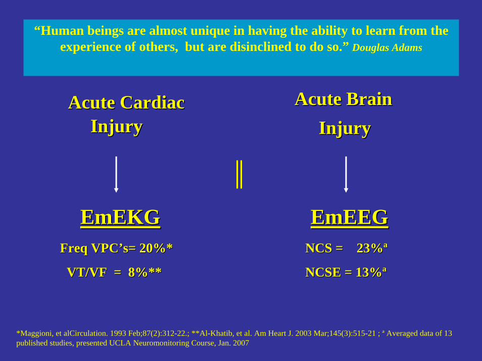
Acute CardiacAcute CardiacInjury Injury
EmEKGEmEKG
Acute Brain Acute Brain InjuryInjury
EmEEGEmEEGFreq Freq VPCVPC’’ss= 20%*= 20%*
VT/VF = 8%**VT/VF = 8%**
NCS = 23%NCS = 23%ªª
NCSE = 13%NCSE = 13%ªª
“Human beings are almost unique in having the ability to learn from the experience of others, but are disinclined to do so.” Douglas Adams
*Maggioni, et alCirculation. 1993 Feb;87(2):312-22.; **Al-Khatib, et al. Am Heart J. 2003 Mar;145(3):515-21 ; ª Averaged data of 13 published studies, presented UCLA Neuromonitoring Course, Jan. 2007
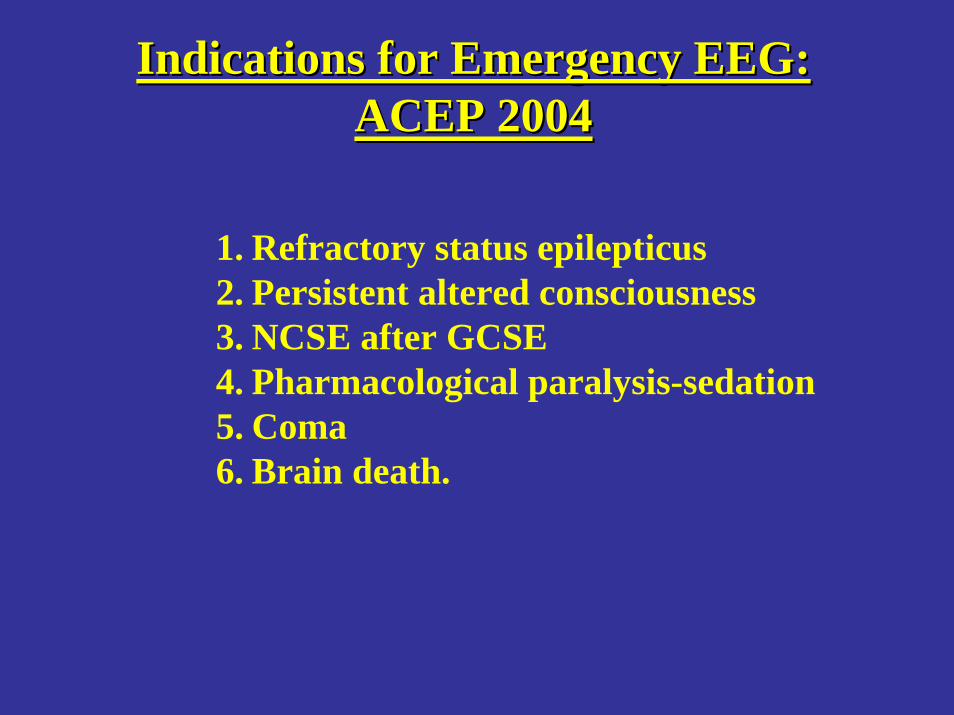
1. Refractory status epilepticus2. Persistent altered consciousness3. NCSE after GCSE4. Pharmacological paralysis-sedation 5. Coma6. Brain death.
Indications for Emergency EEG: Indications for Emergency EEG: ACEP 2004ACEP 2004
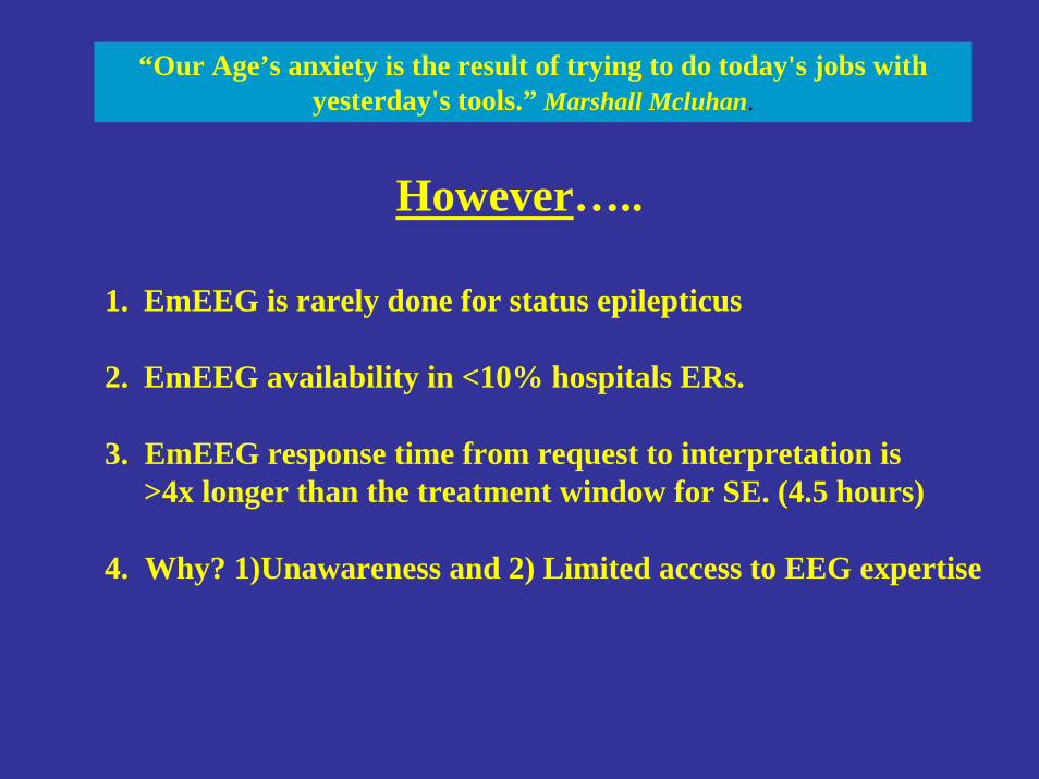
1. EmEEG is rarely done for status epilepticus
2. EmEEG availability in <10% hospitals ERs.
3. EmEEG response time from request to interpretation is >4x longer than the treatment window for SE. (4.5 hours)
4. Why? 1)Unawareness and 2) Limited access to EEG expertise
However…..
“Our Age’s anxiety is the result of trying to do today's jobs with yesterday's tools.” Marshall Mcluhan.
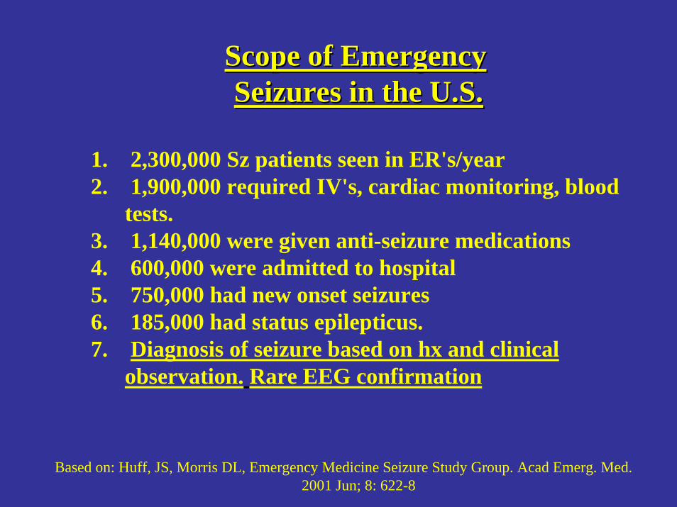
Scope of EmergencyScope of EmergencySeizures in the U.S.Seizures in the U.S.
1. 2,300,000 Sz patients seen in ER's/year 2. 1,900,000 required IV's, cardiac monitoring, blood
tests. 3. 1,140,000 were given anti-seizure medications 4. 600,000 were admitted to hospital5. 750,000 had new onset seizures6. 185,000 had status epilepticus. 7. Diagnosis of seizure based on hx and clinical
observation. Rare EEG confirmation
Based on: Huff, JS, Morris DL, Emergency Medicine Seizure Study Group. Acad Emerg. Med. 2001 Jun; 8: 622-8
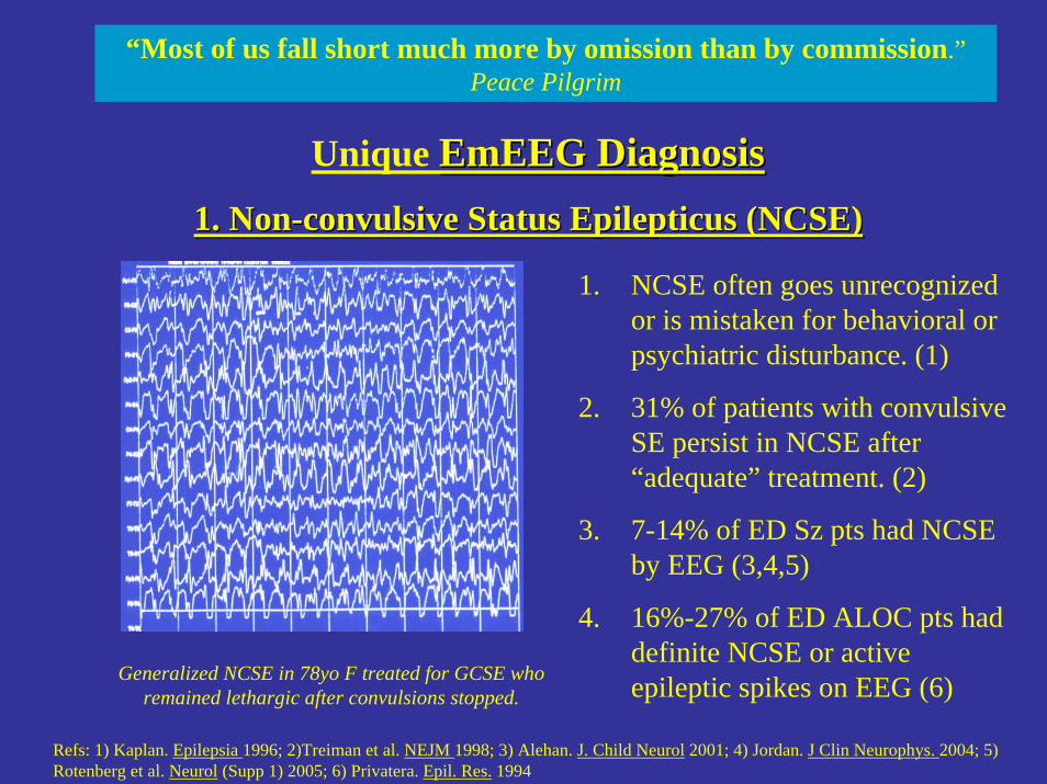
1. Non1. Non--convulsive Status convulsive Status EpilepticusEpilepticus (NCSE)(NCSE)
1. NCSE often goes unrecognized or is mistaken for behavioral or psychiatric disturbance. (1)
2. 31% of patients with convulsive SE persist in NCSE after “adequate” treatment. (2)
3. 7-14% of ED Sz pts had NCSE by EEG (3,4,5)
4. 16%-27% of ED ALOC pts had definite NCSE or active epileptic spikes on EEG (6)
Refs: 1) Kaplan. Epilepsia 1996; 2)Treiman et al. NEJM 1998; 3) Alehan. J. Child Neurol 2001; 4) Jordan. J Clin Neurophys. 2004; 5) Rotenberg et al. Neurol (Supp 1) 2005; 6) Privatera. Epil. Res. 1994
Generalized NCSE in 78yo F treated for GCSE who remained lethargic after convulsions stopped.
Unique EmEEGEmEEG DiagnosisDiagnosis
“Most of us fall short much more by omission than by commission.”Peace Pilgrim
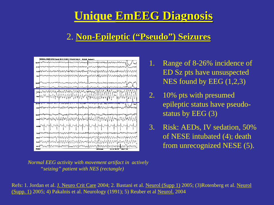
2. NonNon--Epileptic (Epileptic (““PseudoPseudo””) Seizures) Seizures
1. Range of 8-26% incidence of ED Sz pts have unsuspected NES found by EEG (1,2,3)
2. 10% pts with presumed epileptic status have pseudo-status by EEG (3)
3. Risk: AEDs, IV sedation, 50% of NESE intubated (4); death from unrecognized NESE (5).
Refs: 1. Jordan et al. J. Neuro Crit Care 2004; 2. Bastani et al. Neurol (Supp 1) 2005; (3)Rotenberg et al. Neurol(Supp. 1) 2005; 4) Pakalnis et al. Neurology (1991); 5) Reuber et al Neurol. 2004
Normal EEG activity with movement artifact in actively“seizing” patient with NES (rectangle)
Unique EmEEGEmEEG DiagnosisDiagnosis
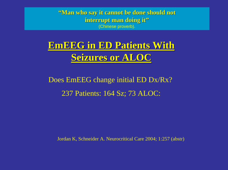
EmEEGEmEEG in ED Patients With in ED Patients With Seizures or ALOCSeizures or ALOC
Does EmEEG change initial ED Dx/Rx?
237 Patients: 164 Sz; 73 ALOC:
Jordan K, Schneider A. Neurocritical Care 2004; 1:257 (abstr)
““Man who say it cannot be done should not Man who say it cannot be done should not interrupt man doing itinterrupt man doing it””
(Chinese proverb).
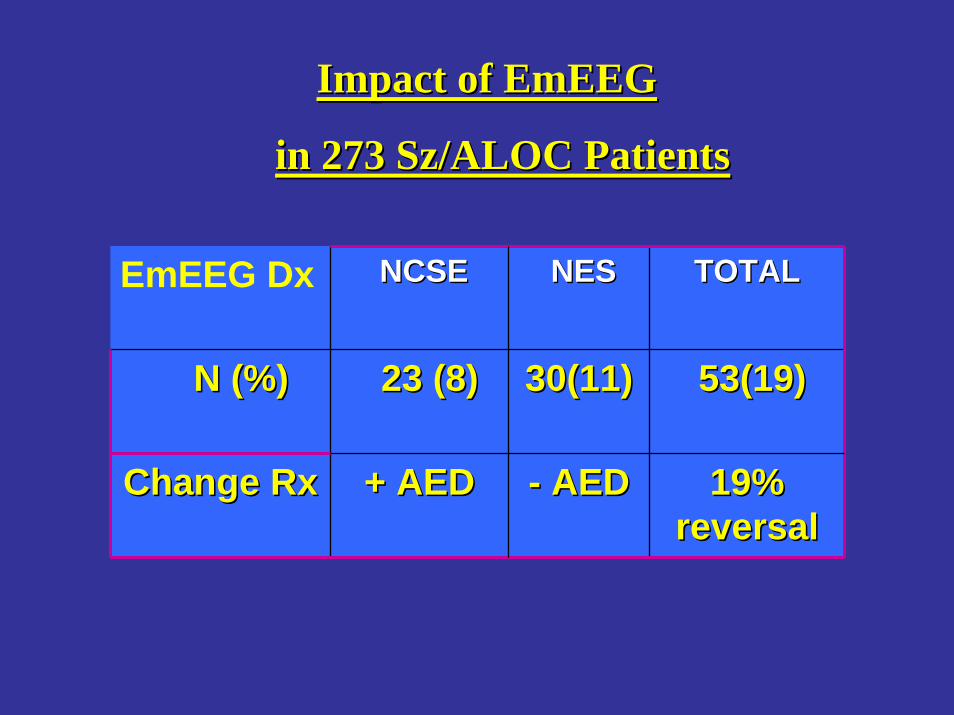
Impact of Impact of EmEEGEmEEG
in 273 in 273 SzSz/ALOC Patients/ALOC Patients
EmEEG Dx NCSENCSE NESNES TOTAL TOTAL
N (%) N (%) 23 (8)23 (8) 30(11) 30(11) 53(19) 53(19)
Change RxChange Rx + AED+ AED -- AEDAED 19% 19% reversalreversal
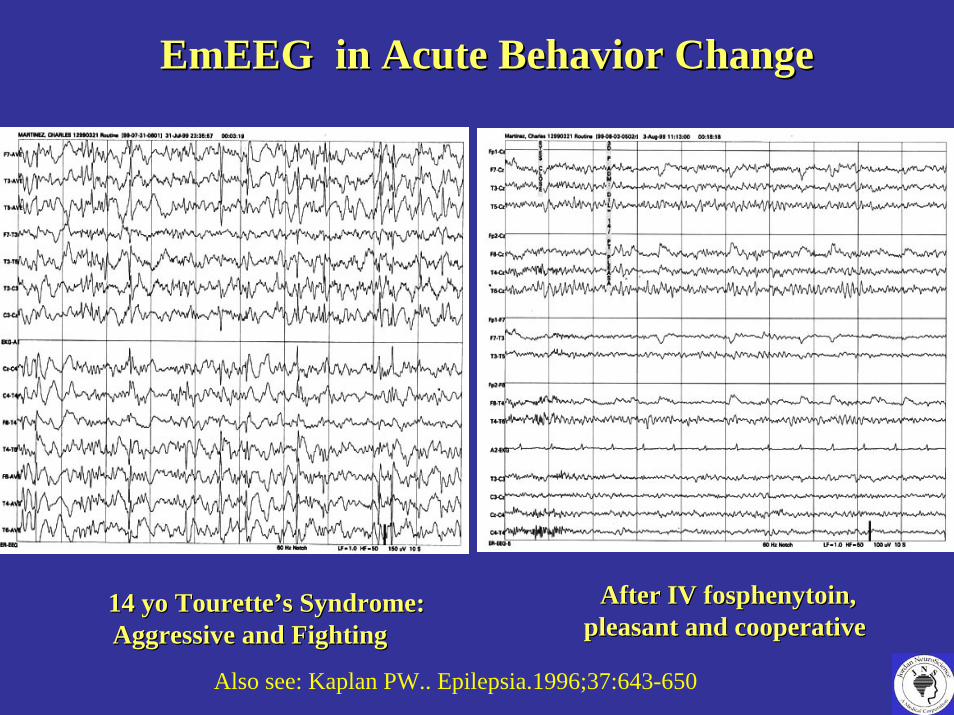
14 14 yoyo TouretteTourette’’ss Syndrome: Syndrome: Aggressive and FightingAggressive and Fighting
EmEEGEmEEG in Acute Behavior Changein Acute Behavior Change
After IV After IV fosphenytoinfosphenytoin, , pleasant and cooperativepleasant and cooperative
Also see: Kaplan PW.. Epilepsia.1996;37:643-650
The Dangerous Delays The Dangerous Delays in Emergency EEGin Emergency EEG
"In delay there lies no plenty."--William Shakespeare
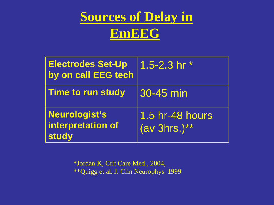
Sources of Delay in EmEEG
Electrodes Set-Up by on call EEG tech
1.5-2.3 hr *
Time to run study 30-45 min
Neurologist’s interpretation of study
1.5 hr-48 hours (av 3hrs.)**
*Jordan K, Crit Care Med., 2004, **Quigg et al. J. Clin Neurophys. 1999
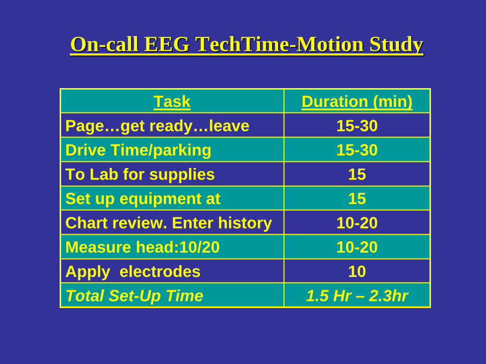
Task Duration (min)Page…get ready…leave
home15-30
Drive Time/parking 15-30To Lab for supplies 15Set up equipment at
bedside15
Chart review. Enter history 10-20Measure head:10/20
method10-20
Apply electrodes 10Total Set-Up Time 1.5 Hr – 2.3hr
OnOn--call EEG call EEG TechTimeTechTime--Motion StudyMotion Study
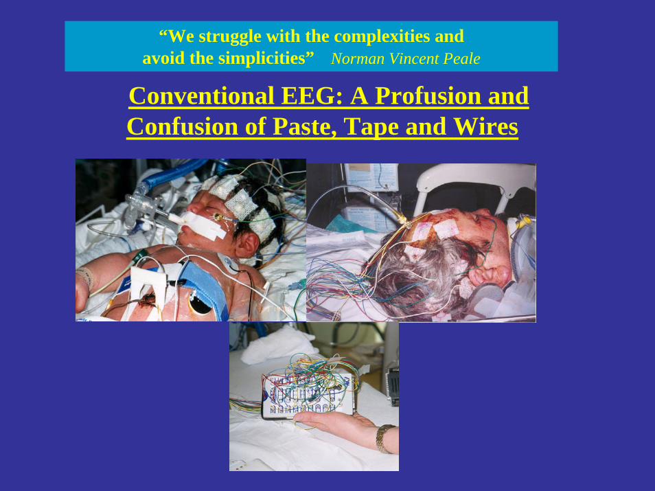
Conventional EEG: A Profusion and Confusion of Paste, Tape and Wires
“We struggle with the complexities and avoid the simplicities” Norman Vincent Peale
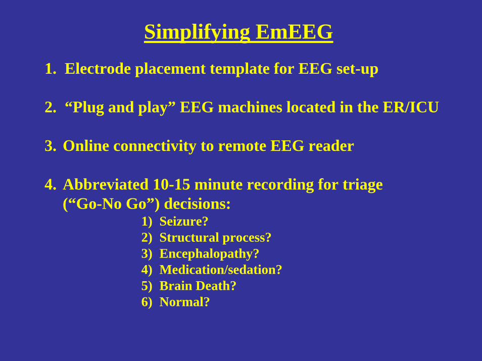
Simplifying EmEEG1. Electrode placement template for EEG set-up
2. “Plug and play” EEG machines located in the ER/ICU
3. Online connectivity to remote EEG reader
4. Abbreviated 10-15 minute recording for triage (“Go-No Go”) decisions:
1) Seizure?2) Structural process?3) Encephalopathy?4) Medication/sedation?5) Brain Death?6) Normal?
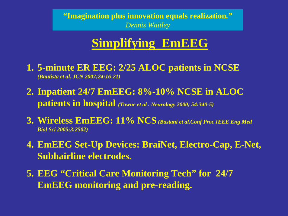
“Imagination plus innovation equals realization.”Dennis Waitley
1. 5-minute ER EEG: 2/25 ALOC patients in NCSE(Bautista et al. JCN 2007;24:16-21)
2. Inpatient 24/7 EmEEG: 8%-10% NCSE in ALOC patients in hospital (Towne et al . Neurology 2000; 54:340-5)
3. Wireless EmEEG: 11% NCS (Bastani et al.Conf Proc IEEE Eng Med Biol Sci 2005;3:2502)
4. EmEEG Set-Up Devices: BraiNet, Electro-Cap, E-Net, Subhairline electrodes.
5. EEG “Critical Care Monitoring Tech” for 24/7 EmEEG monitoring and pre-reading.
Simplifying EmEEG
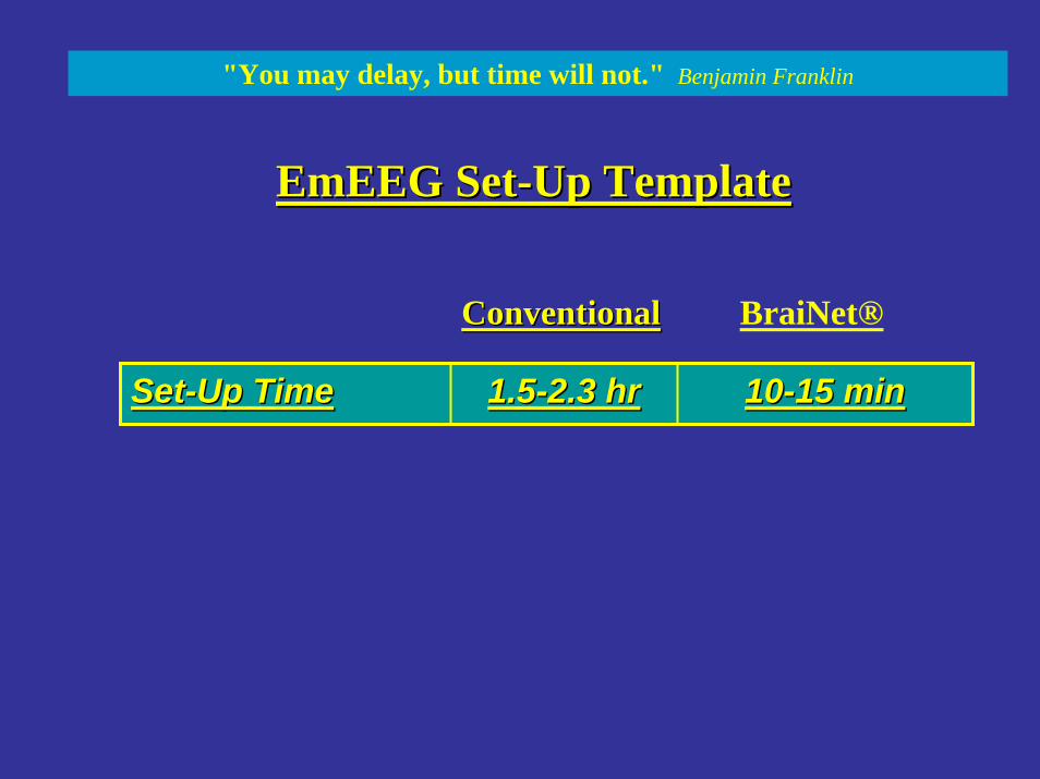
EmEEGEmEEG SetSet--Up Template Up Template
SetSet--Up TimeUp Time 1.51.5--2.3 hr2.3 hr 1010--15 min15 min
ConventionalConventional BraiNet®
"You may delay, but time will not." Benjamin Franklin

Reader paged and EEGSent by Internet
EEG Set-Up with BraiNet®
EmEEGEmEEG in ER: in ER:
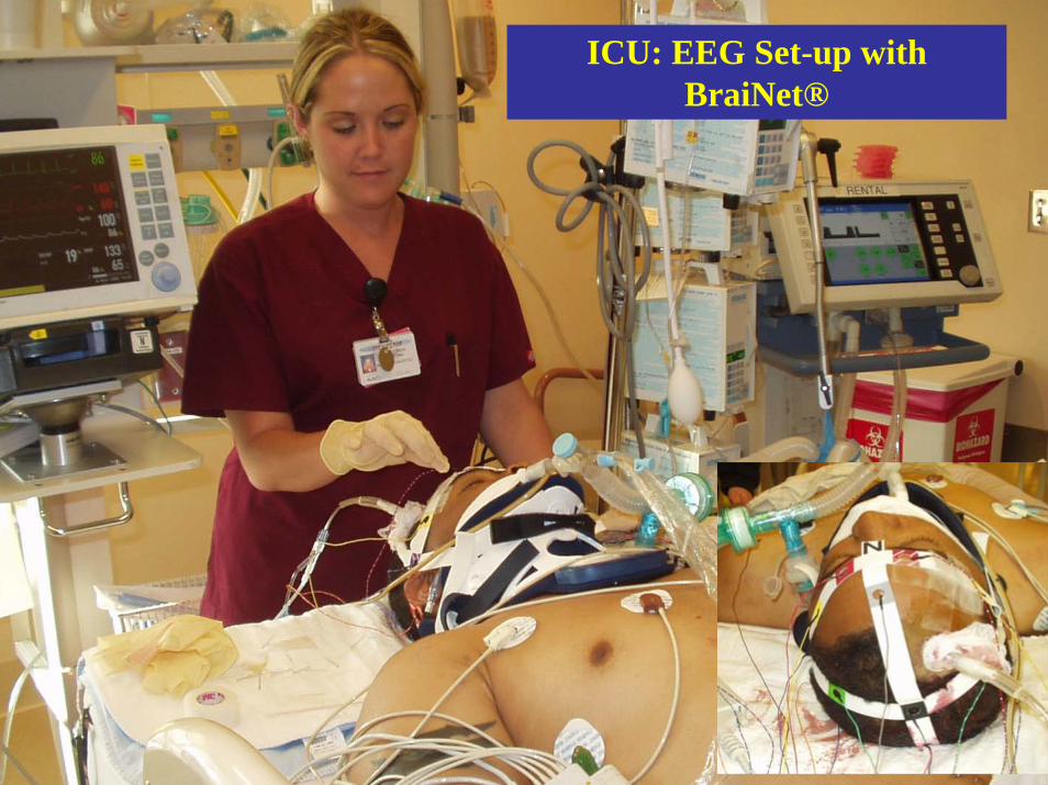
ICU: EEG Set-up with BraiNet®
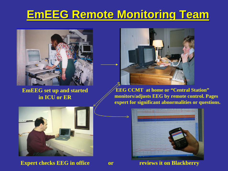
EmEEGEmEEG Remote Monitoring TeamRemote Monitoring Team
EmEEG set up and started in ICU or ER
EEG CCMT at home or “Central Station”monitors/adjusts EEG by remote control. Pages expert for significant abnormalities or questions.
Expert checks EEG in office or reviews it on Blackberry
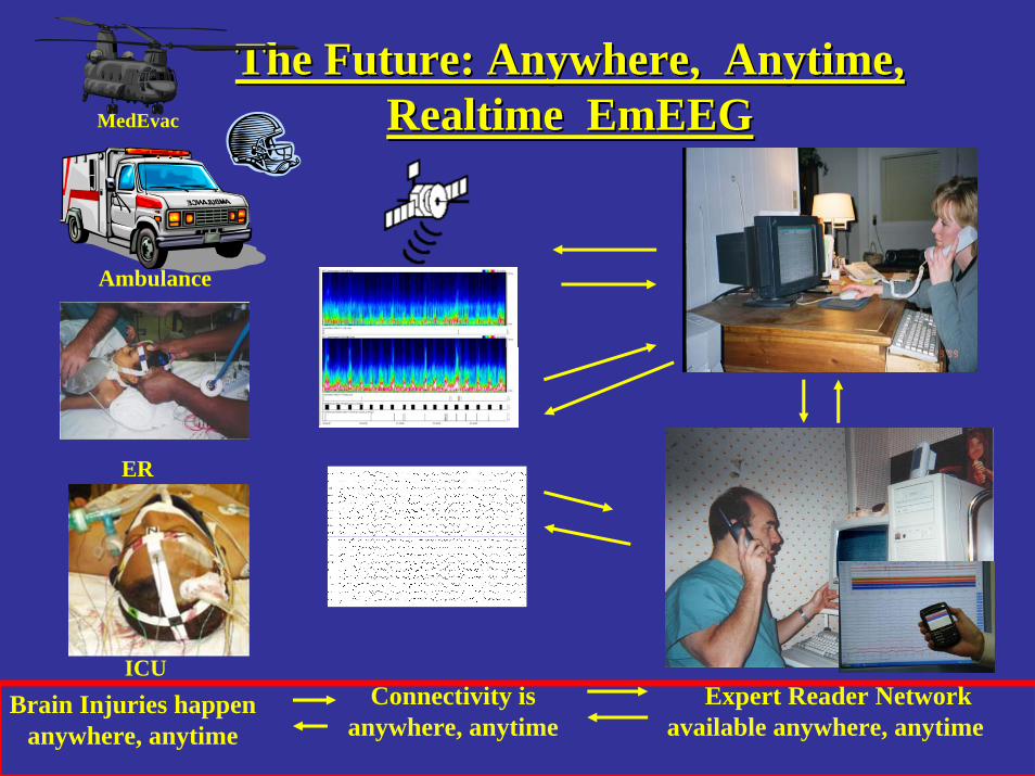
Ambulance
ER
ICU
The Future: Anywhere, Anytime, The Future: Anywhere, Anytime, RealtimeRealtime EmEEGEmEEGMedEvac
Expert Reader Network available anywhere, anytime
Brain Injuries happen anywhere, anytime
Connectivity is anywhere, anytime

“We have brains in our heads. We have feet in our shoes.We can steer ourselves Any direction we choose.”
Dr. Seuss
“If your determination is fixed, I do not counsel you to despair. Few things are impossible to diligence and skill. Great works are performed not by strength, but perseverance.” Samuel Johnson
As Two Great American Philosophers Remind Us:
Emergency EEG
Anywhere, Anytime
Saves Brains!