raft-dependentendocytosisofautocrinemotilityfactor ... ·...
TRANSCRIPT
Raft-dependent Endocytosis of Autocrine Motility FactorIs Phosphatidylinositol 3-Kinase-dependent in BreastCarcinoma Cells*
Received for publication, May 16, 2007, and in revised form, July 25, 2007 Published, JBC Papers in Press, August 8, 2007, DOI 10.1074/jbc.M704069200
Liliana D. Kojic‡, Bharat Joshi‡, Patrick Lajoie‡1, Phuong U. Le§, Michael E. Cox¶, Dmitry A. Turbin�,Sam M. Wiseman**¶�2, and Ivan R. Nabi‡3
From the ‡Department of Cellular and Physiological Sciences, Life Sciences Institute, University of British Columbia, Vancouver,British Columbia V6T 1Z3, §National Research Council, Biotechnology Research Institute, Montreal, Quebec H4P 2R2, ¶ProstateResearch Centre, British Columbia Cancer Agency and University of British Columbia, Vancouver, British Columbia V6H 3Z6,�Genetic Pathology Evaluation Centre, Department of Pathology, Vancouver General Hospital, British Columbia Cancer Agency,and University of British Columbia, Vancouver, British Columbia V6H 3Z6, and the **Department of Surgery, St. Paul’sHospital/University of British Columbia, Vancouver, British Columbia V6T NZ, Canada
Autocrinemotility factor (AMF) is internalized via a receptor-mediated, dynamin-dependent, cholesterol-sensitive raft path-way to the smooth endoplasmic reticulum that is negatively reg-ulated by caveolin-1. Expression of AMF and its receptor(AMFR) is associated with tumor progression and malignancy;however, the extent to which the raft-dependent uptake of AMFis tumor cell-specific has yet to be addressed. By Western blotand cell surface fluorescence-activated cell sorter (FACS) anal-ysis, AMFR expression is increased in tumorigenic MCF7 andmetastatic MDA-231 andMDA-435 breast cancer cell lines rel-ative to dysplastic MCF10A mammary epithelial cells. AMFuptake, determined by FACSmeasurement of protease-insensi-tive internalized fluorescein-conjugated AMF, was increased inMCF7 andMDA-435 cells relative toMCF-10A and caveolin-1-expressing MDA-231 cells. Uptake of fluorescein-conjugatedAMF was dynamin-dependent, methyl-�-cyclodextrin- andgenistein-sensitive, reduced upon overexpression of caveolin-1in MDA-435 cells, and increased upon short hairpin RNAreduction of caveolin-1 in MDA-231 cells. Tissue microarrayanalysis of invasive primary human breast carcinomas showedthat AMFR expression had no impact on survival but did corre-late significantly with expression of phospho-Akt. Phospho-Aktexpression was increased in AMF-internalizing MCF7 andMDA-435 breast carcinoma cells. AMFuptake in these cells wasreduced by phosphatidylinositol 3-kinase inhibition but not byregulators ofmacropinocytosis such as amiloride, phorbol ester,or actin cytoskeleton disruption by cytochalasin D. The raft-de-pendent endocytosis of AMF therefore follows a distinct phos-phatidylinositol 3-kinase-dependent pathway that is up-regu-lated in more aggressive tumor cells.
Autocrine motility factor (AMF)4 is secreted by and stimula-tory to tumor cells. AMF exhibits sequence identity to phos-phoglucose isomerase, a glycolytic enzyme that also exhibitsneurokine and lymphokine activities (1). AMFR expression isassociated with poor survival and prognosis of patients withgastric, colorectal, bladder, and esophageal carcinomas,cutaneous malignant melanoma, and pulmonary adenocar-cinoma (2–8). Importantly, in many of these studies, AMFRwas absent or present at significantly reduced levels in adja-cent normal tissue. Recently, a genome-wide analysis iden-tified AMFR as one of 189 genes that present a high fre-quency of intragenic mutations in breast cancer (9). Alteredexpression of AMFR is therefore associated with breasttumor progression and metastasis.AMFR localizes to cell surface caveolae and internalizes
AMF to the smooth endoplasmic reticulum via a dynamin-de-pendent, raft-mediated endocytic pathway negatively regulatedby caveolin-1 (Cav1) (10–12). Multiple raft-dependent path-ways have been described that vary in their dependence onexpression of Cav1 and dynamin-2 (13, 14). The AMF pathwaycan be distinguished from the caveolae/raft-dependent endo-cytosis of SV40 virus to the endoplasmic reticulum (15) and ofcholera toxin b-subunit to the Golgi apparatus by the insensi-tivity of its uptake to brefeldin A, nocodazole, and a 20 °C tem-perature block (11). Reduced expression of Cav1 upon transfor-mation ofNIH-3T3 cells with ras or abl oncogenes is associatedwith increasedAMFuptake (12). Conversely, AMF transfectionof NIH-3T3 cells induces enhanced transformation and sur-vival via PI3K/Akt signaling and Cav1 down-regulation (16).This suggests that AMF/AMFR expression and signaling areinversely related to Cav1 expression during tumor progression.However, AMF endocytosis in human tumor cells has yet to be* This work was supported in part by a grant from the Canadian Institutes of
Health Research. The costs of publication of this article were defrayed inpart by the payment of page charges. This article must therefore be herebymarked “advertisement” in accordance with 18 U.S.C. Section 1734 solely toindicate this fact.
1 Research student of the Terry Fox Foundation through an award from theNational Cancer Institute of Canada.
2 A Michael Smith Scholar.3 Canadian Institutes of Health Research Investigator. To whom correspond-
ence should be addressed: Dept. of Cellular and Physiological Sciences,University of British Columbia, 2350 Health Sciences Mall, Vancouver, Brit-ish Columbia V6T 1Z3, Canada. E-mail: [email protected].
4 The abbreviations used are: AMF, autocrine motility factor; AMF-FITC, fluo-rescein-conjugated AMF; AMFR, autocrine motility factor receptor; Cav1,caveolin-1; ER, estrogen receptor; FACS, fluorescence-activated cell sorter;HER2, herceptin2; Tf-FITC, FITC-conjugated transferrin; m�CD, methyl-�-cyclodextrin; PI3K, phosphatidylinositol 3-kinase; PR, progesterone recep-tor; TMA, tissue microarray; TPA, phorbol ester12-O tetradecanoylphorbol13-acetate; shRNA, short hairpin RNA; MFI, mean fluorescence intensity;RT, reverse transcription; mAb, monoclonal antibody.
THE JOURNAL OF BIOLOGICAL CHEMISTRY VOL. 282, NO. 40, pp. 29305–29313, October 5, 2007© 2007 by The American Society for Biochemistry and Molecular Biology, Inc. Printed in the U.S.A.
OCTOBER 5, 2007 • VOLUME 282 • NUMBER 40 JOURNAL OF BIOLOGICAL CHEMISTRY 29305
by guest on August 12, 2018
http://ww
w.jbc.org/
Dow
nloaded from
studied, and factors that promote the raft-dependent endocy-tosis of AMF have not been identified.In this study, we show that AMF internalization is reduced in
dysplasticMCF10Amammary cells relative to tumorigenic andmetastatic tumor cell lines and that Cav1 is a critical regulatorof the raft-dependent endocytosis of AMF in breast carcinomacells. Tissue microarray analysis (TMA) of AMFR expressionidentified a highly significant association with p-Akt. AMFstimulates p-Akt expression, and PI3K inhibition prevents raft-dependent AMF uptake in breast carcinoma cells. PI3K istherefore identified as a positive regulator of theAMF raft path-way, indicative of a feedback loop linking AMFR signalingthrough PI3K to AMF uptake.
EXPERIMENTAL PROCEDURES
Cells, Antibodies, and Reagents—MCF7, MDA-231, MDA-435, andMCF10A human breast cell lines (ATCC,Manassas,VA) were maintained in complete RPMI 1640 medium con-taining 10% FBS.Monoclonal rat IgM anti-AMFR (3F3A)was
used as ascites fluid (17). Rabbitanti-Cav1/2 and mouse anti-Cav1antibody were from TransductionLaboratories; rabbit anti-p-Akt andAkt were from Cell Signaling (Dan-vers,MA);Alexa488- andAlexa647-conjugated secondary antibodies,Alexa568-conjugated phalloidin,and rabbit anti-FITC antibodies werefromMolecularProbes (Eugene,OR),and rhodamine red-X anti-rat IgMwas from Jackson ImmunoResearch(West Grove, PA). Commercialrabbit phosphoglucose isomerase(referred to as AMF) was purchasedfrom Sigma (P-9544) and conjugatedto FITC using fluorescein-EX proteinlabeling kit (Molecular Probes).Monoclonal anti-�-actin, methyl-�-cyclodextrin (m�CD), genistein,FITC-conjugated transferrin (Tf-FITC), propidium iodide, goat serum,and Pronase were from Sigma, andLY294002 was fromCalbiochem.Cav1 shRNA Lentiviral Con-
structs—Core sequences for Cav1shRNA constructs (5�-CTGT-TCCCATCCGGGAACAGGGC-AACAT-3�) were cloned intopSHAG-1 plasmid (Dr. G. Han-non, Cold Spring Harbor, NY),and the shRNA cassette was trans-ferred into pLenti6/BLOCK-iTTM-DEST (Invitrogen) that car-ries a blasticidin selection marker.Lentiviral vector from condi-tioned media of HEK293T cellstransfected with Cav1 shRNA-pLenti6/BLOCK-iTTM-DEST (18)
was used to infect MDA-231 cells. A pooled population ofblasticidin-selected MDA-231 cells exhibiting maximalCav1 suppression was used for subsequent experiments.Western Blots and RT-PCR—Cell lysates were prepared as
described (19) and centrifuged for 15 min at 13,000 rpm at4 °C. Supernatant and pellet, the latter passed 20 timesthrough a 21.5-gauge needle, were separated on 10% SDS-PAGE, electroblotted onto nitrocellulose membranes, andrevealed with the indicated primary antibodies, horseradishperoxidase-conjugated secondary antibodies, and chemilu-minescence. Band intensity was quantified by densitometryrelative to �-actin.Total RNA was extracted by the TRIzol method, and RT-
PCR was performed following standard protocols usingSuperScript III reverse transcriptase (Invitrogen), oligo(dT),and primers for AMFR (5�-ATGCCGCTGCTCTTCCTCGA-GCGC-3�; 5�-TCAACCGAAAAACTCGGCAGCCAGCTC-3�) and �-actin (5�-GCCCTTTCTCACTGGTTCTC-3�; 5�-CTTTACACCAGCCTCATGGC-3�).
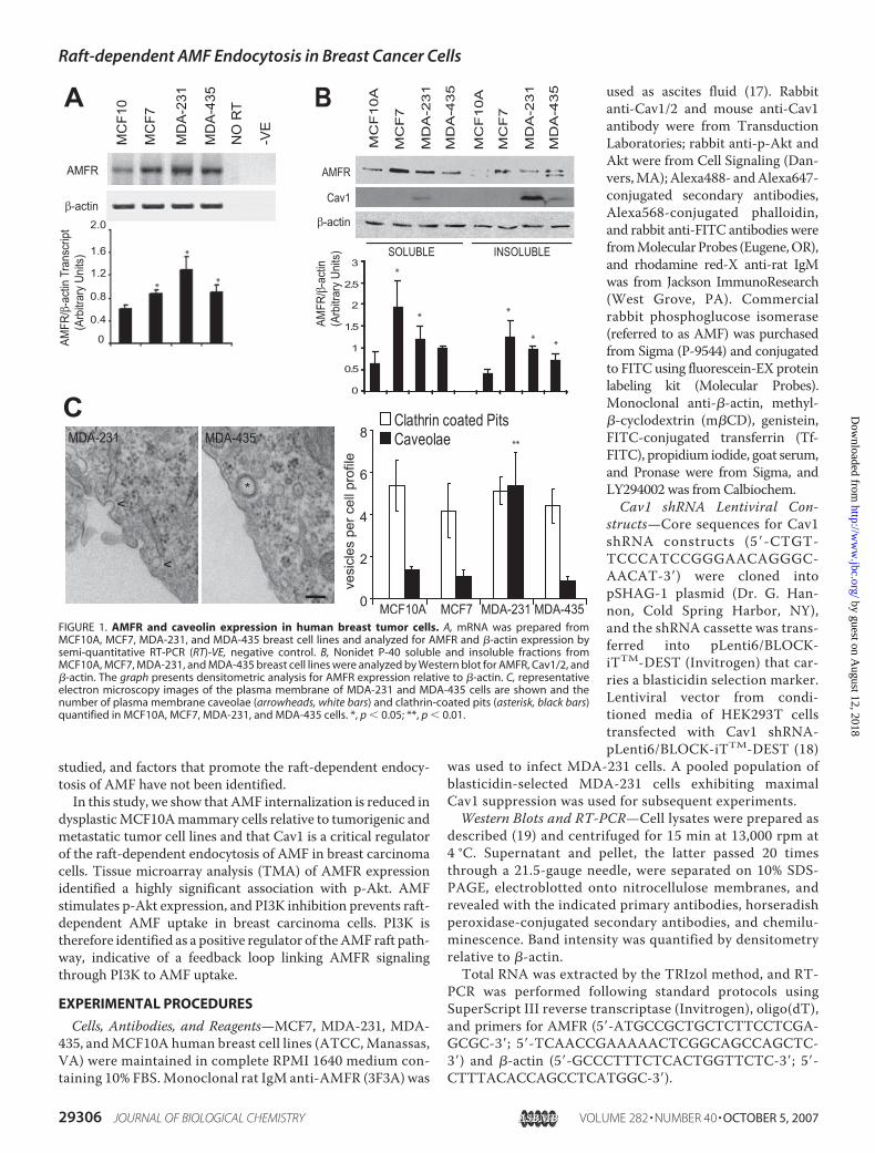
FIGURE 1. AMFR and caveolin expression in human breast tumor cells. A, mRNA was prepared fromMCF10A, MCF7, MDA-231, and MDA-435 breast cell lines and analyzed for AMFR and �-actin expression bysemi-quantitative RT-PCR (RT)-VE, negative control. B, Nonidet P-40 soluble and insoluble fractions fromMCF10A, MCF7, MDA-231, and MDA-435 breast cell lines were analyzed by Western blot for AMFR, Cav1/2, and�-actin. The graph presents densitometric analysis for AMFR expression relative to �-actin. C, representativeelectron microscopy images of the plasma membrane of MDA-231 and MDA-435 cells are shown and thenumber of plasma membrane caveolae (arrowheads, white bars) and clathrin-coated pits (asterisk, black bars)quantified in MCF10A, MCF7, MDA-231, and MDA-435 cells. *, p � 0.05; **, p � 0.01.
Raft-dependent AMF Endocytosis in Breast Cancer Cells
29306 JOURNAL OF BIOLOGICAL CHEMISTRY VOLUME 282 • NUMBER 40 • OCTOBER 5, 2007
by guest on August 12, 2018
http://ww
w.jbc.org/
Dow
nloaded from
Immunofluorescence, FACS, andElectron Microscopy—Immuno-fluorescence, FACS, and electronmicroscopy were performed asdescribed (10, 12). For uptakeanalysis, adherent cells were incu-bated with 25 �g/ml AMF-FITCor 15 �g/ml Tf-FITC for 30 min at37 °C. For FACS analysis, cell sur-face-bound conjugate was removedwith Pronase (400 �g/ml) for 15min. Flow cytometrymeasurementsof at least 50,000 cells usedCellquest software on a FACSCali-bur (BD Biosciences). Adenoviralconstructs and infection were asdescribed (12, 20).Statistical Analysis—RT-PCR,
Western blots, immunofluores-cence, and FACS experiments werereproduced at least three times, andrepresentative blots and images arepresented. FACS experiments wereperformed in duplicate. Unless oth-erwise stated, all values are pre-sented as mean � S.E. of at leastthree independent experiments.Statistical significance was evalu-
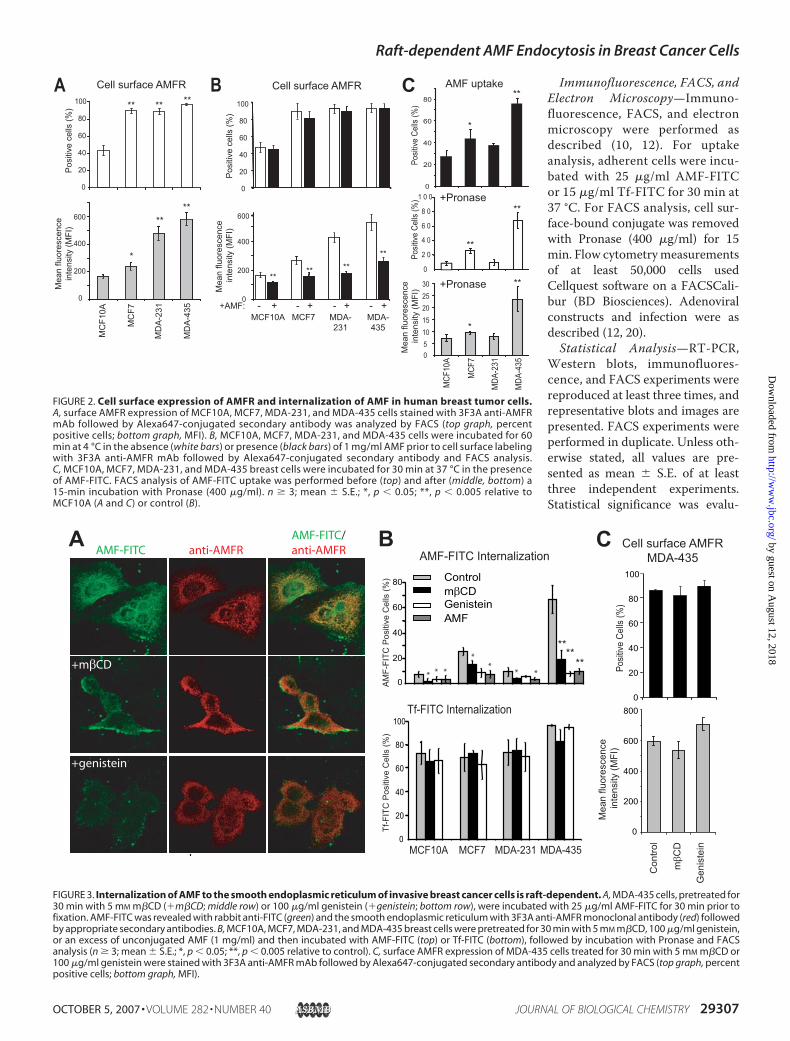
FIGURE 2. Cell surface expression of AMFR and internalization of AMF in human breast tumor cells.A, surface AMFR expression of MCF10A, MCF7, MDA-231, and MDA-435 cells stained with 3F3A anti-AMFRmAb followed by Alexa647-conjugated secondary antibody was analyzed by FACS (top graph, percentpositive cells; bottom graph, MFI). B, MCF10A, MCF7, MDA-231, and MDA-435 cells were incubated for 60min at 4 °C in the absence (white bars) or presence (black bars) of 1 mg/ml AMF prior to cell surface labelingwith 3F3A anti-AMFR mAb followed by Alexa647-conjugated secondary antibody and FACS analysis.C, MCF10A, MCF7, MDA-231, and MDA-435 breast cells were incubated for 30 min at 37 °C in the presenceof AMF-FITC. FACS analysis of AMF-FITC uptake was performed before (top) and after (middle, bottom) a15-min incubation with Pronase (400 �g/ml). n � 3; mean � S.E.; *, p � 0.05; **, p � 0.005 relative toMCF10A (A and C) or control (B).
FIGURE 3. Internalization of AMF to the smooth endoplasmic reticulum of invasive breast cancer cells is raft-dependent. A, MDA-435 cells, pretreated for30 min with 5 mM m�CD (�m�CD; middle row) or 100 �g/ml genistein (�genistein; bottom row), were incubated with 25 �g/ml AMF-FITC for 30 min prior tofixation. AMF-FITC was revealed with rabbit anti-FITC (green) and the smooth endoplasmic reticulum with 3F3A anti-AMFR monoclonal antibody (red) followedby appropriate secondary antibodies. B, MCF10A, MCF7, MDA-231, and MDA-435 breast cells were pretreated for 30 min with 5 mM m�CD, 100 �g/ml genistein,or an excess of unconjugated AMF (1 mg/ml) and then incubated with AMF-FITC (top) or Tf-FITC (bottom), followed by incubation with Pronase and FACSanalysis (n � 3; mean � S.E.; *, p � 0.05; **, p � 0.005 relative to control). C, surface AMFR expression of MDA-435 cells treated for 30 min with 5 mM m�CD or100 �g/ml genistein were stained with 3F3A anti-AMFR mAb followed by Alexa647-conjugated secondary antibody and analyzed by FACS (top graph, percentpositive cells; bottom graph, MFI).
Raft-dependent AMF Endocytosis in Breast Cancer Cells
OCTOBER 5, 2007 • VOLUME 282 • NUMBER 40 JOURNAL OF BIOLOGICAL CHEMISTRY 29307
by guest on August 12, 2018
http://ww
w.jbc.org/
Dow
nloaded from
ated using the Student’s t test for paired comparison; p � 0.05was considered significant.Human Breast Tissue Microarrays—438 sequential archi-
val cases of invasive breast carcinoma, with available paraffinblocks, that had undergone treatment at Vancouver GeneralHospital between 1974 and 1995 were identified for TMAconstruction (21) with adequate tissue present for interpre-tation of 370 cases. The study was approved by the ClinicalResearch Ethics Board of the University of British Columbia.Antibodies used and antigen retrieval methodologies aresummarized in Fig. 5A. Semi-quantitative scoring of TMAsections stained for ER and PR (22), HER2 (23), and p-Akt(24) was as described previously. AMFR was scored positive(1�) if any invasive carcinoma cell staining was observed.For statistical analyses, score data were binarized using
�10% cutoffs for ER and PR and a score of 1� for AMFR and2� (equivalent to strong 3� staining when utilizing the Her-ceptest) for HER2. TMA slides were digitized using a BLISSslide scanner (Bacus Laboratories, Inc., Chicago) and con-nected to a relational data base. They are publicly availableon line. All samples were evaluated and scored, fromscanned images on a computer monitor, by two pathologistsblinded to patient clinical data.Clinical data on all patients was retrospectively collected
from patient hospital charts. Median patient follow-up was 15years, and all patients had newly diagnosed stage I–III invasivebreast cancer. Clinicopathologic data collected includedpatient age and sex, lymph node status (negative versus posi-tive), tumor size, tumor grade, tumor histology, patient follow-up, and survival. All data were logged onto a standardized score
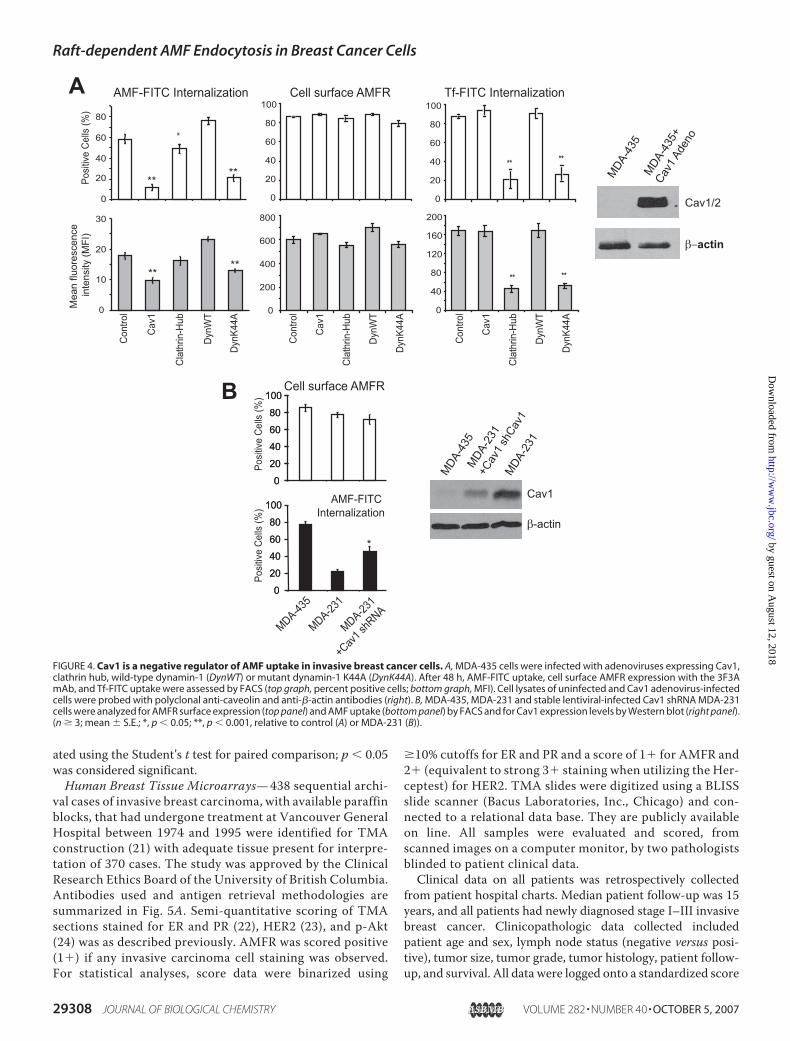
FIGURE 4. Cav1 is a negative regulator of AMF uptake in invasive breast cancer cells. A, MDA-435 cells were infected with adenoviruses expressing Cav1,clathrin hub, wild-type dynamin-1 (DynWT) or mutant dynamin-1 K44A (DynK44A). After 48 h, AMF-FITC uptake, cell surface AMFR expression with the 3F3AmAb, and Tf-FITC uptake were assessed by FACS (top graph, percent positive cells; bottom graph, MFI). Cell lysates of uninfected and Cav1 adenovirus-infectedcells were probed with polyclonal anti-caveolin and anti-�-actin antibodies (right). B, MDA-435, MDA-231 and stable lentiviral-infected Cav1 shRNA MDA-231cells were analyzed for AMFR surface expression (top panel) and AMF uptake (bottom panel) by FACS and for Cav1 expression levels by Western blot (right panel).(n � 3; mean � S.E.; *, p � 0.05; **, p � 0.001, relative to control (A) or MDA-231 (B)).
Raft-dependent AMF Endocytosis in Breast Cancer Cells
29308 JOURNAL OF BIOLOGICAL CHEMISTRY VOLUME 282 • NUMBER 40 • OCTOBER 5, 2007
by guest on August 12, 2018
http://ww
w.jbc.org/
Dow
nloaded from
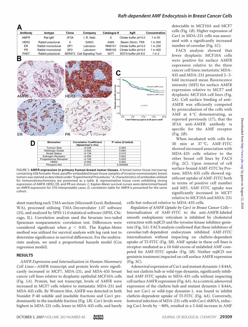
sheet matching each TMA section (Microsoft Excel, Redmond,WA), processed utilizing TMA-Deconvoluter 1.07 software(25), and analyzed by SPSS 11.0 statistical software (SPSS, Chi-cago, IL). Correlation analysis used the bivariate two-tailedSpearman nonparametric correlation test. Differences wereconsidered significant when p � 0.05. The Kaplan-Meiermethod was utilized for survival analysis with log rank test todetermine significance in survival differences. For the multiva-riate analysis, we used a proportional hazards model (Coxregression model).
RESULTS
AMFR Expression and Internalization in Human MammaryCell Lines—AMFR transcript and protein levels were signifi-cantly increased in MCF7, MDA-231, and MDA-435 breastcancer cell lines relative to dysplastic epithelial MCF10A cells(Fig. 1A). Protein, but not transcript, levels of AMFR wereincreased in MCF7 cells relative to metastatic MDA-231 andMDA-435 cells. By Western blot, AMFR was detected in bothNonidet P-40 soluble and insoluble fractions and Cav1 pre-dominantly in the insoluble fraction (Fig. 1B). Cav1 levels werehighest in MDA-231 cells, lesser in MDA-435 cells, and barely
detectable in MCF10A and MCF7cells (Fig. 1B). Higher expression ofCav1 in MDA-231 cells was associ-ated with a significantly increasednumber of caveolae (Fig. 1C).FACS analysis showed that
fewer dysplastic MCF10A cellswere positive for surface AMFRexpression relative to the threecancer cell lines; metastatic MDA-435 and MDA-231 presented 2–3-fold increased mean fluorescenceintensity (MFI) for surface AMFRexpression relative to MCF7 anddysplastic MCF10A cell lines (Fig.2A). Cell surface binding of anti-AMFR was efficiently competedby preincubation of the cells withAMF at 4 °C demonstrating, asreported previously (17), that the3F3A anti-AMFR antibody isspecific for the AMF receptor(Fig. 2B).When incubated with cells for
30 min at 37 °C, AMF-FITCshowed increased association withMDA-435 cells relative to theother breast cell lines by FACS(Fig. 2C). Upon removal of cellsurface-bound AMF-FITC by Pro-nase, MDA-435 cells showed sig-nificant uptake of AMF-FITC bothin terms of positive cell numberand MFI. AMF-FITC uptake wassignificantly increased in MCF7relative toMCF10A andMDA-231
cells but reduced relative to MDA-435 cells.Regulation of AMFR Uptake by Cav1 in Breast Cancer Cells—
Internalization of AMF-FITC to the anti-AMFR-labeledsmooth endoplasmic reticulum is inhibited by cholesterolextraction with m�CD and the tyrosine kinase inhibitor genis-tein (Fig. 3A). FACS analysis confirmed that these inhibitors ofcaveolae/raft-dependent endocytosis inhibited AMF-FITCinternalization without impacting on clathrin-dependentuptake of Tf-FITC (Fig. 3B). AMF uptake in these cell lines isreceptor-mediated as a 10-fold excess of unlabeled AMF com-peted for AMF-FITC uptake (Fig. 3B). Neither m�CD norgenistein treatment impacted on cell surface AMFR expression(Fig. 3C).Adenoviral expression ofCav1 andmutant dynamin-1K44A,
but not clathrin hub or wild-type dynamin, significantly inhib-ited AMF-FITC uptake in MDA-435 cells without impactingcell surfaceAMFRexpression (Fig. 4A). As a control, adenoviralexpression of the clathrin hub and mutant dynamin-1 K44A,but not Cav1 or wild-type dynamin-1, was found to inhibitclathrin-dependent uptake of Tf-FITC (Fig. 4A). Conversely,lentiviral infection ofMDA-231 cells with Cav1 shRNA, reduc-ing Cav1 levels by �80%, resulted in a 2-fold increase in AMF
FIGURE 5. AMFR expression in primary human breast tumor tissues. A breast tumor tissue microarraycontaining 438 formalin-fixed, paraffin-embedded breast tissue samples of invasive nonmetastatic breasttumors was stained as described under “Experimental Procedures.” A, characteristics of antibodies utilizedfor immunohistochemistry are presented as a table. B, representative tissue cores exhibiting strongexpression of AMFR, HER2, ER, and PR are shown. C, Kaplan-Meier survival curves were determined basedon AMFR expression for 370 interpretable cases. D, correlation table for AMFR is presented for the samecohort.
Raft-dependent AMF Endocytosis in Breast Cancer Cells
OCTOBER 5, 2007 • VOLUME 282 • NUMBER 40 JOURNAL OF BIOLOGICAL CHEMISTRY 29309
by guest on August 12, 2018
http://ww
w.jbc.org/
Dow
nloaded from
uptake (Fig. 4B). Cav1 expression therefore negatively regulatesthe clathrin-independent, dynamin-dependent, raft-dependentuptake of AMF in invasive breast cancer cells.Tissue Microarray Analysis of Invasive Human Breast
Carcinomas—From TMA analysis (Fig. 5), 99 of 370 interpret-able cases (27%) of invasive breast cancer expressed AMFR.Increased AMFR expression was not associated (p � 0.414)with disease-specific survival (Fig. 5C). No correlations werefound between AMFR expression and tumor HER2, ER status,tumor size, or histologic grade. AMFR had a weak negativecorrelationwith tumor PR status (p� 0.025) and axillary lymphnode status (p � 0.015) (Fig. 5D). Interestingly, AMFR expres-sion showed a highly significant correlation (p � 0.000065)with p-Akt, previously scored on the same breast cancer TMA(24).Raft-dependent Endocytosis of AMF Is PI3K-dependent—The
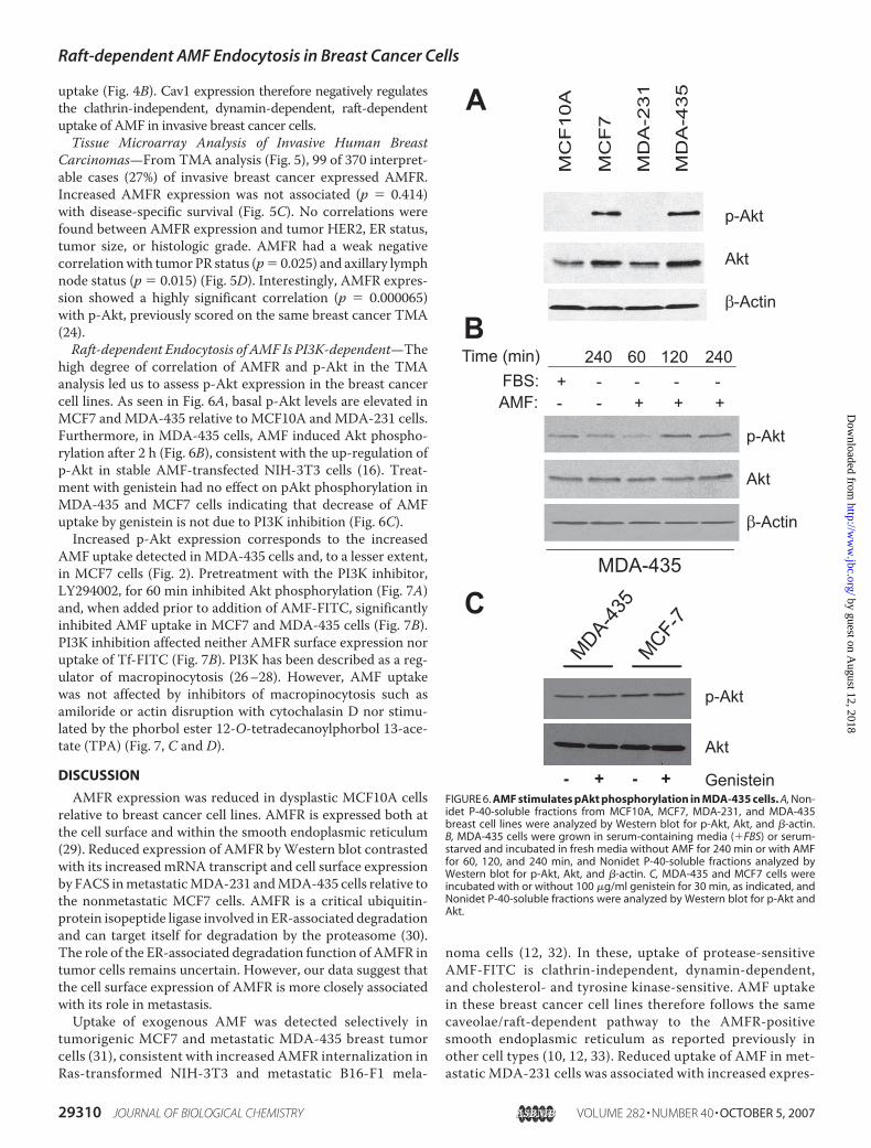
high degree of correlation of AMFR and p-Akt in the TMAanalysis led us to assess p-Akt expression in the breast cancercell lines. As seen in Fig. 6A, basal p-Akt levels are elevated inMCF7 andMDA-435 relative to MCF10A andMDA-231 cells.Furthermore, in MDA-435 cells, AMF induced Akt phospho-rylation after 2 h (Fig. 6B), consistent with the up-regulation ofp-Akt in stable AMF-transfected NIH-3T3 cells (16). Treat-ment with genistein had no effect on pAkt phosphorylation inMDA-435 and MCF7 cells indicating that decrease of AMFuptake by genistein is not due to PI3K inhibition (Fig. 6C).Increased p-Akt expression corresponds to the increased
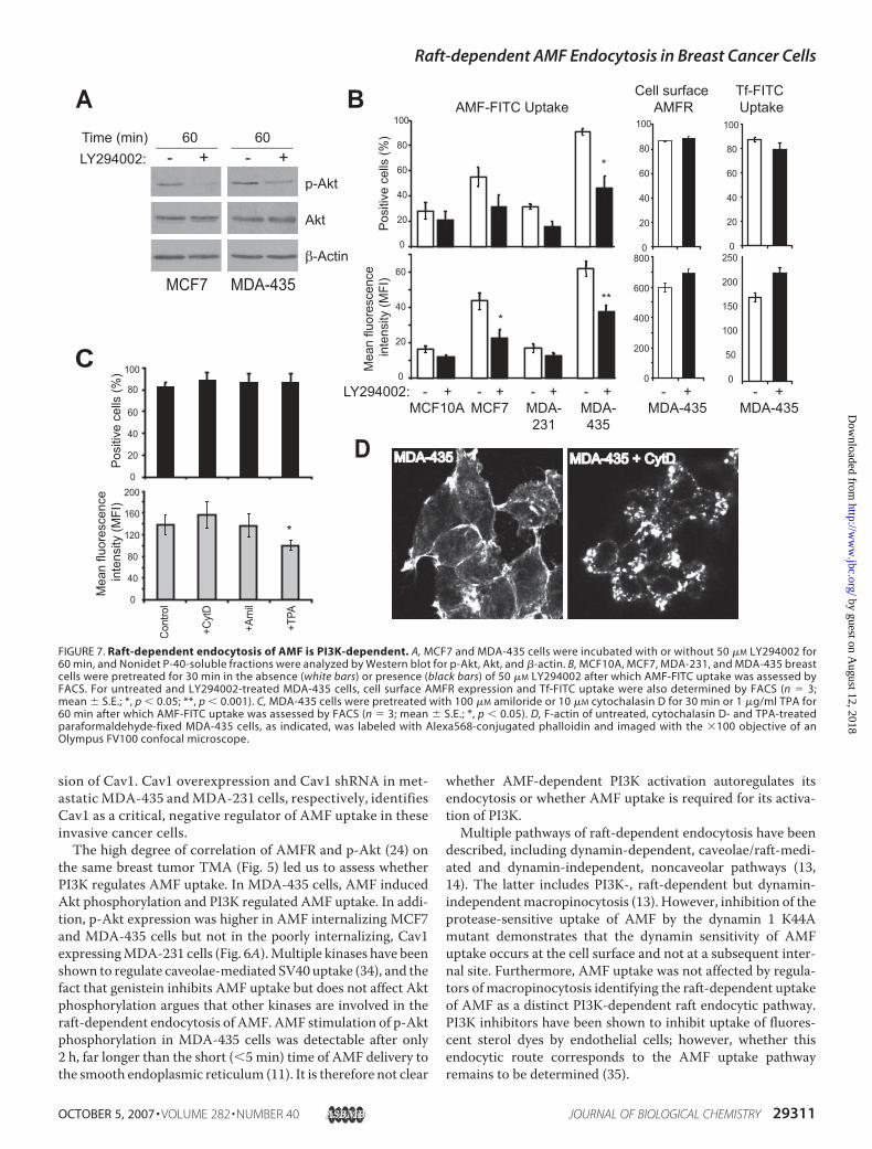
AMF uptake detected in MDA-435 cells and, to a lesser extent,in MCF7 cells (Fig. 2). Pretreatment with the PI3K inhibitor,LY294002, for 60 min inhibited Akt phosphorylation (Fig. 7A)and, when added prior to addition of AMF-FITC, significantlyinhibited AMF uptake in MCF7 and MDA-435 cells (Fig. 7B).PI3K inhibition affected neither AMFR surface expression noruptake of Tf-FITC (Fig. 7B). PI3K has been described as a reg-ulator of macropinocytosis (26–28). However, AMF uptakewas not affected by inhibitors of macropinocytosis such asamiloride or actin disruption with cytochalasin D nor stimu-lated by the phorbol ester 12-O-tetradecanoylphorbol 13-ace-tate (TPA) (Fig. 7, C and D).
DISCUSSION
AMFR expression was reduced in dysplastic MCF10A cellsrelative to breast cancer cell lines. AMFR is expressed both atthe cell surface and within the smooth endoplasmic reticulum(29). Reduced expression of AMFR byWestern blot contrastedwith its increased mRNA transcript and cell surface expressionby FACS inmetastaticMDA-231 andMDA-435 cells relative tothe nonmetastatic MCF7 cells. AMFR is a critical ubiquitin-protein isopeptide ligase involved in ER-associated degradationand can target itself for degradation by the proteasome (30).The role of the ER-associated degradation function of AMFR intumor cells remains uncertain. However, our data suggest thatthe cell surface expression of AMFR is more closely associatedwith its role in metastasis.Uptake of exogenous AMF was detected selectively in
tumorigenic MCF7 and metastatic MDA-435 breast tumorcells (31), consistent with increased AMFR internalization inRas-transformed NIH-3T3 and metastatic B16-F1 mela-
noma cells (12, 32). In these, uptake of protease-sensitiveAMF-FITC is clathrin-independent, dynamin-dependent,and cholesterol- and tyrosine kinase-sensitive. AMF uptakein these breast cancer cell lines therefore follows the samecaveolae/raft-dependent pathway to the AMFR-positivesmooth endoplasmic reticulum as reported previously inother cell types (10, 12, 33). Reduced uptake of AMF in met-astatic MDA-231 cells was associated with increased expres-
FIGURE 6. AMF stimulates pAkt phosphorylation in MDA-435 cells. A, Non-idet P-40-soluble fractions from MCF10A, MCF7, MDA-231, and MDA-435breast cell lines were analyzed by Western blot for p-Akt, Akt, and �-actin.B, MDA-435 cells were grown in serum-containing media (�FBS) or serum-starved and incubated in fresh media without AMF for 240 min or with AMFfor 60, 120, and 240 min, and Nonidet P-40-soluble fractions analyzed byWestern blot for p-Akt, Akt, and �-actin. C, MDA-435 and MCF7 cells wereincubated with or without 100 �g/ml genistein for 30 min, as indicated, andNonidet P-40-soluble fractions were analyzed by Western blot for p-Akt andAkt.
Raft-dependent AMF Endocytosis in Breast Cancer Cells
29310 JOURNAL OF BIOLOGICAL CHEMISTRY VOLUME 282 • NUMBER 40 • OCTOBER 5, 2007
by guest on August 12, 2018
http://ww
w.jbc.org/
Dow
nloaded from
sion of Cav1. Cav1 overexpression and Cav1 shRNA in met-astaticMDA-435 andMDA-231 cells, respectively, identifiesCav1 as a critical, negative regulator of AMF uptake in theseinvasive cancer cells.The high degree of correlation of AMFR and p-Akt (24) on
the same breast tumor TMA (Fig. 5) led us to assess whetherPI3K regulates AMF uptake. In MDA-435 cells, AMF inducedAkt phosphorylation and PI3K regulated AMF uptake. In addi-tion, p-Akt expression was higher in AMF internalizing MCF7and MDA-435 cells but not in the poorly internalizing, Cav1expressingMDA-231 cells (Fig. 6A).Multiple kinases have beenshown to regulate caveolae-mediated SV40 uptake (34), and thefact that genistein inhibits AMF uptake but does not affect Aktphosphorylation argues that other kinases are involved in theraft-dependent endocytosis of AMF. AMF stimulation of p-Aktphosphorylation in MDA-435 cells was detectable after only2 h, far longer than the short (�5 min) time of AMF delivery tothe smooth endoplasmic reticulum (11). It is therefore not clear
whether AMF-dependent PI3K activation autoregulates itsendocytosis or whether AMF uptake is required for its activa-tion of PI3K.Multiple pathways of raft-dependent endocytosis have been
described, including dynamin-dependent, caveolae/raft-medi-ated and dynamin-independent, noncaveolar pathways (13,14). The latter includes PI3K-, raft-dependent but dynamin-independentmacropinocytosis (13). However, inhibition of theprotease-sensitive uptake of AMF by the dynamin 1 K44Amutant demonstrates that the dynamin sensitivity of AMFuptake occurs at the cell surface and not at a subsequent inter-nal site. Furthermore, AMF uptake was not affected by regula-tors of macropinocytosis identifying the raft-dependent uptakeof AMF as a distinct PI3K-dependent raft endocytic pathway.PI3K inhibitors have been shown to inhibit uptake of fluores-cent sterol dyes by endothelial cells; however, whether thisendocytic route corresponds to the AMF uptake pathwayremains to be determined (35).
FIGURE 7. Raft-dependent endocytosis of AMF is PI3K-dependent. A, MCF7 and MDA-435 cells were incubated with or without 50 �M LY294002 for60 min, and Nonidet P-40-soluble fractions were analyzed by Western blot for p-Akt, Akt, and �-actin. B, MCF10A, MCF7, MDA-231, and MDA-435 breastcells were pretreated for 30 min in the absence (white bars) or presence (black bars) of 50 �M LY294002 after which AMF-FITC uptake was assessed byFACS. For untreated and LY294002-treated MDA-435 cells, cell surface AMFR expression and Tf-FITC uptake were also determined by FACS (n � 3;mean � S.E.; *, p � 0.05; **, p � 0.001). C, MDA-435 cells were pretreated with 100 �M amiloride or 10 �M cytochalasin D for 30 min or 1 �g/ml TPA for60 min after which AMF-FITC uptake was assessed by FACS (n � 3; mean � S.E.; *, p � 0.05). D, F-actin of untreated, cytochalasin D- and TPA-treatedparaformaldehyde-fixed MDA-435 cells, as indicated, was labeled with Alexa568-conjugated phalloidin and imaged with the �100 objective of anOlympus FV100 confocal microscope.
Raft-dependent AMF Endocytosis in Breast Cancer Cells
OCTOBER 5, 2007 • VOLUME 282 • NUMBER 40 JOURNAL OF BIOLOGICAL CHEMISTRY 29311
by guest on August 12, 2018
http://ww
w.jbc.org/
Dow
nloaded from
Cholesterol could impact indirectly on macropinocytosisthrough modulation of phosphatidylinositol 4,5-biphosphate-dependent reorganization of the actin cytoskeleton (13, 36).AMF uptake was not affected by disruption of the actincytoskeleton with cytochalasin D in MDA-435 cells (Fig. 7) orMCF7 cells (data not shown). Actin cytoskeleton disruption isassociated with increased mobility of Cav1 and caveolae-medi-ated uptake (37–41). The independence of AMF uptake onactin cytoskeleton integrity further distinguishes this pathwayfrom caveolae-mediated endocytosis.We have shown previously that AMF uptake to the smooth
endoplasmic reticulum is distinct from raft-dependent chol-era toxin b-subunit uptake to the Golgi apparatus and SV40uptake to the endoplasmic reticulum (11). The PI3K depend-ence and actin independence of AMF internalization providefurther evidence for the distinct nature of this dynamin-de-pendent, raft-dependent pathway. Of particular importance,this pathway is shown here to be tumor cell-specific andassociated with enhanced AMFR expression in aggressivetumor cells.AMF-dependent p-Akt activation in MDA-435 cells and
PI3K-dependent AMF uptake is indicative of a feedback loopbetween these two cancer-related proteins functionally link-ing the extensive correlation of AMFR and p-Akt in thebreast tumor TMA. Stable transfection of NIH-3T3 fibro-blasts with AMF, resulting in autocrine activation of AMFR,neoplastic transformation, and resistance to apoptosis, wasfound to be associated with Akt activation and reduced Cav1expression (16). Autocrine activation of AMFR and down-stream PI3K signaling in tumor cells may function to gener-ally enhance AMFR trafficking and plasma membrane turn-over. Akt activation is generally associated with increasedtumor progression and tumor cell invasivity (42) and is anindicator of aggressive tumor behavior and reduced overallsurvival in node-negative breast cancer (43). Overall AMFRpositivity in breast carcinomas did not correlate with patientdisease-specific survival, as reported for p-Akt labeling ofthe same TMA (24). This is consistent with a recent study ofAMF and AMFR expression in breast tumors (44). Coordi-nate overexpression of AMFR and p-Akt may define a cohortof human breast cancers that, although not the most clini-cally aggressive, does represent a significant proportion of allinvasive tumors.Although factors other than Cav1 and p-Akt may regulate
AMF uptake, our data suggest that internalization of cellsurface AMFR may be associated with PI3K-dependent acti-vation of Akt and reduction of Cav1 levels in breast tumorcells. In our invasive breast cancer patient cohort, AMFRtumor expression showed no significant correlation with ERor HER2 expression, but it did present a negative correlationwith PR expression. Thus, individuals with breast tumorsexpressing AMFRmay represent a specific subset of patientswith breast cancer who may have little benefit from HER2targeted or hormonal therapies. The abundant expression ofAMFR by cancer cells in vitro and in vivo suggests that thedynamin and PI3K-dependent, Cav1-regulated, raft-medi-ated endocytosis of AMF may represent a cancer cell-spe-cific endocytic pathway.
Acknowledgment—We thank Mohammad Amraei for assistance.
REFERENCES1. Watanabe, H., Takehana, K., Date, M., Shinozaki, T., and Raz, A. (1996)
Cancer Res. 56, 2960–29632. Hirono, Y., Fushida, S., Yonemura, Y., Yamamoto, H., Watanabe, H., and
Raz, A. (1996) Br. J. Cancer 74, 2003–20073. Nakamori, S., Watanabe, H., Kameyama, M., Imaoka, S., Furukawa, H.,
Ishikawa, O., Sasaki, Y., Kabuto, T., and Raz, A. (1994) Cancer 74,1855–1862
4. Otto, T., Birchmeier, W., Schmidt, U., Hinke, A., Schipper, J., Rubben, H.,and Raz, A. (1994) Cancer Res. 54, 3120–3123
5. Maruyama, K., Watanabe, H., Hitoshi, S., Takayama, T., Gofuku, J., Yano,H., Inoue, M., Tamura, S., Raz, A., and Monden, M. (1995) Int. J. Cancer64, 316–321
6. Nagai, Y., Ishikawa,O.,Miyachi, Y., andWatanabe,H. (1996)Dermatology192, 8–11
7. Takanami, I., Takeuchi, K., Naruke,M., Kodaira, S., Tanaka, F.,Watanabe,H., and Raz, A. (1998) Tumor Biol. 19, 384–389
8. Taniguchi, K., Yonemura, Y., Nojima, N., Hirono, Y., Fushida, S., Fu-jimura, T., Miwa, K., Endo, Y., Yamamoto, H., and Watanabe, H. (1998)Cancer 82, 2112–2122
9. Sjoblom, T., Jones, S., Wood, L. D., Parsons, D. W., Lin, J., Barber, T.,Mandelker, D., Leary, R. J., Ptak, J., Silliman, N., Szabo, S., Buckhaults, P.,Farrell, C., Meeh, P., Markowitz, S. D., Willis, J., Dawson, D., Willson,J. K. V., Gazdar, A. F., Hartigan, J., Wu, L., Liu, C., Parmigiani, G., Park,B.H., Bachman, K. E., Papadopoulos,N., Vogelstein, B., Kinzler, K.W., andVelculescu, V. E. (2006) Science 314, 268–274
10. Benlimame, N., Le, P. U., and Nabi, I. R. (1998) Mol. Biol. Cell 9,1773–1786
11. Le, P. U., and Nabi, I. R. (2003) J. Cell Sci. 116, 1059–107112. Le, P. U., Guay, G., Altschuler, Y., and Nabi, I. R. (2002) J. Biol. Chem. 277,
3371–337913. Kirkham, M., and Parton, R. G. (2005) Biochim. Biophys. Acta 1745,
273–28614. Lajoie, P., and Nabi, I. R. (2007) J. Cell Mol. Med. 11, 644–65315. Pelkmans, L., Kartenbeck, J., and Helenius, A. (2001) Nat. Cell Biol. 3,
473–48316. Tsutsumi, S., Hogan, V., Nabi, I. R., and Raz, A. (2003) Cancer Res. 63,
242–24917. Nabi, I. R., Watanabe, H., and Raz, A. (1990) Cancer Res. 50, 409–41418. Yu, D., Jia, W. W., Gleave, M. E., Nelson, C. C., and Rennie, P. S. (2004)
Prostate 59, 370–38219. Joshi, B., Ordonez-Ercan, D., Dasgupta, P., and Chellappan, S. (2005) On-
cogene 24, 2204–221720. Altschuler, Y., Liu, S., Katz, L., Tang, K., Hardy, S., Brodsky, F., Apodaca,
G., and Mostov, K. (1999) J. Cell Biol. 147, 7–1221. Parker, R. L., Huntsman, D. G., Lesack, D. W., Cupples, J. B., Grant, D. R.,
Akbari, M., and Gilks, C. B. (2002) Am. J. Clin. Pathol. 117, 723–72822. Reiner, A., Neumeister, B., Spona, J., Reiner, G., Schemper,M., and Jakesz,
R. (1990) Cancer Res. 50, 7057–706123. Wiseman, S. M., Makretsov, N., Nielsen, T. O., Gilks, B., Yorida, E.,
Cheang, M., Turbin, D., Gelmon, K., and Huntsman, D. G. (2005) Cancer103, 1770–1777
24. Sutherland, B. W., Kucab, J., Wu, J., Lee, C., Cheang, M. C. U., Yorida, E.,Turbin, D., Dedhar, S., Nelson, C., Pollak,M., LeightonGrimes, H.,Miller,K., Badve, S., Huntsman, D., Blake-Gilks, C., Chen, M., Pallen, C. J., andDunn, S. E. (2005) Oncogene 24, 4281–4292
25. Liu, C. L., Prapong, W., Natkunam, Y., Alizadeh, A., Montgomery, K.,Gilks, C. B., and van de Rijn, M. (2002) Am. J. Pathol. 161, 1557–1565
26. Amyere, M., Payrastre, B., Krause, U., Smissen, P. V. D., Veithen, A., andCourtoy, P. J. (2000)Mol. Biol. Cell 11, 3453–3467
27. Araki, N., Hatae, T., Furukawa, A., and Swanson, J. A. (2003) J. Cell Sci.116, 247–257
28. Zhou, K., Pandol, S., Bokoch, G., and Traynor-Kaplan, A. E. (1998) J. CellSci. 111, 283–294
Raft-dependent AMF Endocytosis in Breast Cancer Cells
29312 JOURNAL OF BIOLOGICAL CHEMISTRY VOLUME 282 • NUMBER 40 • OCTOBER 5, 2007
by guest on August 12, 2018
http://ww
w.jbc.org/
Dow
nloaded from
29. Goetz, J. G., and Nabi, I. R. (2006) Biochem. Soc. Trans. 340, 370–37330. Fang, S., Ferrone, M., Yang, C., Jensen, J. P., Tiwari, S., and Weissman,
A. M. (2001) Proc. Natl. Acad. Sci. U. S. A. 98, 14422–1442731. De Larco, J. E.,Wuertz, B. R., Rosner, K.A., Erickson, S. A., Gamache,D. E.,
Manivel, J. C., and Furcht, L. T. (2001) Am. J. Pathol. 158, 639–64632. Watanabe, H., Nabi, I. R., and Raz, A. (1991) Cancer Res. 51, 2699–270533. Nabi, I. R., and Le, P. U. (2003) J. Cell Biol. 161, 673–67734. Pelkmans, L., Fava, E., Grabner, H., Hannus, M., Habermann, B., Krausz,
E., and Zerial, M. (2005) Nature 436, 78–8635. Niles, W. D., and Malik, A. B. (1999) J. Membr. Biol. 167, 85–10136. Kwik, J., Boyle, S., Fooksman,D.,Margolis, L., Sheetz,M. P., and Edidin,M.
(2003) Proc. Natl. Acad. Sci. U. S. A. 100, 13964–1396937. Conrad, P. A., Smart, E. J., Ying, Y.-S., Anderson, R. G. W., and Bloom,
G. S. (1995) J. Cell Biol. 131, 1421–1433
38. Parton, R. G., Joggerst, B., and Simons, K. (1994) J. Cell Biol. 127,1199–1215
39. Thomsen, P., Roepstorff, K., Stahlhut, M., and van Deurs, B. (2002) Mol.Biol. Cell 13, 238–250
40. Mundy, D. I., Machleidt, T., Ying, Y.-S., Anderson, R. G. W., and Bloom,G. S. (2002) J. Cell Sci. 115, 4327–4339
41. Pelkmans, L., Puntener, D., and Helenius, A. (2002) Science 296,535–539
42. Nicholson, K. M., and Anderson, N. G. (2002) Cell. Signal. 14, 381–39543. Schmitz, K. J., Otterbach, F., Callies, R., Levkau, B., Holscher, M., Hoff-
mann,O., Grabellus, F., Kimmig, R., Schmid, K.W., and Baba, H. A. (2004)Mod. Pathol. 17, 15–21
44. Jiang, W. G., Raz, A., Douglas-Jones, A., and Mansel, R. E. (2006) J. Histo-chem. Cytochem. 54, 231–241
Raft-dependent AMF Endocytosis in Breast Cancer Cells
OCTOBER 5, 2007 • VOLUME 282 • NUMBER 40 JOURNAL OF BIOLOGICAL CHEMISTRY 29313
by guest on August 12, 2018
http://ww
w.jbc.org/
Dow
nloaded from
A. Turbin, Sam M. Wiseman and Ivan R. NabiLiliana D. Kojic, Bharat Joshi, Patrick Lajoie, Phuong U. Le, Michael E. Cox, Dmitry
3-Kinase-dependent in Breast Carcinoma CellsRaft-dependent Endocytosis of Autocrine Motility Factor Is Phosphatidylinositol
doi: 10.1074/jbc.M704069200 originally published online August 8, 20072007, 282:29305-29313.J. Biol. Chem.
10.1074/jbc.M704069200Access the most updated version of this article at doi:
Alerts:
When a correction for this article is posted•
When this article is cited•
to choose from all of JBC's e-mail alertsClick here
http://www.jbc.org/content/282/40/29305.full.html#ref-list-1
This article cites 44 references, 22 of which can be accessed free at
by guest on August 12, 2018
http://ww
w.jbc.org/
Dow
nloaded from