rab11b mediates melanin transfer between donor melanocytes ... · rab11b mediates melanin transfer...
TRANSCRIPT
Rab11b Mediates Melanin Transfer betweenDonor Melanocytes and Acceptor Keratinocytesvia Coupled Exo/EndocytosisAbul K. Tarafder1,2,3,8, Giulia Bolasco3,8, Maria S. Correia1,2, Francisco J.C. Pereira1,2, Lucio Iannone1,2,Alistair N. Hume3, Niall Kirkpatrick4, Mauro Picardo5, Maria R. Torrisi5,6, Ines P. Rodrigues1, Jose S. Ramalho1,Clare E. Futter7, Duarte C. Barral1 and Miguel C. Seabra1,2,3
The transfer of melanin from melanocytes to keratinocytes is a crucial process underlying maintenance of skinpigmentation and photoprotection against UV damage. Here, we present evidence supporting coupledexocytosis of the melanin core, or melanocore, by melanocytes and subsequent endocytosis by keratinocytesas a predominant mechanism of melanin transfer. Electron microscopy analysis of human skin samples revealedthree lines of evidence supporting this: (1) the presence of melanocores in the extracellular space; (2) withinkeratinocytes, melanin was surrounded by a single membrane; and (3) this membrane lacked the melanosomalmembrane protein tyrosinase-related protein 1 (TYRP1). Moreover, co-culture of melanocytes and keratinocytessuggests that melanin exocytosis is specifically induced by keratinocytes. Furthermore, depletion of Rab11b, butnot Rab27a, caused a marked decrease in both keratinocyte-stimulated melanin exocytosis and transfer tokeratinocytes. Thus, we propose that the predominant mechanism of melanin transfer is keratinocyte-inducedexocytosis, mediated by Rab11b through remodeling of the melanosome membrane, followed by subsequentendocytosis by keratinocytes.
Journal of Investigative Dermatology (2014) 134, 1056–1066; doi:10.1038/jid.2013.432; published online 14 November 2013
INTRODUCTIONMelanocytes reside in the basal layer of the epidermis sparselyspread in a 1:40 ratio among keratinocytes (Jimbow et al., 1979),where the two cell types are proposed to interact in a symbioticmanner (Imokawa, 2004; Yamaguchi and Hearing, 2010). Thephotoprotective pigment, melanin, is synthesized in melanocytesand packaged into lysosome-related organelles termedmelanosomes (Marks and Seabra, 2001; Raposo and Marks,2002; Hearing, 2005). Consistent with their unique morphologyand function, melanosomes contain specific integral membrane
proteins such as PMEL (gp100), tyrosinase, and tyrosinase-relatedprotein 1 (TYRP1, gp75) whose sorting and localization havebeen described previously (Raposo et al., 2001). Fully melanizedmelanosomes are transported from their site of synthesis to thecell periphery before they are transferred to keratinocytes andtransported to the apical area of the cell to form a supranuclearcap that prevents DNA photodamage induced by exposure toUV radiation (Scott, 2003; Byers et al., 2007).
Despite its pathophysiological importance, the molecularmechanism underlying melanin transfer remains poorlycharacterized, although several hypotheses (H1–H4) havebeen postulated to date including (Yamamoto and Bhawan,1994; Seiberg, 2001; Van Den Bossche et al., 2006; Singhet al., 2008): (H1) heterophagocytosis of the melanocytedendrite tip; (H2) release of melanosome-loaded vesiclesfrom the melanocyte followed by phagocytosis by thekeratinocyte (Scott et al., 2002; Ando et al., 2011; Wu et al.,2012) (H3) exocytosis of the melanin core from themelanocyte followed by endocytosis by the keratinocyte; and(H4) transfer of melanosomes from the melanocyte filopodia tothe keratinocyte via direct membrane fusion (Singh et al.,2010; Beaumont et al., 2011). The first two mechanismsinvolve melanosome transport within keratinocytes in doublemembrane compartments derived from both the donormelanocyte and the acceptor keratinocyte. In contrast, thelast two hypotheses predict that melanin granules aretransported within keratinocytes in a single membrane-boundstructure derived either from the keratinocyte (H3) or from the
See related commentary on pg 877ORIGINAL ARTICLE
1CEDOC, Faculdade de Ciencias Medicas, Universidade Nova de Lisboa,Lisboa, Portugal; 2Instituto Gulbenkian de Ciencia, Oeiras, Portugal;3Molecular Medicine, National Heart and Lung Institute, Imperial CollegeLondon, London, UK; 4Craniofacial and Orbito-Palpebral Surgery Unit atChelsea and Westminster Hospital, London, UK; 5Laboratorio di FisiopatologiaCutanea, Istituto Dermatologico San Gallicano, IFO, Rome, Italy;6Dipartimento di Medicina Clinica e Molecolare, Azienda OspedalieraSant’Andrea, Universita di Roma ‘La Sapienza’, Rome, Italy and 7Institute ofOphthalmology, University College London, London, UK
Correspondence: Miguel C. Seabra or Duarte C. Barral, CEDOC, Faculdade deCiencias Medicas, FCM, Universidade Nova de Lisboa, 1169-056 Lisboa,Portugal. E-mail: [email protected] or [email protected]
8The first two authors contributed equally to this work.
Received 21 January 2013; revised 30 August 2013; accepted 17 September2013; accepted article preview online 18 October 2013; published online14 November 2013
Abbreviations: LAMP1, lysosomal-associated membrane protein 1; PBS,phosphate-buffered saline; siRNA, small interfering RNA; TYRP1, tyrosinase-related protein 1
1056 Journal of Investigative Dermatology (2014), Volume 134 & 2014 The Society for Investigative Dermatology
melanocyte (H4). Recently, an alternative mechanism hasbeen proposed where melanocytes transfer melanosome-richpackages by a ‘‘shedding’’ mechanism that occurs both at thetips of dendrites and the cell body (Wu et al., 2012).
Despite a number of ultrastructural studies on epidermalcell culture (Okazaki et al., 1976), human hair roots (Mottazand Zelickson, 1967), and skin samples (Yamamoto andBhawan, 1994), molecular evidence for the mechanism ofmelanin transfer has not been forthcoming. Moreover, the lackof a suitable system to reproduce melanin transfer in vitro hasrepresented a major impediment to unraveling the molecularbasis of this process.
Rab proteins are critical regulators of membrane trafficking(Pfeffer, 2001; Zerial and McBride, 2001; Seabra et al., 2002)and have been implicated in a number of processes involvedin skin pigmentation (Wasmeier et al., 2006). Rab32 andRab38 have been implicated in melanosome biogenesis(Wasmeier et al., 2006). Rab27a has a well-characterizedrole in melanosome transport within melanocytes where itregulates the peripheral localization of melanosomes(Bahadoran et al., 2001; Hume et al., 2001; Wu et al.,2001; Strom et al., 2002). Recently, depletion of Rab17 andRab11 have been shown to cause accumulation of pigment inmelanocytes (Beaumont et al., 2011). Hence, it is likely thatRab proteins have a crucial role in regulating melanin transfer.
Here, we have characterized the predominant molecularmechanism of melanin transfer from melanocytes tokeratinocytes based on a combination of in vivo morpho-logical analysis coupled with in vitro models of melanintransfer. We found that melanin exocytosis is mediated byRab11b and that melanocores are subsequently endocytosedby keratinocytes.
RESULTSUltrastructural analysis of human skin reveals melanocorespresent in the extracellular space between melanocytes andkeratinocytes
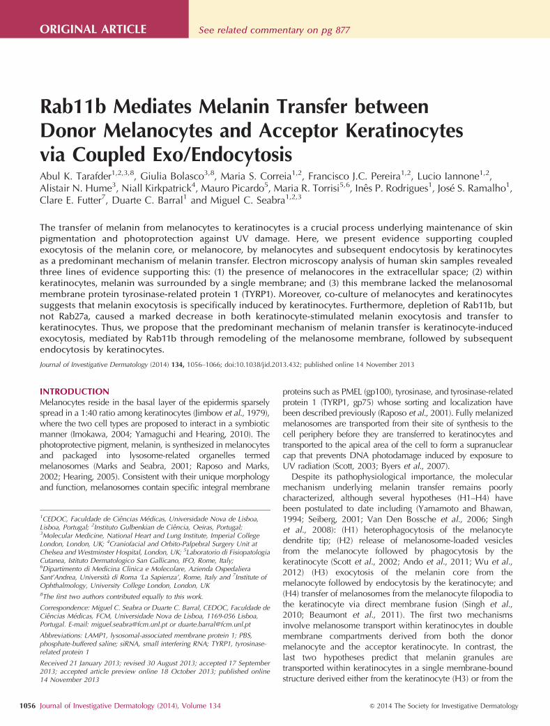
Human skin samples were analyzed by transmission electronmicroscopy and areas with a high concentration of melanin inkeratinocytes were selected for analysis of melanin transfer(Figure 1a). Melanocytes in the basal layer of the epidermiscan be readily distinguished from keratinocytes by the lack ofkeratins, absence of desmosomes at cell–cell junctions(Supplementary Figure S1a–c online), and the presence ofmelanosomes at different stages of maturation (SupplementaryFigure S1c online). Analysis of serial ultrathin sectionsrevealed the presence of melanin granules lacking membranesor associated cytoplasm, herein termed melanocores, in theextracellular space between the melanocyte and the basallamina (Figure 1b1–8). To exclude artifacts or alteration due tochemical fixation with aldehydes, high-pressure freezingfollowed by freeze substitution and conventional embeddingwas also performed on a skin sample, showing similar results.(Supplementary Figure S2a and b online).
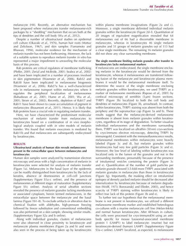
Along with individual granules, clusters of melanocoreswere also observed in close proximity to keratinocyte andmelanocyte plasma membranes (Figure 2a and b) and werealso seen in the process of being taken up by keratinocytes
within plasma membrane invaginations (Figure 2a and c).Moreover, a single membrane delimited individual melaningranules within the keratinocyte (Figure 2d–f). Quantitation of20 images of equivalent magnification revealed that 64melanosomes out of 66 had a discernable membrane inmelanocytes, whereas in keratinocytes 46 single melaningranules and 51 groups of melanin granules out of 113 hada clear single membrane. The remaining 16 melanin granulesdid not show any clear surrounding membrane.
The single membrane limiting melanin granules after transfer tokeratinocytes lacks melanosomal markers
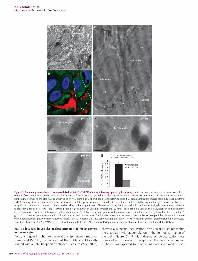
If melanocores are endocytosed, the single membrane enclos-ing melanin in the keratinocyte would be derived from thekeratinocyte, whereas if melanosomes are transferred follow-ing fusion of the melanocyte and keratinocyte plasma mem-branes it would be the melanosomal membrane itself. Todetermine the source of the single membrane enclosingmelanin granules within keratinocytes, we used TYRP1 as amarker of melanosome membranes (Raposo et al., 2001) byconfocal microscopy of semithin (5mm) frozen sections.TYRP1 (Figure 3b) localized to both the cell body anddendrites of melanocytes (Figure 3b, arrowhead). In contrast,within keratinocytes, TYRP1 staining was absent from both theperiphery and the supranuclear cap (Figure 3a and b). Theseresults suggest that the melanocyte-derived melanosomemembrane is absent from melanin granules within keratino-cytes, regardless of its stage of degradation. To better visualizeindividual melanosomes and the membranes surroundingthem, TYRP1 was localized on ultrathin (50 nm) cryo-sectionsby cryo-immuno electron microscopy, detecting TYRP1 byimmunogold. Consistent with the immunofluorescence results,melanosomal membranes within melanocytes were heavilylabeled (Figure 3c and d), but melanin granules withinkeratinocytes had very few gold particles (Figure 3c and e).Moreover, the low level of labeling within keratinocytes waslocalized only in the lumen of the granules and not in thesurrounding membrane, presumably because of the presenceof intraluminal vesicles containing the protein (Figure 3cand e). Quantification of the number of gold particles permelanin granule showed 3.5-fold more TYRP1 associated withmelanin granules in melanocytes than those in keratinocytes(Figure 3g). Importantly, the masking effect on intraluminalepitopes of densely packed melanin should be decreased afterinternalization by keratinocytes because of melanin degrada-tion (Wolff, 1973; Borovansky and Elleder, 2003), and hencescarcity of TYRP1 staining within keratinocytes is likely toreflect true lack of this protein (Figure 3b and e).
To further test our hypothesis that the melanosomal mem-brane is not present in keratinocytes, we utilized a differentmelanosome membrane marker and established heterologousco-cultures with a murine melanocyte cell line (Melan-Ink4a)and normal human keratinocytes. After 48 hours in culture,the cells were processed for cryo-immunoEM using an anti-body specific for mouse lysosomal-associated membraneprotein 1 (LAMP1) to label melanocyte- (murine) but notkeratinocyte-derived (human) LAMP1 (Supplementary FigureS3a–c online). LAMP1 localized, as expected, to melanosome
AK Tarafder et al.Melanosome Transfer via Exo/Endocytosis
www.jidonline.org 1057
membranes in melanocytes, as previously reported(Supplementary Figure S3b online) (Zhou et al., 1993), butmouse LAMP1 was not detected in melanin granules withinkeratinocytes (Supplementary Figure S3c online), thus demon-strating the loss of the membrane after melanin transfer.
The presence of melanocores in the extracellular spacebetween cells, together with the observation that melanintaken up by keratinocytes is surrounded by a single membranelacking melanosomal membrane proteins, suggests that mel-anin exocytosis followed by endocytosis by keratinocytes is amajor mechanism underlying melanin transfer in the skin.
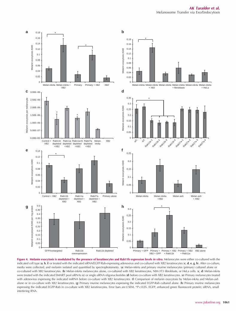
Keratinocytes induce melanin exocytosis by melanocytes in co-cultureConfirmation of this hypothesis required the development of amelanin exocytosis assay. Two melanocyte cell types wereutilized, namely Melan-ink4a and primary murine melano-
cytes derived from C57BL/6 mice. Melanocytes cultured alonedisplayed low levels of melanin exocytosis into tissue culturemedium (Figure 4a). However, when co-cultured with XB2keratinocytes, both melanocyte types showed a 2–3-foldincrease in melanin exocytosis. The increase in melanin exo-cytosis elicited by keratinocytes was specific as co-culture ofmelanocytes with both HeLa and NIH-3T3 fibroblasts did nothave any significant effect on melanin exocytosis (Figure 4b).
Rab11b modulates keratinocyte-induced melanin exocytosis bymelanocytes
To characterize the molecular basis of melanin exocytosisfrom melanocytes, a small interfering RNA (siRNA) screen ofcandidate Rab GTPases was employed. Melan-ink4a melano-cytes were treated with the appropriate siRNA SMART poolsbefore co-culture with XB2 keratinocytes. After 7 days ofco-culture, the quantity of melanin present in tissue culture
Ker
Mel
bFibroblast
Dermis
Epidermis
0
BL
+50 nm +100 nm +150 nm
+350 nm+300 nm+250 nm+200 nm
PM
Figure 1. Transmission electron microscopy (TEM) of ultrathin sections of human skin reveals melanocores in the extracellular space. (a) Low-magnification
TEM image showing the overall structure of the human dermis/epidermis. (b1–8) A series of high-magnification TEM images of serial ultrathin (50nm) sections
of the interface between a melanocyte plasma membrane (arrow) and the BL. Inspection of serial sections reveals the presence of a melanocore devoid of
membrane in the extracellular space. BL, basal lamina; K, keratin filament; Ker, keratinocyte; Mel, melanocyte; PM, plasma membrane. Bars: (a)¼10mm and
(b1–8)¼ 200 nm.
AK Tarafder et al.Melanosome Transfer via Exo/Endocytosis
1058 Journal of Investigative Dermatology (2014), Volume 134
media was assayed and normalized to the number ofmelanocytes present at the end of the assay period. Thisscreen revealed that Rab11b depletion caused a markeddecrease in keratinocyte-induced melanin exocytosis(Figure 4c). Surprisingly, Rab27a depletion had no effect onkeratinocyte-induced melanin exocytosis. To confirm thesefindings, single siRNA oligos designed to specifically silenceRab11b and Rab27a were utilized. Quantitative real-timereverse-transcriptase–PCR analysis revealed that these siRNAsgave an B50% reduction in Rab11b and Rab27a mRNAlevels in melanocytes cultured alone as measured 7 days afterknockdown was initiated (Supplementary Figure S4a and bonline), and no off-target effects on Rab11a expression wereobserved (Supplementary Figure S4c online). All single oligosspecifically designed to deplete Rab11b, but not those
targeting Rab27a, significantly reduced keratinocyte-inducedmelanin exocytosis into tissue culture media to levels seenwith Melan-ink4a cultured alone (Figure 4d).
The effect of Rab11b and Rab27a depletion in primarymurine melanocytes was also tested using adenovirus encod-ing miRNA and a green fluorescent protein reporter to infectmelanocytes. Consistently, depletion of Rab11b but notRab27a caused a reduction of keratinocyte-induced melaninexocytosis to the level seen with primary melanocytes alone(Figure 4e). Furthermore, we confirmed that knockdown bymiRNA was efficient and off-target effects were not seen(Supplementary Figure S4d–f online).
To further verify the surprising finding that Rab27ahad no effect on keratinocyte-induced melanin exocytosis,Melan-ash melanocytes (Rab27a-null) were utilized.Consistent with our findings, no significant difference wasobserved in the keratinocyte-induced increase in melaninexocytosis between Melan-ink4a and Melan-ash melanocytes(Figure 4f).
Next, the effect of Rab overexpression on melanin exocy-tosis was assayed. Primary melanocytes were infected withadenovirus encoding green fluorescent protein–Rab proteins.Overexpression of Rab11a and Rab11b had no effect onmelanin exocytosis from melanocytes cultured alone(Figure 4g). However, when co-cultured with XB2, over-expression of Rab11b caused a 2-fold increase in keratino-cyte-induced melanin exocytosis (Figure 4h). In contrast,overexpression of Rab11a had little effect on keratinocyte-induced melanin exocytosis.
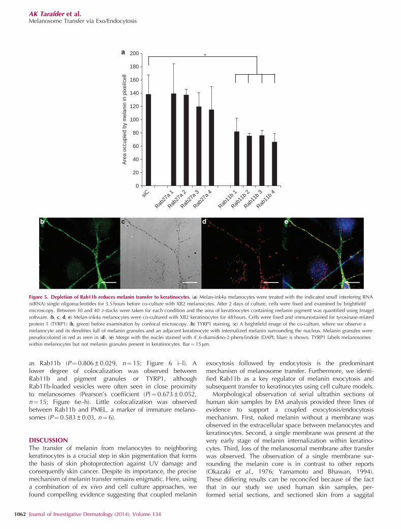
Disruption of melanin exocytosis causes a reduction in melanintransfer to keratinocytesWe showed that depletion of Rab11b, but not Rab27a, causesa decrease in keratinocyte-induced melanin exocytosis. Todetermine whether melanin exocytosis is a predominantmechanism of melanin transfer, the amount of melanintransferred to keratinocytes was assayed in conditions wheremelanin exocytosis was inhibited. Melan-ink4a cells weretreated with single siRNA oligos specifically depleting eitherRab11b or Rab27a and the amount of melanin transferred toXB2 keratinocytes was assayed by brightfield microscopy.Rab11b depletion resulted in an B50% decrease in melanintaken up by keratinocytes with all four siRNAs utilized(Figure 5a). Conversely, Rab27a depletion did not lead to adecrease in melanin uptake by keratinocytes with any of thesingle siRNA oligos (Figure 5a) despite efficient depletion(Supplementary Figure S4b online). These data suggest acorrelation between melanin exocytosis and melanin transferas depletion of Rab11b, but not Rab27a, decreased bothmelanin exocytosis and melanin transfer to keratinocytes.Furthermore, representative images of the co-culture systemclearly show that melanin is taken up by keratinocytes andthat these melanin granules lack TYRP1 staining (Figure 5b–d),again confirming that the melanosomal membrane is notpresent on melanin granules within keratinocytes. Note-worthy, little melanin was transferred when Melan-ink4a wereco-cultured with HeLa or NIH-3T3 fibroblasts (SupplementaryFigure S5 online).
Mel
Keratinocyte
Mel
Keratinocyte
K
nu
K
PMKeratinocyte
c
b
e
f
Figure 2. Melanin granules internalized by keratinocytes are present in single
membrane-bound compartments. (a–c, d–f) Transmission electron microscopy
(TEM) images of ultrathin section of human skin showing two different areas of
melanin-containing keratinocytes in the basal layer. (a, d) Low-magnification
images. (c, b, e, f) High-magnification images of upper and lower boxed areas
in a and d, respectively. (b, c) A cluster of melanin granules is shown: (b) being
uptaken by a keratinocyte membrane ruffle, and (c) in early phase of
internalization within a single membrane. (e, f) Individual melanin granules are
shown that are in the process of being uptaken by a keratinocyte, and are
bounded by a single bilayer (arrows) that is continuous with a keratinocyte
plasma membrane invagination. K, keratin; Mel, melanocyte; nu, nucleus; PM,
plasma membrane. Bars¼ 100 nm.
AK Tarafder et al.Melanosome Transfer via Exo/Endocytosis
www.jidonline.org 1059
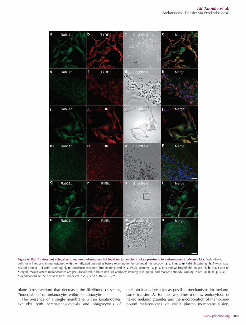
Rab11b localizes in vesicles in close proximity to melanosomesin melanocytes
To try and gain insight into the relationship between melano-somes and Rab11b, we colocalized them. Melan-ink4a cellsstained with a Rab11b-specific antibody (Lapierre et al., 2003)
showed a punctate localization in vesicular structures withinthe cytoplasm with accumulation in the perinuclear region ofthe cell (Figure 6). A high degree of colocalization wasobserved with transferrin receptor in the perinuclear regionof the cell as expected for a recycling endosome marker such
f
Nu
Keratinocyte
K
Melanocyte
Nu
Dermis
ed
PMKeratinocyte
BL
TYRP110 TYRP110
TYRP110
Melanocyte
Melanocyte
4 ***
Melanocytes
TYRP1-positive melanosomesbefore/after intercellular transfer
Keratinocytes
3.53
2.52
1.5
10 n
m P
AG
/mel
anos
ome
10.5
0
Figure 3. Melanin granules lack tyrosinase-related protein 1 (TYRP1) staining following uptake by keratinocytes. (a, b) Confocal analysis of immunolabeled
semithin frozen sections of human skin revealed absence of TYRP1 staining (b, red) in melanin granules within perinuclear melanin cap in keratinocytes (b, anti-
pankeratin, green; a, brightfield). Nuclei are revealed by 4’,6-diamidino-2-phenylindole (DAPI) staining (blue, b). High-magnification images of boxed area show strong
TYRP1 staining of melanosomes within the melanocyte dendrite tip (arrowhead) compared with those transferred to neighboring keratinocytes (arrow). (c) Low
magnification of ultrathin cryosection of human skin. (d, e) Higher magnification of boxed areas in (c) (left hand and right hand, respectively) showing immuno-electron
microscopy analysis of TYRP1 (TYRP1, 10nm protein A gold (PAG)) in ultrathin cryosections (50nm). TYRP1 labeling appears more abundant in both membrane
and intraluminal vesicles in melanosomes within melanocytes (d, f) than in melanin granules after translocation to keratinocytes (e). (g) Quantification of protein A
gold 10nm particles per melanosome in both melanocytes and keratinocytes. The bar chart shows the decrease in the number of gold particles per melanin granule
within keratinocytes (gray) versus melanocytes (black) (n¼ 50 in each case), thus demonstrating the loss of TYRP1 in melanin granules after transfer to keratinocytes.
Error bars shown are±SEM. ***Po0.01. BL, basal lamina; K, keratin; Nu, nucleus; PM, plasma membrane. Bars: a, b¼ 1mm; c¼1mm; d, f¼ 100 nm.
AK Tarafder et al.Melanosome Transfer via Exo/Endocytosis
1060 Journal of Investigative Dermatology (2014), Volume 134
0
0.02
0.04
0.06
0.08
0.1
0.12
0.14
0.16
0.18
Melan-ink4a Melan-ink4a +XB2
Primary Primary + XB2 XB2
Mel
anin
exo
cyto
sis
A34
0 *
*
0
0.02
0.04
0.06
0.08
0.1
0.12
0.14
0.16
0.18
Melan-ink4a Melan-ink4a+ XB2
Melan-ink4a Melan-ink4a+ fibroblasts
Melan-ink4a Melan-ink4a+ HeLa
Mel
anin
exo
cyto
sis
A34
0
*
0
0.05
0.1
0.15
0.2
0.25
0.3
0.35
siC NT
Rab11
b-1
Rab11
b-2
Rab11
b-3
Rab11
b-4
Rab27
a-1
Rab27
a-2
Rab27
a-3
Rab27
a-4
Mel
anin
exo
cyto
sis
A34
0
*
0
0.02
0.04
0.06
0.08
0.1
0.12
0.14
Control + XB2 Rab11bdepleted +
XB2
Rab11adepleted +
XB2
Rab27adepleted +
XB2
Primary alone
Mel
anin
exo
cyto
sis
A34
0
*
0
0.05
0.1
0.15
0.2
0.25
Melan-ink4a Melan-ink4a+XB2
Melan-ash Melan-ash+ XB2
Mel
anin
exo
cyto
sis
A34
0
0
0.05
0.1
0.15
0.2
0.25
0.3
0.35
0.4
0.45
0.5
GFP/nontargeted Rab11boverexpression
Rab11b depleted
Mel
anin
exo
cyto
sed
A34
0 pe
r m
g pr
otei
n
0
0.05
0.1
0.15
0.2
0.25
0.3
Primary + GFP Primary +XB2 + GFP
Primary + XB2+ Rab11b
Primary + XB2+ Rab11a
XB2 alone
Mel
anin
exo
cyto
sis
A34
0
*
0.00E+00
5.00E–07
1.00E–06
1.50E–06
2.00E–06
2.50E–06
3.00E–06
Control +XB2
Rab11bdepleted
+XB2
Rab11adepleted
+XB2
Rab11a+bdepleted
+XB2
Rab27adepleted
+XB2
Melan-ink4a
XB2
Mel
anin
exo
cyto
sis
per
mel
anoc
yte
**
a b
c d
e
g h
f
Figure 4. Melanin exocytosis is modulated by the presence of keratinocytes and Rab11b expression levels in vitro. Melanocytes were either co-cultured with the
indicated cell type (a, b, f) or treated with the indicated siRNA/EGFP-Rab-expressing adenovirus and co-cultured with XB2 keratinocytes (c, d, e, g, h). After co-culture,
media were collected, and melanin isolated and quantified by spectrophotometry. (a) Melan-ink4a and primary murine melanocytes (primary) cultured alone or
co-cultured with XB2 keratinocytes. (b) Melan-ink4a melanocytes alone, co-cultured with XB2 keratinocytes, NIH-3T3 fibroblasts, or HeLa cells. (c, d) Melan-ink4a
were treated with the indicated SMART pool siRNAs (c) or single siRNA oligonucleotides (d) before co-culture with XB2 keratinocytes. (e) Primary melanocytes treated
with adenovirus expressing the indicated miRNA before co-culture with XB2 keratinocytes. (f) Comparison of melanin exocytosis by Melan-ink4a and Melan-ash
alone or in co-culture with XB2 keratinocytes. (g) Primary murine melanocytes expressing the indicated EGFP-Rab cultured alone. (h) Primary murine melanocytes
expressing the indicated EGFP-Rab in co-culture with XB2 keratinocytes. Error bars are±SEM. *Po0.05. EGFP, enhanced green fluorescent protein; siRNA, small
interfering RNA.
AK Tarafder et al.Melanosome Transfer via Exo/Endocytosis
www.jidonline.org 1061
as Rab11b (P¼0.806±0.029, n¼15; Figure 6 i–l). Alower degree of colocalization was observed betweenRab11b and pigment granules or TYRP1, althoughRab11b-loaded vesicles were often seen in close proximityto melanosomes (Pearson’s coefficient (P)¼ 0.673±0.052,n¼15; Figure 6e–h). Little colocalization was observedbetween Rab11b and PMEL, a marker of immature melano-somes (P¼0.583±0.03, n¼ 6).
DISCUSSIONThe transfer of melanin from melanocytes to neighboringkeratinocytes is a crucial step in skin pigmentation that formsthe basis of skin photoprotection against UV damage andconsequently skin cancer. Despite its importance, the precisemechanism of melanin transfer remains enigmatic. Here, usinga combination of ex vivo and cell culture approaches, wefound compelling evidence suggesting that coupled melanin
exocytosis followed by endocytosis is the predominantmechanism of melanosome transfer. Furthermore, we identi-fied Rab11b as a key regulator of melanin exocytosis andsubsequent transfer to keratinocytes using cell culture models.
Morphological observation of serial ultrathin sections ofhuman skin samples by EM analysis provided three lines ofevidence to support a coupled exocytosis/endocytosismechanism. First, naked melanin without a membrane wasobserved in the extracellular space between melanocytes andkeratinocytes. Second, a single membrane was present at thevery early stage of melanin internalization within keratino-cytes. Third, loss of the melanosomal membrane after transferwas observed. The observation of a single membrane sur-rounding the melanin core is in contrast to other reports(Okazaki et al., 1976; Yamamoto and Bhawan, 1994).These differing results can be reconciled because of the factthat in our study we used human skin samples, per-formed serial sections, and sectioned skin from a saggital
0
20
40
60
80
100
120
140
160
180
200
Are
a oc
cupi
ed b
y m
elan
in in
pix
el/c
ell
*
siC
Rab27
a 1
Rab27
a 2
Rab27
a 3
Rab27
a 4
Rab11
b 1
Rab11
b 2
Rab11
b 3
Rab11
b 4
Figure 5. Depletion of Rab11b reduces melanin transfer to keratinocytes. (a) Melan-ink4a melanocytes were treated with the indicated small interfering RNA
(siRNA) single oligonucleotides for 3.5hours before co-culture with XB2 melanocytes. After 2 days of culture, cells were fixed and examined by brightfield
microscopy. Between 30 and 40 z-stacks were taken for each condition and the area of keratinocytes containing melanin pigment was quantified using ImageJ
software. (b, c, d, e) Melan-ink4a melanocytes were co-cultured with XB2 keratinocytes for 48 hours. Cells were fixed and immunostained for tyrosinase-related
protein 1 (TYRP1) (b, green) before examination by confocal microscopy. (b) TYRP1 staining. (c) A brightfield image of the co-culture, where we observe a
melanocyte and its dendrites full of melanin granules and an adjacent keratinocyte with internalized melanin surrounding the nucleus. Melanin granules were
pseudocolored in red as seen in (d). (e) Merge with the nuclei stained with 4’,6-diamidino-2-phenylindole (DAPI; blue) is shown. TYRP1 labels melanosomes
within melanocytes but not melanin granules present in keratinocytes. Bar¼ 15mm.
AK Tarafder et al.Melanosome Transfer via Exo/Endocytosis
1062 Journal of Investigative Dermatology (2014), Volume 134
plane (cross-section) that decreases the likelihood of seeing‘‘indentation’’ of melanocytes within keratinocytes.
The presence of a single membrane within keratinocytesexcludes both hetero-phagocytosis and phagocytosis of
melanin-loaded vesicles as possible mechanisms for melano-some transfer. As for the two other models, endocytosis ofnaked melanin granules and the incorporation of membrane-bound melanosomes via direct plasma membrane fusion,
Rab11b Brightfield
Brightfield
Brightfield
Brightfield
Brightfield
Brightfield Merge
Merge
Merge
Merge
Merge
Merge
Rab11b
Rab11b
Rab11b
Rab11b
Rab11b
TYRP1
TYRP1
TfR
TfR
PMEL
PMEL
Figure 6. Rab11b does not colocalize to mature melanosomes but localizes to vesicles in close proximity to melanosomes in Melan-ink4a. Melan-ink4a
cells were fixed and immunostained with the indicated antibodies before examination by confocal microscopy. (a, e, i, m, q, u) Rab11b staining, (b, f) tyrosinase-
related protein 1 (TYRP1) staining, (j, n) transferrin receptor (TfR) staining, and (r, v) PMEL staining. (c, g, k, o, s and w) Brightfield images. (d, h, l, p, t and x)
Merged images where melanosomes are pseudocolored in blue; Rab11b antibody staining is in green, and marker antibody staining in red. (e–h, m–p, u–x)
Magnifications of the boxed regions indicated in c, k, and s. Bar¼10mm.
AK Tarafder et al.Melanosome Transfer via Exo/Endocytosis
www.jidonline.org 1063
although we did not observe a single event of plasmamembrane fusion between melanocytes and keratinocytes,our morphological observation did not give any indication onthe nature of the membrane surrounding melanin withinkeratinocytes. However, we found molecular evidence forthe loss of the melanosomal membrane during melanintransfer to keratinocytes as we see the absence of the specificmelanosomal membrane markers TYRP1 and LAMP1 inmelanin granules within keratinocytes. Importantly, this arguesagainst melanosome transfer via a direct plasma membranefusion mechanism, although we cannot categorically rule outthe possibility that the melanosomal membrane is rapidlydegraded upon internalization into keratinocytes.
To further investigate the molecular basis of this coupledexocytosis/endocytosis mechanism of melanin transfer, therole of Rab GTPases in the process was probed. Using twomelanocyte cell types, melanin exocytosis into tissue culturemedium was assayed. Co-culture of melanocytes with XB2keratinocytes induced melanin exocytosis, suggesting thatkeratinocyte-derived signals are important for inducing thisprocess. This is consistent with previous research indicatingthat keratinocyte-derived factors are important for promotingmelanogenesis and melanosome transport (Yamaguchi andHearing, 2010). Interestingly, downregulation of Rab11b, butnot Rab27a, in melanocytes caused a marked decrease in bothkeratinocyte-induced melanin exocytosis and transfer tokeratinocytes.
A role for endosomes in transporting melanosomal enzymesto maturing melanosomes has been proposed and transientfusion events between melanosomes and endosomes havebeen observed (Delevoye et al., 2009). Immunofluorescenceanalysis of endogenous Rab11b in melanocytes revealed apunctate distribution throughout the cytoplasm withaccumulation at the perinuclear region of the cell. More-over, Rab11b colocalized with transferrin receptor in theperinuclear region of the cell, suggesting it is predominantlylocalized to recycling endosomes. Interestingly, Rab11b-positive structures were often seen in close proximity tomature melanosomes in the cell periphery, as describedpreviously (Delevoye et al., 2009).
A role for Rab11 and endosomes in exocytosis is notwithout precedent. Rab11b is present on mature synapticvesicles in the brain and has been proposed to function as aswitch between the constitutive and regulated exocytic path-ways (Khvotchev et al., 2003). In cytotoxic T cells, Rab11 hasbeen implicated in the exocytosis of lytic granules, anotherexample of a lysosome-related organelle (Menager et al.,2007). Given that melanocytes are derived from the neuralcrest (Weston, 1991) and can be considered relatives of neu-rons and also because of numerous links between albinismand immunity (i.e., Griscelli syndrome type II) (Stinchcombeet al., 2004), it is plausible that melanocytes would use anexocytic mechanism to transfer melanin in a manner thatparallels synaptic and lytic granule release, perhaps byforming a ‘‘dermatological synapse’’ with keratinocytes.
Surprisingly, knockdown of Rab27a, which leads to B60%depletion, did not affect melanin transfer in contrast to aprevious report (Yoshida-Amano et al., 2012), suggesting that
Rab27a levels are not limiting in this process and possibly thatmelanin transfer can occur at sites in the cell body as well asat peripheral dendrites, as proposed previously (Wu et al.,2012). As one melanocyte may contact up to 40 differentkeratinocytes via its dendrites, it is possible that the role ofRab27a is to allow efficient transfer to many keratinocytesconcomitantly. Indeed, this could explain the phenotype ofthe ashen mouse that displays pigment dilution, rather thancomplete loss of pigmentation, suggesting that some melanintransfer to keratinocytes occurs despite loss of Rab27a (Wilsonet al., 2000).
Hence, our data suggests the following model for melanintransfer: upon stimulation by keratinocytes, mature melano-somes undergo remodeling by peripheral Rab11b-positiverecycling endosomes preparing them for secretion. Afterremodeling, the melanosome fuses with the melanocyteplasma membrane and exocytosis of the melanocore intothe extracellular space between the melanocyte and keratino-cyte occurs at sites that could be described as dermatologicalsynapses. Subsequently, keratinocytes uptake the melanocoreby endocytosis (Supplementary Figure S6 online). Alterna-tively, it is possible that cargo that is ultimately required forexocytosis is delivered concomitantly with melanosomalcargoes via the Rab11b-dependent recycling endosome path-way. Future studies should be directed at characterizingRab11b-mediated melanosome remodeling and the mechan-ism of melanin endocytosis by keratinocytes.
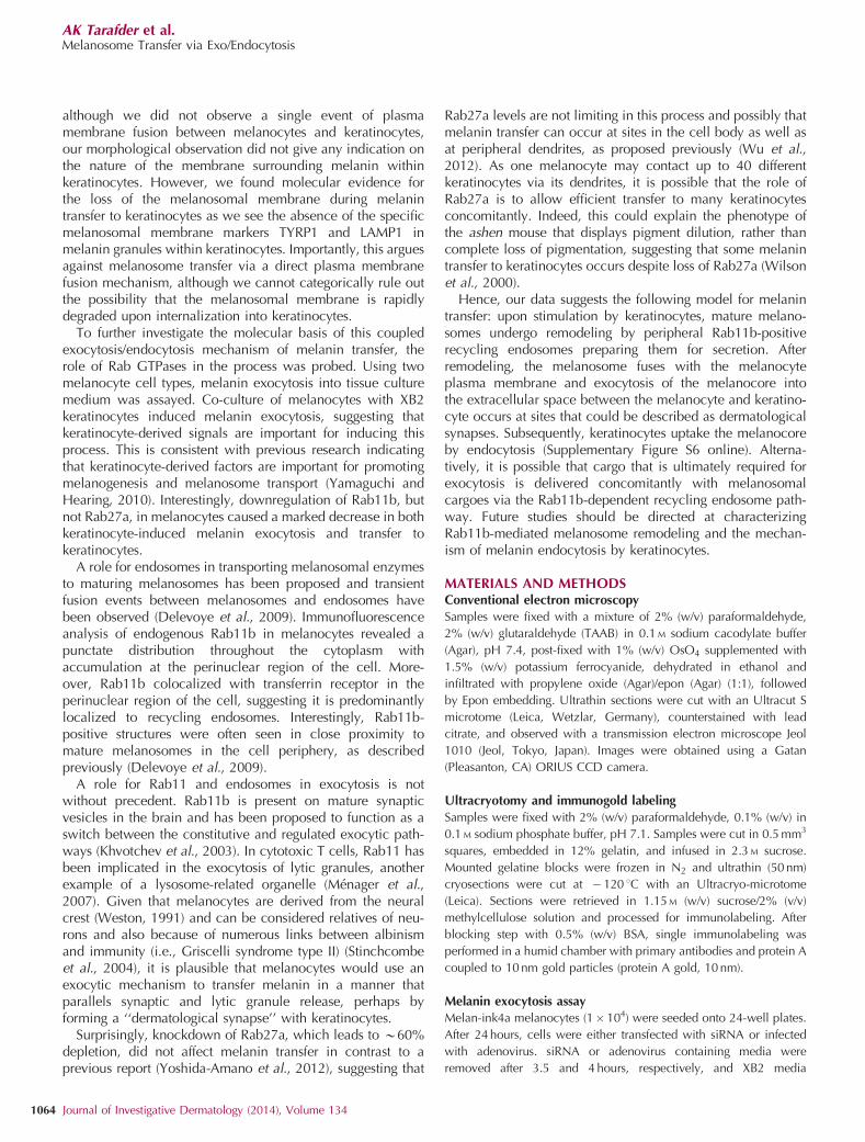
MATERIALS AND METHODSConventional electron microscopy
Samples were fixed with a mixture of 2% (w/v) paraformaldehyde,
2% (w/v) glutaraldehyde (TAAB) in 0.1 M sodium cacodylate buffer
(Agar), pH 7.4, post-fixed with 1% (w/v) OsO4 supplemented with
1.5% (w/v) potassium ferrocyanide, dehydrated in ethanol and
infiltrated with propylene oxide (Agar)/epon (Agar) (1:1), followed
by Epon embedding. Ultrathin sections were cut with an Ultracut S
microtome (Leica, Wetzlar, Germany), counterstained with lead
citrate, and observed with a transmission electron microscope Jeol
1010 (Jeol, Tokyo, Japan). Images were obtained using a Gatan
(Pleasanton, CA) ORIUS CCD camera.
Ultracryotomy and immunogold labeling
Samples were fixed with 2% (w/v) paraformaldehyde, 0.1% (w/v) in
0.1 M sodium phosphate buffer, pH 7.1. Samples were cut in 0.5 mm3
squares, embedded in 12% gelatin, and infused in 2.3 M sucrose.
Mounted gelatine blocks were frozen in N2 and ultrathin (50 nm)
cryosections were cut at � 120 1C with an Ultracryo-microtome
(Leica). Sections were retrieved in 1.15 M (w/v) sucrose/2% (v/v)
methylcellulose solution and processed for immunolabeling. After
blocking step with 0.5% (w/v) BSA, single immunolabeling was
performed in a humid chamber with primary antibodies and protein A
coupled to 10 nm gold particles (protein A gold, 10 nm).
Melanin exocytosis assayMelan-ink4a melanocytes (1� 104) were seeded onto 24-well plates.
After 24 hours, cells were either transfected with siRNA or infected
with adenovirus. siRNA or adenovirus containing media were
removed after 3.5 and 4 hours, respectively, and XB2 media
AK Tarafder et al.Melanosome Transfer via Exo/Endocytosis
1064 Journal of Investigative Dermatology (2014), Volume 134
containing 5� 104 XB2 keratinocytes added. The following day,
200 pM cholera toxin and 200 nM phorbal myristate acetate were
added to the media and co-cultures were incubated for 7 days.
Media containing exocytosed melanin was centrifuged at 800 g for
5 minutes at 4 1C to pellet cell debris. The supernatant was then
centrifuged at 20,000 g for 1 hour at 4 1C to pellet melanin. Melanin
pellets were washed with ethanol/ether (1:1 v/v) and dissolved in
2 M NaOH/20% DMSO at 60 1C for 1 hour. Melanin content was
measured as optical density at 340 nm. For melanocyte counting, a
parallel experiment was performed with the same initial cell
densities and the same silencing conditions. Total cell number
was counted at the end of the assay period and the proportion of
melanocytes determined from the cells co-cultured on coverslips by
immunofluorescence, using an anti-Tyrp1 (TA99) antibody to
specifically stain melanocytes.
Melanin transfer assay
Melan-ink4a melanocytes (2� 104) were seeded on coverslips on 24-
well plates. After 24 hours, siRNA transfection was performed and
XB2 cells (1� 105) in XB2 growth media were added when changing
the siRNA-containing media. Then, 200 pM cholera toxin and 200 nM
phorbal myristate acetate were added the following day. Cells were
co-cultured for 48 hours. Co-cultures were washed 3� with phos-
phate-buffered saline (PBS), and fixed with 4% paraformaldehyde in
PBS for 20 minutes at room temperature. Cells were washed 3� in
PBS and the nucleus visualized by incubation with 4’,6-diamidino-2-
phenylindole for 5 minutes. Images were taken in a Nikon Eclipse
TE2000-S screening microscope (Nikon, Tokyo, Japan) with
the same acquisition settings. To measure melanin uptake, the ImageJ
(NIH, Bethesda, MD) threshold command was applied and corre-
spondent intensity (in pixel) was measured automatically. Nuclei
were counted to ensure similar cell confluency in all samples and to
calculate the amount of melanin internalized per cell. Melanin uptake
reflects the total amount of melanin internalized by XB2 cells in one
coverslip.
Immunoflourescence analysis of cells
Cells grown on coverslips for immunofluorescence were fixed for
15 minutes in 4% paraformaldehyde in PBS for 24 hours.
Excess fixative was removed by extensive washing in PBS and
quenched by incubation in 50 mM NH4Cl for 10 minutes. Fixed cells
were then incubated with diluted primary antibody for 30 minutes,
washed extensively, incubated for 30 minutes with appropriate
Alexa 568-conjugated secondary antibodies (Molecular Probes,
Eugene, OR), washed as before, and mounted in ImmunoFluor
medium (ICN, Eschwege, Germany). All antibody incubations
and washes used 1� PBS, 0.5% BSA, and 0.05% saponin. Cells
were observed using a Leica SP5 confocal microscope, and images
were processed using ImageJ and Adobe Photoshop 5.0 software
(Adobe, San Jose, CA). All images presented are single sections in the
z-plane.
Supplementary methods
For primer sequences and other methods, refer to Supplementary
Materials and Methods online.
CONFLICT OF INTERESTThe authors state no conflict of interest.
ACKNOWLEDGMENTSWe thank Peter Munro (University College London) for his assistancewith high-pressure freezing and freeze substitution. This work was supportedby the Welcome Trust (0575498/Z/04/Z), BBSRC (BB/E021689/1), and Fun-dacao para a Ciencia e a Tecnologia (PTDC/BIA-BCM/111735/2009 andSFRH/BD/65381/2009).
SUPPLEMENTARY MATERIAL
Supplementary material is linked to the online version of the paper at http://www.nature.com/jid
REFERENCES
Ando H, Niki Y, Yoshida M et al. (2011) Involvement of pigment globulescontaining multiple melanosomes in the transfer of melanosomes frommelanocytes to keratinocytes. Cell Logist 1:12–20
Bahadoran P, Aberdam E, Mantoux F et al. (2001) Rab27a: a key tomelanosome transport in human melanocytes. J Cell Biolo 152:843–50
Beaumont KA, Hamilton NA, Moores MT et al. (2011) The recycling endo-some protein Rab17 regulates melanocytic filopodia formation andmelanosome trafficking. Traffic Copenhagen Denmark 12:627–43
Borovansky J, Elleder M (2003) Melanosome degradation: fact or fiction.Pigment Cell Res 16:280–6
Byers HR, Dykstra SG, Boissel SJS (2007) Requirement of dynactinp150(Glued) subunit for the functional integrity of the keratinocytemicroparasol. J Invest Dermatol 127:1736–44
Delevoye C, Hurbain I, Tenza D et al. (2009) AP-1 and KIF13A coordinateendosomal sorting and positioning during melanosome biogenesis. J CellBiol 187:247–64
Van Den Bossche K, Naeyaert J-M, Lambert J (2006) The quest for themechanism of melanin transfer. Traffic Copenhagen Denmark 7:769–78
Hearing VJ (2005) Biogenesis of pigment granules: a sensitive way to regulatemelanocyte function. J Dermatol Sci 37:3–14
Hume AN, Collinson LM, Rapak A et al. (2001) Rab27a regulates theperipheral distribution of melanosomes in melanocytes. J Cell Biol152:795–808
Imokawa G (2004) Autocrine and paracrine regulation of melano-cytes in human skin and in pigmentary disorders. Pigment Cell Res17:96–110
Jimbow K, Oikawa O, Sugiyama S et al. (1979) Comparison of eumelanogen-esis and pheomelanogenesis in retinal and follicular melanocytes; role ofvesiculo-globular bodies in melanosome differentiation. J Invest Dermatol73:278–84
Khvotchev MV, Ren M, Takamori S et al. (2003) Divergent functionsof neuronal Rab11b in Ca2þ -regulated versus constitutive exocytosis.J Neurosci 23:10531–9
Lapierre LA, Dorn MC, Zimmerman CF et al. (2003) Rab11b resides in avesicular compartment distinct from Rab11a in parietal cells and otherepithelial cells. Exp Cell Res. 290:322–31
Marks MS, Seabra MC (2001) The melanosome: membrane dynamics in blackand white. Nat Rev Mol Cell Biol. 2:738–48
Mottaz JH, Zelickson AS (1967) Melanin transfer: a possible phagocyticprocess. J Invest Dermatol 49:605–10
Menager MM, Menasche G, Romao M et al. (2007) Secretory cytotoxic granulematuration and exocytosis require the effector protein hMunc13-4. NatImmunol. 8:257–67
Okazaki K, Uzuka M, Morikawa F et al. (1976) Transfer mechanism ofmelanosomes in epidermal cell culture. J Invest Dermatol 67:541–7
Pfeffer SR (2001) Rab GTPases: specifying and deciphering organelle identityand function. Trends Cell Biol 11:487–91
Raposo G, Marks MS (2002) The dark side of lysosome-related organelles:specialization of the endocytic pathway for melanosome biogenesis.Traffic Copenhagen Denmark 3:237–48
Raposo G, Tenza D, Murphy DM et al. (2001) Distinct protein sorting andlocalization to premelanosomes, melanosomes, and lysosomes in pig-mented melanocytic cells. J Cell Biol 152:809–24
AK Tarafder et al.Melanosome Transfer via Exo/Endocytosis
www.jidonline.org 1065
Scott G (2003) Photo protection begins at the cellular level: microparasols onthe job. J Invest Dermatol 121:viii
Scott G, Leopardi S, Printup S et al. (2002) Filopodia are conduits formelanosome transfer to keratinocytes. J Cell Sci 115(Pt 7):1441–51
Seabra MC, Mules EH, Hume AN (2002) Rab GTPases, intracellular traffic anddisease. Trends Mol Med. 8:23–30
Seiberg M (2001) Keratinocyte-melanocyte interactions during melanosometransfer. Pigment Cell Res 14:236–42
Singh SK, Kurfurst R, Nizard C et al. (2010) Melanin transfer in human skincells is mediated by filopodia–a model for homotypic and heterotypiclysosome-related organelle transfer. FASEB J 24:3756–69
Singh SK, Nizard C, Kurfurst R et al. (2008) The silver locus product (Silv/gp100/Pmel17) as a new tool for the analysis of melanosome transfer inhuman melanocyte-keratinocyte co-culture. Exp Dermatol 17:418–26
Stinchcombe J, Bossi G, Griffiths GM (2004) Linking albinism and immunity:the secrets of secretory lysosomes. Science 305:55–9
Strom M, Hume AN, Tarafder AK et al. (2002) A family of Rab27-bindingproteins. Melanophilin links Rab27a and myosin Va function in melano-some transport. J Biol Chem 277:25423–30
Wasmeier C, Romao M, Plowright L et al. (2006) Rab38 and Rab32 controlpost-Golgi trafficking of melanogenic enzymes. J Cell Biol 175:271–81
Weston JA (1991) Sequential segregation and fate of developmentally restrictedintermediate cell populations in the neural crest lineage. Curr Top DevBiol 25:133–53
Wilson SM, Yip R, Swing D et al. (2000) A mutation in Rab27a causes thevesicle transport defects observed in ashen mice. Proc Natl Acad Sci USA97:7933–8
Wolff K (1973) Melanocyte-keratinocyte interactions in vivo: the fate ofmelanosomes. Yale J Biol Med 46:384–96
Wu X, Rao K, Bowers MB et al. (2001) Rab27a enables myosin Va-dependentmelanosome capture by recruiting the myosin to the organelle. J Cell Sci114:1091–100
Wu XS, Masedunskas A, Weigert R et al. (2012) Melanoregulin regulates ashedding mechanism that drives melanosome transfer from melanocytesto keratinocytes. Proc Natl Acad Sci USA 31:109
Yamaguchi Y, Hearing VJ (2010) Physiological factors that regulate skinpigmentation. Biofactors Oxford England 35:193–9
Yamamoto O, Bhawan J (1994) Three modes of melanosome transfers inCaucasian facial skin: hypothesis based on an ultrastructural study.Pigment Cell Res 7:158–69
Yoshida-Amano Y, Hachiya A, Ohuchi A et al. (2012) Essential roleof RAB27A in determining constitutive human skin colour. PLoS One7:e41160
Zerial M, McBride H (2001) Rab proteins as membrane organizers. Nat RevMol Cell Biol. 2:107–17
Zhou BK, Boissy RE, Pifko-Hirst S et al. (1993) Lysosome-associated membraneprotein-1 (LAMP-1) is the melanocyte vesicular membrane gycoproteinband II. J Invest Dermatol 100:110–4
AK Tarafder et al.Melanosome Transfer via Exo/Endocytosis
1066 Journal of Investigative Dermatology (2014), Volume 134