quantitative structure-hydrophobicity and structure-activity relationships of antibacterial...
TRANSCRIPT
Quantitative Structure-Hydrophobicity and Structure-Activity Relationships of Antibacterial Gramicidin S Analogs
TADASHI KATAYAMA, KAZUYA NAKAO, MIKI AKAMATSU~, TAMIO UENO, AND TOSHIO FUJITA Received April 1, 1994, from the Department of Agricultural Chemistry, Kyoto Universify, Kyoto 606-01, Japan. publication June 6, 1994@.
Accepted for
Abstract 0 The structure-hydrophobicity-antibacterial activity relation- ships of gramicidin S and its analogs retaining a @-pleated sheet structure were examined quantitatively with physicochemical substituent and molecular parameters using regression analyses. Variations in their apparent hydrophobicity in an octanol/buffer (pH 7) system, log P(O/W), were analyzed in terms of the “effective” hydrophobicity and steric parameters of side chain substituents of residues at certain positions in the molecule; however, some of the conformational factors have not been fully defined. For the partitioning into liposomes and the growth inhibitory activity against species of Gram-positive bacteria, the log P(0Nv) value simulated the hydrophobic effects of gramicidin S and its analogs better than substituent parameters. The side chain hydrophobicity was assumed to work together with effects attributed to variations in the entire cyclic peptide structure including conformational components undefined in the structure-log P(0hV) analysis in these activities.
Introduction Gramicidin S (GS) is a symmetric cyclic decapeptide with
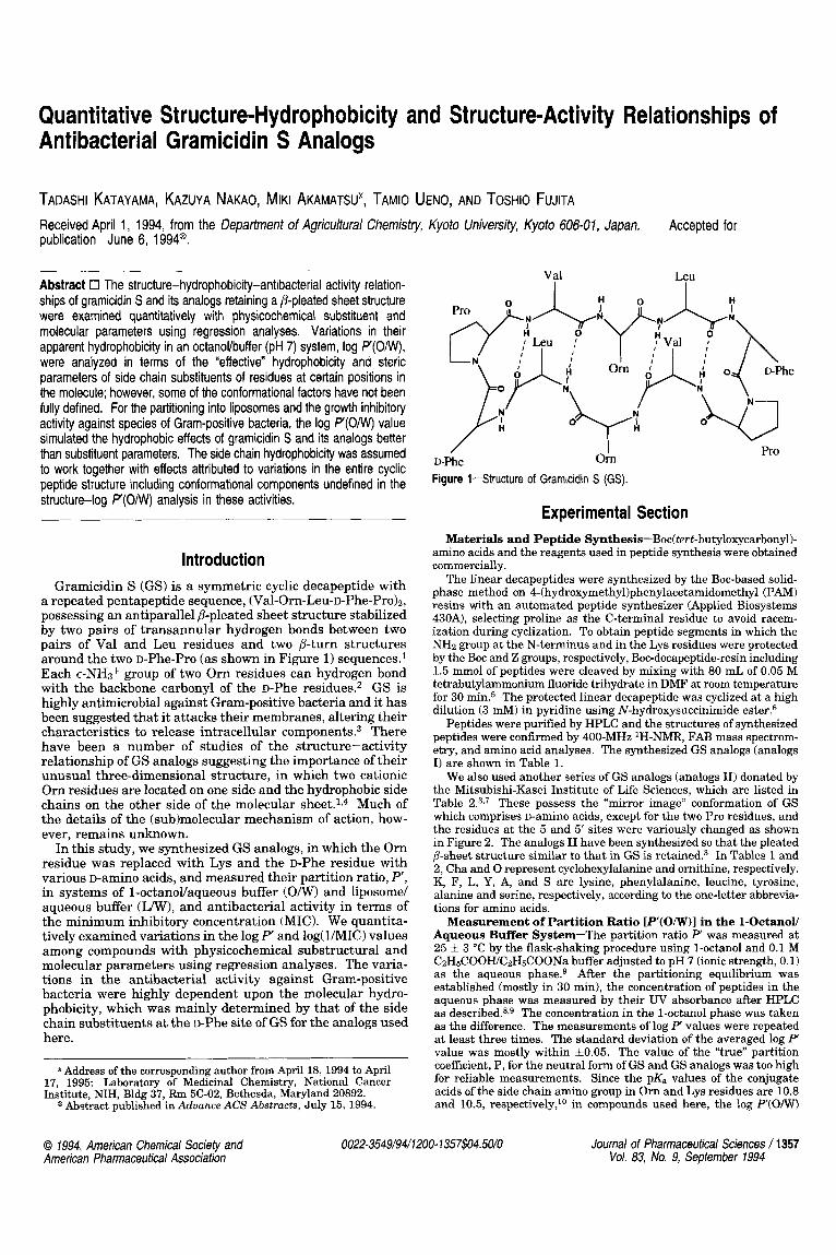
a repeated pentapeptide sequence, (Val-Om-Leu-D-Phe-Pro)z, possessing an antiparallel P-pleated sheet structure stabilized by two pairs of transannular hydrogen bonds between two pairs of Val and Leu residues and two p-turn structures around the two D-Phe-Pro (as shown in Figure 1) sequences1 Each E-NH~+ group of two Orn residues can hydrogen bond with the backbone carbonyl of the D-Phe residues.2 GS is highly antimicrobial against Gram-positive bacteria and it has been suggested that it attacks their membranes, altering their characteristics to release intracellular component^.^ There have been a number of studies of the structure-activity relationship of GS analogs suggesting the importance of their unusual three-dimensional structure, in which two cationic O m residues are located on one side and the hydrophobic side chains on the other side of the molecular ~ h e e t . l , ~ Much of the details of the (sub)molecular mechanism of action, how- ever, remains unknown.
In this study, we synthesized GS analogs, in which the Orn residue was replaced with Lys and the D-Phe residue with various D-amino acids, and measured their partition ratio, P‘, in systems of 1-octanollaqueous buffer (O/W) and liposome/ aqueous buffer (W), and antibacterial activity in terms of the minimum inhibitory concentration (MIC). We quantita- tively examined variations in the log P‘ and log(l/MIC) values among compounds with physicochemical substructural and molecular parameters using regression analyses. The varia- tions in the antibacterial activity against Gram-positive bacteria were highly dependent upon the molecular hydro- phobicity, which was mainly determined by that of the side chain substituents a t the D-Phe site of GS for the analogs used here. ~~ ~
*Address of the corresponding author from April 18, 1994 to April 17, 1995: Laboratory of Medicinal Chemistry, National Cancer Institute, NIH, Bldg 37, Rm 5C-02, Bethesda, Maryland 20892.
@ Abstract published in Aduance ACS Abstracts, July 15, 1994.
Val Leu
I Om
/ D-Phe Figure 1-Structure of Grarnicidin S (GS).
Pro
Experimental Section Materials and Peptide Synthesis-Boc(tert-butyloxycarbony1)-
amino acids and the reagents used in peptide synthesis were obtained commercially.
The linear decapeptides were synthesized by the Boc-based solid- phase method on 4-(hydroxymethyl)phenylacetamidomethyl (PAM) resins with an automated peptide synthesizer (Applied Biosystems 430A), selecting proline as the C-terminal residue to avoid racem- ization during cyclization. To obtain peptide segments in which the NH2 group at the N-terminus and in the Lys residues were protected by the Boc and Z groups, respectively, Boc-decapeptide-resin including 1.5 mmol of peptides were cleaved by mixing with 80 mL of 0.05 M tetrabutylammonium fluoride trihydrate in DMF at room temperature for 30 The protected linear decapeptide was cyclized at a high dilution (3 mM) in pyridine using N-hydroxysuccinimide ester.6
Peptides were purified by HPLC and the structures of synthesized peptides were confirmed by 400-MHz lH-NMR, FAB mass spectrom- etry, and amino acid analyses. The synthesized GS analogs (analogs I) are shown in Table 1.
We also used another series of GS analogs (analogs 11) donated by the Mitsubishi-Kasei Institute of Life Sciences, which are listed in Table 2.3,7 These possess the “mirror image” conformation of GS which comprises D-amino acids, except for the two Pro residues, and the residues at the 5 and 5’ sites were variously changed as shown in Figure 2. The analogs II have been synthesized so that the pleated B-sheet structure similar to that in GS is retained? In Tables 1 and 2, Cha and 0 represent cyclohexylalanine and ornithine, respectively. K, F, L, Y, A, and S are lysine, phenylalanine, leucine, tyrosine, alanine and serine, respectively, according to the one-letter abbrevia- tions for amino acids.
Measurement of Partition Ratio [P‘(O/W)] in the 1-Octanoll Aqueous Buffer System-The partition ratio P‘ was measured at 25 f 3 “C by the flask-shaking procedure using 1-octanol and 0.1 M CzH&OOWCzH&OONa buffer adjusted to pH 7 (ionic strength, 0.1) as the aqueous phase.g After the partitioning equilibrium was established (mostly in 30 min), the concentration of peptides in the aqueous phase was measured by their UV absorbance after HPLC as d e s ~ r i b e d . ~ . ~ The concentration in the 1-octanol phase was taken as the difference. The measurements of log P values were repeated at least three times. The standard deviation of the averaged log P‘ value was mostly within k0.05. The value of the “true” partition coefficient, P, for the neutral form of GS and GS analogs was too high for reliable measurements. Since the pK, values of the conjugate acids of the side chain amino group in O m and Lys residues are 10.8 and 10.5, respectively,1° in compounds used here, the log P(0IW)
0 1994, American Chemical Society and American Pharmaceutical Association
0022-3549/94/1200- 1357$04.50/0 Journal of Pharmaceutical Sciences / 1357 Vol. 83, No. 9, September 1994

Table 1-Structure of GS and the Analogs I and Substituent Parameters’
Cyclo(Va1’ -x ’ -L~u~-Y~-P~o~) ,
Compound X’,?
GS Orn GS(K,DF) Lys GS(K,DL) Lys GS(K,DY) Lys GS(K,DA) Lys GS(K,DS) Lys GS(0,DCha) Orn
- y4.4‘
D-Phe D-Phe
D-Tyr D-Ala
D-Cha
D-Leu
D-Ser
-1.56 1.95 1.99 -1.66 1.95 1.99 -1.66 1.81 1 .85 -1.66 1.20 1.29 -1.66 0.32 0.33 -1.66 -1.49 -0.04 -1.56 3.07e 3.13
PsC(Vd
-0.90 -0.90 -1.44 -0.90 -0.20 -0.48 -1.40‘
a Parameters are either cited from or estimated from those listed in ref 9. An effective hydrophobicity parameter that excluded a component due to steric inhibition on the relative solvation. “Intrinsic” hydrophobicity parameter. Calcu- lated from the Dubois Fs according to PSc = F, - 0.306(3 - n), n being the number of I -H atoms in the substituents. The reference point is shifted so that P,(H) and FSC(H) in glycine is zero. Estimated from the n value of cyclohexyl (2.66) including the group branching factor, n, of CH3 (0.54) and the chain branching factor (-0.13). See refs 16 and 22. ’From ref 19.
Table 2-Structure of the “Mirror Imaged” GS Analogs 11 and Substituent Parameters’
~ y c ~ o ( ~ - ~ e u ’ - D - L ~ S ’ - D - L ~ U ~ - P ~ O ~ - X ~ ) ~
GlY 0.00 0.00 0.00 l l (~A) D-Ala 0.32 0.33 -0.20 W) II(DL) D-Leu 1 .81 1 35 -1.44 II(DF) D-Phe 1.95 1.99 -0.90 Il(DCha) D-Cha 3.07 3.13 -1.40
a See footnote a of Table 1. See footnote c of Table 1. See footnote b of Table 1. dSee footnote d of Table 1.
Pro I D-LYS
Figure 2-Structure of GS Analogs 11.
value measured at pH 7 could be almost entirely due to the ion-pair, in which the counteranion is CzHsCOO-.
Measurement of Partition Ratio [P‘(Lnv)] in the Lecithin Liposomes/Aqueous Buffer System-Lecithin was prepared from fresh egg yolk and purified by the method of Singleton.’l Lecithin, 100 mg, was dissolved in 1 mL of CHC13 and placed in a 30-mL flask under Ar gas. The solvent was removed in uacuo. The lipid film was suspended in pH 7.2 Tris buffer (10 mM Tris-HC1,50 mM NaC1, and 0.2 mM EDTA) and sonicated with a Type 4280 sonicator (Kaijodenki Co., Tokyo, Japan) in an ice-cooled bath under Ar gas to avoid oxidation of lecithin. Bilayered liposomes were separated from the undispersed lecithin by centrifuging the suspension at 15 000 rpm for 30 min and from multilayered liposomes by gel filtration on Sepharose 4B a t 4 T.12 The amount of lecithin was calculated from that of phosphorus by a modification of the Bartlett method.13
The partition ratio, P’(LW), of GS and GS analogs I between the liposomes and the external aqueous phase was measured by equilib- rium dialy~is.’~.’~ Each compound was dissolved in the pH 7.2 Tris- HCl buffer used for the liposome suspension. Using an equilibrium dialysis cell (10 x 1 mL chambers, Sanko Plastic Co., Tokyo, Japan) the liposome suspension (1 mL) was equilibrated with the peptide solution (1 mL) through a cellophane dialysis membrane at 25 “C for 35 h. The partition ratio, P‘(LW), was calculated by eq 1. The
~ ( C O - c’( mole of GS analogkg of lipids P‘‘LMT) = C x C, mole of GS ana logk of buffer
standard deviation of log P‘(L/w) value averaged for six repeats was mostly within fO.l. In eq 1, C is the concentration of compounds in the liposome-free chamber at the equilibrium, C, is concentration of compounds in the control experiment in which the solution of compounds was equilibrated through the membrane with the Tris buffer without liposome, and CL is the concentration of lecithin (kg/ L) in the liposome suspension. The value of 2(C0 - CYCL corresponds with the concentration inside the liposomes.
The log PWW) values of GS and GS(K,DS) were independent of pH in the range of 4-8, in which they exist almost entirely as the ionized form. The log P‘(LW) value was also almost independent of the concentration of lecithin although, for analogs with higher values, there was a trend to slightly increase with an increase in the concentration of lecithin. We selected the log P‘(LIw) value measured at 3.5 x kg of lecithin& for the analysis, because the standard error of the measurement in the lecithin concentration was the smallest and the log P‘WW) value was the most reliable.
Measurement of Antimicrobial Activity-The minimum con- centration of GS and analogs I necessary to completely inhibit the growth of four species of Gram-positive bacteria and of analogs I1 to inhibit another species was measured by serial dilution using a nutrient agar in terms of the minimum inhibitory concentration (MIC in moleshter). Because the set of MIC data changed slightly but significantly among individual runs, we defined the log (1MIC) value relative to that of the GS measured in each run (log RA) as the antimicrobial activity index as shown in eq 2. The standard deviation
] (1)
of the log RA value was f0.17. Natural GS used as the reference completely inhibited the microorganisms used here at a concentration
Substituent Parameters Used for Analyses-According to our previous study of the number of N-acetyl di- and tripeptide a m i d e ~ , ~ the “effective” hydrophobicity on the basis of the octanol-water partitioning of the nonpolar side chain substituents of residues is governed not only by the “intrinsic” hydrophobicity parameter n established from the additive-constitutive nature of log P values for aliphatic compounds16 but also by their steric effects on the relative solvation of partitioning solvents with backbone CONH group^.^ For un-ionizable but polar side chains such as that in serine, the intramolecular hydrogen-bond formation with the backbone CONH and the electronic effect on the backbone solvation of the polar group, both of which increase partitioning into the octanol phase, are to be additionally c~ns idered .~ For ionizable side chains, the effects orig- inating from the ion-pairing should be further taken into account, to evaluate the ”effective” hydrophobicity scale.s
The conformational rigidity of GS is almost completely retained in various solvents as well as upon binding with phospholipids as examined by the NMR and CD m e a s ~ r e m e n t s . ~ J ~ J ~ Although some conformational flexibility in aqueous solution is observed in the GS- (O,G),l8 this compound is not included in this study. We examined the CD spectra for all the GS analogs I used here in MeOH solution (data not shown) and confirmed that their spectral patterns are very similar to that of GS. Thus, we could consider that the conformational rigidity similar to that of GS is mostly held for the analogs I used in this study, if not completely, in various solvent as well as lipid phases. Because one of the residues, the effects of which are to be examined, belongs to the D-series sandwiched by L-residues, the steric effect of D-side chains should be different from that involved in linear “flexible” peptides, in which all residues are of the L-configuration. A situation similar to that of analogs I also applies to analogs II.7 Thus, for the hydrophobic effect of varying substituents a t Orn2*2’ and D-Phe4z4‘ sites
of (2.2-4.6) x M.
1358 / Journal of Pharmaceutical Sciences Vol. 83, No. 9, September 1994
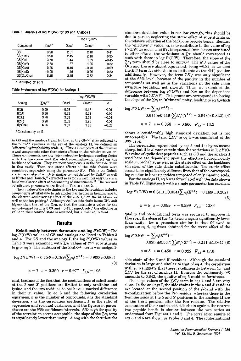
Table 3-Analysis of log P(0MI) for GS and Analogs I
Compound
GS GS(K,DF) GS(K,DL) GS(K,oY) GS(K,DA) GS(K,DS) GS(0,DCha)
E3rly.Y’
3.98 3.98 3.70 2.58 0.66
6.26 -0.08
log P(0Nv)
Obsd
2.51 2.45 1.44 1.37
-0.49 -1.16
3.48
Calcda A
2.10 0.41 2.10 0.35 1.89 -0.45 1.05 0.32
-0.40 -0.09 -0.96 -0.20
3.82 -0.34
a Calculated by eq 3.
Table 4-Analysis of log P(0MI) for Analogs I1
log P(0Nv)
Analog EJq?.x, Obsd Calcda A
IUG) 0.00 -0.26 -0.17 -0.09 II(DA) 0.66 0.33 0.23 0.10
jl(Dcha) 6.26 3.63 3.65 -0.02
II(DL) 3.70 2.05 2.09 -0.04 II(DF) 3.98 2.32 2.26 0.06
a Calculated by eq 5.
in GS and the analogs I and for that at the Gly5v5’ sites adjacent to the L - P ~ o ~ , ~ ’ residues in the set of the analogs 11, we defined an “effective” hydrophobicity scale, JQ. This is a composite of the intrinsic n and components other than steric effects on the relative solvation. These components are the intramolecular hydrogen-bonding factor with the backbone and the electron-withdrawing effect on the backbone solvation. They are most conspicuous in the Ser side chain in this study. Then, the steric effects of the side chains were considered separately using the parameter E S c . This is the Dubois steric parameter,lg which is similar to that defined by Taft,2O as well as Kutter and Hansch,2l corrected so as to represent not only the steric bulk but also the effect of a-branching of substituents.22 The relevant substituent parameters are listed in Tables 1 and 2.
The nl value of the side chains in the Lys and Om residues includes components attributable to intramolecular hydrogen bonding and to the electron-withdrawing effect of the w-NHs+ on the backbone as well as the i~n-pairing.~ Although the Lys side chain is one CH2 unit higher than that of the Om, so that the intrinsic JZ value for the noncationized form is 0.09 and -0.45, respectively: the effective XI value in their ionized state is reversed, but almost equivalent.
Results Relationship between Structure and log P‘(OIW)-The
log W O N ) values of GS and analogs are listed in Tables 3 and 4. For GS and the analogs I, the log P‘(O/W) values in Table 3 were examined with CJCI values of Y4,& substituents to give eq 3. The addition of the Z J C I ( X ~ , ~ ) term was insignifi-
log P.(O/W> = 0.754(zkO.l89)C~@’4 - 0.900(&0.683) (3)
n = 7 s = 0.390 r = 0.977 Fl,5 = 105
cant, because of the fact that the modifications of substituents at the 2 and 2’ positions are limited to only ornithine and lysine, and the two residues do not have a marked difference in their JCI value. In eq 3 and the following correlation equations, n is the number of compounds, s is the standard deviation, r is the correlation coefficient, F is the ratio of regression and residual variances, and the figures in paren- theses are the 95% confidence intervals. Although the quality of the correlation seems acceptable, the slope of the Zn1 term is significantly lower than unity. Along with the fact that the
standard deviation value is not low enough, this should be due in part to neglecting the steric effect of substituents on the relative solvation of the backbone peptide bonds. Because the “effective” JC value, X I , is to contribute to the value of log P‘(O/W) as much, and if it is separated from factors attributed to other effects, the variations in ZJCI should correspond as such with those in log P‘(O/W). Therefore, the slope of the CJCI term should be close to unity.23 The ESc values of the Om and Lys are almost equivalent, being -0.82, so we used the ITSc term for side chain substituents a t the 4(4‘) position additionally. However, the term msc was only significant at the 63% level, because of the paucity in the number of compounds as well as in the variations in the side chain structure (equation not shown). Thus, we examined the difference between log P‘(O/W) and CJCI as the dependent variable with ZE’8c(Y). This procedure corresponds to forcing the slope of the CXI to “ultimate” unity, leading to eq 4,which
log P(0IW) - CZ1(Y494) =
0.614( *0.419)xF,”(P“) - 0.548( k0.822) (4)
n = 7 s = 0.358 r = 0.860 F1,5 = 14.2
shows a considerably high standard deviation but is not unacceptable. The term WSc in eq 4 was significant a t the 98% level.
The correlation represented by eqs 3 and 4 is by no means sharp, but it is almost certain that the variations in log P(O1 W) value of cyclic decapeptides such as GS and the analogs I used here are dependent upon the effective hydrophobicity scale XI, probably, as well as the steric effect on the backbone solvation of the side chain substituents. The steric effect seems to be significantly different from that of the correspond- ing residue in linear peptides composed of only L-amino acids.
For the analogs 11, eq 5 was formulated with the data shown in Table IV. Equation 5 with a single parameter has excellent
log P‘(o/W) = 0 . 6 1 1 ( ~ 0 . 0 5 4 ) ~ ~ ~ , ( X ~ ” ) - 0.169(&0.202) ( 5 )
n = 5 s = 0.088 r = 0.999 F1,3 = 1283
quality and no additional term was required to improve it. However, the slope of the &I term is again significantly lower than unity. By a procedure similar to that followed to generate eq 4, eq 6was obtained for the steric effect of the
logP(0IW) - CnI(x5,’) =
0.695( &0.537)ZE,“(X5”) - 0.21 1( f1.061) (6)
n = 5 s = 0.450 r = 0.922 F1,3 = 17.0
side chain of the 5 and 5‘ residues. Although the standard deviation is large and similar to that of eq 4, the correlation with eq 6 suggests that there is collinearity between ZJCI and wsc for the set of analogs 11. Because the collinearity (r2) amounts to 0.842, the quality of eq 5 could be fortuitous.
The slope values of the msc term in eqs 4 and 6 are very close. In the analogs I, the side chains in the 4 and 4‘ residues are located at the second position of the P-bend with the D-configuration before the Pro residue, whereas those in the D-amino acids at the 5 and 5’ positions in the analogs I1 are at the third position after the Pro residue. The relative orientation of the D-aminO acid side chain against the nearest two peptide bonds is similar between the two series as understood from Figures 1 and 2. The correlation results of eqs 3 and 5 are shown in Tables 3 and 4. The conformational
Journal of Pharmaceutical Sciences / 1359 Vo/. 83, No. 9, Sepfember 1994

Table 5-Analysis of log P ( W ) for GS and Analogs I
log P ( W ) Compound c.nlY,y' Obsd Calcda A
GS 3.98 4.55 4.62 -0.07 GS(KpF) 3.98 4.41 4.60 -0.19 GS(K,oL) 3.70 4.26 4.26 -0.002 GS(K,oY) 2.58 4.35 4.24 0.1 1 GS(K,DA) 0.66 3.68 3.61 0.07
GS(0,oCha) 6.26 5.09 4.95 0.14 GS(K,DS) -0.08 3.33 3.38 -0.05
~~
a Calculated by eq 8.
rigidity of the cyclic structure in each set of analogs could mostly be retained, but the steric effect of each side chain on the relative solvation of peptide bonds as well as on minor flexibilities of the cyclic structure was not well defined. The ring conformation of these cyclic peptides is largely governed by the intramolecular hydrogen bondings. If the conforma- tional rigidity does not hold in each set of analogs so that the hydrogen bonds are disrupted, the variations in the log P'(0l W) values should be much higher and more irregular than those analyzed with eqs 3-6.
Relationship between Structure and log P'(LIW)---It has been suggested that GS and the analogs retaining the P-pleated sheet conformation penetrate liposomal membranes from the side of the four hydrophobic (1, l', 3,3') side chains.18 Two cationic 2 and 2' side chains are exposed in the aqueous phase and the side chain substituents of the 4 and 4' residues in the analogs I on the edges of the P-turned regions project with a bend toward the side opposite that of the hydrophobic side chains. Because the interaction of the side chains with liposomes is dependent on their positions, their effects should be analyzed with separate n~ terms. For the log P'(LIw) values shown in Table 5, eq 7 was formulated with the use of
log P ( W ) = 0.260(&0.068)~~,(Y~,~ ' ) + 3.456kt0.246)
n = 7 s = 0.141 r = 0.975 F,, = 96.4
(7)
ZI values of the 4(4') side chains to give a good correlation. side chains in Table
1 is a composite including the effects of ion-pairing, in which the counterion is CzHsCOO-. The log P'(LIw) value was measured using the Tris-HC1 buffer as the aqueous phase, the counterion of the basic side chain being different from the system in which the X I value was defined. We assumed that the X I values and their counterparts in the liposome/Tris-HC1 system of Om and Lys residues vary in parallel, that is, the XI values can express the effective hydrophobicity scale at least in a relative sense.g Including the ZJCI term for the 2 and 2' side chains into eq 7, however, did not improve the correlation. Because the effect of X2*2' substituents is not significantly involved in the variations in log P'(O/W), the log P(LIw) values seemed to be directly correlated with the log P'(O/W) values. Equation 8 was formulated with a correlation quality
log P'(L/w) = 0.338(fO.O80)log P(ON(r) + 3.775(f0.166) (8 )
The XI value of the Orn2J' and
n = 7 s = 0.128 r = 0.979 Fl,5 = 117
somewhat better than that of eq 7. Although the insignificance of the n~ term for the 2(2') side
chain in eq 7 and the synonymous observation in eq 8 were due to a lack of enough variations in the structure of the 2(2') side chain, eq 7 is not inconsistent with the model for the
Table 6-Relative Antimicrobial Activity Index (log RA) of GS and Analogs I
S. aureus
FDAa Terajima MS 353 M. luteu.9
Obsd Calcdc Obsd Calcdd Obsd Calcde Obsd Calcd'
GS 0.00 -0.19 0.00 0.01 0.00 -0.20 0.00 -0.04 GS(K,DF) -0.24 -0.20 0.06 -0.01 -0.24 -0.21 0.06 4 . 0 6 GS(K,DL) -0.31 -0.39 -0.31 -0.26 -0.31 -0.36 -0.31 4 . 2 6 GS(K,DY) 4 . 2 3 -0.41 0.07 -0.28 -0.23 -0.36 -0.23 -0.28 GS(K,oA) -0.89 -0.76 -0.89 -0.74 -0.89 -0.79 -0.89 -0.66 GS(K,oS) -0.93 -0.89 -0.93 -0.91 -0.63 -0.73 -0.63 -0.79 GS(K,DCha) -0.25 -0.01 0.06 0.25 -0.25 -0.06 0.06 0.15
a S. aureus FDA 209P JC-1. M. luteus ATCC 9341. Calculated by eq 10.
binding of GS analogs with liposomes in which two cationized side chains are left exposed in the aqueous phase. The fact that the slope of the %1(Y4s4') term in eq 5 is considerably smaller than unity is also reasonable in this model. Under the conditions where four (1, l', 3,3') hydrophobic side chains penetrate the liposomal phase, the 4 and 4'side chains locate on the membrane surface. The hydrophobic interaction seems to be of a type occurring with only a partial engulfment within the liposomal phase, so that the hydrations of peptide bonds around the 4 and 4' residues are not so susceptible to their steric effect as to be significantly disrupted. This was sug- gested by the fact that adding the mSc term into eq 7 was insignificant. Because the correlation quality of eq 7 is much better than that of eq 3 in terms of standard deviation, the steric effect of 4 and 4' residues of the type considered in formulating eq 3 need not be considered. The correlation result of eq 8 is summarized in Table 5.
Relationship between Structure and Antibacterial Activity-The antimicrobial activity indices (log RA) of four species of Gram-positive bacteria of GS and the analogs I are listed in Table 6. As described above, the antibacterial activity is thought to be due to expansion and disruption of the cell membrane structure, enhancing its permeability. GS and the antibacterial analogs penetrate the cell membrane with the hydrophobic side of the drug in a manner similar to the partitioning into liposomal membranes. The very low activity against Gram-negative bacteria is due to the very low ability of GS and the analogs to disrupt the outer membrane structure that is not present in Gram-positive ba~ te r i a .~ Thus, the activity indices were analyzed in a manner similar to that for the log P'(L/W). As the counterparts of eqs 7 and 8, eqs 9 and 10 were constructed for the antibacterial activity of the
Calculated by eq 11. Calculated by eq 12. Calculated by eq 13.
log RA = 0.191(f0.110)l0g P'(O/W) - 0.669(f0.228) (10)
n = 7 s = 0.176 r = 0.894 F1,5 = 19.8
set of GS and the analogs I, against Staphylococcus aureus FDA 209P JC-1, respectively. log P'(OIw), as the independent variable in eq 10, gave a better correlation. As indicated above, the variations in log P'(O/W) are not significantly related to the effects of X2,y substituents. In this situation, the log P(O/W) value is a model for the interaction with the bacterial cell membrane that is better than the "effective" hydrophobicity parameter of just the varying substituents. The components in variations of the log P'(O/W) values were not well separated as indicated by the somewhat high
1360 / Journal of Pharmaceutical Sciences Vol. 83, No. 9, September 1994

Table 7-Relative Antimicrobial Activity Index (log RA) of GS Analogs II
8. subtilisp
fit of the 5(5’) side chain substituents with cell membranes could be better than that of the 4(4‘) side chains in analogs I.
Analog Obsd Calcdb
-0.95 -0.94 -0.31 0.02 0.33
-1.04 -0.83 -0.22 -0.1 1
0.35
a 8. subtilis MAR FURG 168. Calculated by eq 14.
standard deviation in eq 3, leaving conformational factors not separated and parameterized well. The conformational effects of the entire cyclic decapeptide skeleton, excluding that of X2,2’ substituents, seem to work together to better simulate the situation.
The above observation was also true of the antibacterial activity against three other Gram-positive bacterial species.
against S. aureus Terajima: log RA = 0.249(~0.121)logP(O/W) - 0.619(f0.250) (11)
n = 7 s = 0.193 r = 0.921 Fl,5 = 28.0
against S. aureus MS 353: log RA = 0.146(fO.l17)10g P(O/W) - 0.564(f0.242) (12)
n = 7 s = 0.181 r = 0.820 Fl,5 = 10.3
against M . luteus ATCC 9341: log RA = 0.204(fO.O93)10g P(O/W) - 0.557(&0.192) (13)
n = 7 s = 0.148 r = 0.930 Fl,5 = 31.7
The counterpart of eqs 11-13 with the XT~(Y~~~) term showed a poorer correlation (s = 0.264 and r = 0.846 compared with those in eq 11, s = 0.217 and r = 0.746 compared with those in eq 12, and s = 0.194 and r = 0.876 compared with those in eq 13). The fact that the observed values of the GS analog with D-serine were smaller than the calculated values from eqs 12 and 13 suggested the electronic inductive effect for the 4 and 4‘ side chain substituents, although the number of compounds is not enough to introduce the electronic term such as the Charton 01 parametee addi t i~nal ly .~~ The slope values of the log P‘(O/W) term in eqs 10-13 are very similar, the average (*standard deviation) being 0.198(&0.042), as are the intercept values, being -0.602(f0.052). These results indi- cated that the modes of the hydrophobic interaction of GS and the analogs I with similar bacterial cell membrane systems are indeed similar.
For the set of analogs 11, the antibacterial activity shown in Table 7 was analyzed to afford eq 14 using log P‘(O/W).
against Bacillus subtilis MAR FURG 168: log RA = 0.356(fO.l29)10g P‘(O/W) - 0.945(+0.277) (14)
n = 5 s = 0.128 r = 0.981 Fl,3 = 77.1
The slope of log ?“(Om) in eq 14 is somewhat higher than the slopes found in eqs 10-13. This might in part be due to the fact that the side chain substituents of the 5 and 5’ residues on the edge of the @-turned moiety tend to lean toward the hydrophobic side chain, contrarily to those of the 4 and 4‘ residues in GS and analogs I. In this situation, the
Discussion From the analyses of log P‘(O/W), the “effective” hydropho-
bicity scale of side chain substituents in the GS-type cyclic peptides was understood to differ at least in two respects from that of those in linear ol igopept ide~.~,~~ The first is the difference attributable to that in the steric effect of side chains on the relative solvation of backbone peptide linkages. The steric effect inhibiting the solvation with l-octanol would be greater for side chains in the conformationally rigid cyclic peptides than for those in flexible analogs. Moreover, the steric effect of side chains in the D-configuration involved in the GS and the analogs should differ from that observed in peptides comprised of only L-amino acids as described above.
The second is due to the difference in the conformational flexibility. For peptides higher than tripeptides, variations in the log P(O/W) are also governed by the ease of intramo- lecular transannular hydrogen-bond formation leading to /?-turns and a-helices.26 The increment attributable t o the /?-turn formation in linear tetra- and pentapeptides has been analyzedz6 by considering an equilibrium between “random” and /?-turned conformations using the equilibrium constant defined by the Chou-Fassman p-turn propensity parameter^.^^ In the octanol phase, the @-turned conformation is predomi- nant. In the water phase, however, “random” structures are preferred. The higher the /?-turn propensity of residues involved in the @-turned domain, the higher the value of log P‘(O/W).26 Among rigid cyclic peptides studied here, the difference in the conformational flexibility between two phases could be minor if any. Although there are observations showing that the ring conformation in the aqueous phase is less rigid than that in lipid phase for some analogs as indicated above,18 the @-pleated sheet structure is stable enough,l* so that the increment due to the difference in the ease of @-turn formation between partitioning solvents seems to be insignificant. Furthermore, the @-turn preference of the D-amino acid residues involved in the p-turn moieties of GS and the analogs appears to differ from the Chou-Fasman propensity of the corresponding L-amino acids.28
Although conditions under which the contribution of hy- drophilic substituents to the molecular log P‘( O N ) value is insignificant exist for GS and the analogs, the log ? ( O N ) value itself simulated the hydrophobic effect of the molecule on partitioning into liposomes as well as the antimicrobial activity better than the “available” fundamental substituent parameters. The analysis of the log P(O/W) or log P‘(0Iw) value of cyclic peptides to formulate empirical correlation equations in terms of (sub)structural and substituent param- eters is believed to be very important for predicting these values so that an explicit model for the structure-activity relationships can be constructed before synthesis. In fact, such analyses of the log P‘(O/W) were not completed for sets of GS and the analogs used here. Factors other than the “effective” hydrophobicity and a component attributed to steric effect remain which may be separated and rationalized by including additional experimentally measured log P‘(0NV) values of newly synthesized analogs within the range of structures that meet the requirements for biological activity.
Cyclic peptidic drugs have attracted growing attention. The number of possible conformers of bioactive linear analogs is markedly reduced by cyclization. Sometimes, pharmacophoric amino acid sequences in linear peptides are more or less forced to arrange in an appropriate conformation to enhance the receptor affinity and s e l e c t i ~ i t y . ~ ~ ~ ~ ~ With the constrained conformations, the importance of the hydrophobicity of amino
Journal of Pharmaceutical Sciences / 1361 Vol. 83, No. 9, September 1994

acid residues at certain positions has been suggested in the enhancement of the receptor affinity by studies of cyclic analogs of enkephalin30 and RGD pep tide^.^^ For the set of GS analogs 11, the antimicrobial activity is also related to the hydrophobicity of the residue at the 5(5') position in terms of their retention time on HPLC7 These previous studies have however mostly been suggestive or qualitative. To show the structure-hydrophobicity-activity relationships of small pep- tides explicitly, quantitative analyses as shown here are preferred, which would complement the findings obtained by computer-aided procedures such as molecular modeling and graphics.
References and Notes 1. Hodgkin, D. C.; Oughton, B. M. Biochem. J . 1957,65,752-756. 2. Krauss, E. M.; Chan, S. I. J . Am. Chem. SOC. 1982,104,6953-
6961. 3. %tsu, T.; Kobayashi, H.; Hirota, T.; Fujita, Y.; Sato, K.; Nagai,
N.; Scheraga, H. A. Macromolecules 1975, 8, U. Biochim. Biophys. Acta 1987,899, 159-170.
4. Dygert, M.; Go, 7~in--7fii I-" .-A.
5. Ueki, M.; Kai, K.; Amemiya, M.; Horino, H.; Oyamada, H. J .
6. Ando, S.; Kato, T.; Izumiya, N. Znt. J . Pept. Protein Res. 1985,
7. Sato, K.; Nagai, U. In Peptide Chemistry 1985; Kiso, Y., Ed.; Protein Research Foundation, Osaka, 1986; pp 217-222.
8. Akamatsu, M.; Yoshida, Y.; Nakamura, H.; Asao, M.; Iwamura, H.; Fujita, T. Quant. Struct.-Act. Relat. 1989, 8, 195-203.
9. Akamatsu, M.; Katayama, T.; Kishimoto, D.; Kurokawa, Y.; Shibata, H.; Ueno, T.; Fujita, T. J . Pharm. Sci., in press.
10. Penin, D. D. Dissociation Constants of Organic Bases in Aqueous Solution: Buttenvorths: London. 1965.
Chem. SOC., Chem. Commun. 1988,414-415.
25, 15-26.
11. Singleton, S.; Gray, M. S.; Brown, M. L.; White, J. L. J . Am. Oil. Chem. SOC. 1965,42, 53-56.
12. Huang, C. H. Biochemistry 1969, 8, 344-352. 13. Shibuva. I.; Honda. H.: Maruo. B. Aer. Biol. Chem. 1967. 31, , . . . -
111-ii4. 14. Englund, P. T.; Huberman, J . A,; Jovin, T. M.; Kornberg, A. J .
Biol. Chem. 1969,244, 3038-3044.
15. Miyoshi, H.; Maeda, H.; Tokutake, N.; Fujita, T. Bull. Chem. Soc. Jpn. 1987,60,4357-4362.
16. Hansch, C.; Leo, A. J . Substituent Constants for Correlation Analysis in Chemistry and Biology; John Wiley and Sons: New York, 1979; pp 17-43.
17. Kawai, M.; Nagai, U. Biopolymers 1978, 17, 1549-1565. 18. Higashijima, T.; Miyazawa, T. Biopolymers 1986, 25, 2295-
2307. 19. MacPhee, J . A.; Panaye, A.; Dubois, J.-E. Tetrahedron 1978,34,
20. Taft, R. W. In Steric Effects in Organic Chemistry; Newman, M. S., Ed.; John Wiley & Sons, New York, 1965; pp 556-675.
21. Kutter, E.; Hansch, C. J . Med. Chem. 1969,12, 647-652. 22. Takayama, C.; Akamatsu, M.; Fujita, T. Quant. Struct.-Act.
Relat. 1985. 4. 149-160.
3553-3562.
23. Hansch, C. InDrug Design, Vol. 1; E. J . Ariens, Ed., Academic
24. Charton, M. Prog. Phys. Org. Chem. 1981,13, 119-251. 25. Topliss, J . G.; Costello, R. J . J . Med. Chem. 1972, 15, 1066-
Press, New York, 1971; pp 271-342.
1068. 26. Akamatsu, M.; Fujita, T. J . Pharm. Sci. 1992, 81, 164-174. 27. Chou, P. Y.; Fasman, G. D. J. Mol. Biol. 1977, 115, 135-175. 28. Sato, K.; Kawai, M.; Nagai, U. Biopolymers 1981, 20, 1921-
1 no" I Y O 4.
29. Cheng, S.; Craig, W. S.; Mullen, D.; Tschopp, J. F.; Dixon, D.; Pierschbacher, M. D. J . Med. Chem. 1994,37, 1-8.
30. Misicka, A,; Lipkowski, A. W.; Horvath, R.; Davis, P.; Yama- mura, H. I.; Porreca, F.; Hruby, V. J. J . Med. Chem. 1994,37, 141-145.
Acknowledgments We are grateful t o Dr. Kazuki Sato of the Mitsubishi Kasei Institute
of Life Sciences for providing GS analogs I1 and Professor Nobutaka Fujii of the Faculty of Pharmaceutical Sciences of this university and Dr. Michinori Waki of the Faculty of Science in Kyushu University for helpful discussions about the syntheses of the GS analogs. We thank Dr. Hideo Kato of Hokuriku Seiyaku Co., Ltd., for the microbial assay and Dr. Kazuo Matsumoto and Messrs. Katsuya Fujikawa and Yasuyuki Yoshihara of Tanabe Seiyaku Co., Ltd., for the measure- ment of mass spectra. We also thank Professor Jun-ichi Oda of the Institute for Chemical Research of this university for the measure- ment of 'H-NMR and Dr. Hideto Miyoshi of this department for his help in the measurement of partition ratio in liposomes/aqueous system.
1362 / Journal of Pharmaceutical Sciences Vol. 83, No. 9, September 1994