quantitative structure-activity relationships (qsar) and molecular modelling in cancer research
TRANSCRIPT
J Cancer Res Clin Oncol (1990) 116:529 537 C~iieer ~esearch Clinical �9 @ Springer-Verlag 1990
Guest editorial*
Quantitative structure-activity relationships (QSAR) and molecular modelling in cancer research Hugo Kubinyi Wirkstoffdesign, Hauptlaboratorium BASF AG, D-6700 Ludwigshafen, Federal Republic of Germany
Received 5 October 1989/Accepted 26 March 1990
Summary. The methods of quantitative structure-activity relationships and molecular modelling that have devel- oped during the last 25 years are nowadays widely ap- plied to describe the relationships between chemical structures of molecules and their biological activities in a quantitative manner. In cancer research also, many at- tempts have been made to understand structure-activity relationships and to design new antitumor drugs on a more rational basis. Owing to the enormous number of publications, only a few, typical examples are reviewed in this editorial. Much emphasis is given to a discussion of the limitations and the inherent problems of all the differ- ent approaches. As a result of the progress in our knowl- edge derived from experimental methods, the computa- tional methods improve in their predictive ability, thus leading to more reliable results. However, the quantita- tive description of structure-activity relationships, as well as the modelling of drug-receptor interactions, are still (and will remain for the near future) very difficult. The transport and the distribution of a drug in a biological system is a function of its lipophilicity and its degree of dissociation or ionization at the pH value of the aqueous phases of the system. The interaction of the drug with its binding site at the receptor is determined by the lipophil- icity pattern, the charge distribution, the electron density, and the polarizability pattern at the surface of the mole- cule. Of course, the three-dimensional structure of the molecule is of utmost importance for its fit to the binding site; however, in the case of a more or less flexible mole- cule, the bound conformation need not be identical to the minimum energy conformation; a less favored conforma- tion may bind to the receptor, when the net interaction energy is large enough to compensate for the energy dif-
* The "Journal of Cancer Research and Clinical Oncology" pub- lishes in loose succession "Editorials" and "Guest editorials" on current and/or controversial problems in experimental and clini- cal oncology. These contributions represent exclusively the per- sonal opinion of the author The Editors
Abbreviations: QSAR, quantitative structure-activity relationships; NMR, nuclear magnetic resonance
ferences, provided the energy barriers between these con- formations are not too large. Not only the ligand, but also the binding site can change its conformation, thus, a flexible fit between both partners occurs. Additional complications arise from different binding modes of chemically closely related molecules, from the insertion of water molecules at the binding site, and from entropy effects. Nature, as well as science, is not trivial. How can we then expect simple answers to complex questions?
Key words: Computational chemistry - Free Wilson anal- ysis - Hansch analysis Molecular dynamics- Molecular model l ing- QSAR Quantitative structure-activity rela- tionships
Introduction
At the turn of our century two different theories devel- oped to explain relationships between chemfcal structure and biological activity: the receptor concept of Paul Ehrlich, who considered biologically active molecules as the keys to certain locks in the biological system (since then called receptors) and the hydrophobicity concept of Hans Horst Meyer and Charles Ernest Overton, stating that unspecific biological actions, like narcotic activity and bactericidal, fungicidal, hemolytic, and toxic activi- ties of drugs are directly related to their partitioning be- havior in oil/water systems. While the receptor theory was only a qualitative approach, the relationships be- tween biological activities and partition coefficients now allowed a quantitative description of biological data.
However, these relationships and related approaches could only be applied to unspecific biological actions. Sixty years later again two different concepts were de- veloped simultaneously: the linear-free-energy-related model (most often called Hansch analysis; Hansch and Fujita 1964) and the Free Wilson model (Free and Wil- son 1964). Both models are based on the fact that the various interactions of a drug with the biological system
530
are determined by the three-dimensional structures of the participating molecules and by their surface properties. When a drug approaches its biological target (which may be an enzyme, a receptor, a nucleic acid or any other binding partner), electrostatic interactions are responsi- ble for the first contact and orientation of both partners. These interactions must override the loss of entropy due to the loss of translational and rotational degrees of free- dom of the ligand when it approaches the binding site. Once a molecule is recognized by its receptor, further at- tracting forces, like dipole interactions and induced di- pole-dipole interactions, add to the binding energy as long as there are no van der Waals repulsions. Where hy- drophobic parts o f the ligand are located at hydrophobic areas of the binding site, the displacement of water mol- ecules f rom both surfaces leads to an additional contribu- tion to the free energy resulting f rom an increase of en- tropy, as these water molecules go from a less mobile to a more mobile state.
Quantitative structure-activity relationships
The Hansch model
The formulation of the Hansch model was a real break- through in quantitative structure-activity relationships. First, Hansch recognized that n-octanol/water partition coefficients are, like some other molecular properties, an additive constitutive molecular property, i.e. they can be estimated from additive increments. The choice of n-octa- nol as reference solvent is justified by several reasons. Be- sides some practical advantages in the experimental mea- surement of such partition coefficients, octanol is compa- rable to a biological membrane. I t is overall lipophilic, but its hydroxyl group can act as a hydrogen donor as well as a hydrogen acceptor. Thus, it may be considered
to be "lipophilic but not hydrophobic" , like biological membranes.
The second and most important contribution was to add several different physicochemical parameters, like li- pophilicity, electronic properties, polarizability and/or steric properties of substituents into one equation. Now it was possible to describe not only unspecific lipophilic- ity-activity relationships but also much more complex drug-receptor interactions in a quantitative manner (Hansch 1971).
Thirdly, Hansch formulated a parabolic equation to describe nonlinear lipophilicity-activity relationships in a quantitative manner. Such nonlinear relationships gener- ally arise f rom the fact that the distribution of an organic compound in a biological mul t icompartment system is determined by its lipophilicity. While hydrophilic com- pounds tend to remain in the aqueous phases, hydropho- bic compounds get lost in membrane and lipid phases. Only compounds of intermediate lipophilicity have a good chance of crossing several aqueous and lipid bar- riers to arrive at their receptor site in reasonable time and concentration (Hansch and Clayton 1973). Some other explanations, like limited binding space, steric and allo- steric interactions, differences in metabolism, limited sol- ubility, micelle formation, or end-product inhibition have been discussed for nonlinear lipophilicity-activity relationships, but none of them is of comparable general- ity like the transport/distribution kinetics of drugs in the biological system.
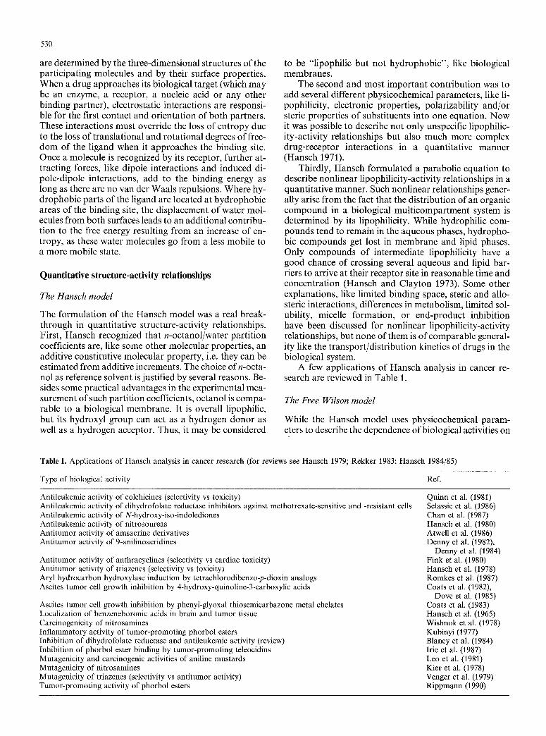
A few applications of Hansch analysis in cancer re- search are reviewed in Table 1.
The Free Wilson model
While the Hansch model uses physicochemical param- eters to describe the dependence of biological activities on
Table 1. Applications of Hansch analysis in cancer research (for reviews see Hansch 1979; Rekker 1983; Hansch 1984/85)
Type of biological activity Ref.
Antileukemic activity of colchicines (selectivity vs toxicity) Antileukemic activity of dihydrofolate reductase inhibitors against methotrexate-sensitive and -resistant cells Antileukemic activity of N-hydroxy-iso-indolediones Antileukemic activity of nitrosoureas Antitumor activity of amsacrine derivatives Antitumor activity of 9-anilinoacridines
Antitumor activity of anthracyclines (selectivity vs cardiac toxicity) Antitumor activity of triazenes (selectivity vs toxicity) Aryl hydrocarbon hydroxylase induction by tetrachlorodibenzo-p-dioxin analogs Ascites tumor cell growth inhibition by 4-hydroxy-quinoline-3-carboxylic acids
Ascites tumor cell growth inhibition by phenyl-glyoxal thiosemicarbazone metal chelates Localization of benzeneboronic acids in brain and tumor tissue Carcinogenicity of nitrosamines Inflammatory activity of tumor-promoting phorbol esters inhibition of dihydrofolate reductase and antileukemic activity (review) Inhibition of phorbol ester binding by tumor-promoting teleocidins Mutagenicity and carcinogenic activities of aniline mustards Mutagenicity of nitrosamines Mutagenicity of triazenes (selectivity vs antitumor activity) Tumor-promoting activity of phorbol esters
Quinn et al. (1981) Selassie et al. (1986) Chan et al. (1987) Hansch et al. (1980) Atwell et al. (1986) Denny et al. (1982),
Denny et al. (1984) Fink et al. (1980) Hansch et al. (1978) Romkes et al. (1987) Coats et al. (1982),
Dove et al. (1985) Coats et al. (1983) Hansch et al. (1965) Wishnok et al. (1978) Kubinyi (1977) Blaney et al. (1984) Irie et al. (1987) Leo et al. (1981) Kier et al. (1978) Venger et al. (1979) Rippmann (1990)
the chemical structure, Free and Wilson formulated a mathematical de novo model (Free and Wilson 1964). In this model it is assumed that within a congeneric series of compounds all substituents and groups add constant contributions to biological activity values, the contribu- tion of the parent structure being constant. These group contributions to biological activity depend only on the site of substitution but not on the presence or absence of other groups or substituents in the molecule. Although this additivity concept is very simple, it has proven its ap- plicability in many cases. In the very first stages of drug design it is a powerful tool to test the possibility of deriv- ing linear structure-activity relationships (provided there is more than one position of structural variation) and to evaluate the effects of different structural changes. On the other hand, a large number of variables are needed, as compared to the number of compounds, and predictions can be made only for compounds with new combinations of the substituents already included in the analysis.
In general, linear Hansch analysis and Free Wilson analysis are theoretically related, which directly leads to the possibility of a combination of both methods. In this approach physicochemical parameters are combined with Free-Wilson-type indicator variables, widening the scope and applicability of both models. Today this mixed approach is the most powerful tool for the quantitative description of complex structure-activity relationships. Even nonlinear terms can be included in such equations (for a review see Kubinyi 1988; examples from cancer re- search are included in Table 1).
Some other approaches, related to Free Wilson anal- ysis as well as to pattern recognition (see Pattern recogni- tion), use a hyperstructure concept. Out of all molecules a hyperstructure is constructed that includes all atoms of the individual compounds; in the next step the contribu- tions of all the atoms of the hyperstructure are derived from a correlation of the structural matrix (i.e. 1 or 0 for each atom in the hyperstructure, depending on the pres- ence or absence of the corresponding atom in the mole- cule) with the biological activities. In the case of flexible molecules and for compounds with different ring systems there is a high degree of ambiguity in the formulation of the hyperstructure.
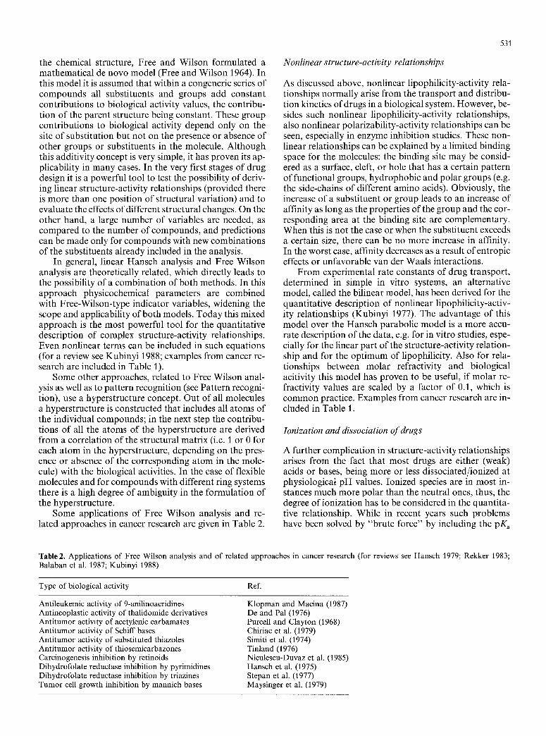
Some applications of Free Wilson analysis and re- lated approaches in cancer research are given in Table 2.
531
Nonlinear structure-activity relationships
As discussed above, nonlinear lipophilicity-activity rela- tionships normally arise from the transport and distribu- tion kinetics of drugs in a biological system. However, be- sides such nonlinear lipophilicity-activity relationships, also nonlinear polarizability-activity relationships can be seen, especially in enzyme inhibition studies. These non- linear relationships can be explained by a limited binding space for the molecules: the binding site may be consid- ered as a surface, cleft, or hole that has a certain pattern of functional groups, hydrophobic and polar groups (e.g. the side-chains of different amino acids). Obviously, the increase of a substituent or group leads to an increase of affinity as long as the properties of the group and the cor- responding area at the binding site are complementary. When this is not the case or when the substituent exceeds a certain size, there can be no more increase in affinity. In the worst case, affinity decreases as a result of entropic effects or unfavorable van der Waals interactions.
F rom experimental rate constants of drug transport, determined in simple in vitro systems, an alternative model, called the bilinear model, has been derived for the quantitative description of nonlinear lipophilicity-activ- ity relationships (Kubinyi 1977). The advantage of this model over the Hansch parabolic model is a more accu- rate description of the data, e.g. for in vitro studies, espe- cially for the linear part of the structure-activity relation- ship and for the optimum of lipophilicity. Also for rela- tionships between molar refractivity and biological acitivity this model has proven to be useful, if molar re- fractivity values are scaled by a factor of 0.1, which is common practice. Examples from cancer research are in- cluded in Table 1.
Ionization and dissociation of drugs
A further complication in structure-activity relationships arises from the fact that most drugs are either (weak) acids or bases, being more or less dissociated/ionized at physiologica~ pH values. Ionized species are in most in- stances much more polar than the neutral ones, thus, the degree of ionization has to be considered in the quantita- tive relationship. While in recent years such problems have been solved by "brute force" by including the pK a
Table2. Applications of Free Wilson analysis and of related approaches in cancer research (for reviews see Hansch 1979; Rekker 1983; Balaban et al. 1987; Kubinyi 1988)
Type of biological activity Ref.
Antileukemic activity of 9-anilinoacridines Antineoplastic activity of thalidomide derivatives Antitumor activity of acetylenic carbamates Antitumor activity of Schiff bases Antitumor activity of substituted thiazoles Antitumor activity of thiosemicarbazones Carcinogenesis inhibition by retinoids Dihydrofolate reductase inhibition by pyrimidines Dihydrofolate reductase inhibition by triazines Tumor cell growth inhibition by mannich bases
Klopman and Macina (1987) De and Pal (1976) Purcell and Clayton (1968) Chiriac et al. (1979) Simiti et al. (1974) Tinland (1976) Niculescu-Duvaz et al. (1985) Hansch et al. (1975) Stepan et al. (1977) Maysinger et al. (1979)

532
values of the compounds as additional parameters, it has been clearly demonstrated that partition coefficients have to be corrected for the degree of ionization (Scherrer and Howard 1977). Such apparent partition coefficients (also called distribution coefficients) are a much better mea- sure of lipophilicity at a given pH value than the combi- nation of the lipophilicity value of the neutral compound and its pK, value.
Of course ion-pair effects, micelle formation of am- phiphilic compounds, and the dependence of saturation solubility on the degree of ionization are additional ef- fects that render the quantitative description of structure- activity relationships more difficult.
Pattern recognition
Pattern recognition is an approach where many different descriptors and variables, not only physicochemical pa- rameters and Free-Wilson-type indicator variables, but also geometrical parameters like distance, topological pa- rameters, etc., i.e. all available information on a group of molecules, are tested for their ability to explain the struc- ture-activity relationships. While Hansch analysis most often starts from a structure-activity hypothesis and in Free Wilson analysis the variables are predetermined by the structural variation, in pattern recognition all the dif- ferent pieces of information are used to derive the struc- ture-activity relationship, most often in a trial-and-error
procedure using a limited number of compounds as a training set. The reliability of the resulting model is then tested by the predictive power for the biological activities of the remaining compounds. While pattern recognition may be a useful tool in large groups of chemically related compounds having the same type of biological activity and acting by the same mechanism of action, this ap- proach is - especially in toxicology and cancer research - often misused to describe biological activities of largely different groups of compounds with totally different mechanism of action. In the case of large screening pro- grams, to select possible candidates out of a huge number of different candidates, this approach may be better than random selection. On the other hand, it is a dangerous procedure: new leads will not be found.
As a general criticism of pattern recognition and hy- perstructure concepts (see The Free Wilson model) it must be stated that, because of the large number of (po- tential) variables, relevant results are not easy to obtain.
Some applications of pattern recognition in cancer re- search are listed in Table 3.
Multivariate statistical methods
Multivariate methods other than multiple regression analysis are sometimes used in QSAR to extract factors from variables or biological activities (principle compo- nent analysis). Such methods are used for the comparison
Table3. Applications of pattern recognition in cancer research (for reviews see Jurs et al. 1983; Nasr et al. 1984; Jurs et al. 1985; Rosenkranz and Klopman 1986)
Type of biological activity Ref.
Antitumor activity of 9-anilinoacridines Antitumor activity of carbamates Antitumor activity of various classes of compounds Carcinogenesis inhibition by retinoids
Carcinogenicity of N-nitroso compounds
Carcinogenicity of polynuclear aromatic hydrocarbons
Prediction of antitumor activity for large-scale screening Prediction of carcinogenicity of various compounds
Henry et al. (1982) Nasr et al. (1985) Nasr et al. (1984) Jurs et al. (1985);
Leavitt and Mass (1985) Jurs et al. (1983);
Dunn and Wold (1981) Miyashita et al. (1981);
Rosenkranz and Klopman (1986) Hodes (1986) Enslein and Craig (1982)
Table 4. Applications of various multivariate methods in cancer research
Type of biological activity (method)" Ref.
Antitumor activity of mitomycins (ALS, DA) Antitumor activity of triazenes (CA) Antitumor activity of triazenes (PC) Binding of polycyclic aromatic hydrocarbons to the
tetrachlorodibenzo-p-dioxin receptor (PLS) Carcinogenicity of various chemicals (CA) Cytostatic activity of naphthoquinones (DA) Mutagenic activity of aminoacridines (CSA) Mutagenic activity of unsaturated aldehydes (PLS) Mutagenicity of halogenated hydrocarbons (PLS)
Moriguchi and Komatsu (1977) Dunn et al. (1976) Ebert et al. (1984) Johnels et al. (1989)
Pet-Edwards et al. (1985) Prakash and Hodnett (1978) McFarland and Gans (1987) Nilsson et al. (1988) Dunn et al. (1984)
a ALS, adaptive least-squares classification; CA, cluster analysis; CSA, cluster significance analysis; DA, discriminant analysis; PC, prin- ciple component analysis; PLS, partial least-squares method
533
of different test models, to correlate large blocks of pa- rameter variables with biological-activity variables by a principle component like procedure (partial least-squares method), to separate most efficiently two or more subsets of compounds, e.g. active and inactive ones (discriminant analysis), and to detect clusters of closely related com- pounds or activities (cluster analysis and cluster signifi- cance analysis). Some applications of these methods in cancer research are listed in Table 4.
Molecular modelling
Conformational analysis
Classical structure-activity models, like Hansch or Free Wilson analysis, focus on physicochemical properties and/or indicator variables for certain structural features; normally a precondition for the application of these methods is a common parent structure of all molecules. Thus, with the exception of steric parameters, the methods neglect the three-dimensional structure and the overall shape of the molecules. However, in the case of specific drug-receptor interactions, the three-dimensional structure of a molecule is of utmost importance for its ability to bind to a certain receptor site.
Most organic molecules are more or less flexible, hav- ing in solution not only three translational and three ro- tational degrees of freedom, but also internal rotational degrees of freedom. Of course, the rotations around a bond depend on energy barriers, determined by the na- ture of the bond and by the size of the substituents of the atoms forming this bond; between the barriers there are one or several energy minima. Thus, for a medium-sized molecule with some rotational degrees of freedom a number of low-energy states (the number being depen- dent on the flexibility or rigidity of the molecule) may be populated simultaneously. Conformations of molecules can be experimentally determined by various diffraction techniques, e.g. neutron, electron or X-ray diffraction (in gas phase or in liquid phase), by microwave spectros- copy, by nuclear magnetic resonance (NMR) or by two- dimensional NMR measurements (in solution), where di- rect information is available on distances between certain atoms, and by x-ray crystallography (in the crystal).
All experimental information is valuable; however, neither the conformation in vacuo, nor in solution, nor in the crystal needs to be identical to the conformation at the receptor site. During the approach of a drug molecule to its binding site conformational changes of the mole- cule and of the binding site may occur (sometimes called induced fit or flexible fit), if the net interaction energy re- sulting from the conformationally modified partners is higher then the interaction energy of the global minimum conformations. Thus, the gain in interaction energy pays at least partially for the transition to these conforma- tions. On the other hand, the barriers between the low-en- ergy conformation and the conformation adopted at the receptor site must not be too high, otherwise the con- formational changes will not take place under physiolog- ical conditions.
The only two experimental techniques that allow the experimental determination of the conformation of
bound ligands are X-ray crystallography of enzyme-in- hibitor complexes and the corresponding two-dimen- sional NMR investigations, where the enzyme has to be deuterated because of the otherwise much more complex spectrum. Alternatives to experimental methods are vari- ous theoretical approaches for the prediction of con- formations and conformational energies. Different force- field calculations, all following the same strategy, are fre- quently used today. For a molecular system the potential energy is described by an empirically derived expression of several terms, e.g. for bond lengths, bond angles, and dihedral angles. The minimum value of each potential term is calibrated at standard values obtained from ex- perimental methods or elaborate quantum-chemical cal- culations. In addition, electrostatic interactions are con- sidered as positive or negative contributions and a Len- nard-Jones type potential function for van der Waals at- traction and repulsion is included. The total energy of a molecular system is then determined by finding the en- ergy minima for all potential energy terms simulta- neously. Different force-field programs differ in their pa- rameter tables of standard terms, in their minimization algorithms, and in the potential energy function.
With the advent of powerful supercomputers, semi- empirical methods become more and more the standard in conformational energy calculations, especially for me- dium-sized molecules (20-100 atoms). In many cases they give more reliable results than force-field calculations at the cost of computing time. Even more time-consuming are ab initio calculations, with the additional complica- tion that approximations may lead to even worse results than a corresponding semiempirical calculation (Clark 1988).
A common disadvantage of all these methods is that only one minimum energy conformation is estimated and that most calculated conformation refer to vacuum. Only from molecular dynamics calculations, treating the molecule in an aqueous environment, do different low- energy conformations result, from which the conforma- tion population in solution can be estimated. Owing to the progress in two-dimensional NMR determination of the conformations of small molecules and medium-sized peptides and proteins, further experimental evidence has become available to test the results of molecular dy- namics simulations.
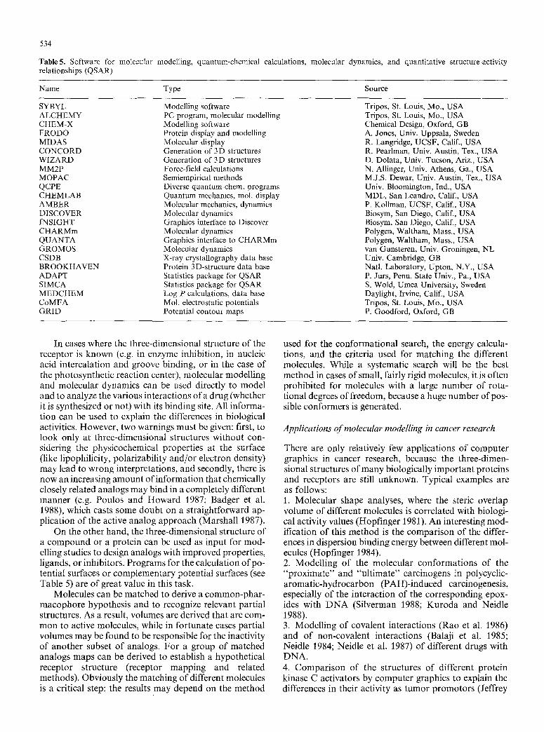
A review of common program packages for molecular modelling, quantum chemistry, and QSAR is given in Table 5.
Techniques of molecular modelling Molecular modelling combines computational tech- niques, like molecular mechanics, molecular dynamics, and quantum-chemical methods, with results from ex- perimentally observed structural data. High-resolution computer graphics is used for the display and interpreta- tion of results.
The techniques of molecular modelling have devel- oped rapidly in the course of the last 10 years. They are now powerful tools for the understanding of structure- activity relationships and for the rational design of new drugs.
534
Table5. Software for molecular modelling, quantum-chemical calculations, molecular dynamics, and quantitative structure-activity relationships (QSAR)
Name Type Source
SYBYL ALCHEMY CHEM-X FRODO MIDAS CONCORD WIZARD MM2P MOPAC QCPE CHEMLAB AMBER DISCOVER INSIGHT CHARMm QUANTA GROMOS CSDB BROOKHAVEN ADAPT SIMCA MEDCHEM CoMFA GRID
Modelling software PC program, molecular modelling Modelling software Protein display and modelling Molecular display Generation of 3D structures Generation of 3 D structures Force-field calculations Semiempirical methods Diverse quantum-chem, programs Quantum mechanics, tool. display Molecular mechanics, dynamics Molecular dynamics Graphics interface to Discover Molecular dynamics Graphics interface to CHARMm Molecular dynamics X-ray crystallography data base Protein 3 D-structure data base Statistics package for QSAR Statistics package for QSAR Log P calculations, data base Mol. electrostatic potentials Potential contour maps
Tripos, St. Louis, Mo., USA Tripos, St. Louis, Mo., USA Chemical Design, Oxford, GB A. Jones, Univ. Uppsala, Sweden R. Langridge, UCSF, Calif., USA R. Pearlman, Univ. Austin, Tex., USA D. Dolata, Univ. Tucson, Ariz., USA N. Allinger, Univ. Athens, Ga., USA M.J.S. Dewar, Univ. Austin, Tex., USA Univ. Bloomington, Ind., USA MDL, San Leandro, Calif., USA P. Kollman, UCSF, Calif., USA Biosym, San Diego, Calif., USA Biosym, San Diego, Calif., USA Polygen, Waltham, Mass., USA Polygen, Waltham, Mass., USA van Gunsteren, Univ. Groningen, NL Univ. Cambridge, GB Natl. Laboratory, Upton, N.Y., USA P. Jurs, Penn. State Univ., Pa., USA S. Wold, Umea University, Sweden Daylight, Irvine, Calif., USA Tripos, St. Louis, Mo., USA P. Goodford, Oxford, GB
In cases where the three-dimensional structure of the receptor is known (e.g. in enzyme inhibition, in nucleic acid intercalation and groove binding, or in the case of the photosynthetic reaction center), molecular modelling and molecular dynamics can be used directly to model and to analyze the various interactions of a drug (whether it is synthesized or not) with its binding site. All informa- tion can be used to explain the differences in biological activities. However, two warnings must be given: first, to look only at three-dimensional structures without con- sidering the physicochemical properties at the surface (like lipophilicity, polarizability and/or electron density) may lead to wrong interpretations, and secondly, there is now an increasing amount of information that chemically closely related analogs may bind in a completely different manner (e.g. Poulos and Howard 1987; Badger et al. 1988), which casts some doubt on a straightforward ap- plication of the active analog approach (Marshall 1987).
On the other hand, the three-dimensional structure of a compound or a protein can be used as input for mod- elling studies to design analogs with improved properties, ligands, or inhibitors. Programs for the calculation of po- tential surfaces or complementary potential surfaces (see Table 5) are of great value in this task.
Molecules can be matched to derive a common-phar- macophore hypothesis and to recognize relevant partial structures. As a result, volumes are derived that are com- mon to active molecules, while in fortunate cases partial volumes may be found to be responsible for the inactivity of another subset of analogs. For a group of matched analogs maps can be derived to establish a hypothetical receptor structure (receptor mapping and related methods). Obviously the matching of different molecules is a critical step: the results may depend on the method
used for the conformational search, the energy calcula- tions, and the criteria used for matching the different molecules. While a systematic search will be the best method in cases of small, fairly rigid molecules, it is often prohibited for molecules with a large number of rota- tional degrees of freedom, because a huge number of pos- sible conformers is generated.
Applications of molecular modelling in cancer research
There are only relatively few applications of computer graphics in cancer research, because the three-dimen- sional structures of many biologically important proteins and receptors are still unknown. Typical examples are as follows: 1. Molecular shape analyses, where the steric overlap volume of different molecules is correlated with biologi- cal activity values (Hopfinger 1981). An interesting mod- ification of this method is the comparison of the differ- ences in dispersion binding energy between different mol- ecules (Hopfinger 1984). 2. Modelling of the molecular conformations of the "proximate" and "ult imate" carcinogens in polycyclic- aromatic-hydrocarbon (PAH)-induced carcinogenesis, especially of the interaction of the corresponding epox- ides with DNA (Silverman 1988; Kuroda and Neidle 1988). 3. Modelling of covalent interactions (Rao et al. 1986) and of non-covalent interactions (Balaji et al. 1985; Neidle 1984; Neidle et al. 1987) of different drugs with DNA. 4. Comparison of the structures of different protein kinase C activators by computer graphics to explain the differences in their activity as tumor promotors (Jeffrey
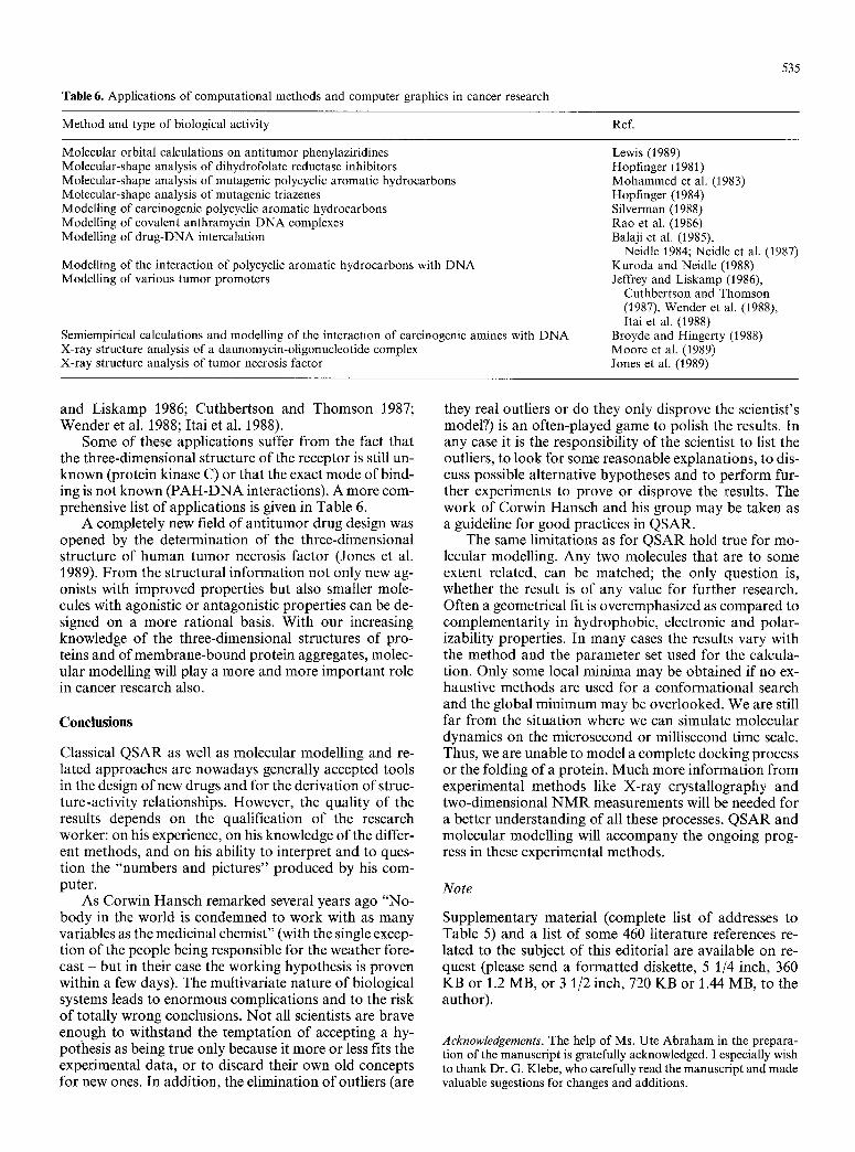
Table 6. Applications of computational methods and computer graphics in cancer research
535
Method and type of biological activity Ref.
Molecular orbital calculations on antitumor phenylaziridines Molecular-shape analysis of dihydrofolate reductase inhibitors Molecular-shape analysis of mutagenic polycyclic aromatic hydrocarbons Molecular-shape analysis of mutagenic triazenes Modelling of carcinogenic polycyclic aromatic hydrocarbons Modelling of covalent anthramycin DNA complexes Modelling of drug-DNA intercalation
Modelling of the interaction of polycyclic aromatic hydrocarbons with DNA Modelling of various tumor promoters
Semiempirical calculations and modelling of the interaction of carcinogenic amines with DNA X-ray structure analysis of a daunomycin-oligonucleotide complex X-ray structure analysis of tumor necrosis factor
Lewis (1989) Hopfinger (1981) Mohammed et al. (1983) Hopfinger (1984) Silverman (1988) Rao et al. (1986) Balaji et al. (1985),
Neidle 1984; Neidle et al. (1987) Kuroda and Neidle (1988) Jeffrey and Liskamp (1986),
Cuthbertson and Thomson (1987), Wender et al. (1988), Itai et al. (1988)
Broyde and Hingerty (1988) Moore et al. (1989) Jones et al. (1989)
and Liskamp 1986; Cuthbertson and Thomson 1987; Wender et al. 1988; Itai et al. 1988).
Some of these applications suffer f rom the fact that the three-dimensional structure of the receptor is still un- known (protein kinase C) or that the exact mode of bind- ing is not known ( P A H - D N A interactions). A more com- prehensive list of applications is given in Table 6.
A completely new field of ant i tumor drug design was opened by the determination of the three-dimensional structure of human tumor necrosis factor (Jones et al. 1989). From the structural information not only new ag- onists with improved properties but also smaller mole- cules with agonistic or antagonistic properties can be de- signed on a more rational basis. With our increasing knowledge of the three-dimensional structures of pro- teins and of membrane-bound protein aggregates, molec- ular modelling will play a more and more important role in cancer research also.
Conclusions
Classical QSAR as well as molecular modelling and re- lated approaches are nowadays generally accepted tools in the design of new drugs and for the derivation of struc- ture-activity relationships. However, the quality of the results depends on the qualification of the research worker: on his experience, on his knowledge of the differ- ent methods, and on his ability to interpret and to ques- tion the "numbers and pictures" produced by his com- puter.
As Corwin Hansch remarked several years ago "No- body in the world is condemned to work with as many variables as the medicinal chemist" (with the single excep- tion of the people being responsible for the weather fore- cast but in their case the working hypothesis is proven within a few days). The multivariate nature of biological systems leads to enormous complications and to the risk of totally wrong conclusions. Not all scientists are brave enough to withstand the temptation of accepting a hy- pothesis as being true only because it more or less fits the experimental data, or to discard their own old concepts for new ones. In addition, the elimination of outliers (are
they real outliers or do they only disprove the scientist's model?) is an often-played game to polish the results. In any case it is the responsibility of the scientist to list the outliers, to look for some reasonable explanations, to dis- cuss possible alternative hypotheses and to perform fur- ther experiments to prove or disprove the results. The work of Corwin Hansch and his group may be taken as a guideline for good practices in QSAR.
The same limitations as for QSAR hold true for mo- lecular modelling. Any two molecules that are to some extent related, can be matched; the only question is, whether the result is of any value for further research. Often a geometrical fit is overemphasized as compared to complementari ty in hydrophobic, electronic and polar- izability properties. In many cases the results vary with the method and the parameter set used for the calcula- tion. Only some local minima may be obtained if no ex- haustive methods are used for a conformational search and the global minimum may be overlooked. We are still far f rom the situation where we can simulate molecular dynamics on the microsecond or millisecond time scale. Thus, we are unable to model a complete docking process or the folding of a protein. Much more information f rom experimental methods like X-ray crystallography and two-dimensional N M R measurements will be needed for a better understanding of all these processes. QSAR and molecular modelling will accompany the ongoing prog- ress in these experimental methods.
Note
Supplementary material (complete list o f addresses to Table 5) and a list of some 460 literature references re- lated to the subject of this editorial are available on re- quest (please send a formatted diskette, 5 1/4 inch, 360 KB or 1.2 MB, or 3 1/2 inch, 720 KB or 1.44 MB, to the author).
Acknowledgements. The help of Ms. Ute Abraham in the prepara- tion of the manuscript is gratefully acknowledged. I especially wish to thank Dr. G. Klebe, who carefully read the manuscript and made valuable sugestions for changes and additions.
536
References
Atwell GJ, Baguley BC, Finlay GJ, Rewcastle GW, Denny AW (1986) Potential antitumor agents: 47. Y-Methylamino analo- gues of amsacrine with in vivo solid tumor activity. J Med Chem 29:1769-1776
Badger J, Minor I, Kremer M J, Oliveira MA, Smith T J, Griffith JP, Guerin DMA, Krishnaswamy S, Luo M, Rossmann MG, McKinlay MA, Diana GD, Dutko FJ, Fancher M, Rueckert RR, Heinz BA (1988) Structural analysis of a series of antiviral agents complexed with human rhinovirus 14. Proc Natl Acad Sci USA 85:3304-3308
Balaban AT, Niculescu-Duvaz I, Simon Z (1987) Topological aspects of QSAR for biologically active molecules. Acta Pharm Jugosl 37:7-35
Balaji VN, Dixon JS, Smith DH, Venkataraghavan R, Murdock KC (1985) Design of anticancer drugs using modeling tech- niques. Ann NY Acad Sci 439:140-161
Blaney JM, Hansch C, Silipo C, Vittoria A (1984) Structure-activity relationships of dihydrofolate reductase inhibitors. Chem Rev 84:333-407
Broyde S, Hingerty BE (1988) Conformations of DNA modified by aromatic amines: Minimized serniempirical potential energy calculations and model building. In: Silverman BD (ed) Com- puter simulation of carcinogenic processes. CRC Press, Boca Raton, F1, pp 117-140
Chan CL, Lien EJ, Tokes ZA (1987) Synthesis, biological evalua- tion, and quantitative structure-activity relationship analysis of 2-hydroxy-lH-isoindolediones as new cyt0static agents. J Med Chem 30:509-514
Chririac A, Dragomir O, Motoc F, Motoc I (1979) A Fujita-Ban analysis of antitumoral activity of Schiff bases. Univ Timisoara [Prepr], Ser Chim; ref CA 93: 187549k
Clark T (1988) Molecular orbital and force-field calculations for structure and energy predictions. In: Jochurn C, Hicks MG, Sunkel J (eds) Physical property prediction in organic chemis- try. Springer, Berlin Heidelberg, New York, pp 95-101
Coats EA, Shah K J, Milstein SR, Genther CS, Nene DM, Roesener J, Schrnidt J, Pleiss M, Wagner E, Baker JK (1982) 4-Hydroxy- quinoline-3-carboxylic acids as inhibitors of cell respiration. 2. Quantitative structure-activity relationship of dehydrogenase enzyme and Ehrlich ascites tumor cell inhibitions. J Med Chem 25:57-63
Coats EA, Milstein SR, Pleiss MA, Roesener JA, Schrnidt J, McDonald J, Reed R (1983) Comparative analysis of cellular respiratory inhibition by substituted phenylglyoxal bis(4- methyl-3-thiosemicarbazone) zinc chelates. Farm Ed Sci 38: 143-152
Cuthbertson AF, Thomson C (1987) Electrostatic potentials of tu- mor promoters. J Mol Graphics 5:92-96
De AU, Pal D (1976) Quantitative structure-activity relationship (QSAR) and rational drug design for some antineoplastic thali- domide and glutarimide derivatives. J Indian Chem Soc 53: 1049 1052
Denny WA, Cain BF, Atwell G J, Hansch C, Panthananickal A, Leo A (1982) Potential antiturnor agents 36. Quantitative relation- ships between experimental antitumor activity, toxicity, and structure for the general class of 9-anilinoacridine antitumor agents. J Med Chem 25:276-315
Denny WA, AtweU GJ, Baguley BC, Rewcastle GW (1984) QSAR in the design of potential antiturnor agents: the example of the 9-anilinoacridines. In: Kuchar M (ed) QSAR Des Bioact Compd. Prous, Barcelona, pp 97-116
Dove S, Coats E, Scharfenberg P, Franke R (1985) 7-Substituted 4- hydroxyquinoline 3-carboxylic acids as inhibitors of dehydro- genase enzymes and of the respiration of Ehrlich ascites tumor cells: multivariate analyses and quantitative structure-activity relationship for polar substituents. J Med Chem 28:447M51
Dunn III W J, Wold S (1981) The carcinogenicity of N-nitroso com- pounds: a SIMCA pattern recognition study. Bioorg Chem 10: 29-45
Dunn III WJ, Greenberg MJ, Callejas SS (1976) Use of cluster anal- ysis in the development of structure-activity relations for antitu- mor triazenes. J Med Chem 19:129%1301
Dunn III WJ, Wold S, Edlund U, Hellberg S, Gasteiger J (1984) Multivariate structure-activity relationships between data from a battery of biological tests and an ensemble of structure de- scriptors: the PLS method. Quant Struct-Act Relat 3:131 137
Ebert C, Lassiani L, Linda P, Nisi C, Alunni S, Clementi S (1984) Chemometric investigation of antiturnor tests. Quant Struct- Act Relat 3:143-147
Enslein K, Craig PN (1982) Carcinogenesis: a predictive structure- activity model. J Toxicol Environ Health 10:521-530
Fink SI, Leo A, Yamakawa M, Hansch C, Quinn FR (1980) The quantitative structure-selectivity relationship of anthracycline antitumor activity and cardiac toxicity. Farm Ed Sci 35: 965- 979
Free Jr SM, Wilson JW (1964) A mathematical contribution to structure-activity studies. J Med Chern 7:395 399
Hansch C (1971) Quantitative structure-activity relationships in drug design. In: Ari6ns EJ (ed) Drug design, vol 1. Academic Press, New York, pp 271-342
Hansch C (1979) QSAR in cancer chemotherapy. Farm Ed Sci 34: 89-104
Hansch C (1984/85) The QSAR paradigm in the design of less toxic molecules. Drug Metab Rev 15:1279-1294
Hansch C, Clayton JM (1973) Lipophilic character and biological activity of drugs. II: the parabolic case. J Pharm Sci 62:1-21
Hansch C, Fujita T (1964) ~-cy-~-analysis. A method for the corre- lation of biological activity and chemical structure. J Am Chem Soc 86:1616-1626
Hansch C, Steward AR, Isawa J (1965) The correlation of localisa- tion rates of benzeneboronic acids in brain and tumor tissue with substituent constants. Mol Pharrnacol 1 : 87-92
Hansch C, Silipo C, Steller EE (1975) Formulation of de novo sub- stituent constants in correlation analysis: inhibition of dihydro- folate reductase by 2,4-diamino-5-(3,4-dichlorophenyl)-6-sub- stituted pyrimidines. J Pharm Sci 64:1186-1191
Hansch C, Hatheway GJ, Quinn FR, Greenberg N (1978) Antitu- mot 1-(X-aryl)-3,3-dialkyltriazenes:2. On the role of correlation analysis in decision making in drug modification toxicity quan- titative structure-activity relationships of 1-(X-phenyl)-3,3- dialkyltriazenes in mice. J Med Chem 21:574-577
Hansch C, Leo A, Schmidt C, Jow PYC, Montgomery JA (1980) Antitumor structure-activity relationships. Nitrosoureas vs L- 1210 leukemia. J Med Chem 23:1095-1101
Henry DR, Jurs PC, Denny WA (1982) Structure-antiturnor activity relationships of 9-anilinoacridines using pattern recognition. J Med Chem 25:899 908
Hodes L (1986) A two-component approach for predicting antitu- mor activity from chemical structure in large scale screening. J Med Chem 29:2207-2212
Hopfinger AJ (1981) A general QSAR for dihydrofolate reductase inhibition by 2,4-diaminotriazines based upon molecular shape analysis. Arch Biochem Biophys 206:153 163
Hopfinger AJ (1984) A QSAR study of the Ames rnutagenicity of l-(X-phenyl)-3,3-dialkyltriazenes using molecular potential en- ergy fields and molecular shape analysis. Quant Struct-Act Re- lat 3:1-5
Irie K, Hagiwara N, Tokuda H, Koshimizu K (1987) Structure-ac- tivity studies of the indole alkaloid tumor promoter teleocidins. Carcinogenesis 8:547-552
Itai A, Kato Y, Tomioka N, Iitaka Y, Endo Y, Hasegawa M, Shudo K, Fujiki H, Sakai S (1988) A receptor model for tumor pro- moters: rational superposition of teleocidines and phorbol esters. Proc Natl Acad Sci USA 85:3688-3692
Jeffrey AM, Liskamp RMJ (1986) Computer-assisted molecular modeling of tumor promoters: rationale for the activity ofphor- bol esters, teleocidin B, and aplysiatoxin. Proc Natl Acad Sci USA 83:241-245
Johnels D, Gillner M, Norden B, Toftgard R, Gustafsson JA (1989) Quantitative structure-activity relationship (QSAR) analysis
537
using the partial least squares (PLS) method: the binding of polycyclic aromatic hydrocarbons (PAH) to the rat liver 2,3,7,8- tetrachlorodibenzo-p-dioxin (TCDD) receptor. Quant Struct- Act Relat 8:83-89
Jones EY, Stuart DI, Walker NPC (1989) Structure of tumour ne- crosis factor. Nature 338:225-228
Jurs PC, Hasan MN, Henry DR, Stouch TR, Whalen-Pedersen EK (1983) Computer-assisted studies of molecular structure and carcinogenic activity. Fundam Appl Toxicol 3:343-349
Jurs PC, Stouch TR, Czerwinski M, Narvaez JN (1985) Computer- assisted studies of molecular structure-biological activity rela- tionships. J Chem Inf Comput Sci 25:296-308
Kier LB, Simons RJ, Hall LH (1978) Structure-activity studies on mutagenicity of nitrosamines using molecular connectivity. J Pharm Sci 67:725-726
Klopman G, Macina OT (1987) Computer-automated structure evaluation of antileukemic 9-anilinoacridines. Mol Pharmacol 31:457-476
Kubinyi H (1977) Quantitative structure activity relationships. 7. The bilinear model, a new model for nonlinear dependence of biological activity on hydrophobic character. J Med Chem 20: 625-629
Kubinyi H (1988) Free Wilson analysis. Theory, applications and its relationship to Hansch analysis. Quant Struct-Act Relat 7:121- 133
Kuroda R, Neidle S (1988) Crystallographic and computer-model- ing studies on the metabolites of polycyclic aromatic hydrocar- bons and their interactions with nucleic acids. In: Silverman BD (ed) Computer simulation of carcinogenic processes. CRC Press, Boca Raton, F1, pp 141-168
Leavitt SA, Mass MJ (1985) Computer-assisted correlation of struc- ture and biological activity in a set of retinoids. Cancer Res 45: 4741-4747
Leo A, Panthananickal A, Hansch C, Theiss J, Shimkin M, An- drews AW (1981) A comparison of mutagenic and carcinogenic activities of aniline mustards. J Med Chem 24:859-864
Lewis DFV (1989) Molecular orbital calculations on tumor-inhibi- tory phenyl aziridines: QSARs. Xenobiotica 19:341 356
Marshall GR (1987) Computer-aided drug design. Annu Rev Phar- macol Toxicol 27:193-213
Maysinger D, Birus M, Movrin M (1979) Structure-activity rela- tionships of isatin N-Mannich bases deduced from the Free- Wilson model. Acta Pharm Jugosl 29:15-18
McFarland JW, Gans DJ (1987) Cluster significance analysis in quantitative structure-activity relationship (QSAR). Pharmaco- chem Libr 10:25-29
Miyashita Y, Seki T, Takahashi Y, Daiba S, Tanaka Y, Yotsui Y, Abe H, Sasaki S (1981) Computer-assisted structure-carcinoge- nicity studies on polycyclic aromatic hydrocarbons by pattern recognition methods. Anal Chim Acta 133:603-613
Mohammad SN, Hopfinger AJ, Bickers DR (1983) Intrinsic muta- genicity of polycyclic aromatic hydrocarbons: a quantitative structure-activity study based upon molecular shape analysis. J Theor Biol 102:323-331
Moore MH, Hunter WN, Langlois d'Estaintot B, Kennard O (1989) DNA-drug interactions. The crystal structure of d(CGATCG) complexed with daunomycin. J Mol Biol 206: 693-705
Moriguchi I, Komatsu K (1977) Adaptive least-squares classifica- tion applied to structure-activity correlation of antitumor mito- mycin derivatives. Chem Pharm Bull 25:2800-2802
Nasr M, Paull KD, Narayanan VL (1984) Computer-assisted struc- ture-activity correlations. Adv Pharmacol Chemother 20: 123- 190
Nasr M, Paull KD, Narayanan VL (1985) Computer-assisted struc- ture-anticancer activity correlations of carbamates and thiocar- bamates. J Pharm Sci 74:831-836
Neidle S (1984) Computer graphics in the study of drug-nucleic acid interactions. Biochem Soc Trans 12:1008-1011
Neidle S, Abraham ZHL, Collier DA, Islam SA (1987) Application of computer-assisted modeling to structure-activity studies with intercalating drugs. Bristol-Myers Cancer Symp 8:83-109
Niculescu-Duvaz I, Simon Z, Voiculet N (1985) QSAR application in chemical carcinogenesis: II. QSAR analysis of a class of car- cinogenesis inhibitors: retinoids. Carcinogenesis 6:479-486
Nilsson LM, Carter RE, Sterner O, Liljefors T (1988) Structure-ac- tivity relationships for unsaturated dialdehydes: 2. A PLS corre- lation of theoretical descriptors for six compounds with muta- genic activity in the Ames Salmonella assay. Quant Struct-Act Relat 7:84-91
Pet-Edwards J, Rosenkranz HS, Chankong V, Haimes YY (1985) Cluster analysis in predicting the carcinogenicity of chemicals using short-term assays. Mutat Res 153:167-185
Poulos TL, Howard AJ (1987) Crystal structures of metyrapone- and phenylimidazole-inhibited complexes of cytochrome P- 450cam. Biochemistry 26:8165 8174
Prakash G, Hodnett EM (1978) Discriminant analysis and struc- ture-activity relationships: 1. Naphthoquinones. J Med Chem 21:369-374
Purcell WP, Clayton JM (1968) Application of regression analyses to antitumor activities of various acetylenic carbamates. J Med Chem 11:199-203
Quinn FR, Neiman Z, Beisler JA (1981) Toxicity quantitative struc- ture-activity relationships of colchicines. J Med Chem 24: 636- 639
Rat SN, Singh UC, Kollman PA (1986) Molecular mechanics sim- ulations on covalent complexes between anthramycin and B DNA. J Med Chem 29:2484-2492
Rekker RF (1983) Quantitative structure-activity relationship stud- ies around cytotoxic drugs. Dev Pharmacol 3:23-46
Rippmann F (1990) Hydrophobicity and tumor promoting activity ofphorbol esters. Quant Struct-Act Relat 9, 1-5
Romkes M, Piskorska-Pliszczynska J, Keys B, Safe S, Fujita T (1987) Quantitative structure-activity relationships analysis of interactions of 2,3,7,8-tetrachlorodibenzo-p-dioxin and 2-sub- stituted analogues with rat, mouse, guinea pig, and hamster cy- tosolic receptor. Cancer Res 47:5108 5111
Rosenkranz HS, Klopman G (1986) Mutagens, carcinogens, and computers. Prog Clin Biol Res [A]209:71-104
Scherrer RA, Howard SM (1977) Use of distribution coefficients in quantitative structure-activity relationships. J Med Chem 20: 53-58
Selassie CD, Strong CD, Hansch C, Delcamp T J, Freisheim JH, Khwaja TA (1986) Comparison of triazines as inhibitors of L1210 dihydrofolate reductase and of L1210 cells sensitive and resistant to methotrexate. Cancer Res 46:744-756
Silverman BD (1988) Molecular conformation and polycyclic aro- matic hydrocarbon (PAH) carcinogenesis. In: Silverman BD (ed) Computer simulation of carcinogenic processes. CRC Press, Boca Raton, F1, pp 91-116
Simiti I, Schwartz I, Coman M (1974) Heterocycles XXXVI. Free- Wilson structure-activity study of some substituted thiazoles. Rev Roum Biochim 11: 139-143; ref CA 82:132793p
Stepan A, Badilescu II, Simon Z (1977) Minimal topological differ- ence (MTD) studies of the structure-activity relationship of triazine derivatives inhibiting dihydrofolate reductase. An Univ Timisoara, Ser Stiinte Fiz-Chim 15: 61-71; refCA 91: 186411s
Tinland B (1976) Structure-activity relationship for antitumor thiosemicarbazones. Farm Ed Sci 31:888-890
Venger BH, Hansch C, Hatheway G J, Amrein YU (1979) Ames test of 1-(X-phenyl)-3,3-dialkyltriazenes. A quantitative structure- activity study. J Med Chem 22:473-476
Wender PA, Cribbs CA, Koehler KF, Sharkey NA, Herald CL, Ka- mano Y, Pettit GR, Blumberg PM (1988) Modeling of the bryo- statins to the phorbol ester pharmacophore in protein kinase C. Proc Natl Acad Sci USA 85:7197 7201
Wishnok JS, Archer MC, Edelman AS, Rand WM (1978) Nitrosa- mine carcinogenicity: a quantitative Hansch-Taft structure-ac- tivity relationship. Chem Biol Interact 20:43-54