quantitative neuromuscular blockade monitoringblockade agents are administered, monitor...
TRANSCRIPT
Quantitative Neuromuscular Blockade
MonitoringMattie Martin BSN, SRNA
The University of Scranton
Objectives
• Review train-of-four and train-of-four ratio
• Define residual neuromuscular blockade and discuss clinical implications
• Define and discuss subjective neuromuscular blockade monitoring
• Define and discuss objective neuromuscular blockade monitoring
• Review of current research and recommendations

Train-of-Four
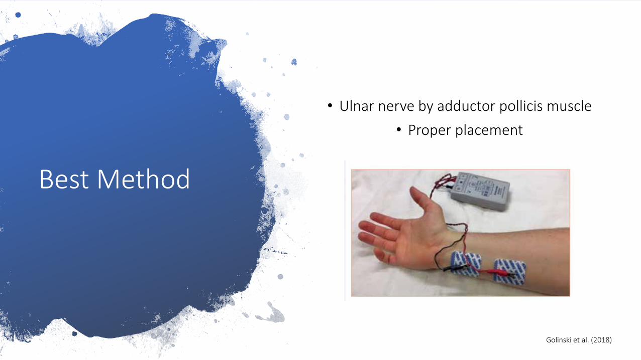
Best Method
• Ulnar nerve by adductor pollicis muscle
• Proper placement
Golinski et al. (2018)
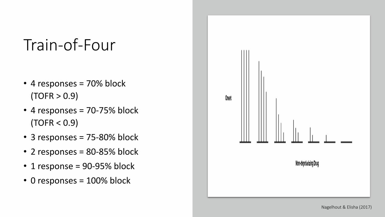
Train-of-Four
• 4 responses = 70% block
(TOFR > 0.9)
• 4 responses = 70-75% block
(TOFR < 0.9)
• 3 responses = 75-80% block
• 2 responses = 80-85% block
• 1 response = 90-95% block
• 0 responses = 100% block
Nagelhout & Elisha (2017)
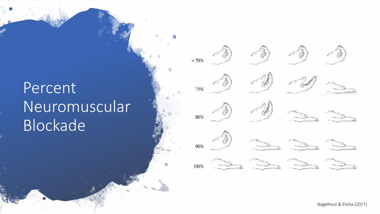
Percent Neuromuscular Blockade
Nagelhout & Elisha (2017)
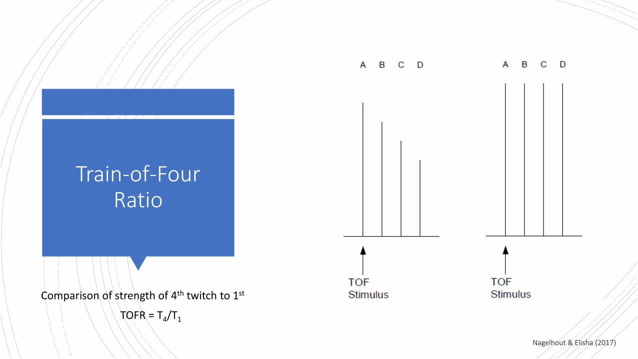
Train-of-Four Ratio
Comparison of strength of 4th twitch to 1st
TOFR = T4/T1
Nagelhout & Elisha (2017)
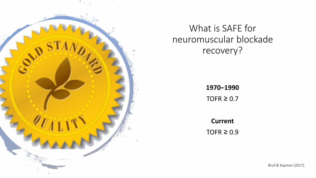
What is SAFE for neuromuscular blockade
recovery?
1970–1990
TOFR ≥ 0.7
Current
TOFR ≥ 0.9
Brull & Kopman (2017)
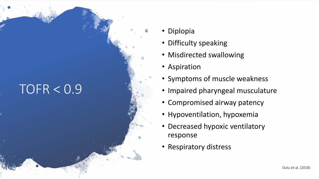
TOFR < 0.9
• Diplopia
• Difficulty speaking
• Misdirected swallowing
• Aspiration
• Symptoms of muscle weakness
• Impaired pharyngeal musculature
• Compromised airway patency
• Hypoventilation, hypoxemia
• Decreased hypoxic ventilatory response
• Respiratory distress
Dutu et al. (2018)
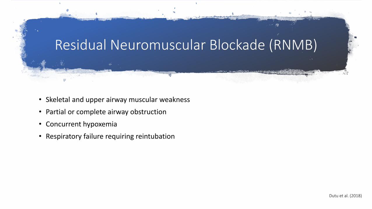
Residual Neuromuscular Blockade (RNMB)
• Skeletal and upper airway muscular weakness
• Partial or complete airway obstruction
• Concurrent hypoxemia
• Respiratory failure requiring reintubation
Dutu et al. (2018)
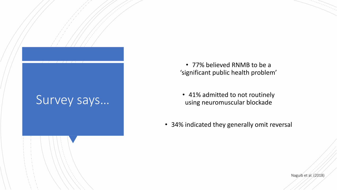
Survey says…
• 77% believed RNMB to be a ‘significant public health problem’
• 41% admitted to not routinely using neuromuscular blockade
• 34% indicated they generally omit reversal
Naguib et al. (2018)
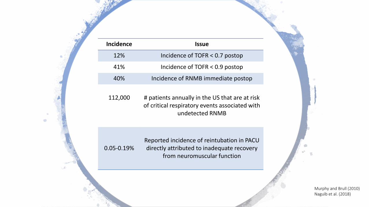
Incidence Issue
12% Incidence of TOFR < 0.7 postop
41% Incidence of TOFR < 0.9 postop
40% Incidence of RNMB immediate postop
112,000 # patients annually in the US that are at risk of critical respiratory events associated with
undetected RNMB
0.05-0.19%Reported incidence of reintubation in PACU directly attributed to inadequate recovery
from neuromuscular function
Murphy and Brull (2010)Naguib et al. (2018)
RECITE Study
• Residual Cuararization and Its Incidence at Tracheal Extubation
• 63.5% RNMB at time of extubation
• 56.5% RNMB at arrival to PACU
Fortier, et al. (2015)
Neuromuscular Blockade
Monitoring
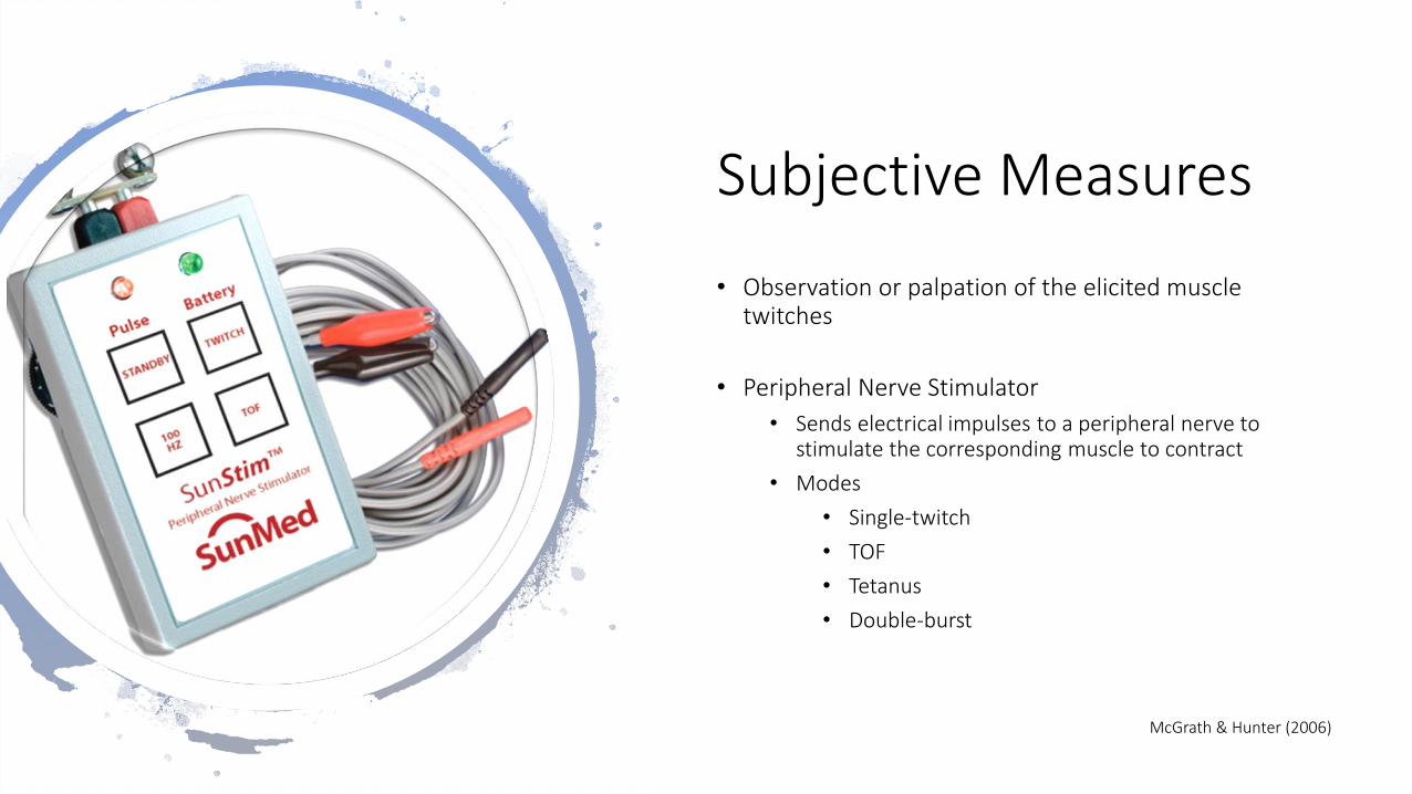
Subjective Measures
• Observation or palpation of the elicited muscle twitches
• Peripheral Nerve Stimulator
• Sends electrical impulses to a peripheral nerve to stimulate the corresponding muscle to contract
• Modes
• Single-twitch
• TOF
• Tetanus
• Double-burst
McGrath & Hunter (2006)
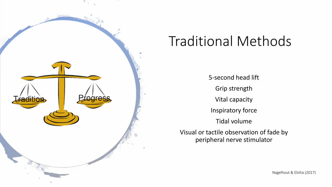
Traditional Methods
5-second head lift
Grip strength
Vital capacity
Inspiratory force
Tidal volume
Visual or tactile observation of fade by peripheral nerve stimulator
Nagelhout & Elisha (2017)
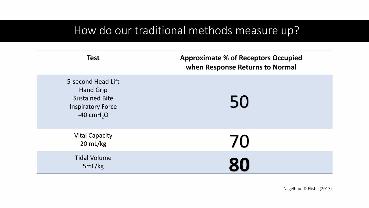
How do our traditional methods measure up?
Test Approximate % of Receptors Occupied when Response Returns to Normal
5-second Head LiftHand Grip
Sustained BiteInspiratory Force
-40 cmH2O
Vital Capacity 20 mL/kg
Tidal Volume 5mL/kg
50
7080
Nagelhout & Elisha (2017)
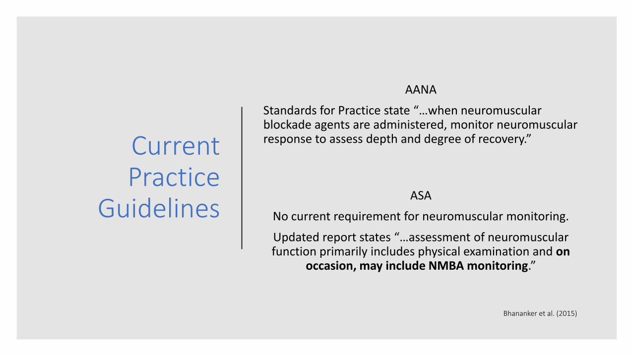
Current Practice
Guidelines
AANA
Standards for Practice state “…when neuromuscular blockade agents are administered, monitor neuromuscular response to assess depth and degree of recovery.”
ASA
No current requirement for neuromuscular monitoring.
Updated report states “…assessment of neuromuscular function primarily includes physical examination and on
occasion, may include NMBA monitoring.”
Bhananker et al. (2015)
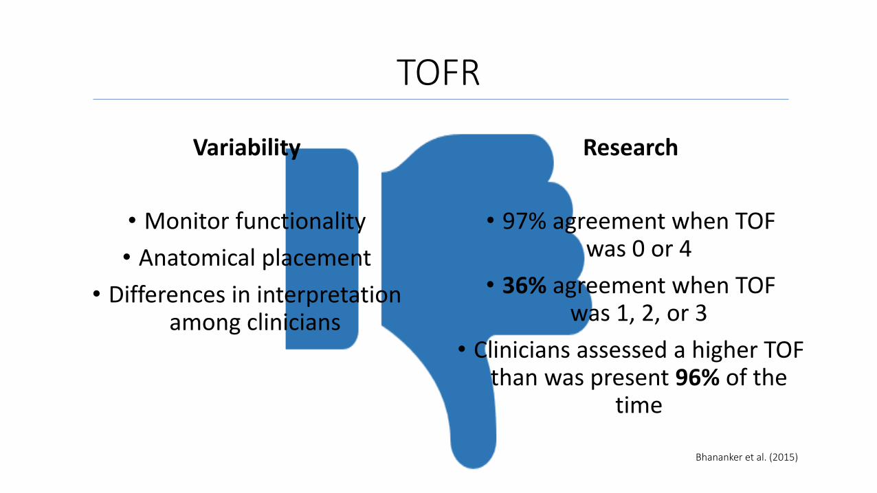
TOFR
Variability
• Monitor functionality
• Anatomical placement
• Differences in interpretation among clinicians
Research
• 97% agreement when TOF was 0 or 4
• 36% agreement when TOF was 1, 2, or 3
• Clinicians assessed a higher TOF than was present 96% of the
time
Bhananker et al. (2015)
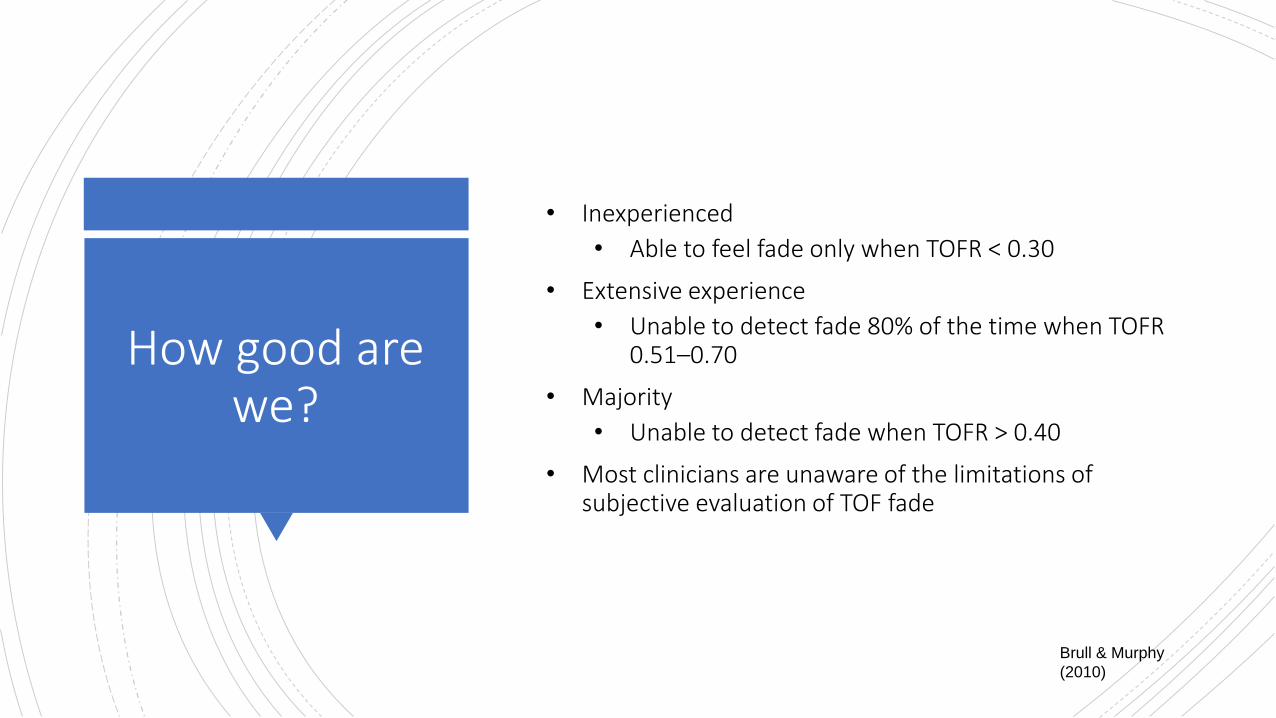
How good are we?
• Inexperienced
• Able to feel fade only when TOFR < 0.30
• Extensive experience
• Unable to detect fade 80% of the time when TOFR 0.51–0.70
• Majority
• Unable to detect fade when TOFR > 0.40
• Most clinicians are unaware of the limitations of subjective evaluation of TOF fade
Brull & Murphy
(2010)
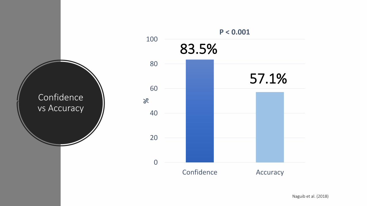
Confidence vs Accuracy
0
20
40
60
80
100
Confidence Accuracy
%
P < 0.001
83.5%
57.1%
Naguib et al. (2018)
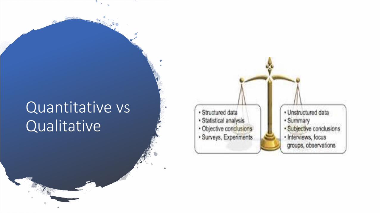
Quantitative vs Qualitative
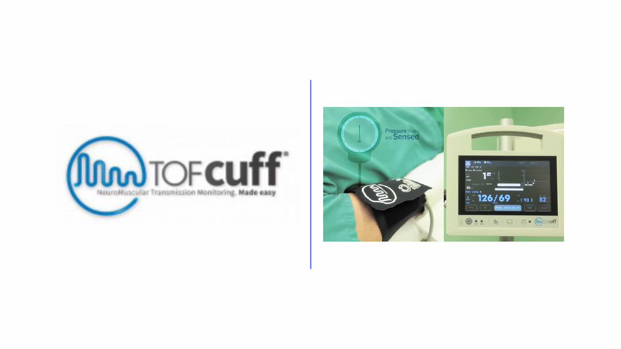
Types of Quantitative Monitors
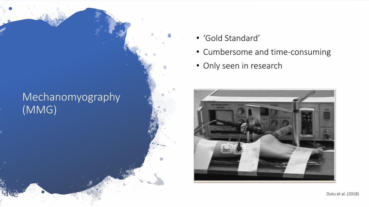
Mechanomyography (MMG)
• ‘Gold Standard’
• Cumbersome and time-consuming
• Only seen in research
Dutu et al. (2018)
MMG
• Thumb placed on a force transducer under mild tension (200-300 g preload)
• Produces an isometric contraction
• Force of contraction is converted to an electrical signal
• Amplitude of the signal is recorded on an interfaced pressure monitor
• Amplitude is proportional to the strength of muscle contraction
• Measurement of TOFR will yield precise and reproducible results
Brull & Kopman (2017)
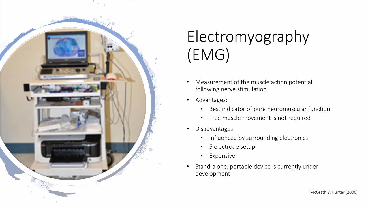
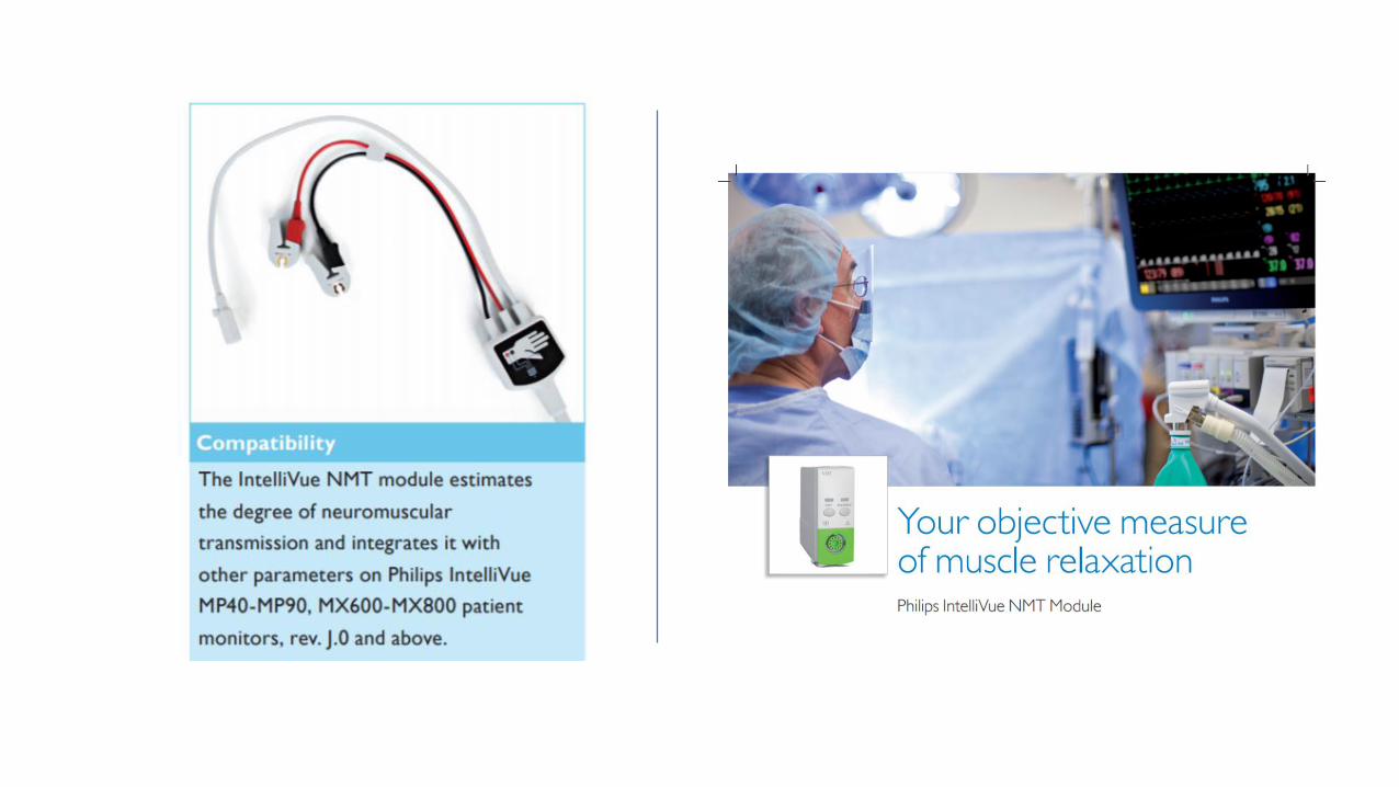
Electromyography (EMG)
• Measurement of the muscle action potential following nerve stimulation
• Advantages:
• Best indicator of pure neuromuscular function
• Free muscle movement is not required
• Disadvantages:
• Influenced by surrounding electronics
• 5 electrode setup
• Expensive
• Stand-alone, portable device is currently under development
McGrath & Hunter (2006)
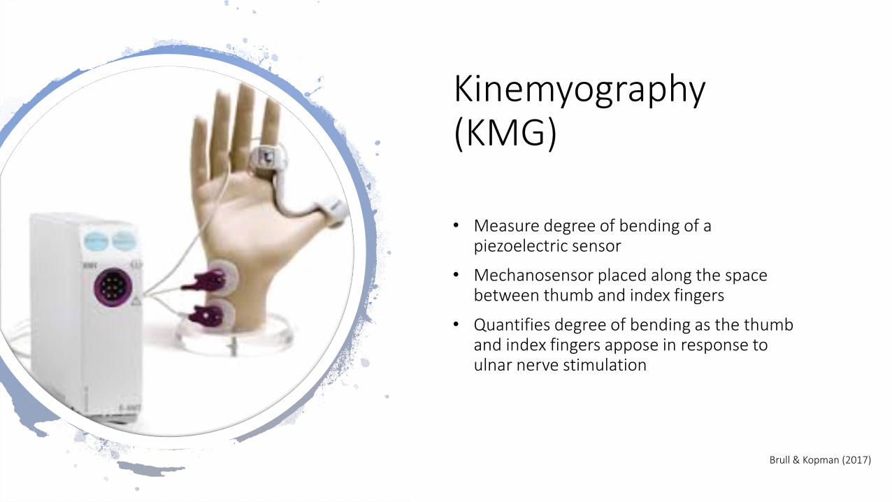
Kinemyography (KMG)
• Measure degree of bending of a piezoelectric sensor
• Mechanosensor placed along the space between thumb and index fingers
• Quantifies degree of bending as the thumb and index fingers appose in response to ulnar nerve stimulation
Brull & Kopman (2017)
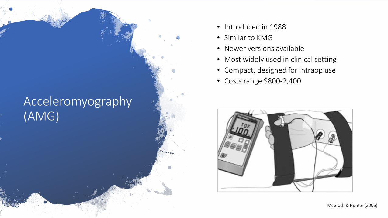
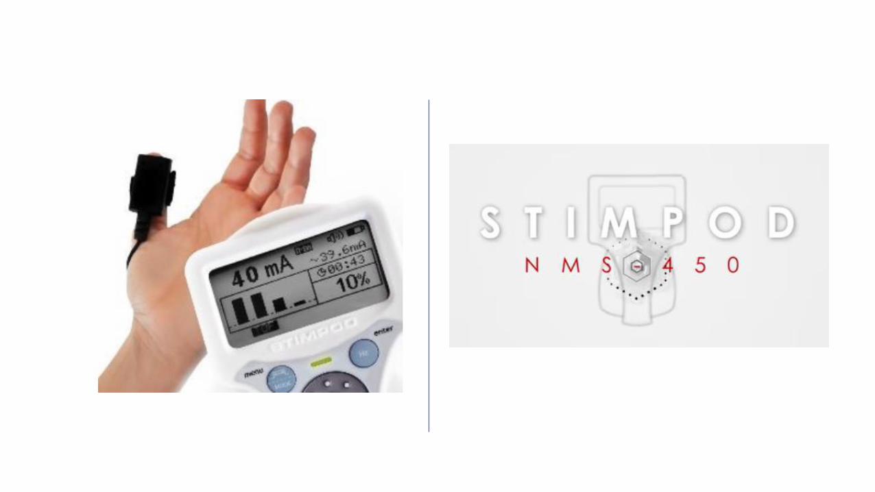
Acceleromyography (AMG)
• Introduced in 1988
• Similar to KMG
• Newer versions available
• Most widely used in clinical setting
• Compact, designed for intraop use
• Costs range $800-2,400
McGrath & Hunter (2006)
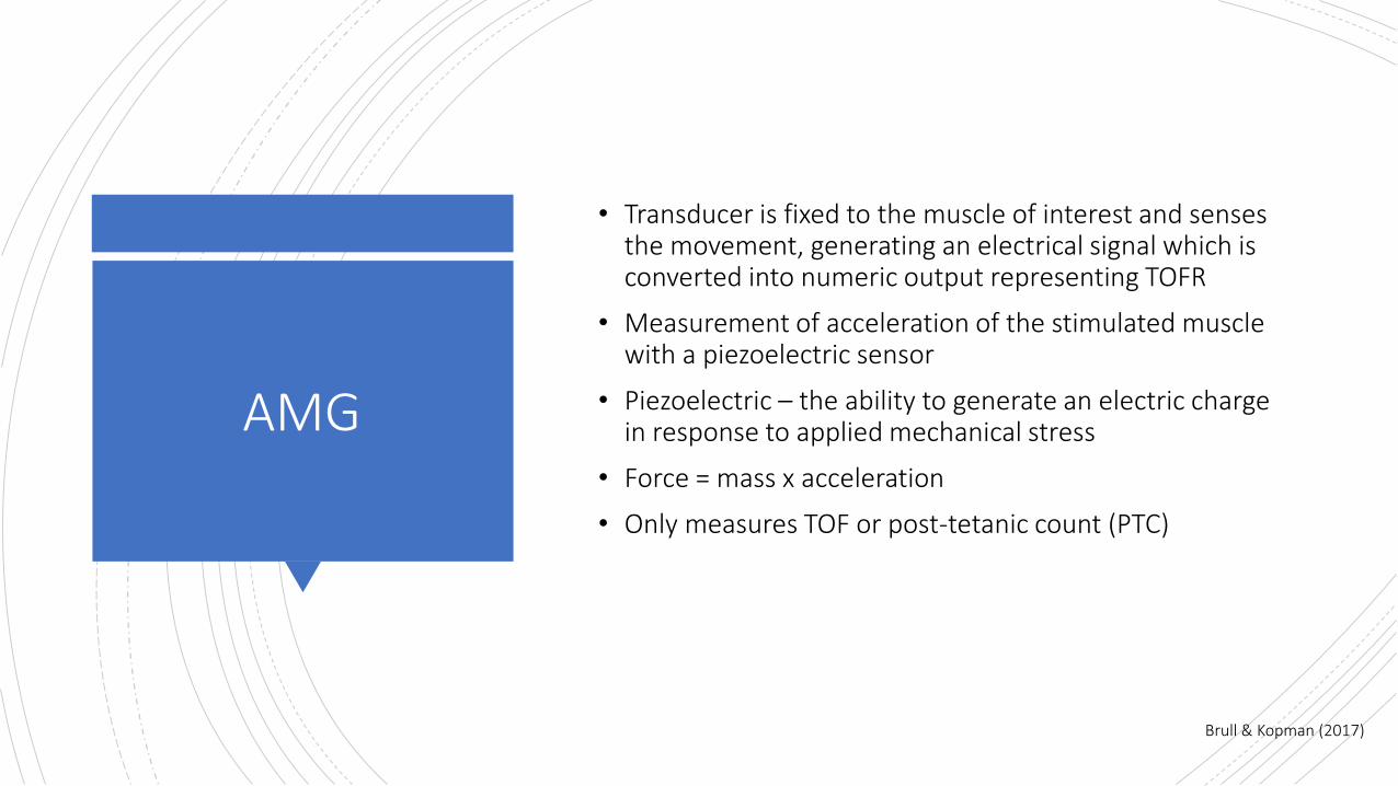
AMG
• Transducer is fixed to the muscle of interest and senses the movement, generating an electrical signal which is converted into numeric output representing TOFR
• Measurement of acceleration of the stimulated muscle with a piezoelectric sensor
• Piezoelectric – the ability to generate an electric charge in response to applied mechanical stress
• Force = mass x acceleration
• Only measures TOF or post-tetanic count (PTC)
Brull & Kopman (2017)
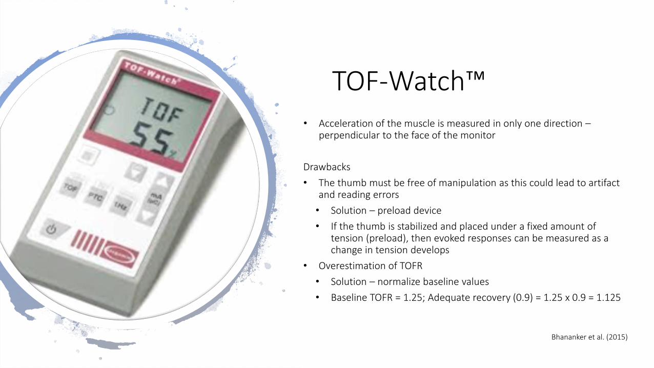
TOF-Watch™• Acceleration of the muscle is measured in only one direction –
perpendicular to the face of the monitor
Drawbacks
• The thumb must be free of manipulation as this could lead to artifact and reading errors
• Solution – preload device
• If the thumb is stabilized and placed under a fixed amount of tension (preload), then evoked responses can be measured as a change in tension develops
• Overestimation of TOFR
• Solution – normalize baseline values
• Baseline TOFR = 1.25; Adequate recovery (0.9) = 1.25 x 0.9 = 1.125
Bhananker et al. (2015)

Staircase Effect
• Repeated indirect stimulation may enhance the evoked mechanical response of muscle
• After spontaneous recovery of the TOFR to ≥ 0.80, T1 frequently returned to values > 150% of control
• 2nd (and often 3rd and 4th) twitch may exceed the 1st
Kopman et al. (2015)
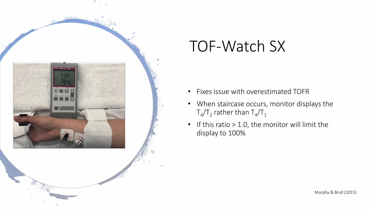
TOF-Watch SX
• Fixes issue with overestimated TOFR
• When staircase occurs, monitor displays the T4/T2 rather than T4/T1
• If this ratio > 1.0, the monitor will limit the display to 100%
Murphy & Brull (2015)
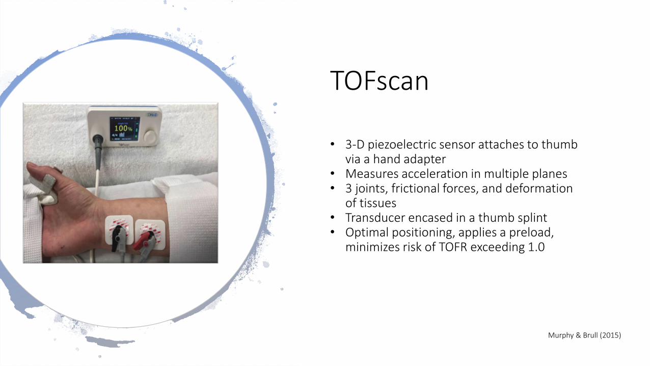
TOFscan
• 3-D piezoelectric sensor attaches to thumb via a hand adapter
• Measures acceleration in multiple planes• 3 joints, frictional forces, and deformation
of tissues• Transducer encased in a thumb splint• Optimal positioning, applies a preload,
minimizes risk of TOFR exceeding 1.0
Murphy & Brull (2015)
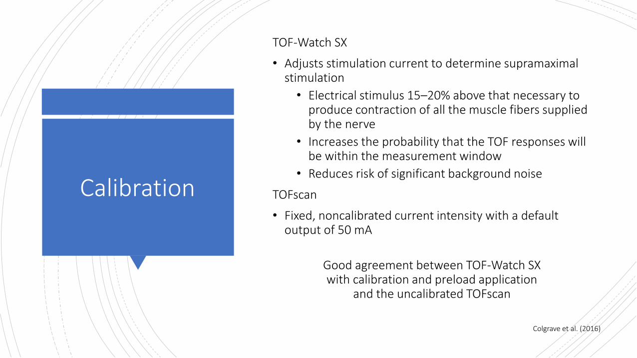
Calibration
TOF-Watch SX
• Adjusts stimulation current to determine supramaximal stimulation
• Electrical stimulus 15–20% above that necessary to produce contraction of all the muscle fibers supplied by the nerve
• Increases the probability that the TOF responses will be within the measurement window
• Reduces risk of significant background noise
TOFscan
• Fixed, noncalibrated current intensity with a default output of 50 mA
Good agreement between TOF-Watch SX with calibration and preload application
and the uncalibrated TOFscan
Colgrave et al. (2016)
Consensus Recommendations
• Whenever a neuromuscular blocker is administered, neuromuscular function must be monitored by observing the evoked muscular response to peripheral nerve stimulation.
• Objective monitoring (documentation of TOFR ≥ 0.90) is the only method of assuring that satisfactory recovery from neuromuscular function has taken place.
Naguib et al. (2018)
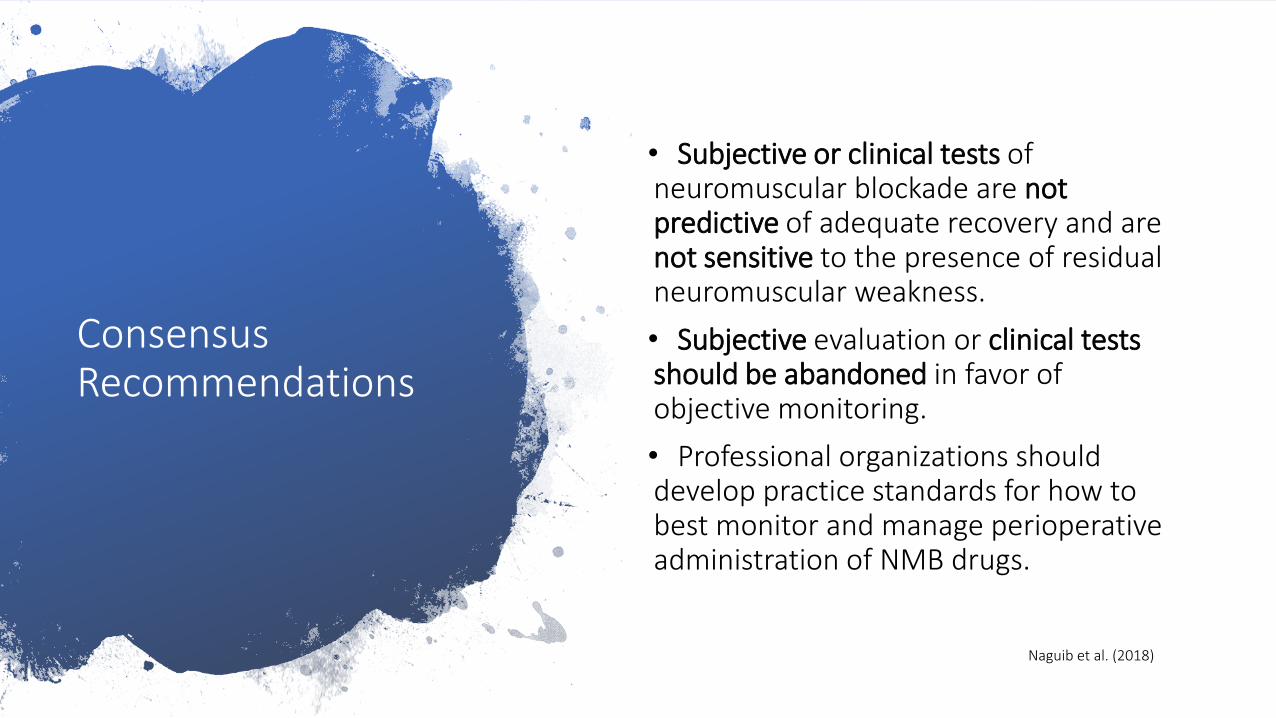
Consensus Recommendations
• Subjective or clinical tests of neuromuscular blockade are not predictive of adequate recovery and are not sensitive to the presence of residual neuromuscular weakness.
• Subjective evaluation or clinical tests should be abandoned in favor of objective monitoring.
• Professional organizations should develop practice standards for how to best monitor and manage perioperative administration of NMB drugs.
Naguib et al. (2018)
References Bhananker, S. M., Treggiari, M. M., Sellers, B. A., Cain, K. C., Ramaiah, R., & Thilen, S. R. (2015). Comparison of train-of-four count by anesthesia providers versus TOF-Watch® SX: a prospective
cohort study. Canadian Journal of Anesthesia/Journal Canadien d’anesthésie, 62(10), 1089–1096. https://doi.org/10.1007/s12630-015-0433-9
Brull, S.J. & Kopman, A.F. (2017). Current status of neuromuscular reversal and monitoring: challenges and opportunities. Anesthesiology, 126(1), 173-190.
Brull, S.J. & Murphy, G.S. (2010). Residual neuromuscular blockade. Lessons unlearned. Part II: methods to reduce the risk of residual weakness. Anesthesia and Analgesia, 111(1) 129-140.
Casper, C. & Viby-Mogensen, J. (2018). Acceleromyography for use in scientific and clinical practice: a systemic review of the evidence. Anesthesiology, 108(6), 1117-1140.
Colegrave, N., Billard, V., Motamed, C., & Bourgain, J.-L. (2016). Comparison of the TOF-ScanTM acceleromyograph to TOF-Watch SXTM: Influence of calibration. Anaesthesia Critical Care & Pain
Medicine, 35(3), 223–227. https://doi.org/10.1016/j.accpm.2016.01.003
Dunworth, B.A., Sandberg, W.S., Morrison, S., Lutz, Wanderer, J.P., O’Donnell, J.M. (2018). Implementation of acceleromyography to increase use of quantitative neuromuscular blockade
monitoring: a quality improvement project. AANA Journal, 86(4), 269-277.
Dutu, M., Ivascu, R., Tudorache, O., Morlova, D., Stanca, A., Negoita, S., et al. (2018). Neuromuscular monitoring: an update. Romanian Journal of Anaesthesia and Intensive Care, 25(1), 55-60.
http://dx.doi.org/10.21454/rjaic.7518.251.nrm
Fortier, L.P., McKeen, D., Turner, K., de Medicis, E., Warriner, B., Jones, P.M., et al. (2015). The RECITE study: a Canadian prospective, multicenter study of the incidence and severity of residual
neuromuscular blockade. Anesthesia and Analgesia, 121(2), 366-372. doi: 10.1213/ANE.0000000000000757.
Golinski, M.A., Wright, S.M., & Crosson, J.A. (2018). Improve patient safety! Recognizing and preventing residual neuromuscular blockade. Supplement to AANA Journal, 86(5), 1-11.
References Kopman, A. F., Kumar, S., Klewicka, M. M., & Neuman, G. G. (2001). The staircase phenomenon: implications for monitoring of neuromuscular transmission. Anesthesiology, 95(2), 403–
407. https://doi.org/10.1097/00000542-200108000-00023
McGrath, C. D. & Hunter, J. M. (2006). Monitoring of neuromuscular block. Continuing Education in Anaesthesia Critical Care & Pain, 6(1), 7-12.
https://doi.org/10.1093/bjaceaccp/mki067
Murphy, G.S. & Brull, S.J. (2010) Residual neuromuscular block: lessons unlearned. Part I: definitions, incidence, and adverse physiologic effects of residual neuromuscular block.
Anesthesia and Analgesia, 111(1), 120-128.
Murphy, G.S., Szokol, J.W., Avram, M.J., Greenberg, S.B., Shear, T.D., Deshur, M., et al. (2018). Comparison of the TOFscan and the TOF-Watch SX during recovery of neuromuscular
function. Anesthesiology, 30, 1-9.
Nagelhout, J.J. & Elisha, S. (2018). Nurse Anesthesia. St. Louis, Missouri: Elsevier.
Naguib, M., Brull, S. J., Hunter, J. M., Kopman, A. F., Fülesdi, B., Johnson, K. B., & Arkes, H. R. (2018). Anesthesiologists’ overconfidence in their perceived knowledge of neuromuscular
monitoring and its relevance to all aspects of medical practice: an international survey. Anesthesia & Analgesia. Advanced online publication.
https://doi.org/10.1213/ANE.0000000000003714
Naguib, M., Brull, S. J., Kopman, A. F., Hunter, J. M., Fülesdi, B., Arkes, H. R., Johnson, K. B. (2018). Consensus statement on perioperative use of neuromuscular monitoring: Anesthesia
& Analgesia, 127(1), 71–80. https://doi.org/10.1213/ANE.0000000000002670