purification and biochemical characterization of a novel … · 2017-05-17 · purification and...
TRANSCRIPT
J. Mater. Environ. Sci. 5 (5) (2014) 1490-1499 Abidi et al.
ISSN : 2028-2508
CODEN: JMESCN
1490
Purification and biochemical characterization of a novel alkaline protease
from Aspergillus niger. Use in Antioxidant peptides production
Ferid Abidi
1* Neyssene Aissaoui
1, Said Lazar
2*, Mohamed Nejib Marzouki
1
1 Laboratoire d’Ingénierie des Protéines & des Mole´cules Bioactives, Institut National des Sciences Applique´es et
Technologie, Université de Carthage, BP 676, 1080 Tunis Cedex,Tunisia 2 Laboratoire de Biochimie, Environnement & Agroalimentaire URAC 36, Universite´ Hassan II Mohammedia-Casablanca,
BP 146, 20650 Mohammedia, Morocco.
Received 9 March 2014; Revised 10 June 2014; Accepted 16 June 2014.
*Corresponding authors: [email protected]/[email protected]; fax: +212 5 23 31 53 53.
Abstract
This work reports the production of a novel alkaline protease from the fungus Aspergillus niger. The protease was purified
from the culture supernatant to homogeneity using ammonium sulfate precipitation, Sephadex G-150 gel filtration and
DEAE-sepharose ion exchange chromatography with a 13.9-fold increase in specific activity. The molecular weight of the
enzyme was estimated to be 32 kDa on SDS-PAGE. The optimum pH and temperature were respectively, 9.0 and 50 °C. The
enzyme stability was investigated over broad range of pH, temperature. The protease maintained considerable activity at the
range of 30–60 °C and pH 7–10. The purified Aspergillus niger Protease (Prot-Asp) was used for the production of bioactive
peptides. Grey mullet by products were hydrolyzed with purified protease in order to obtain peptides with biological
activities. Interestingly, the hydrolysate (GMH) revealed the presence of antioxidant peptides.
Keywords: Protease alkaline; Aspergillus clavatus; protein hydrolysates; antioxidant activity.
Introduction
Proteases constitute a large group of hydrolytic enzymes that catalyze protein hydrolysis and degrade them into
small peptides and amino acids. The relevance of this group of enzymes, rich in structural diversity and
mechanisms of action is reflected in the importance of their applications in industrial processes [1].
Therefore, the industrial demand for proteolytic enzymes, with appropriate specificity and stability to pH,
temperature and chemical agents, continues to motivate the search for new sources [1]. Thermoalkaline proteases
are the most commonly used of the alkaline proteases because they can function at a pH range of 7.0–12.0 and a
temperature range of 35–80 ◦C [2, 3]. Microbial proteases are among the most important hydrolytic enzymes and
have been studied extensively. This group of enzymes represents one of the three largest groups of industrial
enzymes and accounts for approximately 60% of the total enzyme sales in the world [3]. They have numerous
applications in the industrial production of different items, viz. detergents, foods, pharmaceutical, leather,
diagnostics, including waste management and silver recovery [2].
Enzymes of fungal origin offer a distinct advantage over bacterial proteases in terms of ease of downstream
processing [4]. However, to obtain high, commercially viable yields of proteases, the fermentation medium will
be optimized for maximal growth and enzyme production while 30-40% of the production cost of industrial
enzymes is estimated to be the cost of the growth medium [5].
J. Mater. Environ. Sci. 5 (5) (2014) 1490-1499 Abidi et al.
ISSN : 2028-2508
CODEN: JMESCN
1491
Global concern for the environment has attracted researchers for investigation replacement of chemical catalysts
by enzymes in various biochemical processes. Recently, the potential use of thermostable alkaline enzymes has
been catching up fast in a wide range of other biotechnological applications such as bioactive peptide synthesis
[6, 7].
In this study, we describe the purification and characterization of a novel extracellular alkaline protease
produced by Aspergillus niger fungi and its further use in bioactive peptide production.
2. Materials and methods
2.1. Fungal strain
Aspergillus niger employed in this study was provided by the Laboratory of Plant Protection, National Institute of the
Agronomic Research of Tunisia. The strain was isolated from infected citrus, identified and maintained on potato dextrose
agar (PDA) plates at 20 °C.
2.2. Protease production
For production of proteases the fungal culture was carried out on complete medium (Potato Dextrose Broth, PDB). Mycelia
plugs (4 mm Ø) from a 3 days-old PDA culture were transferred to 15 ml of PDB and the culture was grown for 3 days at 25
°C, with shacking at 180 rpm.
The medium used for protease production by strain was composed of: KCl, 1.0 g/l; KH2PO4, 6.7 g/l; K2HPO4, 14.3 g/l;
MgSO4, 0.5 g/l; NaNO3, 4.3 g/l; (NH4)SO4, 1.4 g/l; yeast extract, 2.0 g/l; 2% Soya; 1 ml of oligo elements. The fermentation
conditions of protease production were 25 °C, agitation at 180 rpm and an initial pH of media of 6.5 [8]. The culture was
grown in 500 ml Erlenmeyer flasks containing 200 ml medium. The fermented material was after filtered and centrifuged at
4°C to remove fungi mycelia and the supernatant was used as a crude enzyme.
2.3. Protein assay
Protein was estimated according to the method of Bradford (1976) [9] with bovine serum albumin (BSA) as the standard.
The protein content was calculated from a standard curve (data not shown). During chromatographic purification steps,
protein concentration of each fraction was estimated by measuring its absorbance at 280 nm.
2.4. Proteolytic activity assay
Proteolytic activity was assayed as described by Segers et al. (1994) and Philips et al. (1994) with modifications [10, 11]. The
reaction mixture was made up of 50 µl of diluted enzyme, 100 µl of reaction buffer (100 mM Tris-HCl, pH 9.0) and 50 µl of
5% azocasein (w/v) dissolved in 0.1 M Tris-HCl buffer, pH 9.0. The reaction was carried out at 50°C and stopped by adding
600 µl of 10% (w/v) trichloroacetic acid and left for 15 min on ice, followed by centrifugation at 15,000 g for 10 min to
remove the precipitated protein. Supernatant (600 µl) was neutralized by adding 700 µl of 1 N NaOH and absorbance at 440
nm was recorded with an UV/Visible spectrophotometer (Shimadzu model 1240, Tokyo, Japan). One unit of enzyme activity
was defined as the amount, which yielded an increase A440 of 0.01 in 30 min at 60°C under the assay conditions, as
mentioned above [8].
2.5. Enzyme purification
The culture supernatant containing the extracellular enzyme was first subjected to ammonium sulfate precipitation at 80% of
saturation. The pellet obtained after centrifugation at 7000 rpm for 30 min was suspended in a minimal volume of 20 mM
Tris–HCl buffer, pH 8.0. The partially purified protease was loaded to a Sephadex-G150 (Sigma–Aldrich, USA) column (2.6
J. Mater. Environ. Sci. 5 (5) (2014) 1490-1499 Abidi et al.
ISSN : 2028-2508
CODEN: JMESCN
1492
× 90 cm) coupled to FPLC system and pre-equilibrated with 20 mM Tris–HCl buffer, pH 9.0. Enzyme fractions of 2 ml were
eluted with the same buffer at a flow rate of 30 ml/h. The eluted fractions were monitored continuously at 280 nm for protein
and also assayed individually for protease activity as described above. Fractions with protease activity were pooled. The
active fractions were applied to a DEAE-FF-Sepharose column (2.6 cm × 10 cm) equilibrated with 20 mM Tris–HCl buffer,
pH 8.0. After being washed with the same buffer, bound proteins were eluted with a linear gradient from 0 to 0.5 M NaCl in
the equilibrating buffer. Fractions of 3 ml were collected at a flow rate of 45 ml/h. The fractions with high protease activity
were collected and stored at 4°C for further analysis. Fractions showing maximum activity were further analyzed by
denatured electrophoresis to control purity.
2.6. Sodium dodecyl sulfate-polyacrylamide gel electrophoresis (SDS-PAGE) analysis
Sodium dodecyl sulphate–polyacrylamide gel electrophoresis (SDS–PAGE) was carried out according to the method
suggested by Laemmli [12] using 5% stacking gel and 10% separating gel. Samples were mixed with sample buffer
containing SDS and β-mercaptoethanol and heated at 100 °C before electrophoresis. Protein bands were visualized after
staining the gel with 0.25% Coomassie Brilliant Blue R250 in 30% ethanol-10% acetic acid and destained with 30% ethanol-
10% acetic acid. The molecular weight of protein bands was determined by comparing with the bands of standard molecular
mass marker.
2.7. Effect of pH and temperature on protease activity and stability
Optimum pH was determined by performing standard activity assays in a pH range from 4.0 to 12.0 at 50 °C using suitable
buffers (Acetate buffer, pH 5-6; Tris-HCl buffer, pH 7-9 Glycine-NaOH buffer, pH 10-12).
For the pH stability assay, enzyme was kept at 4 °C for 12 h in different buffers systems at 100 mM, at pH values ranging
from 5.0 to 12.0. Residual proteolytic activity was estimated as described earlier and expressed as percentage of the initial
activity taken as 100%.
The enzymatic assay was carried out at different temperatures (30-80 °C), at pH 9.0, in order to determine optimal
temperature. Thermal inactivation was examined by pre-incubating the enzyme at 40, 50 and 60 °C for 120 min at intervals
of 30 min and the residual activity was measured at 50 °C, pH 9.0 and expressed as percentage of initial activity taken as
100% (non-heated enzyme).
2.8. Fish sample preparation
The grey mullet was purchased from the fish market Tunis City, Tunisia. The samples were packed in polyethylene bags,
placed in ice and transported to the 90 research laboratory within 30 min. The fish muscle was separated, rinsed with distilled
water and then stored in sealed plastic bags at -20°C until used as protease inducer and for peptide production.
2.9. Preparation of grey mullet Hydrolysis (GMH)
The reaction was conducted at pH 8.0, 50 °C using the pH-stat method, as described by Adler-Nissen [13]. The pH of the
mixture was maintained constant during hydrolysis using 1M NaOH. The degree of hydrolysis (DH), defined as the percent
ratio of the number of peptide bonds broken (h) to the total number of peptide bonds in the substrate studied (htot) was
calculated from the amount of base (NaOH) added to keep the pH constant during the hydrolysis as given below:
DH % =
h
htot× 100 =
B × Nb
𝑀𝑃×
1
𝛼×
1
htot× 100
J. Mater. Environ. Sci. 5 (5) (2014) 1490-1499 Abidi et al.
ISSN : 2028-2508
CODEN: JMESCN
1493
Where B is the amount of alkali consumed (ml) to keep the pH constant during hydrolysis, Nb is the normality of the base,
Mp is the mass of the substrate (g) (N × 6.25), htot is the content of peptide bonds and α is the average degree of dissociation
of the α-NH2 groups released during hydrolysis.
The enzymatic reaction was stopped by heating solutions at 80 °C for 20 min followed by centrifugation at 8000 rpm for 30
min. Supernatant was then ultrafiltrated using 10,000 MWCO membranes (Millipore) in order to remove the enzyme and the
non-hydrolysed proteins. The ultrafiltration was conducted using Amicon ultra-15 centrifugal filter devices (Millipore
Corporation, USA). The protein hydrolysate obtained was stored at -20 °C and used for further analyses as described below.
2.10. Antioxidant activity
The DPPH radical-scavenging activity of the hydrolysate was determined according to the method of Yen and Wu [14]. 500
µL of each sample was mixed with 500 µL of 99.5% ethanol and 125 µL of DPPH radical solution (0.02 % in 99.5%
ethanol). The mixture was then incubated at room temperature for 1h in the dark and the reduction of DPPH radicals was
measured at 517 nm using a UV spectrophotometer (Shimadzu model 1240, Tokyo, Japan).
The DPPH-radical scavenging activity was expressed as:
The control was conducted in the same manner, except that distilled water was used instead of sample. Lower absorbance of
the reaction mixture indicated higher free radical scavenging activity. BHA was used as positive control and different peptide
concentrations (0–1.2 mg/ml), was assayed. The EC50 value was defined as the concentration of sample (mg/mL) required to
scavenge 50% of hydroxyl radical. All experiments were carried out in triplicate.
2.11. Metal ion chelating activity
The ability of generated peptides to chelate ferrous ions was assessed using the method of Decker and Welch (1990) with a
minor modification [15]. In the chelation test, 200 µl of hydrolysate solution were mixed with 10 µl of FeCl2 (2 mM) and 600
µl of double distilled water. Subsequently, 20 µl of ferrozine solution (5 mM) were added to the mixture, followed by
vigorous mixing for 2 min. The mixture was vigorously left to stand at room temperature for 20 min, afterward, the color
reduction, due to the chelating of Fe2+
, was recorded by measuring the absorbance at 562 nm. The control was prepared in the
same manner except that distilled water was used instead of the sample. All experiments were carried out in triplicate.
Chelating activity (%) was then calculated as follows:
The EC50 value (required concentration of peptide for 50% chelating activity) was determined by plotting the percentage of
chelating activity versus the different concentrations of peptide (0–1.2 mg/ml). EDTA (ethylenediaminetetraacetic acid) was
used as a positive control.
2.12. Statistical analyses
All the tests were done in triplicate and data were averaged. Standard deviation was also calculated. Student’s t-test was used
to evaluate significant differences (P <0.05) between the means for each sample.
Inhibition (%) =Absorbance control − Absorbance sample
Absorbance control × 100
Chelating Activity % = 1 − A562 Sample
A562 Control × 100
J. Mater. Environ. Sci. 5 (5) (2014) 1490-1499 Abidi et al.
ISSN : 2028-2508
CODEN: JMESCN
1494
3. Results and discussion
3.1. Purification of Aspergillus niger Protease (Prot-Asp)
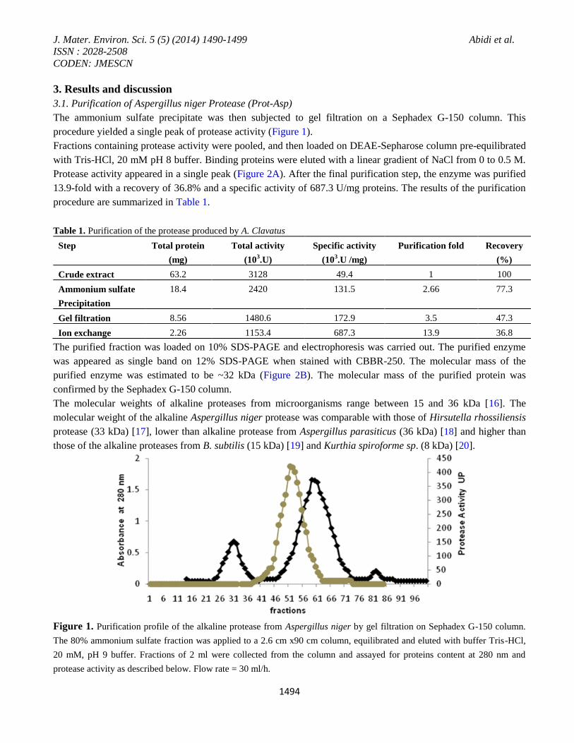
The ammonium sulfate precipitate was then subjected to gel filtration on a Sephadex G-150 column. This
procedure yielded a single peak of protease activity (Figure 1).
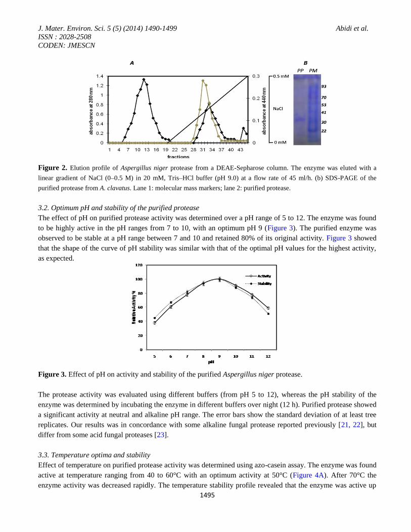
Fractions containing protease activity were pooled, and then loaded on DEAE-Sepharose column pre-equilibrated
with Tris-HCl, 20 mM pH 8 buffer. Binding proteins were eluted with a linear gradient of NaCl from 0 to 0.5 M.
Protease activity appeared in a single peak (Figure 2A). After the final purification step, the enzyme was purified
13.9-fold with a recovery of 36.8% and a specific activity of 687.3 U/mg proteins. The results of the purification
procedure are summarized in Table 1.
Table 1. Purification of the protease produced by A. Clavatus
Step Total protein
(mg)
Total activity
(103.U)
Specific activity
(103.U /mg)
Purification fold Recovery
(%)
Crude extract 63.2 3128 49.4 1 100
Ammonium sulfate
Precipitation
18.4 2420 131.5 2.66 77.3
Gel filtration 8.56 1480.6 172.9 3.5 47.3
Ion exchange 2.26 1153.4 687.3 13.9 36.8
The purified fraction was loaded on 10% SDS-PAGE and electrophoresis was carried out. The purified enzyme
was appeared as single band on 12% SDS-PAGE when stained with CBBR-250. The molecular mass of the
purified enzyme was estimated to be ~32 kDa (Figure 2B). The molecular mass of the purified protein was
confirmed by the Sephadex G-150 column.
The molecular weights of alkaline proteases from microorganisms range between 15 and 36 kDa [16]. The
molecular weight of the alkaline Aspergillus niger protease was comparable with those of Hirsutella rhossiliensis
protease (33 kDa) [17], lower than alkaline protease from Aspergillus parasiticus (36 kDa) [18] and higher than
those of the alkaline proteases from B. subtilis (15 kDa) [19] and Kurthia spiroforme sp. (8 kDa) [20].
Figure 1. Purification profile of the alkaline protease from Aspergillus niger by gel filtration on Sephadex G-150 column.
The 80% ammonium sulfate fraction was applied to a 2.6 cm x90 cm column, equilibrated and eluted with buffer Tris-HCl,
20 mM, pH 9 buffer. Fractions of 2 ml were collected from the column and assayed for proteins content at 280 nm and
protease activity as described below. Flow rate = 30 ml/h.
J. Mater. Environ. Sci. 5 (5) (2014) 1490-1499 Abidi et al.
ISSN : 2028-2508
CODEN: JMESCN
1495
Figure 2. Elution profile of Aspergillus niger protease from a DEAE-Sepharose column. The enzyme was eluted with a
linear gradient of NaCl (0–0.5 M) in 20 mM, Tris–HCl buffer (pH 9.0) at a flow rate of 45 ml/h. (b) SDS-PAGE of the
purified protease from A. clavatus. Lane 1: molecular mass markers; lane 2: purified protease.
3.2. Optimum pH and stability of the purified protease
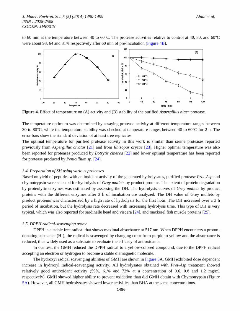
The effect of pH on purified protease activity was determined over a pH range of 5 to 12. The enzyme was found
to be highly active in the pH ranges from 7 to 10, with an optimum pH 9 (Figure 3). The purified enzyme was
observed to be stable at a pH range between 7 and 10 and retained 80% of its original activity. Figure 3 showed
that the shape of the curve of pH stability was similar with that of the optimal pH values for the highest activity,
as expected.
Figure 3. Effect of pH on activity and stability of the purified Aspergillus niger protease.
The protease activity was evaluated using different buffers (from pH 5 to 12), whereas the pH stability of the
enzyme was determined by incubating the enzyme in different buffers over night (12 h). Purified protease showed
a significant activity at neutral and alkaline pH range. The error bars show the standard deviation of at least tree
replicates. Our results was in concordance with some alkaline fungal protease reported previously [21, 22], but
differ from some acid fungal proteases [23].
3.3. Temperature optima and stability
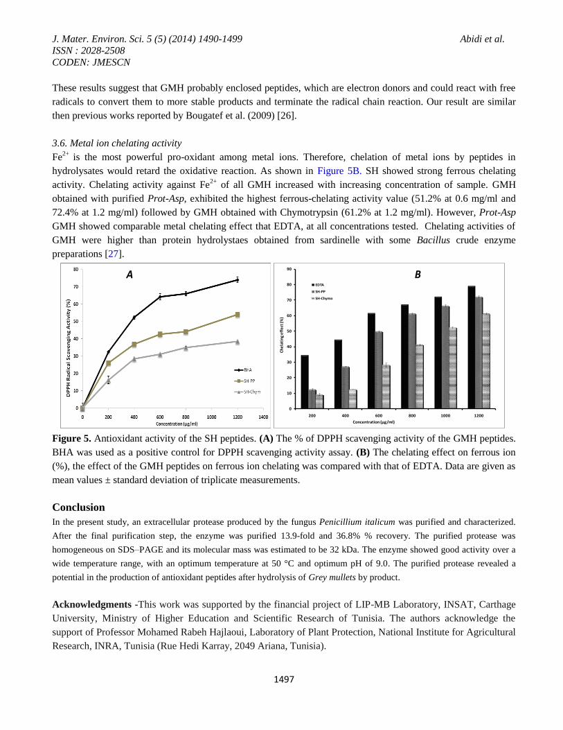
Effect of temperature on purified protease activity was determined using azo-casein assay. The enzyme was found
active at temperature ranging from 40 to 60°C with an optimum activity at 50°C (Figure 4A). After 70°C the
enzyme activity was decreased rapidly. The temperature stability profile revealed that the enzyme was active up
J. Mater. Environ. Sci. 5 (5) (2014) 1490-1499 Abidi et al.
ISSN : 2028-2508
CODEN: JMESCN
1496
to 60 min at the temperature between 40 to 60°C. The protease activities relative to control at 40, 50, and 60°C
were about 98, 64 and 31% respectively after 60 min of pre-incubation (Figure 4B).
Figure 4. Effect of temperature on (A) activity and (B) stability of the purified Aspergillus niger protease.
The temperature optimum was determined by assaying protease activity at different temperature ranges between
30 to 80°C, while the temperature stability was checked at temperature ranges between 40 to 60°C for 2 h. The
error bars show the standard deviation of at least tree replicates.
The optimal temperature for purified protease activity in this work is similar than serine proteases reported
previously from Aspergillus clvatus [21] and from Rhizopus oryzae [23], Higher optimal temperature was also
been reported for proteases produced by Botrytis cinerea [22] and lower optimal temperature has been reported
for protease produced by Penicillium sp. [24].
3.4. Preparation of SH using various proteases
Based on yield of peptides with antioxidant activity of the generated hydrolysates, purified protease Prot-Asp and
chymotrypsin were selected for hydrolysis of Grey mullets by product proteins. The extent of protein degradation
by proteolytic enzymes was estimated by assessing the DH. The hydrolysis curves of Grey mullets by product
proteins with the different enzymes after 3 h of incubation are analyzed. The DH value of Grey mullets by
product proteins was characterized by a high rate of hydrolysis for the first hour. The DH increased over a 3 h
period of incubation, but the hydrolysis rate decreased with increasing hydrolysis time. This type of DH is very
typical, which was also reported for sardinelle head and viscera [24], and mackerel fish muscle proteins [25].
3.5. DPPH radical-scavenging assay
DPPH is a stable free radical that shows maximal absorbance at 517 nm. When DPPH encounters a proton-
donating substance (H+), the radical is scavenged by changing color from purple to yellow and the absorbance is
reduced, thus widely used as a substrate to evaluate the efficacy of antioxidants.
In our test, the GMH reduced the DPPH radical to a yellow-colored compound, due to the DPPH radical
accepting an electron or hydrogen to become a stable diamagnetic molecule.
The hydroxyl radical scavenging abilities of GMH are shown in Figure 5A. GMH exhibited dose dependent
increase in hydroxyl radical-scavenging activity. All hydrolysates obtained with Prot-Asp treatment showed
relatively good antioxidant activity (59%, 61% and 72% at a concentration of 0.6, 0.8 and 1.2 mg/ml
respectively). GMH showed higher ability to prevent oxidation than did GMH obtain with Chymotrypsin (Figure
5A). However, all GMH hydrolysates showed lower activities than BHA at the same concentrations.
J. Mater. Environ. Sci. 5 (5) (2014) 1490-1499 Abidi et al.
ISSN : 2028-2508
CODEN: JMESCN
1497
These results suggest that GMH probably enclosed peptides, which are electron donors and could react with free
radicals to convert them to more stable products and terminate the radical chain reaction. Our result are similar
then previous works reported by Bougatef et al. (2009) [26].
3.6. Metal ion chelating activity
Fe2+
is the most powerful pro-oxidant among metal ions. Therefore, chelation of metal ions by peptides in
hydrolysates would retard the oxidative reaction. As shown in Figure 5B. SH showed strong ferrous chelating
activity. Chelating activity against Fe2+
of all GMH increased with increasing concentration of sample. GMH
obtained with purified Prot-Asp, exhibited the highest ferrous-chelating activity value (51.2% at 0.6 mg/ml and
72.4% at 1.2 mg/ml) followed by GMH obtained with Chymotrypsin (61.2% at 1.2 mg/ml). However, Prot-Asp
GMH showed comparable metal chelating effect that EDTA, at all concentrations tested. Chelating activities of
GMH were higher than protein hydrolystaes obtained from sardinelle with some Bacillus crude enzyme
preparations [27].
Figure 5. Antioxidant activity of the SH peptides. (A) The % of DPPH scavenging activity of the GMH peptides.
BHA was used as a positive control for DPPH scavenging activity assay. (B) The chelating effect on ferrous ion
(%), the effect of the GMH peptides on ferrous ion chelating was compared with that of EDTA. Data are given as
mean values ± standard deviation of triplicate measurements.
Conclusion
In the present study, an extracellular protease produced by the fungus Penicillium italicum was purified and characterized.
After the final purification step, the enzyme was purified 13.9-fold and 36.8% % recovery. The purified protease was
homogeneous on SDS–PAGE and its molecular mass was estimated to be 32 kDa. The enzyme showed good activity over a
wide temperature range, with an optimum temperature at 50 °C and optimum pH of 9.0. The purified protease revealed a
potential in the production of antioxidant peptides after hydrolysis of Grey mullets by product.
Acknowledgments -This work was supported by the financial project of LIP-MB Laboratory, INSAT, Carthage
University, Ministry of Higher Education and Scientific Research of Tunisia. The authors acknowledge the
support of Professor Mohamed Rabeh Hajlaoui, Laboratory of Plant Protection, National Institute for Agricultural
Research, INRA, Tunisia (Rue Hedi Karray, 2049 Ariana, Tunisia).
0
10
20
30
40
50
60
70
80
90
200 400 600 800 1000 1200
Ch
ela
tin
g e
ffe
ct (%
)
Concentration (µg/ml)
EDTA
SH-PP
SH-Chymo
A B
J. Mater. Environ. Sci. 5 (5) (2014) 1490-1499 Abidi et al.
ISSN : 2028-2508
CODEN: JMESCN
1498
References 1. Zanphorlin L.M., Cabral H., Arantes E., Assis D., Juliano L., Juliano M.A., Da-Silva R., Gomes E., Bonilla-
Rodriguez G.O. Process Bioch. 46 (2011) 2137–2143.
2 Rao M.B., Tanksale AM., Ghatge M.S., Deshpande V.V. Microbiol. Mol. Biol. Rev. 62 (1998) 597–635.
3 Beg, K.B. Gupta R. Proc. Biochem. 39 (2003) 2003–2009.
4 Devi K., Rasheedha Banu M. A., Gnanaprabhal G.R., Pradeep B.V., Palaniswamy M. Ind J. Sci. Technol. 1
(2008) 1–6.
5. Laxman S., Santosh S., Dhule V., Jogdand V. Bioch Engineering J. 39 (2008) 510–515.
6. Kechaou E.M., Dumay J., Donnay-Moreno C., Jaouen P., Gouygou J-P., Bergé J.P., Ben Ama R. J. Biosc
and Bioeng. 107 (2009.) 158–164.
7. Da-Yong Z., Bei-Wei Z., Lu Q., Hai-Tao W., Dong-Mei L., Jing-Feng Y., Yoshiyuki M. Food Bioproducts
Processing. 9 (2012) 148–154.
8. Abidi F., Chobert JM., Haertlé T., Marzouki M.N. Process Biochem 46 (2011) 2301–2310.
9. Bradford M.M. Analytical Biochem. 72 (1976) 248–254.
10. Segers R., Butt T.M., Kerry B.R., Peberdy J.F. Microbiology 140 (1994) 2715–2723.
11. Phillips P.K., Prior D., Awes B.D. J Clin Pathol. 3 (1984) 329–331.
12. Laemmli U.K. Nature. 257 (1970) 680–685.
13. Adler-Nissen, J. Elsevier Applied Science Publications, New York, (1986) pp: 57-131.
14. Yen G., Wu J. Food Chem. 65 (1999) 375–379.
15. Decker E.A., Welch B. J. Agric. Food Chem. 38 (1990) 674–677.
16. Kazan D., Denizci A.A., Mine N., Öner K., Erarslan A., J. Ind Microb Biot. 32 (2005) 335–340.
17. Wang B., Liu X., Wu W., Liu X., Li S. Microbiological Research. 164 (2009) 665–673.
18. Anitha, T.S., Palanivelu. P. Protein Expression Purif. 88 (2013) 214–220.
19. Adinarayana K., Ellaiah P., Prasad D.S., AAPS Pharm Sci Technol. 4 (2003) 1–9.
20. Steele D.B., Fiske M.J., Steele B.P., Kelley V.C. Enzyme Microb Technol. 14 (1992) 358–360.
21. Hajji M., Kanoun S., Nasri M., Gharsallah N. Process Biochem. 14 (2007) 791–797.
22. Abidi F, Chobert JM, Haertlé T, Marzouki MN. Process Biochem. 46 (2011) 2301–2310
23. Sushil K, Neeru SS, Mukh RS, Randhir S. Process Biochem. 40 (2005) 1701–1705.
24. Germano S., Pandey A., Osaku C.A., Rocha S.N. Soccol C.R. Enz. Microb Technol. 32 (2003) 246–251.
25. Bougatef A., Nedjar-Arroume N., Manni L., Ravallec R., Barkia A., Guillochon D., Nasri M. Food Chem.
118 (2010) 559–565.
26. Abidi F., Aissaoui N., Gaudin J-C., Chobert J-M., Haertlé T., Marzouki M.N. Appl Biochem Biotechnol. 170
(2013) 231–47.
27. Ben Khaled H., Ktari N., Ghorbel-Bellaaj O., Jridi M., Lassoued I., Nasri M. J. Food Sci. Technol. 14 (2012)
60–68.
(2014) http://www.jmaterenvironsci.com/