punch, shave, snip, excise, freeze,...
TRANSCRIPT

Punch, Shave, Snip, Excise, Freeze, Desiccate? When and How….to do What, Where!
Ted Rosen, MD Professor of Dermatology
Baylor College of Medicine Houston, Texas
Conflict of Interest Disclosure: NONE

Which procedure is best….
•What’s your goal?
•To remove
•To destroy
•To biopsy

Which procedure is best….
•What’s your goal?
•To remove: Always send to pathology
•To destroy: Blind procedure (no histology)
•To biopsy: Adequate width and depth

Width Depth
Cryosurgery None None
Electrodesiccation None None
Snip Yes Maybe
Shave Yes Maybe
Punch Maybe Maybe
Excise Optimum Optimum

Width Depth
Cryosurgery None None
Electrodesiccation None None
Snip Yes Maybe
Shave Yes Maybe
Punch Maybe Maybe
Excise Optimum Optimum
Designed to destroy things Blind procedures: No histological confirmation (unless prior biopsy) Painful, Potential cosmetic abnormality as residual (dyschromia) Particularly difficult in skin of color

Cryosurgery and Electrodesiccation
• Real life examples of how a blind procedure can lead to disaster!
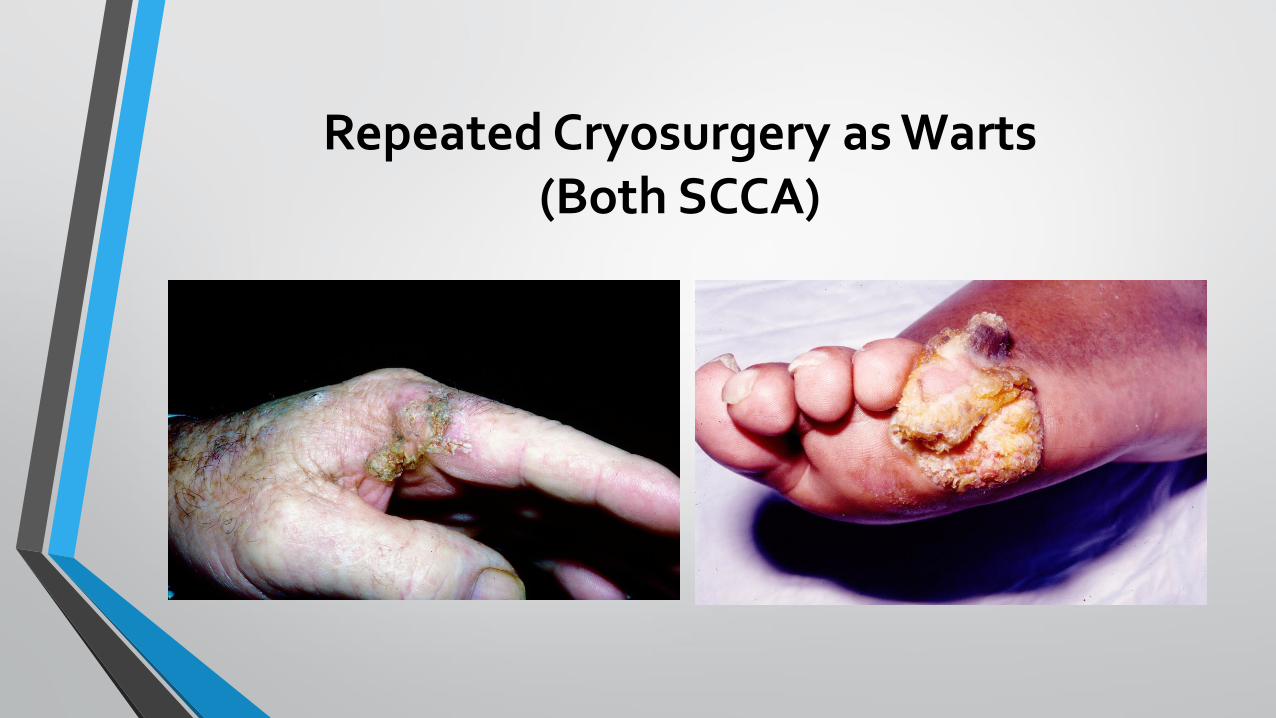
Repeated Cryosurgery as Warts (Both SCCA)

Repeated Cryosurgery as Wart (Both Melanoma)

Width Depth
Cryosurgery None None
Electrodesiccation None None
Snip Yes Maybe
Shave Yes Maybe
Punch Maybe Maybe
Excise Optimum Optimum
Designed to remove things, completely or partially Histological confirmation, but may be insufficient Painful, Potential cosmetic abnormality as residual (depression) Less difficult in skin of color

Snip or Shave May Be Insufficient

Snip or Shave May Be Insufficient
Cutaneous horn may be sign of seborrheic keratosis or wart AND actinic keratosis and squamous cell carcinoma NEED DEPTH to discern nature of underlying pathology

Width Depth
Cryosurgery None None
Electrodesiccation None None
Snip Yes Maybe
Shave Yes Maybe
Punch Maybe Maybe
Excise Optimum Optimum
Designed to sample (biopsy) or remove things Histological confirmation, occasional sample error with punch Painful, Potential cosmetic abnormality as residual (depression) Best in skin of color
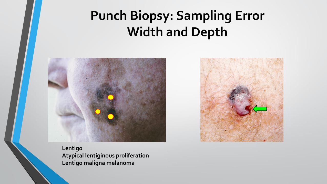
Punch Biopsy: Sampling Error Width and Depth
Lentigo Atypical lentiginous proliferation Lentigo maligna melanoma

Cryosurgery
• Almost always done with liquid nitrogen (about -196oF)
• Almost always done with hand-held sealed canister (vrs Q-tip)
• Warts
• Seborrheic keratoses
• Actinic keratosis
• Adjunct with: keloids, dermatofibroma


Cryosurgery for Actinic Keratoses
Medically appropriate? Risks: Pain, AEs

• Spray until lesion solid white
• Continuous (wider) vrs Intermittent (deeper) spray
• Should take ~20-30 seconds to thaw
• How long to freeze? (EU: 20-40sec; USA much less)
• Thick lesions: 2 freeze-thaw cycles

Cryosurgery
• ADVANTAGES
• Rapid
• Inexpensive
• Easy to learn and perform
• No local anesthesia
• “Good” cosmesis
• Scar improves with time
• DISADVANTAGES
• “Blind” procedure
• Pain and blistering
• Prolonged healing time
• Wound care may be required
• Dyschromia
• Atrophic scar formation

Cryosurgery “Checkerboard”

Cryosurgery Blistering

Cryosurgery Dyschromia

Hemostatic Cryosurgery
Immerse hemostat or needle holder Grasp skin tag (filiform wart) x 10-20 seconds

Electrodesiccation
• Electric current dehydrates tissue
• May use thick or very thin needle
• Small facial lesions: syringoma, DPN, sebaceous hyperplasia, spider/cherry angioma, telangiectasia, venous lake
• Small flat or filiform warts, Skin tags
• Use with curettage: Seborrheic keratosis (before), NMSC (after)

Electrodesiccation
• ADVANTAGES
• Rapid and Inexpensive
• Easy to learn and perform
• Inherent hemostasis
• Local anesthesia optional
• “Good” cosmesis
• Scar improves with time
• DISADVANTAGES
• “Blind” procedure
• Pain and crusting
• Long healing time occasional
• ?Problematic with pacemaker
• Dyschromia potential
• Atrophic scar formation

Examples of Electrodesiccation
Seborrheic keratosis
Facial warts
Then removal of charred lesion by
curettage

Examples of Electrodesiccation
Venous Lake Cherry Angioma

Pacemaker
Thermal Cautery
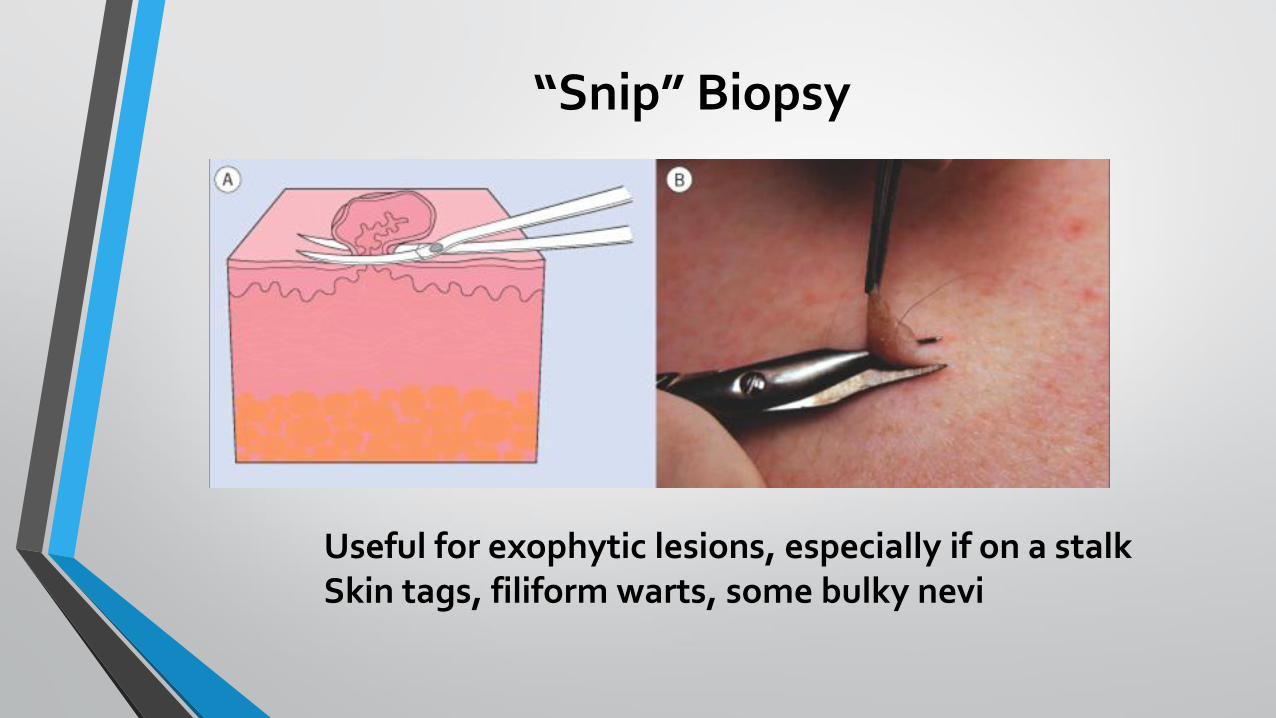
“Snip” Biopsy
Useful for exophytic lesions, especially if on a stalk Skin tags, filiform warts, some bulky nevi

Snip: Skin Tags!


“Snip” Removal
• ADVANTAGES
• Rapid and Inexpensive
• Easy to learn and perform
• Local anesthesia optional (Recommended)
• Specimen available for histologic examination
• “Good” cosmesis
• DISADVANTAGES
• Need for hemostasis afterward
• May miss diagnostic base of the lesion
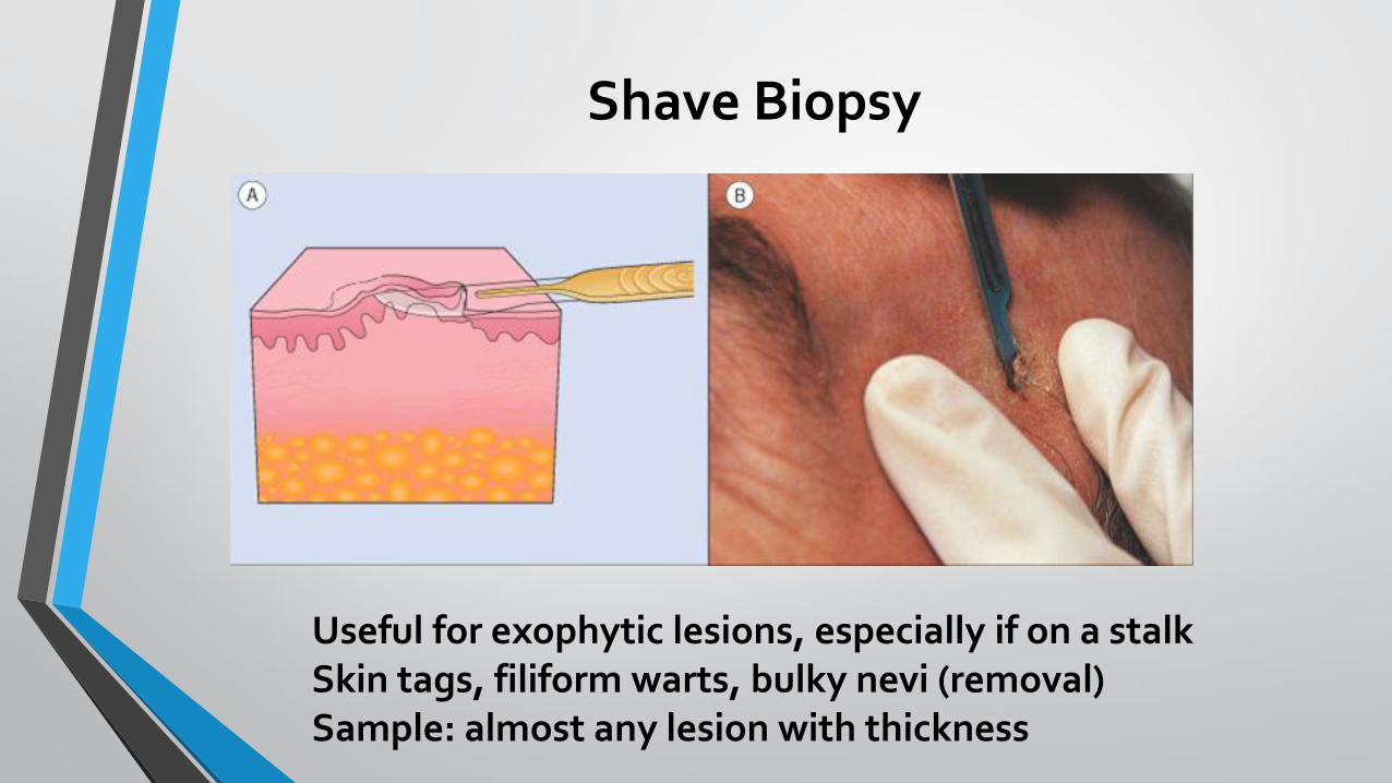
Shave Biopsy
Useful for exophytic lesions, especially if on a stalk Skin tags, filiform warts, bulky nevi (removal) Sample: almost any lesion with thickness


Shave Removal
• ADVANTAGES
• Rapid and Inexpensive
• Easy to learn and perform
• Specimen available for histologic examination
• “Good” cosmesis
• DISADVANTAGES
• Need for local anesthesia
• Need for hemostasis afterward
• May miss diagnostic base of the lesion
• May be unable to judge depth or thickness of lesion**

Snip or Shave May Be Insufficient Need to Obtain Depth for Diagnosis

Use Shaving Blade, Bent, to “Scoop” Shave

“DermaBlade”

Where a Shave Biopsy is NOT Optimum
Nodular Melanoma

Intradermal Injection
x x
x x
x
Ring Block

Anesthesia
• TIPS TO REDUCE PAIN
• Small bore needle (30g)
• Inject slowly
• Warm local anesthetic to near skin temperature
• Buffer with 8.4% sodium bicarbonate (9:1 ratio)
• This reduces shelf life
• Ann Emerg Med 21:16, 1992 Ann Emerg Med 26:121, 1995

Anesthesia • TOXICITY
• Rare with small procedures
• Most common: vaso-vagal rxn (diaphoresis, bradycardia)
• CNS (accidental intravascular)
• Metallic taste, tinnitus, confusion
• Systemic vasoconstriction: BP
• Excess local vasoconstriction
• Warm area
• Arrhythmia
• Allergic rxn: diphenhydramine and/or steroids

Milia (Tiny Cysts)

Milia: Removal


Punch Biopsy (Excision)

Punch Biopsy (Excision)
May obtain hemostasis: Pressure Drysol Monsel’s solution Suture (Punch > 3mm) • Use 6-0 Nylon • 1 suture 3mm • 2 sutures 4mm

Pinching stops bleeding!

Punch Biopsy: Size Varies Larger size: Wider and Deeper Specimen
1mm
2mm
3mm
3.5mm 4mm
4.5mm
5mm
10mm
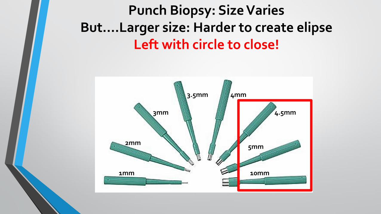
Punch Biopsy: Size Varies But….Larger size: Harder to create elipse
Left with circle to close!
1mm
2mm
3mm
3.5mm 4mm
4.5mm
5mm
10mm

Post-Biopsy Instructions
• Place Bandaid + Ointment
• Vaseline is likely sufficient
• Antibiotic ointment: OTC sufficient (Ear, Nose, Mouth, Eye)
• Bacitracin®, Polysporin®
• Avoid Neosporin® (neomycin sensitivity)
• Latex-free bandaid available
• Wear bandage for first 24-48 hours
• Replace with new cover + ointment daily for one week
• Report: bleeding, pain, swelling, pus
• Then may wash with soap and water, but clean gently BID
• RTC as appropriate for suture removal

Punch Biopsy or Removal
• ADVANTAGES
• Rapid and Inexpensive
• Easy to learn and perform
• Specimen available for histologic examination
• “Good” cosmesis
• DISADVANTAGES
• Need for local anesthesia
• Need for hemostasis afterward
• May miss diagnostic base of the lesion unless punch to hilt
• Risk of hemorrhage, infection
• Risk of scar formation
Punch Biopsy: WHERE?

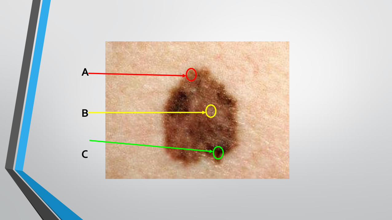
Where to Perform the Biopsy
• Tumor: thickest or most atypical appearing area of lesion
• Annular patch or plaque: active or advancing edge of lesion
• Blister: blister and rim of normal skin
• Vasculitis: newest lesion
• Other plaque lesions: older or most representative portion
• Everything else: right from middle!
A B C

A B C
ANOTHER GOOD OPTION

Where Would You Biopsy?
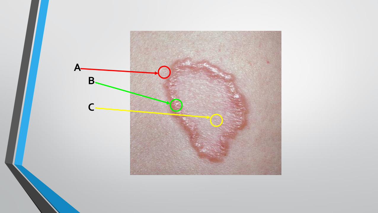
A B C

What’s The Problem With This Biopsy?
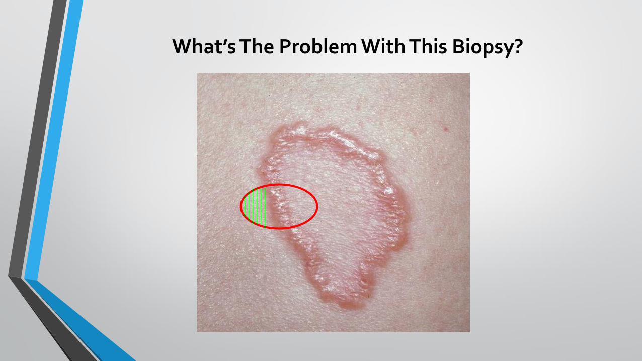
What’s The Problem With This Biopsy?

What’s The Problem With This Biopsy?
Bisect biopsy!
All “cuts” will be parallel to your bisection

What’s The Problem With This Biopsy?
Bisect biopsy!
All “cuts” will be parallel to your bisection

A
B
C

A
B
C

Additional Considerations
•Multiple biopsies? (multiple morphologies)
• Immunofluorescence needed?
• Special media needed for preservation of specimen? (IF, culture)
• Special stains required? (ASK for them)
• Is a culture being sent as well?
You can “split” a specimen (> 4mm)


Multiple Biopsies Reasonable
MULTIPLE MORPHOLOGIES

Surgical Excision

Relaxed skin tension lines

Dimensions
• Length to width ratio: 3:1 to 4:1
• Apical angles = 300

Elliptical Excision (Biopsy)
• ADVANTAGES
• Specimen available for histologic examination
• May allow total removal in one surgical session
• Can check for clear margins
• Facilitates good functional outcome
• DISADVANTAGES
• Requires more skill
• Need for local anesthesia
• Need for hemostasis before closure is accomplished
• Risk of hemorrhage, infection
• Risk of scar formation

Selection of Procedure: Summary
• Cryosurgery or Electrodesiccation
• Diagnosis is not in doubt
• Superficial destruction is goal
• Shave or Snip biopsy
• Lesion elevated above skin surface
• Punch biopsy
• Lesion has depth: dermal, subcutaneous
• Need precise depth of lesion
• Excisional biopsy
• When more than a punch biopsy is needed
• Remove lesion entirely
• Obtain multiple areas (pathology + nearby)