pulmonary manifestations of systemic lupus erythematosus...systemic lupus erythematosus abdul...
TRANSCRIPT
14
Pulmonary Manifestations of Systemic Lupus Erythematosus
Abdul Ghafoor Gari, Amr Telmesani and Raad Alwithenani Umm Al-Qura University
Saudi Arabia
1. Introduction
Systemic lupus erythematosus (SLE) is an autoimmune disease that primarily affects women of childbearing age with 10:1 female to male ratio.(Siegel & Lee, 1973) Any organ can be affected by SLE; pulmonary involvement is usually in the latter course of the disease.(Haupt et al., 1981; Orens et al., 1994; Quadrelli et al., 2009) It is important to note that lung involvement is proportionately more common in men.(Kamen & Strange, 2010) Any part of the pulmonary system can be affected including airways, lung parenchyma, pulmonary vasculature, pleura and diaphragm.(Gross et al., 1972; Haupt et al., 1981; Kamen & Strange, 2010; Orens et al., 1994; Quadrelli et al., 2009; Weinrib et al., 1990) If SLE develops after age 49 years, it has a higher incidence of serositis, pulmonary involvement and mortality.(Boddaert et al., 2004) It is difficult to find out the true prevalence of pulmonary complications of SLE since many cases are due to infections.(Kamen & Strange, 2010) A recent autopsy study of 90 patients diagnosed with SLE, according to the American College of Rheumatology, pleuropulmonary involvement occurred in 98% of the autopsies. (Quadrelli et al., 2009) The most frequent findings were pleuritis (78%), bacterial infections (58%), alveolar hemorrhage (26%), followed by distal airway alterations (21%), opportunistic infections (14%) and pulmonary thromboembolism (8%), both acute and chronic.(Quadrelli et al., 2009) In a larger series, 25% of patients with SLE had clinical and/or radiographic evidence of pulmonary involvement.(Pego-Reigosa et al., 2009)
2. Clinical features
SLE can affect the lungs in many ways. In the next section we will review the pulmonary diseases associated with SLE according to the anatomic involvement.
2.1 Pleural diseases Pleuritis is the most common pleuropulmonary manifestation of SLE.(Orens et al., 1994) It is the initial manifestation in 5% to 10%.(Winslow et al., 1958) Symptoms of pleuritis are present in 45% to 60% of patients with SLE and may be associated with pleural effusion.(Good et al., 1983; Orens et al., 1994; Pines et al., 1985) Pleural effusion in SLE tends to be bilateral, small to moderate in size; however, large effusions may occur.(Bouros et al., 2008) Typical presentation of pleural involvement is pleuritic chest pain (pain that increases with inspiration), dyspnea, and fever. Physical examination may reveal pleural friction rub
www.intechopen.com
Systemic Lupus Erythematosus
314
and signs of pleural effusion. Chest X-ray shows blunting of the costophrenic angle. Some patients may have asymptomatic pleural effusion. Other causes of pleural effusion such as parapneumonic effusion, pulmonary embolism, and heart failure need to be ruled out. Pleural fluid analysis is needed to rule out other etiologies and to confirm the diagnosis of lupus pleuritis. Pleural fluid is exudative (elevated pleural fluid protein and lactate dehydrogenase levels) when analyzed. Cell counts are elevated with predominance of lymphocytes or neutrophils. Pleural fluid glucose level is low, but not as low as in patients with rheumatoid arthritis. Special tests reveal low pleural complement level and positive anti-nuclear antibody (ANA). These tests are not sensitive enough to rule out lupus pleuritis when tests are negative. (Hunder et al., 1972; Small et al., 1982) Pleural fluid ANA titer ≥ 1:160 and pleural fluid / serum ANA ratio of ≥ 1 strongly support the diagnosis.(Good et al., 1983) The finding of lupus erythematosus cells in pleural fluid confirms the diagnosis; however this test is rarely performed.(Kamen & Strange, 2010) Pleural biopsy is rarely needed, if done it will show a peculiar immunofluorescent pattern characterized by staining of nuclei with anti-IgG, anti-IgM and anti-C3.(Pertschuk et al., 1977) Patients with pleural disease usually respond to nonsteriodal anti-inflammatory drugs (NSAIDs). Low doses of oral glucocorticoids hasten the resolution. Small asymptomatic effusions usually resolve without treatment.(Winslow et al., 1958) NSAIDs are sufficient for mild cases; for severe cases or for patients on steroids, giving higher doses of steroid is required.( Orens et al., 1994; Wiedemann & Matthay, 1989) In refractory pleural effusions tetracycline or talc pleurodesis can be an alternative option.( Gilleece et al., 1988; Kaine, 1985; McKnight et al., 1991)
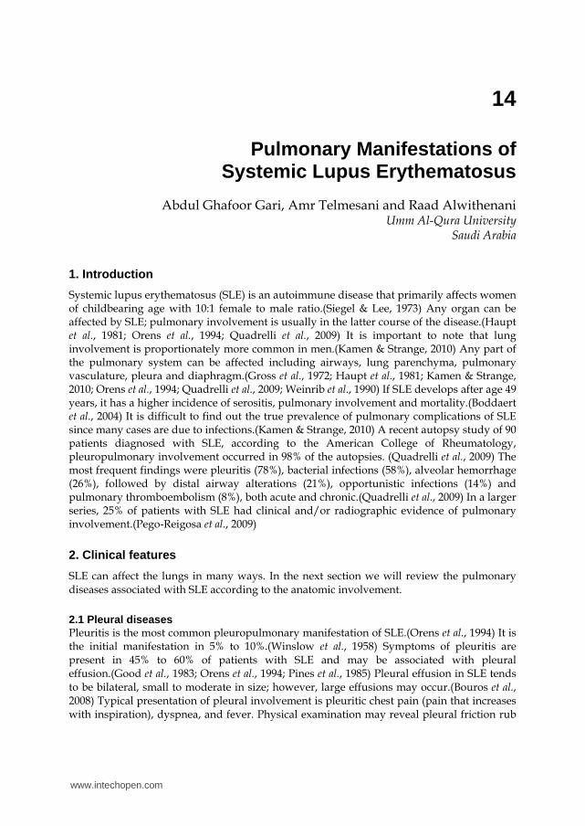
2.2 Parenchymal lung disease 2.2.1 Acute lupus pneumonitis Acute lupus pneumonitis (ALP) is an uncommon but well recognized complication of SLE. There is some controversy over the definition ALP.(Swigris et al., 2008) In two recent series, the prevalence of ALP in patients with SLE was 2% to 8%.( Kim et al., 2000; Mochizuki et al., 1999) It is difficult to estimate the exact prevalence given the significant clinical and radiological overlap between ALP, bacterial pneumonia and alveolar hemorrhage. ALP tends to affect younger patients and those with recent diagnosis of SLE. In 50% of patients with SLE who develop ALP, the pulmonary complication is the initial presentation of lupus.(Matthay et al., 1975) Clinical presentation includes abrupt onset of fever, cough, dyspnea, pleuritic chest pain and occasionally hemoptysis.(Matthay et al., 1975) Physical examination usually reveals signs of hypoxia and bibasilar crackles. Radiographic findings include bilateral alveolar infiltrates with predominance in lower lung fields (figure 1). Pleural effusion occurs in half of the cases.(Matthay et al., 1975) Rarely the initial chest radiograph may be normal or may show pulmonary nodules.(Susanto & Peters, 1997) CT scan of the chest may show diffuse ground glass opacities and areas of consolidation.(Swigris et al., 2008) A fulminant form of ALP may occur during pregnancy.(Comer et al., 1996) The clinical and radiographic features are not specific. Other causes of alveolar infiltrates like infectious pneumonia, alveolar hemorrhage, pulmonary edema, and organizing pneumonia should be considered. It is important to rule out infectious complications. Many of these patients are on systemic steroids and other immunosuppressive medications and are at increased risk of opportunistic infections. Early bronchoscopy and bronchoalveolar lavage (BAL) with or without transbronchial biopsy is mandatory in most cases. BAL should be sent for cell count and differential, bacterial, fungal
www.intechopen.com
Pulmonary Manifestations of Systemic Lupus Erythematosus
315
and viral culture, cytology and for Pneumocystis jiroveci stain. Occasionally a thoracoscopic lung biopsy may be needed. The pathological findings are not specific. The most common findings include diffuse alveolar damage (DAD) with or without alveolar hemorrhage and capillaritis.(Harvey et al., 1954; Keane & Lynch, 2000) Other pathologic features include alveolar wall injury, alveolar edema, hyaline membrane formation, immunoglobulin and complement deposition. There seems to be some association between ALP and anti-Ro/SSa antibodies. One study showed that patients with SLE and pulmonary complications had an 81% positive result for anti-Ro/SSa antibodies, while patients without pulmonary involvement had a 38% positive antibody.(Boulware & Hedgpeth, 1989) A more recent review confirmed this association (Mochizuki et al., 1999) The high frequency of anti-Ro/SSa antibodies raises the possibility of their role in the pathogenesis of ALP.(Cheema & Quismorio, 2000) Prognosis is poor, with mortality reaching up to 50% as reported in an old study.(Matthay et al., 1975) The outcome is worse if ALP occurs postpartum.(Matthay et al., 1975) Eosinophilia or neutrophilia on BAL carries worse prognosis than lymphocytosis.(Witt et al., 1996) Because infectious causes can’t be ruled out, empiric broad spectrum antibiotics should be started immediately and continued until infection is excluded. There are no randomized clinical trials for the treatment of ALP, however it is agreed on that the main treatment is systemic corticosteroids (prednisone 1-1.5 mg/kg/day). If no adequate response within 72 hours, treatment should be with intravenous pulse steroids (1g methylprednisolone daily for three days).(Kamen & Strange, 2010) Additional immunosuppresants such as cyclophosphamide should also be considered. In patients refractory to corticosteroids, intravenous immunoglobulin, plasma exchange or rituximab can be of some help with very little evidence (Eiser & Shanies, 1994; Lim et al., 2006; Pego-Reigosa et al., 2009; Winder et al., 1993)
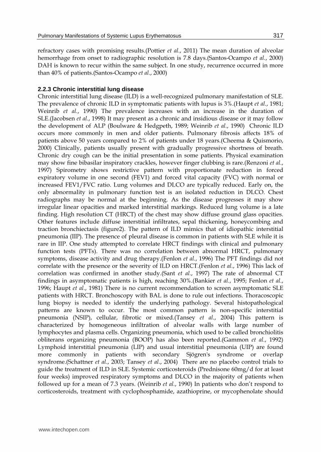
Fig. 1. Chest X-ray showing diffuse alveolar infiltrates in a patient with acute lupus pneumonitis
2.2.2 Diffuse alveolar hemorrhage Diffuse alveolar hemorrhage (DAH) is a rare complication of SLE. ( Badsha et al., 2004; Eagen et al., 1978; Zamora et al., 1997) Its prevalence among SLE patients ranges between
www.intechopen.com
Systemic Lupus Erythematosus
316
<2% and 5.4 % (Santos-Ocampo et al., 2000; Zamora et al., 1997), and the mortality rate ranges between 50% to 90%.( Erickson et al., 1994; Schwab et al., 1993) Usually it occurs in established disease, especially with lupus nephritis.(Zamora et al., 1997) Other extrapulmonary manifestations occur with variable degree.(Abud-Mendoza et al., 1985; Barile et al., 1997; Koh et al., 1997; Liu et al., 1998; Myers & Katzenstein, 1986; Schwab et al., 1993; Zamora et al., 1997) However it can occasionally be the initial presentation of SLE.(Zamora et al., 1997) Risk factors thought to be contributing to the development of DAH are higher titer of circulating anti-DNA antibody, active extra-pulmonary disease, and established SLE diagnosis.(Orens et al., 1994) Clinical presentation of patients with DAH is not specific; symptoms include acute shortness of breath, cough, hemoptysis and fever. The absence of hemoptysis doesn’t rule out DAH. In fact hemoptysis is only present in 54% of patients.(Santos-Ocampo et al., 2000) Fever is present in more than 80% of patients.(Santos-Ocampo et al., 2000) Signs of respiratory distress and hypoxia are noted upon physical examination. Chest radiograph shows bilateral alveolar infiltrates. Unilateral pulmonary infiltrates is noted in up to 18%.(Santos-Ocampo et al., 2000) CT imaging demonstrates new bilateral ground glass opacities and consolidation. Acute drop in hemoglobin is frequently encountered. In most series anemia was noted >90 of all episodes of DAH. (Abud-Mendoza et al., 1985; Barile et al., 1997; Koh et al., 1997; Liu et al., 1998; Myers & Katzenstein, 1986; Schwab et al., 1993; Zamora et al., 1997) If diffusion capacity for carbon monoxide (DLCO) is measured it will be elevated due to the excess hemoglobin in the alveolar spaces. An increase of DLCO by 30% or a value of >130% predicted suggest DAH in the right clinical setting.(Carette et al., 1984; Dweik et al., 1997; Ewan et al., 1976; Harmon & Leatherman, 1988; Leatherman et al., 1984; Young, 1989) Low complement level is found in more than 70% of all episodes of DAH.( Koh et al., 1997; Liu et al., 1998; Myers & Katzenstein, 1986; Santos-Ocampo et al., 2000; Schwab et al., 1993;) Magnetic resonant imaging (MRI) is another imaging study that can suggest the presence of blood in the alveoli given the paramagnetic effect of iron.(Hsu et al., 1992) BAL is mandatory to rule out infection and help in the diagnosis of DAH. BAL can confirm the diagnosis if bloody return increases with serial aliquots. BAL should be evaluated for the presence of hemosiderin-laden macrophages, their presence indicate alveolar hemorrhage. Transbronchial biopsy (TBBx) may be attempted in stable patients. Unfortunately many of these patients require ventilatory support and may not be able to sustain the complication of TBBx. Thoracoscopic lung biopsy is rarely needed. Two pathological patterns have been described. Bland hemorrhage is more common and occurs in 72% while capillaritis occurs in 14% of the times. Both pathological patterns are associated with intra-alveolar hemorrhage and hemosiderin–laden macrophages.(Myers & Katzenstein, 1986; Schwab et al., 1993b; Zamora et al., 1997) IgG , C3 or immune complexes deposition occurs in 50% of the cases.(Myers & Katzenstein, 1986) There are no randomized control trials addressing treatment options for DAH. Supportive care is highly valued since many of these patients end up in the intensive care unit requiring mechanical ventilation. The most acceptable regimen include pulse intravenous steroids (methylprednisolone 1gm per day for three days) followed by 1mg/kg of oral prednisone plus intravenous cyclophosphamide every four weeks.(Schwab et al., 1993a; Swigris et al., 2008) DAH is one of the few indications where plasmapharesis has been shown to be effective, especially in refractory cases. (Erickson et al., 1994; Santos-Ocampo et al., 2000) Plasmapharesis may improve survival in patients who failed treatment with high dose steroids and cyclophosphamide.(Erickson et al., 1994) More recently rituximab has been used in
www.intechopen.com
Pulmonary Manifestations of Systemic Lupus Erythematosus
317
refractory cases with promising results.(Pottier et al., 2011) The mean duration of alveolar hemorrhage from onset to radiographic resolution is 7.8 days.(Santos-Ocampo et al., 2000) DAH is known to recur within the same subject. In one study, recurrence occurred in more than 40% of patients.(Santos-Ocampo et al., 2000)
2.2.3 Chronic interstitial lung disease Chronic interstitial lung disease (ILD) is a well-recognized pulmonary manifestation of SLE. The prevalence of chronic ILD in symptomatic patients with lupus is 3%.(Haupt et al., 1981; Weinrib et al., 1990) The prevalence increases with an increase in the duration of SLE.(Jacobsen et al., 1998) It may present as a chronic and insidious disease or it may follow the development of ALP (Boulware & Hedgpeth, 1989; Weinrib et al., 1990) Chronic ILD occurs more commonly in men and older patients. Pulmonary fibrosis affects 18% of patients above 50 years compared to 2% of patients under 18 years.(Cheema & Quismorio, 2000) Clinically, patients usually present with gradually progressive shortness of breath. Chronic dry cough can be the initial presentation in some patients. Physical examination may show fine bibasilar inspiratory crackles, however finger clubbing is rare.(Renzoni et al., 1997) Spirometry shows restrictive pattern with proportionate reduction in forced expiratory volume in one second (FEV1) and forced vital capacity (FVC) with normal or increased FEV1/FVC ratio. Lung volumes and DLCO are typically reduced. Early on, the only abnormality in pulmonary function test is an isolated reduction in DLCO. Chest radiographs may be normal at the beginning. As the disease progresses it may show irregular linear opacities and marked interstitial markings. Reduced lung volume is a late finding. High resolution CT (HRCT) of the chest may show diffuse ground glass opacities. Other features include diffuse interstitial infiltrates, sepal thickening, honeycombing and traction bronchiectasis (figure2). The pattern of ILD mimics that of idiopathic interstitial pneumonia (IIP). The presence of pleural disease is common in patients with SLE while it is rare in IIP. One study attempted to correlate HRCT findings with clinical and pulmonary function tests (PFTs). There was no correlation between abnormal HRCT, pulmonary symptoms, disease activity and drug therapy.(Fenlon et al., 1996) The PFT findings did not correlate with the presence or the severity of ILD on HRCT.(Fenlon et al., 1996) This lack of correlation was confirmed in another study.(Sant et al., 1997) The rate of abnormal CT findings in asymptomatic patients is high, reaching 30%.(Bankier et al., 1995; Fenlon et al., 1996; Haupt et al., 1981) There is no current recommendation to screen asymptomatic SLE patients with HRCT. Bronchoscopy with BAL is done to rule out infections. Thoracoscopic lung biopsy is needed to identify the underlying pathology. Several histopathological patterns are known to occur. The most common pattern is non-specific interstitial pneumonia (NSIP), cellular, fibrotic or mixed.(Tansey et al., 2004) This pattern is characterized by homogeneous infiltration of alveolar walls with large number of lymphocytes and plasma cells. Organizing pneumonia, which used to be called bronchiolitis obliterans organizing pneumonia (BOOP) has also been reported.(Gammon et al., 1992) Lymphoid interstitial pneumonia (LIP) and usual interstitial pneumonia (UIP) are found more commonly in patients with secondary Sjögren's syndrome or overlap syndrome.(Schattner et al., 2003; Tansey et al., 2004) There are no placebo control trials to guide the treatment of ILD in SLE. Systemic corticosteroids (Prednisone 60mg/d for at least four weeks) improved respiratory symptoms and DLCO in the majority of patients when followed up for a mean of 7.3 years. (Weinrib et al., 1990) In patients who don’t respond to corticosteroids, treatment with cyclophosphamide, azathioprine, or mycophenolate should
www.intechopen.com
Systemic Lupus Erythematosus
318
be considered. Another approach is to start combination therapy; cyclophosphamide and oral glucocorticoids for severe cases and oral steroids with azathioprine for less severe cases.(Swigris et al., 2008) The prognosis of ILD associated with SLE is better than the idiopathic forms.(Renzoni et al., 1997) The course is usually slow and tends to stabilize or improve with time.
2.3 Pulmonary vascular diseases 2.3.1 Thromboembolic disease Patients with SLE are at increased risk of venous thromboembolism (VTE) with a prevalence of 9%.(Gladman & Urowitz, 1980) It is usually related to disease activity. Patients with antiphospholipid antibodies have an even more increased risk reaching up to 35% to 42%. (Love & Santoro, 1990) Antiphospholipid antibodies (aPL) maybe present in up to two thirds of patients with lupus.(Ruiz-Irastorza et al., 2004; Somers et al., 2002) The two major antibodies that constitute aPL are lupus anticoagulant and anticardiolipin antibodies (IgG or IgM). Criteria of diagnosing antiphospholipid syndrome are discussed elsewhere. In addition to VTE, patients with antiphospholipid syndrome are at increased risk of recurrent abortions, pulmonary hypertension (PH), DAH, acute respiratory distress syndrome (ARDS), and cardiac valvular lesions. (Kamen & Strange, 2010; Swigris et al., 2008) If small-vessel occlusion occurs in three or more organs the condition is known as catastrophic antiphospholipid syndrome (CAPS). (Asherson & Cervera, 1995; Asherson et al., 2001; Cervera et al., 2007; Cervera & Asherson, 2008) Cardiopulmonary involvement is common with this syndrome and it usually results in ARDS ( Asherson et al., 2008; Bucciarelli et al., 2006) VTE can occur either acutely (deep vein thrombosis or acute pulmonary embolism) or chronically resulting in chronic thromboembolic pulmonary hypertension (CTEPH). Clinical features and diagnosis of VTE are similar to unprovoked situations. Once VTE develops,
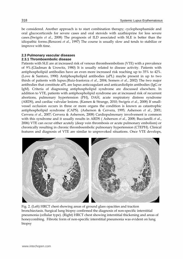
Fig. 2. (Left) HRCT chest showing areas of ground glass opacities and traction bronchiectasis. Surgical lung biopsy confirmed the diagnosis of non-specific interstitial pneumonia (cellular type). (Right) HRCT chest showing interstitial thickening and areas of honeycombing. Fibrotic form of non-specific interstitial pneumonia was evident on lung biopsy
www.intechopen.com
Pulmonary Manifestations of Systemic Lupus Erythematosus
319
long term anticoagulation with warfarin and a target INR of 2.0 to 3.0 is highly recommended. It used to be recommended to achieve a higher target INR, but in one study, high intensity warfarin (target INR 3.0-4.0) was found not superior to moderate intensity warfarin (target INR 2.0-3.0). Moderate intensity warfarin had lower rate of major bleeding.(Crowther et al., 2003) Recommendation for primary prevention is lacking. Some authors use long term low dose aspirin.(Swigris et al., 2008) Patients with CAPS usually require systemic glucocorticoids, immunosuppressants, plasmapharesis and intravenous immunoglobulin in addition to anticoagulation.(Swigris et al., 2008) Mortality rate can reach up to 50%.(Asherson & Cervera, 1995; Asherson et al., 2001, 2008)
2.3.2 Pulmonary hypertension Pulmonary Hypertension (PH) is defined as a mean pulmonary artery pressure (PAP) ≥ 25mmHg at rest. (McLaughlin et al., 2009) The prevalence of PH in SLE patients varies between 0.5% to 15%. (Asherson & Oakley, 1986; Asherson et al., 1990) In one study, 50 consecutive patients with SLE were carefully tested by transthoracic echocardiogram to look particularly for PH, none was found to have any echocardiographic evidence of PH. In that cohort almost one third were found to have an isolated reduction in DLCO, which could be a marker of early pulmonary vascular involvement.(Hodson et al., 1983; Gari et al., 2009) The prevalence is definitely lower than those with scleroderma. Raynaud’s phenomenon occurs in 75% of SLE associated pulmonary arterial hypertension (PAH) compared to only 20% of patients with SLE and no PH.(Matthay et al., 1975) The duration of SLE doesn’t correlate with the development of PAH.(Asherson & Oakley, 1986; Asherson & Cervera, 2007) Clinical presentations of SLE associated PAH is similar to idiopathic pulmonary arterial pulmonary hypertension (IPAH). Symptoms include dyspnea, fatigue, chest pain and lower limb swelling. Physical examination includes jugular venous distension with a large v wave, loud pulmonic component with wide splitting of the second heart sound, murmur of tricuspid regurgitation and/or pulmonic insufficiency, and lower limb edema. Physical findings may be minimal in mild PH. In patients with suspected PH, transthoracic echocardiogram is the best initial diagnostic test. Right ventricular systolic pressure (RVSP) which is an approximation of systolic PAP can only be measured if a tricuspid regurgitation (TR) signal is detected. TR signal is only available in 30% of population. Although PH is more common in SLE patients than general population, other causes of PH need to be ruled out. Tests to evaluate for other causes include HIV, hepatitis B and hepatitis C serology, aPL antibodies, HRCT chest to evaluate for interstitial lung disease, ventilation perfusion scan (V/Q) to look for any evidence of chronic pulmonary emboli leading to CTEPH, and polysomnogram if obstructive sleep apnea is suspected. Eventually right heart catheterization is required to confirm the diagnosis of PAH and to rule out PH secondary to left heart disease. The pathogenesis of SLE associated PAH is not clear; the high prevalence of aPL antibodies suggests that thrombosis may play a role. (Prabu et al., 2009) Histopathologic changes are identical to IPAH and include plexiform lesions, intimal fibrosis, and thickening of the media. In addition, complement and immunoglobulin deposits are found in some patients suggesting that immune deposits may be involved in the pathogenesis.(Quismorio et al., 1984) Several aspects need to be considered when it comes to treating SLE associated PAH. All patients should receive long term anticoagulation especially those with aPL antibodies. Oxygen, diuretics and digoxin should be considered in all patients. PH specific therapies used to treat IPAH are also effective in treating SLE associated PAH. Epoprostenol, bosentan, sildenafil, ambrisentan and tadalafil have all been
www.intechopen.com
Systemic Lupus Erythematosus
320
shown to be effective in treating PAH.( Barst RJ et al.,1996; Galie et al., 2005, 2008, 2009; Rubin et al., 2004 ) PAH specific therapies were found to improve 6-minute walk distance (6MWD) and functional class. Adding immunosuppressants may provide further improvement. Intravenous
cyclophosphamide (monthly for six months) was shown to be effective. It reduced the
systolic PAP when measured by transthoracic echocardiogram, and improved
6MWD.(Gonzalez-Lopez et al., 2004; Jais et al., 2008) Oral glucocorticoids in conjunction with
immunosuppressants lowered PAP and improved 6MWD.(Tanaka et al., 2002; Sanchez et al.,
2006) It is not very clear when to use immunosuppressants in SLE associated PAH. Patients
with mild PH may benefit from immunosuppressive therapy while patients with moderate
to severe PH need PH specific therapy with or without immunosuppressants.(Swigris et al.,
2008) The prognosis of SLE associated PAH is worse than IPAH, with a 5-year survival of
only 17% compared to 68% in patients with IPAH.(Chung et al., 2006) Given the rarity of
PH in patients with SLE, there is no recommendation to screen asymptomatic patients with
echocardiogram. On the other hand, patients with scleroderma should have annual
transthoracic echocardiogram to evaluate for the presence of PH.
2.3.3 Acute reversible hypoxia This is a rare complication of lupus. In one series 27% of hospitalized patients had this
condition.(Abramson et al., 1991) It is characterized by an abrupt onset of unexplained
hypoxia and hypocapnea. Radiographic chest imaging is normal. Ventilation perfusion
(V/Q) scan doesn’t show any evidence of thromboembolism. Arterial blood gases
demonstrates an increase in Alveolar-arterial (A-a) PO2 gradient. The pathogenesis of this
syndrome is not clear, but it is believed to be due to complement activation leading to
leukoaggregation within pulmonary capillaries.(Abramson et al., 1991; Belmont et al., 1994)
Plasma C3a level is markedly elevated if measured during the episode.(Abramson et al.,
1991) Most cases respond quickly to high dose of systemic corticosteroids.(Abramson et al.,
1991; Martinez-Taboada et al., 1995)
2.4 Airway disease 2.4.1 Upper airway involvement Involvement of the upper airways can occur in up to 30% of patients with SLE. A variety of disorders have been described including laryngeal mucosal inflammation or ulceration, cricoarytenoiditis, vocal cord paralysis, and necrotizing vasculitis.( Langford & Van Waes, 1997; Teitel et al., 1992) Patients present with hoarseness and or dyspnea. Severe upper airway obstruction due to angioedema requiring mechanical ventilation has also been reported.(Thong et al., 2001) Angioedema usually present with lips and mouth swelling, dysphagia, odynophagia and breathing difficulty, it could be due to SLE or medications used in SLE like angiotensin-converting enzyme inhibitors.(Agah et al., 1997) Routine chest imaging with Chest x-ray and chest CT is usually normal in patients with upper airway obstruction. Spirometry may show flattening of the inspiratory or expiratory loop or both depending on the location of the obstruction. Specialized imaging of the upper airways with 3-D reconstruction is important to demonstrate the site of obstruction. Direct visualization with fibro-optic laryngoscopy or bronchoscopy is needed to assess for vocal cord mobility. Generally, corticosteroid therapy will be effective in case of laryngeal mucosal inflammation or ulceration, and vocal cord paralysis.(Smith et al., 1977; Teitel et al., 1992). In those who
www.intechopen.com
Pulmonary Manifestations of Systemic Lupus Erythematosus
321
don’t respond to glucocorticoids, infectious causes should be considered. Typical pathogens are Haemophilus influenzae and streptococcus, other rare infections include Histoplasma, coccidioides, cryptococus, blastomycosis and candida.(Toomey et al., 1974)
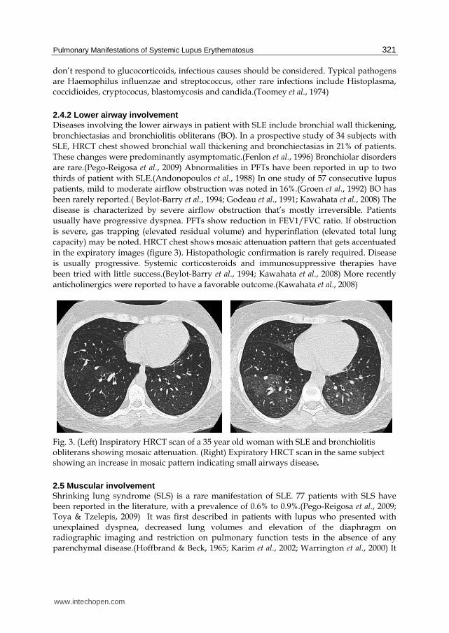
2.4.2 Lower airway involvement Diseases involving the lower airways in patient with SLE include bronchial wall thickening,
bronchiectasias and bronchiolitis obliterans (BO). In a prospective study of 34 subjects with
SLE, HRCT chest showed bronchial wall thickening and bronchiectasias in 21% of patients.
These changes were predominantly asymptomatic.(Fenlon et al., 1996) Bronchiolar disorders
are rare.(Pego-Reigosa et al., 2009) Abnormalities in PFTs have been reported in up to two
thirds of patient with SLE.(Andonopoulos et al., 1988) In one study of 57 consecutive lupus
patients, mild to moderate airflow obstruction was noted in 16%.(Groen et al., 1992) BO has
been rarely reported.( Beylot-Barry et al., 1994; Godeau et al., 1991; Kawahata et al., 2008) The
disease is characterized by severe airflow obstruction that’s mostly irreversible. Patients
usually have progressive dyspnea. PFTs show reduction in FEV1/FVC ratio. If obstruction
is severe, gas trapping (elevated residual volume) and hyperinflation (elevated total lung
capacity) may be noted. HRCT chest shows mosaic attenuation pattern that gets accentuated
in the expiratory images (figure 3). Histopathologic confirmation is rarely required. Disease
is usually progressive. Systemic corticosteroids and immunosuppressive therapies have
been tried with little success.(Beylot-Barry et al., 1994; Kawahata et al., 2008) More recently
anticholinergics were reported to have a favorable outcome.(Kawahata et al., 2008)
Fig. 3. (Left) Inspiratory HRCT scan of a 35 year old woman with SLE and bronchiolitis obliterans showing mosaic attenuation. (Right) Expiratory HRCT scan in the same subject showing an increase in mosaic pattern indicating small airways disease.
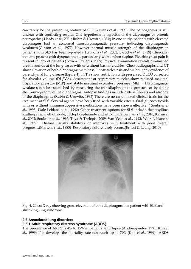
2.5 Muscular involvement Shrinking lung syndrome (SLS) is a rare manifestation of SLE. 77 patients with SLS have been reported in the literature, with a prevalence of 0.6% to 0.9%.(Pego-Reigosa et al., 2009; Toya & Tzelepis, 2009) It was first described in patients with lupus who presented with unexplained dyspnea, decreased lung volumes and elevation of the diaphragm on radiographic imaging and restriction on pulmonary function tests in the absence of any parenchymal disease.(Hoffbrand & Beck, 1965; Karim et al., 2002; Warrington et al., 2000) It
www.intechopen.com
Systemic Lupus Erythematosus
322
can rarely be the presenting feature of SLE.(Stevens et al., 1990) The pathogenesis is still unclear with conflicting results. One hypothesis is myositis of the diaphragm or phrenic neuropathy.( Hardy et al., 2001; Rubin & Urowitz, 1983;) In one study, patients with elevated diaphragms had an abnormal transdiaphragmatic pressure, indicating diaphragmatic weakness.(Gibson et al., 1977) However normal muscle strength of the diaphragm in patients with SLS has been reported.( Hawkins et al., 2001; Laroche et al., 1989) Clinically, patients present with dyspnea that is particularly worse when supine. Pleuritic chest pain is present in 65% of patients.(Toya & Tzelepis, 2009) Physical examination reveals diminished breath sounds at the lung bases with or without basilar crackles. Chest radiographs and CT show elevation of both diaphragms with basal linear atelectasis and without any evidence of parenchymal lung disease (figure 4). PFT’s show restriction with preserved DLCO corrected for alveolar volume (DL/VA). Assessment of respiratory muscles show reduced maximal inspiratory pressure (MIP) and stable maximal expiratory pressure (MEP). Diaphragmatic weakness can be established by measuring the transdiaphragmatic pressure or by doing electromyography of the diaphragms. Autopsy findings include diffuse fibrosis and atrophy of the diaphragms. (Rubin & Urowitz, 1983) There are no randomized clinical trials for the treatment of SLS. Several agents have been tried with variable effects. Oral glucocorticoids with or without immunosuppressive medications have been shown effective. ( Soubrier et al., 1995; Walz-Leblanc et al., 1992) Other treatment options for SLS include theophylline, azathioprine, methotrexate, cyclophosphamide and rituximab.( Benham et al., 2010; Karim et al., 2002; Soubrier et al., 1995; Toya & Tzelepis, 2009; Van Veen et al., 1993; Walz-Leblanc et al., 1992) Disease usually stabilizes or improves with treatment with good overall prognosis.(Martens et al., 1983) Respiratory failure rarely occurs.(Ernest & Leung, 2010)
Fig. 4. Chest X-ray showing gross elevation of both diaphragms in a patient with SLE and shrinking lung syndrome
2.6 Associated lung disorders 2.6.1 Adult respiratory distress syndrome (ARDS) The prevalence of ARDS is 4% to 15% in patients with lupus.(Andonopoulos, 1991; Kim et al., 1999) If it develops the mortality rate can reach up to 70%.(Kim et al., 1999) ARDS
www.intechopen.com
Pulmonary Manifestations of Systemic Lupus Erythematosus
323
related mortality contributes to 30% of all Lupus deaths. The most frequent cause of ARDS is sepsis; other causes include ALP, DAH, and CAPS. In lupus patients, ARDS tend to occur at a younger age and is more progressive than ARDS in non-SLE patients. (Andonopoulos, 1991; Kim et al., 1999; Pego-Reigosa et al., 2009) It is important to identify the underlying cause. Treatment of ARDS is supportive.
2.6.2 Infectious complications SLE can impair the immune system at multiple levels.(Orens et al., 1994; Rudd et al., 1981 ) The clinical significance of this is unknown since the risk of infection in the absence of immunosuppression is negligible. Most patients with infectious complications are on immunosuppressive drugs. Infections account for 30% to 50% of all deaths of SLE.( Bernatsky et al., 2006; Zandman-Goddard & Shoenfeld, 2005;) Bacterial pathogens account for 75% of all infections, mycobacteria 12%, fungal infections 7%, and viruses 5%.(Kinder et al., 2007) Opportunistic infections such as Pneumocystis jiroveci, Nocardia, Aspergillus and Cytomegalovirus have been reported.( Fessler, 2002; Petri, 1998; Zandman-Goddard & Shoenfeld, 2005) Clinical picture is indistinguishable from non-infectious complications such as ALP and DAH, hence aggressive diagnostic approach is recommended with chest imaging, bronchoscopy and BAL. Empiric broad-spectrum antibiotics should be started awaiting identification of an organism. Once a pathogen is isolated treatment should be tailored accordingly. Risk of infection can be reduced by influenza and pneumococcal vaccination.(O'Neill & Isenberg, 2006) Since many patients with SLE require systemic glucocorticoids and immunosuppresants at some point, screening for latent Tuberculosis (TB) is important, especially in high prevalence areas. This can be done via skin testing or interferon gamma release assay (IGRA). For those taking glucocorticoids, induration of 5mm or greater is considered a positive tuberculin skin test. If latent TB is identified treatment is recommended with a nine month course of Isoniazid. The role of Pneumocystis jiroveci pneumonia (PCP) prophylaxis is less clear. It is suggested for those who are on heavy immunosuppression.(Li et al., 2006)
2.6.3 Lung cancer Studies have shown an increased risk of lung cancer in patients with SLE.(Bernatsky et al.,
2006; Pego-Reigosa et al., 2009) Histological pattern is similar to that in general population,
adenocarcinoma being most common. However there is tendency for uncommon thoracic
malignancies such as carcinoids and bronchoalveolar carcinoma.(Bin et al., 2007; Pego-
Reigosa et al., 2009)
2.7 Drug reactions In this section we will cover two aspects of drugs and SLE. First we will briefly discuss
drugs that can cause SLE and the associated pulmonary manifestations. After that we
will elaborate on pulmonary drug toxicity associated with commonly used medications to
treat SLE.
Pulmonary manifestations of drug induced lupus are similar to idiopathic SLE.(Cush &
Goldings, 1985; Yung & Richardson, 1994) Most commonly it presents with pleurisy and
pleural effusion.(Wiedemann & Matthay, 1989) Common drugs include Procainamide and
hydralazine. Newer biologic agents such as entanercept have been reported to cause drug
induced lupus.(Abunasser et al., 2008)
www.intechopen.com
Systemic Lupus Erythematosus
324
Common drugs used to treat lupus and are known to cause pulmonary complications include Methotrexate and Cyclophosphamide. Pulmonary complications related to methotrexate are rare, estimated less than 1%.(Lateef et al., 2005) Methotrexate can cause acute, subacute or chronic lung toxicity. It is usually not dose dependent but rather idiosyncratic.(Imokawa et al., 2000; Ohosone et al., 1997) Subacute pneumonitis is most common and presents with fever, cough and dyspnea. Crackles are usually noted on physical examination. It usually presents within the first year of starting the drug. If left unrecognized it can progress into pulmonary fibrosis in up to 10%. Radiologic findings are not specific. Ground glass opacities and diffuse interstitial infiltrates are frequently noted on HRCT. BAL is needed to rule out infections. Histologic findings include varying degree of inflammation and fibrosis. Ill-defined granulomas, and increased tissue eosinophils have been observed. ( Malik et al., 1996; Sostman et al., 1976) Once diagnosis is made methotrexate needs to be stooped and systemic steroids should be started. Prognosis is usually favorable. Cyclophosphamide lung toxicity is also idiosyncratic. It can present as early onset or late onset pneumonitis.(Malik et al., 1996) Early onset disease appears within the first six months of starting treatment. It presents with non-productive cough, fever and dyspnea. (Pego-Reigosa et al., 2009) CT chest shows bilateral upper lobe predominant ground glass opacities. PFT shows reduction in lung volumes and DLCO. BAL is needed to rule out infections. Discontinuing the drug along with systemic glucocorticoids usually improve symptoms and lung function. Late onset pneumonitis usually occurs after several years of exposure to cyclophosphamide. It is a slowly progressive disease. It presents with progressive dyspnea and dry cough. Chest imaging shows interstitial fibrosis affecting the upper lobes. This condition usually does not respond to steroids. Lung transplantation is an option in appropriate candidates.
3. Assessment of patients with dyspnea
3.1 Assessment of patients with chronic dyspnea The work up of patients with SLE and chronic dyspnea can be lengthy (Figure 5). Chronic dyspnea can be due to a variety of conditions such as interstitial lung disease related to SLE or drugs used to treat lupus, pleural disease, pulmonary hypertension, systolic heart failure, upper airway disease, obliterative bronchiolitis, shrinking lung syndrome or chronic infections. Certain clues on history can be helpful; for example dyspnea increasing in the supine position suggests diaphragmatic involvement due to SLS, dyspnea and hoarseness suggest upper airway involvement, Dyspnea with pleuritic chest pain suggests pleuritis related to SLE. All patients require CXR, HRCT chest and full PFT. If chest imaging is normal with or without isolated reduction in DLCO, then transthoracic echocardiogram should be done to assess for the presence of PH. If PH is detected, patients should not be labeled to have SLE associated PAH until other causes have been ruled out. So hepatitis B and C serology, HIV testing, and V/Q scan should be done. All patients should have right heart catheterization to confirm the presence of PH and to rule out left heart disease. If SLE-PAH is diagnosed PH specific therapies should be started. If chest imaging shows elevation of the diaphragms, especially in the presence of normal DLCO adjusted for alveolar volume, shrinking lung syndrome should be suspected. Electromyography or transdiaphragmatic pressure measurement should be obtained. Either of these two tests may show evidence of diaphragmatic weakness. If confirmed, trial of systemic steroids is advised. The presence of
www.intechopen.com
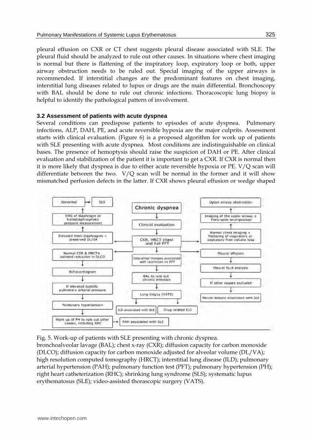
Pulmonary Manifestations of Systemic Lupus Erythematosus
325
pleural effusion on CXR or CT chest suggests pleural disease associated with SLE. The pleural fluid should be analyzed to rule out other causes. In situations where chest imaging is normal but there is flattening of the inspiratory loop, expiratory loop or both, upper airway obstruction needs to be ruled out. Special imaging of the upper airways is recommended. If interstitial changes are the predominant features on chest imaging, interstitial lung diseases related to lupus or drugs are the main differential. Bronchoscopy with BAL should be done to rule out chronic infections. Thoracoscopic lung biopsy is helpful to identify the pathological pattern of involvement.
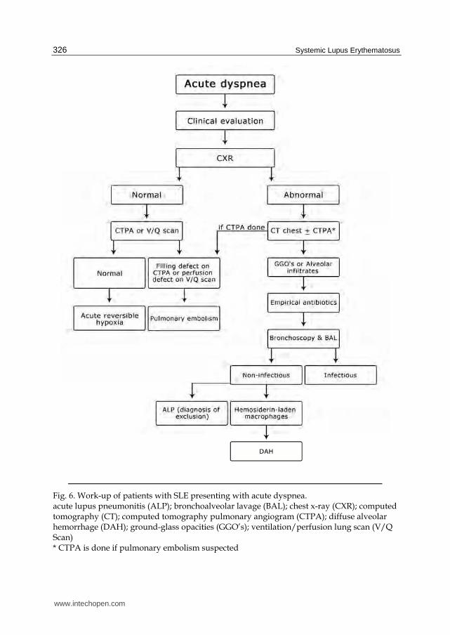
3.2 Assessment of patients with acute dyspnea Several conditions can predispose patients to episodes of acute dyspnea. Pulmonary infections, ALP, DAH, PE, and acute reversible hypoxia are the major culprits. Assessment starts with clinical evaluation. (Figure 6) is a proposed algorithm for work up of patients with SLE presenting with acute dyspnea. Most conditions are indistinguishable on clinical bases. The presence of hemoptysis should raise the suspicion of DAH or PE. After clinical evaluation and stabilization of the patient it is important to get a CXR. If CXR is normal then it is more likely that dyspnea is due to either acute reversible hypoxia or PE. V/Q scan will differentiate between the two. V/Q scan will be normal in the former and it will show mismatched perfusion defects in the latter. If CXR shows pleural effusion or wedge shaped
Fig. 5. Work-up of patients with SLE presenting with chronic dyspnea. bronchoalveolar lavage (BAL); chest x-ray (CXR); diffusion capacity for carbon monoxide (DLCO); diffusion capacity for carbon monoxide adjusted for alveolar volume (DL/VA); high resolution computed tomography (HRCT); interstitial lung disease (ILD); pulmonary arterial hypertension (PAH); pulmonary function test (PFT); pulmonary hypertension (PH); right heart catheterization (RHC); shrinking lung syndrome (SLS); systematic lupus erythematosus (SLE); video-assisted thorascopic surgery (VATS).
www.intechopen.com
Systemic Lupus Erythematosus
326
Fig. 6. Work-up of patients with SLE presenting with acute dyspnea. acute lupus pneumonitis (ALP); bronchoalveolar lavage (BAL); chest x-ray (CXR); computed tomography (CT); computed tomography pulmonary angiogram (CTPA); diffuse alveolar hemorrhage (DAH); ground-glass opacities (GGO’s); ventilation/perfusion lung scan (V/Q Scan) * CTPA is done if pulmonary embolism suspected
www.intechopen.com
Pulmonary Manifestations of Systemic Lupus Erythematosus
327
opacities it is important to get CT pulmonary angiogram to look for evidence of PE. If CXR shows mainly alveolar infiltrates, CT chest should be considered. In these situations bronchoscopy with BAL, with or without TBBX, is highly recommended. The presence of hemosiderin laden macrophages confirms the diagnosis of DAH. If TBBX is performed and it showed features of DAD, then the likely diagnosis is ALP. BAL should be routinely sent for cultures. Empiric antibiotics should be started immediately until the results of cultures are known. It is not unusual to start patients on both broad spectrum antibiotics and systemic corticosteroids while the work up is being actively pursued.
4. Conclusion
SLE can affect many aspects of the pulmonary system. There is significant overlap in the clinical presentation of many SLE associated pulmonary conditions. Aggressive work up is needed early on to identify the underlying etiology.
5. Acknowledgment
The work to produce this chapter was supported by Alzaidi's Chair of research in rheumatic diseases- Umm Alqura University.
6. References
Abramson, S. B., Dobro, J., Eberle, M. A., Benton, M., Reibman, J., Epstein, H. et al. (1991) Acute reversible hypoxemia in systemic lupus erythematosus. Ann Intern Med 114: 941-947.
Abud-Mendoza, C., Diaz-Jouanen, E., & Alarcon-Segovia, D. (1985) Fatal pulmonary hemorrhage in systemic lupus erythematosus. Occurrence without hemoptysis. J Rheumatol 12: 558-561.
Abunasser, J., Forouhar, F. A., & Metersky, M. L. (2008) Etanercept-induced lupus erythematosus presenting as a unilateral pleural effusion. Chest 134: 850- 853.
Agah, R., Bandi, V., & Guntupalli, K. K. (1997) Angioedema: the role of ACE inhibitors and factors associated with poor clinical outcome. Intensive Care Med 23: 793-796.
Andonopoulos, A. P., Constantopoulos, S. H., Galanopoulou, V., Drosos, A. A., Acritidis, N. C., & Moutsopoulos, H. M. (1988) Pulmonary function of nonsmoking patients with systemic lupus erythematosus. Chest 94: 312-315.
Andonopoulos, A. P. (1991) Adult respiratory distress syndrome: an unrecognized premortem event in systemic lupus erythematosus. Br J Rheumatol 30: 346-348.
Asherson, R. A., & Oakley, C. M. (1986) Pulmonary hypertension and systemic lupus erythematosus. J Rheumatol 13: 1-5.
Asherson, R. A., Higenbottam, T. W., Dinh Xuan, A. T., Khamashta, M. A., & Hughes, G. R. (1990) Pulmonary hypertension in a lupus clinic: experience with twenty-four patients. J Rheumatol 17: 1292-1298.
Asherson, R. A., & Cervera, R. (1995) Review: antiphospholipid antibodies and the lung. J Rheumatol 22: 62-66.
www.intechopen.com
Systemic Lupus Erythematosus
328
Asherson, R. A., Cervera, R., Piette, J. C., Shoenfeld, Y., Espinosa, G., Petri, M. A. et al. (2001) Catastrophic antiphospholipid syndrome: clues to the pathogenesis from a series of 80 patients. Medicine (Baltimore) 80: 355-377.
Asherson, R. A., & Cervera, R. (2007) Pulmonary hypertension, antiphospholipid antibodies, and syndromes. Clin Rev Allergy Immunol 32: 153-158.
Asherson, R. A., Cervera, R., Merrill, J. T., & Erkan, D. (2008) Antiphospholipid antibodies and the antiphospholipid syndrome: clinical significance and treatment. Semin Thromb Hemost 34: 256-266.
Badsha, H., Teh, C. L., Kong, K. O., Lian, T. Y., & Chng, H. H. (2004) Pulmonary hemorrhage in systemic lupus erythematosus. Semin Arthritis Rheum 33: 414-421.
Bankier, A. A., Kiener, H. P., Wiesmayr, M. N., Fleischmann, D., Kontrus, M., Herold, C. J. et
al. (1995) Discrete lung involvement in systemic lupus erythematosus: CT
assessment. Radiology 196: 835-840.
Barile, L. A., Jara, L. J., Medina-Rodriguez, F., Garcia-Figueroa, J. L., & Miranda-Limon, J. M.
(1997) Pulmonary hemorrhage in systemic lupus erythematosus. Lupus 6: 445-
448.
Barst RJ, FAU - Rubin, L. J., Rubin LJ, FAU - Long, W. A., Long WA, FAU - McGoon, M. D.
et al. A comparison of continuous intravenous epoprostenol (prostacyclin) with
conventional therapy for primary pulmonary hypertension. The Primary
Pulmonary Hypertension Study Group. - N Engl J Med.1996 Feb 1;334(5):296- 302.
Belmont, H. M., Buyon, J., Giorno, R., & Abramson, S. (1994) Up-regulation of endothelial
cell adhesion molecules characterizes disease activity in systemic lupus
erythematosus. The Shwartzman phenomenon revisited. Arthritis Rheum 37: 376-
383.
Benham, H., Garske, L., Vecchio, P., & Eckert, B. W. (2010) Successful treatment of shrinking
lung syndrome with rituximab in a patient with systemic lupus erythematosus. J
Clin Rheumatol 16: 68-70.
Bernatsky, S., Boivin, J. F., Joseph, L., Manzi, S., Ginzler, E., Gladman, D. D. et al. (2006)
Mortality in systemic lupus erythematosus. Arthritis Rheum 54: 2550-2557.
Beylot-Barry, M., Doutre, M. S., Bletry, O., & Beylot, C. (1994) Lupus bronchiolitis obliterans:
diagnostic difficulties. Rev Med Interne 15: 332-335.
Bin, J., Bernatsky, S., Gordon, C., Boivin, J. F., Ginzler, E., Gladman, D. et al. (2007) Lung
cancer in systemic lupus erythematosus. Lung Cancer 56: 303-306.
Boddaert, J., Huong, D. L., Amoura, Z., Wechsler, B., Godeau, P., & Piette, J. C. (2004) Late-
onset systemic lupus erythematosus: a personal series of 47 patients and pooled
analysis of 714 cases in the literature. Medicine (Baltimore) 83: 348-359.
Boulware, D. W., & Hedgpeth, M. T. (1989) Lupus pneumonitis and anti-SSA(Ro)
antibodies. J Rheumatol 16: 479-481.
Bouros, D., Pneumatikos, I., & Tzouvelekis, A. (2008) Pleural involvement in systemic
autoimmune disorders. Respiration 75: 361-371.
Bucciarelli, S., Espinosa, G., Asherson, R. A., Cervera, R., Claver, G., Gomez-Puerta, J. A. et
al. (2006) The acute respiratory distress syndrome in catastrophic antiphospholipid
syndrome: analysis of a series of 47 patients. Ann Rheum Dis 65: 81-86.
www.intechopen.com
Pulmonary Manifestations of Systemic Lupus Erythematosus
329
Carette, S., Macher, A. M., Nussbaum, A., & Plotz, P. H. (1984) Severe, acute pulmonary
disease in patients with systemic lupus erythematosus: ten years of experience at
the National Institutes of Health. Semin Arthritis Rheum 14: 52-59.
Cervera, R., Bucciarelli, S., Espinosa, G., Gomez-Puerta, J. A., Ramos-Casals, M.,
Shoenfeld, Y. et al. (2007) Catastrophic antiphospholipid syndrome: lessons from
the "CAPS Registry"--a tribute to the late Josep Font. Ann N Y Acad Sci 1108: 448-
456.
Cervera, R., & Asherson, R. A. (2008) Catastrophic antiphospholipid (Asherson's) syndrome. Br J Hosp Med (Lond) 69: 384-387.
Cheema, G. S., & Quismorio, F. P.,Jr. (2000) Interstitial lung disease in systemic lupus erythematosus. Curr Opin Pulm Med 6: 424-429.
Chung, S. M., Lee, C. K., Lee, E. Y., Yoo, B., Lee, S. D., & Moon, H. B. (2006) Clinical aspects of pulmonary hypertension in patients with systemic lupus erythematosus and in patients with idiopathic pulmonary arterial hypertension. Clin Rheumatol 25: 866-872.
Comer, M., D'Cruz, D., Thompson, I., Erskine, K., & Dacre, J. (1996) Pneumonitis in a lupus twin pregnancy: a case report. Lupus 5: 146-148.
Crowther, M. A., Ginsberg, J. S., Julian, J., Denburg, J., Hirsh, J., Douketis, J. et al. (2003) A comparison of two intensities of warfarin for the prevention of recurrent thrombosis in patients with the antiphospholipid antibody syndrome. N Engl J Med 349: 1133-1138.
Cush, J. J., & Goldings, E. A. (1985) Drug-induced lupus: clinical spectrum and pathogenesis. Am J Med Sci 290: 36-45.
Dweik, R. A., Arroliga, A. C., & Cash, J. M. (1997) Alveolar hemorrhage in patients with rheumatic disease. Rheum Dis Clin North Am 23: 395-410.
Eagen, J. W., Memoli, V. A., Roberts, J. L., Matthew, G. R., Schwartz, M. M., & Lewis, E. J. (1978) Pulmonary hemorrhage in systemic lupus erythematosus. Medicine (Baltimore) 57: 545-560.
Eiser, A. R., & Shanies, H. M. (1994) Treatment of lupus interstitial lung disease with intravenous cyclophosphamide. Arthritis Rheum 37: 428-431.
Erickson, R. W., Franklin, W. A., & Emlen, W. (1994) Treatment of hemorrhagic lupus pneumonitis with plasmapheresis. Semin Arthritis Rheum 24: 114-123.
Ernest, D., & Leung, A. (2010) Ventilatory failure in shrinking lung syndrome is associated with reduced chest compliance. Intern Med J 40: 66-68.
Ewan, P. W., Jones, H. A., Rhodes, C. G., & Hughes, J. M. (1976) Detection of intrapulmonary hemorrhage with carbon monoxide uptake. Application in goodpasture's syndrome. N Engl J Med 295: 1391-1396.
Fenlon, H. M., Doran, M., Sant, S. M., & Breatnach, E. (1996) High-resolution chest CT in systemic lupus erythematosus. AJR Am J Roentgenol 166: 301-307.
Fessler, B. J. (2002) Infectious diseases in systemic lupus erythematosus: risk factors, management and prophylaxis. Best Pract Res Clin Rheumatol 16: 281-291.
Galie, N., Ghofrani, H. A., Torbicki, A., Barst, R. J., Rubin, L. J., Badesch, D. et al. (2005) Sildenafil citrate therapy for pulmonary arterial hypertension. N Engl J Med 353: 2148-2157.
Galie, N., Olschewski, H., Oudiz, R. J., Torres, F., Frost, A., Ghofrani, H. A. et al. (2008) Ambrisentan for the treatment of pulmonary arterial hypertension: results of the
www.intechopen.com
Systemic Lupus Erythematosus
330
ambrisentan in pulmonary arterial hypertension, randomized, double-blind, placebo-controlled, multicenter, efficacy (ARIES) study 1 and 2. Circulation 117: 3010-3019.
Galie, N., Brundage, B. H., Ghofrani, H. A., Oudiz, R. J., Simonneau, G., Safdar, Z. et al.
(2009) Tadalafil therapy for pulmonary arterial hypertension. Circulation 119: 2894-
2903.
Gammon, R. B., Bridges, T. A., al-Nezir, H., Alexander, C. B., & Kennedy, J. I.,Jr. (1992)
Bronchiolitis obliterans organizing pneumonia associated with systemic lupus
erythematosus. Chest 102: 1171-1174.
Gari A, Dias B, Khan F, Pope J, Mehta S. (2009) Prevalence of Pulmonary Hypertension in
Unselected Patients With Systemic Lupus Erythematosus in an Academic Tertiary
Care Centre. Chest 136: 55S.
Gibson, C. J., Edmonds, J. P., & Hughes, G. R. (1977) Diaphragm function and lung
involvement in systemic lupus erythematosus. Am J Med 63: 926-932.
Gilleece, M. H., Evans, C. C., & Bucknall, R. C. (1988) Steroid resistant pleural effusion in
systemic lupus erythematosus treated with tetracycline pleurodesis. Ann Rheum Dis
47: 1031-1032.
Gladman, D. D., & Urowitz, M. B. (1980) Venous syndromes and pulmonary embolism in
systemic lupus erythematosus. Ann Rheum Dis 39: 340-343.
Godeau, B., Cormier, C., & Menkes, C. J. (1991) Bronchiolitis obliterans in systemic lupus
erythematosus: beneficial effect of intravenous cyclophosphamide. Ann Rheum Dis
50: 956-958.
Gonzalez-Lopez, L., Cardona-Munoz, E. G., Celis, A., Garcia-de la Torre, I., Orozco-Barocio,
G., Salazar-Paramo, M. et al. (2004) Therapy with intermittent pulse
cyclophosphamide for pulmonary hypertension associated with systemic lupus
erythematosus. Lupus 13: 105-112.
Good, J. T.,Jr, King, T. E., Antony, V. B., & Sahn, S. A. (1983) Lupus pleuritis. Clinical
features and pleural fluid characteristics with special reference to pleural fluid
antinuclear antibodies. Chest 84: 714-718.
Groen, H., ter Borg, E. J., Postma, D. S., Wouda, A. A., van der Mark, T. W., & Kallenberg,
C. G. (1992) Pulmonary function in systemic lupus erythematosus is related
to distinct clinical, serologic, and nailfold capillary patterns. Am J Med 93: 619-
627.
Gross, M., Esterly, J. R., & Earle, R. H. (1972) Pulmonary alterations in systemic lupus
erythematosus. Am Rev Respir Dis 105: 572-577.
Hardy, K., Herry, I., Attali, V., Cadranel, J., & Similowski, T. (2001) Bilateral phrenic
paralysis in a patient with systemic lupus erythematosus. Chest 119: 1274-
1277.
Harmon, K. R., & Leatherman, J. W. (1988) Respiratory manifestations of connective tissue
disease. Semin Respir Infect 3: 258-273.
Harvey, A. M., Shuman, L. E., Tumulty, P. A., Conley, C. L., & Schoenrich, E. H. (1954)
Systemic lupus erythematosus: review of the literature and clinical analysis of 138
cases. Medicine (Baltimore) 33: 291-437.
www.intechopen.com
Pulmonary Manifestations of Systemic Lupus Erythematosus
331
Haupt, H. M., Moore, G. W., & Hutchins, G. M. (1981) The lung in systemic lupus
erythematosus. Analysis of the pathologic changes in 120 patients. Am J Med 71:
791-798.
Hawkins, P., Davison, A. G., Dasgupta, B., & Moxham, J. (2001) Diaphragm strength in
acute systemic lupus erythematosus in a patient with paradoxical abdominal
motion and reduced lung volumes. Thorax 56: 329-330.
Hodson, P., Klemp, P., & Meyers, O. L. (1983) Pulmonary hypertension in systemic lupus
erythematosus: a report of four cases. Clin Exp Rheumatol 1: 241-245.
Hoffbrand, B. I., & Beck, E. R. (1965) "Unexplained" Dyspnoea and Shrinking Lungs in
Systemic Lupus Erythematosus. Br Med J 1: 1273-1277.
Hsu, B. Y., Edwards, D. K.,3rd, & Trambert, M. A. (1992) Pulmonary hemorrhage
complicating systemic lupus erythematosus: role of MR imaging in diagnosis. AJR
Am J Roentgenol 158: 519-520.
Hunder, G. G., McDuffie, F. C., & Hepper, N. G. (1972) Pleural fluid complement in systemic
lupus erythematosus and rheumatoid arthritis. Ann Intern Med 76: 357-363.
Imokawa, S., Colby, T. V., Leslie, K. O., & Helmers, R. A. (2000) Methotrexate pneumonitis:
review of the literature and histopathological findings in nine patients. Eur Respir J
15: 373-381.
Jacobsen, S., Petersen, J., Ullman, S., Junker, P., Voss, A., Rasmussen, J. M. et al. (1998) A
multicentre study of 513 Danish patients with systemic lupus erythematosus. II.
Disease mortality and clinical factors of prognostic value. Clin Rheumatol 17: 478-
484.
Jais, X., Launay, D., Yaici, A., Le Pavec, J., Tcherakian, C., Sitbon, O. et al. (2008)
Immunosuppressive therapy in lupus- and mixed connective tissue disease-
associated pulmonary arterial hypertension: a retrospective analysis of twenty-
three cases. Arthritis Rheum 58: 521-531.
Kaine, J. L. (1985) Refractory massive pleural effusion in systemic lupus erythematosus
treated with talc poudrage. Ann Rheum Dis 44: 61-64.
Kamen, D. L., & Strange, C. (2010) Pulmonary manifestations of systemic lupus
erythematosus. Clin Chest Med 31: 479-488.
Karim, M. Y., Miranda, L. C., Tench, C. M., Gordon, P. A., D'cruz, D. P., Khamashta, M. A.,
& Hughes, G. R. (2002) Presentation and prognosis of the shrinking lung syndrome
in systemic lupus erythematosus. Semin Arthritis Rheum 31: 289-298.
Kawahata, K., Yamaguchi, M., Kanda, H., Komiya, A., Tanaka, R., Dohi, M. et al. (2008)
Severe airflow limitation in two patients with systemic lupus erythematosus: effect
of inhalation of anticholinergics. Mod Rheumatol 18: 52-56.
Keane, M. P., & Lynch, J. P.,3rd. (2000) Pleuropulmonary manifestations of systemic lupus
erythematosus. Thorax 55: 159-166.
Kim, J. S., Lee, K. S., Koh, E. M., Kim, S. Y., Chung, M. P., & Han, J. (2000) Thoracic
involvement of systemic lupus erythematosus: clinical, pathologic, and radiologic
findings. J Comput Assist Tomogr 24: 9-18.
Kim, W. U., Kim, S. I., Yoo, W. H., Park, J. H., Min, J. K., Kim, S. C. et al. (1999) Adult
respiratory distress syndrome in systemic lupus erythematosus: causes and
prognostic factors: a single center, retrospective study. Lupus 8: 552-557.
www.intechopen.com
Systemic Lupus Erythematosus
332
Kinder, B. W., Freemer, M. M., King, T. E.,Jr, Lum, R. F., Nititham, J., Taylor, K. et al. (2007)
Clinical and genetic risk factors for pneumonia in systemic lupus erythematosus.
Arthritis Rheum 56: 2679-2686.
Koh, W. H., Thumboo, J., & Boey, M. L. (1997) Pulmonary haemorrhage in Oriental patients
with systemic lupus erythematosus. Lupus 6: 713-716.
Langford, C. A., & Van Waes, C. (1997) Upper airway obstruction in the rheumatic diseases.
Rheum Dis Clin North Am 23: 345-363.
Laroche, C. M., Mulvey, D. A., Hawkins, P. N., Walport, M. J., Strickland, B., Moxham, J., &
Green, M. (1989) Diaphragm strength in the shrinking lung syndrome of systemic
lupus erythematosus. Q J Med 71: 429-439.
Lateef, O., Shakoor, N., & Balk, R. A. (2005) Methotrexate pulmonary toxicity. Expert Opin
Drug Saf 4: 723-730.
Leatherman, J. W., Davies, S. F., & Hoidal, J. R. (1984) Alveolar hemorrhage syndromes:
diffuse microvascular lung hemorrhage in immune and idiopathic disorders.
Medicine (Baltimore) 63: 343-361.
Li, J., Huang, X. M., Fang, W. G., & Zeng, X. J. (2006) Pneumocystis carinii pneumonia in
patients with connective tissue disease. J Clin Rheumatol 12: 114-117.
Lim, S. W., Gillis, D., Smith, W., Hissaria, P., Greville, H., & Peh, C. A. (2006) Rituximab use
in systemic lupus erythematosus pneumonitis and a review of current reports.
Intern Med J 36: 260-262.
Liu, M. F., Lee, J. H., Weng, T. H., & Lee, Y. Y. (1998) Clinical experience of 13 cases with
severe pulmonary hemorrhage in systemic lupus erythematosus with active
nephritis. Scand J Rheumatol 27: 291-295.
Love, P. E., & Santoro, S. A. (1990) Antiphospholipid antibodies: anticardiolipin and
the lupus anticoagulant in systemic lupus erythematosus (SLE) and in non-
SLE disorders. Prevalence and clinical significance. Ann Intern Med 112: 682-
698.
Malik, S. W., Myers, J. L., DeRemee, R. A., & Specks, U. (1996) Lung toxicity associated with
cyclophosphamide use. Two distinct patterns. Am J Respir Crit Care Med 154: 1851-
1856.
Martens, J., Demedts, M., Vanmeenen, M. T., & Dequeker, J. (1983) Respiratory muscle
dysfunction in systemic lupus erythematosus. Chest 84: 170-175.
Martinez-Taboada, V. M., Blanco, R., Armona, J., Fernandez-Sueiro, J. L., & Rodriguez-
Valverde, V. (1995) Acute reversible hypoxemia in systemic lupus erythematosus: a
new syndrome or an index of disease activity? Lupus 4: 259-262.
Matthay, R. A., Schwarz, M. I., Petty, T. L., Stanford, R. E., Gupta, R. C., Sahn, S. A., &
Steigerwald, J. C. (1975) Pulmonary manifestations of systemic lupus
erythematosus: review of twelve cases of acute lupus pneumonitis. Medicine
(Baltimore) 54: 397-409.
McKnight, K. M., Adair, N. E., & Agudelo, C. A. (1991) Successful use of tetracycline
pleurodesis to treat massive pleural effusion secondary to systemic lupus
erythematosus. Arthritis Rheum 34: 1483-1484.
McLaughlin, V. V., Archer, S. L., Badesch, D. B., Barst, R. J., Farber, H. W., Lindner, J. R. et
al. (2009) ACCF/AHA 2009 expert consensus document on pulmonary
hypertension a report of the American College of Cardiology Foundation Task
www.intechopen.com
Pulmonary Manifestations of Systemic Lupus Erythematosus
333
Force on Expert Consensus Documents and the American Heart Association
developed in collaboration with the American College of Chest Physicians;
American Thoracic Society, Inc.; and the Pulmonary Hypertension Association. J
Am Coll Cardiol 53: 1573-1619.
Mochizuki, T., Aotsuka, S., & Satoh, T. (1999) Clinical and laboratory features of lupus
patients with complicating pulmonary disease. Respir Med 93: 95-101.
Myers, J. L., & Katzenstein, A. A. (1986) Microangiitis in lupus-induced pulmonary
hemorrhage. Am J Clin Pathol 85: 552-556.
Ohosone, Y., Okano, Y., Kameda, H., Fujii, T., Hama, N., Hirakata, M. et al. (1997) Clinical
characteristics of patients with rheumatoid arthritis and methotrexate induced
pneumonitis. J Rheumatol 24: 2299-2303.
O'Neill, S. G., & Isenberg, D. A. (2006) Immunizing patients with systemic lupus
erythematosus: a review of effectiveness and safety. Lupus 15: 778-783.
Orens, J. B., Martinez, F. J., & Lynch, J. P.,3rd. (1994) Pleuropulmonary manifestations of
systemic lupus erythematosus. Rheum Dis Clin North Am 20: 159-193.
Pego-Reigosa, J. M., Medeiros, D. A., & Isenberg, D. A. (2009) Respiratory manifestations of
systemic lupus erythematosus: old and new concepts. Best Pract Res Clin Rheumatol
23: 469-480.
Pertschuk, L. P., Moccia, L. F., Rosen, Y., Lyons, H., Marino, C. M., Rashford, A. A., &
Wollschlager, C. M. (1977) Acute pulmonary complications in systemic lupus
erythematosus. Immunofluorescence and light microscopic study. Am J Clin Pathol
68: 553-557.
Petri, M. (1998) Infection in systemic lupus erythematosus. Rheum Dis Clin North Am 24: 423-
456.
Pines, A., Kaplinsky, N., Olchovsky, D., Rozenman, J., & Frankl, O. (1985) Pleuro-pulmonary
manifestations of systemic lupus erythematosus: clinical features of its subgroups.
Prognostic and therapeutic implications. Chest 88: 129-135.
Pottier, V., Pierrot, M., Subra, J. F., Mercat, A., Kouatchet, A., Parrot, A., & Augusto, J. F.
(2011) Successful rituximab therapy in a lupus patient with diffuse alveolar
haemorrhage. Lupus 20: 656-659.
Prabu, A., Patel, K., Yee, C. S., Nightingale, P., Situnayake, R. D., Thickett, D. R. et al. (2009)
Prevalence and risk factors for pulmonary arterial hypertension in patients with
lupus. Rheumatology (Oxford) 48: 1506-1511.
Quadrelli, S. A., Alvarez, C., Arce, S. C., Paz, L., Sarano, J., Sobrino, E. M., & Manni, J. (2009)
Pulmonary involvement of systemic lupus erythematosus: analysis of 90
necropsies. Lupus 18: 1053-1060.
Quismorio, F. P.,Jr, Sharma, O., Koss, M., Boylen, T., Edmiston, A. W., Thornton, P. J.,
& Tatter, D. (1984) Immunopathologic and clinical studies in pulmonary
hypertension associated with systemic lupus erythematosus. Semin Arthritis Rheum
13: 349-359.
Renzoni, E., Rottoli, P., Coviello, G., Perari, M. G., Galeazzi, M., & Vagliasindi, M. (1997)
Clinical, laboratory and radiological findings in pulmonary fibrosis with and
without connective tissue disease. Clin Rheumatol 16: 570-577.
Rubin, L. A., & Urowitz, M. B. (1983) Shrinking lung syndrome in SLE--a clinical pathologic
study. J Rheumatol 10: 973-976.
www.intechopen.com
Systemic Lupus Erythematosus
334
Rubin, L. J., & American College of Chest Physicians. (2004) Diagnosis and management of
pulmonary arterial hypertension: ACCP evidence-based clinical practice
guidelines. Chest 126: 7S-10S.
Rudd, R. M., Haslam, P. L., & Turner-Warwick, M. (1981) Cryptogenic fibrosing alveolitis. Relationships of pulmonary physiology and bronchoalveolar lavage to response to treatment and prognosis. Am Rev Respir Dis 124: 1-8.
Ruiz-Irastorza, G., Egurbide, M. V., Ugalde, J., & Aguirre, C. (2004) High impact of antiphospholipid syndrome on irreversible organ damage and survival of patients with systemic lupus erythematosus. Arch Intern Med 164: 77-82.
Sanchez, O., Sitbon, O., Jais, X., Simonneau, G., & Humbert, M. (2006) Immunosuppressive therapy in connective tissue diseases-associated pulmonary arterial hypertension. Chest 130: 182-189.
Sant, S. M., Doran, M., Fenelon, H. M., & Breatnach, E. S. (1997) Pleuropulmonary abnormalities in patients with systemic lupus erythematosus: assessment with high resolution computed tomography, chest radiography and pulmonary function tests. Clin Exp Rheumatol 15: 507-513.
Santos-Ocampo, A. S., Mandell, B. F., & Fessler, B. J. (2000) Alveolar hemorrhage in systemic lupus erythematosus: presentation and management. Chest 118: 1083-1090.
Schattner, A., Aviel-Ronen, S., & Mark, E. J. (2003) Accelerated usual interstitial pneumonitis, anti-DNA antibodies and hypocomplementemia. J Intern Med 254: 193-196.
Schwab, E. P., Schumacher, H. R.,Jr, Freundlich, B., & Callegari, P. E. (1993) Pulmonary alveolar hemorrhage in systemic lupus erythematosus. Semin Arthritis Rheum 23: 8-15.
Siegel, M., & Lee, S. L. (1973) The epidemiology of systemic lupus erythematosus. Semin Arthritis Rheum 3: 1-54.
Small, P., Frank, H., Kreisman, H., & Wolkove, N. (1982) An immunological evaluation of pleural effusions in systemic lupus erythematosus. Ann Allergy 49: 101-103.
Smith, G. A., Ward, P. H., & Berci, G. (1977) Laryngeal involvement by systemic lupus erythematosus. Trans Sect Otolaryngol Am Acad Ophthalmol Otolaryngol 84: 124- 128.
Somers, E., Magder, L. S., & Petri, M. (2002) Antiphospholipid antibodies and incidence of venous thrombosis in a cohort of patients with systemic lupus erythematosus. J Rheumatol 29: 2531-2536.
Sostman, H. D., Matthay, R. A., Putman, C. E., & Smith, G. J. (1976) Methotrexate-induced pneumonitis. Medicine (Baltimore) 55: 371-388.
Soubrier, M., Dubost, J. J., Piette, J. C., Urosevic, Z., Rami, S., Oualid, T. et al. (1995) Shrinking lung syndrome in systemic lupus erythematosus. A report of three cases. Rev Rhum Engl Ed 62: 395-398.
Stevens, W. M., Burdon, J. G., Clemens, L. E., & Webb, J. (1990) The 'shrinking lungs syndrome'--an infrequently recognised feature of systemic lupus erythematosus. Aust N Z J Med 20: 67-70.
Susanto, I., & Peters, J. I. (1997) Acute lupus pneumonitis with normal chest radiograph. Chest 111: 1781-1783.
www.intechopen.com
Pulmonary Manifestations of Systemic Lupus Erythematosus
335
Swigris, J. J., Fischer, A., Gillis, J., Meehan, R. T., & Brown, K. K. (2008) Pulmonary and thrombotic manifestations of systemic lupus erythematosus. Chest 133: 271- 280.
Tanaka, E., Harigai, M., Tanaka, M., Kawaguchi, Y., Hara, M., & Kamatani, N. (2002) Pulmonary hypertension in systemic lupus erythematosus: evaluation of clinical characteristics and response to immunosuppressive treatment. J Rheumatol 29: 282-287.
Tansey, D., Wells, A. U., Colby, T. V., Ip, S., Nikolakoupolou, A., du Bois, R. M. et al. (2004) Variations in histological patterns of interstitial pneumonia between connective tissue disorders and their relationship to prognosis. Histopathology 44: 585-596.
Teitel, A. D., MacKenzie, C. R., Stern, R., & Paget, S. A. (1992) Laryngeal involvement in systemic lupus erythematosus. Semin Arthritis Rheum 22: 203-214.
Thong, B. Y., Thumboo, J., Howe, H. S., & Feng, P. H. (2001) Life-threatening angioedema in systemic lupus erythematosus. Lupus 10: 304-308.
Toomey, J. M., Snyder, G. G.,3rd, Maenza, R. M., & Rothfield, N. F. (1974) Acute epiglottitis due to systemic lupus erythematosus. Laryngoscope 84: 522-527.
Toya, S. P., & Tzelepis, G. E. (2009) Association of the shrinking lung syndrome in systemic lupus erythematosus with pleurisy: a systematic review. Semin Arthritis Rheum 39: 30-37.
Van Veen, S., Peeters, A. J., Sterk, P. J., & Breedveld, F. C. (1993) The "shrinking lung syndrome" in SLE, treatment with theophylline. Clin Rheumatol 12: 462- 465.
Walz-Leblanc, B. A., Urowitz, M. B., Gladman, D. D., & Hanly, P. J. (1992) The "shrinking lungs syndrome" in systemic lupus erythematosus--improvement with corticosteroid therapy. J Rheumatol 19: 1970-1972.
Warrington, K. J., Moder, K. G., & Brutinel, W. M. (2000) The shrinking lungs syndrome in systemic lupus erythematosus. Mayo Clin Proc 75: 467-472.
Weinrib, L., Sharma, O. P., & Quismorio, F. P.,Jr. (1990) A long-term study of interstitial lung disease in systemic lupus erythematosus. Semin Arthritis Rheum 20: 48-56.
Wiedemann, H. P., & Matthay, R. A. (1989) Pulmonary manifestations of the collagen vascular diseases. Clin Chest Med 10: 677-722.
Winder, A., Molad, Y., Ostfeld, I., Kenet, G., Pinkhas, J., & Sidi, Y. (1993) Treatment of systemic lupus erythematosus by prolonged administration of high dose intravenous immunoglobulin: report of 2 cases. J Rheumatol 20: 495-498.
Winslow, W. A., Ploss, L. N., & Loitman, B. (1958) Pleuritis in systemic lupus erythematosus: its importance as an early manifestation in diagnosis. Ann Intern Med 49: 70-88.
Witt, C., Dorner, T., Hiepe, F., Borges, A. C., Fietze, I., & Baumann, G. (1996) Diagnosis of alveolitis in interstitial lung manifestation in connective tissue diseases: importance of late inspiratory crackles, 67 gallium scan and bronchoalveolar lavage. Lupus 5: 606-612.
Young, K. R.,Jr. (1989) Pulmonary-renal syndromes. Clin Chest Med 10: 655-675. Yung, R. L., & Richardson, B. C. (1994) Drug-induced lupus. Rheum Dis Clin North Am 20: 61-
86.
www.intechopen.com

Systemic Lupus Erythematosus
336
Zamora, M. R., Warner, M. L., Tuder, R., & Schwarz, M. I. (1997) Diffuse alveolar hemorrhage and systemic lupus erythematosus. Clinical presentation, histology, survival, and outcome. Medicine (Baltimore) 76: 192-202.
Zandman-Goddard, G., & Shoenfeld, Y. (2005) Infections and SLE. Autoimmunity 38: 473-485.
www.intechopen.com
Systemic Lupus ErythematosusEdited by Dr Hani Almoallim
ISBN 978-953-51-0266-3Hard cover, 554 pagesPublisher InTechPublished online 21, March, 2012Published in print edition March, 2012
InTech EuropeUniversity Campus STeP Ri Slavka Krautzeka 83/A 51000 Rijeka, Croatia Phone: +385 (51) 770 447 Fax: +385 (51) 686 166www.intechopen.com
InTech ChinaUnit 405, Office Block, Hotel Equatorial Shanghai No.65, Yan An Road (West), Shanghai, 200040, China
Phone: +86-21-62489820 Fax: +86-21-62489821
This book provides a comprehensive overview of the basic and clinical sciences of Systemic LupusErythematosus. It is suitable for basic scientists looking for detailed coverage of their areas of interest. Itdescribes how advances in molecular biology have increased our understanding of this disease. It is avaluable clinical resource for practicing clinicians from different disciplines including rheumatologists,rheumatology fellows and residents. This book provides convenient access to information you need aboutcytokines, genetics, Fas pathway, toll like receptors and atherogenesis in SLE. Animal models have beenreviewed as well. How to avoid delay in SLE diagnosis and management, in addition to various clinicalmanifestations including pregnancy and SLE have all been explained thoroughly in this book.
How to referenceIn order to correctly reference this scholarly work, feel free to copy and paste the following:
Abdul Ghafoor Gari, Amr Telmesani and Raad Alwithenani (2012). Pulmonary Manifestations of SystemicLupus Erythematosus, Systemic Lupus Erythematosus, Dr Hani Almoallim (Ed.), ISBN: 978-953-51-0266-3,InTech, Available from: http://www.intechopen.com/books/systemic-lupus-erythematosus/pulmonary-manifestations-of-systemic-lupus-erythematosus

© 2012 The Author(s). Licensee IntechOpen. This is an open access articledistributed under the terms of the Creative Commons Attribution 3.0License, which permits unrestricted use, distribution, and reproduction inany medium, provided the original work is properly cited.