pulmonary embolism
TRANSCRIPT
Pulmonary Embolism
WAEL ALHALABI
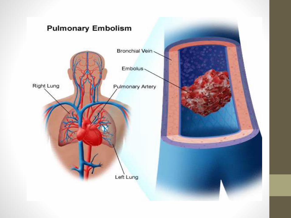
DEFINITION
• Pulmonary thromboembolism results from disruption of a deep venous thrombosis
• or local stasis, causing blockage of pulmonary blood flow beyond the embolus.
• Very large PEs that impede blood flow in both the right and left pulmonary arteries are called saddle emboli.
RISK FACTORS
• Immobilization
• Leg fracture or leg surgery
• Hypercoagulable state (malignancy, pregnancy, genetic)
• Proximal leg deep venous thrombosis
• Stroke
• Orthopedic surgery hip or knee replacement.
• Obesity.
• Owman more than 30 + ocps + smokers
SYMPTOMS/SIGNS
• Tachycardia
• Dyspnea, cough
• Tachypnea
• Pleuritic chest pain
• Hemoptysis
• Hypoxia
FINDINGS/TESTS
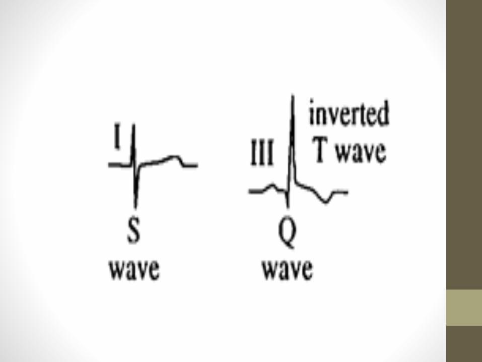
• ECG: S exaggerated in lead I, Q exaggerated and inverted T waves in lead III, or may show diffuse ST changes, and tachycardia
• Chest film: May show an infiltrate or may be normal ( hamptonshump sign , westermarks sign) .
• ABGs: Usually reveal hypoxemia, hypocapnia, and respiratory alkalosis
• D-dimer: Measures products of fibrin degradation (will be elevated)
• Leg ultrasonography (venous duplex): To detect DVT
• Ventilation–perfusion scan (V/Q scan): To look for perfusion defects at
• site of PE
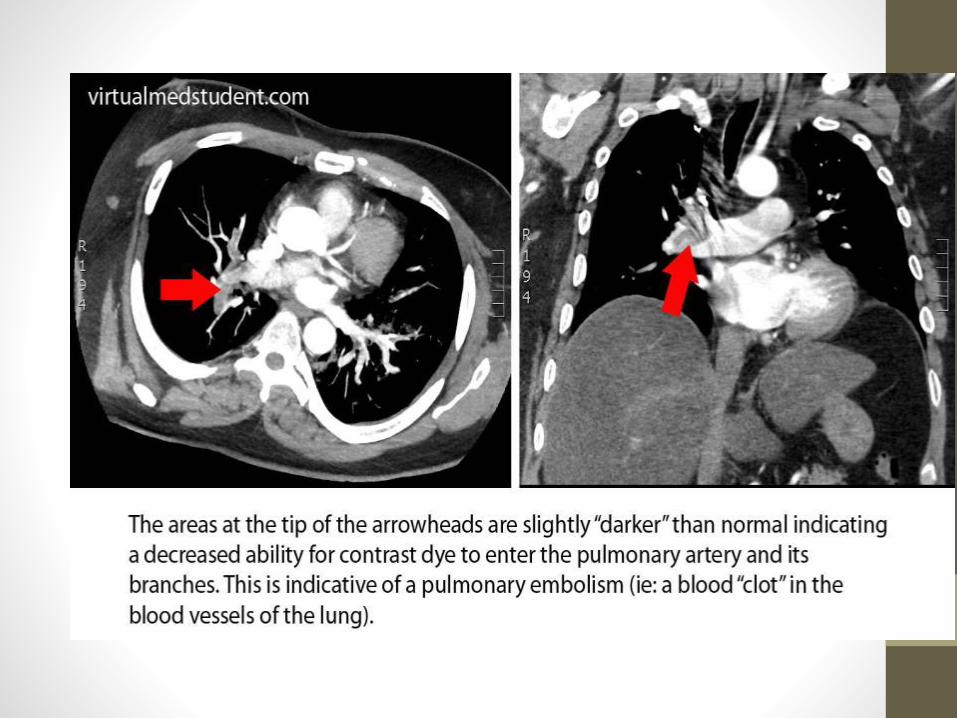
• Helical (spiral) CT: To look for embolus in the pulmonary vasculature.
• Does not detect small emboli.
• Pulmonary angiogram: The gold standard for detection of PE. Invasive test.
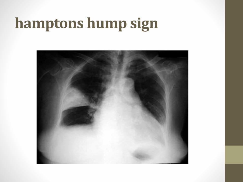
hamptons hump sign
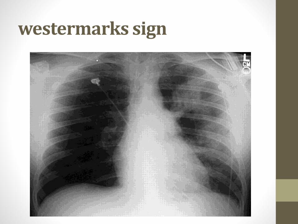
westermarks sign
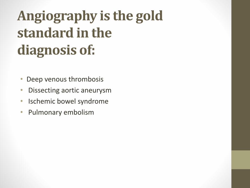
Angiography is the goldstandard in thediagnosis of:
• Deep venous thrombosis
• Dissecting aortic aneurysm
• Ischemic bowel syndrome
• Pulmonary embolism
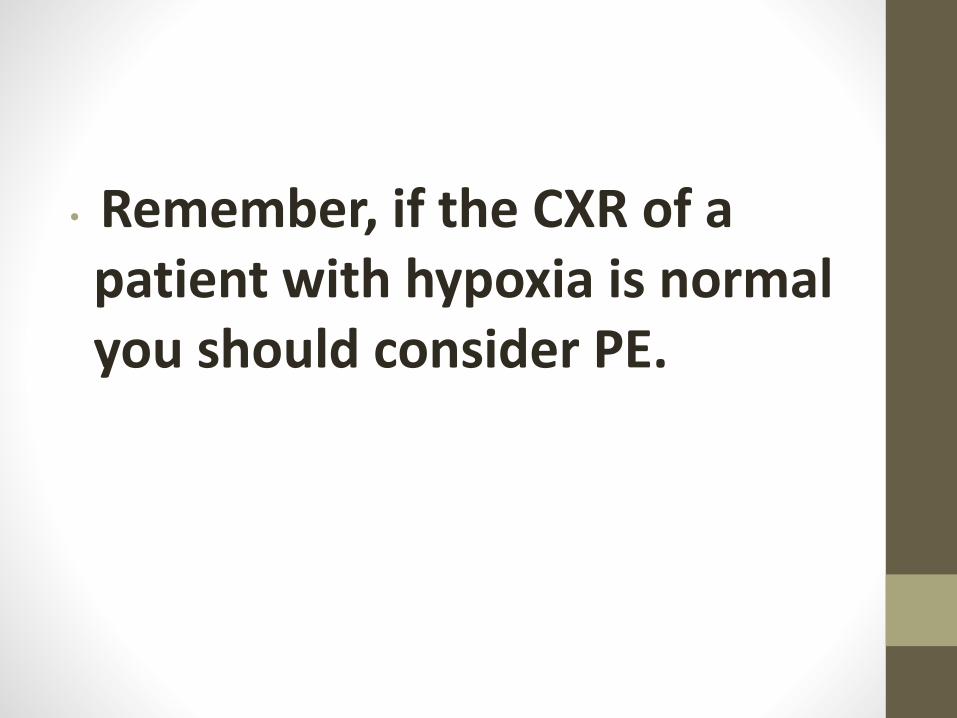
• Remember, if the CXR of a patient with hypoxia is normal you should consider PE.

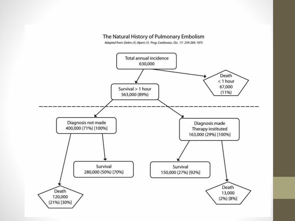
Diagnosis of PE is difficult,
and a high clinical suspicion
should be maintained.
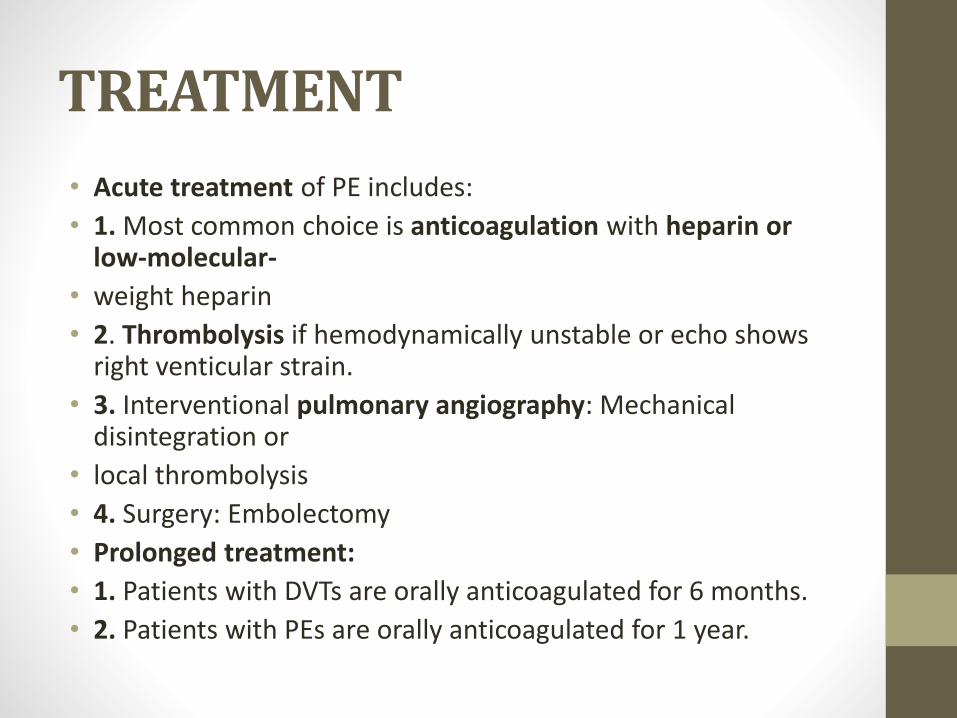
TREATMENT
• Acute treatment of PE includes:
• 1. Most common choice is anticoagulation with heparin or low-molecular-
• weight heparin
• 2. Thrombolysis if hemodynamically unstable or echo shows right venticular strain.
• 3. Interventional pulmonary angiography: Mechanical disintegration or
• local thrombolysis
• 4. Surgery: Embolectomy
• Prolonged treatment:
• 1. Patients with DVTs are orally anticoagulated for 6 months.
• 2. Patients with PEs are orally anticoagulated for 1 year.