protective role of dehydroascorbate in rat liver ischemia-reperfusion injury
TRANSCRIPT
Journal of Surgical Research 123, 215–221 (2005)
Protective Role of Dehydroascorbate in Rat LiverIschemia-Reperfusion Injury
Vincenzo De Tata,1 Stefania Brizzi, Michela Saviozzi, Alessandra Lazzarotti, Vanna Fierabracci,Gino Malvaldi, and Alessandro Casini
Department of Experimental Pathology, Medical Biotechnologies, Infectivology and Epidemiology, University of Pisa,Pisa, Italy
Submitted for publication April 19, 2004
doi:10.1016/j.jss.2004.08.008
Background. Oxidative stress plays an importantrole in liver ischemia/reperfusion (I/R) injury. Thus,enhancing the liver antioxidant capacity could be apromising therapeutic strategy. Ascorbate (AA) is con-sidered the perfect antioxidant, but its therapeuticefficacy is greatly limited by its slow achievement ofhigh intracellular levels. This might be circumventedby administering dehydroascorbate (DHA), which pre-sents a several-fold greater uptake than AA, and un-dergoes rapid intracellular reduction to AA. Thus, ouraim was to assess the protective role of DHA in liverI/R injury.
Materials and methods. Wistar rats (200–300 g bw)were pretreated iv with different doses of AA or DHA20 min before liver ischemia, followed by 6 h reperfu-sion. Liver damage was assessed by biochemical andmorphological indices.
Results. DHA pretreatment induced a rapid increasein liver ascorbate levels, significantly higher thanfindings for AA, without any significant reduction inglutathione levels. Liver damage during I/R in con-trols showed significant increases in serum transami-nases and hepatic thiobarbituric acid reactive sub-stances with alterations of liver morphology. DHAadministration induced a clear, significant protectionagainst I/R injury, whereas liver damage was onlymoderately prevented by AA.
Conclusions. DHA might represent a simple, effec-tive therapeutic option to prevent liver damage asso-ciated with ischemia/reperfusion. © 2004 Elsevier Inc. All
rights reserved.
1 To whom correspondence and reprint requests should be ad-dressed at Dipartimento di Patologia Sperimentale and BMIE,Sezione di Patologia Generale, Università degli Studi di Pisa, ViaRoma, 55–Scuola Medica, 56126 Pisa, Italy. E-mail:
[email protected].215
Key Words: liver; ischemia/reperfusion injury; ascor-bic acid; dehydroascorbic acid; oxidative stress.
INTRODUCTION
Liver ischemia-reperfusion (I/R) injury is a serious,but apparently unavoidable, complication in severalcircumstances such as hepatic resectional surgery(Pringle maneuver), liver transplantation, and hemor-rhagic shock with fluid resuscitation [1, 2]. The conse-quences of I/R injury include liver and remote organfailure in more severe cases, both of which have highrates of morbidity and mortality. Despite improve-ments in organ preservation and surgical techniques,I/R injury remains a significant clinical problem, andthere is considerable interest in its prevention [3].
Various mechanisms have been proposed for thepathogenesis of postischemic tissue injury, but dataobtained by several researchers indicate that the gen-eration of oxygen-derived free-radicals probably repre-sents the most important factor involved [4–6]. Ac-cording to this hypothesis, “reperfusion injury” isassociated with the production of toxic free-radical spe-cies after the resupply of blood (or oxygen) to previ-ously ischemic (or hypoxic) tissues [7]. Enhancing theliver antioxidant capacity can thus be viewed as apromising therapeutic strategy [8]. Approaches involv-ing the administration of various antioxidants havebeen used, but the effectiveness of these interventionshas varied [9–12].
Protection against oxidative stress is afforded byseveral antioxidant systems, including both enzymicand nonenzymic. In view of its biochemical proper-ties (presence in adequate amounts within the cells;capability to react with a variety of free radicals;
suitability to be regenerated in the active form),0022-4804/05 $30.00© 2004 Elsevier Inc. All rights reserved.
216 JOURNAL OF SURGICAL RESEARCH: VOL. 123, NO. 2, FEBRUARY 2005
ascorbic acid is generally considered the perfect an-tioxidant for the cells of nearly all aerobic organisms(for a recent review, see ref. 13). Ascorbate is thoughtto act by scavenging aqueous free-radicals [14 –16] aswell as by reducing lipophilic tocopheroxyl radicalsback to �-tocopherol [17]. Cellular accumulation ofvitamin C is due to transport of both ascorbate (viaNa�-ascorbate symporters such as SVCT1 andSVCT2) [18] and its oxidized metabolite, dehy-droascorbic acid (DHA) [19 –21]. Although ascorbateis the predominant, if not the unique, form in blood,DHA may be produced extracellularly in particularpro-oxidant microenvironments [22, 23]. Experi-ments in neutrophils demonstrated that the rate ofcellular DHA uptake is as much as 30-fold greaterthan that of ascorbate uptake [21, 22]. Once trans-ported, DHA is immediately reduced intracellularlyto ascorbate, thus maintaining a favorable gradientfor the accumulation of ascorbate via this pathway.DHA uptake followed by intracellular reduction canincrease intracellular ascorbate levels 5- to 20-foldwithin minutes. This process, termed ascorbate re-cycling, has been demonstrated experimentally inhuman neutrophils, as well as in other cell types[19 –24].
Recently, Huang et al. [25] demonstrated that DHA,unlike exogenous ascorbate, confers in vivo dose-dependent neuroprotection in a murine model of reper-fused and nonreperfused cerebral ischemia. Here, forthe first time, we show that DHA administration invivo is able to rapidly raise hepatic ascorbate contentwell above physiological levels, and that this increaseresults in a remarkable protection against liver I/Rinjury, significantly higher than that obtained withascorbate treatment.
MATERIALS AND METHODS
Hepatic ischemia/reperfusion injury model. Male Wistar rats(200-300 g bw) were purchased from Harlan Italy (Correzzana, MI,Italy). Rats were housed in a controlled environment at 22 � 1°Cunder a 12-h/12-h light/dark cycle. Food and water were available adlibitum. The experimental protocol followed the Principles of Labo-ratory Animal Care (US NIH Publication No. 83-25, revised 1985) aswell as the recommendations of Italian law for the use of experimen-tal animals (DL No. 116/1992), and was approved by the EthicalCommittee of the University of Pisa Medical School.
Rats were divided into the three following experimental groups:(1) DHA-pretreated rats, receiving intravenously 125, 250, or 500mg/kg dehydroascorbic acid dissolved in a sodium acetate/sodiumbicarbonate buffer, pH 5.5 [25]; (2) ascorbate (AA)-pretreated rats,receiving intravenously the same amount of ascorbic acid; (3)placebo-pretreated rats receiving intravenously equal volumes of thevehicle only. Twenty minutes after intravenous pretreatment, ratswere anesthetized with pentobarbital (50 mg/kg ip); the abdomenwas opened through a midline incision, and the hepatoduodenalligament was exposed. In each rat, the ischemic phase was main-tained through complete occlusion of the ligament using an atrau-matic microvessel clip for 30 min. At the end of the ischemic phase,the clip was removed and liver reperfusion was allowed for different
periods of time (1, 2, 4, or 6 h).Blood samples were collected from the tail vein just before isch-emia and after 0, 1, 2, 4, and 6 h of reperfusion.
Measurement of serum transaminase activities. Serum alaninetransaminase (ALT) and aspartate transaminase (AST) activitieswere measured using commercially available kits (Sigma-Aldrich,Milano, Italy) and the results were obtained as units/liter.
Histological study. Liver samples obtained at various determina-tion points were fixed in 10% buffered formalin and were thenembedded in paraffin, in accordance with standard procedures. Sec-tions (5 �m) were stained with hematoxylin and eosin and examinedmicroscopically.
Assay of TBARS in liver tissue. Liver specimens were homoge-nized with 10 vol of 1.15% KCl, using a Teflon Potter–Elvehjemhomogenizer, and were assayed for thiobarbituric acid-reactivesubstances (TBARS) by the method of Ohkawa et al. [26].
Assay of water-soluble antioxidants in liver tissue. Liver speci-mens were homogenized with a Teflon pestle in 10 vol of 5% TCA andcentrifuged at 12000 g for 20 min. AA, DHA, GSH, and GSSG werequantified on the supernatant by HPLC with electrochemical detec-tion [27].
Liver tissue protein concentration was assayed by the method ofLowry et al. [28] using bovine serum albumin as the standard.
Statistical analysis. Data are expressed as means � SEM. Thestatistical significance of the experimental treatment was assessedby factorial analysis of variance (ANOVA), followed by Tukey’s post-test with multiple comparison. When appropriate, the two-tailedunpaired Student’s t test was used as a method of post-hoc analysisto assess two-by-two differences. A P value �0.05 was consideredsignificant.
RESULTS
Ascorbic Acid and Glutathione Levels in Rat Liver afterPretreatment with Ascorbate or Dehydroascorbate
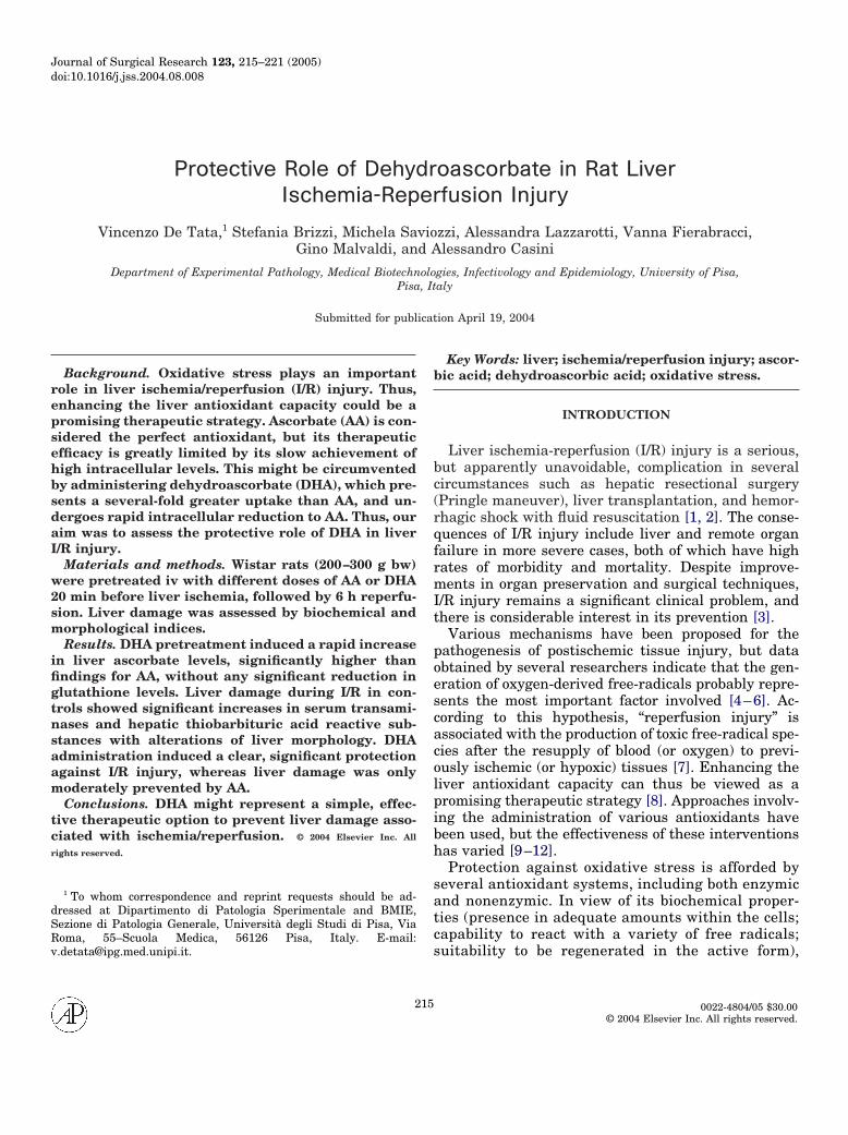
Figure 1 shows the levels of ascorbic acid in the ratliver 20 min after the iv injection of three differentamounts (125, 250, or 500 mg/kg bw, respectively) ofAA or DHA. Since the amount of oxidized ascorbatedetected was always negligible, without any significantdifference between groups, only the levels of the re-duced form will be shown in this as well in all of thefollowing figures. As expected, both treatments causeda significant, dose-dependent, increase in hepaticascorbate intracellular levels, but the effect of DHAwas significantly greater than that of AA for each doseused. At 250 mg/kg, for example, the percentage in-crease compared with untreated controls was �59% forAA and �176% in the case of DHA, respectively.
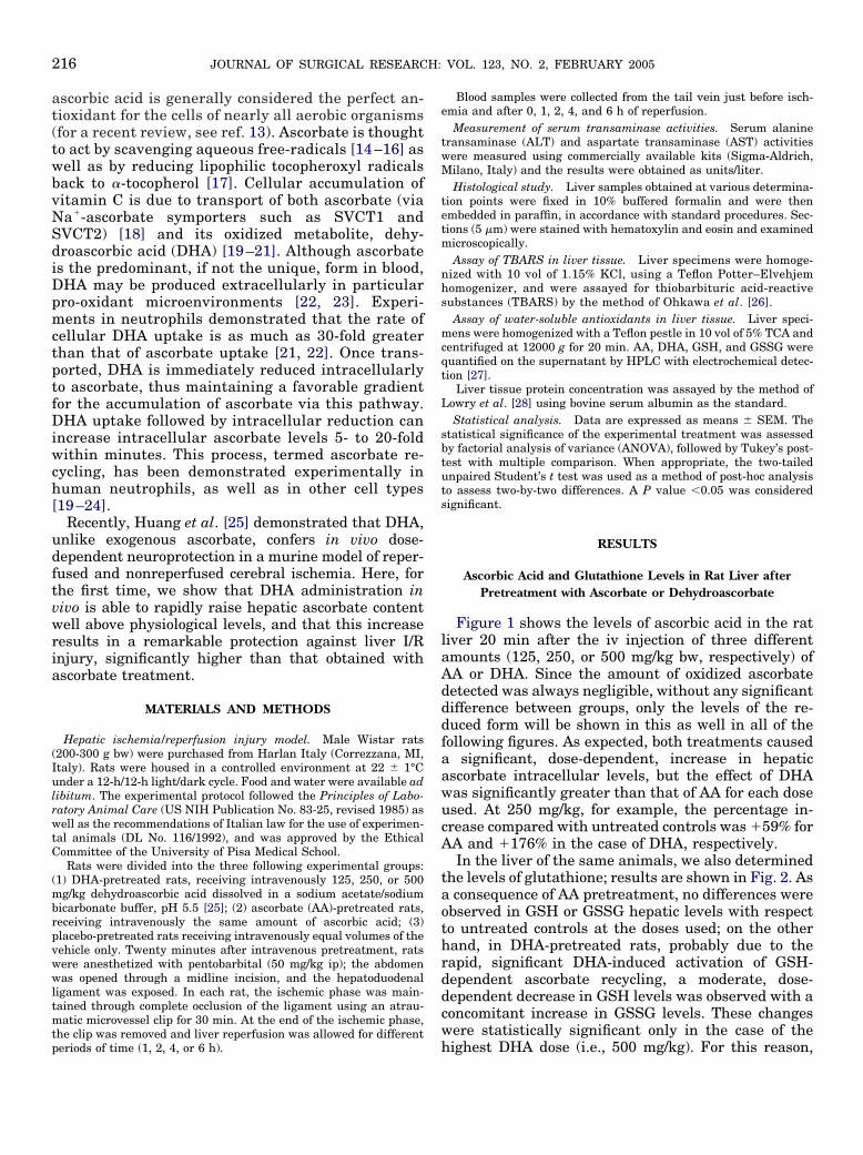
In the liver of the same animals, we also determinedthe levels of glutathione; results are shown in Fig. 2. Asa consequence of AA pretreatment, no differences wereobserved in GSH or GSSG hepatic levels with respectto untreated controls at the doses used; on the otherhand, in DHA-pretreated rats, probably due to therapid, significant DHA-induced activation of GSH-dependent ascorbate recycling, a moderate, dose-dependent decrease in GSH levels was observed with aconcomitant increase in GSSG levels. These changeswere statistically significant only in the case of the
highest DHA dose (i.e., 500 mg/kg). For this reason,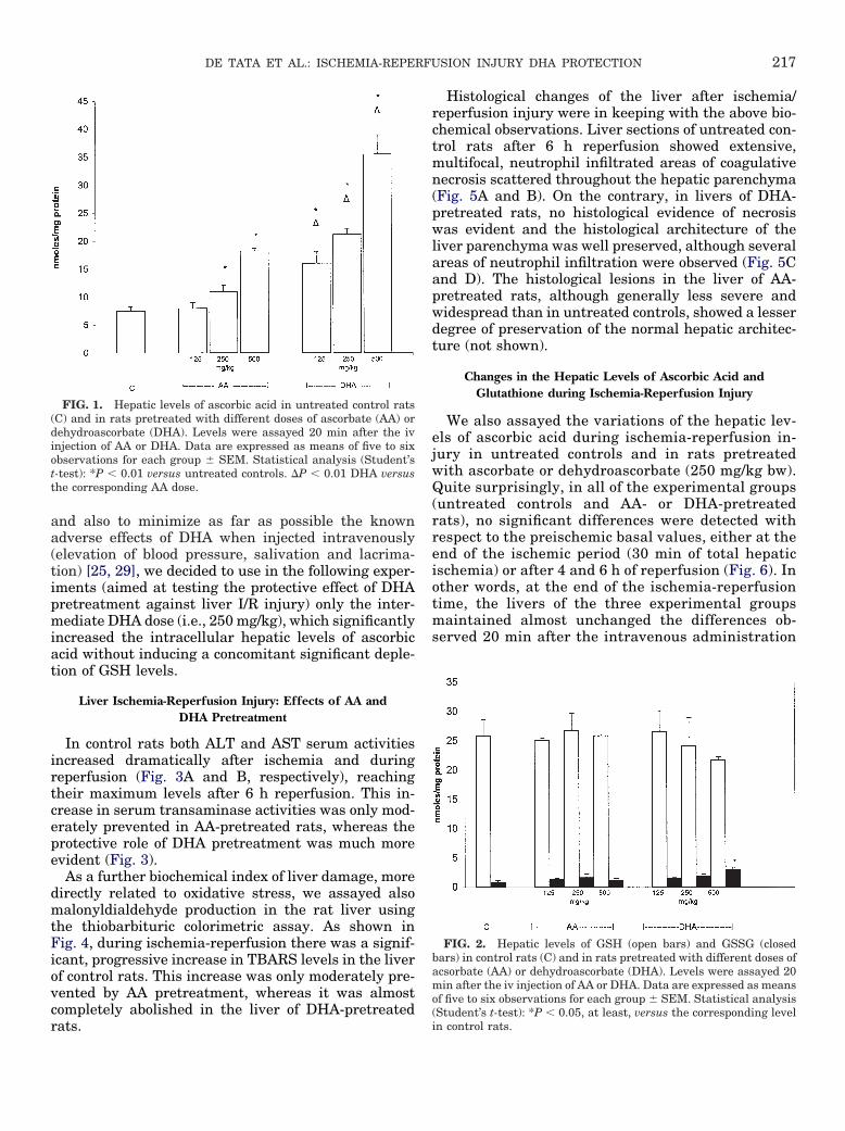
217DE TATA ET AL.: ISCHEMIA-REPERFUSION INJURY DHA PROTECTION
and also to minimize as far as possible the knownadverse effects of DHA when injected intravenously(elevation of blood pressure, salivation and lacrima-tion) [25, 29], we decided to use in the following exper-iments (aimed at testing the protective effect of DHApretreatment against liver I/R injury) only the inter-mediate DHA dose (i.e., 250 mg/kg), which significantlyincreased the intracellular hepatic levels of ascorbicacid without inducing a concomitant significant deple-tion of GSH levels.
Liver Ischemia-Reperfusion Injury: Effects of AA andDHA Pretreatment
In control rats both ALT and AST serum activitiesincreased dramatically after ischemia and duringreperfusion (Fig. 3A and B, respectively), reachingtheir maximum levels after 6 h reperfusion. This in-crease in serum transaminase activities was only mod-erately prevented in AA-pretreated rats, whereas theprotective role of DHA pretreatment was much moreevident (Fig. 3).
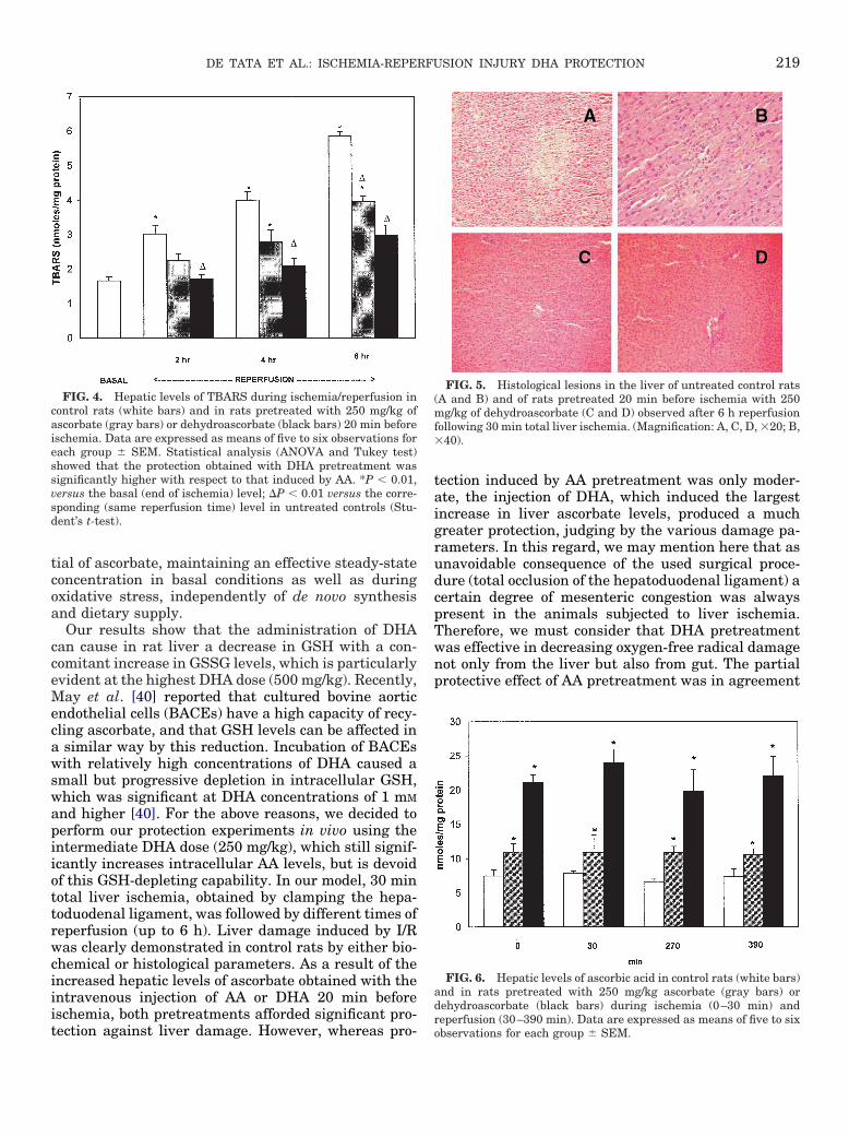
As a further biochemical index of liver damage, moredirectly related to oxidative stress, we assayed alsomalonyldialdehyde production in the rat liver usingthe thiobarbituric colorimetric assay. As shown inFig. 4, during ischemia-reperfusion there was a signif-icant, progressive increase in TBARS levels in the liverof control rats. This increase was only moderately pre-vented by AA pretreatment, whereas it was almostcompletely abolished in the liver of DHA-pretreated
FIG. 1. Hepatic levels of ascorbic acid in untreated control rats(C) and in rats pretreated with different doses of ascorbate (AA) ordehydroascorbate (DHA). Levels were assayed 20 min after the ivinjection of AA or DHA. Data are expressed as means of five to sixobservations for each group � SEM. Statistical analysis (Student’st-test): *P � 0.01 versus untreated controls. �P � 0.01 DHA versusthe corresponding AA dose.
rats.
Histological changes of the liver after ischemia/reperfusion injury were in keeping with the above bio-chemical observations. Liver sections of untreated con-trol rats after 6 h reperfusion showed extensive,multifocal, neutrophil infiltrated areas of coagulativenecrosis scattered throughout the hepatic parenchyma(Fig. 5A and B). On the contrary, in livers of DHA-pretreated rats, no histological evidence of necrosiswas evident and the histological architecture of theliver parenchyma was well preserved, although severalareas of neutrophil infiltration were observed (Fig. 5Cand D). The histological lesions in the liver of AA-pretreated rats, although generally less severe andwidespread than in untreated controls, showed a lesserdegree of preservation of the normal hepatic architec-ture (not shown).
Changes in the Hepatic Levels of Ascorbic Acid andGlutathione during Ischemia-Reperfusion Injury
We also assayed the variations of the hepatic lev-els of ascorbic acid during ischemia-reperfusion in-jury in untreated controls and in rats pretreatedwith ascorbate or dehydroascorbate (250 mg/kg bw).Quite surprisingly, in all of the experimental groups(untreated controls and AA- or DHA-pretreatedrats), no significant differences were detected withrespect to the preischemic basal values, either at theend of the ischemic period (30 min of total hepaticischemia) or after 4 and 6 h of reperfusion (Fig. 6). Inother words, at the end of the ischemia-reperfusiontime, the livers of the three experimental groupsmaintained almost unchanged the differences ob-served 20 min after the intravenous administration
FIG. 2. Hepatic levels of GSH (open bars) and GSSG (closedbars) in control rats (C) and in rats pretreated with different doses ofacsorbate (AA) or dehydroascorbate (DHA). Levels were assayed 20min after the iv injection of AA or DHA. Data are expressed as meansof five to six observations for each group � SEM. Statistical analysis(Student’s t-test): *P � 0.05, at least, versus the corresponding level
in control rats.
218 JOURNAL OF SURGICAL RESEARCH: VOL. 123, NO. 2, FEBRUARY 2005
of AA or DHA, illustrated in Fig. 1. On the otherhand, 30 min of total liver ischemia caused a signif-icant decrease in hepatic GSH levels with a concom-itant increase in GSSG levels (Fig. 7) in all of theexperimental groups (with the result that the totalglutathione content was not significantly modified atthe end of the ischemic time). During reperfusion,GSH levels remained unchanged in the liver of allgroups of rats, whereas GSSG levels underwent asignificant decline (probably due to the leakage of theoxidized form of glutathione from hepatic cells); con-sequently, after 6 h reperfusion the total glutathionecontent was significantly lower in all experimentalgroups.
DISCUSSION
The objective of this study was to evaluate the pos-sibility of preventing the damage associated with liverI/R by the previous administration of the oxidized me-tabolite of vitamin C, DHA. Oxygen-free radicals havelong been indicated as being responsible for I/R-induced liver damage [4–6]. It was subsequently rec-ognized that this increasing production of oxygen rad-icals and postischemic damage of the liver have a two-phase time course: an initial phase of injury (roughlywithin the first 2 h after reperfusion) and a late phase,from approximately 3 to 24 h of blood reflow [5, 30, 31].
FIG. 3. Variation of serum levels of ALT (A) and AST (B) enzymmin) in control rats (�) and in rats pretreated with 250 mg/kg of asexpressed as means of five to six observations for each group � SEM.and AST, the protective effect of DHA was significantly greater thanrats (Student’s t test).
In the initial phase of reperfusion, there is an overpro-
duction of oxygen radicals in activated Kupffer cells[30, 31] and in the mitochondria of hepatocytes andendothelial cells, where oxygen reflow encountershighly reduced respiratory chains [5, 31, 32]. Eventsoccurring during this early phase, including activationof Kupffer cells, initiate a complex inflammatory path-way which culminates in the hepatic accumulation ofneutrophils [30]. Recruited neutrophils directly dam-age hepatocytes by releasing oxidants and proteasesand are responsible for the later phase of liver injuryinduced by I/R (for a review see ref. 33).
AA is one of the water-soluble vitamins. With itsstrong reducing property and its property of scaveng-ing free radicals, AA is well known as a strong antiox-idant agent [13]. However, in a therapeutic perspec-tive, it must be remembered that at least some ofharmful consequences of free-radical production can beefficiently counteracted by AA only at supraphysiologi-cal concentrations [34]. To circumvent the difficulty toreach and maintain this increase in intracellular levelsby the administration of exogenous AA, we used itsoxidized metabolite dehydroascorbate. Here we show,for the first time, that the in vivo iv administration ofDHA rapidly induces a significant dose-dependent in-crease in hepatic intracellular AA levels, in agreementwith previous results obtained in vitro [19–24]. Insidethe cell, there are several enzymatic systems capable ofreducing DHA to AA [35–39], whose main function is
ctivities during liver ischemia (0–30 min) and reperfusion (30–390bate (□) or dehydroascorbate (Œ) 20 min before ischemia. Data aretistical analysis (ANOVA and Tukey test) showed that, for both ALTt afforded by AA. *P � 0.01 versus the corresponding level in control
e acorStatha
believed to be that of restoring the antioxidant poten-
219DE TATA ET AL.: ISCHEMIA-REPERFUSION INJURY DHA PROTECTION
tial of ascorbate, maintaining an effective steady-stateconcentration in basal conditions as well as duringoxidative stress, independently of de novo synthesisand dietary supply.
Our results show that the administration of DHAcan cause in rat liver a decrease in GSH with a con-comitant increase in GSSG levels, which is particularlyevident at the highest DHA dose (500 mg/kg). Recently,May et al. [40] reported that cultured bovine aorticendothelial cells (BACEs) have a high capacity of recy-cling ascorbate, and that GSH levels can be affected ina similar way by this reduction. Incubation of BACEswith relatively high concentrations of DHA caused asmall but progressive depletion in intracellular GSH,which was significant at DHA concentrations of 1 mM
and higher [40]. For the above reasons, we decided toperform our protection experiments in vivo using theintermediate DHA dose (250 mg/kg), which still signif-icantly increases intracellular AA levels, but is devoidof this GSH-depleting capability. In our model, 30 mintotal liver ischemia, obtained by clamping the hepa-toduodenal ligament, was followed by different times ofreperfusion (up to 6 h). Liver damage induced by I/Rwas clearly demonstrated in control rats by either bio-chemical or histological parameters. As a result of theincreased hepatic levels of ascorbate obtained with theintravenous injection of AA or DHA 20 min beforeischemia, both pretreatments afforded significant pro-
FIG. 4. Hepatic levels of TBARS during ischemia/reperfusion incontrol rats (white bars) and in rats pretreated with 250 mg/kg ofascorbate (gray bars) or dehydroascorbate (black bars) 20 min beforeischemia. Data are expressed as means of five to six observations foreach group � SEM. Statistical analysis (ANOVA and Tukey test)showed that the protection obtained with DHA pretreatment wassignificantly higher with respect to that induced by AA. *P � 0.01,versus the basal (end of ischemia) level; �P � 0.01 versus the corre-sponding (same reperfusion time) level in untreated controls (Stu-dent’s t-test).
tection against liver damage. However, whereas pro-
tection induced by AA pretreatment was only moder-ate, the injection of DHA, which induced the largestincrease in liver ascorbate levels, produced a muchgreater protection, judging by the various damage pa-rameters. In this regard, we may mention here that asunavoidable consequence of the used surgical proce-dure (total occlusion of the hepatoduodenal ligament) acertain degree of mesenteric congestion was alwayspresent in the animals subjected to liver ischemia.Therefore, we must consider that DHA pretreatmentwas effective in decreasing oxygen-free radical damagenot only from the liver but also from gut. The partialprotective effect of AA pretreatment was in agreement
FIG. 5. Histological lesions in the liver of untreated control rats(A and B) and of rats pretreated 20 min before ischemia with 250mg/kg of dehydroascorbate (C and D) observed after 6 h reperfusionfollowing 30 min total liver ischemia. (Magnification: A, C, D, �20; B,�40).
FIG. 6. Hepatic levels of ascorbic acid in control rats (white bars)and in rats pretreated with 250 mg/kg ascorbate (gray bars) ordehydroascorbate (black bars) during ischemia (0–30 min) and
reperfusion (30–390 min). Data are expressed as means of five to six observations for each group � SEM.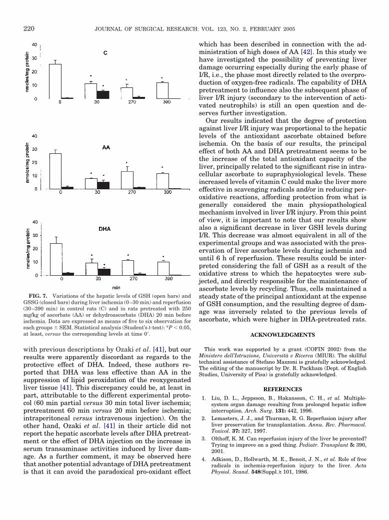
220 JOURNAL OF SURGICAL RESEARCH: VOL. 123, NO. 2, FEBRUARY 2005
with previous descriptions by Ozaki et al. [41], but ourresults were apparently discordant as regards to theprotective effect of DHA. Indeed, these authors re-ported that DHA was less effective than AA in thesuppression of lipid peroxidation of the reoxygenatedliver tissue [41]. This discrepancy could be, at least inpart, attributable to the different experimental proto-col (60 min partial versus 30 min total liver ischemia;pretreatment 60 min versus 20 min before ischemia;intraperitoneal versus intravenous injection). On theother hand, Ozaki et al. [41] in their article did notreport the hepatic ascorbate levels after DHA pretreat-ment or the effect of DHA injection on the increase inserum transaminase activities induced by liver dam-age. As a further comment, it may be observed herethat another potential advantage of DHA pretreatment
FIG. 7. Variations of the hepatic levels of GSH (open bars) andGSSG (closed bars) during liver ischemia (0–30 min) and reperfusion(30–390 min) in control rats (C) and in rats pretreated with 250mg/kg of ascorbate (AA) or dehydroascorbate (DHA) 20 min beforeischemia. Data are expressed as means of five to six observation foreach groups � SEM. Statistical analysis (Student’s t-test): *P � 0.05,at least, versus the corresponding levels at time 0=.
is that it can avoid the paradoxical pro-oxidant effect
which has been described in connection with the ad-ministration of high doses of AA [42]. In this study wehave investigated the possibility of preventing liverdamage occurring especially during the early phase ofI/R, i.e., the phase most directly related to the overpro-duction of oxygen-free radicals. The capability of DHApretreatment to influence also the subsequent phase ofliver I/R injury (secondary to the intervention of acti-vated neutrophils) is still an open question and de-serves further investigation.
Our results indicated that the degree of protectionagainst liver I/R injury was proportional to the hepaticlevels of the antioxidant ascorbate obtained beforeischemia. On the basis of our results, the principaleffect of both AA and DHA pretreatment seems to bethe increase of the total antioxidant capacity of theliver, principally related to the significant rise in intra-cellular ascorbate to supraphysiological levels. Theseincreased levels of vitamin C could make the liver moreeffective in scavenging radicals and/or in reducing per-oxidative reactions, affording protection from what isgenerally considered the main physiopathologicalmechanism involved in liver I/R injury. From this pointof view, it is important to note that our results showalso a significant decrease in liver GSH levels duringI/R. This decrease was almost equivalent in all of theexperimental groups and was associated with the pres-ervation of liver ascorbate levels during ischemia anduntil 6 h of reperfusion. These results could be inter-preted considering the fall of GSH as a result of theoxidative stress to which the hepatocytes were sub-jected, and directly responsible for the maintenance ofascorbate levels by recycling. Thus, cells maintained asteady state of the principal antioxidant at the expenseof GSH consumption, and the resulting degree of dam-age was inversely related to the previous levels ofascorbate, which were higher in DHA-pretreated rats.
ACKNOWLEDGMENTS
This work was supported by a grant (COFIN 2002) from theMinistero dell’Istruzione, Università e Ricerca (MIUR). The skillfultechnical assistance of Stefano Mazzoni is gratefully acknowledged.The editing of the manuscript by Dr. R. Packham (Dept. of EnglishStudies, University of Pisa) is gratefully acknowledged.
REFERENCES1. Liu, D. L., Jeppsson, B., Hakansson, C. H., et al. Multiple-
system organ damage resulting from prolonged hepatic inflowinterruption. Arch. Surg. 131: 442, 1996.
2. Lemasters, J. J., and Thurman, R. G. Reperfusion injury afterliver preservation for transplantation. Annu. Rev. Pharmacol.Toxicol. 37: 327, 1997.
3. Olthoff, K. M. Can reperfusion injury of the liver be prevented?Trying to improve on a good thing. Pediatr. Transplant 5: 390,2001.
4. Adkison, D., Hollwarth, M. E., Benoit, J. N., et al. Role of freeradicals in ischemia-reperfusion injury to the liver. Acta
Physiol. Scand. 548(Suppl.): 101, 1986.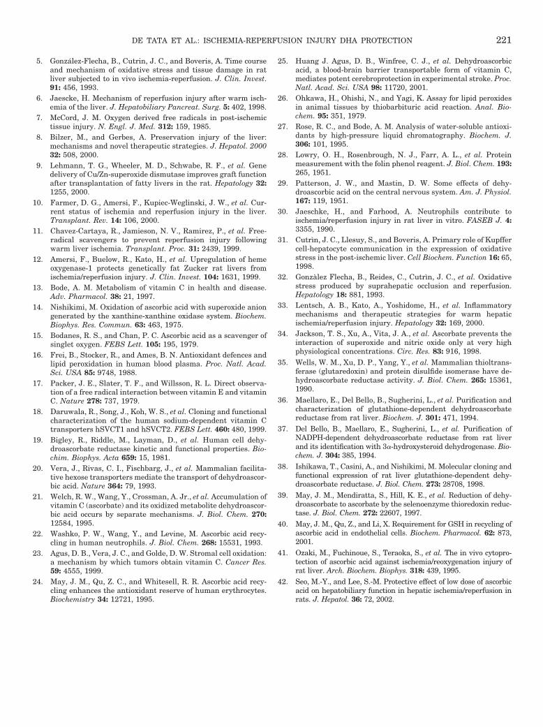
221DE TATA ET AL.: ISCHEMIA-REPERFUSION INJURY DHA PROTECTION
5. González-Flecha, B., Cutrin, J. C., and Boveris, A. Time courseand mechanism of oxidative stress and tissue damage in ratliver subjected to in vivo ischemia-reperfusion. J. Clin. Invest.91: 456, 1993.
6. Jaescke, H. Mechanism of reperfusion injury after warm isch-emia of the liver. J. Hepatobiliary Pancreat. Surg. 5: 402, 1998.
7. McCord, J. M. Oxygen derived free radicals in post-ischemictissue injury. N. Engl. J. Med. 312: 159, 1985.
8. Bilzer, M., and Gerbes, A. Preservation injury of the liver:mechanisms and novel therapeutic strategies. J. Hepatol. 200032: 508, 2000.
9. Lehmann, T. G., Wheeler, M. D., Schwabe, R. F., et al. Genedelivery of Cu/Zn-superoxide dismutase improves graft functionafter transplantation of fatty livers in the rat. Hepatology 32:1255, 2000.
10. Farmer, D. G., Amersi, F., Kupiec-Weglinski, J. W., et al. Cur-rent status of ischemia and reperfusion injury in the liver.Transplant. Rev. 14: 106, 2000.
11. Chavez-Cartaya, R., Jamieson, N. V., Ramirez, P., et al. Free-radical scavengers to prevent reperfusion injury followingwarm liver ischemia. Transplant. Proc. 31: 2439, 1999.
12. Amersi, F., Buelow, R., Kato, H., et al. Upregulation of hemeoxygenase-1 protects genetically fat Zucker rat livers fromischemia/reperfusion injury. J. Clin. Invest. 104: 1631, 1999.
13. Bode, A. M. Metabolism of vitamin C in health and disease.Adv. Pharmacol. 38: 21, 1997.
14. Nishikimi, M. Oxidation of ascorbic acid with superoxide aniongenerated by the xanthine-xanthine oxidase system. Biochem.Biophys. Res. Commun. 63: 463, 1975.
15. Bodanes, R. S., and Chan, P. C. Ascorbic acid as a scavenger ofsinglet oxygen. FEBS Lett. 105: 195, 1979.
16. Frei, B., Stocker, R., and Ames, B. N. Antioxidant defences andlipid peroxidation in human blood plasma. Proc. Natl. Acad.Sci. USA 85: 9748, 1988.
17. Packer, J. E., Slater, T. F., and Willsson, R. L. Direct observa-tion of a free radical interaction between vitamin E and vitaminC. Nature 278: 737, 1979.
18. Daruwala, R., Song, J., Koh, W. S., et al. Cloning and functionalcharacterization of the human sodium-dependent vitamin Ctransporters hSVCT1 and hSVCT2. FEBS Lett. 460: 480, 1999.
19. Bigley, R., Riddle, M., Layman, D., et al. Human cell dehy-droascorbate reductase kinetic and functional properties. Bio-chim. Biophys. Acta 659: 15, 1981.
20. Vera, J., Rivas, C. I., Fischbarg, J., et al. Mammalian facilita-tive hexose transporters mediate the transport of dehydroascor-bic acid. Nature 364: 79, 1993.
21. Welch, R. W., Wang, Y., Crossman, A. Jr., et al. Accumulation ofvitamin C (ascorbate) and its oxidized metabolite dehydroascor-bic acid occurs by separate mechanisms. J. Biol. Chem. 270:12584, 1995.
22. Washko, P. W., Wang, Y., and Levine, M. Ascorbic acid recy-cling in human neutrophils. J. Biol. Chem. 268: 15531, 1993.
23. Agus, D. B., Vera, J. C., and Golde, D. W. Stromal cell oxidation:a mechanism by which tumors obtain vitamin C. Cancer Res.59: 4555, 1999.
24. May, J. M., Qu, Z. C., and Whitesell, R. R. Ascorbic acid recy-cling enhances the antioxidant reserve of human erythrocytes.
Biochemistry 34: 12721, 1995.25. Huang J. Agus, D. B., Winfree, C. J., et al. Dehydroascorbicacid, a blood-brain barrier transportable form of vitamin C,mediates potent cerebroprotection in experimental stroke. Proc.Natl. Acad. Sci. USA 98: 11720, 2001.
26. Ohkawa, H., Ohishi, N., and Yagi, K. Assay for lipid peroxidesin animal tissues by thiobarbituric acid reaction. Anal. Bio-chem. 95: 351, 1979.
27. Rose, R. C., and Bode, A. M. Analysis of water-soluble antioxi-dants by high-pressure liquid chromatography. Biochem. J.306: 101, 1995.
28. Lowry, O. H., Rosenbrough, N. J., Farr, A. L., et al. Proteinmeasurement with the folin phenol reagent. J. Biol. Chem. 193:265, 1951.
29. Patterson, J. W., and Mastin, D. W. Some effects of dehy-droascorbic acid on the central nervous system. Am. J. Physiol.167: 119, 1951.
30. Jaeschke, H., and Farhood, A. Neutrophils contribute toischemia/reperfusion injury in rat liver in vitro. FASEB J. 4:3355, 1990.
31. Cutrìn, J. C., Llesuy, S., and Boveris, A. Primary role of Kupffercell-hepatocyte communication in the expression of oxidativestress in the post-ischemic liver. Cell Biochem. Function 16: 65,1998.
32. Gonzàlez Flecha, B., Reides, C., Cutrìn, J. C., et al. Oxidativestress produced by suprahepatic occlusion and reperfusion.Hepatology 18: 881, 1993.
33. Lentsch, A. B., Kato, A., Yoshidome, H., et al. Inflammatorymechanisms and therapeutic strategies for warm hepaticischemia/reperfusion injury. Hepatology 32: 169, 2000.
34. Jackson, T. S., Xu, A., Vita, J. A., et al. Ascorbate prevents theinteraction of superoxide and nitric oxide only at very highphysiological concentrations. Circ. Res. 83: 916, 1998.
35. Wells, W. M., Xu, D. P., Yang, Y., et al. Mammalian thioltrans-ferase (glutaredoxin) and protein disulfide isomerase have de-hydroascorbate reductase activity. J. Biol. Chem. 265: 15361,1990.
36. Maellaro, E., Del Bello, B., Sugherini, L., et al. Purification andcharacterization of glutathione-dependent dehydroascorbatereductase from rat liver. Biochem. J. 301: 471, 1994.
37. Del Bello, B., Maellaro, E., Sugherini, L., et al. Purification ofNADPH-dependent dehydroascorbate reductase from rat liverand its identification with 3�-hydroxysteroid dehydrogenase. Bio-chem. J. 304: 385, 1994.
38. Ishikawa, T., Casini, A., and Nishikimi, M. Molecular cloning andfunctional expression of rat liver glutathione-dependent dehy-droascorbate reductase. J. Biol. Chem. 273: 28708, 1998.
39. May, J. M., Mendiratta, S., Hill, K. E., et al. Reduction of dehy-droascorbate to ascorbate by the selenoenzyme thioredoxin reduc-tase. J. Biol. Chem. 272: 22607, 1997.
40. May, J. M., Qu, Z., and Li, X. Requirement for GSH in recycling ofascorbic acid in endothelial cells. Biochem. Pharmacol. 62: 873,2001.
41. Ozaki, M., Fuchinoue, S., Teraoka, S., et al. The in vivo cytopro-tection of ascorbic acid against ischemia/reoxygenation injury ofrat liver. Arch. Biochem. Biophys. 318: 439, 1995.
42. Seo, M.-Y., and Lee, S.-M. Protective effect of low dose of ascorbicacid on hepatobiliary function in hepatic ischemia/reperfusion in
rats. J. Hepatol. 36: 72, 2002.